Hyperbaric Oxygen Prevents Early Death Caused by Experimental Cerebral Malaria Yara C. Blanco 1,2 , Alessandro S. Farias 1 , Uta Goelnitz 3 , Stefanie C. P. Lopes 1,2 , Wagner W. Arrais-Silva 2 , Bruna O. Carvalho 1,2 , Roge ´ rio Amino 4 , Gerhard Wunderlich 3 , Leonilda M. B. Santos 1 , Selma Giorgio 2 , Fabio T. M. Costa 1,2 * 1 Department of Microbiology & Immunology, State University of Campinas – UNICAMP, Campinas, Sa ˜o Paulo, Brazil, 2 Department of Parasitology, UNICAMP, State University of Campinas, Campinas, Sa ˜o Paulo, Brazil, 3 Department of Parasitology – ICB, University of Sa ˜o Paulo – USP, Sa ˜o Paulo, Sa ˜ o Paulo, Brazil, 4 Department of Biochemistry, Federal University of Sa ˜o Paulo – UNIFESP, Sa ˜o Paulo, Sa ˜ o Paulo, Brazil Abstract Background: Cerebral malaria (CM) is a syndrome characterized by neurological signs, seizures and coma. Despite the fact that CM presents similarities with cerebral stroke, few studies have focused on new supportive therapies for the disease. Hyperbaric oxygen (HBO) therapy has been successfully used in patients with numerous brain disorders such as stroke, migraine and atherosclerosis. Methodology/Principal Findings: C57BL/6 mice infected with Plasmodium berghei ANKA (PbA) were exposed to daily doses of HBO (100% O 2 , 3.0 ATA, 1–2 h per day) in conditions well-tolerated by humans and animals, before or after parasite establishment. Cumulative survival analyses demonstrated that HBO therapy protected 50% of PbA-infected mice and delayed CM-specific neurological signs when administrated after patent parasitemia. Pressurized oxygen therapy reduced peripheral parasitemia, expression of TNF-a, IFN-c and IL-10 mRNA levels and percentage of cd and ab CD4 + and CD8 + T lymphocytes sequestered in mice brains, thus resulting in a reduction of blood-brain barrier (BBB) dysfunction and hypothermia. Conclusions/Significance: The data presented here is the first indication that HBO treatment could be used as supportive therapy, perhaps in association with neuroprotective drugs, to prevent CM clinical outcomes, including death. Citation: Blanco YC, Farias AS, Goelnitz U, Lopes SCP, Arrais-Silva WW, et al. (2008) Hyperbaric Oxygen Prevents Early Death Caused by Experimental Cerebral Malaria. PLoS ONE 3(9): e3126. doi:10.1371/journal.pone.0003126 Editor: Mauricio Martins Rodrigues, Federal University of Sa ˜ o Paulo, Brazil Received March 22, 2007; Accepted August 14, 2008; Published September 4, 2008 Copyright: ß 2008 Blanco et al. This is an open-access article distributed under the terms of the Creative Commons Attribution License, which permits unrestricted use, distribution, and reproduction in any medium, provided the original author and source are credited. Funding: This work was supported by Fundac ¸a ˜o de Amparo a ` Pesquisa do Estado de Sa ˜o Paulo (FAPESP), grant nu 2004/00638-6, and from Conselho Nacional de Desenvolvimento Cientı ´fico e Tecnolo ´ gico (CNPq). YCB, WWA, ASF and SCPL were supported by Coordenac ¸a ˜o de Aperfeic ¸oamento de Pessoal de Nı ´vel Superior (CAPES), and UG was sponsored by a FAPESP fellowship. GW, SG and FTMC are CNPq fellows. Competing Interests: The authors have declared that no competing interests exist. * E-mail: [email protected] Introduction Cerebral malaria (CM) causes 1–2 million deaths annually; mainly in sub-Saharan African children aged 2–6. It is estimated that 250,000 children that do not succumb to CM will develop neurocognitive impairments per year [1] and most CM patients die before the beneficial effects of drug treatment are observed [2]; thus indicating the need to explore new supportive therapies. CM is a multi-factorial syndrome characterized by neurological signs, seizures and coma, which can, in turn, lead to death. This syndrome can be associated with a loss of cerebrospinal fluid spaces and ischemia [3], alterations in cerebral blood flow velocity [4], a decrease in cerebral oxygen consumption in CM comatose patients [5] and an increase in the lactate levels of the cerebrospinal fluid [6] which decreases after patients recover consciousness [7]. Recent imaging and postmortem analyses have revealed the presence of Durck granulomas, blood-brain barrier (BBB) dysfunction and diffuse cerebral edema with multiple petechial hemorrhages and ischemic changes in the brain of adults with CM [8,9]. Although the CM pathogenic process is controversial and still not fully understood, evidence suggests that the host’s immune system plays a major role in expressing certain cytokines, e.g. TNF-a and IFN-c, and activating immunocompetent cells [10– 15]. In fact, recent immunological analyses have shown that, unlike individuals with mild and severe non-cerebral malaria, CM patients present elevated levels of a specific cluster of cytokines, which include TGF-b, TNF-a, IL-1b and IL-10 [16]. Hyperbaric oxygen therapy (HBO; pO 2 = 760 mmHg) has been successfully used against bacterial and fungal infections and as an adjunct therapy in surgeries [17–19]. In addition, reports have recently shown that HBO therapy transiently suppresses the inflammatory process of ischemic wounding and trauma [20,21]. Indeed, immunological analyses have revealed that HBO therapy significantly decreases the levels of TNF-a and IL-1b secreted by monocytes and macrophage collected from rats or from human peripheral blood after stimulation with LPS [22,23]. In an experimental model for ischemia, HBO reduces immunocompe- tent cell sequestration and the synthesis of TNF-a [24]; probably by decreasing ICAM-1 expression levels [25]. Moreover, HBO PLoS ONE | www.plosone.org 1 September 2008 | Volume 3 | Issue 9 | e3126

Welcome message from author
This document is posted to help you gain knowledge. Please leave a comment to let me know what you think about it! Share it to your friends and learn new things together.
Transcript
Hyperbaric Oxygen Prevents Early Death Caused byExperimental Cerebral MalariaYara C. Blanco1,2, Alessandro S. Farias1, Uta Goelnitz3, Stefanie C. P. Lopes1,2, Wagner W. Arrais-Silva2,
Bruna O. Carvalho1,2, Rogerio Amino4, Gerhard Wunderlich3, Leonilda M. B. Santos1, Selma Giorgio2,
Fabio T. M. Costa1,2*
1 Department of Microbiology & Immunology, State University of Campinas – UNICAMP, Campinas, Sao Paulo, Brazil, 2 Department of Parasitology, UNICAMP, State
University of Campinas, Campinas, Sao Paulo, Brazil, 3 Department of Parasitology – ICB, University of Sao Paulo – USP, Sao Paulo, Sao Paulo, Brazil, 4 Department of
Biochemistry, Federal University of Sao Paulo – UNIFESP, Sao Paulo, Sao Paulo, Brazil
Abstract
Background: Cerebral malaria (CM) is a syndrome characterized by neurological signs, seizures and coma. Despite the factthat CM presents similarities with cerebral stroke, few studies have focused on new supportive therapies for the disease.Hyperbaric oxygen (HBO) therapy has been successfully used in patients with numerous brain disorders such as stroke,migraine and atherosclerosis.
Methodology/Principal Findings: C57BL/6 mice infected with Plasmodium berghei ANKA (PbA) were exposed to daily dosesof HBO (100% O2, 3.0 ATA, 1–2 h per day) in conditions well-tolerated by humans and animals, before or after parasiteestablishment. Cumulative survival analyses demonstrated that HBO therapy protected 50% of PbA-infected mice anddelayed CM-specific neurological signs when administrated after patent parasitemia. Pressurized oxygen therapy reducedperipheral parasitemia, expression of TNF-a, IFN-c and IL-10 mRNA levels and percentage of cd and ab CD4+ and CD8+ Tlymphocytes sequestered in mice brains, thus resulting in a reduction of blood-brain barrier (BBB) dysfunction andhypothermia.
Conclusions/Significance: The data presented here is the first indication that HBO treatment could be used as supportivetherapy, perhaps in association with neuroprotective drugs, to prevent CM clinical outcomes, including death.
Citation: Blanco YC, Farias AS, Goelnitz U, Lopes SCP, Arrais-Silva WW, et al. (2008) Hyperbaric Oxygen Prevents Early Death Caused by Experimental CerebralMalaria. PLoS ONE 3(9): e3126. doi:10.1371/journal.pone.0003126
Editor: Mauricio Martins Rodrigues, Federal University of Sao Paulo, Brazil
Received March 22, 2007; Accepted August 14, 2008; Published September 4, 2008
Copyright: � 2008 Blanco et al. This is an open-access article distributed under the terms of the Creative Commons Attribution License, which permitsunrestricted use, distribution, and reproduction in any medium, provided the original author and source are credited.
Funding: This work was supported by Fundacao de Amparo a Pesquisa do Estado de Sao Paulo (FAPESP), grant nu 2004/00638-6, and from Conselho Nacional deDesenvolvimento Cientıfico e Tecnologico (CNPq). YCB, WWA, ASF and SCPL were supported by Coordenacao de Aperfeicoamento de Pessoal de Nıvel Superior(CAPES), and UG was sponsored by a FAPESP fellowship. GW, SG and FTMC are CNPq fellows.
Competing Interests: The authors have declared that no competing interests exist.
* E-mail: [email protected]
Introduction
Cerebral malaria (CM) causes 1–2 million deaths annually;
mainly in sub-Saharan African children aged 2–6. It is estimated
that 250,000 children that do not succumb to CM will develop
neurocognitive impairments per year [1] and most CM patients
die before the beneficial effects of drug treatment are observed [2];
thus indicating the need to explore new supportive therapies.
CM is a multi-factorial syndrome characterized by neurological
signs, seizures and coma, which can, in turn, lead to death. This
syndrome can be associated with a loss of cerebrospinal fluid
spaces and ischemia [3], alterations in cerebral blood flow velocity
[4], a decrease in cerebral oxygen consumption in CM comatose
patients [5] and an increase in the lactate levels of the
cerebrospinal fluid [6] which decreases after patients recover
consciousness [7]. Recent imaging and postmortem analyses have
revealed the presence of Durck granulomas, blood-brain barrier
(BBB) dysfunction and diffuse cerebral edema with multiple
petechial hemorrhages and ischemic changes in the brain of adults
with CM [8,9].
Although the CM pathogenic process is controversial and still
not fully understood, evidence suggests that the host’s immune
system plays a major role in expressing certain cytokines, e.g.
TNF-a and IFN-c, and activating immunocompetent cells [10–
15]. In fact, recent immunological analyses have shown that,
unlike individuals with mild and severe non-cerebral malaria, CM
patients present elevated levels of a specific cluster of cytokines,
which include TGF-b, TNF-a, IL-1b and IL-10 [16].
Hyperbaric oxygen therapy (HBO; pO2 = 760 mmHg) has been
successfully used against bacterial and fungal infections and as an
adjunct therapy in surgeries [17–19]. In addition, reports have
recently shown that HBO therapy transiently suppresses the
inflammatory process of ischemic wounding and trauma [20,21].
Indeed, immunological analyses have revealed that HBO therapy
significantly decreases the levels of TNF-a and IL-1b secreted by
monocytes and macrophage collected from rats or from human
peripheral blood after stimulation with LPS [22,23]. In an
experimental model for ischemia, HBO reduces immunocompe-
tent cell sequestration and the synthesis of TNF-a [24]; probably
by decreasing ICAM-1 expression levels [25]. Moreover, HBO
PLoS ONE | www.plosone.org 1 September 2008 | Volume 3 | Issue 9 | e3126
reduces the expression of the cyclooxygenase-2 (COX-2) mRNA,
an enzyme involved in inflammation, and the hypoxia-inducible
factor-1a (HIF-1a), a transcriptional factor associated with low
oxygen concentrations [26,27]. HBO therapy has been used in
patients with numerous brain disorders such as stroke, migraine
and atherosclerosis, due to its capacity to decrease cerebral edema
and brain infarction while maintaining BBB integrity, reducing
neuronal death and improving blood flow in damaged areas of the
brain [28]. Nevertheless, depending on the protocol used for
treatment, HBO therapy has potential side effects associated to ear
and sinus barotraumas, myopia and convulsion [29].
In an early study, HBO was observed to alter the parasitemia
levels of mice infected with a non-cerebral line of Plasmodium berghei
[30]. However, the HBO effect on the entire curve of parasitemia,
on the clinical symptoms and on the mechanisms of the illness
were not further investigated. Moreover, although the pathological
process involved in CM displays some features in common with
brain stroke, the effect of HBO on CM, to our knowledge, has
never been assessed. Here we show that in conditions also suitable
for human use, HBO therapy prevents CM clinical symptoms in
C57BL/6 mice infected with P. berghei ANKA, a model widely
used for experimental cerebral malaria (ECM) [31].
Methods
Mice and parasitesC57BL/6 mice (7–10 weeks old) were purchased from the
University of Sao Paulo (Sao Paulo, SP, Brazil) and maintained in
our specific pathogen-free animal facility. All experiments and
procedures were approved by the UNICAMP Committee for
Ethics in Animal Research (Protocol No. 857-1).
Two different strains of P. berghei were used: the cloned line of P.
berghei ANKA (PbA) and P. berghei NK-65 (PbNK-65), respectively
an ECM- and non-ECM-causing strain; kindly provided by Dr.
Laurent Renia (Singapore Immunology Network, Agency for
Science, Technology and Research, Biopolis, Singapore) and Dr.
Nobuko Yoshida (Federal University of Sao Paulo, Sao Paulo, SP,
Brazil), respectively. The blood stage forms of both parasites were
stored in liquid nitrogen after in vivo passages in C57BL/6 mice
according to the protocol described elsewhere [31]. Mice were
infected intraperitoneally (i.p.) with 106 infected red blood cells
(iRBC) and parasitemia and the neurological signs for CM were
monitored daily.
Hyperbaric oxygen treatmentGroups of 8–10 PbA-infected mice were exposed daily to 100%
oxygen at a pressure of 3.0 atmospheres (ATA) for 1 h per day in a
hyperbaric animal research chamber (Research Chamber, model
HB 1300B, Sechrist, USA) from day 0 to 10 post-infection (11-day
exposure), or for 2 h from day 4–7 post-infection (4-day exposure).
The chamber was pressurized and decompressed at a rate of 0.5
ATA/min as described elsewhere [32]. For the 11-day exposure
protocol, mice were previously exposed to HBO for 1 h before
PbA infection, whereas for the 4-day exposure protocol, PbA-
infected mice were randomly selected and placed in the hyperbaric
chamber. To determine the effect of 100% oxygen (hyperoxia),
regardless of pressurization, PbA-infected mice were submitted to
the 11-day exposure protocol, but at 1.0 ATA (normobaric)
instead of 3.0 ATA. Infected mice in the control group (non-
exposed) were left in an airy room. The temperature inside the
hyperbaric chamber was 21uC, the same as in the room, and was
measured with the aid of a high-pressure resistant thermometer
(model TB-0261, Instrucamp, Brazil). For the direct HBO effect
assays, normal red blood cells (nRBC) or iRBC were collected
from a naıve mouse or a PbA-infected animal on day 6 post-
infection (12% parasitemia), and then diluted in an RPMI 1640
medium (Sigma, USA) supplemented with 10% of fetal bovine
serum (Hyclone, USA). One mL of nRBC or iRBC (107/mL) were
plated in five replicates on a 24 well-plate and exposed to HBO
(100% O2, 3.0 ATA) in a hyperbaric chamber for up to 6 hours.
Parasitemia, temperature and red blood cell densityassessment
The percentage of parasitemia was determined by counting the
number of iRBC in at least 1,000 erythrocytes in Giemsa-stained
blood smears. The mice’s corporal temperature and the density of
red blood cells (DRBC/mL6109) were evaluated daily, starting on
day 21 post-infection (p.i.), by rectal introduction of a precision
digital thermometer (model TE-300, Instrucamp, Brazil), and with
the aid of a Neubauer chamber, respectively. In the in vitro assays,
DRBC were counted from 0 hour. The percentage of RBC
density relative to day 21 p.i. or to 0 hour was calculated with the
following formula: [(DRBC per mL6109 of a determined day p.i.
or hour/DRBC per mL6109 on day 21 p.i. or at 0 h)6100].
Measuring cytokine gene expression in the brainThe expression of several cytokine genes was evaluated by real-
time quantitative reserve transcription-PCR (RT-qPCR) in the
brain of PbA-infected animals removed on day 7 p.i.. Mice brains
were frozen with crushed liquid nitrogen placed in the TrizolTM
reagent (Invitrogen, USA) according to the protocol described by
the manufacturer. Shortly, after the addition of 1 mL of TrizolTM
(Invitrogen, USA) in 40 mg of the brain powder, 0.2 mL of
chloroform was added and the lysate was vigorously mixed. The
sample was centrifuged at 12,0006g for 15 min and the aqueous
phase was transferred to a new tube. The RNA was precipitated
by adding 0.5 mL of isopropanol followed by a centrifugation at
12,0006 g, then washed with 1 mL of 75% ethanol and
resuspended in RNAse free water. RNA was then treated with
Deoxyribonuclease I (Fermentas, Canada) in order to degrade
contaminating genomic DNA. The cDNA was synthesized using
approximately 2 mg of the total RNA with the aid of the High
Capacity cDNA Reverse Transcription Kit (Applied Biosystems,
USA) according to the protocol provided by the manufacturer.
The polymerase chain reaction was performed with an ABI Prism
7500 (Applied Biosystems, USA) and the reactions were carried
out in 25 mL volume and in the presence of the TaqMan PCR
Master MixTM (Applied Biosystems, USA) and different sets of
oligonucleotides and probes for the amplification of the b-actin,
IFN-c, TNF-a, IL-1b, IL-6 and IL-10 genes. These corresponded
(respectively) to the following reference numbers (Applied
Biosystems, USA): Rn00667869_m1, Mm00443258_m1,
Mm00443285_m1, Mm00434228_m1, Mm00446190_m1 and
Mm00439616_m1. Expression levels of cytokine genes in PbA-
infected animals were represented as a relative copy numbers by
using the delta threshold cycle method (22DCt) [33].
Purification of brain-sequestered T cells (BST)Adherent leukocytes were isolated from mice brains as described
elsewhere [14]. Briefly, on day 7 p.i., PbA-infected mice were
perfused intracardially with PBS to remove both circulating and
non-adherent RBC and leukocytes. Brains were collected and
crushed in an RPMI-1640 medium (Sigma, USA) supplemented
with 10% fetal bovine serum (Hyclone, USA) and gentamycin.
The cellular suspension was collected and centrifuged at 15,0006g for 5 min. The pellet was resuspended with 10 mL of an HEPES
buffer (Sigma, USA) and supplemented with collagenase (Roche,
Hyperbaric Oxygen in ECM
PLoS ONE | www.plosone.org 2 September 2008 | Volume 3 | Issue 9 | e3126
USA) and DNase I (Roche, Germany). The mixture was stirred at
room temperature for 30 min. The tissue extract was passed
through sterile gauze and centrifuged at 5,0006 g for 30 s to
remove debris. The supernatant was deposited on a 30%
PercollTM (GE Healthcare, Sweden) gradient and centrifuged at
3,0006 g for 10 min. The pellet was collected and residual RBC
were removed by an ACK lysis buffer. BST were resuspended in
PBS containing 5% FBS and counted.
Immunolabeling and flow cytometry analysis of BSTCells were stained with appropriate dilutions of the following
fluorochrome-labeled monoclonal antibodies (mAbs): FITC/anti-
CD4 (clone H129-19), FITC/anti-CD8 (clone 53-6.7), PE/anti-
TCR cd (clone GL3) and APC/anti TCR ab (clone H57-597) and
then washed with PBS, fixed and analyzed by flow cytometry in a
FACSCantoTM device (Becton Dickinson, USA). All these
reagents were purchased from Pharmingen/Becton-Dickinson
(USA). Analyses were performed after recording 10,000 events
for each sample using DivaTM software. BST were identified by
their size (forward light scatter) and granulosity (side light scatter)
as previously described [34].
Evaluating Blood-brain barrier dysfunctionBlood-brain barrier (BBB) integrity was assessed in PbA-infected
mice on day 7 p.i. by i.v. injection of Evans Blue (1% in saline) in
the retro-orbital plexus as previously described [35]. One hour
after injection, mice brains were extracted and photographed
using a digital camera (Nikon, USA). Brain staining was quantified
by measuring the brightness intensity using the red channel in a
delimited circular area of 12,294 pixels2 with the aid of the
ImageJTM software (http://rsb.info.nih.gov/ij). The brightness
intensity of mice brain was inversely proportional to the levels of
Evans Blue staining.
Statistical analysisThe statistical significance between control and experimental
groups were determined with the Log-Rank test for the cumulative
survival experiments. The Mann-Whitney U test was used to
compare parasitemia levels, the drop in relative temperature, the
relative RBC density, BBB integrity and parasite and cytokine
gene expression among brains collected from both naıve animals
and infected mice. Calculations were performed using BioEstatTM
version 3.0 (CNPq, Brazil) and PrismTM version 3.02 (Graphpad,
USA) software. Values were considered significant when P,0.05.
Results
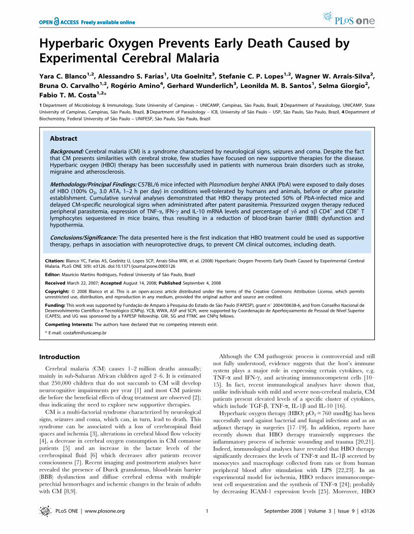
HBO effects on ECM associated mortality and on parasitedevelopment
To evaluate the neuroprotective effect of pressurized oxygen,
two groups of 10 mice each were infected with PbA. One of these
groups was submitted daily to HBO conditions (100% O2, 3.0
ATA, 1 hour) during 11 consecutive days. As shown on Figure 1A,
100% of PbA-infected mice not exposed to HBO exhibited CM-
specific neurological signs within 5 to 8 days after infection and
died of fatal cerebral malaria in the following 24 hours; most
(80%) died on day 7 p.i.. All animals from this group were dead by
day 9 p.i.. In contrast to the non-exposed animals, 50% of the
Figure 1. HBO’s effect on the survival and the parasitedevelopment in P. berghei-infected mice. (A) Groups of 10 miceinfected i.p. with 106 iRBC were exposed or not to HBO (100% O2, 3.0ATA) for 1 h from day 0 to 10. Pressurized oxygen significantlyprotected mice against CM neurological symptoms (P,0.0005).Neurological signs of CM appeared on days 5–10 with death occurringapproximately 24 h after onset (shaded area). Parasitemia levels wereassessed daily in mice infected with (B) P. berghei ANKA (PbA; cerebralline) or (C) P. berghei NK-65 (PbNK-65; non-cerebral line) regardless of
exposure to HBO. HBO significantly (P,0.05) reduced the parasiteburden on days 4–6 and 4–13 p.i., respectively in PbA- and PbNK-65-infected mice when compared to non-exposed animals.doi:10.1371/journal.pone.0003126.g001
Hyperbaric Oxygen in ECM
PLoS ONE | www.plosone.org 3 September 2008 | Volume 3 | Issue 9 | e3126
mice from the HBO group did not develop CM symptoms and
survived. In the HBO group, CM neurological signs began to
appear later and the mortality rate increased slowly throughout
days 7–10, representing 10, 20, 10 and 10%, respectively, on days
7–10. Of note, 1 animal (10%) died on day 14 and 4 (40%) on day
19 post-infection. Cumulative survival statistical analyses clearly
demonstrated that HBO therapy had a significant (P,0.0005)
neuroprotective effect against ECM. As expected, in the mice that
did not develop CM, parasite burden progressed and mice died as
a result of hyperparasitemia (Figure 1B).
As previously reported, HBO therapy inhibits the development
of Leishmania amazonensis and of a non-cerebral line of P. berghei
[30,32,36]. To further explore the effects of HBO, we monitored
the parasitemia levels of infected mice exposed daily, or not, to
HBO (11-day exposure protocol) for up to 19 days. We observed
that HBO significantly (P,0.05) reduced the parasite burden of
PbA-infected mice on days 4, 5 and 6 p.i., when compared to non-
exposed animals (Figure 1B). However, since 100% of non-
exposed PbA-infected mice died, we decided to evaluate whether
the reduction on parasitemia levels in HBO exposed animals could
be sustained over longer periods. Mice infected with P. berghei NK-
65, a non-cerebral strain that displays similar parasitemia levels,
were submitted to pressurized oxygen sessions as in the 11-day
exposure protocol (Figure 1C). As observed in PbA-infected
animals submitted to pressurized oxygen, a significant (P,0.05)
decrease in PbNK-65 development was observed on day 4–13 p.i..
Nevertheless, no correlation was found between mice that
presented a reduction of parasitemia levels with protection or
attenuation of the neurological symptoms (Table S1).
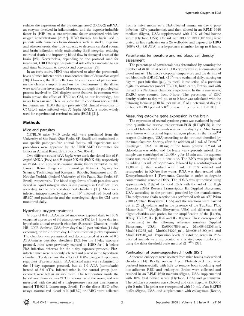
Because we observed that HBO had a significant effect on the
parasite burden in the infections of PbA and PbNK-65, we
addressed the question as to whether pressurized oxygen therapy
could damage normal red blood cells (nRBC) or inhibit parasite
development directly. For this purpose, normal RBC (nRBC)
collected from a naıve mouse were exposed to pressurized oxygen
(100% O2, 3 ATA) during 4 or 6 hours. The relative percentage of
nRBC density was not significantly altered (P.0.05) after direct
exposure to HBO for up to 6 hours (data not shown),
demonstrating that HBO therapy was not toxic to healthy
erythrocytes in these conditions. Next, to evaluate HBO’s effect
directly on parasite development, infected RBC (iRBC) from a
PbA-infected mouse were collected and exposed to HBO (100%
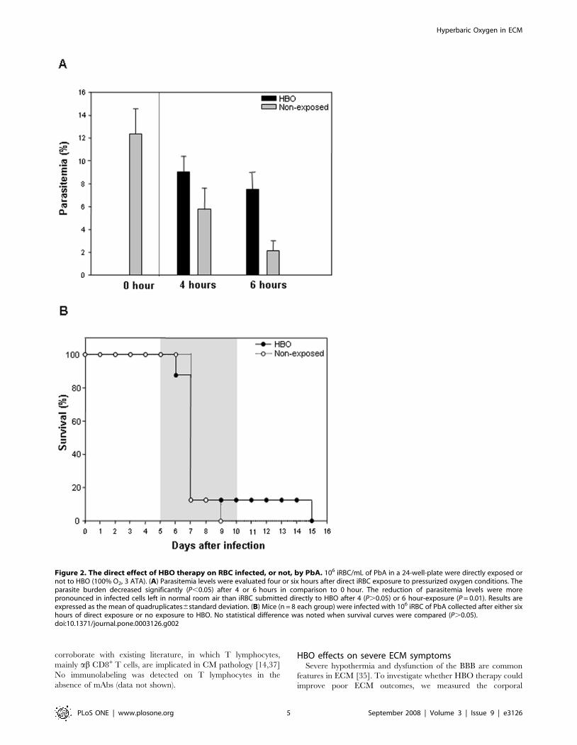
O2, 3 ATA). Figure 2A shows a significant reduction (P,0.05) on
parasite development after 4 and 6 hours in comparison to 0 hour,
regardless of exposure to pressurized oxygen. However, when we
compared the reduction on parasitemia levels of iRBC left in room
air or exposed to HBO, we noticed a significant (P = 0.01) and
more pronounced reduction of the non-exposed iRBC than of the
infected cells directly exposed to HBO up to 6 hours. Inhibition of
parasite development was also observed after 4 hours of exposure;
however, no statistical difference was found (P.0.05). Then, to
assess whether these iRBC were still able to induce CM
neurological signs, we collected 106 iRBC exposed directly to
HBO or left outside the hyperbaric chamber for 6 hours and
injected them in susceptible mice. As shown on Figure 2B, mice
infected with iRBC directly exposed to HBO or with the cells left
outside the chamber did not present significant differences
(P.0.05) when the survival curves were compared. Taken
together, these data suggest that 6 hours of HBO exposure do
not directly affect PbA-infected erythrocytes nor alter their ability
to induce CM clinical symptoms.
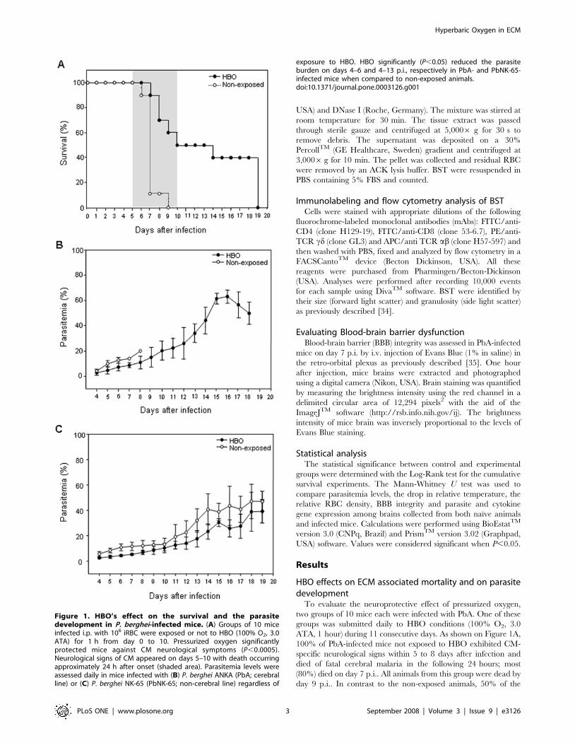
Next, to investigate whether pressurized oxygen could have an
effect when parasitemia was already patent (4%), we randomly
selected half of the PbA-infected mice on day 4 p.i. and exposed
them to daily HBO sessions (100% O2, 3.0 ATA, 2 hours per day)
until day 7 (Figure 3A). As expected, non-treated mice started to
display CM clinical features early on day 5 and 6 and began dying
within 20–24 hours on days 5 (10%) and 6 (10%), though the
majority (80%) died on day 7 p.i.. All mice were dead by day 7.
Notably, hyperbaric oxygen significantly delayed (P,0.01) CM
specific mortality by up to two days, when compared to non-
exposed animals, and reduced the rate of mortality on day 7 from
80% to 40% (Figure 3A). Moreover, two HBO-exposed mice
(20%) only exhibited CM neurological signs on days 8 and 9,
dying within 24 hours on days 9 and 10. This shows that HBO is
capable of interfering significantly with the manifestation of the
CM clinical symptoms, including death, even when administrated
after parasite establishment. As observed in the 11-day exposure
protocol, the administration of pressurized oxygen starting on day
4 p.i. (4-day-exposure) in PbA-infected mice reduced the
parasitemia levels (P,0.01) significantly on days 4–6 (data not
shown).
To confirm that only pressurized oxygen had neuroprotective
effects, PbA-infected mice were submitted to the 11-day exposure
protocol, but using 1.0 ATA as the atmospheric air pressure
(Figure 3B). In this assay, no significant difference (P.0.05) was
observed after cumulative survival analyses between infected
animals exposed to HBO-1.0 ATA and the control mice. Of note,
most of the non-exposed mice began to present CM symptoms and
died earlier than the HBO-1.0 ATA treated animals. Although a
minimal beneficial effect was observed after the administration of
100% oxygen (hyperoxia) under normobaric conditions, this was
not enough to protect or even delay CM neurological symptoms,
thus demonstrating that HBO’s neuroprotective effect does not
rely solely on the administration of 100% oxygen.
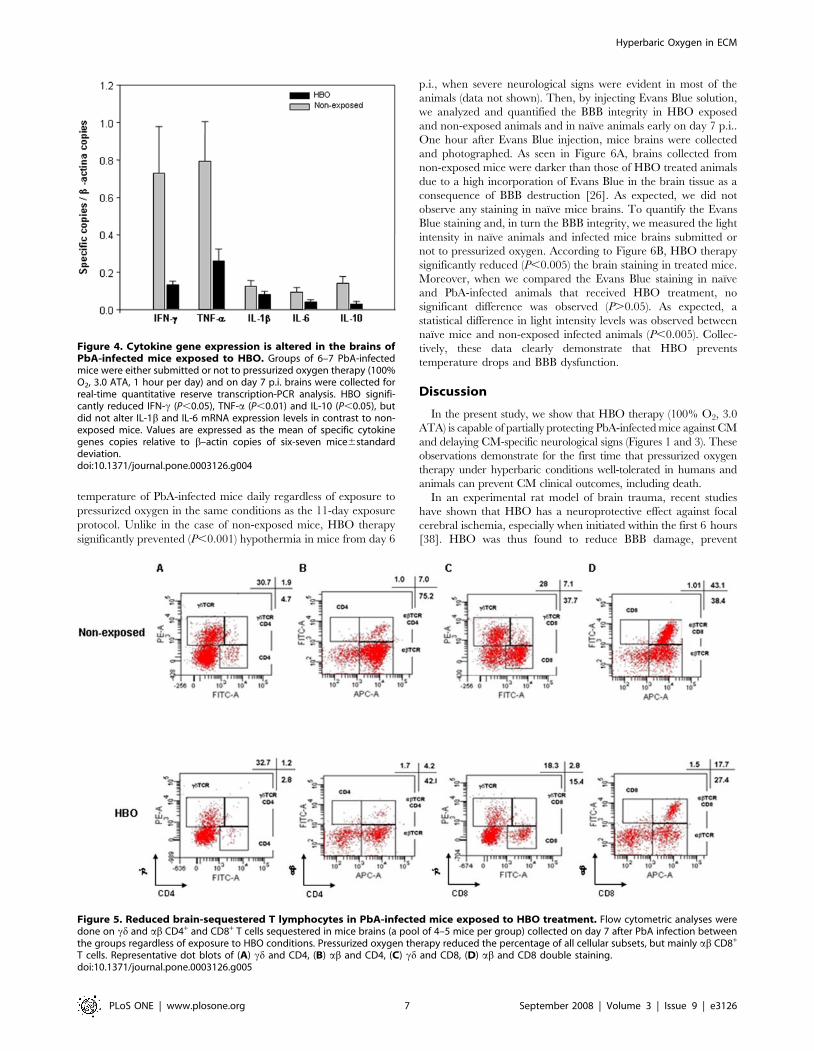
The effect of HBO on cytokine expression levels andadherent T cells in the brain
Based on the anti-inflammatory features of the HBO treatment
reported in ischemic models [21,26] and since the up-regulation of
pro-inflammatory cytokines (IFN-c, TNF-a and IL-1b) [10–12]
and the participation of CD4+ and CD8+ T lymphocytes [14,37] is
essential for CM pathology to occur, we examined the mRNA
levels of different cytokines in the brain of PbA-infected mice
scarified on day 7 p.i.. According to Figure 4, after RT-qPCR
analysis the mRNA levels of IFN-c (P,0.05), TNF-a (P,0.01) and
IL-10 (P,0.05) significantly decreased in the brain of mice
submitted to the 11-day exposure HBO protocol in comparison to
non-exposed animals. No significant difference (P.0.05) was
noted in the mRNA levels of IL-1b and IL-6. RT-negative controls
did not generate a detectable amplification product. All cDNA
samples resulted in a product when the b-actin set of oligonucle-
otides and specific probe were present. Regardless of exposure to
HBO, animals that presented an increase in the expression of IFN-
c mRNA also presented elevated levels of TNF-a and IL-10.
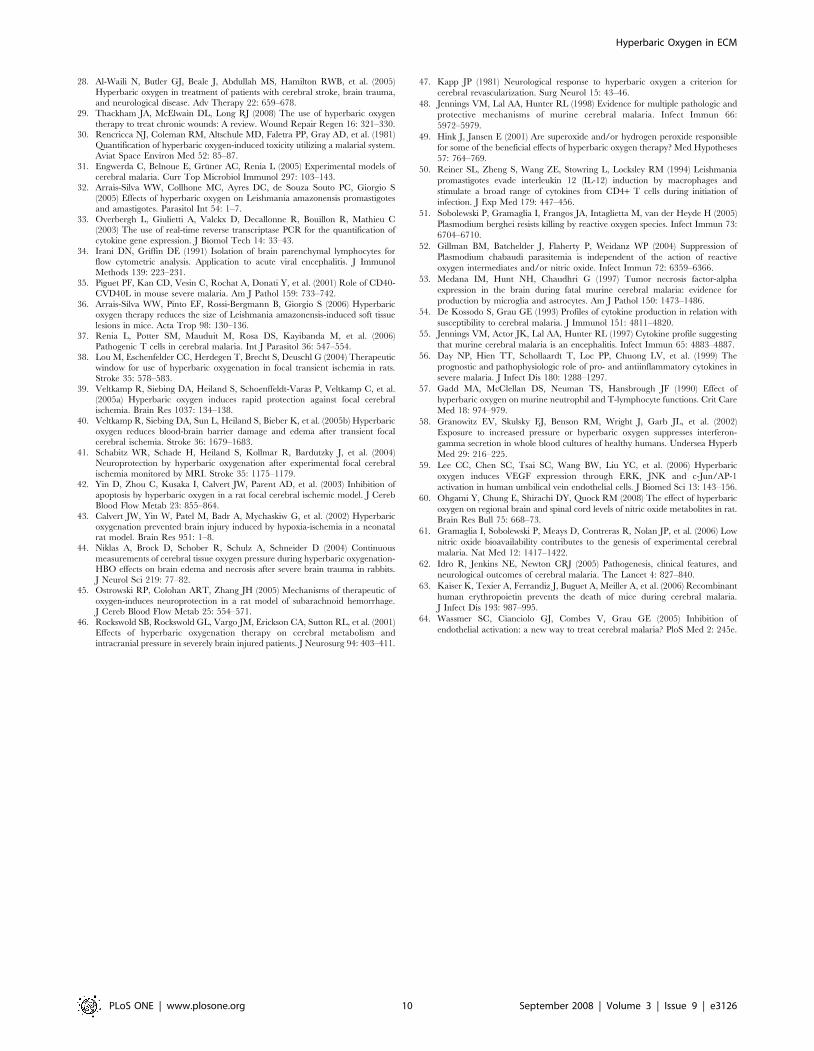
Next, we asked whether the neuroprotective effect of the
pressurized oxygen therapy could be associated to the percentage
of cd and ab T lymphocytes sequestered in mice brains collected
on day 7 p.i. (Figure 5). As compared with brains of non-exposed
animals, HBO treatment reduced about 1.6 fold the percentage of
both cd (1.9 vs. 1.2%) and ab (7.0 vs. 4.2%) CD4+ T cells between
the pools of mice of these two groups (Figure 5A–B). However, a
more pronounced decline, about 2.5 fold, was observed on the
percentage of both cd (7.1 vs. 2.8%) and ab (43.1 vs. 17.7%) CD8+
T lymphocytes in the mice exposed to HBO in contrast to the non-
exposed animals (Figure 5C–D). Taken together, our data
demonstrate that HBO’s neuroprotective effect is related to the
reduction of the T cells sequestered in mice brains; and
Hyperbaric Oxygen in ECM
PLoS ONE | www.plosone.org 4 September 2008 | Volume 3 | Issue 9 | e3126
corroborate with existing literature, in which T lymphocytes,
mainly ab CD8+ T cells, are implicated in CM pathology [14,37]
No immunolabeling was detected on T lymphocytes in the
absence of mAbs (data not shown).
HBO effects on severe ECM symptomsSevere hypothermia and dysfunction of the BBB are common
features in ECM [35]. To investigate whether HBO therapy could
improve poor ECM outcomes, we measured the corporal
Figure 2. The direct effect of HBO therapy on RBC infected, or not, by PbA. 106 iRBC/mL of PbA in a 24-well-plate were directly exposed ornot to HBO (100% O2, 3 ATA). (A) Parasitemia levels were evaluated four or six hours after direct iRBC exposure to pressurized oxygen conditions. Theparasite burden decreased significantly (P,0.05) after 4 or 6 hours in comparison to 0 hour. The reduction of parasitemia levels were morepronounced in infected cells left in normal room air than iRBC submitted directly to HBO after 4 (P.0.05) or 6 hour-exposure (P = 0.01). Results areexpressed as the mean of quadruplicates6standard deviation. (B) Mice (n = 8 each group) were infected with 106 iRBC of PbA collected after either sixhours of direct exposure or no exposure to HBO. No statistical difference was noted when survival curves were compared (P.0.05).doi:10.1371/journal.pone.0003126.g002
Hyperbaric Oxygen in ECM
PLoS ONE | www.plosone.org 5 September 2008 | Volume 3 | Issue 9 | e3126
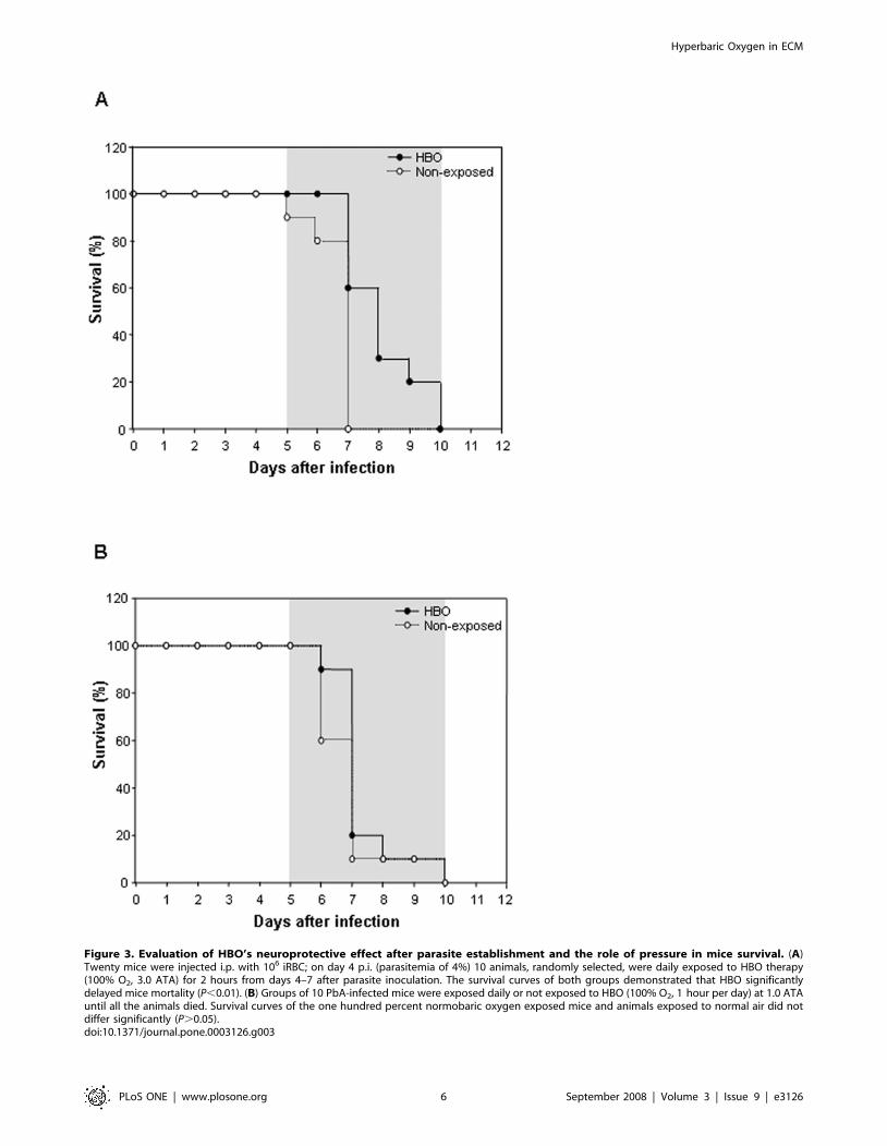
Figure 3. Evaluation of HBO’s neuroprotective effect after parasite establishment and the role of pressure in mice survival. (A)Twenty mice were injected i.p. with 106 iRBC; on day 4 p.i. (parasitemia of 4%) 10 animals, randomly selected, were daily exposed to HBO therapy(100% O2, 3.0 ATA) for 2 hours from days 4–7 after parasite inoculation. The survival curves of both groups demonstrated that HBO significantlydelayed mice mortality (P,0.01). (B) Groups of 10 PbA-infected mice were exposed daily or not exposed to HBO (100% O2, 1 hour per day) at 1.0 ATAuntil all the animals died. Survival curves of the one hundred percent normobaric oxygen exposed mice and animals exposed to normal air did notdiffer significantly (P.0.05).doi:10.1371/journal.pone.0003126.g003
Hyperbaric Oxygen in ECM
PLoS ONE | www.plosone.org 6 September 2008 | Volume 3 | Issue 9 | e3126
temperature of PbA-infected mice daily regardless of exposure to
pressurized oxygen in the same conditions as the 11-day exposure
protocol. Unlike in the case of non-exposed mice, HBO therapy
significantly prevented (P,0.001) hypothermia in mice from day 6
p.i., when severe neurological signs were evident in most of the
animals (data not shown). Then, by injecting Evans Blue solution,
we analyzed and quantified the BBB integrity in HBO exposed
and non-exposed animals and in naıve animals early on day 7 p.i..
One hour after Evans Blue injection, mice brains were collected
and photographed. As seen in Figure 6A, brains collected from
non-exposed mice were darker than those of HBO treated animals
due to a high incorporation of Evans Blue in the brain tissue as a
consequence of BBB destruction [26]. As expected, we did not
observe any staining in naıve mice brains. To quantify the Evans
Blue staining and, in turn the BBB integrity, we measured the light
intensity in naıve animals and infected mice brains submitted or
not to pressurized oxygen. According to Figure 6B, HBO therapy
significantly reduced (P,0.005) the brain staining in treated mice.
Moreover, when we compared the Evans Blue staining in naıve
and PbA-infected animals that received HBO treatment, no
significant difference was observed (P.0.05). As expected, a
statistical difference in light intensity levels was observed between
naıve mice and non-exposed infected animals (P,0.005). Collec-
tively, these data clearly demonstrate that HBO prevents
temperature drops and BBB dysfunction.
Discussion
In the present study, we show that HBO therapy (100% O2, 3.0
ATA) is capable of partially protecting PbA-infected mice against CM
and delaying CM-specific neurological signs (Figures 1 and 3). These
observations demonstrate for the first time that pressurized oxygen
therapy under hyperbaric conditions well-tolerated in humans and
animals can prevent CM clinical outcomes, including death.
In an experimental rat model of brain trauma, recent studies
have shown that HBO has a neuroprotective effect against focal
cerebral ischemia, especially when initiated within the first 6 hours
[38]. HBO was thus found to reduce BBB damage, prevent
Figure 4. Cytokine gene expression is altered in the brains ofPbA-infected mice exposed to HBO. Groups of 6–7 PbA-infectedmice were either submitted or not to pressurized oxygen therapy (100%O2, 3.0 ATA, 1 hour per day) and on day 7 p.i. brains were collected forreal-time quantitative reserve transcription-PCR analysis. HBO signifi-cantly reduced IFN-c (P,0.05), TNF-a (P,0.01) and IL-10 (P,0.05), butdid not alter IL-1b and IL-6 mRNA expression levels in contrast to non-exposed mice. Values are expressed as the mean of specific cytokinegenes copies relative to b–actin copies of six-seven mice6standarddeviation.doi:10.1371/journal.pone.0003126.g004
Figure 5. Reduced brain-sequestered T lymphocytes in PbA-infected mice exposed to HBO treatment. Flow cytometric analyses weredone on cd and ab CD4+ and CD8+ T cells sequestered in mice brains (a pool of 4–5 mice per group) collected on day 7 after PbA infection betweenthe groups regardless of exposure to HBO conditions. Pressurized oxygen therapy reduced the percentage of all cellular subsets, but mainly ab CD8+
T cells. Representative dot blots of (A) cd and CD4, (B) ab and CD4, (C) cd and CD8, (D) ab and CD8 double staining.doi:10.1371/journal.pone.0003126.g005
Hyperbaric Oxygen in ECM
PLoS ONE | www.plosone.org 7 September 2008 | Volume 3 | Issue 9 | e3126

apoptosis and maintain lipid oxidation levels stable [39–42].
HBO’s neuroprotection was also observed in neonatal rats after
the induction of the ischemic process [43]. Rabbits exposed to
pressurized oxygen for 90 min during 3 consecutive days
presented a significant reduction in the edema area of the brain
and cerebral necrosis [44]. In addition, the preservation of BBB,
the reduction in HIF-1a levels, and decreased apoptosis and
neuronal damage were observed in a rat model for subarachnoid
hemorrhage after exposure to HBO [45]. In humans, exposure of
thirty-seven brain-injured patients to sixty minutes of HBO
treatment every 24 hours increased the cerebral metabolic oxygen
rate and reduced cerebrospinal lactate levels [46]. In another
study, 10 out of 22 patients with cerebral infarction presented an
amelioration of their motor function, while 7 of these patients
experienced improved revascularization after pressurized oxygen
sessions [47].
When comparing exposed animals with non-exposed animals,
we noticed a significant reduction on the parasitemia levels of
PbA-infected mice exposed to HBO (11-day exposure protocol)
during infection (4–6 p.i.; Figure 1B). PbNK-65-infected mice
exposed to HBO in the same conditions also presented a
significant reduction of their parasite burden on day 4–13 p.i.
(Figure 1C). These findings are in line with a recent study in which
daily sessions of 100% pressurized oxygen at 2.5 ATA significantly
reduced the size of Leishmania amazonensis induced lesions and the
parasite development in infected mice [36]. Nevertheless, as in
ECM parasites in the brain are necessary, but not sufficient, to
neurological symptoms appearing [15], the lack of correlation
between survival and the reduction of parasitemia levels, measured
daily until the death of PbA-infected animals exposed to HBO,
might be related to the fact that parasitemia levels probably do not
determine the parasite load in the brain. Indeed, methods aimed at
inducing protection against ECM often do not reduce parasitemia
levels [48].
Also, direct exposure to HBO for up to 6 hours observed in our
in vitro analyses was not harmful to normal or PbA-infected
erythrocytes (data not shown and Figure 2), differing from
previous studies where direct exposure of L. amazonensis promas-
tigotes to HBO for up to 6 hours significantly decreased parasite
viability [32]. However, as it is assumed that HBO increases the
levels of reactive oxygen intermediates (ROI) [49], we believe that
the disparity of these two protozoan parasites in terms of HBO
susceptibility might be linked to differential killing mediated by
reactive oxygen intermediates (ROI). In fact, it has been shown
that Leishmania parasite killing is sensitive to ROI, whereas PbA-
infected erythrocytes are resistant to killing by ROI, even at
supraphysiological doses, and ROI are not essential for controlling
Plasmodium sp. parasitemia [50–52].
We have also shown that the neuroprotective effects of daily
hyperbaric sessions rely on the combination of hyperoxia and
pressure at 3.0 ATA (Figure 1A), as ECM-specific mortality of
PbA-infected mice submitted to 100% oxygen pressurized at 1.0
ATA did not differ significantly from the non-exposed animals
(Figure 3B). In an experimental model for cerebral ischemia, HBO
neuroprotection was not achieved in animals submitted to pure
oxygen at only 1.0 ATA [39,40], and human stimulated
monocyte-macrophages cultured in hyperoxia did not present
changes in their cytokine expression levels [23]. More importantly,
in a study of 12 CM comatose patients who breathed 95% oxygen,
no improvement in the consciousness levels were observed in any
of the individuals [7].
Brain macrophages from adults and children who died of CM
had higher levels of immunological markers that are normally not
upregulated [9], such as IFN-c, IL-1b, IL-10 and TNF-a[10,11,16] neuroprotection in ECM is often associated with the
reduction of IFN-c, and TNF-a levels [53–55]. IL-10 is higher in
severe malaria patients from different regions despite the fact that
CM individuals presented lower levels of IL-10 in comparison to
the non-cerebral malaria group [16,56] Furthermore, CD8+ ab T
cells migrating to the brain have been implicated in cytotoxicity
and BBB disruption, thus contributing to ECM mortality [14,15].
Here, we showed that HBO therapy reduced IFN-c, TNF-a and
IL-10 mRNA expression levels in the brain and the percentage of
Figure 6. HBO preserves integrity of the blood-brain barrier inPbA-infected mice. Four PbA-infected mice, representative of eachgroup (n = 8) exposed or not to HBO treatment (100% O2, 3.0 ATA,1 hour per day), received i.v. injections of 1% Evans Blue solution earlyon day 7 p.i.. (A) One hour after Evans Blue injection, brains of naıveanimals, PbA-infected mice and HBO-treated PbA-infected mice werecollected and photographed (n = 4 of each group). (B) The BBBdysfunction of naıve mice or PbA-infected animals, regardless ofsubmission to hyperbaric conditions, was determined by brain stainingquantification with the aid of the ImageJTM software (n = 4 of eachgroup). HBO significantly reduced (P,0.005) the staining in the brainsof infected-mice in comparison to non-treated animals. No statisticaldifference (P.0.05) was noticed between naıve and HBO-treatedinfected mice and brains collected from non-treated infected micewere significantly (P,0.005) darker than naıve animals. Results areexpressed as the mean of brightness intensity of each delimited brainarea of six mice6standard deviation.doi:10.1371/journal.pone.0003126.g006
Hyperbaric Oxygen in ECM
PLoS ONE | www.plosone.org 8 September 2008 | Volume 3 | Issue 9 | e3126

brain-sequestered CD4+ and CD8+ cd and ab T lymphocytes
(Figures 4–5). Moreover, the reduction in the IL-10 levels in PbA-
infected mice exposed to HBO might be associated with the
decrease in expression of IFN-c and TNF-a. These data are in line
with the fact that pressurized oxygen is able to inhibit synthesis of
cytokines, such as TNF-a and IFN-c, T lymphocyte proliferation,
decrease the migration of immunocompetent cells and improve
tissue transplantation by down-regulating lymphoid system
functions [19,22,23,28,57,58].
Finally, when we assessed the HBO effects on cerebral
outcomes, we noticed a significant reduction in hypothermia (data
not shown) and in the BBB breakdown (Figure 6) in mice exposed
to pressurized oxygen. This corroborates previous findings where
HBO (100% O2, 2.8–3.0 ATA) prevented BBB permeability and
functionality in animals submitted to a brain injury [31,36]. Based
on these observations, it is plausible to assume that HBO prevents
BBB breakdown and then avoids vascular leakage by down-
regulating the inflammatory immune response in ECM, but
mainly, by reducing the percentage of brain-sequestered CD8+ T
lymphocytes [10]. Therefore, we cannot rule out that other
mechanisms are also involved in HBO neuroprotective effects in
ECM, as HBO also inhibits ICAM-1 expression and neuronal
apoptosis and upregulates the expression of vascular endothelial
growth factor (VEGF), which is involved in angiogenesis in human
endothelial cells [22,23,25,28,59]. Also, HBO led to an increase in
the brain levels of nitric oxide (NO) [60], a molecule that
contributes to protection against ECM [61].
In summary, we have presented evidence of the beneficial
effects induced by HBO therapy against ECM. We also
demonstrated that the administration of pressurized oxygen down-
regulates IFN-c, TNF-a and IL-10 cytokine expression and the
migration to the brain of T lymphocytes, preventing BBB
breakdown and severe mice hypothermia without directly affecting
iRBC viability and infectivity. Since complementary therapies
such as steroids, sodium bicarbonate and heparin are deleterious
in CM, and treatment with an anti-TNF-a monoclonal can
worsen neurological symptoms [62]. The data presented here
create promising perspectives for further investigation of addition-
al HBO’s neuroprotective mechanisms and to consider it as a new
supportive therapy that could act alone or in association with
conventional treatment or with recently discovered neuroprotec-
tive or anti-inflammatory molecules to improve poor CM
outcomes [63,64].
Supporting Information
Table S1
Found at: doi:10.1371/journal.pone.0003126.s001 (0.01 MB
PDF)
Acknowledgments
Many thanks to Dr. Lindsay Ann Pirrit for revising the English, to Dr.
Laurent Renia for critical reading of the manuscript and to Dr. Lucio H.
Freitas-Junior for delightful discussions.
Author Contributions
Conceived and designed the experiments: YCB UG WWAS FTMC.
Performed the experiments: YCB ASF UG SCPL BOC. Analyzed the
data: YCB ASF UG SCPL WWAS RA GW LMBS SG FTMC.
Contributed reagents/materials/analysis tools: RA GW LMBS SG FTMC.
Wrote the paper: FTMC.
References
1. Carter JA, Ross AJ, Neville BG, Obiero E, Katana K, et al. (2005)
Developmental impairments following severe falciparum malaria in children.
Trop Med Int Health 10: 3–10.
2. Newton CRJC, Krishna S (1998) Severe Falciparum malaria in children: current
understanding of pathophysiology and supportive treatment. Pharmacol Ther
79: 1–53.
3. Newton CR, Peshu N, Kendall B, Kirkham FJ, Sowunmi A, et al. (1994) Brain
swelling and ischaemia in Kenyans with cerebral malaria. Arch Dis Child 70:
281–287.
4. Newton CR, Marsh K, Peshu N, Kirkham FJ (1996) Perturbations of cerebral
hemodynamics in Kenyans with cerebral malaria. Pediatr Neurol 15: 41–49.
5. Pongponratn E, Riganti M, Punpoowong B, Aikawa M (1991) Microvascular
sequestration of parasitized erythrocytes in human falciparum malaria: a
pathological study. Am J Trop Med Hyg 44: 168–175.
6. White NJ, Warrell DA, Looareesuwan S, Chanthavanich P, Phillips RE, et al.
(1985) Pathophysiological and prognostic significance of cerebrospinal-fluid
lactate in cerebral malaria. Lancet 1: 776–778.
7. Warrell DA, White NJ, Veall N, Looareesuwan S, Chanthavanich P, et al.
(1988) Cerebral anaerobic glycolysis and reduced cerebral oxygen transport in
human cerebral malaria. Lancet 2: 534–538.
8. Patankar TF, Karnad DR, Shetty PG, Desai AP, Prasad SR (2002) Adult
cerebral malaria: prognostic importance of imaging findings and correlation with
postmortem findings. Radiology 224: 811–816.
9. Medana IM, Turner GD (2006) Human cerebral malaria and the blood-brain
barrier. Int J Parasitol 36: 555–568.
10. Hunt NH, Grau GE (2003) Cytokines: accelerators and brakes in the
pathogenesis of cerebral malaria. Trends Immunol 24: 491–499.
11. Taylor TE, Fu WJ, Carr RA, Whitten RO, Mueller JS, et al. (2004)
Differentiating the pathologies of cerebral malaria by postmortem parasite
counts. Nat Med 10: 143–145.
12. Rudin W, Favre N, Bordmann G, Ryffel B (1997) Interferon-c is essential for the
development of cerebral malaria. Eur J Immunol 27: 810–815.
13. Hermsen CC, Crommert JV, Fredix H, Sauerwein RW, Eling WM (1997)
Circulating tumour necrosis factor alpha is not involved in the development of
cerebral malaria in Plasmodium berghei-infected C57BL mice. Parasite
Immunol 19: 571–577.
14. Belnoue E, Kayibanda M, Vigario AM, Deschemin JC, van Rooijen N, et al.
(2002) On the pathogenic role of brain-sequestered ab CD8+ T cells in
experimental cerebral malaria. J Immunol 169: 6369–6375.
15. Nitcheu J, Bonduelle O, Combadiere C, Tefit M, Seilhean D, et al. (2003)
Perforin-dependent brain-infiltrating cytotoxic CD8+ T lymphocytes mediate
experimental cerebral malaria pathogenesis. J Immunol 170: 2221–2228.
16. Prakash D, Fesel C, Jain R, Cazenave PA, Mishra GC, et al. (2006) Clusters of
cytokines determine malaria severity in Plasmodium falciparum-infected patients
from endemic areas of Central India. J Infect Dis 194: 198–207.
17. Gudewicz TM, Mader JT, Davis CP (1987) Combined effects of hyperbaric
oxygen and antifungal agents on the growth of Candida albicans. Aviat Space
Environ Med 58: 673–678.
18. Park MK, Myers RA, Marzella L (1992) Oxygen tensions and infections:
modulation of microbial growth, activity of antimicrobial agents, and
immunologic responses. Clin Infect Dis 14: 720–740.
19. Kaide CG, Khandelwal S (2008) Hyperbaric oxygen: applications in infectious
disease. Emerg Med Clin North Am 26: 571–595.
20. Al-Waili NS, Butler GJ (2006) Effects of hyperbaric oxygen on inflammatory
response to wound and trauma: possible mechanism of action. The Scientific
World Journal 6: 425–441.
21. Zhang Q, Chang Q, Cox RA, Gong X, Gould LJ (2008) Hyperbaric oxygen
attenuates apoptosis and decreases inflammation in an ischemic wound model.
J Invest Dermatol 128: 2102–2112.
22. Lahat N, Bitterman H, Yaniv N, Kinarty A, Bitterman N (1995) Exposure to
hyperbaric oxygen induces tumour necrosis factor-alpha (TNF-a) secretion from
rat macrophages. Clin Exp Immum 102: 655–659.
23. Benson RM, Minter LM, Osborne BA, Granowitz EV (2003) Hyperbaric
oxygen inhibits stimulus-induced proinflammatory cytokine synthesis by
human blood-derived monocyte-macrophages. Clin Exp Immunol 134: 57–
62.
24. Yang ZJ, Bosco G, Montante A, Ou XI, Camporesi EM (2001) Hyperbaric O2
reduces intestinal ischemia-reperfusion-induced TNF-a production and lung
neutrophils sequestration. Eur J Appl Physiol 85: 96–103.
25. Buras JA, Stahl GL, Svoboda KK, Reenstra WR (2000) Hyperbaric oxygen
downregulates ICAM-1 expression induced by hypoxia and hypoglycemia: the
role of NOS. Am J Physiol Cell Physiol 278: 292–302.
26. Yin W, Badr AE, Mychaskiw G, Zhang JH (2002) Down regulation of COX-2 is
involved in hyperbaric oxygen treatment in a rat transient focal cerebral
ischemia model. Brain Res 926: 165–171.
27. Li Y, Zhou C, Calvert JW, Colohan AR, Zhang JH (2005) Multiple effects of
hyperbaric oxygen on the expression of HIF-1 alpha and apoptotic genes in a
global ischemia-hypotension rat model. Exp Neurol 191: 198–210.
Hyperbaric Oxygen in ECM
PLoS ONE | www.plosone.org 9 September 2008 | Volume 3 | Issue 9 | e3126
28. Al-Waili N, Butler GJ, Beale J, Abdullah MS, Hamilton RWB, et al. (2005)
Hyperbaric oxygen in treatment of patients with cerebral stroke, brain trauma,and neurological disease. Adv Therapy 22: 659–678.
29. Thackham JA, McElwain DL, Long RJ (2008) The use of hyperbaric oxygen
therapy to treat chronic wounds: A review. Wound Repair Regen 16: 321–330.30. Rencricca NJ, Coleman RM, Altschule MD, Faletra PP, Gray AD, et al. (1981)
Quantification of hyperbaric oxygen-induced toxicity utilizing a malarial system.Aviat Space Environ Med 52: 85–87.
31. Engwerda C, Belnoue E, Gruner AC, Renia L (2005) Experimental models of
cerebral malaria. Curr Top Microbiol Immunol 297: 103–143.32. Arrais-Silva WW, Collhone MC, Ayres DC, de Souza Souto PC, Giorgio S
(2005) Effects of hyperbaric oxygen on Leishmania amazonensis promastigotesand amastigotes. Parasitol Int 54: 1–7.
33. Overbergh L, Giulietti A, Valckx D, Decallonne R, Bouillon R, Mathieu C(2003) The use of real-time reverse transcriptase PCR for the quantification of
cytokine gene expression. J Biomol Tech 14: 33–43.
34. Irani DN, Griffin DE (1991) Isolation of brain parenchymal lymphocytes forflow cytometric analysis. Application to acute viral encephalitis. J Immunol
Methods 139: 223–231.35. Piguet PF, Kan CD, Vesin C, Rochat A, Donati Y, et al. (2001) Role of CD40-
CVD40L in mouse severe malaria. Am J Pathol 159: 733–742.
36. Arrais-Silva WW, Pinto EF, Rossi-Bergmann B, Giorgio S (2006) Hyperbaricoxygen therapy reduces the size of Leishmania amazonensis-induced soft tissue
lesions in mice. Acta Trop 98: 130–136.37. Renia L, Potter SM, Mauduit M, Rosa DS, Kayibanda M, et al. (2006)
Pathogenic T cells in cerebral malaria. Int J Parasitol 36: 547–554.38. Lou M, Eschenfelder CC, Herdegen T, Brecht S, Deuschl G (2004) Therapeutic
window for use of hyperbaric oxygenation in focal transient ischemia in rats.
Stroke 35: 578–583.39. Veltkamp R, Siebing DA, Heiland S, Schoenffeldt-Varas P, Veltkamp C, et al.
(2005a) Hyperbaric oxygen induces rapid protection against focal cerebralischemia. Brain Res 1037: 134–138.
40. Veltkamp R, Siebing DA, Sun L, Heiland S, Bieber K, et al. (2005b) Hyperbaric
oxygen reduces blood-brain barrier damage and edema after transient focalcerebral ischemia. Stroke 36: 1679–1683.
41. Schabitz WR, Schade H, Heiland S, Kollmar R, Bardutzky J, et al. (2004)Neuroprotection by hyperbaric oxygenation after experimental focal cerebral
ischemia monitored by MRI. Stroke 35: 1175–1179.42. Yin D, Zhou C, Kusaka I, Calvert JW, Parent AD, et al. (2003) Inhibition of
apoptosis by hyperbaric oxygen in a rat focal cerebral ischemic model. J Cereb
Blood Flow Metab 23: 855–864.43. Calvert JW, Yin W, Patel M, Badr A, Mychaskiw G, et al. (2002) Hyperbaric
oxygenation prevented brain injury induced by hypoxia-ischemia in a neonatalrat model. Brain Res 951: 1–8.
44. Niklas A, Brock D, Schober R, Schulz A, Schneider D (2004) Continuous
measurements of cerebral tissue oxygen pressure during hyperbaric oxygenation-HBO effects on brain edema and necrosis after severe brain trauma in rabbits.
J Neurol Sci 219: 77–82.45. Ostrowski RP, Colohan ART, Zhang JH (2005) Mechanisms of therapeutic of
oxygen-induces neuroprotection in a rat model of subarachnoid hemorrhage.J Cereb Blood Flow Metab 25: 554–571.
46. Rockswold SB, Rockswold GL, Vargo JM, Erickson CA, Sutton RL, et al. (2001)
Effects of hyperbaric oxygenation therapy on cerebral metabolism andintracranial pressure in severely brain injured patients. J Neurosurg 94: 403–411.
47. Kapp JP (1981) Neurological response to hyperbaric oxygen a criterion for
cerebral revascularization. Surg Neurol 15: 43–46.
48. Jennings VM, Lal AA, Hunter RL (1998) Evidence for multiple pathologic and
protective mechanisms of murine cerebral malaria. Infect Immun 66:
5972–5979.
49. Hink J, Jansen E (2001) Are superoxide and/or hydrogen peroxide responsible
for some of the beneficial effects of hyperbaric oxygen therapy? Med Hypotheses
57: 764–769.
50. Reiner SL, Zheng S, Wang ZE, Stowring L, Locksley RM (1994) Leishmania
promastigotes evade interleukin 12 (IL-12) induction by macrophages and
stimulate a broad range of cytokines from CD4+ T cells during initiation of
infection. J Exp Med 179: 447–456.
51. Sobolewski P, Gramaglia I, Frangos JA, Intaglietta M, van der Heyde H (2005)
Plasmodium berghei resists killing by reactive oxygen species. Infect Immun 73:
6704–6710.
52. Gillman BM, Batchelder J, Flaherty P, Weidanz WP (2004) Suppression of
Plasmodium chabaudi parasitemia is independent of the action of reactive
oxygen intermediates and/or nitric oxide. Infect Immun 72: 6359–6366.
53. Medana IM, Hunt NH, Chaudhri G (1997) Tumor necrosis factor-alpha
expression in the brain during fatal murine cerebral malaria: evidence for
production by microglia and astrocytes. Am J Pathol 150: 1473–1486.
54. De Kossodo S, Grau GE (1993) Profiles of cytokine production in relation with
susceptibility to cerebral malaria. J Immunol 151: 4811–4820.
55. Jennings VM, Actor JK, Lal AA, Hunter RL (1997) Cytokine profile suggesting
that murine cerebral malaria is an encephalitis. Infect Immun 65: 4883–4887.
56. Day NP, Hien TT, Schollaardt T, Loc PP, Chuong LV, et al. (1999) The
prognostic and pathophysiologic role of pro- and antiinflammatory cytokines in
severe malaria. J Infect Dis 180: 1288–1297.
57. Gadd MA, McClellan DS, Neuman TS, Hansbrough JF (1990) Effect of
hyperbaric oxygen on murine neutrophil and T-lymphocyte functions. Crit Care
Med 18: 974–979.
58. Granowitz EV, Skulsky EJ, Benson RM, Wright J, Garb JL, et al. (2002)
Exposure to increased pressure or hyperbaric oxygen suppresses interferon-
gamma secretion in whole blood cultures of healthy humans. Undersea Hyperb
Med 29: 216–225.
59. Lee CC, Chen SC, Tsai SC, Wang BW, Liu YC, et al. (2006) Hyperbaric
oxygen induces VEGF expression through ERK, JNK and c-Jun/AP-1
activation in human umbilical vein endothelial cells. J Biomed Sci 13: 143–156.
60. Ohgami Y, Chung E, Shirachi DY, Quock RM (2008) The effect of hyperbaric
oxygen on regional brain and spinal cord levels of nitric oxide metabolites in rat.
Brain Res Bull 75: 668–73.
61. Gramaglia I, Sobolewski P, Meays D, Contreras R, Nolan JP, et al. (2006) Low
nitric oxide bioavailability contributes to the genesis of experimental cerebral
malaria. Nat Med 12: 1417–1422.
62. Idro R, Jenkins NE, Newton CRJ (2005) Pathogenesis, clinical features, and
neurological outcomes of cerebral malaria. The Lancet 4: 827–840.
63. Kaiser K, Texier A, Ferrandiz J, Buguet A, Meiller A, et al. (2006) Recombinant
human erythropoietin prevents the death of mice during cerebral malaria.
J Infect Dis 193: 987–995.
64. Wassmer SC, Cianciolo GJ, Combes V, Grau GE (2005) Inhibition of
endothelial activation: a new way to treat cerebral malaria? PloS Med 2: 245e.
Hyperbaric Oxygen in ECM
PLoS ONE | www.plosone.org 10 September 2008 | Volume 3 | Issue 9 | e3126
Related Documents


