Hydroxyurea-Stalled Replication Forks Become Progressively Inactivated and Require Two Different RAD51-Mediated Pathways for Restart and Repair Eva Petermann 1 , Manuel Luís Orta 1,2 , Natalia Issaeva 3,4 , Niklas Schultz 3 , and Thomas Helleday 1,3,∗ 1 Gray Institute for Radiation Oncology and Biology, University of Oxford, Oxford OX3 7DQ, UK 2 Department of Cell Biology, University of Seville, E-41012 Seville, Spain 3 Department of Genetics Microbiology and Toxicology, Stockholm University, SE-106 91 Stockholm, Sweden Summary Faithful DNA replication is essential to all life. Hydroxyurea (HU) depletes the cells of dNTPs, which initially results in stalled replication forks that, after prolonged treatment, collapse into DSBs. Here, we report that stalled replication forks are efficiently restarted in a RAD51-dependent process that does not trigger homologous recombination (HR). The XRCC3 protein, which is required for RAD51 foci formation, is also required for replication restart of HU-stalled forks, suggesting that RAD51- mediated strand invasion supports fork restart. In contrast, replication forks collapsed by prolonged replication blocks do not restart, and global replication is rescued by new origin firing. We find that RAD51-dependent HR is triggered for repair of collapsed replication forks, without apparent restart. In conclusion, our data suggest that restart of stalled replication forks and HR repair of collapsed replication forks require two distinct RAD51-mediated pathways. Abstract Graphical Abstract— © 2010 ELL & Excerpta Medica. ∗ Corresponding author [email protected]. 4 Present address: Departments of Radiation Oncology and Environmental Health Sciences, University of Michigan, Ann Arbor, MI 48109-5637, USA This document was posted here by permission of the publisher. At the time of deposit, it included all changes made during peer review, copyediting, and publishing. The U.S. National Library of Medicine is responsible for all links within the document and for incorporating any publisher-supplied amendments or retractions issued subsequently. The published journal article, guaranteed to be such by Elsevier, is available for free, on ScienceDirect. Sponsored document from Molecular Cell Published as: Mol Cell. 2010 February 26; 37(4): 492–502. Sponsored Document Sponsored Document Sponsored Document

Welcome message from author
This document is posted to help you gain knowledge. Please leave a comment to let me know what you think about it! Share it to your friends and learn new things together.
Transcript
Hydroxyurea-Stalled Replication Forks Become ProgressivelyInactivated and Require Two Different RAD51-Mediated Pathwaysfor Restart and Repair
Eva Petermann1, Manuel Luís Orta1,2, Natalia Issaeva3,4, Niklas Schultz3, and ThomasHelleday1,3,∗1Gray Institute for Radiation Oncology and Biology, University of Oxford, Oxford OX3 7DQ, UK2Department of Cell Biology, University of Seville, E-41012 Seville, Spain3Department of Genetics Microbiology and Toxicology, Stockholm University, SE-106 91Stockholm, Sweden
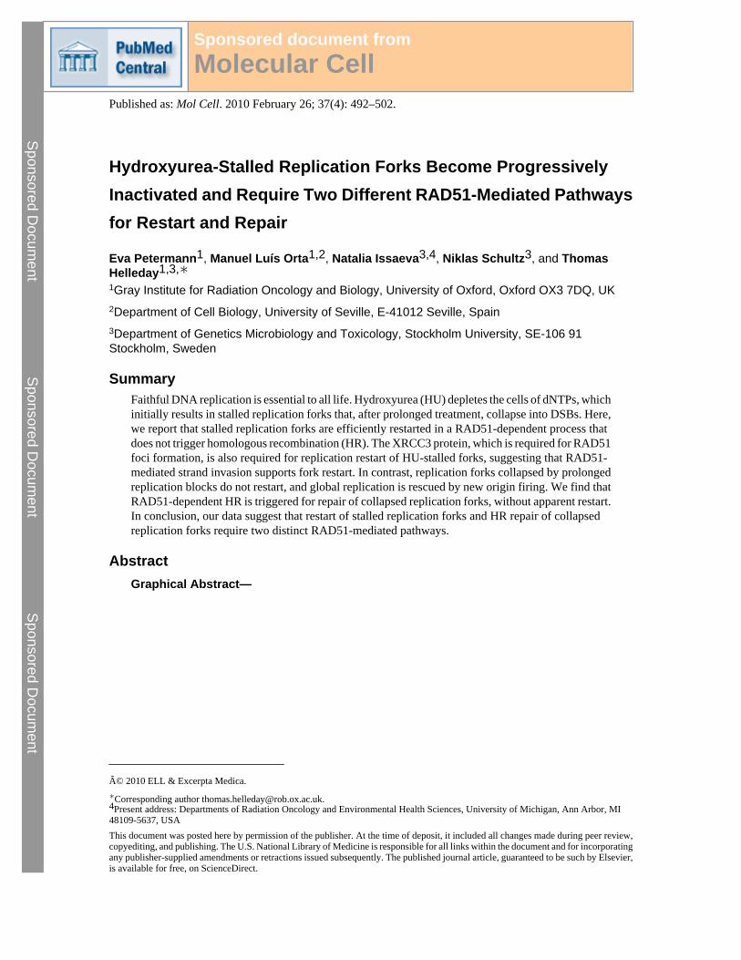
SummaryFaithful DNA replication is essential to all life. Hydroxyurea (HU) depletes the cells of dNTPs, whichinitially results in stalled replication forks that, after prolonged treatment, collapse into DSBs. Here,we report that stalled replication forks are efficiently restarted in a RAD51-dependent process thatdoes not trigger homologous recombination (HR). The XRCC3 protein, which is required for RAD51foci formation, is also required for replication restart of HU-stalled forks, suggesting that RAD51-mediated strand invasion supports fork restart. In contrast, replication forks collapsed by prolongedreplication blocks do not restart, and global replication is rescued by new origin firing. We find thatRAD51-dependent HR is triggered for repair of collapsed replication forks, without apparent restart.In conclusion, our data suggest that restart of stalled replication forks and HR repair of collapsedreplication forks require two distinct RAD51-mediated pathways.
AbstractGraphical Abstract—
© 2010 ELL & Excerpta Medica.∗Corresponding author [email protected] address: Departments of Radiation Oncology and Environmental Health Sciences, University of Michigan, Ann Arbor, MI48109-5637, USAThis document was posted here by permission of the publisher. At the time of deposit, it included all changes made during peer review,copyediting, and publishing. The U.S. National Library of Medicine is responsible for all links within the document and for incorporatingany publisher-supplied amendments or retractions issued subsequently. The published journal article, guaranteed to be such by Elsevier,is available for free, on ScienceDirect.
Sponsored document fromMolecular Cell
Published as: Mol Cell. 2010 February 26; 37(4): 492–502.
Sponsored Docum
ent Sponsored D
ocument
Sponsored Docum
ent
Highlights—► RAD51 promotes replication fork restart after short hydroxyurea (HU) blocks ►RAD51-mediated fork restart does not create homologous recombination (HR) products ► LongHU blocks lead to fork collapse; replication is restarted by new origin firing ► Collapsed replicationforks are repaired by classical RAD51-dependent HR
KeywordsDNA
IntroductionFaithful DNA replication is essential to maintain genome integrity and prevent theaccumulation of cancer-promoting mutations. Replication forks encounter numerous obstacleson the DNA template, which can lead to fork stalling or fork collapse—that is, the dissociationof the replication machinery and the generation of replication-dependent DNA double-strandbreaks (DSBs). The restart of stalled replication forks is best characterized in Escherichiacoli, where reactivation is essential because of the lack of backup origins. Forks in E. coli arereactivated by recombination-dependent or -independent pathways catalyzed by the RuvABCor PriA and PriC proteins, respectively (Heller and Marians, 2006). These proteins are notconserved in eukaryotes, and the extent and mechanisms of eukaryotic replication forkreactivation are not well characterized. In mammalian cells, agents that stall or collapsereplication forks, such as hydroxyurea (HU), thymidine, and camptothecin, strongly inducehomologous recombination (HR), which promotes the survival of these treatments (Arnaudeauet al., 2001; Lundin et al., 2002; Saintigny et al., 2001), suggesting that recombination-dependent replication restart mechanisms might also be used by higher eukaryotes. If forks arekept stalled for more than 12 hr, increasing amounts of fork-associated DSBs are generated(Saintigny et al., 2001) in a process dependent on the structure-specific endonuclease MUS81(Hanada et al., 2007). This supports a model of replication fork restart via recombination,initiated by a one-ended DSB, which is similar to the RuvABC-mediated mechanism(Helleday, 2003; Heller and Marians, 2006). MUS81-dependent DSBs only start to appear aftermany hours of HU treatment, and the formation of RAD51 foci at stalled forks is independent
Petermann et al. Page 2
Published as: Mol Cell. 2010 February 26; 37(4): 492–502.
Sponsored Docum
ent Sponsored D
ocument
Sponsored Docum
ent
of DSB formation (Hanada et al., 2007), suggesting that RAD51 might be involved in adifferent fork restart mechanism avoiding DSB formation, especially after short replicationblocks (Helleday, 2003). RAD51, the eukaryotic RecA homolog, is an essential HR factor thatcatalyzes homology search and strand exchange (Baumann et al., 1996; Li and Heyer, 2008).RAD51 promotes survival of replication stress and prevents accumulation of replication-associated DSBs (Lundin et al., 2003; Sonoda et al., 1998). The formation of RAD51presynaptic filaments and resulting nuclear foci in response to HU is mediated by the RAD51paralogs, including XRCC3 (Bishop et al., 1998).
Here, we analyze replication restart after different lengths of HU blocks and the roles playedby RAD51 in this process. Our data suggest that RAD51 has distinct early and late roles duringreplication blocks, facilitating replication fork restart when forks are still viable and repairingfork-associated DNA damage after forks have collapsed and global replication is rescued bynew origin firing.
ResultsReplication Forks Become Inactivated during Prolonged Replication Blocks
To understand the fate of replication forks following replication blocks, we analyzed the restartof replication forks after different periods of HU treatment using the DNA fiber technique(Henry-Mowatt et al., 2003). HU depletes deoxyribonucleotide pools and immediately stallsreplication forks (Bianchi et al., 1986). U2OS cells were pulse-labeled with 5-chlorodeoxyuridine (CldU) for 20 min, washed and blocked in HU for 2 hr, washed again andpulse-labeled with 5-iododeoxyuridine (IdU) for 1, 2, or 24 hr (Figure 1A). Afterward, DNAspreads were prepared and analyzed by immunofluorescence (Figure 1B). To quantifyreplication fork restart, the amount of stalled forks was related to the total number of replicationtracks labeled with CldU (Figure 1C). Surprisingly, we found that, although most forksrestarted after release from a 1 or 2 hr HU block, most forks remained stalled after release from24 hr HU block. Instead of restarting forks, replication tracks labeled only with IdU appearedthat seemed to result from new initiation events (Figures 1B and 1C). Because it was previouslyshown that nucleotide incorporation resumes between 12 and 18 hr of HU blocks (Hanadaet al., 2007), we included IdU during the HU treatment to determine whether the apparent lackof restart was due to forks moving large distances during the treatment, resulting in the twolabels becoming separated (see Figure S1A available online). Most forks moved less than 6μm (15.5 kb) during the 24-hr block (Figures S1B and S1C). CldU tracks in less than 6 μmdistance from IdU tracks were therefore not considered as stalled forks. Taking this intoconsideration, our data suggest that most forks become inactivated after long times in HU.
Replication Restarts by Firing of New Origins after Long Replication BlocksTo test whether the new initiation events observed during restart from 24 hr HU blocks occurredin cells that had been replicating before the HU block, or only in cells newly entering S phase,cells were pulse-labeled like before, but then fixed and immunostained to reveal replicationfoci (Figure 1D). Most cells that had contained active forks before the HU treatment did resumereplication after removal of the drug (Figure 1E). As expected, a large number of cells alsonewly entered S phase and were only labeled with IdU (Figure 1F). These observations suggestthat most of the replication restart observed after release from long HU blocks does not involveelongation of existing forks, but instead the firing of new replication origins. Fork inactivationand new origin firing after 24 hr HU treatment was not dependent on the processing of stalledforks into DSBs, because both were not affected by siRNA depletion of MUS81 (Figures S1D–S1F).
Petermann et al. Page 3
Published as: Mol Cell. 2010 February 26; 37(4): 492–502.
Sponsored Docum
ent Sponsored D
ocument
Sponsored Docum
ent
Replication Forks Accumulate Damage after Long Replication BlocksTo confirm the inactivation of replication forks by long HU blocks, we measured the formationof the phosphorylated histone variant H2AX (γH2AX) at inactivated forks. γH2AXaccumulates quickly during HU blocks (Figures 2A and 2B), even before DSB induction isobserved (Figure 2C) (Saintigny et al., 2001). The γH2AX signal colocalized with RPA foci,suggesting that it marks regions of extensive single-stranded DNA at stalled forks (Figure 2A).We released cells from the HU block for 1 hr and measured how much γH2AX remained afterreplication had resumed (Figure 2B). We found that γH2AX rapidly disappeared after releasefrom the 2 hr HU block. In contrast, γH2AX foci persisted after release from 24 hr HU block,at times when more DSB were induced (Figure 2B, C). Persisting γH2AX foci colocalized withstalled or inactivated replication forks (Figure 2D). These observations show that DNA damageaccumulates at stalled forks with increasing lengths of HU treatments and that this DNAdamage persists in cells released from long HU blocks.
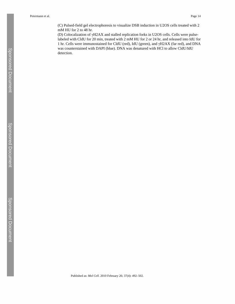
RAD51 Foci and HR Are Induced Late during Replication BlocksTo analyze the response of RAD51 and HR at early and late times of replication blocks, weperformed time courses of RAD51 foci formation and HR frequencies induced by HUtreatment. Cells were treated with HU for different times, and the percentages of cellscontaining more than 10 RAD51 foci were quantified. RAD51 foci are induced after 24 hr, butnot after 1 or 2 hr HU treatment in U2OS cells (Figures 3A and 3B). To analyze the time courseof HR induction by HU treatments, we used the SPD8 cell line, which carries a recombinationreporter in the hprt gene. HR by either unequal sister chromatid exchange, intrachromatidexchange, single-strand annealing, or gene conversion can lead to restoration of the wild-typehprt gene encoding a functional HGPRT protein (Helleday et al., 1998). We found that HR isinduced by HU treatments of 24, but not by treatments of 1 or 2 hr (Figure 3C). This findingsuggested that HR is not active while replication forks restart, but is induced when replicationforks become inactivated. Because we used SPD8 cells to measure HR, we confirmed that theydisplay similar progressive replication fork inactivation as do U2OS cells (Figures S2A–S2D).Forks become inactivated considerably earlier in SPD8 cells, accompanied by earlier RAD51foci formation (Figure S2E), which colocalize with stalled replication forks (Figure S2F). DSBinduction occurred earlier in SPD8 cells as well, although there was no difference to U2OScells at 2 hr (Figure S2G). These observations confirm that HR activation occurs late duringreplication blocks and coincides with or is preceded by replication fork inactivation and DSBformation.
RAD51 Promotes Early Fork RestartBecause RAD51 foci have been suggested to require several kilobases of single-stranded DNA(Raderschall et al., 1999), we tested whether shorter HU treatments lead to a form of RAD51recruitment not detectable as foci. Coimmunoprecipitation with CldU-labeled replication sitesshowed that RAD51 localizes to chromatin after short HU treatments in U2OS cells and evenin the absence of HU (Figure 4A). These observations suggest that RAD51 and HR might playdifferent early and late roles at stalled replication forks. A possible role of RAD51 during shortreplication blocks would be to promote the restart of stalled replication forks. To test this idea,we determined the effect of RAD51 depletion on the restart of individual replication forks afterHU blocks. We depleted U2OS cells of RAD51 using siRNA (Figure 4B). We found thatRAD51 depletion increases the number of forks that do not resume replication after releasefrom 2 hr HU block (Figures 4C and 4D). To confirm specificity of the siRNA treatment,RAD51 siRNA-treated cells were cotransfected with expression vectors encoding wild-type(RAD51-WT) or siRNA targeting resistant (RAD51-Res) RAD51 (Figure 4B). Re-expressionof RAD51 protein reduced percentages of stalled forks after release from 2 hr HU treatment
Petermann et al. Page 4
Published as: Mol Cell. 2010 February 26; 37(4): 492–502.
Sponsored Docum
ent Sponsored D
ocument
Sponsored Docum
ent
back to control levels (Figures 4C and 4D). These data show that RAD51 is required toreactivate a subset of replication forks after short HU blocks.
XRCC3 Promotes Fork Restart after Short Replication BlocksThe XRCC3 protein is required for RAD51 ssDNA complex formation (Bishop et al., 1998).To test whether RAD51 complex formation, which is required for strand invasion (Baumannet al., 1996), is involved in replication restart, we analyzed whether XRCC3 is also requiredfor early restart of replication forks by depleting U2OS cells of XRCC3 using siRNA(Figure 5A). Forty-eight hours later, cells were pulse-labeled with CldU, blocked with HU,and released into IdU for 1 hr. We found that, like RAD51 depletion, XRCC3 depletionincreases the number of forks that do not resume replication after release from 2 hr HU block(Figures 5B and 5C). To confirm the specificity of the XRCC3 depletion, we used twoindividual siRNA duplexes targeting different sequences in the XRCC3 mRNA, with similarresults (Figure 5C). These observations demonstrate that, like RAD51, XRCC3 is required toreactivate stalled replication forks.
The S Phase Checkpoint Suppresses New Origin Firing at Stalled or Collapsed ReplicationForks
Although reduced fork restart after long HU blocks was accompanied by increased new originfiring, origin firing was not elevated when RAD51-depled cells were released from short HUblocks (Figure 6A). To test whether this might be due to origin suppression by the S phasecheckpoint, we analyzed replication restart in the presence or absence of the Chk1 inhibitorCEP-3891 (Figure 6B). We found that Chk1 inhibition alone did reduce fork restart to the sameextent as RAD51 depletion, which is in agreement with previous reports that Chk1 stabilizesstalled replication forks (Feijoo et al., 2001; Zachos et al., 2003). Chk1 inhibition alsodramatically increased new origin firing after release from 2 hr HU block (Figure 6B). Theseeffects were specific to HU treatment (Figure S3). When RAD51 depletion and Chk1 inhibitionwere combined, we observed an additive effect on both fork stalling and new origin firing(Figure 6B). This finding suggests that failed fork restart due to RAD51 depletion can triggernew origin firing, but after short HU blocks, the checkpoint prevents this from being asignificant mechanism of replication restart. Even after release from 24 hr HU treatment, whennew origin firing was increased by 10 fold, Chk1 inhibition during the time of restart increasednew origin firing even further (Figure 6C), suggesting that the S phase checkpoint stillsuppresses a large amount of origin firing after release from long HU blocks. Chk1 inhibitorwas only present during the last hour of HU block and did not affect fork restart (Figure 6C).
RAD51 Promotes Repair of Collapsed Replication Forks after Long Replication BlocksOur data suggested that RAD51 promotes replication fork restart after short replication blocks,when forks are still competent to restart. Next, we wanted to analyze the effect of RAD51 onfork restart after long HU blocks, when RAD51 foci accumulate and HR is activated. Weconfirmed that control cells, but not RAD51-depleted cells, formed RAD51 foci in responseto 24 hr HU block (Figure 7A). We used the DNA fiber technique to determine the effect ofRAD51 depletion and lack of RAD51 foci on fork restart after 24 hr HU block. We found that,in contrast to release from short HU treatments, RAD51 depletion does not decrease the numberof forks that restart after release from long HU blocks (Figure 7B). RAD51 depletion did notaffect new origin firing (Figure 7B). However RAD51-depleted cells repair DNA damageinduced by long HU treatment less efficiently than do control cells, as demonstrated by thehigher amounts of DSB remaining in RAD51-depleted cells up to 48 hr after release from HUblock (Figures 7C and 7D). Similarly, a larger number of RAD51-depleted cells still containedγH2AX signal up to 48 hr after release from HU (Figure S4). These observations suggest thatthere is no correlation between the ability of cells to form RAD51 foci and the ability to restart
Petermann et al. Page 5
Published as: Mol Cell. 2010 February 26; 37(4): 492–502.
Sponsored Docum
ent Sponsored D
ocument
Sponsored Docum
ent
replication forks. Rather, RAD51 foci formation coincides with replication fork inactivation,DSB formation, and the requirement for RAD51 for DNA repair. Taken together, these datasupport the idea that RAD51 protein promotes fork restart without forming foci, whereasRAD51 foci formation is a step in the recombination process that repairs collapsed forks.
DiscussionWe have shown that most replication forks resume progression after short replication bocksby HU but do not restart after long HU blocks in either U2OS or SPD8 cells. Our data suggestthat stalled replication forks retain the ability to restart for some time before becominginactivated in a process that coincides with accumulation of DNA damage and DSB formation.In fission yeast, the replication checkpoint controls the structural integrity of stalled replicationforks and prevents the formation of recombination foci (Alabert et al., 2009; Barlow andRothstein, 2009; Meister et al., 2005). The checkpoint also controls MUS81 activity, and HU-stalled forks are processed into DSBs in checkpoint-deficient mutants only (Froget et al., 2008;Kai et al., 2005). In mammalian cells, the ATR- and Chk1-dependent checkpoint keepsreplication foci active and prevents excessive DSB formation during replication blocks,suggesting that similar control mechanisms exist (Feijoo et al., 2001; Sorensen et al., 2005;Zachos et al., 2003). Fork collapse in mammalian cells could therefore result from checkpointadaptation or a leaky checkpoint that allows gradual disintegration or collapse of stalledreplication forks. It was recently shown that vertebrate Polo-like kinase 1 mediates adaptationof the replication and G2/M checkpoints (Syljuasen et al., 2006; Yoo et al., 2004) and can alsoconstitutively down-regulate the S phase checkpoint (Trenz et al., 2008). We observe reducedfork restart if Chk1 is inhibited during short HU blocks, supporting the idea that the S phasecheckpoint stabilizes stalled replication forks. However, checkpoint inhibition during a 2 hrHU treatment inactivates a much smaller number of forks, compared with a 24 hr HU treatment,suggesting that the length of the replication block plays an important role in fork inactivation.Cleavage of forks into DSBs does not seem to be the primary cause of fork inactivation, becauseMUS81 depletion did not affect fork restart in U2OS cells, and fork inactivation after shortHU blocks is much higher in SPD8 cells, without more DSB formation. However, fork cleavageinto DSBs might be a result of fork inactivation and prerequisite for replication fork repair.
It has been proposed that eukaryotic forks might not necessarily need to restart becausereplication can be completed from adjacent origins (Branzei and Foiani, 2007; Paulsen andCimprich, 2007). We now show that new origin firing can indeed be a major mechanism bywhich mammalian cells resume replication. Stalled forks do not need to be converted into DSBsto trigger new origin firing, because MUS81-depleted cells also display increased origin firingafter release from long HU blocks. This finding is in agreement with previous reports thatglobally reduced replication fork speeds alone can increase origin firing (Anglana et al., 2003;Ge et al., 2007). Ge and co-workers (2007) also showed that replication inhibition can triggernew origin firing locally within active replication clusters. However, our data suggest that,after short HU treatments, any large-scale increase in new origin firing is prevented by theChk1-mediated S phase checkpoint. This also applies to new origin firing after direct forkcollapse induced by camptothecin (Seiler et al., 2007 and data not shown). However, after longHU treatments, suppression of new origin firing by the checkpoint is clearly incomplete, whichmay lend some support to the idea that checkpoint adaptation occurs. Alternatively or inaddition, the sheer extent of replication fork inactivation could allow so many dormant originsto fire that a larger number can escape the checkpoint.
Our observations that replication forks do not restart after prolonged HU treatments are indisagreement with previously published observations observing fork restart after 24 hr HU(Hanada et al., 2007). These data were obtained using mouse embryonic stem cells, and wecannot exclude differences between different cell systems; it is noteworthy that both U2OS
Petermann et al. Page 6
Published as: Mol Cell. 2010 February 26; 37(4): 492–502.
Sponsored Docum
ent Sponsored D
ocument
Sponsored Docum
ent
and SPD8 are transformed cell lines. Interestingly, it has been reported that p53 protects cellsfrom HU-induced DSBs (Kumari et al., 2004), which agrees with the different kinetics of forkinactivation in p53-proficient U2OS and p53-deficient SPD8 cells.
In E. coli, Holliday Junctions are formed as intermediates for restart of stalled replication forks(McGlynn and Lloyd, 2002). RecA, the RAD51 homolog in E. coli, is required for theformation of these Holliday Junctions in the absence of dnaB (Seigneur et al., 2000). Here, wesuggest that RAD51 plays a similar role in mammalian cells. In such a model, RAD51 wouldcoat ssDNA regions occurring surrounding a stalled replication fork and would then invadethe homologous molecule, which would facilitate the formation of a Holliday Junction, oftenreferred to as a “chicken foot” (Figure 7E). RAD51 could stabilize this structure by bindingthe ssDNA tail of paired nascent strands or possibly the dsDNA of the reversed fork, if this isnot prevented by BRCA2 (Carreira et al., 2009). Such a chicken foot structure may be morestable and may facilitate replication restart from the DNA end, which could potentially also bemediated by RAD51-dependent strand invasion. This type of end-induced replication shouldtheoretically be able to cause a recombination event in a recombination reporter. To detectrecombination in this reporter requires an unequal recombination event occurring over severalkilobases of DNA, and the recombination tract involved in restarting a replication fork is likelyrestricted to a very short piece of DNA because the Holliday Junction would preventrecombination at a distant site. The benefit of RAD51-mediated restart of forks stalled byreplication inhibitors might be that the Holliday Junction intermediate can serve as a substratefor origin-independent replisome loading, as appears to be the case in bacteria (Heller andMarians, 2006).
Our data suggest that the HR induced by HU (Lundin et al., 2002; Saintigny et al., 2001) doesnot restart replication forks but performs postreplication repair of collapsed forks. In line withthis argument, a recent report showed that RAD51-dependent mechanisms removespontaneously accumulated ssDNA foci preferentially during the G2 phase of the cell cycle(Su et al., 2008), suggesting that HR is temporally separated from DNA replication that createsthese foci. It has been previously shown that HR also preferentially repairs direct DSBs duringlate S/G2 phase (Rothkamm et al., 2003; Takata et al., 1998; Vispe et al., 1998), but thisobservation could be due to the fact that HR can repair DSBs only if two sister chromatids arepresent, which is more likely after replication has been completed. If a replication fork froman adjacent origin arrived at an unrepaired collapsed fork, this would result in a two-endedDSB that could also be a substrate for nonhomologous end joining. This would explain the HUsensitivity of nonhomologous end joining mutant cells previously reported (Lundin et al., 2002;Saintigny et al., 2001).
Taken together, our data suggest that RAD51 has distinct early and late roles during replicationblocks and promotes the restart of stalled forks and the repair of collapsed forks by differentmechanisms.
Experimental ProceduresCell Lines and Reagents
U2OS cells were obtained from ATCC. SPD8 cells have been described elsewhere (Helledayet al., 1998). Cells were confirmed to be free of Mycoplasma infection and grown in Dulbecco'smodified Eagle's Medium with 10% fetal bovine serum in a humidified atmosphere containing5% CO2. CEP-3891 was obtained from Cephalon.
Petermann et al. Page 7
Published as: Mol Cell. 2010 February 26; 37(4): 492–502.
Sponsored Docum
ent Sponsored D
ocument
Sponsored Docum
ent
DNA Fiber AnalysisU2OS cells were pulse-labeled with 25 μM CldU for 20 min, washed three times with medium,incubated in 2 mM HU for times indicated, washed three times with medium, and pulse-labeledwith 250 μM IdU for 1 hr. Labeled cells were harvested, and DNA fiber spreads were preparedas described elsewhere (Henry-Mowatt et al., 2003). CldU was detected by incubating acid-treated fiber spreads with rat anti-BrdU monoclonal antibody (1:1000; AbD Serotec) for 1 hr.Slides were fixed with 4% PFA and incubated with AlexaFluor 555–conjugated goat anti–ratIgG (1:500; Molecular Probes) for 1.5 hr. IdU was detected using mouse anti-BrdU monoclonalantibody (1:1000; Becton Dickinson) overnight at 4°C and AlexaFluor 488–conjugated goatanti–mouse IgG (1:500; Molecular Probes) for 1.5 hr. Fibers were examined using a BioradRadiance confocal microscope with a 60× oil immersion objective. For quantification ofreplication structures, at least 250 structures were counted per experiment. The lengths of red(AF 555) or green (AF 488) labeled patches were measured using the ImageJ software (NationalInstitutes of Health; http://rsbweb.nih.gov/ij/) and arbitrary length values were converted intomicrometers using the scale bars created by the microscope.
ImmunofluorescencePrimary antibodies were rabbit polyclonal anti-RAD51 (H92, Santa Cruz Biotechnology,1:500–1:1000), rabbit polyclonal anti-RPA70 (a kind gift from Prof. Rolf Knippers, Konstanz,Germany; 1:1000), mouse monoclonal and rabbit polyclonal anti–phospho-Histone H2AX(Ser139) (both Upstate Biotechnology; 1:1000), rat monoclonal anti-BrdU (AbD Serotec;1:400) to detect CldU, and mouse monoclonal anti-BrdU (1:50; Becton Dickinson) to detectIdU. Secondary antibodies were anti–rabbit IgG AlexaFluor 555 or AlexaFluor 647, anti–mouse IgG AlexaFluor 488, and anti–rat IgG AlexaFluor 555 (Molecular Probes). Forcolocalization with replication forks, primary and secondary antibodies against phospho-Histone H2AX were fixed for 10 min with 2% PFA before DNA denaturation with 2 M HClfor 40 min and immunostaining for thymidine analogs. DNA was counterstained with DAPI.
Pulsed-Field Gel ElectrophoresisCells (2.5 × 10⁶) were treated with 2 mM HU for the times indicated. For DSB repairexperiments, U2OS cells were transfected with control or RAD51 siRNA for 36 hr, then treatedwith 2 mM HU for 24 hr, and released into fresh medium to allow repair. Afterward, cells weretrypsinized and melted into 1.0% InCert-Agarose (BMA) inserts. Subsequently, agarose insertswere digested in 0.5 M EDTA-1% N-laurylsarcosyl-proteinase K (1 mg/ml) at 50°C for 48 hrand washed four times in TE buffer. The inserts were loaded onto a separation gel (1.0%chromosomal-grade agarose; Bio-Rad). Separation was performed on a CHEF DR IIIequipment (BioRad; 120 field angle, 240 s switch time, 4 V cm−1, 14°C) for 24 hr. Gels werestained with ethidium bromide, and DSBs were quantified (chromosome fragments >2 Mbp).Densitometric analysis was performed using the PCBASS 2.0 software.
Recombination in SPD8 CellsSPD8 cells were grown in the presence of 6-thioguanine to suppress spontaneousrecombination. Cells (1.5 × 10⁶) cells were treated with 0.5 mM HU for times indicated andrecovered in medium for 48 hr. HPRT+ revertants were selected by plating 3 × 10⁵ cells in thepresence of HAsT (50 μM hypoxanthine, 10 μM L-azaserine, and 5 μM thymidine). Todetermine cloning efficiency, two dishes were plated with 500 cells each. Colonies were stainedwith methylene blue following 7 (for cloning efficiency) or 10 (for reversion) days ofincubation (see Supplemental Data for more detailed protocol).
Petermann et al. Page 8
Published as: Mol Cell. 2010 February 26; 37(4): 492–502.
Sponsored Docum
ent Sponsored D
ocument
Sponsored Docum
ent
CldU Coimmunoprecipitation of Proteins Present at Stalled Replication ForksU2OS cells (2 × 10⁶) were treated with 2 mM HU for 3 hr. HU was removed, and cells werelabeled with 100 μM CldU for 40 min. Cells were cross-linked in 1% PFA for 15 min. Thecytoplasmic protein fraction was removed by incubation in hypotonic buffer (10 mM HEPES[pH 7], 50 mM NaCl, 0.3 M sucrose, 0.5% TX-100, and protease inhibitor cocktail [Roche])for 10 min on ice and centrifugation at 1500 g for 5 min. Nuclear soluble fraction was removedby incubation with nuclear buffer (10 mM HEPES [pH 7], 200 mM NaCl, 1 mM EDTA, 0.5%NP-40, and protease inhibitor cocktail) for 10 min on ice and centrifugation at 13,000 rpm for2 min. Pellets were resuspended in lysis buffer (10 mM HEPES [pH 7], 500 mM NaCl, 1 mMEDTA, 1% NP-40, and protease inhibitor cocktail), sonicated, and centrifuged for 30 s at13,000 rpm, and the supernatant was transferred to a new tube. Total protein (150 μg) was usedfor IP with 2 μg anti-CldU antibody (rat-anti-BrdU; OBT0030F AbD Serotec) and 20 μl ofProtein A/G-PLUS agarose (Santa Cruz Biotechnology). The IP reaction was washed twicewith nuclear buffer and twice with washing buffer (10 mM HEPES and 0.1mM EDTA proteaseinhibitor cocktail), incubated in 2× sample loading buffer (100 mM Tris HCl [pH 6.8], 100mM DTT, 4% SDS, 0.2% bromophenol blue, and 20% glycerol) for 30 min at 90°C, and wasused for Western Blot with rabbit polyclonal anti-Rad51 (H92, Santa Cruz Biotechnology;1:500), rabbit polyclonal anti-H3 (Fl-136, Santa Cruz Biotechnology; 1:500), and mousemonoclonal anti-γH2AX (3F2, Abcam; 1:2000).
siRNA TreatmentsiRNA against human Rad51 (Ito et al., 2005), XRCC3 (siGENOME SMARTpool D-012067),and MUS81 (siGENOME SMARTpool D-016143) were purchased from Dharmacon.Individual XRCC3 siRNA #1 and #2 were Dharmacon siGenome D-012067-01 and −04.“Allstars negative control siRNA” was purchased from QIAGEN. Cells were transfected with50 nM siRNA using Dharmafect 1 reagent (Dharmacon) or Lipofectamine2000 (Invitrogen)for pulsed-field gel electrophoresis. Cells were cultured for 48 hr prior to DNA labeling andHU treatments. Depletion was confirmed by Western Blot using rabbit anti-Rad51 (1:1000;H-92, Santa Cruz), rabbit anti-XRCC3 (1:5000; Novus Biologicals), mouse anti-MUS81(1:500; MTA30 2G10/3, Santa Cruz), and mouse anti-αTubulin (1:5000; Sigma). For re-expression of Rad51, 50 nM RAD51 siRNA and 78 pM (1 μg) of RAD51 pcDNA3.1/V5/His-TOPO construct (Sorensen et al., 2005) were cotransfected using Lipofectamine2000(Invitrogen) 48 hr before labeling (see Supplemental Data for more detailed protocol).
Statistical AnalysisThe means and standard deviations of two to ten independent repeats are shown. Error bars are1× standard deviation. Statistical significance of differences between means was determinedusing the Student's t test (one-tailed and paired, or using two-sample with equal variance forunpaired arrays).
ReferencesAlabertC.BiancoJ.N.PaseroP.Differential regulation of homologous recombination at DNA breaks and
replication forks by the Mrc1 branch of the S-phase checkpointEMBO J.2820091131114119322196AnglanaM.ApiouF.BensimonA.DebatisseM.Dynamics of DNA replication in mammalian somatic cells:
nucleotide pool modulates origin choice and interorigin spacingCell114200338539412914702ArnaudeauC.LundinC.HelledayT.DNA double-strand breaks associated with replication forks are
predominantly repaired by homologous recombination involving an exchange mechanism inmammalian cellsJ. Mol. Biol.30720011235124511292338
BarlowJ.H.RothsteinR.Rad52 recruitment is DNA replication independent and regulated by Cdc28 andthe Mec1 kinaseEMBO J.2820091121113019262568
Petermann et al. Page 9
Published as: Mol Cell. 2010 February 26; 37(4): 492–502.
Sponsored Docum
ent Sponsored D
ocument
Sponsored Docum
ent
BaumannP.BensonF.E.WestS.C.Human Rad51 protein promotes ATP-dependent homologous pairingand strand transfer reactions in vitroCell8719967577668929543
BianchiV.PontisE.ReichardP.Changes of deoxyribonucleoside triphosphate pools induced byhydroxyurea and their relation to DNA synthesisJ. Biol. Chem.261198616037160423536919
BishopD.K.EarU.BhattacharyyaA.CalderoneC.BeckettM.WeichselbaumR.R.ShinoharaA.Xrcc3 isrequired for assembly of Rad51 complexes in vivoJ. Biol. Chem.273199821482214889705276
BranzeiD.FoianiM.Interplay of replication checkpoints and repair proteins at stalled replicationforksDNA Repair (Amst.)62007994100317382606
CarreiraA.HilarioJ.AmitaniI.BaskinR.J.ShivjiM.K.VenkitaramanA.R.KowalczykowskiS.C.The BRCrepeats of BRCA2 modulate the DNA-binding selectivity of RAD51Cell13620091032104319303847
FeijooC.Hall-JacksonC.WuR.JenkinsD.LeitchJ.GilbertD.M.SmytheC.Activation of mammalian Chk1during DNA replication arrest: a role for Chk1 in the intra-S phase checkpoint monitoring replicationorigin firingJ. Cell Biol.154200191392311535615
FrogetB.BlaisonneauJ.LambertS.BaldacciG.Cleavage of stalled forks by fission yeast Mus81/Eme1 inabsence of DNA replication checkpointMol. Biol. Cell19200844545618032583
GeX.Q.JacksonD.A.BlowJ.J.Dormant origins licensed by excess Mcm2-7 are required for human cellsto survive replicative stressGenes Dev.2120073331334118079179
HanadaK.BudzowskaM.DaviesS.L.vanDrunenE.OnizawaH.BeverlooH.B.MaasA.EssersJ.HicksonI.D.KanaarR.The structure-specificendonuclease Mus81 contributes to replication restart by generating double-strand DNA breaksNat.Struct. Mol. Biol.1420071096110417934473
HelledayT.Pathways for mitotic homologous recombination in mammalian cellsMutat. Res.532200310311514643432
HelledayT.ArnaudeauC.JenssenD.A partial hprt gene duplication generated by non-homologousrecombination in V79 Chinese hamster cells is eliminated by homologous recombinationJ. Mol. Biol.27919986876949642052
HellerR.C.MariansK.J.Replisome assembly and the direct restart of stalled replication forksNat. Rev.Mol. Cell Biol.7200693294317139333
Henry-MowattJ.JacksonD.MassonJ.Y.JohnsonP.A.ClementsP.M.BensonF.E.ThompsonL.H.TakedaS.WestS.C.CaldecottK.W.XRCC3 and Rad51 modulate replication fork progression on damagedvertebrate chromosomesMol. Cell1120031109111712718895
ItoM.YamamotoS.NimuraK.HiraokaK.TamaiK.KanedaY.Rad51 siRNA delivered by HVJ envelopevector enhances the anti-cancer effect of cisplatinJ. Gene Med.720051044105215756713
KaiM.BoddyM.N.RussellP.WangT.S.Replication checkpoint kinase Cds1 regulates Mus81 to preservegenome integrity during replication stressGenes Dev.19200591993215805465
KumariA.SchultzN.HelledayT.p53 protects from replication-associated DNA double-strand breaks inmammalian cellsOncogene2320042324232914743204
LiX.HeyerW.D.Homologous recombination in DNA repair and DNA damage toleranceCell Res.1820089911318166982
LundinC.ErixonK.ArnaudeauC.SchultzN.JenssenD.MeuthM.HelledayT.Different roles fornonhomologous end joining and homologous recombination following replication arrest inmammalian cellsMol. Cell. Biol.2220025869587812138197
LundinC.SchultzN.ArnaudeauC.MohindraA.HansenL.T.HelledayT.RAD51 is involved in repair ofdamage associated with DNA replication in mammalian cellsJ. Mol. Biol.328200352153512706714
McGlynnP.LloydR.G.Recombinational repair and restart of damaged replication forksNat. Rev. Mol.Cell Biol.3200285987012415303
MeisterP.TaddeiA.VernisL.PoidevinM.GasserS.M.BaldacciG.Temporal separation of replication andrecombination requires the intra-S checkpointJ. Cell Biol.168200553754415716375
PaulsenR.D.CimprichK.A.The ATR pathway: fine-tuning the forkDNA Repair (Amst.)6200795396617531546
Petermann et al. Page 10
Published as: Mol Cell. 2010 February 26; 37(4): 492–502.
Sponsored Docum
ent Sponsored D
ocument
Sponsored Docum
ent
RaderschallE.GolubE.I.HaafT.Nuclear foci of mammalian recombination proteins are located at single-stranded DNA regions formed after DNA damageProc. Natl. Acad. Sci.USA9619991921192610051570
RothkammK.KrugerI.ThompsonL.H.LobrichM.Pathways of DNA double-strand break repair during themammalian cell cycleMol. Cell. Biol.2320035706571512897142
SaintignyY.DelacoteF.VaresG.PetitotF.LambertS.AverbeckD.LopezB.S.Characterization ofhomologous recombination induced by replication inhibition in mammalian cellsEMBO J.2020013861387011447127
SeigneurM.EhrlichS.D.MichelB.RuvABC-dependent double-strand breaks in dnaBts mutants requirerecAMol. Microbiol.38200056557411069680
SeilerJ.A.ContiC.SyedA.AladjemM.I.PommierY.The intra-S-phase checkpoint affects both DNAreplication initiation and elongation: single-cell and -DNA fiber analysesMol. Cell. Biol.2720075806581817515603
SonodaE.SasakiM.S.BuersteddeJ.M.BezzubovaO.ShinoharaA.OgawaH.TakataM.Yamaguchi-IwaiY.TakedaS.Rad51-deficient vertebrate cells accumulate chromosomal breaks prior to celldeathEMBO J.1719985986089430650
SorensenC.S.HansenL.T.DziegielewskiJ.SyljuasenR.G.LundinC.BartekJ.HelledayT.The cell-cyclecheckpoint kinase Chk1 is required for mammalian homologous recombination repairNat. Cell Biol.7200519520115665856
SuX.BernalJ.A.VenkitaramanA.R.Cell-cycle coordination between DNA replication and recombinationrevealed by a vertebrate N-end rule degron-Rad51Nat. Struct. Mol. Biol.1520081049105818794841
SyljuasenR.G.JensenS.BartekJ.LukasJ.Adaptation to the ionizing radiation-induced G2 checkpointoccurs in human cells and depends on checkpoint kinase 1 and Polo-like kinase 1 kinasesCancer Res.662006102531025717079442
TakataM.SasakiM.S.SonodaE.MorrisonC.HashimotoM.UtsumiH.Yamaguchi-IwaiY.ShinoharaA.TakedaS.Homologous recombination and non-homologous end-joiningpathways of DNA double-strand break repair have overlapping roles in the maintenance ofchromosomal integrity in vertebrate cellsEMBO J.171998549755089736627
TrenzK.ErricoA.CostanzoV.Plx1 is required for chromosomal DNA replication under stressfulconditionsEMBO J.27200887688518309293
VispeS.CazauxC.LescaC.DefaisM.Overexpression of Rad51 protein stimulates homologousrecombination and increases resistance of mammalian cells to ionizing radiationNucleic Acids Res.261998285928649611228
WuL.HicksonI.D.The Bloom's syndrome helicase suppresses crossing over during homologousrecombinationNature426200387087414685245
YooH.Y.KumagaiA.ShevchenkoA.ShevchenkoA.DunphyW.G.Adaptation of a DNA replicationcheckpoint response depends upon inactivation of Claspin by the Polo-likekinaseCell117200457558815163406
ZachosG.RaineyM.D.GillespieD.A.Chk1-deficient tumour cells are viable but exhibit multiplecheckpoint and survival defectsEMBO J.22200371372312554671
Supplemental InformationRefer to Web version on PubMed Central for supplementary material.
AcknowledgmentsWe thank Drs. Claus Storgaard Sørensen, Stephen Trusko, and Rolf Knippers for materials. We thank the MedicalResearch Council, Cancer Research UK, the Swedish Cancer Society, the Swedish Children's Cancer Foundation theSwedish Research Council, and the Swedish Pain Relief Foundation for supporting this work financially. M.L.O. wassupported by an FPU Fellowship from the Ministerio de Educacion y Ciencia (Spain).
Petermann et al. Page 11
Published as: Mol Cell. 2010 February 26; 37(4): 492–502.
Sponsored Docum
ent Sponsored D
ocument
Sponsored Docum
ent
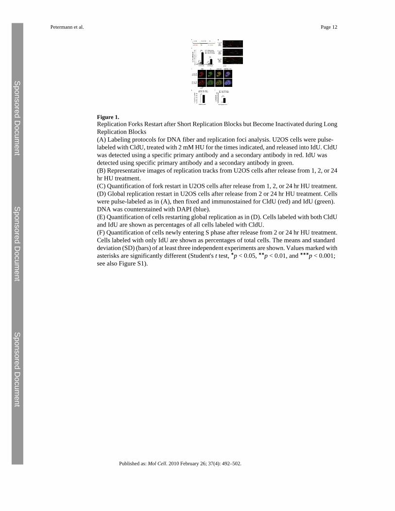
Figure 1.Replication Forks Restart after Short Replication Blocks but Become Inactivated during LongReplication Blocks(A) Labeling protocols for DNA fiber and replication foci analysis. U2OS cells were pulse-labeled with CldU, treated with 2 mM HU for the times indicated, and released into IdU. CldUwas detected using a specific primary antibody and a secondary antibody in red. IdU wasdetected using specific primary antibody and a secondary antibody in green.(B) Representative images of replication tracks from U2OS cells after release from 1, 2, or 24hr HU treatment.(C) Quantification of fork restart in U2OS cells after release from 1, 2, or 24 hr HU treatment.(D) Global replication restart in U2OS cells after release from 2 or 24 hr HU treatment. Cellswere pulse-labeled as in (A), then fixed and immunostained for CldU (red) and IdU (green).DNA was counterstained with DAPI (blue).(E) Quantification of cells restarting global replication as in (D). Cells labeled with both CldUand IdU are shown as percentages of all cells labeled with CldU.(F) Quantification of cells newly entering S phase after release from 2 or 24 hr HU treatment.Cells labeled with only IdU are shown as percentages of total cells. The means and standarddeviation (SD) (bars) of at least three independent experiments are shown. Values marked withasterisks are significantly different (Student's t test, ∗p < 0.05, ∗∗p < 0.01, and ∗∗∗p < 0.001;see also Figure S1).
Petermann et al. Page 12
Published as: Mol Cell. 2010 February 26; 37(4): 492–502.
Sponsored Docum
ent Sponsored D
ocument
Sponsored Docum
ent
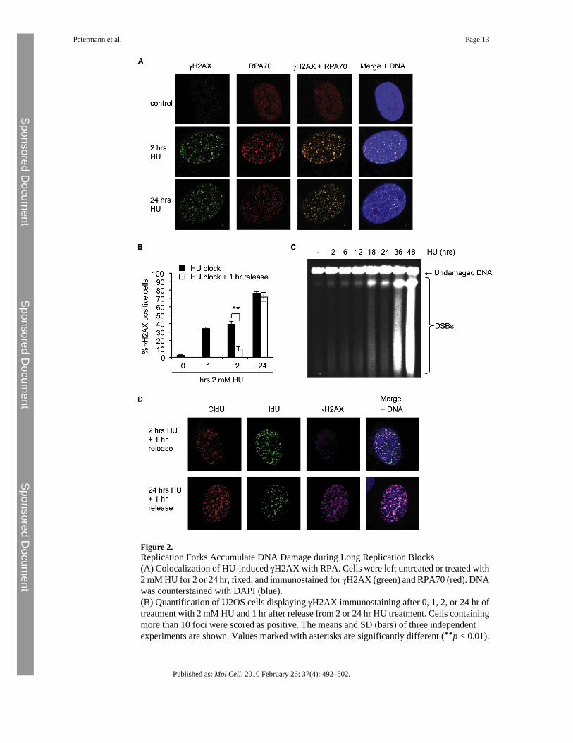
Figure 2.Replication Forks Accumulate DNA Damage during Long Replication Blocks(A) Colocalization of HU-induced γH2AX with RPA. Cells were left untreated or treated with2 mM HU for 2 or 24 hr, fixed, and immunostained for γH2AX (green) and RPA70 (red). DNAwas counterstained with DAPI (blue).(B) Quantification of U2OS cells displaying γH2AX immunostaining after 0, 1, 2, or 24 hr oftreatment with 2 mM HU and 1 hr after release from 2 or 24 hr HU treatment. Cells containingmore than 10 foci were scored as positive. The means and SD (bars) of three independentexperiments are shown. Values marked with asterisks are significantly different (∗∗p < 0.01).
Petermann et al. Page 13
Published as: Mol Cell. 2010 February 26; 37(4): 492–502.
Sponsored Docum
ent Sponsored D
ocument
Sponsored Docum
ent
(C) Pulsed-field gel electrophoresis to visualize DSB induction in U2OS cells treated with 2mM HU for 2 to 48 hr.(D) Colocalization of γH2AX and stalled replication forks in U2OS cells. Cells were pulse-labeled with CldU for 20 min, treated with 2 mM HU for 2 or 24 hr, and released into IdU for1 hr. Cells were immunostained for CldU (red), IdU (green), and γH2AX (far red), and DNAwas counterstained with DAPI (blue). DNA was denatured with HCl to allow CldU/IdUdetection.
Petermann et al. Page 14
Published as: Mol Cell. 2010 February 26; 37(4): 492–502.
Sponsored Docum
ent Sponsored D
ocument
Sponsored Docum
ent
Figure 3.Late Activation of Homologous Recombination during HU Block(A) HU-induced RAD51 foci in U2OS cells. Cells were left untreated or treated with 2 mMHU for 1, 2, or 24 hr, fixed, and immunostained for RAD51 (red). DNA was counterstainedwith DAPI (blue).(B) Quantification of HU-induced RAD51 foci in U2OS cells. Cells were treated with 2 mMHU for the times indicated and immunostained for RAD51. Cells containing >10 RAD51 fociwere scored as positive. The means and range (bars) of two independent experiments are shown.(C) Recombination frequencies in SPD8 cells induced by HU. Cells were treated with 2 mMHU for the times indicated and recovered for 48 hr. HPRT+ revertants were quantified 7 dayslater. The means and SD (bars) of four to ten independent experiments are shown. Valuesmarked with asterisks are significantly different from control (∗p < 0.05, ∗∗p < 0.01,and ∗∗∗p < 0.001; see also Figure S2).
Petermann et al. Page 15
Published as: Mol Cell. 2010 February 26; 37(4): 492–502.
Sponsored Docum
ent Sponsored D
ocument
Sponsored Docum
ent
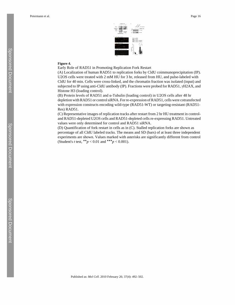
Figure 4.Early Role of RAD51 in Promoting Replication Fork Restart(A) Localization of human RAD51 to replication forks by CldU coimmunoprecipitation (IP).U2OS cells were treated with 2 mM HU for 3 hr, released from HU, and pulse-labeled withCldU for 40 min. Cells were cross-linked, and the chromatin fraction was isolated (input) andsubjected to IP using anti-CldU antibody (IP). Fractions were probed for RAD51, γH2AX, andHistone H3 (loading control).(B) Protein levels of RAD51 and α-Tubulin (loading control) in U2OS cells after 48 hrdepletion with RAD51 or control siRNA. For re-expression of RAD51, cells were cotransfectedwith expression constructs encoding wild-type (RAD51-WT) or targeting-resistant (RAD51-Res) RAD51.(C) Representative images of replication tracks after restart from 2 hr HU treatment in control-and RAD51-depleted U2OS cells and RAD51-depleted cells re-expressing RAD51. Untreatedvalues were only determined for control and RAD51 siRNA.(D) Quantification of fork restart in cells as in (C). Stalled replication forks are shown aspercentage of all CldU labeled tracks. The means and SD (bars) of at least three independentexperiments are shown. Values marked with asterisks are significantly different from control(Student's t test, ∗∗p < 0.01 and ∗∗∗p < 0.001).
Petermann et al. Page 16
Published as: Mol Cell. 2010 February 26; 37(4): 492–502.
Sponsored Docum
ent Sponsored D
ocument
Sponsored Docum
ent
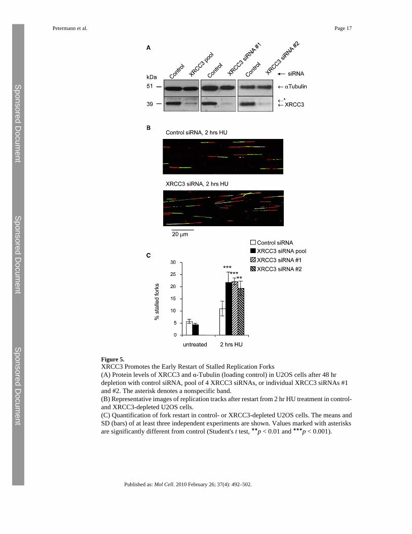
Figure 5.XRCC3 Promotes the Early Restart of Stalled Replication Forks(A) Protein levels of XRCC3 and α-Tubulin (loading control) in U2OS cells after 48 hrdepletion with control siRNA, pool of 4 XRCC3 siRNAs, or individual XRCC3 siRNAs #1and #2. The asterisk denotes a nonspecific band.(B) Representative images of replication tracks after restart from 2 hr HU treatment in control-and XRCC3-depleted U2OS cells.(C) Quantification of fork restart in control- or XRCC3-depleted U2OS cells. The means andSD (bars) of at least three independent experiments are shown. Values marked with asterisksare significantly different from control (Student's t test, ∗∗p < 0.01 and ∗∗∗p < 0.001).
Petermann et al. Page 17
Published as: Mol Cell. 2010 February 26; 37(4): 492–502.
Sponsored Docum
ent Sponsored D
ocument
Sponsored Docum
ent
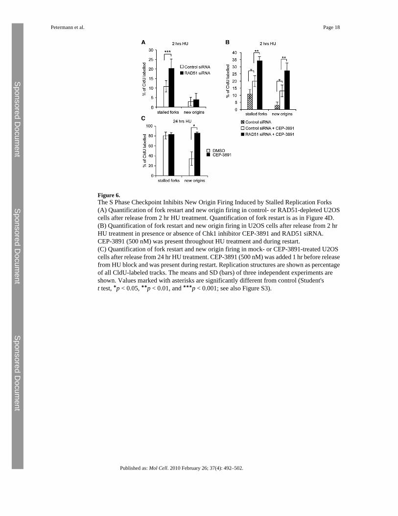
Figure 6.The S Phase Checkpoint Inhibits New Origin Firing Induced by Stalled Replication Forks(A) Quantification of fork restart and new origin firing in control- or RAD51-depleted U2OScells after release from 2 hr HU treatment. Quantification of fork restart is as in Figure 4D.(B) Quantification of fork restart and new origin firing in U2OS cells after release from 2 hrHU treatment in presence or absence of Chk1 inhibitor CEP-3891 and RAD51 siRNA.CEP-3891 (500 nM) was present throughout HU treatment and during restart.(C) Quantification of fork restart and new origin firing in mock- or CEP-3891-treated U2OScells after release from 24 hr HU treatment. CEP-3891 (500 nM) was added 1 hr before releasefrom HU block and was present during restart. Replication structures are shown as percentageof all CldU-labeled tracks. The means and SD (bars) of three independent experiments areshown. Values marked with asterisks are significantly different from control (Student'st test, ∗p < 0.05, ∗∗p < 0.01, and ∗∗∗p < 0.001; see also Figure S3).
Petermann et al. Page 18
Published as: Mol Cell. 2010 February 26; 37(4): 492–502.
Sponsored Docum
ent Sponsored D
ocument
Sponsored Docum
ent
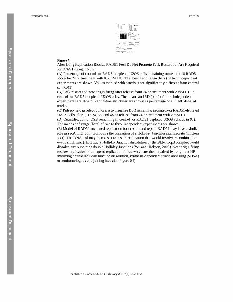
Figure 7.After Long Replication Blocks, RAD51 Foci Do Not Promote Fork Restart but Are Requiredfor DNA Damage Repair(A) Percentage of control- or RAD51-depleted U2OS cells containing more than 10 RAD51foci after 24 hr treatment with 0.5 mM HU. The means and range (bars) of two independentexperiments are shown. Values marked with asterisks are significantly different from control(p < 0.01).(B) Fork restart and new origin firing after release from 24 hr treatment with 2 mM HU incontrol- or RAD51-depleted U2OS cells. The means and SD (bars) of three independentexperiments are shown. Replication structures are shown as percentage of all CldU-labeledtracks.(C) Pulsed-field gel electrophoresis to visualize DSB remaining in control- or RAD51-depletedU2OS cells after 0, 12 24, 36, and 48 hr release from 24 hr treatment with 2 mM HU.(D) Quantification of DSB remaining in control- or RAD51-depleted U2OS cells as in (C).The means and range (bars) of two to three independent experiments are shown.(E) Model of RAD51-mediated replication fork restart and repair. RAD51 may have a similarrole as recA in E. coli, promoting the formation of a Holliday Junction intermediate (chickenfoot). The DNA end may then assist to restart replication that would involve recombinationover a small area (short tract). Holliday Junction dissolution by the BLM-Top3 complex woulddissolve any remaining double Holliday Junctions (Wu and Hickson, 2003). New origin firingrescues replication of collapsed replication forks, which are then repaired by long tract HRinvolving double Holliday Junction dissolution, synthesis-dependent strand annealing (SDSA)or nonhomologous end joining (see also Figure S4).
Petermann et al. Page 19
Published as: Mol Cell. 2010 February 26; 37(4): 492–502.
Sponsored Docum
ent Sponsored D
ocument
Sponsored Docum
ent
Related Documents