Hydrothermal growth of ZnO nanostructures This content has been downloaded from IOPscience. Please scroll down to see the full text. Download details: IP Address: 219.150.117.116 This content was downloaded on 30/09/2013 at 02:19 Please note that terms and conditions apply. 2009 Sci. Technol. Adv. Mater. 10 013001 (http://iopscience.iop.org/1468-6996/10/1/013001) View the table of contents for this issue, or go to the journal homepage for more Home Search Collections Journals About Contact us My IOPscience

Welcome message from author
This document is posted to help you gain knowledge. Please leave a comment to let me know what you think about it! Share it to your friends and learn new things together.
Transcript

Hydrothermal growth of ZnO nanostructures
This content has been downloaded from IOPscience. Please scroll down to see the full text.
Download details:
IP Address: 219.150.117.116
This content was downloaded on 30/09/2013 at 02:19
Please note that terms and conditions apply.
2009 Sci. Technol. Adv. Mater. 10 013001
(http://iopscience.iop.org/1468-6996/10/1/013001)
View the table of contents for this issue, or go to the journal homepage for more
Home Search Collections Journals About Contact us My IOPscience

IOP PUBLISHING SCIENCE AND TECHNOLOGY OF ADVANCED MATERIALS
Sci. Technol. Adv. Mater. 10 (2009) 013001 (18pp) doi:10.1088/1468-6996/10/1/013001
TOPICAL REVIEW
Hydrothermal growth of ZnOnanostructuresSunandan Baruah and Joydeep Dutta
Centre of Excellence in Nanotechnology at the Asian Institute of Technology,PO Box 4, Klong Luang, Pathumthani 12120, Thailand
E-mail: [email protected]
Received 5 October 2008Accepted for publication 14 November 2008Published 13 January 2009Online at stacks.iop.org/STAM/10/013001
AbstractOne-dimensional nanostructures exhibit interesting electronic and optical properties due totheir low dimensionality leading to quantum confinement effects. ZnO has received lot ofattention as a nanostructured material because of unique properties rendering it suitable forvarious applications. Amongst the different methods of synthesis of ZnO nanostructures, thehydrothermal method is attractive for its simplicity and environment friendly conditions. Thisreview summarizes the conditions leading to the growth of different ZnO nanostructures usinghydrothermal technique. Doping of ZnO nanostructures through hydrothermal method are alsohighlighted.
Keywords: ZnO, hydrothermal, nanostructures, synthesis, doping
(Some figures in this article are in colour only in the electronic version)
1. Introduction
Nanostructured ZnO materials have received considerableinterest from scientists due to their remarkable performancein electronics, optics and photonics. As early as the 1960s,synthesis of ZnO thin films was an active field because ofapplications in sensors, transducers and as photocatalysts. Inthe last few decades, study of one-dimensional material hasgained importance in nanoscience and nanotechnology. Withreduction in size, novel electrical, mechanical, chemical andoptical properties are introduced resulting from surface andquantum confinement effects.
ZnO is a significant technological material. The absenceof a centre of symmetry in its wurtzite structure, alongwith large electromechanical coupling, results in strongpiezoelectric and pyroelectric properties. ZnO is thereforewidely used in mechanical actuators and piezoelectricsensors. In addition, ZnO is a wide band-gap (3.37 eV)compound semiconductor that is appropriate for shortwavelength optoelectronic applications. The high excitonbinding energy (60 meV) in ZnO crystal allows efficient
excitonic emission at room temperature. ZnO is transparentto visible light and its conductivity can be increased throughdoping. ZnO nanostructures have a wide range of hightechnology applications like surface acoustic wave filters [1],photonic crystals [2], photodetectors [3], light emittingdiodes [4], photodiodes [5], gas sensors [6], optical modulatorwaveguides [7], solar cells [8, 9] and varistors [10]. ZnO isalso receiving a lot of attention because of its antibacterialproperty and its bactericidal efficacy has been reported toincrease as the particle size decreases [11].
The discovery of carbon nanotubes by Iijima [12] in1991 has initiated active research leading to the growthand characterization of one-dimensional nanowires ofelemental and compound semiconductors such as Si [13],Ge [14], InP [15], GaAs [16] and ZnO [17–19]. Differentnanostructures of ZnO have been reported such as nanowiresand nanorods [20], nanocombs [21], nanorings [22],nanoloops and nanohelices [23], nanobows [24],nanobelts [25] and nanocages [26]. These structureshave been successfully synthesized under explicit growthconditions [27].
1468-6996/09/013001+18$30.00 1 © 2009 National Institute for Materials Science Printed in the UK

Sci. Technol. Adv. Mater. 10 (2009) 013001 Topical Review
Figure 1. The wurtzite structure model of ZnO.
ZnO nanostructures can be grown either in solution orfrom gaseous phase. The gas phase synthesis methods areexpensive and complicated. The solution phase synthesis isusually done in water. The hydrothermal process of growingZnO nanostructures has gained immense popularity due to itssimplicity and tolerable growth conditions. As synthesis iscarried out in aqueous solution, the growth temperatures areless than the boiling point of water.
2. Zinc oxide: crystal structure
The ZnO crystal is hexagonal wurtzite and exhibits partialpolar characteristics [27] with lattice parameters a = 0.3296and c = 0.52065 nm.
The structure of ZnO can be described as a numberof alternating planes composed of tetrahedrally coordinatedO2− and Zn2+ stacked alternately along the c-axis, as shownin figure 1. The tetrahedral coordination in ZnO results inpiezoelectric and pyroelectric properties due to the absenceof inversion symmetry. Another important characteristic ofZnO is polar surfaces. The most common polar surface isthe basal plane (0001). One end of the basal polar planeterminates with partially positive Zn lattice sites and the otherend terminates in partially negative oxygen lattice sites. Theoppositely charged ions produce positively charged Zn-(0001)and negatively charged O-(0001) surfaces, resulting in anormal dipole moment and spontaneous polarization along thec-axis as well as a variance in surface energy. To maintaina stable structure, the polar surfaces generally have facetsor exhibit massive surface reconstructions, but ZnO ± (0001)
surfaces are exceptions: they are atomically flat, stable andexhibit no reconstruction [1, 2]. Efforts to understand thesuperior stability of the ZnO ± (0001) polar surfaces are atthe forefront of research in today’s surface physics [3–6]. Theother two most commonly observed facets for ZnO are {2110}
and {0110}, which are non-polar and have lower energy thanthe {0001} facets.
Figure 2. Growth morphologies of ZnO nanostructures withcorresponding facets (reproduced with permission from [27]© 2004 IOP).
3. Zinc oxide: growth structures
ZnO exhibits a varied range of novel structures. Thesestructures can be grown by tuning the growth rates alongthree fast growing directions:
⟨2110
⟩(±[1210], ±[2110],
±[1120]);⟨0110
⟩(±[0110], ±[1010], ±[1100]) and ±[0001].
The relative surface activities of various growth facets undergiven conditions determine the surface morphology of thegrown structure. Macroscopically, a crystal has differentkinetic parameters for different crystal planes, which areemphasized under controlled growth conditions. Thus, afteran initial period of nucleation and incubation, a crystallitewill commonly develop into a three-dimensional object withwell-defined, low-index crystallographic faces. Figures 2(a),(b) and (d) show a few typical growth morphologies of 1Dnanostructures of ZnO. These structures tend to maximize theareas of the {2110} and {0110} facets because of the lowerenergy. The morphology shown in figure 2(b) is dominatedby the polar surfaces, which can be grown by introducingplanar defects parallel to the polar surfaces. Occasional planardefects and twins can be observed parallel to the (0001) plane,but dislocations are hardly seen [27].
2

Sci. Technol. Adv. Mater. 10 (2009) 013001 Topical Review
Figure 3. TEM images of the as synthesized ZnO nanoparticles using zinc nitrate hexahydrate in an autoclave at a temperature of 120 ◦C.(Reproduced with permission from [46] © 2006 Elsevier.)
Table 1. Partial charge distribution in ZAH derived precursorclusters. (Reproduced with permission for [28] © 2006 Springer)
Precursor clusters δZn δAc
Zn(Ac)2 · 2H2O 0.471 −0.227Zn(Ac)2 0.469 −0.235Zn4O(Ac)6 0.467 −0.245Zn10O4(Ac)12 0.465 −0.254Zn5(OH)8(Ac)2 · 2H2O 0.463 −0.269Zn10O4(Ac)12 · H2O · 7EtOH 0.429 −0.462EtOZnAc 0.411 −0.567
4. Zinc oxide nanostructures—synthesis methods
The synthesis methods of different zinc oxide nanostructurescan broadly be classified as follows:
a. Solution phase synthesis: In the solution phase synthesis,the growth process is carried out in a liquid. Normallyaqueous solutions are used and the process is thenreferred to as hydrothermal growth process. Some of thesolution phase synthesis processes are
1. Zinc Acetate Hydrate (ZAH) derived nano-colloidalsol-gel route [28].
2. ZAH in alcoholic solutions with sodium hydroxide(NaOH) or tetra methyl ammonium hydroxide(TMAH) [29–31].
3. Template assisted growth [32].4. Spray pyrolysis for growth of thin films [33, 34].5. Electrophoresis [35].
b. Gas phase synthesis: Gas phase synthesis uses gaseousenvironment in closed chambers. Normally the synthesisis carried out at high temperatures from 500 ◦C to1500 ◦C. Some commonly used gas phase methods are
1. Vapour phase transport, which includes vapour solid(VS) and vapour liquid solid (VLS) growth [36–39].
2. Physical vapour deposition [40].3. Chemical vapour deposition [41].4. Metal organic chemical vapour deposition
(MOCVD) [42].5. Thermal oxidation of pure Zn and condensation [43].6. Microwave assisted thermal decomposition [44].
3

Sci. Technol. Adv. Mater. 10 (2009) 013001 Topical Review
Figure 4. Variation in particle size and yield of the ZnO nanopowders with growth temperature and pH of the growth solution.Region A: heterogeneous solution; region B: homogeneoussolution. (Reproduced with permission from [47] © 2000 Elsevier.)
4.1. ZAH based sol-gel synthesis of ZnO nanostructures
The sol-gel method has gained a lot of popularity as it offerscontrolled consolidation, shape modulation and patterningof the nanostructures. Concentrated ethanolic zinc acetatehydrate (ZAH) suspension when refluxed and distilled formsa transparent sol. Small ZnO nanoparticles of dimensionsaround 5 nm can be grown under high concentrationconditions by addition of hydroxides (e.g. LiOH, NaOH,etc) [28]. There are reports of modifications of the ZAHdehydration or dissolution and subsequent condensation forthe growth of ZnO nanostructures [29–31].
Table 2. Morphologies obtained using different templates.(Reproduced with permission from [48] © 1999 Elsevier.)
Particle propertiesAdditives Morphology Size (nm)
Tributylamine Rod-like 200–300Triethylamine Rod-like 100–300Triethanolamie Spindle-like 100–300Diisopropylamine Rod-like 200–400Ammonium phosphate Rod-like 200–5001, 6-Hexadianol Rod-like 300–700Triethyldiethylnol Rod-like 100–300Isopropylamine Rod or sheet-likeCyclohexylamine Sheet-like 300–500n-Butylamine Sheet-like 200–400Ammonium chloride Sheet 50–200Hexamethylenetetramine Snow-flake like 20–50Ethylene glycol Ellipse 40–100Ethanolamine Polyhedron 50–200
Figure 5. Attachment of hexamine to the non polar facets of thezincite crystal allows the growth of the crystal in the (0001)direction. (a) hexagonal ZnO crystal (b) possible attachment ofhexamine on to the non polar facets leaving the polar face exposedallowing further crystal growth along the c-direction. (Reproducedwith permission from [57] © 2006 Springer.)
Figure 6. SEM image of ZnO nanorods grown using zinc nitrateand hexamine after seeding with ZnO nanoparticles. Inset: close upof the rods. (Reproduced with permission from authors [51].)
The search for primary clusters to serve as buildingblocks for various nanostructures has been going on forquite some time. The isolation and identification of primaryclusters is an area of active research. The synthesis of theprimary structures depend on various conditions like initial
4

Sci. Technol. Adv. Mater. 10 (2009) 013001 Topical Review
Figure 7. Images of ZnO homocentric bundles obtained in block copolymers systems: (a) SEM image: products in surfactant L64 (b) TEMimage: products in L64. Inset: diffraction pattern. (c) and (d) SEM images: products in F68. (Reproduced with permission from [73] © 2007Elsevier.)
concentration of the salt, the synthesis temperature, heatingtime as well as nature of the solvent. The ZnO clusters canbe members of any of the three different families mentionedbelow [28]:
1. Tetrahedral oxy-acetate Zn4O(Ac)6.2. Ethoxy acetate (EtOZnAc)n .3. Hydroxy-double salt (Zn − HDS)Zn5(OH)8(Ac)2
(H2O)2.
When Zn(Ac)2 is heated in alcohol, the followingreaction takes place initially
4Zn(Ac)2 · 2H2Oheat
−−−−−→ Zn4O(Ac)6 + 7H2O + 2HAc.(1)
Zn4O(Ac)6 is also called basic zinc acetate. The by-productslike H2O, acetic acid etc, can be removed by distillation.ZAH forms a larger homologue Zn10O4(Ac)12 when it isdehydrated in the presence of acetanhydride and refluxed inEtOH. Continuous refluxing of ZAH sols can result in ZnOnanoparticles [45]. Zn4O(Ac)6 can be considered as a welldesigned molecular model of ZnO.
Ethoxy acetates (EtOZnAc)n are formed in solutionshaving Zn10O4(Ac)12 clusters. These species are formedgradually with time and after several weeks, large single
Table 3. Difference reaction for growth of ZnO nanorods.Reproduced with permission from [78] © 2007 Elsevier.
Sample NH4OH (g) Reaction time (h)
A1 1.5 12A2 1.5 24A3 3.5 12A4 3.5 24
crystals can be separated. The oxy-acetate clusters mostprobably generate intermediary zinc acetate monomers asshown by the following reactions:
4Zn4O(Ac)6 −→ Zn10O4(Ac)12 + 6Zn(Ac)2, (2)
Zn4O(Ac)6 + Zn10O4(Ac)12 −→ Zn13O5(Ac)16 + Zn(Ac)2.
(3)These Zn(Ac)2 monomers can lead to formation of zincethoxy acetate (EtOZnAc)n crystals and acetic acid.
Hydroxy-double salt (Zn-HDS)Zn5(OH)8(Ac)2(H2O)2
is formed by the titration of ZAH solutions with an aqueoussolution of sodium hydroxide. Zn-HDS has been detectedin precipitates of preheated alcoholic ZAH sols. At hightemperatures and after prolonged refluxing, Zn-HDS can beeasily transformed into ZnO nanoparticles.
5

Sci. Technol. Adv. Mater. 10 (2009) 013001 Topical Review
Table 4. Morphology and shape of difference ZnO nonostructures atvarying pH. (Reproduced with permission from [82] © 2008Elsevier.)
pH Morphology Shape
9.0 Coalescence of Zno Coalescence Budding flower9.5 Nano-particles ↑
10.0 Zno nanorod Blossom10.5 Separation sharp rods Separation Chestnut but
or echinoid11.0 Separation of thick rods ↓ Dense chestnut
bur11.5 Coalescence of rods Gingko leaves11.8 Archetype of thick rods Coalescence Dendelion
Figure 8. Growth habits of hexagonal prism- and pyramid-like ZnOcrystals. (Reproduced with permission from [73] © 2007 Elsevier.)
The occurrence of hydroxy double salts during the growthof ZnO nanoparticles may be due to the following:
1. H2O induced reorganization of the tetrahedral species(primary and secondary) formed during initial nucleation.Hydroxyl ions also play a role.
Zn4O(Ac)6 + Zn(Ac)2 + 9H2O
−→ 2Zn5(OH)8(Ac)2(H2O)2 + 6HAc. (4)
Zn10O4(Ac)12 + 16H2O
−→ 2Zn5(OH)8(Ac)2(H2O)2 + 8HAc. (5)
Zn10O4(Ac)12 + 16H2O + 8OH−
−→ 2Zn5(OH)8(Ac)2(H2O)2 + 8Ac−. (6)
2. The continuous liberation of zinc acetate during thegrowth of the ZnO nanoparticles may be another reasonfor the growth of the hydroxy double salts.
Figure 9. SEM images of the array of ZnO obelisk shapednanorods grown on glass substrate. (Reproduced with permissionfrom [75] © 2004 Elsevier.)
4.2. Stability of ZAH derived structures
Spanhel [28] carried out partial charge calculations using theHenry–Livage model. The partial charge values for the ZAHstructures are shown in table 1.
It can be observed that the tetrahedral oxy-acetate clustersare slightly more stable than the zinc acetate hydrate. Further,the stability of Zn10O4(Ac)12 precursor is strongly increasedin the presence of H2O and EtOH. Once a certain amountof water is present, there is a spontaneous formation of zinchydroxy double salts (Zn-HDS). This is because of the higherstability of the Zn-HDS monomer with respect to the nakedoxy-acetate clusters. As seen from the table 1, the most stableprecursor cluster is the zinc ethoxy-acetate. An importantpoint to note is that Zn2+ ions do not exist as free ions inalcoholic ZAH solutions as a strong chemical bond existsbetween Zn2+ ions and Ac ligands in all the compounds intable 1.
4.3. ZnO nanostructures through hydrothermal growth
4.3.1 Nanoparticles. Even though the organometallicsynthesis of ZnO nanoparticles in alcoholic medium hasreceived wider acceptance for reasons of faster nucleationand growth as compared to water, still scattered reports ofhydrothermal synthesis in aqueous medium are available in
6

Sci. Technol. Adv. Mater. 10 (2009) 013001 Topical Review
Figure 10. Illustration of the crystal structure of the obelisk shapedZnO nanorods. (Reproduced with permission from [75] © 2004Elsevier.)
the literature. Baruwati et al [46] have reported the aqueoussynthesis of ZnO nanoparticles using zinc nitrate hexahydrate.Synthesis was carried out in an autoclave at a temperatureof 120 ◦C after adjusting the pH to 7.5 using ammoniumhydroxide. After washing, the particles were dried at 80 ◦Covernight to obtain the powder form. The as synthesizedparticles are shown in the transmission electron microscope(TEM) images in figure 3.
Lu et al [47] successfully prepared crystalline ZnOpowder through a hydrothermal process using ammonia asthe base source. With Zn(NO3)2 as the source of Zn2+ ions,growth was carried out at 100 ◦C, 150 ◦C and 200 ◦C for 2 hand the effect of growth temperature and pH was studied.Figure 4 shows the variation of particle size of the ZnOpowder and its yield as functions of growth temperature andpH. With pH < 11 in region A of figure 4, the zinc hydroxideprecursors are dissolved partially and the ZnO powder isnucleated in a heterogeneous system. On the other hand,in region B with pH> 11, all zinc hydroxide precursorsare dissolved and a clear solution is formed so that ZnOpowder is nucleated in a homogeneous solution. Differentnucleation states thus take place in regions A and B withhigher probability of nucleation in the heterogeneous solution.
Chen et al [48] synthesized nanoparticles of differentmorphologies using ZnCl2 and NaOH in a hydrothermalgrowth process using different organic compounds as templateagents. A significant change in the morphology was observedas the synthesis temperature was increased with the particleschanging from rod like to polyhedral. It was reportedthat the morphology also changed with the addition ofdifferent organic templates to the reaction mixture whenthe temperature was maintained at 160 ◦C. The variousmorphologies along with the templates used are listed intable 2.
A very simple procedure to prepare ZnO nanoparticles ata very high pH ∼ 14 using tetramethylammonium hydroxide(TMAH) as a precipitating agent was suggested by Musicet al [49]. Nanoparticles sized from 10 to 20 nm wereprecipitated at room temperature by adding TMAH to anethanolic solution of zinc acetate dehydrate. Addition of waterto the ethanolic solution prior to adding TMAH yielded ZnOsnowflakes.
Vishwanathan and Gupta [50] have shown thatsupercritical water can also be a good reaction mediumfor the hydrothermal synthesis of ZnO nanoparticles.Spherical ZnO nanoparticles were synthesized by oxidationof zinc acetate in supercritical water in a continuous tubularreactor. Particle size and morphology can be controlled byvarying conditions like temperature, pressure or the reactionatmosphere. Nanoparticles with diameters ranging between39 and 320 nm were synthesized using this method.
The synthesis time has been significantly reduced throughthe use of microwave irradiation and the ZnO nanocrystallitesthus formed were observed to be more defective than the onessynthesized over a few hours of hydrolysis [51]. Nanoparticleswith inherent defects are capable of exhibiting visible lightphotocatalysis even without doping with transition metals,which is the normally followed method.
4.3.2 Nanowires and nanorods. Andres-Vergés et al [52]first reported the hydrothermal method of growing ZnOnanostructures. However, this could not instil much interesttill Vayssieres et al [53] successfully used the method forthe controlled fabrication of ZnO nanowires on glass andSi substrates by the thermal decomposition of methenamineand zinc nitrate. To initiate the growth from the substrate, athin layer of ZnO nanoparticles was grown on the substrate.Methenamine, also known as hexamethylenetetramine (HMT)or hexamine is a highly water soluble, non-ionic tetradentatecyclic tertiary amine. Thermal degradation of HMT releaseshydroxyl ions which react with Zn2+ ions to form ZnO [54].This can be summarized in the following equations:
(CH2)6N4 + 6H2O ↔ 6HCHO + 4NH3, (7)
NH3 + H2O ↔ NH+4 + OH−, (8)
2OH− + Zn2+→ ZnO(s) + H2O. (9)
It is a general acceptance that the role of HMT is to supply thehydroxyl ions to drive the precipitation reaction [55]. Apartfrom that, some also opine that HMT acts as a buffer as the
7

Sci. Technol. Adv. Mater. 10 (2009) 013001 Topical Review
Figure 11. SEM images of the ZnO nanostructures on Zn foil with condition mentioned in table 3: (a) A1 (b) A2 (c) A3 (d) A4.(Reproduced with permission from [78] © 2007 Elsevier.)
rate of its hydrolysis decreases with increasing pH and viceversa [56]. Ashfold et al [55] have demonstrated that the rateof decomposition of HMT is independent of the reaction thatyields ZnO indicating that HMT does act as a kinetic buffer.In oxide formation, the phase that is thermodynamically lessstable will precipitate out faster [56]. In the initial growthstage, the pH and the concentration of Zn2+ ions is such thatthe ZnO growth will be through Zn(OH)2. With the gradualincrease in the pH and the decrease in the concentration ofthe Zn ions, Zn(OH)2 becomes thermodynamically unstableand the Zn(OH)2 formed on the substrate will start dissolving.Further growth of the nanostructures will have to be throughdirect deposition of ZnO [55].
The contribution of HMT in the growth process of ZnOnanowires has also been discussed by Sugunan et al [57]in a totally different approach. It was proposed that HMT,being a long chain polymer and a nonpolar chelating agent,will preferentially attach to the non polar facets of the zincitecrystal, thereby cutting off the access of Zn2+ ions to themleaving only the polar (001) face for epitaxial growth. HMTtherefore acts more like a shape-inducing polymer surfactantrather than as a buffer. Figure 5 shows the mechanism ofattachment of hexamine on the nonpolar facets.
There is a lot of literature reporting investigation onthe growth and properties of ZnO nanorods synthesizedusing zinc nitrate and HMT such as the effect of substratesand seed layers on the morphology of nanorods andnanotubes [58–60], growth on different substrates [20] andthe control of aspect ratio through the addition of citrateanions [61]. Pal and Santiago [62] have managed to controlthe morphology of ZnO nanostructures by varying the amountof a soft surfactant, ethylenediamine and the pH of thereaction mixture of zinc acetate, sodium hydroxide and thesurfactant. Homogenous growth was observed at a pH of12 with inhomogeneity creeping in as the pH decreases. A10% concentrated ethylenediamine gave higher aspect ratiothan 5% concentration. Figure 6 shows scanning electronmicroscope (SEM) images of hydrothermally grown ZnOnanorods using zinc nitrate and hexamine.
ZnO nanowires and nanorods have been successfullysynthesized using different surfactants and numerous reportsare available. Tang et al [63] synthesized ZnO nanorodsusing zinc acetylacetonate Zn(acac)2 · H2O as a single sourceprecursor and investigated the growth of the rods in thepresence of four different surfactants: polyvinyl alcohol(PVA), polyethylene glycol (PEG), sodium dodecyl sulphate
8

Sci. Technol. Adv. Mater. 10 (2009) 013001 Topical Review
Figure 12. SEM images of the flower-like ZnO nanostructures at different magnifications. (Reproduced with permission from [79] © 2007Elsevier.)
(SDS) and cetyltrimethyl ammonium bromide (CTAB). Theuse of PVA resulted in more regular and defect free rods thanPEG, SDS and CTAB. Li et al [64] has reported the growth oftapered ZnO nanorods with diameter decreasing from 400 nmat the body to about 80 nm at the tip from CTAB assistedhydrothermal growth. Zinc acetate dehydrate was used asthe precursor and the pH was adjusted to 13 using KOH.The use of carbamide CO(NH2)2 as a surfactant [65] in ahydrothermal growth with ZnSO4 and NaOH yielded highlycrystalline ZnO nanobelts. Chen et al [66] studied the effectof potassium iodide (KI) as a surfactant in the crystallizationof ZnO nanorod clusters from a chemical bath containing zincnitrate hexahydrate Zn(NO3)2 · 6H2O and hydrazine hydrateN2H4 · H2O. They obtained step growth of hexagonal nanorodclusters, and this morphology is attributed to the presence ofiodine ions in the growth bath. Stepped columns of ZnO werealso observed using CTAB as the surfactant [67]. Experimentsfurther showed that the source of Zn ions can also affect themorphology of the end product. Ni et al [67] obtained ZnOnanorods in place of stepped columns simply by changing thesource of Zn ions from Zn(Ac)2 to ZnCl2 and keeping all otherconditions same. This may be due to the change in pH of thegrowth bath.
Water soluble amphiphilic block copolymers,PEO-PPO-PEO, due to their unique properties are beingused as templates for the ingenious morphologies ofinorganic materials such as nanoparticles, mesoporousmaterials and hierarchically ordered oxides [68–72]. Zhanget al [73] managed to control the shape of bundles of ZnO
nanostructures through a macromolecular surfactant (L64and F68) assisted hydrothermal growth route. The bundles ofZnO nanostructures are shown in figure 7.
It was deduced that the nanobundle superstructures ofZnO result from a two-step mechanism in aqueous solutionswith initial nucleation followed by the growth of the nanorodsaround these nuclei. Overall size and arm lengths are probablycontrolled by the growth velocity of ZnO particles and themicelle dimension of PEO–PPO–PEO copolymers. ZnO is apolar crystal, O2−
is in hexagonal closest packing, and eachZn2+ lies within a tetrahedral group of four oxygen ions [74].Zn and O atoms are stacked alternatively along the c-axisand the top face (0001) consists of tetrahedral zinc having aterminal OH ligand as shown in figure 8. The formation ofhexagonal prism and pyramid like ZnO crystals is attributedto the difference in the growth velocities of various crystalfacets. The growth velocities under hydrothermal conditionsalong the different directions are known to follow the patternV (0001) > V (1011) > V (1010) [73]. The relative growthrate of these crystal faces will determine the final shapeand aspect ratio of the ZnO nanostructures. The preferentialgrowth along the (0001) polar direction proceeds unabatedand the ZnO nanorods would appear in the end products.Although the chemical reaction is relatively simple, thegrowth process of ZnO nanobundles is quite complicated. Itcan be presumed that the faster the growth rate, the quickerthe disappearance of the plane. The (0001) plane disappearsdue to its high growth velocity leading to the pointed shapeat the end of the c-axis. As a consequence of the slow growth
9

Sci. Technol. Adv. Mater. 10 (2009) 013001 Topical Review
Figure 13. SEM images of ZnO nanoflowers synthesized usingzinc chloride and ammonia. (Reproduced with permission from [80]© 2007 Elsevier.)
of the (1010) plane, the crystal remains to form the hexagonalprisms, while the (1011) plane corresponds to the formationof the hexagonal pyramid like tips.
Wang et al [75] has shown that it is possible toefficiently synthesize large two-dimensional arrays of obeliskshaped ZnO nanorods on quartz or glass substrates througha simple hydrothermal deposition method with zinc nitrate,ammonia and ammonium hydroxide as the precursors.The single-crystalline obelisk rods with diameters rangingbetween 300 and 400 nm and lengths of about 5 µm weregrown in Teflon vessel at 95 ◦C for 30 min and the SEMimages are shown in figure 9.
The mechanism for the formation of ZnO crystals usingammonia can be summarized in the following equations[76, 77].
NH3 + H2O ⇔ NH3 · H2O ⇔ NH+4 + HO−, (10)
Zn2+ + NH3 → Zn(NH3)42+, (11)
Zn(NH3)42+ + OH−
→ ZnO. (12)
or ZnO may also form from the complex ion Zn(OH)42+ as
below:Zn2+ + OH−
→ Zn(OH)42−, (13)
ZnOH42+
→ ZnO. (14)
The complex ions Zn(NH3)42+ and Zn(OH)4
2− formed dueto the mixing of zinc nitrate, ammonia and ammoniumhydroxide, dehydrate to initiate heterogeneous nucleationof ZnO on the substrate. The possible formation of theobelisk-shaped single-crystalline rods is attributed to multiplelayered structures as explained in figure 10.
The presence of Zn2+ containing salts is not anecessity for the growth of ZnO nanostructures through thehydrothermal growth method. Li et al [78] has successfullygrown large scale arrays of ZnO nanorods on zinc foil withoutthe assistance of any template, oxidant or coating of metaloxide layers, simply by dipping the foil into a 25% aqueoussolution of ammonia (NH4OH) and heating at a temperature80 ◦C in a Teflon-lined stainless steel autoclave. Four differentsamples were prepared by varying the concentration ofammonia in a 80 ml growth bath and the growth duration asshown in table 3. The SEM images are shown in figure 11.
It was observed that the thickness, density andmorphology of the ZnO nanorod arrays are affected by thealkalinity of the solution in the growth bath. The sharp tips ofthe nanorods are probably due to the concentration gradientof Zn2+ from the base to the tip. The increase in growth timeonly led to thicker rods with the lengths remaining almostcomparable. With the increase in concentration of ammonia,no growth was observed till 12 h but a very dense growth ofuneven pointed rods was observed when the growth durationwas increased to 24 h.
4.3.3 Flower-like and cabbage-like nanostructures.Flower-like structures are very typical of ZnO and canbe synthesized using simple hydrothermal methods. Wahabet al [79] has reported the growth of flower-like ZnO usinghydrothermal method. The flowers, composed of hexagonalZnO nanorods, were synthesized at a temperature of 90 ◦Cusing zinc acetate dehydrate and sodium hydroxide inaqueous solutions. The flowers, with nanorod petals as longas 2–4 µm, could be synthesized in just 30 min. SEM imagesof the nanoflowers are shown in figure 12. All the pointednanorods emerged from a single centre thereby acquiring thespherical shape.
Flower-like ZnO nanostructures with nanosheet petalswas synthesized by Shao et al [80] through a hydrothermalroute using zinc chloride and ammonia as the reactants. Theflowers, averaging in size of about 30 µm, are composedof sheets of thickness about 150–250 nm. SEM images(figure 13) reveal numerous well distributed nanostructuresformed all over the copper plate substrate.
Li et al [81] has reported the growth of flower-like andcabbage-like nanostructures using CTAB as the surfactant in ahydrothermal growth process at temperatures of 120, 150 and180 ◦C. They obtained flower-like micro and nanostructuresconsisting of ZnO nanorods at a temperature of 120 ◦C.
10

Sci. Technol. Adv. Mater. 10 (2009) 013001 Topical Review
Figure 14. SEM images of as-synthesized ZnO micro and nanostructures prepared by CTAB-assisted hydrothermal growth at varioustemperatures (a) 120 ◦C (b) 150 ◦C and (c) 180 ◦C. Inset: magnified images. (Reproduced with permission from [81] © 2008 Elsevier.)
As the synthesis temperature was increased to 150 and 180 ◦C,cabbage-like structures were formed due to the repeatedgrowth of two-dimensional ZnO nanosheets. The thicknessof the nanosheets obtained at 150 ◦C was about 50 nm and at180 ◦C about 100 nm. The nanostructures obtained are shownin figure 14. The authors have tried to explain the growthof the nanostructures with the schematic diagram shown infigure 15.
CTAB being a cationic surfactant reduces the surfacetension and as such inhibits the formation of a new phase.CTAB acts as a transport medium to transport Zn(OH)4
2−
growth units which come together to form individual rod-likestructures at 120 ◦C which then self-assemble into theZnO nanoflowers. However, at temperatures of 150 and180 ◦C, two-dimensional nanosheets are formed which thenself-assemble into the cabbages.
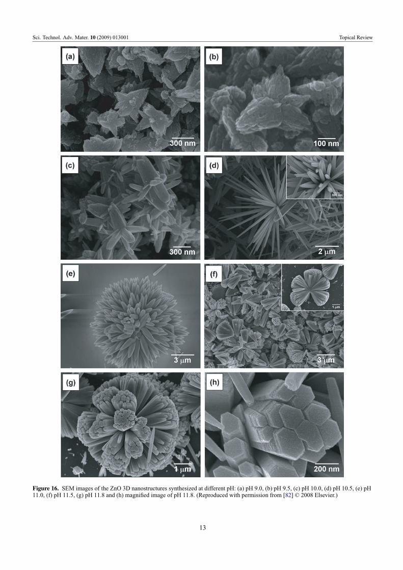
Three-dimensional ZnO nanostructures have also beensuccessfully synthesized through a simple hydrothermal
process using ammonia and zinc acetate dihydrate at95 ◦C [82]. Different morphologies were observed when thepH of the growth solution was changed between 9 and 11.8.The SEM micrographs are shown in figure 16 and a summaryof the results is presented in table 4.
4.4. Hydrothermal synthesis using microwaves
Another method of synthesis of nanostructures which isreceiving a lot of interest lately is the use of microwaveheating in place of conventional heating. Use of microwavesfor synthesis was first reported in 1986 by Gedye et al [83]following which there has been an increase in the use ofmicrowaves for nanostructure synthesis [51, 84, 85]. Maet al [86] have synthesized ZnO micro and nanostructuresusing zinc nitrate hexahydrate and pyridine in a hydrothermalprocess through microwave irradiation at 90 ◦C for 10 min.Adjusting the concentration of pyridine in the reaction
11

Sci. Technol. Adv. Mater. 10 (2009) 013001 Topical Review
Figure 15. Schematic illustration explaining the growth of ZnO micro and nanostructures synthesized through CTAB-assisted hydrothermalprocess at different temperatures (a) 120 ◦C and (b) 150 ◦C and 180 ◦C. (Reproduced with permission from [81] © 2008 Elsevier.)
bath, various morphologies of ZnO could be synthesizedlike hexagonal columns, linked hexagonal needles, hollowstructures and hexagonal nanorings. Some of the structuresare shown in figure 17.
4.5. Chemical double dip technique
Vijayan et al [87] have successfully grown thin films of ZnOusing a double dip technique using sodium zincate (Na2ZnO2)as the first dipping solution followed by a second dipping inhot water. The conversion of sodium zincate into zinc oxide inpresence of water follows the simple reaction
Na2ZnO2 + H2O → ZnO + 2NaOH. (15)
Polycrystalline thin films with high preferential orientationalong the (002) plane could be obtained using the double diptechnique. The microstructural parameters were dependent
upon the temperature of the hot water bath, the pH as well asthe amount of Zn(SO4)2 used for preparing the sodium zincatesolution.
5. Crystal structure of hydrothermally grown ZnOnanostructures
The hydrothermally grown ZnO nanostructures are singlecrystalline and exhibit the wurtzite structure of ZnO. X-raydiffraction (XRD) analyses for different ZnO nanostructuresgrown through hydrothermal methods revealed the hexagonalwurtzite structure, and the peaks could be indexed accordingto JCPDS card No. 79-2205 with a = 0.3249 nm and c =
0.5205 nm [46, 77, 81]. Some of the XRD results are shownin figure 18.
Further structural characterizations can be performedusing high resolution transmission electron microscopy(HRTEM). Fast Fourier transforms (FFT) and inverse FFT
12
Sci. Technol. Adv. Mater. 10 (2009) 013001 Topical Review
Figure 16. SEM images of the ZnO 3D nanostructures synthesized at different pH: (a) pH 9.0, (b) pH 9.5, (c) pH 10.0, (d) pH 10.5, (e) pH11.0, (f) pH 11.5, (g) pH 11.8 and (h) magnified image of pH 11.8. (Reproduced with permission from [82] © 2008 Elsevier.)
13

Sci. Technol. Adv. Mater. 10 (2009) 013001 Topical Review
Figure 17. TEM micrographs of the samples synthesized through microwave irradiation in aqueous solution of Zn(NO3)2 and pyridine at90 ◦C for 10 min. (Reproduced with permission from [86] © 2007 Elsevier.)
Figure 18. Typical x-ray diffraction (XRD) patterns for different ZnO nanostructures prepared using hydrothermal methods(a) nanoparticles, (b) nanorods and (c) nanoflowers. The indexed peaks correspond to the hexagonal wurtzite structure [46, 80, 81].(Reproduced with permission from [46] © 2006 Elsevier, [79] © 2007 Elsevier and [80] © 2007 Elsevier.)
14

Sci. Technol. Adv. Mater. 10 (2009) 013001 Topical Review
Figure 19. (A) High resolution TEM image of ZnO nanoparticles (inset: magnification of the squared area.) (B) Fast Fourier transformdone on the squared area (C), (D) Inverse fast Fourier transform of (B). (Reproduced with permission from Baruah et al [51].)
Figure 20. (a) Low resolution TEM image (inset: SAED pattern). (b) High resolution TEM image showing lattice fringes (inset: SAEDpattern). (reproduced with permission from [79] © 2007 Elsevier.)
of HRTEM images of ZnO nanoparticles [51] confirmed thewurtzite structure. One of the imaged particles is shown infigure 19(B) and the corresponding FFT and Inverse FFTs areshown in figures 19(C)–(E). Lattice spacings of 0.28 nm and0.16 nm indicate the presence of the (100) and (110) planes.
Another method of confirming the crystal structure isthrough the use of selected area electron diffraction (SAED)setup which comes as an accessory with TEMs. The SAEDpattern for ZnO nanorods synthesized through a hydrothermalmethod [79] is shown in figure 20 along with low andhigh magnification TEM images. Figure 20(a) shows a lowresolution TEM image of pointed tip ZnO nanorods. Thecorresponding SAED pattern (inset) confirmed that the rodsare single crystalline and grew along the [0001] direction.
The lattice fringe spacing in the HRTEM imageis 0.52 nm which corresponds to the hexagonal wurtzitestructure of ZnO and indicates that the growth occurred alongthe [0001] direction. The SAED pattern (inset) agrees with theHRTEM observation [79].
6. Doping of ZnO nanostructures throughhydrothermal routes
Doping of semiconducting nanostructured materials is theprimary technique for controlling properties like bandgap,electrical conductivity and ferromagnetism. Transition metaldoping of II–VI and III–V semiconductors is generating alot of interest from researchers for possible applications in
15

Sci. Technol. Adv. Mater. 10 (2009) 013001 Topical Review
Figure 21. SEM images of ZnO nanorods grown hydrothermally (A) undoped (B) doped using 10% Co solution (C) doped using 10% CoCr solution and (D) doped using 10% Mn solution. (Reproduced with permission from [99] © 2007 Elsevier.)
spintronics and visible light photocatalysis. A lot of work hasbeen done in the area of transition metal doping of ZnO singlecrystals and thin films [88–92]. However, a few reports on thesynthesis and characterization of ZnO nanostructures dopedwith different impurities like Al, Sn, Ga, In, Sb, Cu, etc areavailable in the literature [93–98]. Li et al [99] has synthesizedZnO nanorods doped with manganese (Mn), chromium (Cr)and cobalt (Co) in a hydrothermal synthesis using zinc nitrate,transition metal nitrate and hexamethylenetetramine. It wasobserved that the doping was good with Mn (8% doping froma 10% solution) and Co (17% doping from a 10% solution)dopants and poor with Cr (4% doping from a 10% solution).The morphology of the doped ZnO nanorods was differentfrom that of the undoped nanorods, the SEM images of whichare shown in figure 21.
Yuhas et al [100] has communicated the successfuldoping of ZnO nanorods with Co by the thermaldecomposition of zinc acetate and cobalt (II) acetate inrefluxing trioctylamine. The reaction temperature was setat 310 ◦C and the reaction was allowed to continue from45 to 180 min to obtain rods of different diameters. Thehydrothermal synthesis of Al-doped ZnO nanopowders wasreported by Piticescu et al [101]. The synthesis was carriedout in a 2L computer-controlled Teflon autoclave using KOH
as a mineralizing agent. Indium doping of ZnO nanorods wasdone after the rods were grown hydrothermally through a postdeposition thermal annealing in an inert atmosphere [102].Indium doping during the hydrothermal synthesis could notbe carried out due to the formation of In(OH)3 phase. Dopingwas carried out successfully for 0.5, 1 and 2%.
7. Conclusion
Hydrothermal synthesis of ZnO nanostructures is simpleand efficient and it is receiving a lot of attention of late.Various additives are used in aqueous medium to successfullysynthesize ZnO nanostructures of different morphologies, amixture of zinc nitrate and hexamine are the most popular.ZnO nanostructures are attractive candidates for applicationsin a host of devices like solar cells, sensors, detectors,energy generators as well as artificial structures for tissueengineering. ZnO nanostructures are being increasingly usedas photocatalysts for degrading harmful contaminants likepesticides from ground water. Within the next decade,ZnO nanostructures are most likely to move into industrialapplications.
16

Sci. Technol. Adv. Mater. 10 (2009) 013001 Topical Review
Acknowledgments
The authors would like to acknowledge partial financialsupport from the National Nanotechnology Center, belongingto the National Science and Technology Development Agency(NSTDA), Ministry of Science and Technology (MOST),Thailand and the Centre of Excellence in Nanotechnology atthe Asian Institute of Technology.
Reference
[1] Emanetoglu N W, Gorla C, Liu Y, Liang S and Lu Y 1999Mater. Sci. Semicond. Process 2 247
[2] Chen Y, Bagnall D and Yao T 2000 Mater. Sci. Eng. B 75 190[3] Liang S, Sheng H, Liu Y, Hio Z, Lu Y and Chen H 2001
J. Cryst. Growth 225 110[4] Saito N, Haneda H, Sekiguchi T, Ohashi N, Sakaguchi I and
Koumoto K 2002 Adv. Mater. 14 418[5] Lee J Y, Choi Y S, Kim J H, Park M O and Im S 2002 Thin
Solid Films 403 533[6] Mitra A, Chatterjee A P and Maiti H S 1998 Mater. Lett. 35 33[7] Koch M H, Timbrell P Y and Lamb R N 1995 Semicond. Sci.
Technol. 10 1523[8] Gratzel M 2005 MRS Bull. 30 39374[9] Baxter J B, Walker A M, van Ommering K and Aydil E S 2006
Nanotechnology 17 S304[10] Lin Y, Zhang Z, Tang Z, Yuan F and Li J 1999 Adv. Mater.
Opt. Electron. 9 205[11] Padmavathy N and Vijayaraghavan R 2008 Sci. Technol. Adv.
Mater. 9 035004[12] Iijima S 1991 Nature 354 56[13] Cui Y, Lauhon L J and Gudiksen M S 2001 Appl. Phys. Lett.
78 2214[14] Burghard G M, Kim G T, Dusberg G S, Chiu P W, Krstic V,
Roth S and Han W Q 2001 J. Appl. Phys. 90 5747[15] Duan X, Huang Y, Cui Y, Wang J and Lieber C M 2001 Nature
409 66[16] Bai Z G, Yu D P, Zhang H Z, Ding Y, Gai S Q, Hang X Z,
Hiong Q L and Feng G C 1999 Chem. Phys. Lett. 303 311[17] Huang M H, Wu Y, Feick H, Tran N, Webe E and Yang P 2001
Adv. Mater. 13 113[18] Huang M H, Mao S, Feick H, Yan H, Wu Y, Kind H, Weber E,
Russo R and Yang P 2001 Science 292 1897[19] Shi G, Mo C M, Cai W L and Zhang L D 2005 Solid State
Commun. 115 253[20] Baruah S, Thanachayanont C and Dutta J 2008 Sci. Technol.
Adv. Mater. 9 025009[21] Huang Y, Zhang Y, Bai X, He J, Liu J and Zhang X 2006
J. Nanosci. Nanotechnol. 6 2566[22] Hughes W L and Wang Z L 2005 Appl. Phys. Lett. 86 043106[23] Kong X Y and Wang Z L 2003 Nano Lett. 3 1625[24] Hughes W L and Wang Z L 2004 J. Am. Chem. Soc. 126 6703[25] Sun T, Qiu J and Liang C 2008 J. Phys. Chem. C 112 715[26] Snure M and Tiwari A 2007 J. Nanosci. Nanotechnol. 7 485[27] Wang Z L 2004 J. Phys.: Condens. Matter 16 R829[28] Spanhel L 2006 J. Sol–Gel Sci. Technol. 39 7[29] Ma X, Zhang H, Ji Y, Xu J and Yang D 2005 Mater. Lett.
59 3393[30] Kohls M, Bonnani M and Spanhel L 2002 Appl. Phys. Lett.
81 3858[31] Xu H Y, Wang H, Zhang Y C, Wang S, Zhu M and Yan H 2003
Cryst. Res. Technol. 38 429[32] Shingubara S 2003 J. Nanoparticle Res. 5 17[33] Krunks M and Mellikov E 1995 Thin Solid Films 270 33[34] Ayouchi R, Martin F, Leinen D and Ramos-Barrado J R 2003
J. Cryst. Growth 247 497
[35] Wang Y C, Leu I C, Chung Y W and Hon M H 2006Nanotechnology 17 4445
[36] Tang C C, Fan S S, Lamy de la, Chapelle M and Li P 2001Chem. Phys. Lett. 333 12
[37] Pan Z W, Dai Z R and Wang Z L 2001 Science 291 1947[38] Wang Z and Li H L 2002 Appl. Phys. A 74 201[39] Miao L, Ieda Y, Tanemura S, Cao Y G, Tanemura M,
Hayashi Y, Toh S and Kaneko K 2007 Sci. Technol. Adv.Mater. 8 443
[40] Dalal S H, Baptista D L, Teo K B K, Lacerda R G, JeffersonD A and Milne W I 2006 Nanotechnology 17 4811
[41] Satoh Y, Ohshio S and Saitoh H 2005 Sci. Technol. Adv. Mater.6 215
[42] Yasuda T and Segawa Y 2004 Phys. Status Solidi b 241 676[43] Li Z W and Gao W 2007 Thin Solid Films 515 3323[44] Lagashetty A, Havanoor V, Basavaraja S, Balaji S D and
Venkataraman A 2007 Sci. Technol. Adv. Mater. 8 484[45] Tokumoto M, Briois V, Santili C V and Pulcinelli S H 2003
J. Sol–Gel Sci. Technol. 26 547[46] Baruwati B, Kumar D K and Manorama S V 2006 Sensors
Actuators B 119 676[47] Lu C H and Yeh C H 2000 Ceram. Int. 26 351[48] Chen D, Jiao X and Cheng G 2000 Solid State Commun.
113 363[49] Music S, Popovic S, Maljkovic M and Dragcevic D 2002
J. Alloys Compd. 347 324[50] Vishwanathan R and Gupta R B 2003 J. Supercritical Fluids
27 187[51] Baruah S, Rafique R F and Dutta J 2008 Nano at press[52] Andres-Vergés M, Mifsud A and Serna C J 1990 J. Chem. Soc.
Faraday Trans. 86 959[53] Vayssieres L, Keis K and Lindquist S E 2001 J. Phys. Chem.
B 105 3350[54] Schmidt-Mende L and MacManus-Driscoll J L 2007 Mater.
Today 10 40[55] Ashfold M N R, Doherty R P, Ndifor-Angwafor N G, Riley
D J and Sun Y 2007 Thin Solid Films 515 8679[56] Govender K, Boyle D S, Kenway P B and O’Brien P 2004
J. Mater. Chem. 14 2575[57] Sugunan A, Warad H C, Boman M and Dutta J 2006
J. Sol–Gel Sci. Technol. 39 49[58] Vayssieres L, Keis K, Lindquist S E and Hagfeldt A 2001
J. Phys. Chem. B 105 3350[59] Vayssieres L 2003 Adv. Mater. 15 464[60] Sun Y, Riley D J and Ashfold M N R 2006 J. Phys. Chem. B
110 15186[61] Tian Z R, Voigt J A, Liu J, Mckenzie B, Mcdermott M J,
Rodriguez M A, Konishi H and Xu H 2003 Nat. Mater.2 821
[62] Pal U and Santiago P 2005 J. Phys. Chem. B 109 15317[63] Tang L, Bao X-B, Zhou H and Yuan A-H 2008 Physica
E 40 924[64] Li F, Hu L, Li Z and Huang X 2007 J. Alloys Compd. 465 L14[65] Zhang X Y, Dai J Y, Ong H C, Wang N, Chan H L W and
Choy C L 2004 Chem. Phys. Lett. 393 17[66] Chen Y, Yu R, Shi Q, Qin J and Zheng F 2007 Mater. Lett.
61 4438[67] Ni Y H, Wei X-W, Ma X and Hong J-M 2005 J. Cryst. Growth
283 48[68] Zhao D Y, Feng J L, Huo Q S, Melosh N, Fredrickson G H,
Chmelka B F and Stucky G D 1998 Science 279 548[69] Yang P D, Deng T, Zhao D Y, Feng P Y, Pine D, Chmelka B F,
Whitesides G M and Stucky G D 1998 Science 282 2244[70] Melosh N A, Davidson P and Chmelka B F 2000 J. Am. Chem.
Soc. 122 823[71] Yu C Z, Tian B, Fan J, Stucky G D and Zhao D Y 2002 J. Am.
Chem. Soc. 124 4556[72] Zhang R, Liu J, Han B X, He J, Liu Z M and Zhang J L 2003
Langmuir 19 8611
17

Sci. Technol. Adv. Mater. 10 (2009) 013001 Topical Review
[73] Zhang Z and Mu J 2007 J. Colloid Interface Sci. 307 79[74] Peterson R B, Fields C L and Gregg B A 2004 Langmuir
20 5114[75] Wang Z, Qian X-F, Yin J and Zhu Z-K 2004 J. Solid State
Chem. 177 2148[76] Li W J, Shi E W, Zhong W Z and Yin Z W 1999 J. Cryst.
Growth 203 186[77] Zhang J, Sun L D, Yin J L, Su H L, Liao C S and Yan C H
2002 Chem. Mater. 14 4172[78] Li Z, Huang X, Liu J, Li Y, Ji X and Li G 2007 Mater. Lett.
61 4362[79] Wahab R, Ansari S G, Kim Y S, Seo H K, Kim G S, Khang G
and Shin H-S 2007 Mater. Res. Bull. 42 1640[80] Shao S, Jia P, Liu S and Bai W 2008 Mater. Lett. 62 1200[81] Li F, Hu L, Li Z and Huang X 2008 J. Alloys Compd. 465 L14[82] Jang J M, Kim S D, Choi H M, Kim J Y and Jung W G 2008
Mater. Chem. Phys. doi:10.1016/j.matchemphys.2008.07.108
[83] Gedye R, Smith F, Westaway K, Humera A, Baldisera L,Laberge L and Rousell L 1986 Tetrahedron Lett. 27 279
[84] Zhu Y J, Wang W W, Qi R J and Hu X L 2004 Angew. Chem.,Int. Ed. Engl. 43 1410
[85] Tsuji M, Hashimoto M, Nishizawa Y, Kubokawa Mand Tsuji T 2005 Chem. Eur. J. 11 440
[86] Ma M-G, Zhu Y J, Cheng G F and Huang Y H 2007 Mater.Lett. 62 507
[87] Vijayan T A, Chandramohan R, Valanarasu S, Thirumalai J,Venkateswaran S, Mahalingam T and Srkumar S R 2008Sci. Technol. Adv. Mater. 9 035007
[88] Jung S W, An S-J, Yi G C, Jung C U, Lee S-I and Cho S 2002Appl. Phys. Lett. 80 4561
[89] Cheng X M and Chien C L 2003 J. Appl. Phys. 93 7876[90] Yoon S W, Cho S-B, We S C, Yoon S, Suh B J, Song H K and
Shin Y J 2003 J. Appl. Phys. 93 7879[91] Fukumura T, Jin Z, Kawasaki M, Shono T, Hasegawa T and
Koinuma H 2001 Appl. Phys. Lett. 78 958[92] Robers B K, Pakhomov A B, Shutthanandan V S and Krishnan
K M 2005 J. Appl. Phys. 97 10D310[93] Bae S Y, Na C W, Kang J H and Part J 2005 J. Phys. Chem.
B 109 2526[94] Cimitan S, Albonetti S, Forni L, Peri F and Lazzari D 2009
J. Colloid Interface Sci. 329 73[95] Majumder S B, Jain M, Dobal P S and Katiyar R S 2003
Mater. Sci. Eng. B 103 16[96] Xu C, Kim M, Chun J and Kim D 2005 Appl. Phys. Lett. 86
133107[97] Zuo J, Xu C, Zhang L, Xu B and Wu R 2001 J. Raman
Spectrosc. 32 979[98] Agne T, Guan Z, Li X M, Wolf H, Wichert T, Natter H and
Hempelmann R 2003 Appl. Phys. Lett. 83 1204[99] Li D, Liu Z T, Leung Y H, Djurisic A B, Xie M H
and Chan W K 2008 J. Phys. Chem. Solids 69 616[100] Yuhas B D, Zitoun D O, Pauzauskie P J, He R and Yang P
2006 Angew. Chem., Intl. Ed. Engl. 45 420[101] Piticescu R R, Piticescu R M and Monty C J 2006 J. Eur.
Ceram. Soc. 26 2979[102] Morales A E, Zaldivar M H and Pal U 2006 Opt. Mater.
29 100
18
Related Documents