Hydrogel Thin Film with Swelling-Induced Wrinkling Patterns for High-Throughput Generation of Multicellular Spheroids Ziqi Zhao, † Jianjun Gu, † Yening Zhao, † Ying Guan, † X. X. Zhu, ‡ and Yongjun Zhang* ,† † State Key Laboratory of Medicinal Chemical Biology and Key Laboratory of Functional Polymer Materials, The Co-Innovation Center of Chemistry and Chemical Engineering of Tianjin, Institute of Polymer Chemistry, College of Chemistry, Nankai University, Tianjin 300071, China ‡ Department of Chemistry, Universite ́ de Montre ́ al, C. P. 6128, Succursale Centre-ville, Montreal, Quebec H3C 3J7, Canada ABSTRACT: Three-dimensional (3D) multicellular sphe- roids (MCSs) mimic the structure and function of real tissue much better than the conventional 2D cell monolayers, however, their application was severely hindered by difficulties in their generation. An ideal method for MCS fabrication should produce spheroids with narrow size distribution and allow for control over their size. The method should also be simple, cheap, and scalable. Here, we use patterned non- adhesive poly(2-hydroxyethyl methacrylate) hydrogel films to guide the self-assembly of cells. The films were fabricated directly in the wells of cell culture plates. They were patterned spontaneously by swelling in water, without the use of any template or specialized facilities. When cell suspension is added, the cells settle down by gravity to the bottom. Because of the presence of the wrinkling pattern composed of uniformed microcaves, the cells accumulate to the center of the microcaves and gradually self-assemble into MCSs. Using this method, monodisperse MCSs were generated. The size of the spheroids can be facilely controlled by the number of cells seeded. The method is compatible with the conventional monolayer cell culture method. Thousands of spheroids can be generated in a single well. We expect this method will pave the way for the application of MCSs in various biomedical areas. ■ INTRODUCTION The majority of human cells live in a 3D environment surrounded by other cells and ECM (extracellular matrix) and the cell−cell and cell−ECM interactions play a significant role in modulating their differentiation, proliferation, and migration. When cultured in vitro as 2D cell monolayers, however, these interactions are largely lost. Therefore, tissue-specific properties are often lost. 1 In contrast, complex in vivo cell phenotypes can be preserved in 3D multicellular spheroids (MCSs) due to the improved cell−cell and cell−matrix interactions in these models. 2,3 Because MCS can model the in vivo environment more accurately, they found many important applications in biomedical researches, including drug screening 4−6 and tumor studies. 1,7,8 In addition, they hold great promise to be used as building blocks in organ printing for the construction of hybrid artificial organs. 9,10 Unfortunately the application of MCSs was severely hindered by difficulties in their generation. 1,10−12 Up to now, a lot of techniques have been developed to generate MCSs. 1 For many of their applications, it is important that the MCSs generated are uniform in shape and size. 10,13,14 Previous studies show that spheroid size of embryonic stem cell is an important parameter in their differentiation. 15 Control of spheroid size is also extremely important from the viewpoint of their central necrosis. 16 Some methods, 11,17−21 for example, the nonadhesive substratum method, 11,17 or the method using an intercellular linker, 20,21 afford little control over the size and homogeneity of the MCSs, despite that they show advantages such as simple to perform and ease to scale up. In contrast, control of spheroid size is easy for the hanging drop method; however, it is cumbersome for massive production. 1 To control the spheroid size, many authors used micro- structures to guide the multicellular self-assembly. 22 As an example, Kotov et al. 23,24 fabricated hydrogel scaffolds with inverted colloidal crystal structure in which HepG2 spheroids with a narrow size distribution were successfully generated. A more widely exploited microstructure is microwell, which is fabricated using microfabrication techniques, such as micro- molding, 25 soft lithography, 26,27 and contact-lithographic process. 28 Using this method, MCSs with well-controlled size, cell composition, and geometry were generated from various types of cells, including hepatocyte, 29,30 human embryonic stem cells (hESCs), 31 HepG2, 25,27,32 embryonic stem (ES) cells, 15,26,33−35 human umbilical vein endothelial cells (HU- VECs), 36 pancreatic islet cells, 28,37 human and murine pluripotent stem cells, 38 etc. Although this method is extremely powerful, 1 it still suffers from some limits. For example, the microstructures were usually fabricated using a template (mask or mold), and the fabrication of the template is complicated Received: May 19, 2014 Revised: July 29, 2014 Published: July 29, 2014 Article pubs.acs.org/Biomac © 2014 American Chemical Society 3306 dx.doi.org/10.1021/bm500722g | Biomacromolecules 2014, 15, 3306−3312 Downloaded via PURDUE UNIV on April 15, 2020 at 03:48:21 (UTC). See https://pubs.acs.org/sharingguidelines for options on how to legitimately share published articles.

Welcome message from author
This document is posted to help you gain knowledge. Please leave a comment to let me know what you think about it! Share it to your friends and learn new things together.
Transcript
Hydrogel Thin Film with Swelling-Induced Wrinkling Patterns forHigh-Throughput Generation of Multicellular SpheroidsZiqi Zhao,† Jianjun Gu,† Yening Zhao,† Ying Guan,† X. X. Zhu,‡ and Yongjun Zhang*,†
†State Key Laboratory of Medicinal Chemical Biology and Key Laboratory of Functional Polymer Materials, The Co-InnovationCenter of Chemistry and Chemical Engineering of Tianjin, Institute of Polymer Chemistry, College of Chemistry, Nankai University,Tianjin 300071, China‡Department of Chemistry, Universite de Montreal, C. P. 6128, Succursale Centre-ville, Montreal, Quebec H3C 3J7, Canada
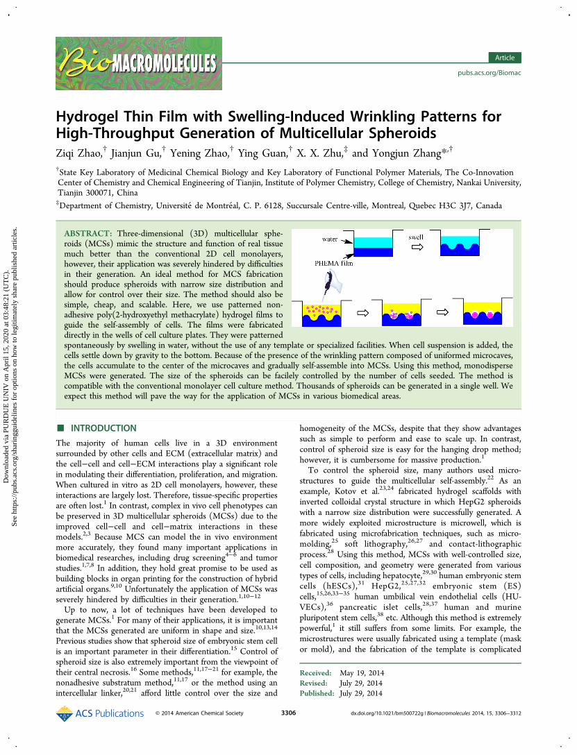
ABSTRACT: Three-dimensional (3D) multicellular sphe-roids (MCSs) mimic the structure and function of real tissuemuch better than the conventional 2D cell monolayers,however, their application was severely hindered by difficultiesin their generation. An ideal method for MCS fabricationshould produce spheroids with narrow size distribution andallow for control over their size. The method should also besimple, cheap, and scalable. Here, we use patterned non-adhesive poly(2-hydroxyethyl methacrylate) hydrogel films toguide the self-assembly of cells. The films were fabricateddirectly in the wells of cell culture plates. They were patternedspontaneously by swelling in water, without the use of any template or specialized facilities. When cell suspension is added, thecells settle down by gravity to the bottom. Because of the presence of the wrinkling pattern composed of uniformed microcaves,the cells accumulate to the center of the microcaves and gradually self-assemble into MCSs. Using this method, monodisperseMCSs were generated. The size of the spheroids can be facilely controlled by the number of cells seeded. The method iscompatible with the conventional monolayer cell culture method. Thousands of spheroids can be generated in a single well. Weexpect this method will pave the way for the application of MCSs in various biomedical areas.
■ INTRODUCTION
The majority of human cells live in a 3D environmentsurrounded by other cells and ECM (extracellular matrix) andthe cell−cell and cell−ECM interactions play a significant rolein modulating their differentiation, proliferation, and migration.When cultured in vitro as 2D cell monolayers, however, theseinteractions are largely lost. Therefore, tissue-specific propertiesare often lost.1 In contrast, complex in vivo cell phenotypes canbe preserved in 3D multicellular spheroids (MCSs) due to theimproved cell−cell and cell−matrix interactions in thesemodels.2,3 Because MCS can model the in vivo environmentmore accurately, they found many important applications inbiomedical researches, including drug screening4−6 and tumorstudies.1,7,8 In addition, they hold great promise to be used asbuilding blocks in organ printing for the construction of hybridartificial organs.9,10 Unfortunately the application of MCSs wasseverely hindered by difficulties in their generation.1,10−12
Up to now, a lot of techniques have been developed togenerate MCSs.1 For many of their applications, it is importantthat the MCSs generated are uniform in shape and size.10,13,14
Previous studies show that spheroid size of embryonic stem cellis an important parameter in their differentiation.15 Control ofspheroid size is also extremely important from the viewpoint oftheir central necrosis.16 Some methods,11,17−21 for example, thenonadhesive substratum method,11,17 or the method using anintercellular linker,20,21 afford little control over the size and
homogeneity of the MCSs, despite that they show advantagessuch as simple to perform and ease to scale up. In contrast,control of spheroid size is easy for the hanging drop method;however, it is cumbersome for massive production.1
To control the spheroid size, many authors used micro-structures to guide the multicellular self-assembly.22 As anexample, Kotov et al.23,24 fabricated hydrogel scaffolds withinverted colloidal crystal structure in which HepG2 spheroidswith a narrow size distribution were successfully generated. Amore widely exploited microstructure is microwell, which isfabricated using microfabrication techniques, such as micro-molding,25 soft lithography,26,27 and contact-lithographicprocess.28 Using this method, MCSs with well-controlled size,cell composition, and geometry were generated from varioustypes of cells, including hepatocyte,29,30 human embryonic stemcells (hESCs),31 HepG2,25,27,32 embryonic stem (ES)cells,15,26,33−35 human umbilical vein endothelial cells (HU-VECs),36 pancreatic islet cells,28,37 human and murinepluripotent stem cells,38 etc. Although this method is extremelypowerful,1 it still suffers from some limits. For example, themicrostructures were usually fabricated using a template (maskor mold), and the fabrication of the template is complicated
Received: May 19, 2014Revised: July 29, 2014Published: July 29, 2014
Article
pubs.acs.org/Biomac
© 2014 American Chemical Society 3306 dx.doi.org/10.1021/bm500722g | Biomacromolecules 2014, 15, 3306−3312
Dow
nloa
ded
via
PUR
DU
E U
NIV
on
Apr
il 15
, 202
0 at
03:
48:2
1 (U
TC
).Se
e ht
tps:
//pub
s.ac
s.or
g/sh
arin
ggui
delin
es f
or o
ptio
ns o
n ho
w to
legi
timat
ely
shar
e pu
blis
hed
artic
les.
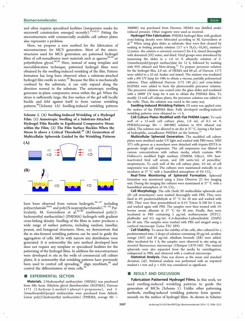
and often requires specialized facilities (inexpensive masks formicrowell construction emerged recently).25,26,33 Fitting themicrostructures with commercially available cell culture platesalso represents a problem.Here, we propose a new method for the fabrication of
microstructures for MCS generation. Most of the micro-structures used for MCS generation are patterned hydrogelfilms of cell-nonadhesive inert materials such as agarose36,38 orpolyethylene glycol.26,28 Here, instead of using template andmicrofabrication technique, patterned hydrogel films wereobtained by the swelling-induced wrinkling of the film. Patternformation has long been observed when a substrate-attachedhydrogel film swells in water.39 Because the film is mechanicallyconfined by the substrate, it can only expand along thedirection normal to the substrate. The anisotropic swellinggenerates in-plane compressive stress within the gel. When thestress is sufficiently large, the free surface of the gel will locallybuckle and fold against itself to form various wrinklingpatterns.40(Scheme 1A) Swelling-induced wrinkling patterns
have been observed from various hydrogels,39−47 includingpolyacrylamide39,41 and poly(N-isopropylacrylamide).42−44 Par-ticularly, M. Guvendiren et al.45,46 synthesized poly(2-hydroxyethyl methacrylate) (PHEMA) hydrogels with gradientcross-linking density. Swelling of the films in water produces awide range of surface patterns, including random, lamellar,peanut, and hexagonal structures. Here, we demonstrate thatthe in situ-formed wrinkling patterns can be used to guide theaggregation of cells. MCSs with narrow size distribution weregenerated. It is noteworthy the new method developed heredoes not require any template or specialized facilities for thepatterning of the hydrogel films. In addition the microstructureswere developed in situ in the wells of commercial cell cultureplates. It is noteworthy that wrinkling patterns have previouslybeen used to control cell spreading,48 align myoblasts,49 andcontrol the differentiation of stem cells.50
■ EXPERIMENTAL SECTIONMaterials. 2-Hydroxyethyl methacrylate (HEMA) was purchased
from Alfa Aesar. Ethylene glycol dimethacrylate (EGDMA), Darocur1173 (2-hydroxy-2-methyl-1-phenyl-1-propanone), and 3-(trimethoxysilyl)propyl methacrylate were purchased from Aldrich.Linear poly(2-hydroxyethyl methacrylate) (PHEMA, average Mv ∼
300000) was purchased from Heowns. HEMA was distilled underreduced pressure. Other reagents were used as received.
Hydrogel Film Fabrication. PHEMA hydrogel films with gradientcross-linking density were fabricated according to M. Guvendiren etal.45 When using glass slides as substrate, they were first cleaned bysoaking in boiling piranha solution (3:7 v/v H2O2−H2SO4 mixture)(Caution: this solution is extremely corrosive!) for 4 h, rinsed thoroughlywith deionized (DI) water, and dried. Vinyl groups were introduced byimmersing the slides in a 1.0 wt % ethanolic solution of 3-(trimethoxysilyl)propyl methacrylate for 12 h, followed by washingwith 95% ethanol and blow-drying.51 To prepare precursor solutionfor the hydrogel film, 2.0 mL of HEMA and 60 μL of Darocur 1173were added to a 10 mL beaker and mixed. The mixture was irradiatedwith a 6W UV lamp for 190s to obtain a viscous, partially polymerizedsolution. Then additional Darocur 1173 (40 μL) and cross-linkerEGDMA were added to form the photocurable precursor solution.The precursor solution was coated onto the glass slides and irradiatedwith a 500W UV lamp for 4 min to obtain the PHEMA films. Tomodify 12-well cell culture plates, the precursor solution was added tothe wells. Then, the solution was cured in the same way.
Swelling-Induced Wrinkling Pattern. DI water was applied ontothe surface of the PHEMA films. Fully developed swelling-inducedwrinkling patterns were obtained in ∼1 h.
Cell Culture Plates Modified with Flat PHEMA Layer. To eachwell of a 12-well cell culture plate, 1.0 mL of 0.5 wt %PHEMA(average Mv ∼ 300 000) solution in 95% ethanol wasadded. The solution was allowed to air-dry at 37 °C, leaving a flat layerof hydrophilic, nonadhesive PHEMA on the bottom.11
Multicellular Spheroid Generation. The modified cell cultureplates were sterilized under UV light and washed with PBS twice. NIH3T3 cells grown as a monolayer were detached with trypsin-EDTA togenerate single-cell suspension. The cell suspension was diluted tovarious concentrations with culture media, which contains 90%Dulbecco’s modified Eagle medium (DMEM, Gibco), 10% heat-inactivated fetal calf serum, and 100 units/mL of penicillin/streptomycin. To each well of the cell culture plate, 3.0 mL of cellsuspension was added. The cultures were maintained statically in anincubator at 37 °C with a humidified atmosphere of 5% CO2.
Real-Time Monitoring of Spheroid Formation. Spheroidformation was monitored using a Zeiss Observer Z1 live imagingunit. During the imaging the cultures were maintained at 37 °C with ahumidified atmosphere of 5% CO2.
Cell Morphology. The cells (both 3D multicellular spheroids and2D cell monolayers) were washed thoroughly with PBS. They werefixed in 4% paraformaldehyde at 37 °C for 20 min and washed withPBS. They were then permeabilized in 0.1% Triton X-100 for 5 minand washed again with PBS. The samples were then treated with 1%bovine serum albumin at 37 °C for 30 min. Finally, they wereincubated in PBS containing 5 μg/mL isothiocyanate (FITC)-phalloidin and 0.2 mg/mL 4′,6-diamidino-2-phenylindole (DAPI)for 20 min. The samples were washed with PBS and imaged using aconfocal microscope (Leica TSC SP8).
Cell Viability. To assess the viability of the cells, after cultured for apredetermined time, 3 drops of solution containing 50 μg/mL acridineorange (AO) and 50 μg/mL ethidium bromide (EB) were added.After incubated for 1 h, the samples were observed in situ using aninverted fluorescence microscope (Olympus LX70-140). The stainedspheroids were also separated from the media by centrifugation,redispersed in PBS, and observed with a confocal microscope.
Statistical Analysis. Data was shown as the mean and standarddeviation (sd). Statistical analysis was performed with an unpairedstudent’s t test and p < 0.05 was considered as significant.
■ RESULT AND DISCUSSION
Fabrication Patterned Hydrogel Films. In this work, weused swelling-induced wrinkling patterns to guide thegeneration of MCSs (Scheme 1). Unlike other patterningmethods, swelling-induced wrinkling patterns form sponta-neously on the surface of hydrogel films. As shown in Scheme
Scheme 1. (A) Swelling-Induced Wrinkling of a HydrogelFilm: (1) Anisotropic Swelling of a Substrate-AttachedHydrogel Film Results in Equibiaxial Compressive Stresswithin the Film; (2) The Film Surface Buckles When theStress Is above a Critical Threshold.39 (B) Generation ofMulticellular Spheroids Guided by the Wrinkling Patterns
Biomacromolecules Article
dx.doi.org/10.1021/bm500722g | Biomacromolecules 2014, 15, 3306−33123307
1A, because the hydrogel films are fixed on a rigid substrate,when swelling in water, the films can only expand along thedirection normal to the substrate. The unidirectional swellinggenerates a biaxial compressive stress within the gel. When thestress is sufficiently large, the free gel surface locally buckles andfolds against itself to form various wrinkling patterns.41
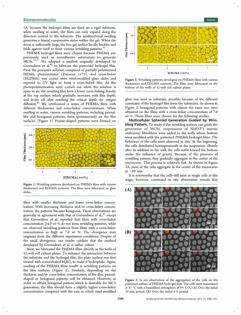
PHEMA hydrogel films were chosen because PHEMA waspreviously used as nonadhesive substratum to generateMCSs.11,17 We adopted a method originally developed byGuvendiren et al.45 to fabricate the patterned hydrogel film.First, the precursor solution composed of partially polymerizedHEMA, photoinitiator (Darocur 1173), and cross-linker(EGDMA) was coated onto vinyl-modified glass slides andexposed to UV light to form a cross-linked film. As thephotopolymerization were carried out when the solution isopen to air, the resulting film have a lower cross-linking densityat the top surface, which gradually increases with film depth,and levels off after reaching the critical depth for oxygendiffusion.45 We synthesized a series of PHEMA films withdifferent thicknesses and cross-linker concentrations. Whenswelling in water, various wrinkling patterns, including peanut-like and hexagonal patterns, form spontaneously on the filmsurfaces. (Figure 1) Peanut-shaped patterns were formed on
films with smaller thickness and lower cross-linker concen-tration. With increasing thickness and/or cross-linker concen-tration, the patterns become hexagonal. These observations aregenerally in agreement with that of Guvendiren et al.,45 exceptthat Guvendiren et al. reported that films with cross-linkerconcentration ≥4.0 wt % do not form wrinkling patterns, whilewe observed wrinkling patterns from films with a cross-linkerconcentration as high as 7.0 wt %. The divergence mayoriginate from the different experiment conditions. Despite ofthe small divergence, our results confirm that the methoddeveloped by Guvendiren et al. is rather robust.Next, we fabricated the PHEMA films directly in the wells of
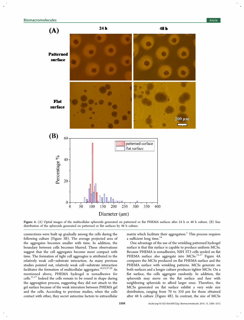
12-well cell culture plates. To enhance the interaction betweenthe substrate and the hydrogel film, the plate surface was firsttreated with concentrated H2SO4 to make it hydrophilic. Again,swelling of the PHEMA films results in wrinkling patterns onthe film surfaces (Figure 2). Similarly, depending on thethickness and/or cross-linker concentration of the film, peanut-shaped or hexagonal patterns will be obtained. However, inorder to obtain hexagonal pattern which is desirable for MCSgeneration, the film should have a slightly higher cross-linkerconcentration compared with the case in which vinyl-modified
glass was used as substrate, possibly because of the differentconstraint of the hydrogel film from the substrates. As shown inFigure 2, hexagonal patterns with almost the same size wereobtained on the films with a cross-linker concentration of 7.0wt %. These films were chosen for the following studies.
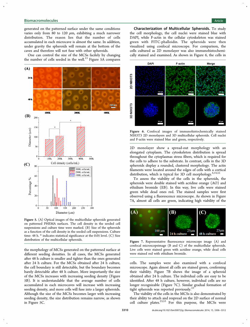
Multicellular Spheroid Generation Guided by Wrin-kling Pattern. To study if the wrinkling pattern can guide thegeneration of MCSs, suspensions of NIH3T3 murineembryonic fibroblasts were added to the wells whose bottomwere modified with the patterned PHEMA hydrogel films. Thebehaviors of the cells were observed in situ. At the beginning,the cells distributed homogeneously in the suspension. Shortlyafter its addition to the well, the cells settle toward the bottomunder the influence of gravity. Because of the presence ofwrinkling pattern, they gradually aggregate in the center of themicrocaves. This process is relatively fast. As shown in Figure3A, most of the cells aggregate in the center of the microcavesin ∼10 min.It is noteworthy that the cells still exist as single cells at this
stage; however, continued in situ observation reveals thatFigure 1. Wrinkling patterns developed on PHEMA films with variousthicknesses and EDGMA contents. The films were fabricated on glassslides.
Figure 2. Wrinkling patterns developed on PHEMA films with variousthicknesses and EDGMA contents. The films were fabricated on thebottom of the wells of 12-well cell culture plates.
Figure 3. In situ observation of the aggregation of the cells on thepatterned surface of PHEMA hydrogel film. The cells were maintainedat 37 °C with a humidified atmosphere of 5% CO2. (A) Over the initial10 min period. (B) Over the initial 6 h period.
Biomacromolecules Article
dx.doi.org/10.1021/bm500722g | Biomacromolecules 2014, 15, 3306−33123308
connections were built up gradually among the cells during thefollowing culture (Figure 3B). The average projected area ofthe aggregates becomes smaller with time. In addition, theboundary between cells becomes blurred. These observationssuggest that the cell aggregates become more compact withtime. The formation of tight cell aggreagtes is attributed to therelatively weak cell−substrate interaction. As many previousstudies pointed out, relatively weak cell−substrate interactionfacilitates the formation of multicellular aggregates.18,23,27,28 Asmentioned above, PHEMA hydrogel is nonadhesive forcells.11,17 Indeed the cells remain to be round in shape duringthe aggreagtion process, suggesting they did not attach to thegel surface because of the weak interation between PHEMA geland the cells. According to previous studies, when the cellscontact with other, they secret autocrine factors to extracellular
matrix which facilitate their aggregation.1 This process requiresa sufficient long time.14
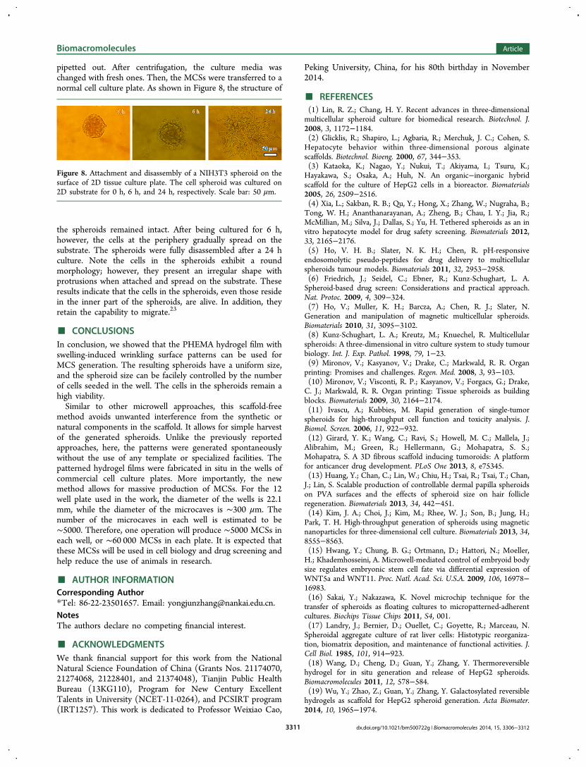
One advantage of the use of the wrinkling patterned hydrogelsurface is that this surface is capable to produce uniform MCSs.Because PHEMA is nonadhesive, NIH 3T3 cells seeded on flatPHEMA surface also aggregate into MCSs.11,17 Figure 4Acompares the MCSs produced on flat PHEMA surface and thePHEMA surface with wrinkling patterns. MCSs generate onboth surfaces and a longer culture produces tighter MCSs. On aflat surface, the cells aggregate randomly. In addition, thespheroids may move on the flat surface and fuse withneighboring spheroids to afford larger ones. Therefore, theMCSs generated on flat surface exhibit a very wide sizedistribution, ranging from 70 to 310 μm for those obtainedafter 48 h culture (Figure 4B). In contrast, the size of MCSs
Figure 4. (A) Optial images of the multicellular spheroids generated on patterned or flat PHEMA surfaces after 24 h or 48 h culture. (B) Sizedistribution of the spheroids generated on patterned or flat surfaces by 48 h culture.
Biomacromolecules Article
dx.doi.org/10.1021/bm500722g | Biomacromolecules 2014, 15, 3306−33123309
generated on the patterned surface under the same conditionsvaries only from 80 to 120 μm, exhibiting a much narrowerdistribution. The reason lies that the number of cellsaccumulated in each microcave is almost the same. In addition,under gravity the spheroids will remain at the bottom of thecaves and therefore will not fuse with other spheroids.One can control the size of the MCSs facilely by changing
the number of cells seeded in the well.13 Figure 5A compares
the morphology of MCSs generated on the patterned surface atdifferent seeding densities. In all cases, the MCSs generatedafter 48 h culture is smaller and tighter than the ones generatedafter 24 h culture. For the MCSs obtained after 24 h culture,the cell boundary is still detectable, but the boundary becomesbarely detectable after 48 h culture. More importantly the sizeof the MCSs increases with increasing seeding density (Figure5B). It is understandable that the average number of cellsaccumulated in each microcaves will increase with increasingseeding density, and more cells will fuse into a larger spheroids.Although the size of the MCSs becomes larger with increasingseeding density, the size distribution remains narrow, as shownin Figure 5C.
Characterization of Multicellular Spheroids. To studythe cell morphology, the cell nuclei were stained blue withDAPI, while F-actin in the cellular cytoskeleton was stainedgreen with FITC-phalloidin. The spheroids were thenvisualized using confocal microscope. For comparison, thecells cultured as 2D monolayer was also immunohistochemi-cally stained and examined. As shown in Figure 6, the cells in
2D monolayer show a spread-out morphology with anelongated cytoplasm. The cytoskeleton distribution is spreadthroughout the cytoplasmas stress fibers, which is required forthe cells to adhere to the substrate. In contrast, cells in the 3Dspheroids display a rounded, clustered morphology. The actinfilaments were located around the edges of cells with a corticaldistribution, which is typical for 3D cell morphology.4,14,52
To assess the viability of the cells in the spheroids, thespheroids were double stained with acridine orange (AO) andethidium bromide (EB). In this way, live cells were stainedgreen while dead ones red. The stained samples were firstobserved using a fluorescence microscope. As shown in Figure7A, almost all cells are green, indicating high viability of the
cells. The samples were also examined with a confocalmicroscope. Again almost all cells are stained green, confirmingtheir viability. Figure 7B shows the image of a spheroidobtained after 24 h culture. The individual cells are easy to beidentified. After 48 h culture, however, individual cells are nolonger recognizable (Figure 7C). Similar gradual formation oftight spheroids was reported previously.23
The viability of the cells in the MCSs is also demonstrated bytheir ability to attach and respread on the 2D surface of normalcell culture plates.23,53 For this purpose, the MCSs were
Figure 5. (A) Optical images of the multicellular spheroids generatedon patterned PHEMA surfaces. The cell density in the seeded cellsuspensions and culture time were marked. (B) Size of the spheroidsas a function of the cell density in the seeded cell suspensions. Culturetime: 48 h. * indicates statistical significance at the 0.05 level. (C) Sizedistribution of the multicellular spheroids.
Figure 6. Confocal images of immunohistochemically stainedNIH3T3 2D monolayers and 3D multicellular spheroids. Cell nucleiand F-actin were stained blue and green, respectively.
Figure 7. Representative fluorescence microscope image (A) andconfocal microscopeimage (B and C) of the multicellular spheroids.Live cells were stained green with acridine orange, while dead cellswere stained red with ethidium bromide.
Biomacromolecules Article
dx.doi.org/10.1021/bm500722g | Biomacromolecules 2014, 15, 3306−33123310
pipetted out. After centrifugation, the culture media waschanged with fresh ones. Then, the MCSs were transferred to anormal cell culture plate. As shown in Figure 8, the structure of
the spheroids remained intact. After being cultured for 6 h,however, the cells at the periphery gradually spread on thesubstrate. The spheroids were fully disassembled after a 24 hculture. Note the cells in the spheroids exhibit a roundmorphology; however, they present an irregular shape withprotrusions when attached and spread on the substrate. Theseresults indicate that the cells in the spheroids, even those residein the inner part of the spheroids, are alive. In addition, theyretain the capability to migrate.23
■ CONCLUSIONSIn conclusion, we showed that the PHEMA hydrogel film withswelling-induced wrinkling surface patterns can be used forMCS generation. The resulting spheroids have a uniform size,and the spheroid size can be facilely controlled by the numberof cells seeded in the well. The cells in the spheroids remain ahigh viability.Similar to other microwell approaches, this scaffold-free
method avoids unwanted interference from the synthetic ornatural components in the scaffold. It allows for simple harvestof the generated spheroids. Unlike the previously reportedapproaches, here, the patterns were generated spontaneouslywithout the use of any template or specialized facilities. Thepatterned hydrogel films were fabricated in situ in the wells ofcommercial cell culture plates. More importantly, the newmethod allows for massive production of MCSs. For the 12well plate used in the work, the diameter of the wells is 22.1mm, while the diameter of the microcaves is ∼300 μm. Thenumber of the microcaves in each well is estimated to be∼5000. Therefore, one operation will produce ∼5000 MCSs ineach well, or ∼60 000 MCSs in each plate. It is expected thatthese MCSs will be used in cell biology and drug screening andhelp reduce the use of animals in research.
■ AUTHOR INFORMATIONCorresponding Author*Tel: 86-22-23501657. Email: [email protected] authors declare no competing financial interest.
■ ACKNOWLEDGMENTSWe thank financial support for this work from the NationalNatural Science Foundation of China (Grants Nos. 21174070,21274068, 21228401, and 21374048), Tianjin Public HealthBureau (13KG110), Program for New Century ExcellentTalents in University (NCET-11-0264), and PCSIRT program(IRT1257). This work is dedicated to Professor Weixiao Cao,
Peking University, China, for his 80th birthday in November2014.
■ REFERENCES(1) Lin, R. Z.; Chang, H. Y. Recent advances in three-dimensionalmulticellular spheroid culture for biomedical research. Biotechnol. J.2008, 3, 1172−1184.(2) Glicklis, R.; Shapiro, L.; Agbaria, R.; Merchuk, J. C.; Cohen, S.Hepatocyte behavior within three-dimensional porous alginatescaffolds. Biotechnol. Bioeng. 2000, 67, 344−353.(3) Kataoka, K.; Nagao, Y.; Nukui, T.; Akiyama, I.; Tsuru, K.;Hayakawa, S.; Osaka, A.; Huh, N. An organic−inorganic hybridscaffold for the culture of HepG2 cells in a bioreactor. Biomaterials2005, 26, 2509−2516.(4) Xia, L.; Sakban, R. B.; Qu, Y.; Hong, X.; Zhang, W.; Nugraha, B.;Tong, W. H.; Ananthanarayanan, A.; Zheng, B.; Chau, I. Y.; Jia, R.;McMillian, M.; Silva, J.; Dallas, S.; Yu, H. Tethered spheroids as an invitro hepatocyte model for drug safety screening. Biomaterials 2012,33, 2165−2176.(5) Ho, V. H. B.; Slater, N. K. H.; Chen, R. pH-responsiveendosomolytic pseudo-peptides for drug delivery to multicellularspheroids tumour models. Biomaterials 2011, 32, 2953−2958.(6) Friedrich, J.; Seidel, C.; Ebner, R.; Kunz-Schughart, L. A.Spheroid-based drug screen: Considerations and practical approach.Nat. Protoc. 2009, 4, 309−324.(7) Ho, V.; Muller, K. H.; Barcza, A.; Chen, R. J.; Slater, N.Generation and manipulation of magnetic multicellular spheroids.Biomaterials 2010, 31, 3095−3102.(8) Kunz-Schughart, L. A.; Kreutz, M.; Knuechel, R. Multicellularspheroids: A three-dimensional in vitro culture system to study tumourbiology. Int. J. Exp. Pathol. 1998, 79, 1−23.(9) Mironov, V.; Kasyanov, V.; Drake, C.; Markwald, R. R. Organprinting: Promises and challenges. Regen. Med. 2008, 3, 93−103.(10) Mironov, V.; Visconti, R. P.; Kasyanov, V.; Forgacs, G.; Drake,C. J.; Markwald, R. R. Organ printing: Tissue spheroids as buildingblocks. Biomaterials 2009, 30, 2164−2174.(11) Ivascu, A.; Kubbies, M. Rapid generation of single-tumorspheroids for high-throughput cell function and toxicity analysis. J.Biomol. Screen. 2006, 11, 922−932.(12) Girard, Y. K.; Wang, C.; Ravi, S.; Howell, M. C.; Mallela, J.;Alibrahim, M.; Green, R.; Hellermann, G.; Mohapatra, S. S.;Mohapatra, S. A 3D fibrous scaffold inducing tumoroids: A platformfor anticancer drug development. PLoS One 2013, 8, e75345.(13) Huang, Y.; Chan, C.; Lin, W.; Chiu, H.; Tsai, R.; Tsai, T.; Chan,J.; Lin, S. Scalable production of controllable dermal papilla spheroidson PVA surfaces and the effects of spheroid size on hair follicleregeneration. Biomaterials 2013, 34, 442−451.(14) Kim, J. A.; Choi, J.; Kim, M.; Rhee, W. J.; Son, B.; Jung, H.;Park, T. H. High-throughput generation of spheroids using magneticnanoparticles for three-dimensional cell culture. Biomaterials 2013, 34,8555−8563.(15) Hwang, Y.; Chung, B. G.; Ortmann, D.; Hattori, N.; Moeller,H.; Khademhosseini, A. Microwell-mediated control of embryoid bodysize regulates embryonic stem cell fate via differential expression ofWNT5a and WNT11. Proc. Natl. Acad. Sci. U.S.A. 2009, 106, 16978−16983.(16) Sakai, Y.; Nakazawa, K. Novel microchip technique for thetransfer of spheroids as floating cultures to micropatterned-adherentcultures. Biochips Tissue Chips 2011, S4, 001.(17) Landry, J.; Bernier, D.; Ouellet, C.; Goyette, R.; Marceau, N.Spheroidal aggregate culture of rat liver cells: Histotypic reorganiza-tion, biomatrix deposition, and maintenance of functional activities. J.Cell Biol. 1985, 101, 914−923.(18) Wang, D.; Cheng, D.; Guan, Y.; Zhang, Y. Thermoreversiblehydrogel for in situ generation and release of HepG2 spheroids.Biomacromolecules 2011, 12, 578−584.(19) Wu, Y.; Zhao, Z.; Guan, Y.; Zhang, Y. Galactosylated reversiblehydrogels as scaffold for HepG2 spheroid generation. Acta Biomater.2014, 10, 1965−1974.
Figure 8. Attachment and disassembly of a NIH3T3 spheroid on thesurface of 2D tissue culture plate. The cell spheroid was cultured on2D substrate for 0 h, 6 h, and 24 h, respectively. Scale bar: 50 μm.
Biomacromolecules Article
dx.doi.org/10.1021/bm500722g | Biomacromolecules 2014, 15, 3306−33123311
(20) Mo, X.; Li, Q.; Lui, L. W. Y.; Zheng, B.; Kang, C. H.; Nugraha,B.; Yue, Z.; Jia, R. R.; Fu, H. X.; Choudhury, D.; Arooz, T.; Yan, J.;Lim, C. T.; Shen, S.; Tan, C. H.; Yu, H. Rapid construction ofmechanically-confined multi-cellular structures using dendrimericintercellular linker. Biomaterials 2010, 31, 7455−7467.(21) Zhao, D.; Ong, S.; Yue, Z.; Jiang, Z.; Toh, Y.; Khan, M.; Shi, J.;Tan, C.; Chen, J. P.; Yu, H. Dendrimer hydrazides as multivalenttransient intercellular linkers. Biomaterials 2008, 29, 3693−3702.(22) Chan, H. F.; Zhang, Y.; Ho, Y.; Chiu, Y.; Jung, Y.; Leong, K. W.Rapid formation of multicellular spheroids in double-emulsiondroplets with controllable microenvironment. Sci. Rep. 2013, 3, 3462.(23) Lee, J.; Cuddihy, M. J.; Cater, G. M.; Kotov, N. A. Engineeringliver tissue spheroids with inverted colloidal crystal scaffolds.Biomaterials 2009, 30, 4687−4694.(24) Zhang, Y. J.; Wang, S. P.; Eghtedari, M.; Motamedi, M.; Kotov,N. A. Inverted-colloidal-crystal hydrogel matrices as three-dimensionalcell scaffolds. Adv. Funct. Mater. 2005, 15, 725−731.(25) Fukuda, J.; Khademhosseini, A.; Yeo, Y.; Yang, X.; Yeh, J.; Eng,G.; Blumling, J.; Wang, C.; Kohane, D. S.; Langer, R. Micromolding ofphotocrosslinkable chitosan hydrogel for spheroid microarray and co-cultures. Biomaterials 2006, 27, 5259−5267.(26) Karp, J. M.; Yeh, J.; Eng, G.; Fukuda, J.; Blumling, J.; Suh, K.;Cheng, J.; Mahdavi, A.; Borenstein, J.; Langer, R.; Khademhosseini, A.Controlling size, shape, and homogeneity of embryoid bodies usingpoly(ethylene glycol) microwells. Lab Chip 2007, 7, 786−794.(27) Tekin, H.; Anaya, M.; Brigham, M. D.; Nauman, C.; Langer, R.;Khademhosseini, A. Stimuli-responsive microwells for formation andretrieval of cell aggregates. Lab Chip 2010, 10, 2411−2418.(28) Bernard, A. B.; Lin, C.; Anseth, K. S. A microwell cell cultureplatform for the aggregation of pancreatic β-cells. Tissue Eng. C 2012,18, 583−592.(29) Fukuda, J.; Sakai, Y.; Nakazawa, K. Novel hepatocyte culturesystem developed using microfabrication and collagen/polyethyleneglycol microcontact printing. Biomaterials 2006, 27, 1061−1070.(30) Wong, S. F.; No, D. Y.; Choi, Y. Y.; Kim, D. S.; Chung, B. G.;Lee, S. Concave microwell based size-controllable hepatosphere as athree-dimensional liver tissue model. Biomaterials 2011, 32, 8087−8096.(31) Mohr, J. C.; de Pablo, J. J.; Palecek, S. P. 3-D microwell cultureof human embryonic stem cells. Biomaterials 2006, 27, 6032−6042.(32) Lee, K. H.; No, D. Y.; Kim, S.; Ryoo, J. H.; Wong, S. F.; Lee, S.Diffusion-mediated in situ alginate encapsulation of cell spheroidsusing microscale concave well and nanoporous membrane. Lab Chip2011, 11, 1168−1173.(33) Park, J.; Cho, C. H.; Parashurama, N.; Li, Y.; Berthiaume, F.;Toner, M.; Tilles, A. W.; Yarmush, M. L. Microfabrication-basedmodulation of embryonic stem cell differentiation. Lab Chip 2007, 7,1018−1028.(34) Ungrin, M. D.; Joshi, C.; Nica, A.; Bauwens, C.; Zandstra, P. W.Reproducible, ultra-high-throughput formation of multicellular organ-ization from single cell suspension-derived human embryonic stem cellaggregates. PLoS One 2008, 3, e1565.(35) Moeller, H.; Mian, M. K.; Shrivastava, S.; Chung, B. G.;Khademhosseini, A. A microwell array system for stem cell culture.Biomaterials 2008, 29, 752−763.(36) Napolitano, A. P.; Chai, P.; Dean, D. M.; Morgan, J. R.Dynamics of the self-assembly of complex cellular aggregates onmicromolded nonadhesive hydrogels. Tissue Eng. 2007, 13, 2087−2094.(37) Lee, B. R.; Hwang, J. W.; Choi, Y. Y.; Wong, S. F.; Hwang, Y.H.; Lee, D. Y.; Lee, S. In situ formation and collagen-alginatecomposite encapsulation of pancreatic islet spheroids. Biomaterials2012, 33, 837−845.(38) Dahlmann, J.; Kensah, G.; Kempf, H.; Skvorc, D.; Gawol, A.;Elliott, D. A.; Drager, G.; Zweigerdt, R.; Martin, U.; Gruh, I. The useof agarose microwells for scalable embryoid body formation andcardiac differentiation of human and murine pluripotent stem cells.Biomaterials 2013, 34, 2463−2471.
(39) Tanaka, T.; Sun, S.; Hirokawa, Y.; Katayama, S.; Kucera, J.;Hirose, Y.; Amiya, T. Mechanical instability of gels at the phasetransition. Nature 1987, 325, 796−798.(40) Dervaux, J.; Ben Amar, M. Mechanical instabilities of gels. Annu.Rev. Conden. Matter Phys. 2012, 3, 311−332.(41) Trujillo, V.; Kim, J.; Hayward, R. C. Creasing instability ofsurface-attached hydrogels. Soft Matter 2008, 4, 564−569.(42) Ortiz, O.; Vidyasagar, A.; Wang, J.; Toomey, R. Surfaceinstabilities in ultrathin, cross-linked poly(N-isopropylacrylamide)coatings. Langmuir 2010, 26, 17489−17494.(43) Yoon, J.; Kim, J.; Hayward, R. C. Nucleation, growth, andhysteresis of surface creases on swelled polymer gels. Soft Matter 2010,6, 5807−5816.(44) Kim, J.; Yoon, J.; Hayward, R. C. Dynamic display ofbiomolecular patterns through an elastic creasing instability ofstimuli-responsive hydrogels. Nat. Mater. 2010, 9, 159−164.(45) Guvendiren, M.; Yang, S.; Burdick, J. A. Swelling-inducedsurface patterns in hydrogels with gradient crosslinking density. Adv.Funct. Mater. 2009, 19, 3038−3045.(46) Guvendiren, M.; Burdick, J. A.; Yang, S. Kinetic study ofswelling-induced surface pattern formation and ordering in hydrogelfilms with depth-wise crosslinking gradient. Soft Matter 2010, 6, 2044−2049.(47) Han, Q.; Li, C.; Guan, Y.; Zhu, X. X.; Zhang, Y. Swelling-induced surface instability of a hydrogen-bonded LBL film and its self-healing. Polymer 2014, 55, 2197−2204.(48) Jiang, X.; Takayama, S.; Qian, X.; Ostuni, E.; Wu, H.; Bowden,N.; LeDuc, P.; Ingber, D. E.; Whitesides, G. M. Controllingmammalian cell spreading and cytoskeletal arrangement withconveniently fabricated continuous wavy features on poly-(dimethylsiloxane). Langmuir 2002, 18, 3273−3280.(49) Lam, M. T.; Sim, S.; Zhu, X.; Takayama, S. The effect ofcontinuous wavy micropatterns on silicone substrates on the alignmentof skeletal muscle myoblasts and myotubes. Biomaterials 2006, 27,4340−4347.(50) Guvendiren, M.; Burdick, J. A. The control of stem cellmorphology and differentiation by hydrogel surface wrinkles.Biomaterials 2010, 31, 6511−6518.(51) Nayak, S.; Debord, S. B.; Lyon, L. A. Investigations into thedeswelling dynamics and thermodynamics of thermoresponsivemicrogel composite films. Langmuir 2003, 19, 7374−7379.(52) Xu, X.; Gurski, L. A.; Zhang, C.; Harrington, D. A.; Farach-Carson, M. C.; Jia, X. Recreating the tumor microenvironment in abilayer, hyaluronic acid hydrogel construct for the growth of prostatecancer spheroids. Biomaterials 2012, 33, 9049−9060.(53) Xu, J. S.; Ma, M. W.; Purcell, W. M. Characterization of somecytotoxic endpoints using rat liver and HepG2 spheroids as in vitromodels and their application in hepatotoxicity studies. II. Spheroid cellspreading inhibition as a new cytotoxic marker. Toxicol. Appl.Pharmacol. 2003, 189, 112−119.
Biomacromolecules Article
dx.doi.org/10.1021/bm500722g | Biomacromolecules 2014, 15, 3306−33123312
Related Documents