HYDRAGEL 7 HR Violet Acide / Acid Violet Ref. 4102 HYDRAGEL 15 HR Violet Acide / Acid Violet Ref. 4122 HYDRAGEL 7 HR Amidoschwarz / Amidoblack Ref. 4105 HYDRAGEL 15 HR Amidoschwarz / Amidoblack Ref. 4125 2013/01

Welcome message from author
This document is posted to help you gain knowledge. Please leave a comment to let me know what you think about it! Share it to your friends and learn new things together.
Transcript
HYDRAGEL 7 HR Violet Acide / Acid Violet
Ref. 4102
HYDRAGEL 15 HR Violet Acide / Acid Violet
Ref. 4122
HYDRAGEL 7 HR Amidoschwarz / Amidoblack
Ref. 4105
HYDRAGEL 15 HR Amidoschwarz / Amidoblack
Ref. 4125
2013/01
HYDRAGEL 7 & 15 HR - 2013/01
- 9 -
INTENDED USEThe HYDRAGEL 7 HR and HYDRAGEL 15 HR kits are designed for multi-fractionation of proteins from human sera or other biological fluids, such asurine and cerebrospinal fluid (CSF), by electrophoresis on alkaline buffered (pH 8.8) agarose gels. The kits are used in conjunction with the semi-automated HYDRASYS instrument. The electrophoretic separations are evaluated visually for protein pattern abnormalities. Densitometry can providesemi-quantitative, relative values.
Each agarose gel is intended to run :• 7 samples in the HYDRAGEL 7 HR kit,• 15 samples in the HYDRAGEL 15 HR kit.
For In Vitro Diagnostic Use.
NOTE : In this instruction sheet, the name "HYDRASYS" is used for both semi-automated HYDRASYS and HYDRASYS 2 instruments.
PRINCIPLE OF THE TEST
Protein electrophoresis is a well established technique routinely used in clinical laboratories for screening of serum and other biological fluids forprotein abnormalities. It is based on the principle of zone electrophoresis performed on a suitable support medium. Agarose has been developed intoa versatile and effective support medium. Although most clinical laboratories are satisfied with the "traditional" five-zone pattern of serum proteins, highresolution techniques can yield additional diagnostic information.On HYDRAGEL 7 HR and HYDRAGEL 15 HR gels, serum, urine or cerebrospinal fluid (CSF) proteins separate into about ten fractions. Each fractioncontains one or more proteins.The composition of the gel, the electrophoretic conditions and the choice of the stain allow an excellent resolution and a high sensitivity particularly inthe gamma-zone. To accommodate customer’s preferences, the electrophoretic protein separations can be stained either with acid violet oramidoblack. The stained separations are evaluated visually for pattern abnormalities. The visual observations can be complemented by densitometryto obtain semi-quantitative, relative values of the individual or combined protein fractions.
REAGENTS AND MATERIALS SUPPLIED IN THE HYDRAGEL 7 HR AND HYDRAGEL 15 HR KITS
WARNING : See the safety data sheets.
| ITEMS | PN 4102 | PN 4122 | PN 4105 | PN 4125 || Agarose Gels (ready to use) | 10 gels | 10 gels | 10 gels | 10 gels || Buffered Strips (ready to use) | 10 packs of 2 each | 10 packs of 2 each | 10 packs of 2 each | 10 packs of 2 each || Staining solution diluent (stock solution) | | | 1 vial, 60 mL | 1 vial, 60 mL || Amidoblack Stain (stock solution) | | | 1 vial, 20 mL | 1 vial, 20 mL || Acid Violet Stain (stock solution) | 1 vial, 75 mL | 1 vial, 75 mL | | || Applicators (ready to use) | 1 pack of 10 (7 teeth) |1 pack of 10 (15 teeth) | 1 pack of 10 (7 teeth) | 1 pack of 10 (15 teeth) || Filter Papers | 1 pack of 10 | 1 pack of 10 | 1 pack of 10 | 1 pack of 10 |FOR OPTIMAL RESULTS :All reagents from the same kit must be always used together and according to the package insert instructions.PLEASE READ THE PACKAGE INSERT CAREFULLY.
1. AGAROSE GELSPreparationAgarose gels are ready to use. Each gel contains : agarose ; buffer solution pH 8.8 ± 0.5 ; additives, nonhazardous at concentrations used, necessaryfor optimum performance.UseSupport medium for protein electrophoresis.Storage, stability and signs of deteriorationStore the gels horizontally in the original protective packaging at room temperature (15 to 30 °C) or refrigerated (2 to 8 °C). (The arrow on the front ofthe kit box must be pointing upwards).Avoid obvious temperature fluctuations during storage (e.g., do not store close to a window or a heat source). The gels are stable until the expirationdate indicated on the kit package or the gel package labels. DO NOT FREEZE.Discard gel when:(i) crystals or precipitate form on the gel surface or the gel texture becomes very soft (all these result from freezing the gel),(ii) bacterial or mold growth is indicated,(iii) abnormal liquid quantity is present in the gel box (as a result of buffer exudation from the gel due to improper storage conditions).
2. BUFFERED STRIPSPreparationBuffered sponge strips are ready to use. Each contains : buffer solution pH 8.7 ± 0.5 ; additives, nonhazardous at concentrations used, necessary foroptimum performance.UseBuffered strips function as electrophoresis buffer reservoir and ensure contact between the gel and electrodes.
SEBIA INSTRUCTIONS - English
HYDRAGEL 7 & 15 HR - 2013/01
- 10 -
Storage, stability and signs of deteriorationStore the buffered strips horizontally in the original protective packaging at room temperature or refrigerated. (The arrow on the front of the kit boxmust be pointing upwards).They are stable until the expiration date indicated on the kit package or buffered strips package label.DO NOT FREEZE.Discard buffered strips if the package is opened and the strips dry out.
3. STAINING SOLUTION DILUENT (with PN 4105 and 4125)PreparationThe stock staining solution diluent must be used as described in paragraph " AMIDOBLACK STAIN ".It contains an acidic solution pH ≈ 2.UseFor the preparation of the amidoblack staining solution.Storage, stability and signs of deteriorationStore the stock staining solution diluent at room temperature or refrigerated. It is stable until the expiration date indicated on the kit package or stainingsolution diluent vial labels. DO NOT FREEZE.Do not add any sodium azide.
4. AMIDOBLACK STAIN (with PN 4105 and 4125)PreparationThe amidoblack concentrated stain is a visquous solution which may gelify. The integrity of the stock staining solution is not altered by the increase inviscosity or solidification.In all cases, to obtain a perfect reconstitution of the stain, we advise you to respect the following procedure:1. Add 15 mL of stain diluent to the concentrated amidoblack vial.2. Close carefully the vial.3. Shake very vigorously the vial during approximately 5 seconds.4. Pour this solution in the container for staining solution processing.5. Repeat this step twice, three times if necessary.6. Pour the remaining diluent in the container and complete the volume to 300 mL with distilled or deionized water.7. Mix contents of stain cubitainer well for 5 to 10 minutes.The staining solution is ready to use.NOTE : An incomplete reconstitution of the stain will lead to an under-evaluation of albumin fraction (low percentage or white hole inside the fraction).After dilution, the working staining solution contains : acidic solution pH ≈ 2 ; amidoblack ; ethylene-glycol ; additives, nonhazardous at concentrationsused, necessary for optimum performance.UseFor staining gels with electrophoretic protein separations.IMPORTANT : The staining solution is designed to stain only 10 gels. Change the solution after 10 staining steps.Storage, stability and signs of deteriorationStore both stock and working staining solutions at room temperature or refrigerated in closed containers to prevent evaporation. Stock staining solutionis stable until the expiration date indicated on the kit package or staining vial labels.Working staining solution is stable for 1 month. Its stability may be extended for 3 months if the working solution is refrigerated. The closed containermust be stored refrigerated immediately after each use.Do not store the working staining solution close to a heat source.
5. ACID VIOLET STAIN (with PN 4102 and 4122)PreparationThe vial of the stock acid violet stain to be diluted up to 300 mL with distilled or deionized water.After dilution, the working stain solution contains : acidic solution pH ≈ 2 ; acid violet ; ethylene-glycol ; additives, nonhazardous at concentrations used,necessary for optimum performance.UseFor staining gels with electrophoretic protein separations.IMPORTANT : The staining solution is designed to stain only 10 gels. Change the solution after 10 staining steps.Storage, stability and signs of deteriorationStore both stock and working stain solutions at room temperature or refrigerated in closed containers to prevent evaporation. Stock stain solution isstable until the expiration date indicated on the kit package or stain vial labels. Working stain solution is stable for 6 months.
6. APPLICATORSUsePrecut, single use applicators for sample application.StorageStore the applicators in a dry place at room temperature or refrigerated.
7. FILTER PAPERSUsePrecut, single use, thin absorbent paper pads for blotting excessive moisture off the gel surface before sample application.StorageStore the thin filter papers in a dry place at room temperature or refrigerated.
HYDRAGEL 7 & 15 HR - 2013/01
- 11 -
REAGENTS REQUIRED BUT NOT SUPPLIED
WARNING : See the safety data sheets.
1. SALINEPreparationMake 0.15 M (0.9 g/dL) NaCl solution in distilled or deionized water.UseTo dilute samples.Storage, stability and signs of deteriorationStore at room temperature or refrigerated. Discard after 3 months or if it changes its appearance, e.g., becomes cloudy due to microbial contamination.For longer storage periods, add sodium azide, 0.1 g/dL.
2. DESTAINING SOLUTIONPreparationEach vial of stock Destaining Solution (SEBIA, PN 4540, 10 vials 100 mL each) to be diluted up to 100 liters with distilled or deionized water. It isconvenient to dilute only 5 mL of the stock solution to 5 liters, the volume of the destaining solution container. After dilution, the working destaining solution contains an acidic solution pH ≈ 2.UseFor destaining, that is removal of excess and background stain from the gels and for rinsing the staining chamber after cleaning with wash solution.To neutralize the acidity of the destaining solution, pour 15 mL of a 50 % solution of Sodium Hydroxide, into the empty waste container.Storage, stability and signs of deteriorationStore the stock destaining solution at room temperature or refrigerated. It is stable until the expiration date indicated on the kit package or destainingsolution vial labels. Working destaining solution is stable for one week at room temperature in a closed bottle. Do not add any sodium azide.Discard working destaining solution if it changes its appearance, e.g., becomes cloudy due to microbial contamination.To prevent microbial proliferation in the diluted destaining solution to be stored more than one week, add 5 µL/dL of ProClin 300.Working destaining solution added with ProClin is stable in a closed bottle at room temperature or refrigerated until the expiration date indicated on thekit package or destaining solution vial labels.
3. HYDRASYS WASH SOLUTIONPreparationEach vial of the stock HYDRASYS Wash Solution (SEBIA, PN 4541, 10 vials, 80 mL each) to be diluted up to 5 liters with distilled or deionized water.After dilution, the working wash solution contains : buffer solution pH 8.7 ± 0.5.UseIt serves for cleaning of the HYDRASYS Staining Compartment. Use periodically, e.g., if the instrument is used daily, wash the staining compartmentweekly.See the package insert for directions to use.Storage, stability and signs of deteriorationStore the stock and working wash solutions in closed containers at room temperature or refrigerated. They are stable until the expiration date indicatedon the wash solution vial label.Discard working wash solution if it changes its appearance, e.g., becomes cloudy due to microbial contamination.
4. FLUIDILPreparationFluidil (SEBIA, PN 4587, 5 mL) is ready to use.UseTo dilute samples with hindered diffusion through the sample applicator teeth (for example, viscous or turbid samples, e.g., sera containing cryoglobulinor cryogel, samples with polymerized Ig M, …) or giving low intensity electrophoretic pattern.Storage, stability and signs of deteriorationStore at room temperature or refrigerated. It is stable until the expiration date indicated on the Fluidil vial label.Fluidil must be free of precipitate.
NOTES :The assays that were performed for the validation of reagents demonstrated that, for the different solutions and using an adapted equipment for thereconstitution volume, a variation of ± 5 % on the final volume has no adverse effect on the analysis.The distilled or deionized water used to reconstitute solutions, must be free of bacterial proliferation and mold (use a 0.22 µm filter) and have aresistivity higher than 10 Megohms x cm.
EQUIPMENT AND ACCESSORIES REQUIRED
1. HYDRASYS System SEBIA: HYDRASYS 2 SCAN PN 1200, HYDRASYS 2 PN 1201, HYDRASYS 2 SCAN FOCUSING PN 1202, HYDRASYS 2FOCUSING PN 1203, HYDRASYS PN 1210 or PN 1211 or HYDRASYS FOCUSING PN 1212.
2. Micropipettor, either manual or automated, such as HYDRAPLUS SEBIA, PN 1216, HYDRAPLUS 2 SEBIA, PN 1217 or ASSIST SEBIA, PN 1218,for an alternative way of loading the sample applicators.
3. Wet Storage, PN 1270, Chamber supplied with HYDRASYS.4. Container Kit supplied with HYDRASYS.5. Gel holder for half-gels SEBIA, PN 1278.6. Pipettes: 10 µL and 200 µL.7. Densitometer / scanner capable of scanning 82 x 51 mm or 82 x 102 mm gel plates : HYRYS SEBIA, GELSCAN SEBIA, DVSE SEBIA or
PHORESIS software for flat-bed scanner. Refer to manufacturer’s instructions for operation and calibration procedures.8. Control materials.
HYDRAGEL 7 & 15 HR - 2013/01
- 12 -
SAMPLES FOR ANALYSIS
Samples for qualitative analysis on HYDRAGEL 7/15 HR may be serum, urine or cerebrospinal fluid (CSF).Serum proteins may also be quantified after staining with acid violet or amidoblack stain.When monitoring the same patient with the analysis of serum, cerebrospinal fluid or urine (concentrated or not) samples, it is recommended to performalways the same HYDRAGEL 7 / 15 HR procedure using one of the 7/15 HR1, 7/15 HR2 or 7/15 HR3 corresponding migration programs.
Sample collection and storageFresh samples are recommended for analysis. They must be collected according to established procedures used in clinical laboratory testing. Ifneeded, store samples at 2 to 8 °C for up to one week.For longer storage periods, keep samples frozen ; frozen samples are stable for at least one month.Thawed samples may give slight application marks due to protein or lipoprotein denaturation.Freezing serum and CSF samples with sodium azide, 0.02 g/dL improves the storage stability.Freezing urine samples with HEPES 0.1 M (pH 6.75) and sodium azide, 0.02 g/dL improves the storage stability.CAUTION : Do not use boric acid as a preservative.
Sample preparation1. Sample preparation for qualitative analysis (with acid violet staining)
The samples are prepared the same way for staining with either acid violet or amidoblack, unless otherwise noted.SerumApply undiluted serum samples. Treatment of serum samples with Fluidil :The treatment of serum samples with Fluidil must be applied in the following cases :- Serum samples with hindered diffusion through the sample applicator teeth, for example, viscous or turbid sera after storage at 2 to 8 °C or after
freezing (particularly those containing cryoglobulin or cryogel). - Serum samples with polymerized Ig M.- Serum samples giving low intensity electrophoretic pattern.In such cases, add 25 μL Fluidil to 75 μL serum and vortex for 15 seconds. Then follow the standard procedure.CSF or serum/CSF pairsApply samples with a total protein concentration of about 1.0 g/dL.IMPORTANT : Serum and CSF sample pairs must be adjusted to have identical total protein concentration.UrinesAnalysis is performed on unconcentrated urine. Concentrate urine if higher sensitivity is needed. Bands are detected when the proteinconcentration per band is about ≥ 2 mg/dL.
- Unconcentrated urines : Apply neat urine.- Concentrated urines : Analysis is performed on samples previously concentrated to a total protein concentration of about 0.5 g/dL.
IMPORTANT: Some urines contain a high salt concentration. This may cause gel deformation during migration and consequently, distortion of themigration profiles. If such a distortion makes interpretation inaccurate, the urine should be dialyzed to remove the salts.NOTE : Diffusion of urine samples into the applicator tips may be hindered when the urine (neat or concentrated) is turbid. It is recommended toremove the particulates by centrifugation (e.g., 10 minutes at 3,000 rpm) or filtration (e.g., 0.45 µm syringe filter).
2. Serum preparation for semi-quantitative analysis (with amidoblack or acid violet staining and 7/15 HR1 program)Amidoblack stainingUse undiluted serum samples.Acid violet stainingUse serum samples previously diluted 4 times (1 vol./3 vol.) in saline. Then follow the standard procedure.
Sample to avoidDo not use hemolysed serum samples. Hemolysis increases alpha-2 and beta zones.Avoid plasma samples. Fibrinogen gives a band close to the application point which might be taken for a monoclonal immunoglobulin.
PROCEDURE
The HYDRASYS system is a semi-automated multi-parameter instrument. The automated steps include processing of HYDRAGEL agarose gels inthe following sequence: sample application, electrophoretic migration, drying, staining, destaining and final drying. The manual steps include handlingsamples and gels, and setting up the instrument for operation.READ CAREFULLY HYDRASYS / HYDRASYS 2 INSTRUCTION MANUAL.
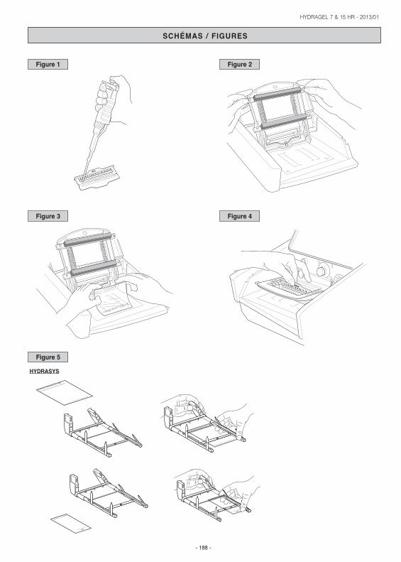
I. MIGRATION SET UP1. Switch on HYDRASYS instrument.2. Place one applicator on a flat surface with the well numbers in the right-side-up position (Fig. 1).
- Apply 10 µL of sample in each well. Load each applicator within 2 minutes.- Place the applicator into the wet storage chamber with the teeth up [handle it by the plastic tooth protection frame]. Let the samples diffuse
into the teeth for 5 minutes after the last sample application. For later use (up to 8 hours), keep the entire chamber under refrigeration. See wet chamber package insert for further details.
3. Open the lid of the Migration Module and raise the electrode and applicator carriers.WARNING: Never close the lid while the carriers are raised !
4. Select migration program from the instrument menu (left side of the keyboard).- "7/15 HR1" migration program : For undiluted or diluted serum on HYDRAGEL 7 HR and HYDRAGEL 15 HR,- "7/15 HR2" migration program : For diluted serum, concentrated CSF and concentrated urines on HYDRAGEL 7 HR and HYDRAGEL
15 HR,- "7/15 HR3" migration program : For unconcentrated urines on HYDRAGEL 7 HR and HYDRAGEL 15 HR.
5. Remove buffered strips from the package ; handle them by the plastic ends. Engage the punched ends of the strip's plastic backing to the pinson the electrode carrier ; the strip's plastic backing must face the carrier (Fig. 2).
HYDRAGEL 7 & 15 HR - 2013/01
- 13 -
6. Unpack the HYDRAGEL plate.- Roll quickly and uniformly one thin filter paper onto the gel surface to absorb the excess of liquid. Remove the paper immediately.
WARNING: Do not leave the filter paper for a too long contact with the gel to avoid its dehydration.- Pool 120 µL of distilled or deionized water for HYDRAGEL 7 HR or 200 µL for HYDRAGEL 15 HR on the lower third of the frame printed on
the Temperature Control Plate of the migration module.- Place the gel plate (the gel side up) with its edge against the stop at the bottom of the printed frame (Fig. 3).- Bend the gel and ease it down onto the water pool (Fig. 3). Ensure that no air bubbles are trapped, water is spread underneath the entire
gel plate and the gel is lined up with the printed frame.7. Lower both carriers down. In this position the buffered strips do not touch the gel. DO NOT FORCE THE CARRIERS ALL THE WAY DOWN.8. Remove the applicator from the wet chamber. Handle it by the protection frame.
- Snap off the applicator teeth's protection frame.- Place the applicator into position No 5 on the carrier.IMPORTANT: The numbers printed on the applicator must face the operator (Fig. 4).
9. Close the lid of the migration module.10. Start the procedure immediately by pressing the green arrow "START" key on the left side of the keyboard.
IMPORTANT: Make sure that the ventilation air inlet on the right side of the instrument is not blocked.
MIGRATION - DESCRIPTION OF THE AUTOMATED STEPS• The two carriers are lowered so that buffered strips and applicator contact the gel surface.• Sample applicator carrier rises up.• "7/15 HR1" and "7/15 HR2" migration programs : Migration is carried out under 255 V constant current, for HYDRAGEL 7 HR and HYDRAGEL
15 HR, at 20 °C, controlled by Peltier effect, until 75 Vh have accumulated, for about 18 minutes.• "7/15 HR3" migration program : Migration is carried out under 255 V constant current, for HYDRAGEL 7 HR and HYDRAGEL 15 HR, at 20 °C,
controlled by Peltier effect, until 40 Vh have accumulated, for about 10 minutes.• The electrode carrier rises to disconnect the electrodes.• The temperature of the control plate rises to 60 °C for 9 minutes to dry the gel.• The control plate is cooled down ; when it reaches 50 °C, an audible beep signals that the migration module lid unlocks. The plate temperature
remains at 50 °C until the lid is opened. Then, the temperature keeps decreasing until it reaches 20 °C (in less than 5 minutes) after which a newmigration run may start.NOTE: The migration module lid remains closed during all migration steps.
II. GEL PROCESSING SET-UP1. Open the lid.2. Remove the applicator and discard.3. Raise both carriers, remove the buffered strips by their plastic ends and discard.4. Remove the dried gel film for further processing.5. After each use, wipe the electrodes and the temperature control plate with a soft wet tissue.6. Open the Gel Holder. Lay it flat and position the dried gel (with gel side facing up) into the grooves of the two rods and close the holder. Make
sure that the film is correctly positioned inside the holder (Fig. 5).7. Place the gel holder into the Gel Processing / Staining Module.
IMPORTANT: Before starting the gel processing / staining program, check the following:- the staining container is filled with 300 mL of staining solution ;- the destaining container contains at least 1 liter of destaining solution ;- the waste container is empty.For reagent line connection: refer to the information displayed on the screen of the instrument (select key: REAGENT LINES).IMPORTANT: Do not forget to block up the unused lines.
8. Select "HR ACID VIOLET" or "HR AMIDO" staining program from the instrument menu and start the run by pressing the "START" key (greenarrow on the right side of the keyboard).
During staining, destaining and drying steps, the compartment remains locked.After cooling step, an audible beep signals that the compartment unlocks (the ventilation is maintained until the gel holder is removed).
III. GEL PROCESSING COMPLETION1. Remove the gel holder from the compartment, open it and remove the dried gel.
NOTE : After gel staining / destaining and before densitometry / scanning, a gel may be put through an additional wash step, if needed, tofurther clarify the gel background and to remove any residual stain that may appear as blue spots. Wash the gel using the "WASHISOENZ/GEL" program.
2. If needed, clean the back side (the plastic support side) of the dry film with a damp soft paper.3. Interpret the separations visually for pattern abnormalities. The visual observations may be completed by densitometry to obtain semi-
quantitative, relative values for the individual or combined protein fractions. Scan using a densitometer / scanner by selecting the appropriatescanning program.
IMPORTANT : Only gels obtained with the 7/15 HR3 migration program can be scanned with the HR3 scanning program of the scanner equippedwith the SEBIA PHORESIS software (VS ≥ 8.51).
QUALITY CONTROLIt is advised to include a normal sample into each run of samples.
* US customers : Follow federal, state and local guidelines for quality control.
HYDRAGEL 7 & 15 HR - 2013/01
- 14 -
RESULTS
Values (quantitative analysis)Densitometer scanning of stained electrophoregrams yields relative concentrations (percentages) of individual protein zones.
Normal values (mean ± 2 SD) for individual major electrophoretic serum protein zones on HYDRAGEL 7/15 HR gels have been established from ahealthy population of 225 adults (men and women):
| FRACTION | HYRYS - GELSCAN | PHORESIS || Albumin | 56.2 - 69.0 | 57.5 - 67.9 || Alpha-1 globulins | 1.2 - 3.2 | 1.0 - 3.8 || Alpha-2 globulins | 7.8 - 13.4 | 7.2 - 12.8 || Beta globulins | 7.4 - 13.4 | 7.6 - 13.6 || Gamma globulins | 9.8 - 18.6 | 10.3 - 18.3 |It is recommended each laboratory establishes its own normal values.
InterpretationInterpretation is qualitative. As an aid in interpretation see BIBLIOGRAPHY.
SERUM PROTEINS The interpretation is made by comparing the electrophoregrams of the clinical sample and a normal control. In the case of an increased, decreasedor additional fraction, it is usually necessary to confirm the observed changes by other tests such as quantification of specific proteins,immunoelectrophoresis, or immunofixation.Among the very numerous plasma proteins, only about fifteen exist in a sufficient quantity to contribute to the staining intensity of the bands observedon HYDRAGEL 7/15 HR gels. The value of HYDRAGEL 7/15 HR gels is primarily in the detection of faint monoclonal or oligoclonal bands.
CEREBROSPINAL FLUID PROTEINSThe cerebrospinal fluid (CSF) is a biological liquid with a low protein concentration ; in a normal CSF, it is about 30 ± 15 mg/dL. The majority of CSFproteins originate in serum from where they are filtered and transported through the blood - CSF barrier. For analysis on HYDRAGEL 7/15 HR gels,the CSF must be concentrated up to about 1 g/dL.
A normal CSF pattern shows the following order of zones:• Prealbumin, or transthyretin, the fastest fraction ;• Albumin, the prevalent fraction representing about 75 % of the total proteins ;• Alpha 1 zone, consisting primarily to alpha-1 antitrypsin ;• Alpha 2 zone, contains primarily high molecular weight proteins ; it is usually very weak since such proteins cannot pass through the blood - CSF
barrier ;• Beta fraction, containing transferrin ;• Beta-2 zone, containing primarily carbohydrate (sialic acid) deficient "CSF specific" transferrin also called the Tau protein ;• Gamma zone, contains essentially Ig G, and sometimes Ig A and Ig M.
An increase of immunoglobulins synthesized within the central nervous system (intrathecal synthesis of Ig’s) has been reported in association withvarious disorders of the central nervous system including demyelinating, inflammatory and infectious disorders and auto-immune reactions.The intrathecal Ig CSF synthesis is often found associated with reduced heterogeneity of the Ig’s which manifests itself as "oligoclonal banding"discerned in high resolution electrophoretic patterns.A distinct characteristic of intrathecal Ig CSF synthesis is the presence of oligoclonal Ig bands in CSF and absence of such banding in the serum.
Therefore, it is necessary :• To analyze the CSF & serum pairs collected from the same patient at the same time while excluding any treatment which could affect the sample’s
concentration of immunoglobulins,• To apply identical quantities of proteins (about 1 g/dL) with each of the 2 samples.• To subject any abnormal band detected in the gamma zone of CSF, without any corresponding fraction in the serum, to immunofixation analysis.The immunological confirmation of the Ig character of such CSF band is necessary since bands other than immunoglobulins may be found in thegamma zone (e.g., tau fraction, carbonic anhydrase, post gamma, or CRP). The latter bands do not have the same diagnostic significance as the " true ", immunoglobulin type oligoclonal bands.
URINEUrine electrophoresis is performed in order to detect monoclonal components and other urinary proteins.HYDRAGEL 7/15 HR is used as a screening test for urinary protein. It allows the detection of monoclonal components (which must be confirmed andidentified by immunofixation), and the detection of proteins of tubular, glomerular or mixed origin (which must be characterized with suitable methods,such as molecular sieving in SDS buffer, nephelometric immunoassay or immunofixation with appropriate antibodies).
CONFIRMATORY TESTSWhen compared to a normal pattern, increased, decreased or additional fractions are observed, confirmatory tests might be needed, such asidentification and quantification of specific proteins by immunochemical procedures. SEBIA offers a number of specialized tests for the use inconjunction with the HYDRASYS system, for example :• monoclonal proteins in serum and urine :
HYDRAGEL 1 IF SEBIA (PN 4301 / 4801), HYDRAGEL 2 IF SEBIA (PN 4302 / 4802), HYDRAGEL 4 IF SEBIA (PN 4304 / 4804 and 4308 / 4808)and HYDRAGEL 9 IF SEBIA (PN 4309 /4809),
• oligoclonal (Ig’s) banding in CSF :HYDRAGEL 3 CSF SEBIA (PN 4350 / 4850) and HYDRAGEL 6 CSF SEBIA (PN 4351 / 4851),
HYDRAGEL 7 & 15 HR - 2013/01
- 15 -
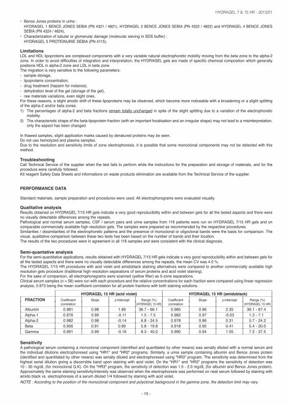
• Bence Jones proteins in urine :HYDRAGEL 1 BENCE JONES SEBIA (PN 4321 / 4821), HYDRAGEL 2 BENCE JONES SEBIA (PN 4322 / 4822) and HYDRAGEL 4 BENCE JONESSEBIA (PN 4324 / 4824),
• Characterization of tubular or glomerular damage (molecular sieving in SDS buffer) :HYDRAGEL 5 PROTEINURIE SEBIA (PN 4115).
LimitationsLDL and HDL lipoproteins are complexed components with a very variable natural electrophoretic mobility moving from the beta zone to the alpha-2zone. In order to avoid difficulties of integration and interpretation, the HYDRAGEL gels are made of specific chemical composition which generallypositions HDL in alpha-2 zone and LDL in beta zone.The migration is very sensitive to the following parameters:- sample storage,- lipoproteins concentration,- drug treatment (heparin for instance),- dehydration level of the gel (storage of the gel),- raw materials variations, even slight ones.For these reasons, a slight anodic shift of these lipoproteins may be observed, which become more noticeable with a broadening or a slight splittingof the alpha-2 and/or beta zones.1) The percentages of alpha-2 and beta fractions remain totally unchanged in spite of the slight splitting due to a variation of the electrophoretic
mobility.2) The characteristic shape of the beta-lipoprotein fraction (with an important focalisation and an irregular shape) may not lead to a misinterpretation,
only the aspect has been changed.
In thawed samples, slight application marks caused by denatured proteins may be seen.Do not use hemolyzed and plasma samples.Due to the resolution and sensitivity limits of zone electrophoresis, it is possible that some monoclonal components may not be detected with thismethod.
TroubleshootingCall Technical Service of the supplier when the test fails to perform while the instructions for the preparation and storage of materials, and for theprocedure were carefully followed.Kit reagent Safety Data Sheets and informations on waste products elimination are available from the Technical Service of the supplier.
PERFORMANCE DATA
Standard materials, sample preparation and procedures were used. All electrophoregrams were evaluated visually.
Qualitative analysisResults obtained on HYDRAGEL 7/15 HR gels indicate a very good reproducibility within and between gels for all the tested aspects and there wereno visually detectable differences among the repeats.Pathological and normal serum samples, CSF / serum pairs and urine samples from 116 patients were run on HYDRAGEL 7/15 HR gels and oncomparable commercially available high resolution gels. The samples were prepared as recommended by the respective procedures.Similarities / dissimilarities of the electrophoretic patterns and the presence of monoclonal or oligoclonal bands were the basis for comparison. Thevisual, qualitative comparison between these two tests has been based on the number of bands and their location.The results of the two procedures were in agreement in all 116 samples and were consistent with the clinical diagnosis.
Semi-quantative analysisFor the semi-quantitative applications, results obtained with HYDRAGEL 7/15 HR gels indicate a very good reproducibility within and between gels forall the tested aspects and there were no visually detectable differences among the repeats, the mean CV was 4.0 %.The HYDRAGEL 7/15 HR procedures with acid violet and amidoblack staining alternatives were compared to another commercially available highresolution gels procedure (traditional high resolution separations of serum proteins and acid violet staining).For the sake of comparison, all electrophoregrams were scanned (yellow filter) as 5-zone separations.Clinical serum samples (n = 56) were run with each procedure and the relative concentrations for each fraction were compared using linear regressionanalysis, 0.973 being the mean coefficient correlation for all protein fractions with both staining solutions.
| HYDRAGEL 15 HR (acid violet) | HYDRAGEL 15 HR (amidoblack) || FRACTION | Coefficient | Slope | y-intercept | Range (%) | Coefficient | Slope | y-intercept | Range (%) || | correlation | | |(HYDRAGEL 15 HR) | correlation | | | (HYDRAGEL 15 HR) || Albumin | 0.981 | 0.98 | 1.89 | 36.7 - 66.1 | 0.985 | 0.96 | 2.35 | 36.1 - 67.4 || Alpha-1 | 0.976 | 0.99 | -0.11 | 1.5 - 7.5 | 0.982 | 0.97 | -0.03 | 1.3 - 7.1 || Alpha-2 | 0.982 | 0.98 | -0.14 | 6.8 - 24.5 | 0.978 | 0.96 | 0.31 | 5.7 - 24.2 || Beta | 0.956 | 0.91 | 0.90 | 5.8 - 19.8 | 0.918 | 0.95 | 0.41 | 5.4 - 20.0 || Gamma | 0.991 | 0.99 | -0.16 | 8.3 - 40.0 | 0.990 | 0.94 | 1.05 | 7.2 - 37.5 |SensitivityA pathological serum containing a monoclonal component (identified and quantitated by other means) was serially diluted with a normal serum andthe individual dilutions electrophoresed using "HR1" and "HR2" programs. Similarly, a urine sample containing albumin and Bence Jones protein(identified and quantitated by other means) was serially diluted and electrophoresed using "HR3" program. The sensitivity was determined from thehighest serial dilution giving a discernible band upon staining with acid violet. On the "HR1" and "HR2" programs the sensitivity of detection was10 - 30 mg/dL (for monoclonal G,K). On the "HR3" program, the sensitivity of detection was 1.5 - 2.0 mg/dL (for albumin and Bence Jones protein).Approximately the same staining sensitivity/intensity was observed when the electrophoresis was performed on neat serum followed by staining withamido black vs. electrophoresis of a serum diluted 1/4 followed by staining with acid violet.NOTE : According to the position of the monoclonal component and polyclonal background in the gamma zone, the detection limit may vary.
HYDRAGEL 7 & 15 HR - 2013/01
- 16 -
LinearityThe assays were linear to at least 5.8 g/dL albumin and 3.9 g/dL Ig G for either staining procedure.
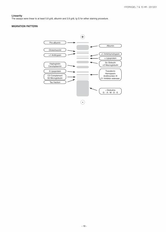
MIGRATION PATTERN
HYDRAGEL 7 & 15 HR - 2013/01
- 187 -
(1) Cameron JS. 1987. The nephrotic syndrome. Am J Kidney Dis, 10 : 157-171.(2) Chopin N. 1991. Étude de la protéinurie. Inf Tech Biol, 1 : 23-28.(3) Cohen R, François B, Sabot JF, Bernard P, Picq JP, Adeleine P. 1985. Étude comparative de l’immunoélectrophorèse urinaire et de quatre index
de sélectivité glomérulaire au cours des glomérulopathies chroniques. Path Biol, 33 : 23-26.(4) Davenport RD, Keren DF. 1988. Oligoclonal bands in cerebrospinal fluids, significance of corresponding bands in serum for diagnosis of multiple
sclerosis. Clin Chem, 34 : 764-765.(5) Joachim GR, Cameron JS, Chwartz M, Belker EL. 1964. Selectivity of protein excretion in patients with the nephrotic syndrome. J Clin Invest,
43, 2332-2346.(6) Keren DF, “High resolution electrophoresis and immunofixation techniques and interpretation”, Butterworth-Heinemann, Woburn, MA, USA, 1994, 397 pp.(7) Laurell DB. 1972. Comparison and variation of the gel electrophoresis fractions of plasma cerebrospinal fluid and urine. Scand J Clin Lab
Investigation, 29 : 71-82.(8) Le Carrer D. 1990. Protéinuries : Mise au point sur leur exploration biologique en 1990. L’Eurobiologiste, 190 : 395-405.(9) Le Carrer D, Chopin N. 1994. Profil protéique urinaire : Proposition d’un protocole d’exploration biologique des protéinuries. Revue française des
laboratoires, 269 : 29-37.(10) Le Carrer D, Nicolas A, Ducasse L. 1992. L’analyse des protéinuries au laboratoire de biologie en 1992. Revue française des laboratoires, 225 : 41-47.(11) Linstedt G, Lindberg PA. 1974. Loss of tubular proteinuria pattern during urine concentration with a commercial membrane filter cell. Clin Chem
Acta, 56 : 125-126.(12) Olsson JE, Link M. 1973. Immunoglobulin abnormalities in multiple sclerosis. 1973. Arch Neurol, 28 : 392-399.(13) Papadopoulos NM, Costello R, Kay AD, Cuttler NR, Papoport SL. 1984. Combined immunochemical and electrophoretic determination of protein
in paired serum and cerebrospinal fluid samples. Clin Chem, 30 : 1814-1816.(14) Pearson SD, Wu JT. Sensitive, specific detection of oligoclonal banding in cerebrospinal fluid by agarose gel electrophoresis. 1989. Clin Chem,
35 : 1997.(15) Philipon C. 1989. Protéines urinaires : Intérêt clinique et interprétation. Technique et Biologie, 6 : 239-249.(16) Waller KV, Ward KM, Mahan JD, Wismatt DK. 1989. Current concepts in proteinuria. Clin Chem, 35 : 755-765.(17) Wendling A. Procédures de diagnostic ou de dépistage : Justification et validité d’un test de diagnostic ou de dépistage-sensibilité-spécificité.
Impact-Internat, 1986 ; Sept : 93-97.
BIBLIOGRAPHIE / BIBLIOGRAPHY
HYDRAGEL 7 & 15 HR - 2013/01
- 188 -
1 2 3 4 5 6 7 8 9 10 11 12 13 14 15
SCHÉMAS / FIGURES
1 2 3 4 5 6 7 8 9 10 11 12 13 14 15
sebia1 2 3 4 5 6 7 8 9 10 11 12
Figure 1 Figure 2
Figure 3 Figure 4
sebia
HYDRAGEL 7 PROTEIN(E)
1 2 3 4 5 6 7
sebia
HYDRAGEL PROTEIN(E) 15/30
16 17 18 19 20 21 22 23 24 25 26 27 28 29 30
1 2 3 4 5 6 7 8 9 10 11 12 13 14 15
sebia
HYDRAGEL 7 PROTEIN(E)
1 2 3 4 5 6 7
sebia
HYDRAGEL PROTEIN(E) 15/30
16 17 18 19 20 21 22 23 24 25 26 27 28 29 30
1 2 3 4 5 6 7 8 9 10 11 12 13 14 15
Figure 5
HYDRASYS
HYDRAGEL 7 & 15 HR - 2013/01
- 189 -
SCHÉMAS / FIGURES
HYDRAGEL PROTEIN(E) 15/30
16 17 18 19 20 21 22 23 24 25 26 27 28 29 30
1 2 3 4 5 6 7 8 9 10 11 12 13 14 15
sebia
1 2 3
4 5
6 7
8 9
10 11 12 13 14 15HYDRAGEL PROTEIN(E) 15/30
16 17 18 19 20 21 22 23 24 25 26 27 28 29 30
sebia
sebiasebia
HYDRAGEL 7 PROTEIN(E)
1 2 3 4 5 6 7
sebia
HYDRAGEL 7 PROTEIN(E)
1 2 3 4 5 6 7
HYDRASYS 2
Related Documents