Hyaluronan-CD44-ERK1/2 Regulate Human Aortic Smooth Muscle Cell Motility during Aging * Received for publication, November 5, 2007, and in revised form, December 12, 2007 Published, JBC Papers in Press, December 12, 2007, DOI 10.1074/jbc.M709051200 Davide Vigetti ‡ , Manuela Viola ‡ , Eugenia Karousou ‡ , Manuela Rizzi ‡ , Paola Moretto ‡ , Anna Genasetti ‡ , Moira Clerici ‡ , Vincent C. Hascall § , Giancarlo De Luca ‡1 , and Alberto Passi ‡1 From the ‡ Dip. di Scienze Biomediche Sperimentali e Cliniche, Universita ` degli Studi dell’Insubria, via J. H. Dunant 5, 21100 Varese, Italy and the § Department of Biomedical Engineering and Orthopaedic Research Center/ND20, The Cleveland Clinic Foundation, Cleveland, Ohio 44195 The glycosaminoglycan hyaluronan (HA) modulates cell prolif- eration and migration, and it is involved in several human vascular pathologies including atherosclerosis and vascular restenosis. Dur- ing intima layer thickening, HA increases dramatically in the neointima extracellular matrix. Aging is one of the major risk fac- tors for the insurgence of vascular diseases, in which smooth mus- cle cells (SMCs) play a role by determining neointima formation through their migration and proliferation. Therefore, we estab- lished an in vitro aging model consisting of sequential passages of human aortic smooth muscle cells (AoSMCs). Comparing young and aged cells, we found that, during the aging process in vitro, HA synthesis significantly increases, as do HA synthetic enzymes (i.e. HAS2 and HAS3), the precursor synthetic enzyme (UDP-glucose dehydrogenase), and the HA receptor CD44. In aged cells, we also observed increased CD44 signaling that consisted of higher levels of phosphorylated MAP kinase ERK1/2. Further, aged AoSMCs migrated faster than young cells, and such migration could be mod- ulated by HA, which alters the ERK1/2 phosphorylation. HA oligo- saccharides of 6.8 kDa and an anti-CD44 blocking antibody pre- vented ERK1/2 phosphorylation and inhibited AoSMCs migration. These results indicate that, during aging, HA can modulate cell migration involving CD44-mediated signaling through ERK1/2. These data suggest that age-related HA accumulation could pro- mote SMC migration and intima thickening during vascular neointima formation. Hyaluronan (HA) 2 is a linear, unsulfated glycosaminoglycan (GAG) that is composed of repeating units of D-glucuronic acid and N-acetylglucosamine linked together through alternating 1,4 and 1,3 glycosidic bonds. The amount and the molecular weight of HA are important factors that regulate the physio- pathological effects that this molecule displays on cells (1). In mammals, three specific HA synthases (HAS1, -2, and -3) and three hyaluronidases (HYAL1, 2, and PH20) regulate HA syn- thesis and degradation with specific biochemical properties and distributions in adult as well as in embryonic tissues (2, 3). Therefore, these enzymes have a critical role in HA metabolism and are responsible for HA balance in the extracellular matrix (ECM). Hydrated HA makes the ECM an ideal environment in which cells can move and proliferate. Moreover, HA is an important space filling molecule as is evident in the vitreous humor, the dermis and the synovial fluid of joints. Besides its chemical and mechanical properties, HA interacts with several receptors at the cellular level that specifically trigger various signal trans- duction responses (4). The HA receptor CD44 is expressed on the surface of most cells, including immune system cells, and it mediates cell adhesion, proliferation and migration (5). Recep- tor for HA-mediated motility (RHAMM) mediates cellular motility (6). Lyve-1 is the specific HA receptor of the lymphatic system although very recent evidences indicate a more complex function of this protein unrelated to HA (7). HA Receptor for Endocytosis (HARE) mediates the endocytosis of HA (8, 9). Recently, Toll Like Receptors 4 and 2 (TLR4/2) were shown to recognize HA fragments (10) and modulate the inflammation response in the lung (11). Although it has a simple structure, it is clear that HA can modulate many cellular responses, and the amount of this GAG has to be strictly regulated. Cells during different stress responses synthesize HA cable-like structures that have a role in inflammation, acting as an adherent for monocytes and other immune system cells (12–15). A large body of evidence describes a direct correlation between the amounts of HA and degree of malignancy in cancers. Consid- ering the role of HA in cell proliferation and migration, the correlation between malignancy and HA content opened a new insight in tumor biology (16). In vascular pathology, HA accumulates during the formation of neointima, which reduces the vessel diameter and is a crucial event for the pathological outcome (17). In the first steps of its formation, neointima tissue is composed mainly of proliferat- ing smooth muscle cells (SMCs) that migrated from the tunica media of the vessel after a mechanical or other undefined injury. In this context, HA and CD44 have a pivotal role to * This work was supported by Ministero dell’Università e della Ricerca (PRIN) (to D. V.), University of Insubria (FAR) (to D. V., A. P., M. V., and G. D. L.), and by Centro Interuniversitario di Biotecnologie (CIB) (to A. P.). The costs of publication of this article were defrayed in part by the payment of page charges. This article must therefore be hereby marked “advertisement” in accordance with 18 U.S.C. Section 1734 solely to indicate this fact. 1 To whom correspondence should be addressed: Dipartimento di Scienze Biomediche Sperimentali e Cliniche, Universita ` degli Studi dell’Insubria, via J. H. Dunant 5, 21100 Varese, Italy. Tel.: 39-0332-217142; Fax: 39-0332- 217119; E-mail: [email protected]. 2 The abbreviations used are: HA, hyaluronan; GAG, glycosaminoglycan; RT- PCR, reverse transcriptase-polymerase chain reaction; HAS, HA syn- thase(s); HYAL, hyaluronidase(s); FACE, fluorophore-assisted carbohydrate electrophoresis; AMAC, 2-aminoacridone; HWHA, high molecular weight HA; UGDH, UDP-glucose dehydrogenase; CS-4S, chondroitin 4 sulfate disaccharide; CS-6S, chondroitin 6 sulfate disaccharide; SM actin, -smooth muscle actin; AoSMCs, aortic smooth muscle cells; ERK, extracel- lular signal-regulated kinase; PBS, phosphate-buffered saline; FBS, fetal bovine serum; ECM, extracellular matrix. THE JOURNAL OF BIOLOGICAL CHEMISTRY VOL. 283, NO. 7, pp. 4448 –4458, February 15, 2008 © 2008 by The American Society for Biochemistry and Molecular Biology, Inc. Printed in the U.S.A. 4448 JOURNAL OF BIOLOGICAL CHEMISTRY VOLUME 283 • NUMBER 7 • FEBRUARY 15, 2008 by guest on March 27, 2016 http://www.jbc.org/ Downloaded from

Welcome message from author
This document is posted to help you gain knowledge. Please leave a comment to let me know what you think about it! Share it to your friends and learn new things together.
Transcript
Hyaluronan-CD44-ERK1/2 Regulate Human Aortic SmoothMuscle Cell Motility during Aging*
Received for publication, November 5, 2007, and in revised form, December 12, 2007 Published, JBC Papers in Press, December 12, 2007, DOI 10.1074/jbc.M709051200
Davide Vigetti‡, Manuela Viola‡, Eugenia Karousou‡, Manuela Rizzi‡, Paola Moretto‡, Anna Genasetti‡,Moira Clerici‡, Vincent C. Hascall§, Giancarlo De Luca‡1, and Alberto Passi‡1
From the ‡Dip. di Scienze Biomediche Sperimentali e Cliniche, Universita degli Studi dell’Insubria, via J. H. Dunant 5,21100 Varese, Italy and the §Department of Biomedical Engineering and Orthopaedic Research Center/ND20,The Cleveland Clinic Foundation, Cleveland, Ohio 44195
The glycosaminoglycan hyaluronan (HA) modulates cell prolif-eration andmigration, and it is involved in several human vascularpathologies includingatherosclerosis andvascular restenosis.Dur-ing intima layer thickening, HA increases dramatically in theneointima extracellular matrix. Aging is one of the major risk fac-tors for the insurgence of vascular diseases, in which smoothmus-cle cells (SMCs) play a role by determining neointima formationthrough their migration and proliferation. Therefore, we estab-lished an in vitro aging model consisting of sequential passages ofhuman aortic smooth muscle cells (AoSMCs). Comparing youngand aged cells, we found that, during the aging process in vitro, HAsynthesis significantly increases, as do HA synthetic enzymes (i.e.HAS2 and HAS3), the precursor synthetic enzyme (UDP-glucosedehydrogenase), and the HA receptor CD44. In aged cells, we alsoobserved increased CD44 signaling that consisted of higher levelsof phosphorylated MAP kinase ERK1/2. Further, aged AoSMCsmigratedfaster thanyoungcells, andsuchmigrationcouldbemod-ulatedbyHA,whichalters theERK1/2phosphorylation.HAoligo-saccharides of 6.8 kDa and an anti-CD44 blocking antibody pre-ventedERK1/2phosphorylationandinhibitedAoSMCsmigration.These results indicate that, during aging, HA can modulate cellmigration involving CD44-mediated signaling through ERK1/2.These data suggest that age-related HA accumulation could pro-mote SMC migration and intima thickening during vascularneointima formation.
Hyaluronan (HA)2 is a linear, unsulfated glycosaminoglycan(GAG) that is composed of repeating units of D-glucuronic acid
and N-acetylglucosamine linked together through alternating�1,4 and �1,3 glycosidic bonds. The amount and themolecularweight of HA are important factors that regulate the physio-pathological effects that this molecule displays on cells (1). Inmammals, three specific HA synthases (HAS1, -2, and -3) andthree hyaluronidases (HYAL1, 2, and PH20) regulate HA syn-thesis anddegradationwith specific biochemical properties anddistributions in adult as well as in embryonic tissues (2, 3).Therefore, these enzymes have a critical role inHAmetabolismand are responsible for HA balance in the extracellular matrix(ECM).HydratedHAmakes the ECMan ideal environment inwhich
cells can move and proliferate. Moreover, HA is an importantspace filling molecule as is evident in the vitreous humor, thedermis and the synovial fluid of joints. Besides its chemical andmechanical properties, HA interacts with several receptors atthe cellular level that specifically trigger various signal trans-duction responses (4). The HA receptor CD44 is expressed onthe surface of most cells, including immune system cells, and itmediates cell adhesion, proliferation and migration (5). Recep-tor for HA-mediated motility (RHAMM) mediates cellularmotility (6). Lyve-1 is the specific HA receptor of the lymphaticsystem although very recent evidences indicate amore complexfunction of this protein unrelated to HA (7). HA Receptor forEndocytosis (HARE) mediates the endocytosis of HA (8, 9).Recently, Toll Like Receptors 4 and 2 (TLR4/2) were shown torecognize HA fragments (10) and modulate the inflammationresponse in the lung (11). Although it has a simple structure, itis clear that HA canmodulate many cellular responses, and theamount of this GAG has to be strictly regulated. Cells duringdifferent stress responses synthesize HA cable-like structuresthat have a role in inflammation, acting as an adherent formonocytes and other immune system cells (12–15). A largebody of evidence describes a direct correlation between theamounts of HA and degree of malignancy in cancers. Consid-ering the role of HA in cell proliferation and migration, thecorrelation betweenmalignancy andHA content opened a newinsight in tumor biology (16).In vascular pathology, HA accumulates during the formation
of neointima, which reduces the vessel diameter and is a crucialevent for the pathological outcome (17). In the first steps of itsformation, neointima tissue is composed mainly of proliferat-ing smooth muscle cells (SMCs) that migrated from the tunicamedia of the vessel after a mechanical or other undefinedinjury. In this context, HA and CD44 have a pivotal role to
* This work was supported by Ministero dell’Università e della Ricerca (PRIN)(to D. V.), University of Insubria (FAR) (to D. V., A. P., M. V., and G. D. L.), andby Centro Interuniversitario di Biotecnologie (CIB) (to A. P.). The costs ofpublication of this article were defrayed in part by the payment of pagecharges. This article must therefore be hereby marked “advertisement” inaccordance with 18 U.S.C. Section 1734 solely to indicate this fact.
1 To whom correspondence should be addressed: Dipartimento di ScienzeBiomediche Sperimentali e Cliniche, Universita degli Studi dell’Insubria, viaJ. H. Dunant 5, 21100 Varese, Italy. Tel.: 39-0332-217142; Fax: 39-0332-217119; E-mail: [email protected].
2 The abbreviations used are: HA, hyaluronan; GAG, glycosaminoglycan; RT-PCR, reverse transcriptase-polymerase chain reaction; HAS, HA syn-thase(s); HYAL, hyaluronidase(s); FACE, fluorophore-assisted carbohydrateelectrophoresis; AMAC, 2-aminoacridone; HWHA, high molecular weightHA; UGDH, UDP-glucose dehydrogenase; �CS-4S, chondroitin 4 sulfatedisaccharide; �CS-6S, chondroitin 6 sulfate disaccharide; �SM actin,�-smooth muscle actin; AoSMCs, aortic smooth muscle cells; ERK, extracel-lular signal-regulated kinase; PBS, phosphate-buffered saline; FBS, fetalbovine serum; ECM, extracellular matrix.
THE JOURNAL OF BIOLOGICAL CHEMISTRY VOL. 283, NO. 7, pp. 4448 –4458, February 15, 2008© 2008 by The American Society for Biochemistry and Molecular Biology, Inc. Printed in the U.S.A.
4448 JOURNAL OF BIOLOGICAL CHEMISTRY VOLUME 283 • NUMBER 7 • FEBRUARY 15, 2008
by guest on March 27, 2016
http://ww
w.jbc.org/
Dow
nloaded from
induce SMC proliferation andmigration (18). Transgenic miceoverexpressing HAS2 have high HA levels in the vasculatureand developed more serious atherosclerotic lesions comparedwith controls (19). Moreover, in other experiments, CD44knock-out mice showed slower SMC proliferation rates thancontrol mice and less severe atherosclerotic lesions (5).As aging is one of the major risk factors for the insurgence of
vascular pathology (20), we have established a cell model for invitro aging in order to investigate age related modifications ingene expression and cellular migration in primary human aor-tic SMCs (AoSMCs). Recently, we used this aging model todescribe age related changes in expression and activity ofmatrix metalloproteinase 2 (21). In this study, we describe theage-related changes in HA content, including the gene expres-sion of enzymes involved in HA metabolism, and its effect onthe migration properties of young and aged AoSMCs. The dataobtained suggest that HA can regulate cell migration through aCD44 pathway that modulates ERK1/2 phosphorylation.
EXPERIMENTAL PROCEDURES
Cell Culture—Human primary aortic smooth muscle cells(AoSMCs, donor age 17, male) were purchased from Cambrexand grown in SmGm2 complete culture medium (Cambrex)supplemented with 5% fetal bovine serum (FBS). This cell cul-turemediumwas used in all the experiments. The culturesweremaintained in an atmosphere of humidified 95% air, 5% CO2 at37 °C. Whenever cultures became nearly confluent, the cellswere trypsinized, counted using trypan blue exclusion, and sub-cultured. Population doublings (PDs) were determined at eachpassage and were calculated as follows: PD � log (number ofcells obtained/initial number of cells)/log 2. In the experiments,4 PD and 18 PD cells were used and are referred to as young andaged AoSMCs, respectively.HAQuantification—HA and chondroitin sulfate were quan-
tified by means of fluorophore-assisted carbohydrate electro-phoresis (FACE) and HPLC analysis (22). Briefly, GAGs werepurified starting from 300 �l of conditioned culture medium ofyoung and agedAoSMCsby proteinaseK (Fynnzyme) digestionand ethanol precipitation. GAGs associated with the cell layerwere analyzed by resuspending the AoSMCs in 0.1 M ammo-nium acetate, pH 7, disrupting the cells with sonication, digest-ing with proteinase K, and recovering the GAGs by ethanolprecipitation. GAGs were digested with hyaluronidase SD(Seikagaku) and by chondroitinase ABC (Seikagaku). Disaccha-rides obtained by the enzymatic digestions were fluorotaggedwith 2-aminoacridone (AMAC, Molecular Probes). AMAC-tagged disaccharides were separated and quantified with FACEand HPLC.Determination of HAMolecular Size—Gel filtration chroma-
tography was done on AMAC derivatized GAGs purified fromyoung and aged AoSMC conditioned culture media as previ-ously described (23) using an FPLC (Amersham Biosciences)apparatus. Sample peaks were identified and quantified bycomparing the absorbance at 280 nm with standard proteins.The identification and quantification of GAGs was done oneluted fractions (2ml each) by digestion with hyaluronidase SDand chondroitinase ABC and analysis as described above.
HA Localization—Immunofluorescence experiments weredone as previously described by de la Motte et al. (12). Briefly,young and aged AoSMCs grown on coverslips were fixed incold methanol for 15 min. The coverslips were preincubatedwith PBS containing 2% FBS and then incubated in the samesolution containing biotinylated HA-binding protein (Seika-gaku) (5�g/ml) and aCD44monoclonal antibody (cloneA3D8,Sigma; 10�g/ml) overnight at 4 °C. The coverslips werewashedwith PBS, and then incubated with a solution containing fluo-rescein-tagged streptavidin (1:500) and Texas Red-conjugatedanti-mouse Ig (H�L) (1:500) in PBS containing 2% FBS. Afterwashing in PBS, the coverslips were mounted using mountingmedium containing 4,6-diamidino-2-phenylindole (DAPI)(Vector Laboratories). The slides were then sealed with nailpolish and images were obtained using a fluorescence micro-scope (Olympus).Quantitative and Semiquantitative RT-PCR—Total RNA
fromboth young and agedAoSMCswas extracted usingTRIzolreagent (Invitrogen) following the manufacturer’s protocol.Total RNA from human normal fetal aorta (Stratagene) andtotal RNA from human adult aorta (Ambion) were used toquantify the gene expression in vivo. To remove DNA contam-ination, DNase (Ambion) treatment was done in all samples.One �g of total RNA was retrotranscribed using the HighCapacity cDNA synthesis kit (Applied Biosystems) for 2 h at37 °C. Quantitative RT-PCR was done on an Abi Prism 7000instrument (Applied Biosystems) usingTaqmanUniversal PCRMasterMix (Applied Biosystems) following themanufacturer’sinstructions. Probes and primers were from TaqMan geneexpression assay reagents (Applied Biosystems). The followingTaqMan gene expression assays were used: HAS1(Hs00155410), HAS2 (Hs00193435), HAS3 (Hs00193436),HYAL2 (Hs00186841), PH20 (Hs00162139), smooth muscle�-actin (Hs00224622), ICAM (Hs00164932), VCAM(Hs00174239), CD44 (Hs00174139), RHAMM (Hs00234864),UDP-glucose dehydrogenase (UGDH) (Hs00163365) andRNAseP (Hs00706565_s1). Fluorescent signals generated dur-ing PCR amplifications were monitored and analyzed with AbiPrism 7000 SDS software (Applied Biosystems). Comparison ofthe amount of each gene transcript among different sampleswasmade using RNAseP as the reference. In order to determinethe efficiency of each Taqman gene expression assay, standardcurves were generated by serial dilution of cDNA, and quanti-tative evaluations of target and housekeeping gene levels wereobtained by measuring threshold cycle numbers (Ct). As thedifferences among efficiencies of eachTaqman gene expressionassay were �0.1, we used the relative quantification method��Cts to quantify gene expression (24).The Taqman assay for HYAL1 detects mRNAs coding for
both the active and the inactive splicing variants. Therefore, wedesigned two primers (5�-ACTTTCTAAGCCCCAACTA-CACC-3� and 5�-GGTTCTTGTATTTTCCCAGCTC-3�) todetect only the active HYAL1 wild-type transcript (25) incanonical semiquantitative RT-PCR experiments using RED-Taq (Sigma). Moreover, to detect HYAL3 we designed5�-TGTCCCAGGATGACCTTGTG-3� and 5�-TCTAC-CCCTCAGGGATTCCA-3�-specific primers, and to detectHYAL4 we designed 5�-GCGCCCAGTTACCTTCACTT-3�
Hyaluronan during Smooth Muscle Cell Aging
FEBRUARY 15, 2008 • VOLUME 283 • NUMBER 7 JOURNAL OF BIOLOGICAL CHEMISTRY 4449
by guest on March 27, 2016
http://ww
w.jbc.org/
Dow
nloaded from
and 5�-GCAACTTAAATATTCCAATAAGGAGGA-3� prim-ers. For standardization, �-actin was also amplified by using5�-GGCACCCAGCACAATGAAG-3� and 5�-GCCGATCCA-CACGGAGTACT-3� primers.Detection of Hyaluronidase Activity by Zymography—Cell
lysates from young and aged AoSMC cultures were obtained bydisrupting the cells with sonication in PBS with 0.1% TritonX-100. Medium samples were concentrated 10� in a filtrationapparatus with a cut-off of 10,000Da. Aliquots of these sampleswere electrophoresed in a 12% polyacrylamide gel containing 1mg/ml of HA (26). The gel was rinsed for 2 h in 2.5% TritonX-100 and incubated overnight at 37 °C in 50 mM citric acid-Na2HPO4, 0.15 M NaCl, pH 5, or in 0.1 M sodium formate, 0.15MNaCl, pH 3.5. Each gel was treated with 0.1mg/ml Pronase in20mMTris-HCl, pH 8.0 for 2 h at 37 °C and stained with AlcianBlue.The electrophoretic assay of hyaluronidase activity was also
used as described (27). Briefly, 50�g of HA fromhuman umbil-ical cord (Sigma) was incubated with young or aged AoSMC-conditioned medium or with 30 �g of cell lysate proteins pre-pared by exposing young or aged AoSMC cultures to a PBSbuffer containing 0.5% Triton X-100. After incubation at 37 °Cat pH 3.5 (using formate buffer, see above) or at pH 5 (usingcitric acid-Na2HPO4buffer, see above), the sampleswere boiledand electrophoresed in standard 0.5% agarose Tris acetate-EDTAgels, andHAwas stainedwith 0.005%Stains-All (Sigma).Migration Assay—Cell migration was measured with the
Transwell (Costar) system, which allows cells to migratethrough 8-�m pore size polycarbonate membranes asdescribed previously (21). Briefly, 105 cells were resuspended inserum-free SmGm2 and added to the upper chamber of Tran-swells. The lower chamber was filled with 1.5 ml serum-freeSmGm2 or 1.5 ml serum-free SmGm2 containing 10 �g/ml ofpurified, low endotoxin 34-mer HA oligosaccharide (28), or 10�g/ml of high molecular weight HA (Healon, Amersham Bio-sciences), or anti CD44 monoclonal antibodies (clone A3D8 at15 �g/ml or clone BRIC235 at 5 �g/ml), or 15 �g/ml of anisotype-matched control antibody against tubulin (cloneDM1A, Sigma), or 15 �M (final concentration) of U0126(Sigma), or 10 ng/ml of PDGF-BB (Euroclone), or 20% FBS(Euroclone). In control experiments, HWHAwas digested with30 milliunits of chondroitinase ABC (Seikagaku) at 37 °C for16 h to obtainHAdisaccharides. After 6 h, filterswere removed,and cells remaining on the upper surface of themembranewereremoved with a cell scraper. The cells present beneath themembrane were fixed with cold methanol and stained withCrystal violet. Cells were counted in 10 high-powermicroscopefields.Western Blotting—Young and aged AoSMC cultures were
lysed in ice-cold PBS buffer containing 0.5% Triton X-100, anda phosphatase and protease inhibitor mixture (Roche AppliedScience) by means of sonication. Total protein contents weredetermined by the Bradford method. 30 �g of proteins wereseparated by SDS-PAGE under reducing conditions and blot-ted onto a polyvinylidene difluoride membrane (Millipore).Membranes were probed with specific anti-phospho-ERK1/2or anti-ERK1/2 antibodies (Cell Signaling Technology) at thedilutions reported by the manufacturer. Signals were revealed
using secondary peroxidase-conjugated antibodies, and thebands visualized by chemoluminescence (Amersham Bio-sciences). Developed films were scanned using a GS-700 Imag-ingDensitometer (Bio-Rad) and quantified usingQuantityOnesoftware (Bio-Rad).Statistical Analyses—Unpaired Student’s t tests were done
for statistical analyses. Probability values of p � 0.01 were con-sidered statistically significant. Experiments were done in trip-licate, and data are expressed as means � S.E.
RESULTS
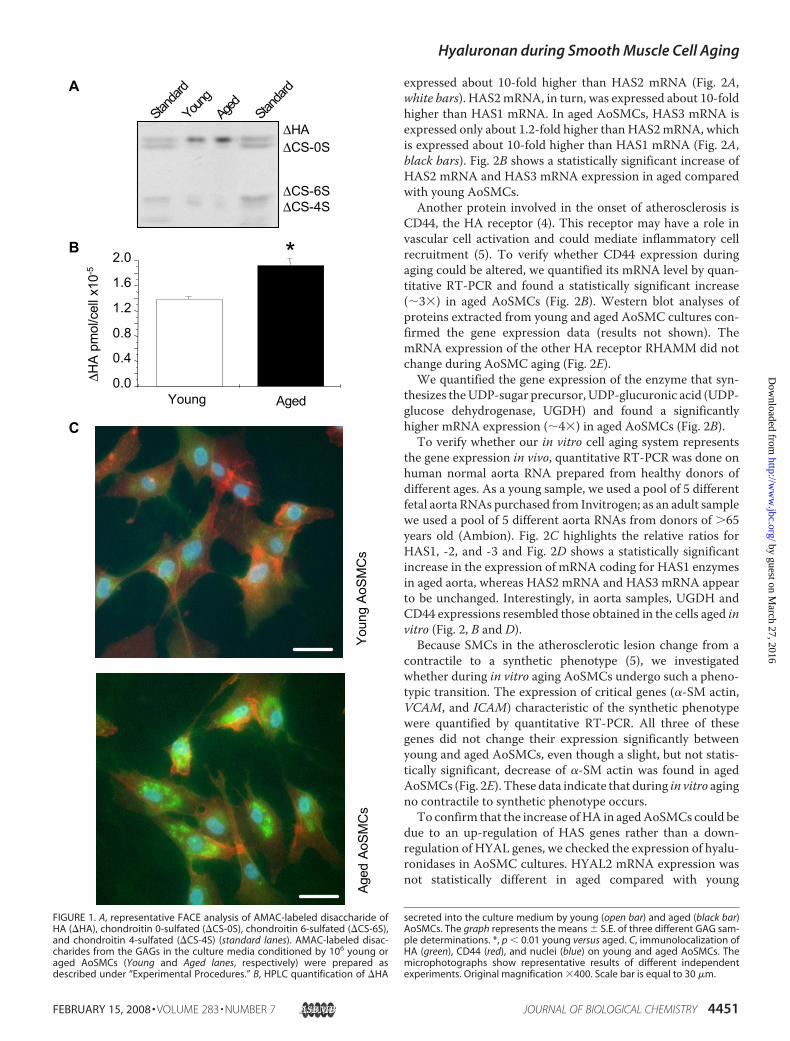
HA Quantification and Characterization—As previouslydescribed (21), we cultured commercial AoSMCs (Cambrex) incomplete-SmGm2 medium (Cambrex) for several populationdoublings (PD) and consider as “young,” cells with 4 PD afterthawing, and as “aged,” cells with 18 PD after thawing. Duringcell passages, AoSMCs began to show senescence associated�-galactosidase activity, which is considered a typical senes-cence marker (29), increasing from about 10% in youngAoSMCs to about 90% in aged cells indicating a substantialabsence of senescence in the cells that we defined as young (21).Moreover, the expression of cyclin p16INK4a, another senes-cence marker, was significantly higher in aged than in youngAoSMCs (21). Moreover, we exclude any dedifferentiationevents because a SMC specific marker, � SM actin, and thedistribution of actin filaments did not change during cell pas-sages (21).By FACE analyses, we found that aged cells secrete more HA
into the culture medium (Fig. 1A). These findings were con-firmed by quantitative HPLC analyses of disaccharides, whichmeasured a significant 20% increase of HA secreted into theculture medium of aged relative to young AoSMCs (Fig. 1B).Moreover, the HPLC analyses also showed that the ratios ofdisaccharides obtained from chondroitin 4 and 6 sulfate (�CS-4S/�CS-6S) decreased during aging from 7.0 � 0.5 in youngAoSMCs to 5.4 � 0.4 in aged cells (data not shown). The rela-tive increase of chondroitin 6-sulfate is known to be linkedwiththe aging process as well as with growth relative to puberty incartilage (30, 31).Qualitative assessment of immunofluorescence of cultures
showed that HA associated with AoSMCs was higher in agedthan in young cells (Fig. 1C). Interestingly, some HA in agedAoSMCs was distributed in spots within cells, as previouslyreported in mitotic rat aortic SMCs (14, 32).As theHAmolecular weight is an important factor inHA cell
responses, we characterized the size distribution ofHA releasedin cell culture medium by young and aged AoSMC cultureswith gel filtration. The average HAmolecular weight was about2.2 � 106 Da in the medium of both young and aged cells(results not shown).Gene Expression Studies of the HA-metabolizing Enzymes—
Quantitative RT-PCR was used to characterize the differencesin the expression of HA metabolizing enzymes between youngand aged AoSMC cultures. We quantified transcripts codingfor the HA synthetic enzymes (HAS1, -2, and -3) and thedegrading enzymes (HYAL1, -2, and PH20) usingTaqman geneexpression assays. In young AoSMCs, HAS3mRNA is themostabundant transcript for the HA synthetic enzymes, and is
Hyaluronan during Smooth Muscle Cell Aging
4450 JOURNAL OF BIOLOGICAL CHEMISTRY VOLUME 283 • NUMBER 7 • FEBRUARY 15, 2008
by guest on March 27, 2016
http://ww
w.jbc.org/
Dow
nloaded from
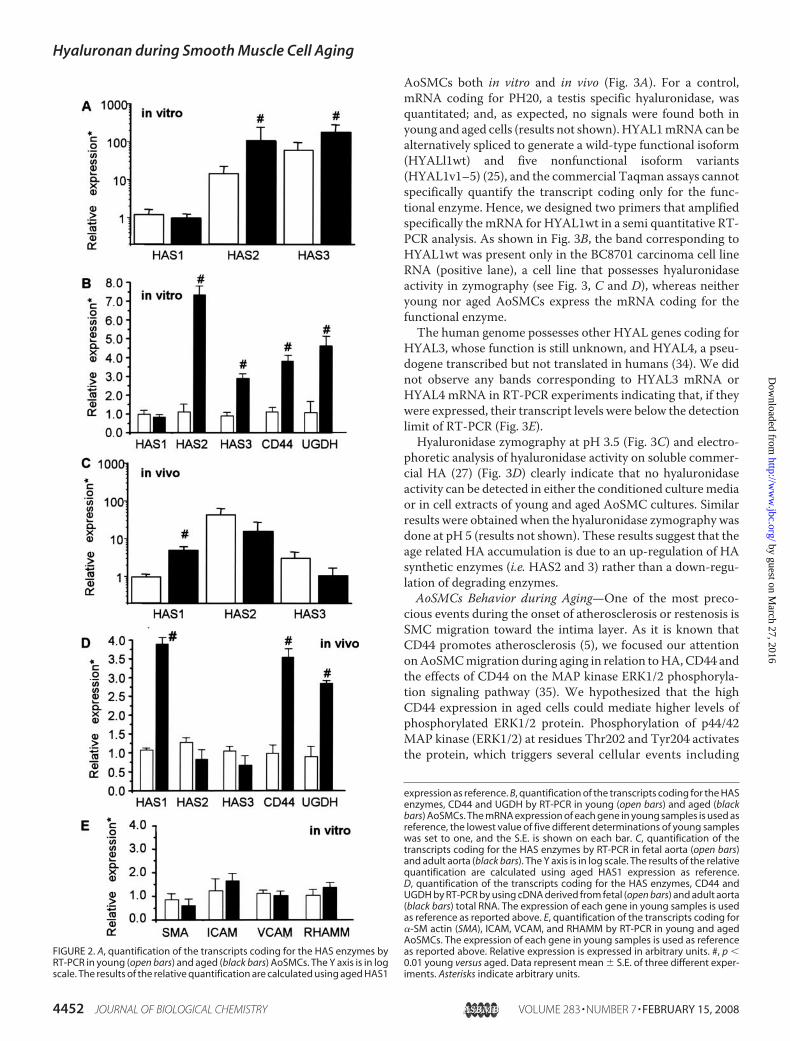
expressed about 10-fold higher than HAS2 mRNA (Fig. 2A,white bars). HAS2mRNA, in turn, was expressed about 10-foldhigher than HAS1 mRNA. In aged AoSMCs, HAS3 mRNA isexpressed only about 1.2-fold higher than HAS2mRNA, whichis expressed about 10-fold higher than HAS1 mRNA (Fig. 2A,black bars). Fig. 2B shows a statistically significant increase ofHAS2 mRNA and HAS3 mRNA expression in aged comparedwith young AoSMCs.Another protein involved in the onset of atherosclerosis is
CD44, the HA receptor (4). This receptor may have a role invascular cell activation and could mediate inflammatory cellrecruitment (5). To verify whether CD44 expression duringaging could be altered, we quantified its mRNA level by quan-titative RT-PCR and found a statistically significant increase(�3�) in aged AoSMCs (Fig. 2B). Western blot analyses ofproteins extracted from young and aged AoSMC cultures con-firmed the gene expression data (results not shown). ThemRNA expression of the other HA receptor RHAMM did notchange during AoSMC aging (Fig. 2E).We quantified the gene expression of the enzyme that syn-
thesizes theUDP-sugar precursor, UDP-glucuronic acid (UDP-glucose dehydrogenase, UGDH) and found a significantlyhigher mRNA expression (�4�) in aged AoSMCs (Fig. 2B).To verify whether our in vitro cell aging system represents
the gene expression in vivo, quantitative RT-PCR was done onhuman normal aorta RNA prepared from healthy donors ofdifferent ages. As a young sample, we used a pool of 5 differentfetal aorta RNAs purchased from Invitrogen; as an adult samplewe used a pool of 5 different aorta RNAs from donors of 65years old (Ambion). Fig. 2C highlights the relative ratios forHAS1, -2, and -3 and Fig. 2D shows a statistically significantincrease in the expression of mRNA coding for HAS1 enzymesin aged aorta, whereas HAS2 mRNA and HAS3 mRNA appearto be unchanged. Interestingly, in aorta samples, UGDH andCD44 expressions resembled those obtained in the cells aged invitro (Fig. 2, B and D).Because SMCs in the atherosclerotic lesion change from a
contractile to a synthetic phenotype (5), we investigatedwhether during in vitro aging AoSMCs undergo such a pheno-typic transition. The expression of critical genes (�-SM actin,VCAM, and ICAM) characteristic of the synthetic phenotypewere quantified by quantitative RT-PCR. All three of thesegenes did not change their expression significantly betweenyoung and aged AoSMCs, even though a slight, but not statis-tically significant, decrease of �-SM actin was found in agedAoSMCs (Fig. 2E). These data indicate that during in vitro agingno contractile to synthetic phenotype occurs.To confirm that the increase ofHA in agedAoSMCs could be
due to an up-regulation of HAS genes rather than a down-regulation of HYAL genes, we checked the expression of hyalu-ronidases in AoSMC cultures. HYAL2 mRNA expression wasnot statistically different in aged compared with young
Stand
ard
Aged
Youn
gSta
ndard
∆HA∆CS-0S
∆CS-6S∆CS-4S
A
C
Aged
AoSM
Cs
Youn
gAo
SMC
s
∆HA
pmol
/cel
lx10
-5
B
0.0
0.4
0.8
1.2
1.6
2.0 *
Young Aged
FIGURE 1. A, representative FACE analysis of AMAC-labeled disaccharide ofHA (�HA), chondroitin 0-sulfated (�CS-0S), chondroitin 6-sulfated (�CS-6S),and chondroitin 4-sulfated (�CS-4S) (standard lanes). AMAC-labeled disac-charides from the GAGs in the culture media conditioned by 106 young oraged AoSMCs (Young and Aged lanes, respectively) were prepared asdescribed under “Experimental Procedures.” B, HPLC quantification of �HA
secreted into the culture medium by young (open bar) and aged (black bar)AoSMCs. The graph represents the means � S.E. of three different GAG sam-ple determinations. *, p � 0.01 young versus aged. C, immunolocalization ofHA (green), CD44 (red), and nuclei (blue) on young and aged AoSMCs. Themicrophotographs show representative results of different independentexperiments. Original magnification �400. Scale bar is equal to 30 �m.
Hyaluronan during Smooth Muscle Cell Aging
FEBRUARY 15, 2008 • VOLUME 283 • NUMBER 7 JOURNAL OF BIOLOGICAL CHEMISTRY 4451
by guest on March 27, 2016
http://ww
w.jbc.org/
Dow
nloaded from
AoSMCs both in vitro and in vivo (Fig. 3A). For a control,mRNA coding for PH20, a testis specific hyaluronidase, wasquantitated; and, as expected, no signals were found both inyoung and aged cells (results not shown).HYAL1mRNAcan bealternatively spliced to generate a wild-type functional isoform(HYALl1wt) and five nonfunctional isoform variants(HYAL1v1–5) (25), and the commercial Taqman assays cannotspecifically quantify the transcript coding only for the func-tional enzyme. Hence, we designed two primers that amplifiedspecifically themRNA for HYAL1wt in a semi quantitative RT-PCR analysis. As shown in Fig. 3B, the band corresponding toHYAL1wt was present only in the BC8701 carcinoma cell lineRNA (positive lane), a cell line that possesses hyaluronidaseactivity in zymography (see Fig. 3, C and D), whereas neitheryoung nor aged AoSMCs express the mRNA coding for thefunctional enzyme.The human genome possesses other HYAL genes coding for
HYAL3, whose function is still unknown, and HYAL4, a pseu-dogene transcribed but not translated in humans (34). We didnot observe any bands corresponding to HYAL3 mRNA orHYAL4 mRNA in RT-PCR experiments indicating that, if theywere expressed, their transcript levels were below the detectionlimit of RT-PCR (Fig. 3E).Hyaluronidase zymography at pH 3.5 (Fig. 3C) and electro-
phoretic analysis of hyaluronidase activity on soluble commer-cial HA (27) (Fig. 3D) clearly indicate that no hyaluronidaseactivity can be detected in either the conditioned culturemediaor in cell extracts of young and aged AoSMC cultures. Similarresults were obtained when the hyaluronidase zymography wasdone at pH 5 (results not shown). These results suggest that theage related HA accumulation is due to an up-regulation of HAsynthetic enzymes (i.e. HAS2 and 3) rather than a down-regu-lation of degrading enzymes.AoSMCs Behavior during Aging—One of the most preco-
cious events during the onset of atherosclerosis or restenosis isSMC migration toward the intima layer. As it is known thatCD44 promotes atherosclerosis (5), we focused our attentiononAoSMCmigration during aging in relation toHA,CD44 andthe effects of CD44 on the MAP kinase ERK1/2 phosphoryla-tion signaling pathway (35). We hypothesized that the highCD44 expression in aged cells could mediate higher levels ofphosphorylated ERK1/2 protein. Phosphorylation of p44/42MAP kinase (ERK1/2) at residues Thr202 and Tyr204 activatesthe protein, which triggers several cellular events including
FIGURE 2. A, quantification of the transcripts coding for the HAS enzymes byRT-PCR in young (open bars) and aged (black bars) AoSMCs. The Y axis is in logscale. The results of the relative quantification are calculated using aged HAS1
expression as reference. B, quantification of the transcripts coding for the HASenzymes, CD44 and UGDH by RT-PCR in young (open bars) and aged (blackbars) AoSMCs. The mRNA expression of each gene in young samples is used asreference, the lowest value of five different determinations of young sampleswas set to one, and the S.E. is shown on each bar. C, quantification of thetranscripts coding for the HAS enzymes by RT-PCR in fetal aorta (open bars)and adult aorta (black bars). The Y axis is in log scale. The results of the relativequantification are calculated using aged HAS1 expression as reference.D, quantification of the transcripts coding for the HAS enzymes, CD44 andUGDH by RT-PCR by using cDNA derived from fetal (open bars) and adult aorta(black bars) total RNA. The expression of each gene in young samples is usedas reference as reported above. E, quantification of the transcripts coding for�-SM actin (SMA), ICAM, VCAM, and RHAMM by RT-PCR in young and agedAoSMCs. The expression of each gene in young samples is used as referenceas reported above. Relative expression is expressed in arbitrary units. #, p �0.01 young versus aged. Data represent mean � S.E. of three different exper-iments. Asterisks indicate arbitrary units.
Hyaluronan during Smooth Muscle Cell Aging
4452 JOURNAL OF BIOLOGICAL CHEMISTRY VOLUME 283 • NUMBER 7 • FEBRUARY 15, 2008
by guest on March 27, 2016
http://ww
w.jbc.org/
Dow
nloaded from
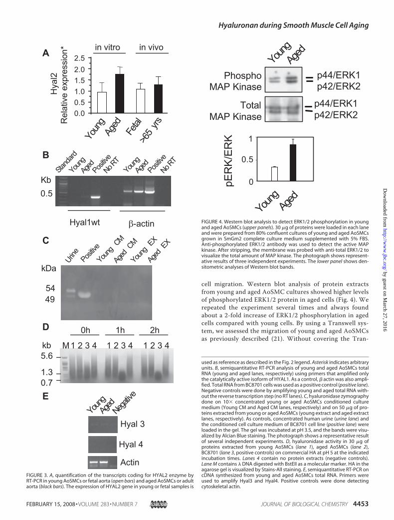
cell migration. Western blot analysis of protein extractsfrom young and aged AoSMC cultures showed higher levelsof phosphorylated ERK1/2 protein in aged cells (Fig. 4). Werepeated the experiment several times and always foundabout a 2-fold increase of ERK1/2 phosphorylation in agedcells compared with young cells. By using a Transwell sys-tem, we assessed the migration of young and aged AoSMCsas previously described (21). Without covering the Tran-
>65
yrs
Hya
l2R
elat
ive
expr
essi
on*
0.0
1.01.52.02.5
0.5
Youn
gAg
edFe
tal
in vitro in vivoA
BAg
ed
Youn
g
Positiv
eNo R
T
Aged
Youn
g
Positiv
eNo R
T
Stand
ard
β-actinHyal1wt
0.5Kb
Aged
CM
Young
CM
Positive
5449
Urine
Aged
EX
Young
EX
kDa
C
0h 1h 2h
1 2 3 4 1 2 3 4 1 2 3 4M
D
5.6
1.30.7
kb
Young
Aged
Negative
Hyal 3
Hyal 4
Actin
E
FIGURE 3. A, quantification of the transcripts coding for HYAL2 enzyme byRT-PCR in young AoSMCs or fetal aorta (open bars) and aged AoSMCs or adultaorta (black bars). The expression of HYAL2 gene in young or fetal samples is
used as reference as described in the Fig. 2 legend. Asterisk indicates arbitraryunits. B, semiquantitative RT-PCR analysis of young and aged AoSMCs totalRNA (young and aged lanes, respectively) using primers that amplified onlythe catalytically active isoform of HYAL1. As a control, � actin was also ampli-fied. Total RNA from BC8701 cells was used as a positive control (positive lane).Negative controls were done by amplifying young and aged total RNA with-out the reverse transcription step (no RT lanes). C, hyaluronidase zymographydone on 10� concentrated young or aged AoSMCs conditioned culturemedium (Young CM and Aged CM lanes, respectively) and on 50 �g of pro-teins extracted from young or aged AoSMCs (young extract and aged extractlanes, respectively). As controls, concentrated human urine (urine lane) andthe conditioned cell culture medium of BC8701 cell line (positive lane) wereloaded in the gel. The gel was incubated at pH 3.5, and the bands were visu-alized by Alcian Blue staining. The photograph shows a representative resultof several independent experiments. D, hyaluronidase activity in 30 �g ofproteins extracted from young AoSMCs (lane 1), aged AoSMCs (lane 2),BC8701 (lane 3, positive controls) on commercial HA at pH 5 at the indicatedincubation times. Lanes 4 contain no protein extracts (negative controls).Lane M contains � DNA digested with BstEII as a molecular marker. HA in theagarose gel is visualized by Stains-All staining. E, semiquantitative RT-PCR oncDNA synthesized from young and aged AoSMCs total RNA. Primers wereused to amplify Hyal3 and Hyal4. Positive controls were done detectingcytoskeletal actin.
Young
Aged
p44/ERK1p42/ERK2
p44/ERK1p42/ERK2
PhosphoMAP Kinase
TotalMAP Kinase
Youn
gAg
ed
pER
K/E
RK
0
0.5
1
FIGURE 4. Western blot analysis to detect ERK1/2 phosphorylation in youngand aged AoSMCs (upper panels). 30 �g of proteins were loaded in each laneand were prepared from 80% confluent cultures of young and aged AoSMCsgrown in SmGm2 complete culture medium supplemented with 5% FBS.Anti-phosphorylated ERK1/2 antibody was used to detect the active MAPkinase. After stripping, the membrane was probed with anti-total ERK1/2 tovisualize the total amount of MAP kinase. The photograph shows represent-ative results of three independent experiments. The lower panel shows den-sitometric analyses of Western blot bands.
Hyaluronan during Smooth Muscle Cell Aging
FEBRUARY 15, 2008 • VOLUME 283 • NUMBER 7 JOURNAL OF BIOLOGICAL CHEMISTRY 4453
by guest on March 27, 2016
http://ww
w.jbc.org/
Dow
nloaded from
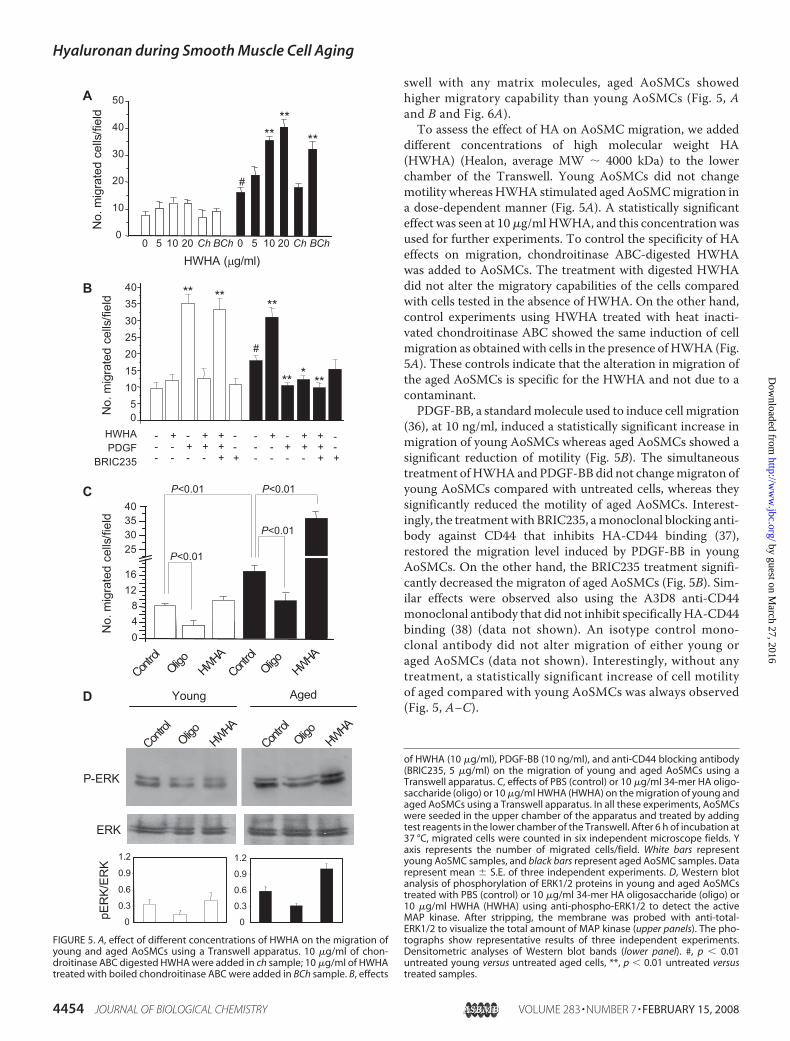
swell with any matrix molecules, aged AoSMCs showedhigher migratory capability than young AoSMCs (Fig. 5, Aand B and Fig. 6A).To assess the effect of HA on AoSMC migration, we added
different concentrations of high molecular weight HA(HWHA) (Healon, average MW � 4000 kDa) to the lowerchamber of the Transwell. Young AoSMCs did not changemotility whereas HWHA stimulated agedAoSMCmigration ina dose-dependent manner (Fig. 5A). A statistically significanteffect was seen at 10�g/mlHWHA, and this concentrationwasused for further experiments. To control the specificity of HAeffects on migration, chondroitinase ABC-digested HWHAwas added to AoSMCs. The treatment with digested HWHAdid not alter the migratory capabilities of the cells comparedwith cells tested in the absence of HWHA. On the other hand,control experiments using HWHA treated with heat inacti-vated chondroitinase ABC showed the same induction of cellmigration as obtainedwith cells in the presence of HWHA (Fig.5A). These controls indicate that the alteration in migration ofthe aged AoSMCs is specific for the HWHA and not due to acontaminant.PDGF-BB, a standardmolecule used to induce cell migration
(36), at 10 ng/ml, induced a statistically significant increase inmigration of young AoSMCs whereas aged AoSMCs showed asignificant reduction of motility (Fig. 5B). The simultaneoustreatment ofHWHAand PDGF-BB did not changemigraton ofyoung AoSMCs compared with untreated cells, whereas theysignificantly reduced the motility of aged AoSMCs. Interest-ingly, the treatmentwith BRIC235, amonoclonal blocking anti-body against CD44 that inhibits HA-CD44 binding (37),restored the migration level induced by PDGF-BB in youngAoSMCs. On the other hand, the BRIC235 treatment signifi-cantly decreased the migraton of aged AoSMCs (Fig. 5B). Sim-ilar effects were observed also using the A3D8 anti-CD44monoclonal antibody that did not inhibit specificallyHA-CD44binding (38) (data not shown). An isotype control mono-clonal antibody did not alter migration of either young oraged AoSMCs (data not shown). Interestingly, without anytreatment, a statistically significant increase of cell motilityof aged compared with young AoSMCs was always observed(Fig. 5, A–C).
Control
048
1216
25303540
HWHAOligo
Control
HWHAOligo
No.
mig
rate
dce
lls/fi
eld
P<0.01
P<0.01 P<0.01
P<0.01
B
0 0
05
10152025303540
No.
mig
rate
dce
lls/fi
eld
HWHAPDGF
BRIC235
- + - + + - + - + +- - + + + - - + + +- - - - + - - - - +
****
**
** ***
#
C
ERK
P-ERK
Control
HWHAOligo
Control
HWHAOligo
Young Aged
0.3
0.6
0.9
1.2
0.3
0.6
0.9
1.2
pER
K/ER
K
D
A
0 5 10 200
10
20
30
40
50
0 5 10 20
HWHA (µg/ml)
No.
mig
rate
dce
lls/fi
eld
****
#
Ch BCh
**
Ch BCh
-+
--+
-
FIGURE 5. A, effect of different concentrations of HWHA on the migration ofyoung and aged AoSMCs using a Transwell apparatus. 10 �g/ml of chon-droitinase ABC digested HWHA were added in ch sample; 10 �g/ml of HWHAtreated with boiled chondroitinase ABC were added in BCh sample. B, effects
of HWHA (10 �g/ml), PDGF-BB (10 ng/ml), and anti-CD44 blocking antibody(BRIC235, 5 �g/ml) on the migration of young and aged AoSMCs using aTranswell apparatus. C, effects of PBS (control) or 10 �g/ml 34-mer HA oligo-saccharide (oligo) or 10 �g/ml HWHA (HWHA) on the migration of young andaged AoSMCs using a Transwell apparatus. In all these experiments, AoSMCswere seeded in the upper chamber of the apparatus and treated by addingtest reagents in the lower chamber of the Transwell. After 6 h of incubation at37 °C, migrated cells were counted in six independent microscope fields. Yaxis represents the number of migrated cells/field. White bars representyoung AoSMC samples, and black bars represent aged AoSMC samples. Datarepresent mean � S.E. of three independent experiments. D, Western blotanalysis of phosphorylation of ERK1/2 proteins in young and aged AoSMCstreated with PBS (control) or 10 �g/ml 34-mer HA oligosaccharide (oligo) or10 �g/ml HWHA (HWHA) using anti-phospho-ERK1/2 to detect the activeMAP kinase. After stripping, the membrane was probed with anti-total-ERK1/2 to visualize the total amount of MAP kinase (upper panels). The pho-tographs show representative results of three independent experiments.Densitometric analyses of Western blot bands (lower panel). #, p � 0.01untreated young versus untreated aged cells, **, p � 0.01 untreated versustreated samples.
Hyaluronan during Smooth Muscle Cell Aging
4454 JOURNAL OF BIOLOGICAL CHEMISTRY VOLUME 283 • NUMBER 7 • FEBRUARY 15, 2008
by guest on March 27, 2016
http://ww
w.jbc.org/
Dow
nloaded from
As HA of different molecular weight can modify cellmigration (38), we added a 34-mer HA oligosaccharide (akind gift from Akira Asari) or HWHA to the Transwell. Asshown in Fig. 5C, 34-mer oligosaccharide reduced themigra-tion of both young and aged AoSMCs. In contrast, asdescribed above, HWHA did not significantly change themigration of young cells, but increased the migration of agedAoSMCs about 2-fold.
The migratory capability of the cells could depend uponphosphorylation of ERK1/2 protein. Thirty minutes treatmentof young and aged AoSMCs with 10 �g/ml of 34-mer oligosac-charide or 10 �g/ml of HWHA altered ERK1/2 phosphoryla-tion (Fig. 5D). In young AoSMCs, the 34-mer oligosaccharidecaused a decrease in ERK1/2 phosphorylation whereas HWHAdid not modify ERK1/2 phosphorylation. Similarly, 34-mer oli-gosaccharide caused a decrease in ERK1/2 phosphorylation inaged cells, whereas, interestingly, the treatment of agedAoSMCs with HWHA induced a marked increase of phospho-rylation of ERK1/2 (Fig. 5D).As the expression of CD44 varied during cell aging, we tested
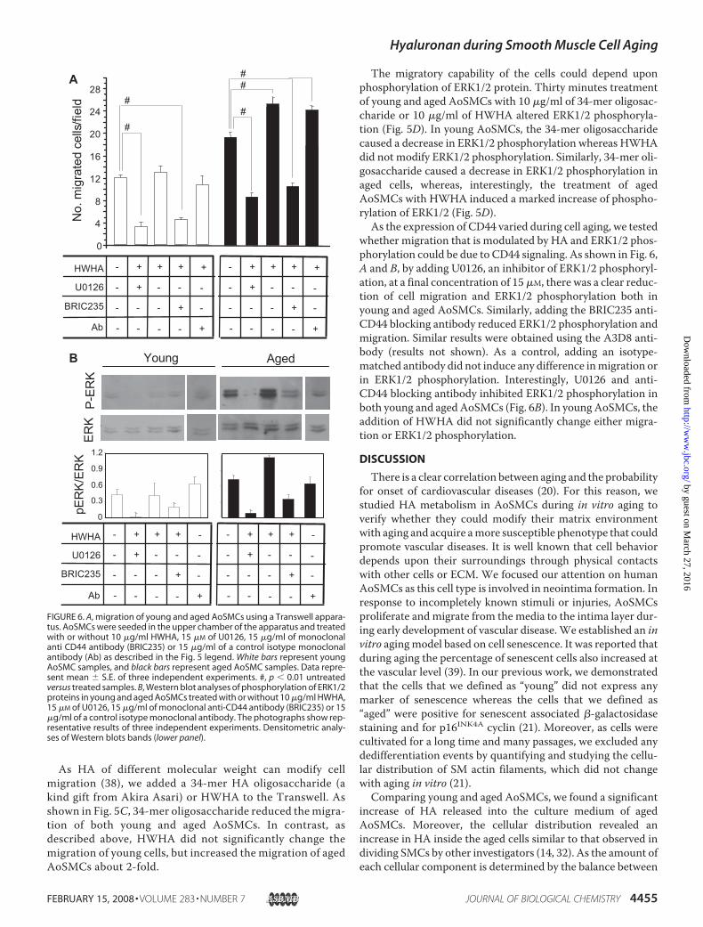
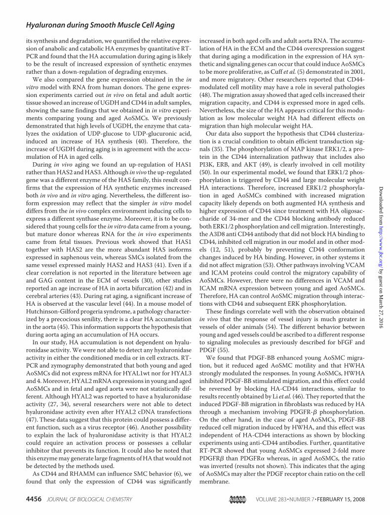
whether migration that is modulated by HA and ERK1/2 phos-phorylation could be due to CD44 signaling. As shown in Fig. 6,A and B, by adding U0126, an inhibitor of ERK1/2 phosphoryl-ation, at a final concentration of 15�M, there was a clear reduc-tion of cell migration and ERK1/2 phosphorylation both inyoung and aged AoSMCs. Similarly, adding the BRIC235 anti-CD44 blocking antibody reduced ERK1/2 phosphorylation andmigration. Similar results were obtained using the A3D8 anti-body (results not shown). As a control, adding an isotype-matched antibody did not induce any difference inmigration orin ERK1/2 phosphorylation. Interestingly, U0126 and anti-CD44 blocking antibody inhibited ERK1/2 phosphorylation inboth young and aged AoSMCs (Fig. 6B). In young AoSMCs, theaddition of HWHA did not significantly change either migra-tion or ERK1/2 phosphorylation.
DISCUSSION
There is a clear correlation between aging and the probabilityfor onset of cardiovascular diseases (20). For this reason, westudied HA metabolism in AoSMCs during in vitro aging toverify whether they could modify their matrix environmentwith aging and acquire amore susceptible phenotype that couldpromote vascular diseases. It is well known that cell behaviordepends upon their surroundings through physical contactswith other cells or ECM. We focused our attention on humanAoSMCs as this cell type is involved in neointima formation. Inresponse to incompletely known stimuli or injuries, AoSMCsproliferate and migrate from the media to the intima layer dur-ing early development of vascular disease.We established an invitro agingmodel based on cell senescence. It was reported thatduring aging the percentage of senescent cells also increased atthe vascular level (39). In our previous work, we demonstratedthat the cells that we defined as “young” did not express anymarker of senescence whereas the cells that we defined as“aged” were positive for senescent associated �-galactosidasestaining and for p16INK4A cyclin (21). Moreover, as cells werecultivated for a long time and many passages, we excluded anydedifferentiation events by quantifying and studying the cellu-lar distribution of SM actin filaments, which did not changewith aging in vitro (21).
Comparing young and aged AoSMCs, we found a significantincrease of HA released into the culture medium of agedAoSMCs. Moreover, the cellular distribution revealed anincrease in HA inside the aged cells similar to that observed individing SMCs by other investigators (14, 32). As the amount ofeach cellular component is determined by the balance between
0
4
8
12
16
20
24
28N
o.m
igra
ted
cells
/fiel
dA
#
##
##
HWHA
U0126
BRIC235 +---
--+-
+++-
- - - - +
-
-
+
Ab
+---
--+-
+++-
- - - - +
-
-
+
HWHA
U0126
BRIC235 +---
--+-
+++-
Young AgedB
ERK
P-ER
K
0
0.3
0.6
0.9
1.2
pER
K/E
RK
- - - - +
-
-
-
Ab
+---
--+-
+++-
- - - - +
-
-
-
FIGURE 6. A, migration of young and aged AoSMCs using a Transwell appara-tus. AoSMCs were seeded in the upper chamber of the apparatus and treatedwith or without 10 �g/ml HWHA, 15 �M of U0126, 15 �g/ml of monoclonalanti CD44 antibody (BRIC235) or 15 �g/ml of a control isotype monoclonalantibody (Ab) as described in the Fig. 5 legend. White bars represent youngAoSMC samples, and black bars represent aged AoSMC samples. Data repre-sent mean � S.E. of three independent experiments. #, p � 0.01 untreatedversus treated samples. B, Western blot analyses of phosphorylation of ERK1/2proteins in young and aged AoSMCs treated with or without 10 �g/ml HWHA,15 �M of U0126, 15 �g/ml of monoclonal anti-CD44 antibody (BRIC235) or 15�g/ml of a control isotype monoclonal antibody. The photographs show rep-resentative results of three independent experiments. Densitometric analy-ses of Western blots bands (lower panel).
Hyaluronan during Smooth Muscle Cell Aging
FEBRUARY 15, 2008 • VOLUME 283 • NUMBER 7 JOURNAL OF BIOLOGICAL CHEMISTRY 4455
by guest on March 27, 2016
http://ww
w.jbc.org/
Dow
nloaded from
its synthesis and degradation, we quantified the relative expres-sion of anabolic and catabolic HA enzymes by quantitative RT-PCR and found that the HA accumulation during aging is likelyto be the result of increased expression of synthetic enzymesrather than a down-regulation of degrading enzymes.We also compared the gene expression obtained in the in
vitro model with RNA from human donors. The gene expres-sion experiments carried out in vivo on fetal and adult aortictissue showed an increase ofUGDHandCD44 in adult samples,showing the same findings that we obtained in in vitro experi-ments comparing young and aged AoSMCs. We previouslydemonstrated that high levels of UGDH, the enzyme that cata-lyzes the oxidation of UDP-glucose to UDP-glucuronic acid,induced an increase of HA synthesis (40). Therefore, theincrease of UGDH during aging is in agreement with the accu-mulation of HA in aged cells.During in vivo aging we found an up-regulation of HAS1
rather thanHAS2 andHAS3. Although in vivo the up-regulatedgene was a different enzyme of the HAS family, this result con-firms that the expression of HA synthetic enzymes increasedboth in vivo and in vitro aging. Nevertheless, the different iso-form expression may reflect that the simpler in vitro modeldiffers from the in vivo complex environment inducing cells toexpress a different synthase enzyme. Moreover, it is to be con-sidered that young cells for the in vitro data came from a young,but mature donor whereas RNA for the in vivo experimentscame from fetal tissues. Previous work showed that HAS1together with HAS2 are the more abundant HAS isoformsexpressed in saphenous vein, whereas SMCs isolated from thesame vessel expressed mainly HAS2 and HAS3 (41). Even if aclear correlation is not reported in the literature between ageand GAG content in the ECM of vessels (30), other studiesreported an age increase of HA in aorta bifurcation (42) and incerebral arteries (43). During rat aging, a significant increase ofHA is observed at the vascular level (44). In a mouse model ofHutchinson-Gilford progeria syndrome, a pathology character-ized by a precocious senility, there is a clear HA accumulationin the aorta (45). This information supports the hypothesis thatduring aorta aging an accumulation of HA occurs.In our study, HA accumulation is not dependent on hyalu-
ronidase activity.We were not able to detect any hyaluronidaseactivity in either the conditioned media or in cell extracts. RT-PCR and zymography demonstrated that both young and agedAoSMCs did not express mRNA for HYAL1wt nor for HYAL3and 4.Moreover,HYAL2mRNAexpressions in young and agedAoSMCs and in fetal and aged aorta were not statistically dif-ferent. Although HYAL2 was reported to have a hyaluronidaseactivity (27, 34), several researchers were not able to detecthyaluronidase activity even after HYAL2 cDNA transfections(47). These data suggest that this protein could possess a differ-ent function, such as a virus receptor (46). Another possibilityto explain the lack of hyaluronidase activity is that HYAL2could require an activation process or possesses a cellularinhibitor that prevents its function. It could also be noted thatthis enzymemay generate large fragments ofHA thatwould notbe detected by the methods used.As CD44 and RHAMM can influence SMC behavior (6), we
found that only the expression of CD44 was significantly
increased in both aged cells and adult aorta RNA. The accumu-lation of HA in the ECM and the CD44 overexpression suggestthat during aging a modification in the expression of HA syn-thetic and signaling genes can occur that could induceAoSMCsto bemore proliferative, as Cuff et al. (5) demonstrated in 2001,and more migratory. Other researchers reported that CD44-modulated cell motility may have a role in several pathologies(48). Themigration assay showed that aged cells increased theirmigration capacity, and CD44 is expressed more in aged cells.Nevertheless, the size of the HA appears critical for this modu-lation as low molecular weight HA had different effects onmigration than high molecular weight HA.Our data also support the hypothesis that CD44 clusteriza-
tion is a crucial condition to obtain efficient transduction sig-nals (35). The phosphorylation of MAP kinase ERK1/2, a pro-tein in the CD44 internalization pathway that includes alsoPI3K, ERB, and AKT (49), is clearly involved in cell motility(50). In our experimental model, we found that ERK1/2 phos-phorylation is triggered by CD44 and large molecular weightHA interactions. Therefore, increased ERK1/2 phosphoryla-tion in aged AoSMCs combined with increased migrationcapacity likely depends on both augmented HA synthesis andhigher expression of CD44 since treatment with HA oligosac-charide of 34-mer and the CD44 blocking antibody reducedboth ERK1/2 phosphorylation and cellmigration. Interestingly,the A3D8 anti CD44 antibody that did not block HA binding toCD44, inhibited cell migration in our model and in other mod-els (12, 51), probably by preventing CD44 conformationchanges induced by HA binding. However, in other systems itdid not affect migration (53). Other pathways involving VCAMand ICAM proteins could control the migratory capability ofAoSMCs. However, there were no differences in VCAM andICAM mRNA expression between young and aged AoSMCs.Therefore, HA can control AoSMCmigration through interac-tions with CD44 and subsequent ERK phosphorylation.These findings correlate well with the observation obtained
in vivo that the response of vessel injury is much greater invessels of older animals (54). The different behavior betweenyoung and aged vessels could be ascribed to a different responseto signaling molecules as previously described for bFGF andPDGF (55).We found that PDGF-BB enhanced young AoSMC migra-
tion, but it reduced aged AoSMC motility and that HWHAstrongly modulated the responses. In young AoSMCs, HWHAinhibited PDGF-BB stimulated migration, and this effect couldbe reversed by blocking HA-CD44 interactions, similar toresults recently obtained by Li et al. (46). They reported that theinduced PDGF-BBmigration in fibroblasts was reduced by HAthrough a mechanism involving PDGFR-� phosphorylation.On the other hand, in the case of aged AoSMCs, PDGF-BBreduced cell migration induced by HWHA, and this effect wasindependent of HA-CD44 interactions as shown by blockingexperiments using anti-CD44 antibodies. Further, quantitativeRT-PCR showed that young AoSMCs expressed 2-fold morePDGFR� than PDGFR� whereas, in aged AoSMCs, the ratiowas inverted (results not shown). This indicates that the agingof AoSMCsmay alter the PDGF receptor chain ratio on the cellmembrane.
Hyaluronan during Smooth Muscle Cell Aging
4456 JOURNAL OF BIOLOGICAL CHEMISTRY VOLUME 283 • NUMBER 7 • FEBRUARY 15, 2008
by guest on March 27, 2016
http://ww
w.jbc.org/
Dow
nloaded from
The high migratory capability of aged AoSMCs fits well withtheir high expression of CD44. However, it is not clear whethercells extracted from aged animals have higher migration ratesthan cells extracted from young animals. Controversial resultswere obtained from SMCs prepared from young and agedhuman or animal models. Aged human SMCs showed a limitedmigration (33) whereas aged rat SMCs showed an increase ofmigration (52). However, it is possible that the microenviron-ment surrounding cells influences the cell behavior. For exam-ple, we previously reported that themigration of aged AoSMCson a gelatin rich matrix was different due to differences in met-alloproteinase 2 activity (20). The absence of an externalmatrixin the model used in the experiments described in this reporthighlights the specific role of HA in AoSMC migration.In conclusion we demonstrated that during in vitro aging,
AoSMCs accumulate HA by increasing HA synthetic enzymes.Moreover, we provided evidence that HWHA induces migra-tion of agedAoSMCsprobably through aCD44-mediated path-way by controlling the activity of MAP kinase ERK1/2.
Acknowledgments—We thank Dr. Akira Asari (Glycoscience Labora-tories, Inc.) for the 34-mer HA oligosaccharide, Dr. Frances Spring(National Blood Service, UK) for Bric235 monoclonal antibodies, Dr.Raffaella Cinquetti for aorta RNAs. The authors gratefully acknowl-edge the “Centro Grandi Attrezzature per la Ricerca Biomedica” Uni-versita degli Studi dell’Insubria, for the instrument facility.
REFERENCES1. Toole, B. P., Wight, T. N., and Tammi, M. I. (2002) J. Biol. Chem. 277,
4593–45962. Itano, N., Sawai, T., Yoshida, M., Lenas, P., Yamada, Y., Imagawa, M.,
Shinomura, T., Hamaguchi, M., Yoshida, Y., Ohnuki, Y., Miyauchi, S.,Spicer, A. P., McDonald, J. A., and Kimata, K. (1999) J. Biol. Chem. 274,25085–25092
3. Tien, J. Y., and Spicer, A. P. (2005) Dev. Dyn. 233, 130–1414. Turley, E. A., Noble, P.W., and Bourguignon, L. Y.W. (2002) J. Biol. Chem.
277, 4589–45925. Cuff, C. A., Kothapalli, D., Azonobi, I., Chun, S., Zhang, Y., Belkin, R., Yeh,
C., Secreto, A., Assoian, R. K., Rader, D. J., and Pure, E. (2001) J. Clin.Investig. 108, 1031–1040
6. Nedvetzki, S., Gonen, E., Assayag, N., Reich, R., Williams, R. O., Thur-mond, R. L., Huang, J., Neudecker, B. A.,Wang, F., Turley, E. A., andNaor,D. (2004) Proc. Natl. Acad. Sci. U. S. A. 101, 18081–18086
7. Johnson, L. A., Prevo, R., Clasper, S., and Jackson, D. G. (2007) J. Biol.Chem. 282, 33671–33680
8. Harris, E. N., Weigel, J. A., and Weigel, P. H. (2004) J. Biol. Chem. 279,36201–36209
9. Harris, E. N., Kyosseva, S. V., Weigel, J. A., andWeigel, P. H. (2007) J. Biol.Chem. 282, 2785–9277
10. Termeer, C., Benedix, F., Sleeman, J., Fieber, C., Voith, U., Ahrens, T.,Miyake, K., Freudenberg, M., Galanos, C., and Simon, J. C. (2002) J. Exp.Med. 195, 99–111
11. Jiang, D., Liang, J., Fan, J., Yu, S., Chen, S., Luo, Y., Prestwich, G. D.,Mascarenhas, M. M., Garg, H. G., Quinn, D. A., Homer, R. J., Goldstein,D. R., Bucala, R., Lee, P. J., Medzhitov, R., and Noble, P. W. (2005) Nat.Med. 11, 1173–1179
12. de la Motte, C. A., Hascall, V. C., Drazba, J., Bandyopadhyay, S. K., andStrong, S. A. (2003) Am. J. Pathol. 163, 121–133
13. Majors, A. K., Austin, R. C., de laMotte, C. A., Pyeritz, R. E., Hascall, V. C.,Kessler, S. P., Sen, G., and Strong, S. A. (2003) J. Biol. Chem. 278,47223–47231
14. Hascall, V. C., Majors, A. K., De La Motte, C. A., Evanko, S. P., Wang,A., Drazba, J. A., Strong, S. A., and Wight, T. N. (2004) Biochim. Bio-
phys. Acta 1673, 3–1215. Wang, A., and Hascall, V. C. (2004) J. Biol. Chem. 279, 10279–1028516. Heldin, P. (2003) Braz. J. Med. Biol. Res. 36, 967–97317. Riessen, R., Wight, T. N., Pastore, C., Henley, C., and Isner, J. M. (1996)
Circulation 93, 1141–114718. Tzircotis, G., Thorne, R. F., and Isacke, C. M. (2005) J. Cell Sci. 118,
5119–512819. Chai, S., Chai, Q., Danielsen, C. C., Hjorth, P., Nyengaard, J. R., Ledet, T.,
Yamaguchi, Y., Rasmussen, L. M., and Wogensen, L. (2005) Circ. Res. 96,583–591
20. Spagnoli, L. G., Orlandi, A., Mauriello, A., Santeusanio, G., Angelis, C.,Lucreziotti, R., and Ramacci, M. T. (1991) Atherosclerosis 89, 11–24
21. Vigetti, D., Moretto, P., Viola, M., Genasetti, A., Rizzi, M., Karousou, E.,Pallotti, F., De Luca, G., and Passi, A. (2006) FASEB J. 20, 1118–1130
22. Karousou, E. G., Militsopoulou, M., Porta, G., De Luca, G., Hascall, V. C.,and Passi, A. (2004) Electrophoresis 25, 2919–2925
23. Raio, L., Cromi, A., Grezzi, F., Passi, A., Karousou, E., Viola,M., Vigetti, D.,De Luca, G., and Bolis, P. (2005)Matrix Biol. 24, 166–174
24. Winer, J., Jung, C. K., Shackel, I., and Williams, P. M. (1999) Anal. Bio-chem. 270, 41–49
25. Lokeshwar, V. B., Schroeder, G. L., Carey, R. I., Soloway,M. S., and Iida, N.(2002) J. Biol. Chem. 277, 33654–33663
26. Miura, R. O., Yamagata, S., Miura, Y., Harada, T., and Yamagata, T. (1995)Anal. Biochem. 225, 333–340
27. Lepperdinger, G., Strobl, B., and Kreil, G. (1998) J. Biol. Chem. 273,22466–22470
28. Tawada, A., Masa, T., Oonuki, Y., Watanabe, A., Matsuzaki, Y., and Asari,A. (2002) Glycobiology 12, 421–426
29. Dimri, G. P., Lee, X., Basile, G., Acosta, M., Scott, G., Roskelley, C., Me-drano, E. E., Linskens, M., Rubelj, I., and Pereira-Smith, O. (1995) Proc.Natl. Acad. Sci. U. S. A. 92, 9363–9367
30. Tovar, A. M., Cesar, D. C., Leta, G. C., and Mourao, P. A. (1998) Arterio-scler. Thromb. Vasc. Biol. 18, 604–614
31. Plaas, A. H., Wong-Palms, S., Roughley, P. J., Midura, R. J., and Hascall,V. C. (1997) J. Biol. Chem. 272, 20603–20610
32. Evanko, S. P., and Wight, T. N. (1999) J. Histochem. Cytochem. 47,1331–1342
33. Ruiz-Torres, A., Lozano, R.,Melon, J., andCarraro, R. (2003) J. Gerontol. ABiol. Sci. Med. Sci. 58, B1074–B1077
34. Harada, H., and Takahashi, M. (2007) J. Biol. Chem. 282, 5597–560735. Toole, B. P. (2004) Nat. Rev. Cancer. 4, 528–53936. Arita, Y., Kihara, S., Ouchi, N., Maeda, K., Kuriyama, H., Okamoto, Y.,
Kumada, M., Hotta, K., Nishida, M., Takahashi, M., Nakamura, T., Shi-momura, I., Muraguchi, M., Ohmoto, Y., Funahashi, T., and Matsuzawa,Y. (2002) Circulation 105, 2893–2898
37. Liao, H. X., Lee, D. M., Leveque, M. C., and Haynes, B. F. (1995) J. Immu-nol. 155, 3938–3945
38. Slevin, M., Kumar, S., and Gaffney, J. (2002) J. Biol. Chem. 277,41046–41059
39. Martin, G. M., Ogburn, C. E., andWight, T. N. (1983) Am. J. Pathol. 110,236–245
40. Vigetti, D., Ori, M., Viola, M., Genasetti, A., Karousou, E., Rizzi, M., Pal-lotti, F., Nardi, I., Hascall, V. C., De Luca, G., and Passi, A. (2006) J. Biol.Chem. 281, 8254–8263
41. van den Boom, M., Sarbia, M., vonWnuck Lipinski, K., Mann, P., Meyer-Kirchrath, J., Rauch, B. H., Grabitz, K., Levkau, B., Schror, K., and Fischer,J. W. (2006) Circ. Res. 98, 36–44
42. Stuhlsatz, H. W., Loffler, H., Mohanaradhakrishnan, V., Cosma, S., andGreiling, H. (1982) J. Clin. Chem. Clin. Biochem. 20, 713–721
43. Murata, K., and Yokoyama, Y. (1989) Atherosclerosis. 78, 69–7944. Chajara, A., Delpech, B., Courel, M. N., Leroy, M., Basuyau, J. P., and
Levesque, H. (1998) Atherosclerosis 138, 53–6445. Varga, R., Eriksson, M., Erdos, M. R., Olive, M., Harten, I., Kolodgie, F.,
Capell, B. C., Cheng, J., Faddah, D., Perkins, S., Avallone, H., San, H., Qu,X., Ganesh, S., Gordon, L. B., Virmani, R., Wight, T. N., Nabel, E. G., andCollins, F. S. (2006) Proc. Natl. Acad. Sci. U. S. A. 103, 3250–3255
46. Li, L., Heldin, C. H., andHeldin, P. (2006) J. Biol. Chem. 281, 26512–2651947. Rai, S. K., Duh, F. M., Vigdorovich, V., Danilkovitch-Miagkova, A., Lerman,
Hyaluronan during Smooth Muscle Cell Aging
FEBRUARY 15, 2008 • VOLUME 283 • NUMBER 7 JOURNAL OF BIOLOGICAL CHEMISTRY 4457
by guest on March 27, 2016
http://ww
w.jbc.org/
Dow
nloaded from
M. I., andMiller, A. D. (2001) Proc. Natl. Acad. Sci. U. S. A. 98, 4443–444848. Isacke, C. M., and Yarwood, H. (2002) Int. J. Biochem. Cell Biol. 34,
718–72149. Bourguignon, L. Y., Gilad, E., Rothman, K., and Peyrollier, K. (2005) J. Biol.
Chem. 280, 11961–1197250. Cox, B. D., Natarajan,M., Stettner, M. R., and Gladson, C. L. (2006) J. Cell.
Biochem. 99, 35–5251. Clark, R. A. F., Lin, F., Greiling, D., An, J., and Couchman, J. R. (2004)
J. Investig. Dermatol. 122, 266–27752. Li, Z., Cheng,H., Lederer,W. J., Froehlich, J., and Lakatta, E. G. (1997)Exp.
Mol. Pathol. 64, 1–1153. Kitayama, J., Juji, T., Atomi, Y., Kuroda, A., Muto, T., Kobayashi, M.,
Mitsui, Y., and Minami, M. (1993) J. Immunol. 151, 1663–167254. Hariri, R. J., Alonso, D. R., Hajjar, D. P., Coletti, D., and Weksler, M. E.
(1986) J. Exp. Med. 164, 1171–117855. Lundberg, M. S., and Crow, M. T. (1999) Exp. Gerontol. 34, 549–557
Hyaluronan during Smooth Muscle Cell Aging
4458 JOURNAL OF BIOLOGICAL CHEMISTRY VOLUME 283 • NUMBER 7 • FEBRUARY 15, 2008
by guest on March 27, 2016
http://ww
w.jbc.org/
Dow
nloaded from
PassiAnna Genasetti, Moira Clerici, Vincent C. Hascall, Giancarlo De Luca and Alberto Davide Vigetti, Manuela Viola, Eugenia Karousou, Manuela Rizzi, Paola Moretto,
during AgingHyaluronan-CD44-ERK1/2 Regulate Human Aortic Smooth Muscle Cell Motility
doi: 10.1074/jbc.M709051200 originally published online December 12, 20072008, 283:4448-4458.J. Biol. Chem.
10.1074/jbc.M709051200Access the most updated version of this article at doi:
Alerts:
When a correction for this article is posted•
When this article is cited•
to choose from all of JBC's e-mail alertsClick here
http://www.jbc.org/content/283/7/4448.full.html#ref-list-1
This article cites 55 references, 34 of which can be accessed free at
by guest on March 27, 2016
http://ww
w.jbc.org/
Dow
nloaded from
Related Documents