Expression, purification, and characterization of recombinant human transferrin from rice (Oryza sativa L.) Deshui Zhang a, * , Somen Nandi a,1 , Paula Bryan a,2 , Steve Pettit b , Diane Nguyen a , Mary Ann Santos b , Ning Huang a,b, ** a Ventria Bioscience, 2860 W Covell Blvd., Suite 1, Davis, CA 95616, United States b InVitria, 2120 Milestone Dr., Suite 102, Fort Collins, CO 80525, United States a r t i c l e i n f o Article history: Received 23 March 2010 and in revise d form 21 April 2010 Available online 4 May 2010 Keywords: Human serum transferrin Recombin ant protein Rice (Oryza sativa L.) Serum-free cell culture medium a b s t r a c t Tra ns fer rinis an ess ential ing redien t use d in cell cultu re me diadue to its cru cia l rol e in re gulat ingcellu lar iron uptake, transport, and utilization. It is also a promising drug carrier used to increase a drug’s ther- apeutic index via the unique transferrin receptor-mediated endocytosis pathway. Due to the high risk of con taminat ion wit h bloo d–bo rne path ogen s fromthe use of hum an or ani mal plas ma- derived tran sferr in, reco mbi nan t tran sferr in is pref erred for use as a replacem ent for native tran sferrin. We expressed reco m- binant human transferrin in rice ( Oryza sativa L.) at a high lev el of 1% seed dry weig ht (10 g/k g). The reco mbi nan t human transferrin was able to be extr acted wit h salin e buff ers and then pur ified by a one step anion exchange chromatographic process to greater than 95% purity. The rice-derived recombi- nant human transferrin was shown to be not only structurally similar to the native human transferrin, but als o funct ion all y the same as na tiv e tra nsf er rin in terms of revers ibl e iro n bin din g and pro mo tin g cel l growth and productivity. These results indicate that rice-derived recombinant human transferrin should be a safe and low cost alternative to human or animal plasma-derived transferrin for use in cell culture- based biopharmaceutical production of protein therapeutics and vaccines. Ó 2010 Elsevier Inc. All rights reserved. Introduction Iron is an essential element used by all eukaryotic organisms and most microorganisms as a cofactor for numerous proteins or enzymes involved in respiration, DNA synthesis, and many other critical metabolic processes [1] . Cellular iron deficiency can arrest cell proliferation and even cause cell death, whereas the excessive iron will be toxic to cells by reacting with oxygen via the Fenton reaction to produce highly reactive hydroxyl radicals that cause oxidative damage to cells [1,2] . To overcome the dual challenges of iron deficiency and over- load, a family of i ron carr ier gly cop rote ins coll ecti vel y called tran s- fer rin s ha s ev olv ed in ne ar ly all or ga ni sms to tig ht ly contro l cellula r iron uptake , stor age , and tran spo rt to ma inta in cell ular iron homeostasis [3] . The tran sferrin pro tein family includes seru m transfe rrin (TF) 3 ; lactoferrin (LF) found in mammalian extracellular secretions such as milk, tears, and pancreatic fluid; melanotransfer- rin (mTF) which is present on the surface of melanocytes and in liver and intestinal epithelium; and ovotransferrin (oTF) found in bird and reptile oviduct secretions and egg white. While all members of the transferrin protein family can bind iron to control free iron level, TF is currently the only protein that has been proven to be able to transport iron into cells [1]. TF is a sing le-c hai n gly copr ote in of 679 amino acid residues including 38 cysteine residues which are all disulfide bonded. TF con sist s of twohomo log oushalves , eachcompr isin g abou t 340ami- no acid residues and sharing about 40% sequence identity [1,4,5] . Th e two ho mo lo go us ha lv es ar e shown by X- ra y cry sta llo gr ap hy to 1046-5928/$ - see front matter Ó 2010 Elsevier Inc. All rights reserved. doi: 10.1016/j.pep.2010.04.019 * Corresponding author. Fax: +1 5307921427. ** Corre spon ding author at: Vent ria Biosc ienc e, 286 0 W Covel l Blvd. , Suit e 1, Davis, CA 95616, United States. Fax: +1 5307921427. E-mai l addr esses: [email protected] (D. Zhan g), [email protected] (N. Huang). 1 Pres ent addre ss: Depar tment of Mole cular and Cellu lar Biol ogy, Univers ity of California, Davis, CA 95616, United States. 2 Pres ent address: Life Technolo gies, 6055 Sunol Blvd ., Pleasanto n, CA 945 66, United States. 3 Abbreviations used : TF, transferri n; hTF, human trans ferri n; TFR, transfer rin receptor; LF, lactoferrin; HRP, horseradish peroxidase; IEF, isoelectric focusing; PCR, polymerase chain reaction; MALDI, matrix-assisted laser desorption ionization; ELISA, enzyme-linked immunoso rbent assay; TFA, trifluoroacetic acid; ACN, acetonitrile; SDS, sodium dodecyl sulfate; CAPS, N-cyclohexyl-3-aminopropan esulfonic acid; BCIP/ NBT, 5-bromo, 4-chloro, 3-indolylphosphate (BCIP)/nitro blue tetrazolium chloride (NBT); PAGE, polyacrylamide gel electrophoresis; TBST, Tris buffered saline tween-20; PBS, phosphate-buffered saline; DEAE resin, diethyl amino ethane; Q resin, quater- nary amine; PVDF, polyvinylidene difluoride; EDTA, ethylenediaminetetraacetic acid; FBS , feta l bov ine ser um; ITS E, a mixture of ins uli n, tra nsf err in, sel eni te, and ethanolami ne; DMEM, Dulbecco’ s modi fied Eagl e mediu m; PNGase F, pepti de-N- glycosidase F; RP-HPLC, reversed-phase high-performance liquid chromatography. Protein Expression and Purification 74 (2010) 69–79 Contents lists available at ScienceDirect Protein Expression and Purification journal homepage: www.elsevier.com/locate/yprep

Welcome message from author
This document is posted to help you gain knowledge. Please leave a comment to let me know what you think about it! Share it to your friends and learn new things together.
Transcript
8/6/2019 Human Transferrin in Rice
http://slidepdf.com/reader/full/human-transferrin-in-rice 1/11

8/6/2019 Human Transferrin in Rice
http://slidepdf.com/reader/full/human-transferrin-in-rice 2/11
fold into two distinct globular lobes called N- and C-terminal lobes
[1,4]. Each lobe comprises two dissimilar domains (N1 and N2 in
the N-lobe;C1 and C2in the C-lobe) separated bya deepcleft, where
theiron binding siteis located. Theiron-bindingligands in eachlobe
are identical, whichinvolves the side chains of an aspartic acid, two
tyrosines, a histidine and two oxygen molecules from a synergistic
carbonate anion [1,2,4–6].
The cellular iron uptake and transport is normally driven by a
TF/TF receptor (TFR)-mediated endocytotic process [1]. When TF
is free of iron (apo-TF), the two domains of each TF lobe (N1, N2
and C1, C2) remain apart from each other, forming a large water-
filled cleft for easy access by the ferric iron. The apo-TF can then
binds one (monoferric TF) or two iron molecules (diferric TF or
holo-TF) by the coordination of iron-binding ligands at the extra-
cellular pH of 7.4. The diferric TF then binds to TFR on the cell sur-
face in a way that the TF C-lobe binds laterally to the helical
domain of the dimeric TFR ectodomain while the TF N-lobe is sand-
wiched between the bulk of the dimeric TFR ectodomain and the
cell membrane [7,8]. This TF–TFR complex is then endocytosed into
the early endosome, where the acidic environment (pH 5.5) trig-
gers the conformational change of TF–TFR and the subsequent re-
lease of iron from TF by first protonating and dissociating the
synergistic anion followed by protonating iron binding-related
His and/or Tyr ligands [1,6]. Finally, the apo-TF–TFR complex is
recycled to the cell surface, where the neutral extracellular pH will
dissociate the complex and release the TF for re-use.
The TF–TFR complex-mediated endocytosis pathway of iron
transport is not only biologically significant for maintaining cellu-
lar iron homeostasis, but also has important pharmaceutical appli-
cations. TF is a requisite component of almost all serum-free cell
culture media to ensure iron delivery for propagating cells and
maintaining sustained growth in mammalian culture for the pro-
duction of therapeutic proteins and vaccines [9–12]. In addition,
TF has also been actively pursued as a drug-delivery vehicle due
to its unique receptor-mediated endocytosis pathway as well as
its added advantages of being biodegradable, nontoxic, and nonim-
munogenic [13–15]. TF not only can deliver anti-cancer drugs intoprimary proliferating malignant cells where the TFR is abundantly
expressed [14], but also can deliver drugs to the brain by crossing
the blood–brain barrier (BBB), which is a major barrier for admin-
istrated drugs to reach the central nervous system (CNS)
[13,15,16]. TF can also be exploited for the oral delivery of pro-
tein-based therapeutics [17,18], as TF is resistant to proteolytic
degradation and TFR is abundantly expressed in human gastroin-
testinal (GI) epithelium [17,19].
With the increasing concerns over the risk of transmission of
infectious pathogenic agents from the use of human or animal
plasma-derived TFs in both cell culture and drug delivery applica-
tions, recombinant transferrin (rTF) is preferred to native TF [20].
Recombinant human TF (rhTF) has long been pursued in a variety
of expression systems [21], but proves to be challenging largelydue to hTF’s complicated structural characteristics as described
above. The commonly used Escherichia coli system for production
of recombinant proteins has proved to be impractical for producing
rhTF, as the expressed rhTF protein remains in insoluble inclusion
bodies and the yield of functionally active rhTF after renaturation
is very limited [22]. Although both the insect cell (baculovirus)
[23] and mammalian cell [21] expression systems have been
shown to be able to express the bioactive rhTF, neither of them
can provide enough quantity to be commercially available because
of the low expression level and the high cost of production.
Recently, bioactive rhTF has been expressed in Saccharomyces cere-
visiae [24] and become commercially available. This yeast-derived
rhTF, however, still remains very expensive (Millipore, Billerica,
MA). To address the problems of both the shortage and the highcost of rhTF, alternative expression systems need to be explored.
With the advancement of plant molecular biology in general
and the improvement of plant transformation techniques in partic-
ular, plant hosts have become a powerful system to produce re-
combinant proteins cost-effectively and on a large scale [25–28].
In this paper, we report the high level expression of rhTF in rice
grains, and the purification as well as the biochemical and func-
tional characterization of rhTF. The expression level of rhTF is esti-
mated at 1% seed dry weight. The rhTF was able to be extracted
with saline buffer and purified by a one step anion exchange chro-
matographic process to greater than 95% purity. The rice-derived
rhTF was shown to display similar structural characteristics and
biologic functionalities to that of native hTF.
Materials and methods
Materials
All DNA restriction enzymes and modified enzymes used for
developing the expression vector were purchased from New Eng-
land Biolabs (Beverly, MA). All the plant cell culture medium com-
ponents, antibiotics, and the Extract-N-Amp Plant PCR kit used in
this study were purchased from Sigma (St. Louis, MO). The 4–20%Tris–glycine gels, 6% TBE urea gels, and pH 3–10 IEF gels were pur-
chased from Invitrogen (Carlsbad, CA). The rabbit anti-hTF anti-
body used for both western and dot immunoblot detections was
from Abcam (Cambridge, MA). The hTF ELISA assay kit was pur-
chased from Bethyl Labs (Montgomery, TX). Chromatographic
media Phenyl Sepharose 6 FF, Q (quaternary amine) Sepharose FF
and DEAE (diethyl amino ethane) Sepharose FF were obtained from
GE (Piscataway, NJ). The native hTF and the yeast-derived agly-
cosylated rhTF were from Sigma (St. Louis, MO) and Millipore
(Billerica, MA), respectively. The peptide-N-glycosidase F was from
Sigma. DMEM/F12 medium was obtained from SAFC Bioscience
(Lenexa, KS). FBS and ITSE cocktail were purchased from Invitrogen
(Carlsbad, CA). All other chemicals and reagents were analytic
grade or purer.
Development of hTF expression vector and plant transformation
To obtain high level expression of rhTF in rice seeds, the mature
hTF protein amino acid sequence (under Swiss-Prot Accession No.
P02787) was back translated to a nucleotide sequence with the co-
dons optimized towards the codon-usage preference of rice genes
(http://www.kazusa.or.jp/codon). At the same time, internal re-
peats and other features that might affect mRNA stability or trans-
lation efficiency were avoided. Compared to the native gene
sequence for mature hTF, nucleotides in 339 of a total of 679 co-
dons were modified in the codon-optimized nucleotide sequence
encoding hTF without altering the amino acid sequence, and the
G + C content was increased to 65% from50% in the native hTF genesequence. To facilitate the subcloning of hTF gene into an expres-
sion vector, the MlyI blunt-cutting restriction site that allows a
cut right before the first nucleotide of the hTF gene was engi-
neered, while two consecutive stop codons followed by an XhoI
restriction site were engineered after the last genetic codon of
hTF gene. The entire gene sequence was synthesized by the com-
pany DNA2.0 (Menlo Park, CA), and subcloned into a pUC-derived
plasmid, creating an intermediate plasmid designated as pVB23.
The expression plasmid pAPI405 comprising rice seed storage
protein glutelin 1 gene promoter (Gt1) and its signal peptide
encoding sequence (GenBank Accession No. Y00687), the GUS
(beta-glucuronidase) reporter gene, and the nopaline synthase
(nos) gene terminator of Agrobacterium tumefaciens was used to
prepare a vector for the expression of rhTF. To replace the GUS re-porter gene with the synthesized hTF gene, the plasmid pAPI405
70 D. Zhang et al./ Protein Expression and Purification 74 (2010) 69–79

8/6/2019 Human Transferrin in Rice
http://slidepdf.com/reader/full/human-transferrin-in-rice 3/11
was digested with NaeI and XhoI to remove the GUS reporter gene
while the plasmid pVB23 was digested with MlyI and XhoI to re-
cover the hTF gene. Then, these two restriction fragments were
gel purified and fused together by ligation with T4 DNA ligase, cre-
ating the hTF expression vector pVB24 (Fig. 1).
The plasmid pAPI146 was used to provide a selection marker in
plant transformation. The pAPI146 consists of the hpt (hygromycin
B phosphor-transferase) gene encoding the hygromycin B-resistant
protein under the control of rice beta-glucanase 9 gene promoter,
which restricts the expression of hpt gene only in rice calli [29].
The linear expression cassette DNA fragments comprising the
region from promoter to terminator (without the superfluous
backbone plasmid sequence) in both pVB24 and pAPI146 plasmids
were prepared by double digestion with EcoRI and HindIII, and used
for transformation. Microprojectile bombardment-mediated trans-
formation of embryonic calli induced from the mature seeds of two
cultivars, Tapei309 and Bengal (Oryza sativa L. subsp. Japonica), was
performed as described before [29]. Before the regenerated trans-
genic seedlings were transferred to soil, PCR analysis of the plants
were conducted with primers specific to the hTF gene using the Ex-
tract-N-Amp Plant PCR kit (Sigma, St. Louis, MO), and plants shown
as negative were discarded. The regenerated transgenic plants are
referred to as R 0 plants or transgenic events, and their progeny in
successive generations are designated as R 1, R 2, etc.
Expression analysis of recombinant hTF from transgenic rice
To identify transgenic events expressing rhTF, pooled R 1 seeds
from each transgenic event (R 0) were analyzed because of the ge-
netic segregation of hemizygous hTF gene in the selfed R 1 seeds.
Eight R 1 seeds from each transgenic event were randomly picked,
dehusked, and placed into eight wells within the same column of
a 96 deep-well plate. Five hundred microliters of PBS buffer (pH
7.4) and two 2 mm diameter steel beads were dispensed into each
well. Then, a homogeneous extract was produced by agitating the
plate with a Geno/Grinder 2000 (SPEX CertiPrep, Metuchen, NJ) for
20 min at 1300 strokes/min followed by centrifugation with a
microplate centrifuge at 1800 g for 20 min at 4 °C. Equal amounts
of supernatant extract of each seed from the same transgenic event
were pooled. Two microliters of the pooled protein extracts from
each transgenic event were spotted onto a nitrocellulose mem-
brane. The blot was blocked in 5% non-fat milk in Tris buffered sal-
ine tween-20 (TBST) buffer for 1 h, and then incubated with rabbit
anti-hTF antibody (Abcam, Cambridge, MA) in TBST buffer at a con-
centration of 1 lg/ml for 1 h followed by washing four times
(5 min each) with TBST buffer. Then, the blots were incubated with
1:20,000 diluted anti-rabbit HRP (horseradish peroxidase)-conju-
gated antibody (Bio-Rad, Hercules, CA) in TBST buffer for 1 h fol-
lowed by three washes, 5 min each in TBST buffer, and one wash
in TBS buffer for 5 min. The dot blots were then incubated with
the enhanced chemiluminescence (ECL) reagent (Perice Biotech-nology, Rockford, IL) for 5 min, and then exposed to X-ray film
for signal detection.
The seed protein extracts from positive transgenic plants iden-
tified by immuno-dot blot were resolved on a 4–20% Tris–glycine
SDS–PAGE gel (Invitrogen, Carlsbad, CA), electro-blotted onto a
0.45 lm nitrocellulose membrane for 1 h at 100 V in a Bio-Rad Pro-
tean System (Bio-Rad, Hercules, CA). The subsequent western blot
detection procedure was the same as described for dot immuno-
blot except that the secondary antibody was the anti-rabbit alka-
line phosphatase-conjugated antibody (Bio-Rad, Hercules, CA) at
a 1:4000 dilution and that the blot was developed with BCIP/NBT
substrate (Sigma, St. Louis, MO).
To investigate the tissue-specificity of rhTF expression, proteins
were extracted from roots, stems, leaves, leaf sheaths, anthers with
pollens, grain husks, pistils, immature seeds, and mature seeds,
respectively, with PBS buffer (pH 7.4), resolved on a 4–20% Tris–
glycine SDS–PAGE gel, and immuno-detected by anti-hTF antibody
using the method as described above.
Quantification of rhTF was performed by ELISA (enzyme-linked
immunosorbent assay) with a hTF ELISA assay kit (Bethyl Labs,
Montgomery, TX) by following the manufacturer’s instructions, ex-
cept that the purified hTF from Sigma was used to produce the
standard curve.
Extraction and purification of rhTF
To find the optimal extraction condition for rhTF, the effect of
temperature, buffer pH, ionic strength, and mixing time on protein
extraction was investigated using 100 mg of rice seed flour in each
treatment. The temperature effect on rhTF extraction was exam-
ined by extracting 100 mg of rice seed flour in 1 ml of PBS buffer,
pH 7.4 at room temperature (RT), 37, 40, and 60 °C, respectively,
for 1 h. The effect of buffer pH on rhTF extraction was determined
by extracting 100 mg of rice seed flour in each Eppendorf tube with
1 ml of 25 mM sodium acetate at pH 4.5, 5.0, 6.0; 25 mM Tris–HCl
at pH 7.0, 7.5, 8.0, 9.0; and 25 mM CAPS, pH 10.0 for 1 h at RT. The
ionic strength effect on rhTF extraction was determined by extract-
ing 100 mg of rice flour in each of 1 ml 25 mM Tris–HCl, pH 8.0
with 100, 200, and 500 mM sodium chloride for 1 h at RT. The time
effect on rhTF extraction was determined by extracting 100 mg of
rice flour in 1 ml of 25 mM Tris–HCl, pH 8.0 for 10, 30, 60, and
120 min, respectively. After extraction, all samples were centri-
fuged at 13,000 g for 20 min at RT, and the supernatants were as-
sayed to estimate the total soluble protein (TSP) and rhTF proteincontent.
The purification of rhTF protein was tested with hydrophobic
interaction chromatography (HIC) medium Phenyl Sepharose 6
FF, anion exchange chromatography media Q (quaternary amine)
and DEAE (diethyl amino ethane) Sepharose FF (GE, Piscataway,
NJ), respectively, using the Biologic LP chromatography system
(Bio-Rad, Hercules, CA). Each type of chromatography media was
packed to 5 cm high in a 1Â 10 cm Bio-Rad Econo column. The
purification of rhTF protein using Phenyl Sepharose resin was car-
ried out essentially as described in Ref. [23]. For the purification of
rhTF protein with anion exchange chromatography, the seed crude
total proteins were extracted with 25 mM Tris–HCl buffer, pH 7.5
at a ratio of 1:10 of flour to buffer (g/ml) for 30 min at RT followed
by centrifugation at 15,000 g for 30 min. The supernatant was fil-tered through a 0.2lm filter, and then loaded onto a DEAE or Q Se-
pharose column pre-equilibrated with 25 mM Tris–HCl buffer, pH
7.5. After the column was washed with 25 mM Tris–HCl buffer,
pH 7.5 to the UV and conductivity baseline, the rhTF protein was
eluted either by linear gradient from 0 to 100 mM NaCl in
25 mM Tris–HCl buffer, pH 7.5 or by a step elution with 40 mM
NaCl in 25 mM Tris–HCl buffer, pH 7.5.
Amino-terminal sequence analysis
The purified rhTF was resolved on a 4–20% Tris–glycine SDS–
PAGE gel (Invitrogen, Carlsbad, CA) and electro-blotted onto a PVDF
membrane (Bio-Rad, Hercules, CA) in 50 mM CAPS buffer, pH 10.0.
The blot was stained with 0.1% Ponceau S in 0.1% acetic acid for5 min, and destained with 0.1% acetic acid and ddH2O. The protein
Gt1 Promoter SP hTF T-NOS
Fig. 1. Schematic diagram of the gene construct for the expression of hTF protein.
Gt1 promoter, rice seed storage protein glutelin gene promoter; SP, Gt1 signal
peptide; hTF, human transferrin; T-Nos, nopaline synthase gene terminator of Agrobacterium tumefaciens.
D. Zhang et al./ Protein Expression and Purification 74 (2010) 69–79 71

8/6/2019 Human Transferrin in Rice
http://slidepdf.com/reader/full/human-transferrin-in-rice 4/11
band correspondingto rhTF wasexcisedand sequenced using an ABI
494-HTProciseEdman Sequencer at theMolecular Structure Facility
at the University of California, Davis, CA, US.
Matrix-assisted laser desorption ionization (MALDI) analysis of rhTF
Three sources of TFs, rice-derived rhTF, yeast-derived aglycosy-
lated rhTF (Millipore, Billerica, MA), and native hTF (Sigma, St.
Louis, MO), were all dialyzed against 50 mM sodium acetate,
5 mM EDTA, pH 4.9 overnight followed by dialyses in ddH2O tode-
plete iron that was bound to TFs. These iron-free or apo-TFs were
further desalted using ZipTip™l-C18 pipette tips (Millipore, Bille-
rica, MA), eluted with a solution of 70% acetonitrile (ACN), 0.2% for-
mic acid, and 5 mg/ml MALDI matrix (a-cyano-4-hydroxycinnamic
acid), and spotted onto the MALDI target and analyzed with an
Applied Biosystems 4700 Proteomics Analyzer (Applied Biosys-
tems Inc., Foster City, CA) at the Molecular Structure Facility at
the University of California, Davis, CA, US.
PNGase F digestion of rhTF
To evaluate the glycosylation status of rice-derived rhTF, the
purified rhTF protein was subjected to digestion with peptide-N-
glycosidase F (PNGase F) (Sigma, St. Louis, MO). The yeast-derived
aglycosylated rhTF (Millipore, Billerica, MA) and native hTF (Sigma,
St. Louis, MO) were also included for comparison. All TFs were de-
salted and buffer exchanged for 20 mM ammonium bicarbonate,
pH 8.6 using 10 kDa MWCO Microcon spin columns (Millipore,
Billerica, MA) to a final TF concentration of 0.5 mg/ml. Then,
45ll of each type of TF was transferred into an Eppendorf tube
followed by adding 5 ll of 10Â denaturant (0.2% SDS, 10 mM
2-mercaptoethanol, 20 mM ammonium bicarbonate, pH 8.6) and
boiling for 10 min. After the samples were cooled to RT, 5 ll of
15% Triton X-100 was added followed by the addition of 5 ll
(2.5 units) PNGase F to remove the glycans from TFs. The reaction
was carried out at 37 °C overnight (16 h) and analyzed by resolving
15ll of each reaction on 4–20% Tris–glycine SDS–PAGE gel (Invit-rogen, Carlsbad, CA) and staining with LabSafe Gel Blue (G Biosci-
ences, St. Louise, MO).
Analysis of the isoelectric point of rhTF
The isoelectric point of rice-derived apo-rhTF was determined
using a pre-cast Novex IEF (isoelectric focusing) gel, pH 3–10
(Invitrogen, Carlsbad, CA) according to manufacturer’s instruction.
The native apo-hTF (Sigma, St. Louis, MO) and the yeast-derived
aglycosylated apo-rhTF (Millipore, Billerica, MA) were also loaded
on the gel for comparison. The running condition was 100 V for
1 h, 200 V for 1 h, and 300 V for 30 min. The gel was then fixed
in 136 mM sulfosalicylic acid and 11.5% trichloroacetic acid (TCA)
for 30 min and then stained with 0.1% Coomassie Brilliant BlueR-250 followed by destaining.
RP-HPLC analysis of rhTF
Both native apo-hTF (Sigma, St. Louis, MO) and rice-derived
apo-rhTF were prepared in buffer A containing 0.1% trifluoroacetic
acid (TFA) and 5% ACN at a concentration of 50 lg/ml and filtered
through a 0.2 lm syringe filter (PALL, Port Washington, NY). Then
2.5lg of each protein sample was injected into a pre-equilibrated
Zorbax 3000SB-C8 column (Aglient, Santa Clara, CA) with buffer A
using a Beckman Coulter System Gold 126 solvent module (Beck-
man, Fullerton, CA). The column was washed with three column
volume of buffer A, and then run with a gradient from buffer A
to 100% buffer B containing 0.04% TFA and 95% ACN in 12 columnvolume.
Iron-binding assay of rhTF
To test the reversible iron binding capacity of rice-derived rhTF,
the purified rhTF was first dialyzed against 50 mM sodium acetate,
5 mM EDTA, pH 4.9 overnight followed by sequential dialysis in
ddH2O two times for 2 h each and 25 mM Tris–HCl, pH 7.5 three
times for 2 h each to remove the iron that was bound to rhTF. Then,
the apo-rhTF at a concentration of 5 mg/ml in 25 mMTris–HCl buf-
fer, pH 7.4 + 10 mM NaHCO3 was titrated with increasing amount
of iron (III)–nitrilotriacetate (Fe3+–NTA). The spectra were scanned
from 700 to 380 nm after each addition of Fe3+–NTA, and the read-
ing was corrected for dilution. The iron-saturated rhTF was dia-
lyzed in 25 mM Tris–HCl buffer, pH 7.5 overnight with three
buffer changes to remove the unbound iron, resulting in the
holo-rhTF. The iron-binding status of rhTF with different iron sat-
uration levels was assayed by examining the mobility of rhTF on
the urea–PAGE gel with the method as described in Refs.
[30,31,48]. Approximately 2 lg of each TF sample was mixed with
equal volume of 2Â sample buffer (89 mM Tris–borate, pH 8.4, 7 M
urea, 50% sucrose, 0.01% bromophenol blue), loaded onto a Novex
pre-cast 6% TBE–urea–PAGE gel (7 M urea) (Invitrogen, Carlsbad,
CA), and electrophoresed in a buffer containing 89 mM Tris–borate,
20 mM EDTA, pH 8.4 for 2 h at 170 V. The gel was stained with
Coomassie blue.
Cell growth and antibody productivity assay of rhTF
The rice-derived rhTF was compared to the native holo-hTF
(Sigma, St. Louis, MO) to test its effect on proliferation and produc-
tivity of hybridoma cells under serum-free conditions. The log
phase Sp2/0-derived hybridoma cells AE1 (ATCC HB-72) were pre-
pared by growing in DMEM/F12 medium+ 1% FBS + ITSE supplement
(insulin 10lg/ml, TF 5.5lg/ml, sodium selenite 0.0067lg/ml,
ethanolamine 2.0 lg/ml (Invitrogen, Carlsbad, CA)). The cells were
then washed three times with DMEM/F12 without supplements to
remove FBS and TF, and seeded in serum-free assay medium(DMEM/F12) supplemented with ISE (no TF) and 1 g/L Cellastim™
(recombinant human albumin) (InVitria, Fort Collins, CO) at
0.8Â 105 viable cells/ml. A dose response study was carried out
by adding rhTF or its native counterpart hTF (Sigma, St. Louis,
MO) into assay medium at concentrations of 0.03, 0.1, 0.3, 1.0,
5.0, and 30lg/ml and examining their cell proliferation effect after
three days of growth in a humidified incubator, 37 °C, 6% CO2. The
negative control was the same assay medium without any added
TF, while 10% FBS and ITSE cocktail (Invitrogen, Carlsbad, CA) in as-
say medium were positive controls. The assay was carried out in
duplicate 1 ml stationary cultures for each condition. The concen-
tration of viable cells was determined by a Guava PCA cell counter.
The cell proliferation effect of rhTF was further evaluated by using
cell growth curve. The AE1 cells were grown in assay medium with
the addition of rhTF or native hTF at 10 lg/ml, and the concentra-
tion of viable cells was determined every day for six days.
The cell productivity of rhTF was assayed by quantifying the
amount of antibody produced in hybridoma cells at day 6 through
ELISA. After cells and debris were removed from the media by cen-
trifugation, the antibody quantity was measured using by ELISA as
instructed by the manufacturer (Bethyl Labs, Montgomery, TX).
Results and discussion
Expression analysis of rhTF
From the particle bombardment transformation of two rice cul-
tivars, Bengal and Taipei 309, by using linear rhTF gene expressioncassette DNA (Fig. 1), we obtained 568 regenerated plants (R 0), of
72 D. Zhang et al./ Protein Expression and Purification 74 (2010) 69–79
8/6/2019 Human Transferrin in Rice
http://slidepdf.com/reader/full/human-transferrin-in-rice 5/11
which 555 plants were confirmed to contain the rhTF gene by PCR
analysis (data not shown). In total, 195 independent transgenic rice
plants were fertile and produced seeds (R 1).
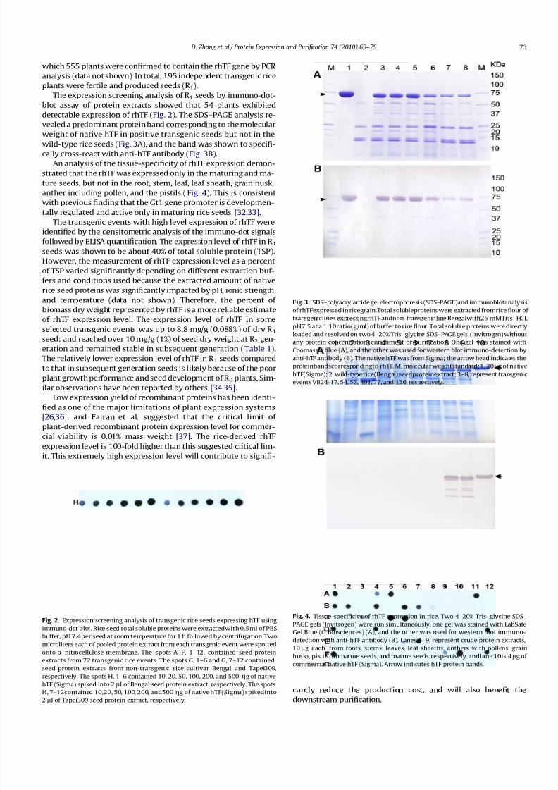
The expression screening analysis of R 1 seeds by immuno-dot-
blot assay of protein extracts showed that 54 plants exhibited
detectable expression of rhTF (Fig. 2). The SDS–PAGE analysis re-
vealed a predominant protein band corresponding to the molecular
weight of native hTF in positive transgenic seeds but not in the
wild-type rice seeds (Fig. 3A), and the band was shown to specifi-
cally cross-react with anti-hTF antibody (Fig. 3B).
An analysis of the tissue-specificity of rhTF expression demon-
strated that the rhTF was expressed only in the maturing and ma-
ture seeds, but not in the root, stem, leaf, leaf sheath, grain husk,
anther including pollen, and the pistils (Fig. 4). This is consistent
with previous finding that the Gt1 gene promoter is developmen-
tally regulated and active only in maturing rice seeds [32,33].
The transgenic events with high level expression of rhTF were
identified by the densitometric analysis of the immuno-dot signals
followed by ELISA quantification. The expression level of rhTF in R 1seeds was shown to be about 40% of total soluble protein (TSP).
However, the measurement of rhTF expression level as a percent
of TSP varied significantly depending on different extraction buf-
fers and conditions used because the extracted amount of native
rice seed proteins was significantly impacted by pH, ionic strength,
and temperature (data not shown). Therefore, the percent of
biomass dry weight represented by rhTF is a more reliable estimate
of rhTF expression level. The expression level of rhTF in some
selected transgenic events was up to 8.8 mg/g (0.088%) of dry R 1seed; and reached over 10 mg/g (1%) of seed dry weight at R 2 gen-
eration and remained stable in subsequent generation (Table 1).
The relatively lower expression level of rhTF in R 1 seeds compared
to that in subsequent generation seeds is likely because of the poor
plant growth performance and seed development of R 0 plants. Sim-
ilar observations have been reported by others [34,35].
Low expression yield of recombinant proteins has been identi-
fied as one of the major limitations of plant expression systems
[26,36], and Farran et al. suggested that the critical limit of plant-derived recombinant protein expression level for commer-
cial viability is 0.01% mass weight [37]. The rice-derived rhTF
expression level is 100-fold higher than this suggested critical lim-
it. This extremely high expression level will contribute to signifi-
cantly reduce the production cost, and will also benefit thedownstream purification.
Fig. 2. Expression screening analysis of transgenic rice seeds expressing hTF using
immuno-dot blot. Rice seed total soluble proteins were extractedwith 0.5ml of PBS
buffer, pH 7.4per seed at room temperature for 1 h followed by centrifugation.Two
microliters each of pooled protein extract from each transgenic event were spotted
onto a nitrocellulose membrane. The spots A–F, 1–12, contained seed protein
extracts from 72 transgenic rice events. The spots G, 1–6 and G, 7–12 contained
seed protein extracts from non-transgenic rice cultivar Bengal and Tapei309,
respectively. The spots H, 1–6 contained 10, 20, 50, 100, 200, and 500 gg of native
hTF (Sigma) spiked into 2 ll of Bengal seed protein extract, respectively. The spots
H, 7–12contained 10,20, 50, 100, 200, and500gg of native hTF(Sigma) spikedinto2ll of Tapei309 seed protein extract, respectively.
Fig. 3. SDS–polyacrylamide gel electrophoresis (SDS–PAGE)and immunoblotanalysis
of rhTFexpressed in ricegrain.Total solubleproteins were extracted fromrice flour of
transgeniclines expressingrhTF andnon-transgenic line Bengalwith25 mMTris–HCl,
pH7.5 at a 1:10ratio(g/ml) of buffer to rice flour. Total soluble proteins were directly
loaded and resolved on two 4–20% Tris–glycine SDS–PAGE gels (Invitrogen) without
any protein concentration, enrichment or purification. One gel was stained with
Coomassie blue (A), and the other was used for western blot immuno-detection by
anti-hTF antibody (B). The native hTF was from Sigma; the arrow head indicates the
proteinbandscorrespondingto rhTF. M, molecular weightstandard;1, 20lg of native
hTF(Sigma); 2, wild-type rice(Bengal) seedproteinextract; 3–8, represent transgenic
events VB24–17, 54, 57, 401, 77, and 136, respectively.
Fig. 4. Tissue-specificity of rhTF expression in rice. Two 4–20% Tris–glycine SDS–
PAGE gels (Invitrogen) were run simultaneously, one gel was stained with LabSafe
Gel Blue (G Biosciences) (A), and the other was used for western blot immuno-
detection with anti-hTF antibody (B). Lanes 1–9, represent crude protein extracts,
10lg each, from roots, stems, leaves, leaf sheaths, anthers with pollens, grain
husks, pistils, immature seeds, and mature seeds,respectively, andlane 10is 4 lg of
commercial native hTF (Sigma). Arrow indicates hTF protein bands.
D. Zhang et al./ Protein Expression and Purification 74 (2010) 69–79 73

8/6/2019 Human Transferrin in Rice
http://slidepdf.com/reader/full/human-transferrin-in-rice 6/11
Extraction and purification of rhTF
Identification of the optimal extraction conditions for rhTF is
important for developing a purification procedure that allows to
increase protein purity and to reduce purification costs. The effect
of buffer pH onrhTF extractability was tested in a range from4.5 to
10.0. It was shown that while the amount of TSP increased with the
increase in pH, the extracted rhTF protein was shown to increase
with increase in pH from 4.5 to 7.0 but no substantial difference
in the pH range from 7.0 to 10.0 (data not presented). Comparisonof the effect of extraction time showed that 30 min extraction was
already able to exact the maximum amount of rhTF. Neither the
salt concentration nor the extraction temperature showed a signif-
icant effect on the rhTF extractability (data not shown). These re-
sults indicated that extraction of rhTF from rice flour with
25 mM Tris–HCl, pH 7.5 for 30 min at RT was the optimal condition
to maximize the extraction of rhTF while minimizing the extrac-
tion of rice native proteins.
To develop a cost-effective procedure for purification of rhTF,
different chromatography media and conditions were tested. The
HIC column with a Phenyl Sepharose was shown to be able to pur-
ify rhTF at a purity of 90%. However, the requirement of a step of
precipitating rice native proteins with ammonium sulfate before
loading the protein extracts onto the Phenyl Sepharose column
could reduce the yield of rhTF and also add the purification cost.
The weak anion exchange chromatography DEAE showed that
the rhTF bound to the DEAE resin in the extraction buffer 25 mM
Tris–HCl, pH 7.5 without the need of buffer exchange, while some
rice proteins leaked out of the resin into the flow-through fractions
during loading and washing. The rhTF could then be eluted from
the DEAE resin with 40 mM NaCl in 25 mM Tris–HCl, pH 7.5, and
was at a purity of greater than 95% based on the SDS–PAGE
(Fig. 5). The purification of rhTF by the strong anion exchange
chromatography Q Sepharose resin showed a very similar chro-
matographic profile to that of DEAE-Sepharose column. However,
the Q Sepharose resin bound rhTF protein more strongly than DEAE
Sepharose resin, and the rhTF protein needed to be eluted with
higher concentration of salts, resulting in coeluting more rice pro-
teins. With the DEAE chromatography, we purified rhTF with four
batches of 100 g seed flour and each batch consistently yielded the
recovery rate of rhTF to 60% calculated on the basis of protein mass
as determined by ELISA. These results showed that a one-column
DEAE chromatography method can effectively purify rhTF from
rice grain protein extracts. The ease of purifying rhTF with a single
purification step is presumably enabled by both the high expres-
sion level of rhTF and the relatively simple protein composition
of the rice grain [38], because either of them will lead to a higher
enrichment of target protein in the starting material for purifica-
tion, which can help simplify the purification process and reduce
the cost. The ease and low cost of purification of recombinant pro-
teins from rice grains have also been shown in our prior work on
recombinant lactoferrin [39] and lysozyme [40,41].
Biochemical characterization of rhTF
Amino (N)-terminal sequence analysis
Since a rice seed storage protein signal sequence targeting to
the protein body in endosperm was fused to the N-terminus of
the rhTF, N-terminal sequencing of rhTF was carried out to exam-
ine whether the rice signal sequence was cleaved correctly. Eleven
sequencer cycles were analyzed, and the N-terminal sequence of
rhTF was revealed as V-P-D-K-T-V-R-W-Xc-A-V, which is identical
to nhTF except that the expected cysteine amino acid residue at cy-
cle 9 was not determined. The undetected cysteine is expected be-
cause cysteine, without special modification, cannot be detected by
N-terminal sequencing. This result indicates that the rice signal se-
quence before the mature rhTF protein was correctly removed at
the expected position.
Molecular weight of rhTF
The MALDI analysis was carried out to estimate the molecular
weight of rice-derived rhTF. A close-up view of the MALDI spec-
trum of rhTF revealed a peak comprising two small split peaks
on top with molecular weights of 75,255.6 and 76,573.8 Da,
respectively (Fig. 6). This MALDI spectrum is similar to that of
the yeast-derived aglycosylated rhTF but different from the N-gly-
cosylated nhTF spectrum, which showed a single peak of 80,000 Da
Table 1
Quantification of rhTF expression levels over three generations in rice grains.
Generation VB24–17 VB24–54 VB24–57
n Mean ± STD n Mean± STD n Mean± STD
R 1a 8 8.8 ± 0.9 8 8.0 ± 0.8 8 7.7 ± 0.3
R 2b 59 10.2 ± 1.7 64 10.0 ± 1.7 76 10.1 ± 2.1
R 3c 10 10.5 ± 1.8 10 10.5 ± 1.4 15 10.1 ± 1.6
a Eight R 1 positive seeds from each transgenic event were assayed.b One gram of pooled R 2 seeds from a single TF-positive R 1 plant was assayed.c One gram of pooled R 3 seeds from each single homozygous R 2 plant was
assayed.
Fig. 5. SDS–polyacrylamide gel electrophoresis (SDS–PAGE) of protein extracts,
different fraction pools from the purification of rice-derived rhTF. CK1 and CK2
represent native hTF (Sigma) and yeast-derived aglycosylated rhTF (Millipore),
respectively; M, molecular weight marker; 1, crude grainextract containing rhTF; 2,
flow-through from DEAE-Sepharose column; 3, eluate from DEAE-Sepharose
column; 4, fractions from high salt cleaning of DEAE-Sepharose column after theelution of rhTF; arrow, protein bands corresponding to hTF.
76573.811.3E+4
10075255.61
90
100
80
90
70
50
60
40
50
% I n
t e n s i t y
30
10
20
10
69752.0 72758.6 75765.2 78771.8 81778.4 84785.0
Mass(m/z)
Fig. 6. MALDImass spectra of purified rice-derivedrhTF (a close-up view). The apo-
rhTFs was desalted using ZipTip™l-C18 pipette tips (Millipore, Billerica, MA),
eluted with a solution of 70% acetonitrile (ACN), 0.2% formic acid, and 5 mg/ml
MALDI matrix (a-cyano-4-hydroxycinnamic acid), and analyzed with an AppliedBiosystems 4700 Proteomics Analyzer.
74 D. Zhang et al./ Protein Expression and Purification 74 (2010) 69–79
8/6/2019 Human Transferrin in Rice
http://slidepdf.com/reader/full/human-transferrin-in-rice 7/11
mass (data not shown). The mass for the first split small peak of the
rice-derived rhTF is close to the calculated mass of non-N-glycosyl-
ated nhTF (75,181.4 Da) with a mass shift of just 74.2 Da, and the
mass for the second split small peak showed a mass increase of 1392.4 Da. The size discrepancy between rhTF and N-glycosylated
nhTF as revealed by MALDI is consistent with the finding as shown
in the SDS–PAGE gel analysis of rhTF (Fig. 5). Furthermore, the rice-
derived rhTF molecular weight as revealed by MALDI is similar to
that of the yeast-derived aglycosylated rhTF, suggesting that the
rice-derived rhTF may not be N-glycosylated.
Glycosylation modifications
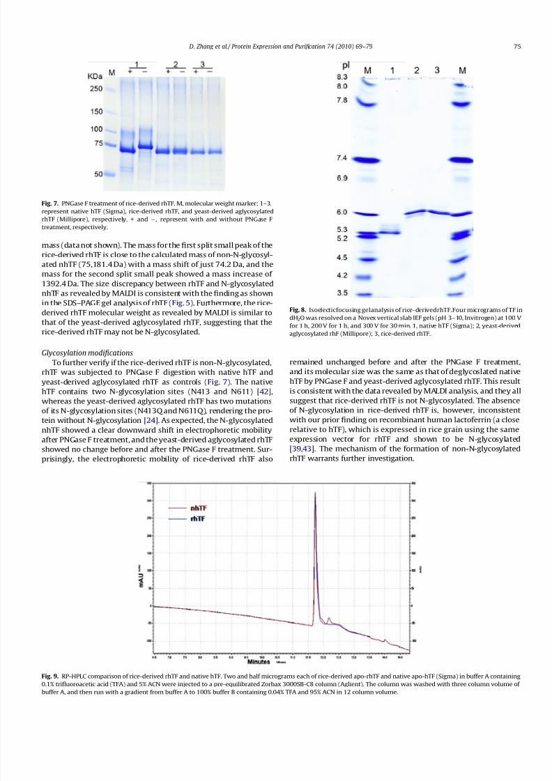
To further verify if the rice-derived rhTF is non-N-glycosylated,
rhTF was subjected to PNGase F digestion with native hTF and
yeast-derived aglycosylated rhTF as controls (Fig. 7). The native
hTF contains two N-glycosylation sites (N413 and N611) [42],
whereas the yeast-derived aglycosylated rhTF has two mutations
of its N-glycosylation sites (N413Q and N611Q), rendering the pro-tein without N-glycosylation [24]. As expected, the N-glycosylated
nhTF showed a clear downward shift in electrophoretic mobility
after PNGase F treatment, and the yeast-derived aglycosylated rhTF
showed no change before and after the PNGase F treatment. Sur-
prisingly, the electrophoretic mobility of rice-derived rhTF also
remained unchanged before and after the PNGase F treatment,
and its molecular size was the same as that of deglycoslated native
hTF by PNGase F and yeast-derived aglycosylated rhTF. This result
is consistent with the data revealed by MALDI analysis, and they all
suggest that rice-derived rhTF is not N-glycosylated. The absence
of N-glycosylation in rice-derived rhTF is, however, inconsistentwith our prior finding on recombinant human lactoferrin (a close
relative to hTF), which is expressed in rice grain using the same
expression vector for rhTF and shown to be N-glycosylated
[39,43]. The mechanism of the formation of non-N-glycosylated
rhTF warrants further investigation.
Fig. 7. PNGase F treatment of rice-derived rhTF. M, molecular weight marker; 1–3,
represent native hTF (Sigma), rice-derived rhTF, and yeast-derived aglycosylated
rhTF (Millipore), respectively. + and À, represent with and without PNGase F
treatment, respectively.
Fig. 8. Isoelecticfocusing gelanalysis of rice-derivedrhTF.Four micrograms of TF in
dH2O was resolved on a Novex vertical slab IEF gels (pH 3–10, Invitrogen) at 100 V
for 1 h, 200 V for 1 h, and 300 V for 30 min. 1, native hTF (Sigma); 2, yeast-derived
aglycosylated rhF (Millipore); 3, rice-derived rhTF.
Fig. 9. RP-HPLC comparison of rice-derived rhTF and native hTF. Two and half micrograms each of rice-derived apo-rhTF and native apo-hTF (Sigma) in buffer A containing
0.1% trifluoroacetic acid (TFA) and 5% ACN were injected to a pre-equilibrated Zorbax 3000SB-C8 column (Aglient). The column was washed with three column volume of buffer A, and then run with a gradient from buffer A to 100% buffer B containing 0.04% TFA and 95% ACN in 12 column volume.
D. Zhang et al./ Protein Expression and Purification 74 (2010) 69–79 75

8/6/2019 Human Transferrin in Rice
http://slidepdf.com/reader/full/human-transferrin-in-rice 8/11
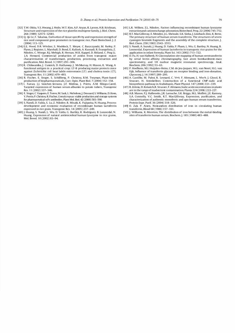
Isoelectric focusing point of rhTF
The isoelectric point (pI) of rice-derived rhTF was shown to be
6.3, which is same as the pI of yeast-derived aglycosylated rhTF
but one unit higher than the pI of the native hTF (5.3) ( Fig. 8).
The pI discrepancy between rhTF and native hTF is due to the neg-
atively charged sialic acid residues present in the native hTF but
absent in both rice-derived and yeast-derived rhTFs. The native
hTF has two N-linked oligosaccharide chains, and each chain termi-
nates in two or three antennae, each with terminal sialic acid res-
idues [42,44]. It has been reported that loss of the sialic acid
residues leads to a cathodic shift of the pI of TF molecules [45].
The yeast-derived aglycosylated rhTF has no N-linked glycans
and sialic acid residues. The rhTF expressed in rice grain is not ex-
pected to have sialic acids either, as plants are presumably not
capable of synthesizing sialic acids or at best just contain negligible
amounts [46,47].
Conformation of rhTF
The conformation and integrity of rice-derived apo-rhTF was as-
sessed by comparing with the apo-nhTF using reverse phase liquid
chromatography (RP-HPLC). RP-HPLC resolved both the rhTF and
nhTF into a major peak corresponding to their respective monomer
form of the molecule, and the two peaks were shown to have the
same retention time (Fig. 9), suggesting that rice-derived rhTF
has similar conformational structure as nhTF.
Biologic function assay of rhTF
Iron-binding assay
The biologic function of TF can be measured by assessing its
ability to bind and release iron reversibly. The purified partially-
iron-saturated (pis) rhTF from rice grains showed a salmon-pink
color, a characteristic color of iron-bound TF, suggesting that rhTF
Fig. 10. Iron-binding characteristics of rice-derived rhTF. (A) Color appearance of rhTF (5 mg/ml) withdifferent iron saturationlevels. 1, partially-iron-saturated (pis) rhTF; 2,
apo-rhTF made frompurifiedpis-rhTF;3, holo-rhTFmade from apo-rhTF.(B) Urea–PAGE gel (Invitrogen)analysis of rhTF with differentiron saturation levels. TheTF samples
fromleft to right on the gel are the same as in panel A. (C) Urea–PAGE gel analysis of rice-derived rhTF andthe comparison with commercial sources of hTF. Two micrograms
of each protein sample were resolved on a pre-cast 6% TBE–urea gel (Invitrogen) under 170 V for 2 h. 1, native apo-hTF (Sigma); 2, yeast-derived aglycosylated apo-rhf
(Millipore); 3, rice-derived apo-rhTF; 4, native holo-hTF (Sigma); 5, yeast-derived aglycosylated holo-rhf (Millipore); 6, rice-derived holo-rhTF. (D) Visible spectra of rhTF in
response to different concentrations of ferric iron. Apo-rhTF (5 mg/ml in 25 mM Tris–HCl buffer, pH 7.5 + 10 mM NaHCO3) was titrated with increasing amounts of iron (III)–nitrilotriacetate (Fe3+–NTA). The visible spectra were scanned from 700 to 380 nm after each addition of Fe3+–NTA, and the reading was corrected for dilution.
76 D. Zhang et al./ Protein Expression and Purification 74 (2010) 69–79

8/6/2019 Human Transferrin in Rice
http://slidepdf.com/reader/full/human-transferrin-in-rice 9/11
has already bound iron in rice grains. After being dialyzed against
50 mM sodium acetate, 5 mM EDTA, pH 4.9 overnight followed by
sequential dialysis in ddH2O and 25 mMTris–HCl, pH 7.5, the pink-
ish rhTF became colorless (Fig. 10A), an indication of iron release
from the pis-rhTF, resulting in the conversion of pis-rhTF into
apo-rhTF. Spectrophotometric titration of this apo-rhTF with iron
(Fe3+–NTA) showed a broad peak in the region from 465 to
470 nm, and the peak grew in size as the rhTF was gradually satu-
rated with the increasing concentration of iron (Fig. 10D). At the
same time, the pink color also gradually showed up in the titrated
rhTF solution and became darker when rhTF was saturated with
iron (Fig. 10A). The saturation of apo-rhTF with iron resulted in
the production of holo-rhTF.
To evaluate the iron-binding status of purified pis-rhTF and its
derived apo- and holo-isoforms after iron depletion and saturation,
these rhTF samples were subjected to a urea–PAGE gel electropho-
resis analysis. The apo- and holo-rhTF both showed a single band
but with slower and faster electrophoretic mobility, respectively,
in the urea–PAGE gel (Fig. 10B). The slower and faster migrating
forms of rhTF reflected the conformational change of rhTF without
or with bound iron [24,30]. The pis-rhTF showed three bands in the
urea–PAGE gel; the slowest and the fastest bands corresponded to
the apo- and holo-forms of rhTF, respectively, whereas the middle
band represented the monoferric form of rhTF. The coexistence of
apo-, holo- and monoferric-rhTF in the purified rhTF indicated that
rhTF had been indeed partially saturated with iron in the rice grain.
0
5
10
15
20
25
30
0 1 2 3 4 5 6 7
V i a b l e c e l l s 1 0 5
day
NoTransferrin
Human
Plasma TF
rhTF
0
25
50
75
100
125
150
No TF Human PlasmaTF rhTF
A n t i b o d y u g / m l
A
B
C
2
4
6
8
10
12
14
16
Treatments
V i a b l e c e l l s / m
l ( 1 0 ^ 5 )
0.03ug/ml
0.1ug/ml
0.3ug/ml
1ug/ml
5ug/ml
30ug/ml
Fig. 11. Effect of rhTF on cell growth and antibody production. (A) Growth of hybridoma cells in serum-free media supplemented with no hTF, 0.03, 0.1, 0.3, 1, 5 or 30lg/ml
native hTF (holo-form, from Sigma), rice-derived rhTF, ITSE or 10% FBS (Invitrogen). Shown is viable cell concentration after three days. (B) Growth of Sp2/0 hybridoma in
serum-free medium. The six-day growth curve of Sp2/0 hybridoma in serum-free medium supplemented with either 10 lg/ml native hTF or rice-derived rhTF, or
unsupplemented medium. (C) Increase in antibody production by hybridoma cells in serum-free medium supplied with TF. The concentration of antibody after six days of culture is shown. The medium was supplemented with either 10 lg/ml of rice-derived rhTF or native hTF.
D. Zhang et al./ Protein Expression and Purification 74 (2010) 69–79 77

8/6/2019 Human Transferrin in Rice
http://slidepdf.com/reader/full/human-transferrin-in-rice 10/11
The monoferric form of rhTF was further inferred to have an iron
bound in C-lobe of rhTF because the band was shown to be closer
to the apo-rhTF, which is a characteristic of C-terminal monoferric
TF [30,48]. In normal serum with an iron concentration insufficient
to saturate TF, the two monoferric forms of hTF (C- and N-terminal)
can be revealed in the urea–PAGE gel because both N- and C-termi-
nal iron-binding sites are occupied with iron although the N-termi-
nal site is normally preferentially occupied [49,50]. However, when
the serum is dialyzed against a buffer at pH 7.4, iron is found to
preferentially bind to the C-terminal site so that the N-terminal
monoferric TF is undetectable in the urea–PAGE gel [50]. Similarly,
the rice-derived rhTF was extracted and purified at pH 7.5 followed
by a step of diafiltration at pH 7.5 to concentrate and buffer ex-
change, and thus these conditions could cause the C-terminal
iron-binding site of rhTF to be predominantly occupied with iron,
resulting in the absence of the band corresponding to N-terminal
monoferric-rhTF.
The electrophoretic mobility of rice-derived apo- and holo-rhTF
in urea–PAGE gel was compared with that of native hTF and the
yeast-derived aglycosylated rhTF (Fig. 10 C). It was shown that
the rice-derived apo- or holo-rhTF migrated with the same mobil-
ity exhibited by their corresponding form of yeast-derived agly-
cosylated rhTF. These results showed that rice-derived rhTF was
able to bind and release iron reversibly. However, both apo- and
holo-native hTF exhibited faster mobility compared to their
respective counterpart of recombinant hTF. The faster electropho-
retic mobility of native hTF is associated with its possession of neg-
atively charged sialic acid residues that are absent in both rice- and
yeast-derived rhTFs.
Effect of rhTF on cell growth and antibody production
Rice-derived pis-rhTF was shown to have an equivalent dose re-
sponse as native holo-hTF for the proliferation of hybridoma cells
(Fig. 11A). Less than saturating levels of activity were observed at
concentrationsfrom 0.03 to 1 lg/ml with similarEC50 valueof about
0.3lg/ml. Likewise, a similarmaximumeffectwas observed at 5 and
30lg/ml that supported cell proliferation to 12.0Â 105 cells/ml.Themaximumeffectwas similarto theITSE cocktailcontrol contain-
ing5.5 lg/ml nativehTF. In addition, hybridomacellsgrownin med-
ium with either rice-derived rhTF or native hTF showed similar
growth curves (Fig. 11 B), supporting that rhTF has the same prolif-
eration effectas native hTF. Similar effects of rhTF and native hTF on
production of antibody were also seen (Fig. 11 C). These data show
that pis-rhTF is equivalent to the native holo-form of hTF in
stimulating cell growth and antibody production. Likely, the par-
tially-iron-saturated rhTF quickly becomes iron saturated due to
the presence of iron in the medium.
In summary, we have expressed hTF in rice grain at a high level
of 10 g/kg of seed dry weight (1%). The recombinant hTF protein
can be extracted with salt-water buffer and purified using a single
chromatographic step on DEAE-Sepharose column. The rice-de-rived rhTF was shown to be non-N-glycosylated by MALDI and
PNGase F enzyme digestion analyses although the entire amino
acid sequence of rhTF including its N-glycosylation sites were not
genetically modified. The rice-derived rhTF was proved to be not
only structurally similar to the native hTF, but also functionally
the same as native hTF in terms of reversibly binding iron and pro-
moting cell growth and productivity. The rice-derived rhTF will be
a safe and low cost alternative to human or animal plasma-derived
TF for use in biopharmaceutical cell culture, and it is available un-
der trade name, Optiferrin from InVitria.
Acknowledgments
This work was supported in part by NIH Grant GM086916 fromthe National Institute of General Medical Sciences (to D.Z.). The
authors thank Dr. Michael Barnet of InVitria for reviewing the
manuscript; Javier Herrera for helping grow transgenic plants in
greenhouse; Tanya Tanner for technical support.
References
[1] H.M. Baker, B.F. Anderson, E.N. Baker, Dealing with iron: common structural
principles in proteins that transport iron and heme, Proc. Natl. Acad. Sci. USA
100 (2003) 3579–3583.[2] M.W. Hentze, M.U. Muckenthaler, N.C. Andrews, Balancing acts: molecular
control of mammalian iron metabolism, Cell 117 (2004) 285–297.
[3] J. Williams, Theevolution of transferrin, Trends Biochem.Soc. 7 (1982)394–397.
[4] M. Hirose, The structural mechanism for iron uptake and release by
transferrins, Biosci. Biotechnol. Biochem. 64 (2000) 1328–1336.
[5] J. Wally, S.K. Buchanan, A structural comparison of human serum transferrin
and human lactoferrin, Biometals 20 (2007) 249–262.
[6] Q.-Y. He, A. Mason, Molecular aspects of release of iron from transferrin, in:
D.M. Templeton (Ed.), Molecular and Cellular Iron Transport, CRC Press,
2002, pp. 95–124.
[7] Y. Cheng, O. Zak, P. Aisen, S.C. Harrison, T. Walz, Structure of the human
transferrin receptor–transferrin complex, Cell 116 (2004) 565–576.
[8] Y. Cheng, O. Zak, P. Aisen, S.C. Harrison, T. Walz, Single particle reconstruction
of the human apo-transferrin–transferrin receptor complex, J. Struct. Biol. 152
(2005) 204–210.
[9] D. Barnes, G. Sato, Serum-free cell culture: a unifying approach, Cell 22 (1980)
649–655.
[10] J. Laskey, I. Webb, H.M. Schulma, P. Ponka, Evidence that transferrin supports
cell proliferation by supplyingiron for DNA synthesis,Exp. Cell Res. 176 (1988)87–95.
[11] S. Mortellaro, M. Devine, Advance in animal-free manufacturing of
biopharmaceuticals, Biopharm. Int. 20 (Supp.) (2007) 30–37.
[12] M.D. Sharath, S.B. Rinderknecht, J.M. Weiler, Human immunoglobulin
biosynthesis in a serum-free medium, J. Lab. Clin. Med. 103 (1984) 739–748.
[13] H. Li, Z.M. Qian, Transferrin/transferrin receptor-mediated drug delivery, Med.
Res. Rev. 22 (2002) 225–250.
[14] Z.M. Qian, H. Li, H. Sun, K. Ho, Targeted drug delivery via the transferrin
receptor-mediated endocytosis pathway, Pharmacol. Rev. 54 (2002) 561–587.
[15] V. Soni,S.K. Jain,D.V. Kohli, Potential of transferrinand transferrinconjugatesof
liposomes in drug delivery and targeting, Am. J. Drug Deliv. 3 (2005) 155–170.
[16] W.M. Pardridge, Molecular Trojanhorses for blood–brain barrier drug delivery,
Discov. Med. 6 (2006) 139–143.
[17] Y. Bai, D.K. Ann, W.C. Shen, Recombinant granulocyte colony-stimulating
factor-transferrin fusion protein as an oral myelopoietic agent, Proc. Natl.
Acad. Sci. USA 102 (2005) 7292–7296.
[18] A. Widera, F. Norouziyan, W.C. Shen, Mechanisms of TfR-mediated transcytosis
and sorting in epithelial cells and applications toward drug delivery, Adv.
Drug. Deliv. Rev. 55 (2003) 1439–1466.
[19] D. Banerjee, P.R. Flanagan, J. Cluett, L.S. Valberg, Transferrin receptors in the
human gastrointestinal tract. Relationship to body iron stores,
Gastroenterology 91 (1986) 861–869.
[20] J. Keenan, D. Pearson, L. O’Driscoll, P. Gammell, M. Clynes, Evaluation of
recombinant human transferrin(DeltaFerrin(TM)) as an ironchelatorin serum-
free media for mammalian cell culture, Cytotechnology 51 (2006) 29–37.
[21] R.T.M. MacGillivray, A.B. Mason, Transferrins, in: D.M. Templeton (Ed.),
Molecular and Cellular Iron Transport, Marcel Dekker, New York, 2002, pp.
41–70.
[22] P. Hoefkens, M.H. de Smit, N.M. de Jeu-Jaspars, M.I. Huijskes-Heins, G. de Jong,
H.G. van Eijk, Isolation, renaturation and partial characterization of
recombinant human transferrin and its half molecules from Escherichia coli,Int. J. Biochem. Cell Biol. 28 (1996) 975–982.
[23] S.A. Ali, H.C. Joao, R. Csonga, F. Hammerschmid, A. Steinkasserer, High-yield
productionof functionally active human serumtransferrin using a baculovirus
expression system, and its structural characterization, Biochem. J. 319 (Pt. 1)
(1996) 191–195.
[24] P.J. Sargent, S. Farnaud, R. Cammack, H.M. Zoller, R.W. Evans, Characterisationof recombinant unglycosylated human serum transferrin purified from
Saccharomyces cerevisiae, Biometals 19 (2006) 513–519.
[25] H. Daniell, S.J. Streatfield, K. Wycoff, Medical molecular farming: production of
antibodies, biopharmaceuticals and edible vaccines in plants, Trends Plant Sci.
6 (2001) 219–226.
[26] D. Lienard, C. Sourrouille, V. Gomord, L. Faye, Pharming and transgenic plants,
Biotechnol. Annu. Rev. 13 (2007) 115–147.
[27] R.M. Twyman, S. Schillberg, R. Fischer, Transgenic plants in the
biopharmaceutical market, Expert Opin. Emerg. Drugs 10 (2005) 185–218.
[28] N. Huang, D. Yang, ExpressTec: high level expression of biopharmaceuticals in
cereal grains, in: K. J. (Ed.), Modern Biopharmaceuticals, Wiley VCH, 2005, pp.
931–947.
[29] N. Huang, L. Wu, S. Nandi, E. Bowman, J. Huang, T. Sutliff, R.L. Rodriguez, The
tissue-specific activity of a rice beta-glucanase promoter (Gns9) is used to
select rice transformants, Plant Sci. 161 (2001) 589–595.
[30] R.W. Evans, J. Williams, The electrophoresis of transferrins in urea/
polyacrylamide gels, Biochem. J. 189 (1980) 541–546.
[31] D.G. Makey, U.S. Seal, The detection of four molecular forms of human
transferrin during the iron binding process, Biochim. Biophys. Acta 453 (1976)250–256.
78 D. Zhang et al./ Protein Expression and Purification 74 (2010) 69–79
8/6/2019 Human Transferrin in Rice
http://slidepdf.com/reader/full/human-transferrin-in-rice 11/11
[32] T.W. Okita, Y.S. Hwang, J. Hnilo, W.T. Kim, A.P. Aryan, R. Larson, H.B. Krishnan,
Structure and expression of the rice glutelin multigene family, J. Biol. Chem.
264 (1989) 12573–12581.
[33] Q. Qu Le, F. Takaiwa, Evaluation of tissue specificity and expression strength of
rice seed component gene promoters in transgenic rice, Plant Biotechnol. J. 2
(2004) 113–125.
[34] E.E. Hood, D.R. Witcher, S. Maddock, T. Meyer, C. Baszczynski, M. Bailey, P.
Flynn, J. Register, L. Marshall, D. Bond, E. Kulisek, A. Kusnadi, R. Evangelista, Z.
Nikolov, C. Wooge, R.J. Mehigh, R. Hernan, W.K. Kappel, D. Ritland, C. Ping Li,
J.A. Howard, Commercial production of avidin from transgenic maize:
characterization of transformant, production, processing, extraction andpurification, Mol. Breed. 3 (1997) 291–306.
[35] R. Chikwamba, J. Cunnick, D. Hathaway, J. McMurray, H. Mason, K. Wang, A
functional antigen in a practical crop: LT-B producing maize protects mice
against Escherichia coli heat labile enterotoxin (LT) and cholera toxin (CT),
Transgenic Res. 11 (2002) 479–493.
[36] R. Fischer, E. Stoger, S. Schillberg, P. Christou, R.M. Twyman, Plant-based
production of biopharmaceuticals, Curr. Opin. Plant Biol. 7 (2004) 152–158.
[37] I. Farran, J.J. Sánchez-Serrano, J.F. Medina, J. Prieto, A.M. Mingo-Castel,
Targeted expression of human serum albumin to potato tubers, Transgenic
Res. 11 (2002) 337–346.
[38] E. Stoger, C. Vaquero, E. Torres, M. Sack, L. Nicholson, J. Drossard, S. Williams, D. Keen,
Y. Perrin, P. Christou, R. Fischer, Cerealcropsas viable production and storage systems
for pharmaceutical scFv antibodies, Plant Mol. Biol. 42 (2000) 583–590.
[39] S. Nandi, D. Yalda, S. Lu, Z. Nikolov, R. Misaki, K. Fujiyama, N. Huang, Process
development and economic evaluation of recombinant human lactoferrin
expressed in rice grain, Transgenic Res. 14 (2005) 237–249.
[40] J. Huang, S. Nandi, L. Wu, D. Yalda, G. Bartley, R. Rodriguez, B. Lonnerdal, N.
Huang, Expression of natural antimicrobial human lysozyme in rice grains,
Mol. Breed. 10 (2002) 83–94.
[41] L.R. Wilken, Z.L. Nikolov, Factors influencing recombinant human lysozyme
extractionand cationexchange adsorption, Biotechnol. Prog. 22 (2006) 745–752.
[42] R.T. MacGillivray, E. Mendez, J.G. Shewale, S.K. Sinha, J. Lineback-Zins, K. Brew,
The primary structure of human serum transferrin. The structures of seven
cyanogen bromide fragments and the assembly of the complete structure, J.
Biol. Chem. 258 (1983) 3543–3553.
[43] S. Nandi, A. Suzuki, J. Huang, D. Yalda, P. Pham, L. Wu, G. Bartley, N. Huang, B.
Lonnerdal, Expression of human lactoferrin in transgenic rice grains for the
application in infant formula, Plant Sci. 163 (2002) 713–722.
[44] D. Fu, H. van Halbeek, N-Glycosylation site mapping of human serotransferrin
by serial lectin affinity chromatography, fast atom bombardment-massspectrometry, and 1H nuclear magnetic resonance spectroscopy, Anal.
Biochem. 206 (1992) 53–63.
[45] P. Hoefkens, M.I. Huijskes-Heins, C.M. de Jeu-Jaspars, W.L. van Noort, H.G. van
Eijk, Influence of transferrin glycans on receptor binding and iron-donation,
Glycoconj. J. 14 (1997) 289–295.
[46] A. Castilho, M. Pabst, R. Leonard, C. Veit, F. Altmann, L. Mach, J. Glossl, R.
Strasser, H. Steinkellner, Construction of a functional CMP-sialic acid
biosynthesis pathway in Arabidopsis, Plant Physiol. 147 (2008) 331–339.
[47] R. Zeleny, D. Kolarich,R. Strasser, F. Altmann,Sialic acidconcentrations in plants
are in the range of inadvertent contamination, Planta 224 (2006) 222–227.
[48] A.B. Mason, P.J. Halbrooks, J.R. Larouche, S.K. Briggs, M.L. Moffett, J.E. Ramsey,
S.A. Connolly, V.C. Smith, R.T. MacGillivray, Expression, purification, and
characterization of authentic monoferric and apo-human serum transferrins,
Protein Expr. Purif. 36 (2004) 318–326.
[49] O. Zak, P. Aisen, Nonrandom distribution of iron in circulating human
transferrin, Blood 68 (1986) 157–161.
[50] J. Williams, K. Moreton, The distribution of iron between the metal-binding
sites of transferrin human serum, Biochem. J. 185 (1980) 483–488.
D. Zhang et al./ Protein Expression and Purification 74 (2010) 69–79 79
Related Documents