University of Wollongong Research Online Australian Institute for Innovative Materials - Papers Australian Institute for Innovative Materials 2017 Human skin interactive self-powered wearable piezoelectric bio-e-skin by electrospun poly-l-lactic acid nanofibers for non-invasive physiological signal monitoring Ayesha Sultana Jadavpur University Sujoy Ghosh Jadavpur University Vitor Sencadas University of Wollongong, [email protected] Tian Zheng University of Wollongong, [email protected] Michael J. Higgins University of Wollongong, [email protected] See next page for additional authors Research Online is the open access institutional repository for the University of Wollongong. For further information contact the UOW Library: [email protected] Publication Details Sultana, A., Ghosh, S. Kumar., Sencadas, V., Zheng, T., Higgins, M. J., Middya, T. Ranjan. & Mandal, D. (2017). Human skin interactive self-powered wearable piezoelectric bio-e-skin by electrospun poly-l-lactic acid nanofibers for non-invasive physiological signal monitoring. Journal of Materials Chemistry B, 5 (35), 7352-7359.

Welcome message from author
This document is posted to help you gain knowledge. Please leave a comment to let me know what you think about it! Share it to your friends and learn new things together.
Transcript

University of WollongongResearch Online
Australian Institute for Innovative Materials - Papers Australian Institute for Innovative Materials
2017
Human skin interactive self-powered wearablepiezoelectric bio-e-skin by electrospun poly-l-lacticacid nanofibers for non-invasive physiologicalsignal monitoringAyesha SultanaJadavpur University
Sujoy GhoshJadavpur University
Vitor SencadasUniversity of Wollongong, [email protected]
Tian ZhengUniversity of Wollongong, [email protected]
Michael J. HigginsUniversity of Wollongong, [email protected]
See next page for additional authors
Research Online is the open access institutional repository for the University of Wollongong. For further information contact the UOW Library:[email protected]
Publication DetailsSultana, A., Ghosh, S. Kumar., Sencadas, V., Zheng, T., Higgins, M. J., Middya, T. Ranjan. & Mandal, D. (2017). Human skininteractive self-powered wearable piezoelectric bio-e-skin by electrospun poly-l-lactic acid nanofibers for non-invasive physiologicalsignal monitoring. Journal of Materials Chemistry B, 5 (35), 7352-7359.
Human skin interactive self-powered wearable piezoelectric bio-e-skin byelectrospun poly-l-lactic acid nanofibers for non-invasive physiologicalsignal monitoring
AbstractFlexible and wearable piezoelectric bio e-skin (PBio-e-skin) based on electrospun poly(l-lactic acid) PLLAnanofiber membrane is demonstrated for non-invasive human physiological signal monitoring and detectingdynamic tactile stimuli. The molecular orientations of the CO dipoles by electrospinning technique result in alongitudinal piezoelectric charge co-efficient (d 33 ) value of ∼(3 ± 1) pm V -1 realized by piezoresponse forcemicroscopy, allowing the PBio-e-skin for pressure sensing applications. The robust mechanical strength(Young's modulus ∼50 MPa) of nanofiber membrane ensures PBio-e-skin's superior operational stability over375000 cycles. Owing to the superior mechanosensitivity of ∼22 V N -1 , PBio-e-skin has the ability tomeasure subtle movement of muscle in the internal organs such as esophagus, trachea, motion of joints andarterial pressure by recognition of strains on human skin. This flexible and light weight PBio-e-skin preciselydetects vital signs and provides important clinical insights without using any external power source.Eventually, the low cost, environmental friendly PBio-e-skin will have a huge impact in a broad range ofapplications including self-powered wearable health care systems, human-machine interfacing devices,artificial intelligence and prosthetic skin.
DisciplinesEngineering | Physical Sciences and Mathematics
Publication DetailsSultana, A., Ghosh, S. Kumar., Sencadas, V., Zheng, T., Higgins, M. J., Middya, T. Ranjan. & Mandal, D.(2017). Human skin interactive self-powered wearable piezoelectric bio-e-skin by electrospun poly-l-lacticacid nanofibers for non-invasive physiological signal monitoring. Journal of Materials Chemistry B, 5 (35),7352-7359.
AuthorsAyesha Sultana, Sujoy Ghosh, Vitor Sencadas, Tian Zheng, Michael J. Higgins, Tapas Middya, and DipankarMandal
This journal article is available at Research Online: http://ro.uow.edu.au/aiimpapers/2751
http://pubs.rsc.org/en/content/articlelanding/2017/tb/c7tb01439b#!divAbstract
Human skin interactive self-powered wearable piezoelectric bio-e-skin by
electrospun poly-l-lactic acid nanofibers for non-invasive physiological
signal monitoring
Ayesha Sultana†, Sujoy Kumar Ghosh
†, Vitor Sencadas
‡,#, Tian Zheng
a, Michael Higgins
a,
Tapas Ranjan Middya†, Dipankar Mandal
†,*
†Organic Nano-Piezoelectric Device Laboratory, Department of Physics, Jadavpur
University, Kolkata 700032, India
‡Australian Centre of Excellence for Electromaterials Science (ACES), University of
Wollongong, NSW 2522, Australia
#School of Mechanical, Materials and Mechatronics Engineering, University of Wollongong,
Wollongong, NSW 2522, Australia
aARC Centre of Excellence for Electromaterials Science, Intelligent Polymer Research
Institute/AIIM Faculty, Innovation Campus, University of Wollongong, Squires Way, NSW,
Australia
*Corresponding Author
Dr. Dipankar Mandal
E-mail: [email protected];[email protected]
Fax: +91-33-2413-8917
Tel.: +91 33241466662880
http://pubs.rsc.org/en/content/articlelanding/2017/tb/c7tb01439b#!divAbstract
Abstract
Flexible and wearable piezoelectric bio e-skin (PBio-e-skin) based on electrospun poly(L-
lactide) PLLA nanofiber membrane is demonstrated for human physiological signal
monitoring and detecting dynamic tactile stimuli. The molecular orientations of the C=O
dipoles by electrospinning technique results in a longitudinal piezoelectric charge co-
efficient, d33 value of ~ (3±1) pm/V realized by piezoresponse force microscopy, allow the
PBio-e-skin for pressure sensing applications. The robust mechanical strength (Young’s
modulus ~ 50 MPa) of nanofiber membrane ensures PBio-e-skin’s operational stability (over
375,000 cycles). Owing to the superior mechanical sensitivity of ~ 22 V/N, PBio-e-skin has
the ability to measure subtle movement of muscle in the internal organs such as esophagus,
trachea, motion of joints and arterial pressure by recognition of strains on human skin. This
flexible and light weight PBio-e-skin can be valuable for monitoring vital signs in detecting
cardiovascular diseases, assessing health status and, also for voice recognition. Thus, the low
cost, environmental friendly PBio-e-skin can be used for non-invasive, artery pulse wave
monitoring, which may lead to the use of flexible PBio-e-skin in health monitoring and
diagnostics incardiovascular medicine.
DOI: 10.1038
Keywords: piezoelectric, PLLA, electrospinning, e-skin, flexible, human motion detection,
wearable, nanofiber, health monitoring
Introduction
Skin-mountable, stretchable and wearable/epidermal electronic devices for human motion
detection,1,2
flexible electronic skin (e-skin),3 human physiological and behavioural signal
detection,4,5
human body temperature monitoring6 and personal healthcare,
7 have recently
received enormous attention due to their facile interaction with skin or organs of the human
body. These flexible sensors can be attached into clothing or directly mounted on the human
http://pubs.rsc.org/en/content/articlelanding/2017/tb/c7tb01439b#!divAbstract
skin for the real-time monitoring of human activities and physiological measurements with
high efficiency. They must fulfil various minimum necessities including large stretchability,
lightweight, flexibility, durability, biocompatibility for stable contact with human skin with
minimal irritation.
So far design and development of wearable sensor systems for measuring and quantifying
physical signals are based on force-driven changes in resistivity,8,9
capacitance,10,11
piezo-
and tribo-electricity.12,13
In spite of the possibility and superior performance, wide spread
applications of some of these methods is perhaps limited by structure complexity, sensitivity,
dependence on external power source and fabrication of high-quality materials.14
One
confront for making pressure sensors that meet the aforementioned demands of high
stretchability, ultrahigh sensitivity and biocompatibility is to use piezoelectric nanofiber
membranes. In this context, poly(L-lactide) (PLLA) is the one which is biocompatible,
biodegradable, optically active polymer and continuously drawing increasing attention due to
its medical usefulness in tissue engineering scaffolds.15-17
Many biological systems exhibit
unique uniaxial symmetry where both direct and inverse piezoelectric effects are present but
only d14 and d25 (with same magnitude and opposite sign) shear piezoelectric constants are
finite. PLLA molecular chain consists of chiral molecules where –CO-O polar group joined
to an asymmetric carbon atom and oriented in helical conformation in the molecules. When
the helix is sheared through its side chain –C=O dipole makes a slight rotation that changes
the polarization of the chain molecule.18,15
Polarization appears in a direction perpendicular to
the plane of applied stress. The sum of these rotation results in electric polarization.19,15
Owing to its bio- piezoelectricity, PLLA is a suitable material for in-vivo applications where
biomaterial based self-powered system is particularly relevant.20,21
In previous, several
biomaterials such as, collagen, chitin, cellulose, gelatin, M13 bacteriophage and many others

http://pubs.rsc.org/en/content/articlelanding/2017/tb/c7tb01439b#!divAbstract
possessing longitudinal piezoelectric coefficient (d33) have been considered as engineering
effective materials for developing self-powered systems.22-26
The thermodynamically stable conformation of PLLA is the α-crystalline form, where the
C=O dipoles are randomly oriented along the main chain, resulting non-polarity of PLLA in
this form. In order to induce piezoelectricity the chains must be stretched to transform the α-
form into β-crystalline form where random orientation of molecular chains is aligned along
stretched direction.27
The electrospinning process applies stretching force into the molecular
chains of PLLA under high electric field and thus aligns molecular dipoles resulting net
polarization, in a single step.28
Herein, a simple structured, easy to fabricate, wearable, lightweight, cost-effective
piezoelectric bio-e-skin was prepared for pressure monitoring. This device is capable of
working in a multimodal manner for human physiological signal monitoring, sports
performance monitoring and also for voice recognition. It asserts a sensitivity of 22 V/N as
well as a pressure detection extent down to 18 Pa for detection of feather light pressure. In
loading-unloading cycle a negligible change in output signal was experimentally noticed over
375,000 cycles, suggesting its performance stability and durability. The flexible e-skin thus
evidenced the capability of monitoring human physiological signals such as throat and wrist
pulse and muscle movement when a person is speaking, which might extend their possible
applications for real-time disease diagnosis and voice recognition.
Experimental Section
Materials. Poly (L-lactic acid) (PLLA, Purasorb PL18, = 217 – 225 kDa) from Corbion
(Netherlands), N,N-dimethylformamide (DMF, from Merck), dichloromethane (MC, from
http://pubs.rsc.org/en/content/articlelanding/2017/tb/c7tb01439b#!divAbstract
Sigma-Aldrich), Copper (Cu)–nickel (Ni) plated fine knit polyester fabric (Coatex Industries,
India) and poly(dimethylsiloxane) (PDMS) (SYLGARD, 184 SILICONE ELASTOMER).
PLLA electrospun membranes preparation: PLLA was dissolved in a mixed solvent of
DMF and dichloromethane (3/7 v/v) to achieve a polymer concentration of 10 wt% of the
solution. The polymer solution was dissolved at room temperature with the help of a
magnetic stirrer until complete polymer dissolution.29
The polymer solution was placed in a
commercial glass syringe (10 mL) fitted with a steel needle with 500 µm of inner diameter.
Electrospinning was conducted at 1.25 kV.cm-1
with a high voltage power supply from
Gamma High Voltage. A syringe pump (from KD Scientific) was used to feed the polymer
solutions into the needle tip at 0.5 mL.h-1
. The electrospun fibres were collected in ground
collecting plate placed at 20 cm apart from the needle. All experiments were conducted at 21
± 2 ºC and a relative humidity of 43 ± 5%.
Piezoelectric bio-e-skin (PBio-e-skin) fabrication. The simple structured PBio-e-skin was
fabricated by the following method. At first a (2.5 × 2) cm2 area was cut from electrospun
PLLA membrane with thickness of ~ 40 µm. Then top and bottom electrodes were prepared
by attaching Ni−Cu−Ni plated polyester fabric on either side with effective contact area of (2
× 1.5) cm2. Then conducting copper wires were attached on both electrodes. Finally, it was
sandwiched between two layers (thickness of ~0.5 mm) of) PDMS which were prepared by
10:1 PDMS curing agent ratio and cured at 60 oC for 2 h.
Characterization
The morphological features of the electrospun fibers were imaged by a Field Emission
Scanning Electron Microscope (FE-SEM, FEI, INSPECT F50) operated at an acceleration
voltage of 20 kV. Fourier transform infrared (FT-IR) spectra were recorded (Bruker Tensor
http://pubs.rsc.org/en/content/articlelanding/2017/tb/c7tb01439b#!divAbstract
II) to characterizethe different bands within the NFs. The topography images were obtained
in AC mode with an MFP3D atomic force microscope (AFM) (Asylum Research, CA). Scan
rate was set as 0.5 Hz. Dual AC Resonance Tracking Mode piezoresponse force microscopy
(PFM) was used for measuring piezoresponse locally. A Pt/Ir coated silicon tip with force
constant of 2.8 N/m and gold layer on the substrate was used as top and bottom electrodes.
Contact frequency varied from 320 kHz to 340 kHz due to slightly changes on sample surface
from point to point. A small AC voltage (200 mV) was applied to oscillate the tip during
measurement. A sweeping DC bias (Frequency = 0.2 Hz) in the range of ± 25 V was applied
to the tip. 5 cycles of sweeping triangle/square waves (frequency = 0.2 Hz) were applied to
the tip to offer the fiber with essential electric field. Igor Pro 6.36 Software was used to
obtain the butterfly loops. Stress-strain measurement was carried out using universal testing
machine (Tinius Olsen H50KS). PBio-e-skin was fabricated by simply sandwiching the
nanofiber mat between two conducting Ni−Cu−Ni plated polyester fabric substrates.
Electrical contacts for recording open-circuit voltagewere made by Cu-wire leads from PBio-
e-skin connected through a National Instrument (NI) Data acquisition (DAQ) device (NI,
USB 6000) using a sampling rate of 1000 samplesper second, interfaced with a computer
with a standaloneprogram made by using LabVIEW software and current measurement were
carried out with KEITHLEY 6485 picoammeter.
Results and Discussion
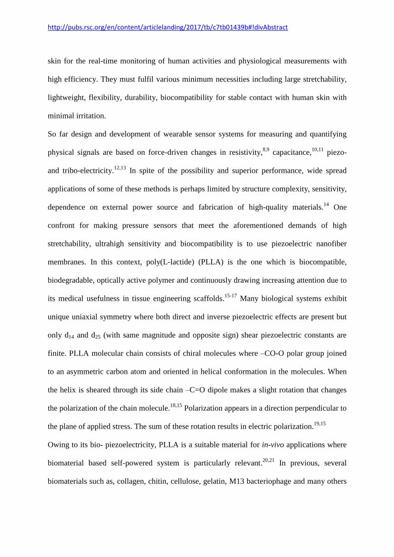
The schematic diagram in Fig. 1a shows a PLLA nanofiber fabrication process by
electrospining technique. The resulting PLLA nanofiber mat is used in flexible PBio-e-skin
(2 ×1.5 cm2) design with splendid capability to bend in desirable shape (Fig. 1b). The
morphology of as-electrospun nanofibers of PLLA is shown in Fig. 1c. The nanofibers are
entangled with one another and form a network-structured fiber mat. All fibers evidence

http://pubs.rsc.org/en/content/articlelanding/2017/tb/c7tb01439b#!divAbstract
smooth and bead free morphology with an average fiber diameter found to be 450 nm (inset
of Fig. 1c). Fig. 1d shows the FTIR spectra of nanofibers. The bands at 871, 1088, 1184,
1755 cm-1
are assigned to the C-C backbone stretching, symmetric and asymmetric stretching
of C-O-C group and stretching vibration of C=O which are responsible for piezo- and ferro-
electricty in PLLA nanofibers.30,31
All the bands of different wavenumbers are labelled in
Fig. 1d.
As the piezoelectric output performance of the nanofibers is greatly affected by their
mechanical properties in addition to piezoelectric coefficient value, thus, tensile stress-strain
behaviour of PLLA nanofibers has been studied as shown in Fig. 2a. Firstly, when a small
external stress (<8 kPa) is applied, the non-woven nanofibers are forced to align along stress
direction. This forced orientation results in a nonlinear elastic behaviour. With continual
increase of stress, the stress-strain curve exhibits a linear elasticity. Thus, the PLLA fibers
have ultimate tensile stress of 6.8 MPa, tensile strain of 42.3% with Young’s modulus value
of ~ 50 MPa. PLLA nanofiber membrane with the robust mechanical strength could be
applied as promising material towards real time applications.
http://pubs.rsc.org/en/content/articlelanding/2017/tb/c7tb01439b#!divAbstract
Fig. 1 (a) Schematic illustration of the experimental setup for preparation of PLLA
nanofibers. (b) The digital photograph of fabricated PBio-e-skin from PLLA nanofiber mat
showing its flexibility by human fingers. (c) FE-SEM micrograph and corresponding fiber
diameter distribution plot in the inset. (d) FT-IR spectra from 2000 to 800 cm-1
frequency
region.
To quantify local piezo- and ferro-electricity of PLLA nanofiber piezoelectric hysteresis
loops composed of phase voltage and amplitude voltage loops, PFM was conducted in a
single polymeric fiber deposited on a gold substrate. It assesses the dipole orientation and
10 µm
(c)
200 400 600 8000
10
20
30
Fre
qu
ency
Diameter (nm)
2000 1600 1200 800
as(
C-O
-C)
s(
C-O
-C)
(C
=C
H3)
as(
C-O
-C)+
r as(
CH
3)
(C
H)
s(
CH
3)
(C
-H)+
(CO
C)
(C
-H)
as(
CH
3)
Ab
sorb
an
ce (
a.u
.)
Wavenumber (cm-1)
(C
=O
)0.0
25
(a)
Pressure
1 cm
(b)
(d)

http://pubs.rsc.org/en/content/articlelanding/2017/tb/c7tb01439b#!divAbstract
mechanical fashion of the nanofiber when an electrical signal was applied by a metal-coated
AFM tip and scanning the sample.
Fig. 2 (a) Stress-strain curve, (b) AFM topography, (c) PFM phase-voltage and (d)
amplitude-voltage hysteresis loops of the PLLA nanofibers.
The AFM topography investigation (Fig. 2b) shows that the PLLA nanofiber has height of
650 nm (supporting information, Fig. S1). Excellent phase and amplitude responses (Fig. 2c–
d) from one single nanofiber have been observed as a function of applied voltage between –
25 V to +25 V to the AFM conductive cantilever tip with respect to ground. The PFM phase
response demonstrates a clear and almost rectangular hysteresis in the phase versus voltage
-30 -20 -10 0 10 20 30
0
50
100
150
200
250
dc bias voltage (V)
Ph
ase
(d
egre
e)
(c)
0 20 40 60 80
0
2
4
6
Str
ess
(N/m
m2)
Strain (%)
(a)
-30 -20 -10 0 10 20 30
20
40
60
dc bias voltage (V)
Am
pli
tud
e (p
m)
(d)
728.36 nm
0.0 nm
(b)

http://pubs.rsc.org/en/content/articlelanding/2017/tb/c7tb01439b#!divAbstract
diagram (Fig. 2c), with a phase contrast of 180
o at bias voltage of 25 V. In response to
sweeping DC voltage the hysteretic switching of the phase signal by 180o is assigned to the
switching of the direction of polarization of the C=O dipoles, which implies the evidence of
nano-scale ferroelectricity of the PLLA nanofibers. The PFM amplitude response is hysteretic
and the shape resembles the piezoelectric “butterfly loop” (Fig. 2d). Each point of this loop
includes information about piezoelectric deformation (εp = d33E, where d33 is the longitudinal
piezoelectric co-efficient) under corresponding applied voltage/electric field (E). The
estimated effective piezoelectric coefficient from the slope of the curves of amplitude versus
voltage is d33 ~ 3±1 pm/V.32,33
The observations of hysteretic phase switching and butterfly
shaped amplitude loops reveal a clear evidence of ferro– and piezo–electric properties in the
PLLA nanofiber mat.
Thus, we have fabricated PLLA nanofibers based piezoelectric bio e-skin (PBio-e-skin)
which could also be used for mechanical energy harvesting and piezotronics applications.
The performance of the PBio-e-skin as a function of various deformation frequencies was
investigated since mechanical energies from environment is irregular and varies in frequency.
The corresponding output voltage and current are shown in Fig. 3a–b. As the frequency of the
applied stress (~0.3 MPa) increases, the open circuit voltage (Voc) increases because the
electrons are flowing to reach equilibrium in a shorter time.34
http://pubs.rsc.org/en/content/articlelanding/2017/tb/c7tb01439b#!divAbstract
0 2
-0.5
0.0
0.5
1.0
4 6 8 10 12 14 16
18 Hz
15 Hz
14 Hz
12 Hz5 Hz2 Hz
19 Hz
1 Hz
a)
Volt
age
(V)
Time (sec)
0 2
-6
-4
-2
0
2
4
6
4 6 8 1010 12 14 16
b)
1 Hz 2 Hz5 Hz
12 Hz 14 Hz15 Hz
18 Hz19 Hz
Cu
rren
t (n
A)
Time (sec)
-0.60981.3287
1.2
Piezopotential (V)
-0.6-0.4-0.200.20.40.60.81.0
0.000020.2646
0.25 0.2 0.15 0.005
Displacement (µm)
0.1
105
106
107
0.0
0.3
0.6
0.9
1.2
0
1
2
3
4
5
6
Cu
rren
t (n
A)
V
olt
age
(V)
Resistance ()
Resistance () 10
510
610
70.00
0.02
0.04
0.06
0.08
Resistance ()
Pow
er (
W/c
m2)
0.000 0.005 0.010 0.015 0.0200.00
0.08
0.16
0.24
0.32
0.40
Volt
age
(V)
Applied Force (N)
c) d)
e) f)
g) h)
http://pubs.rsc.org/en/content/articlelanding/2017/tb/c7tb01439b#!divAbstract
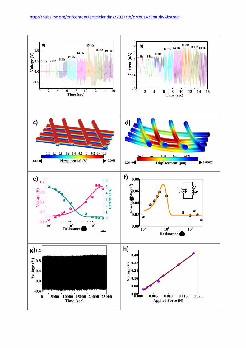
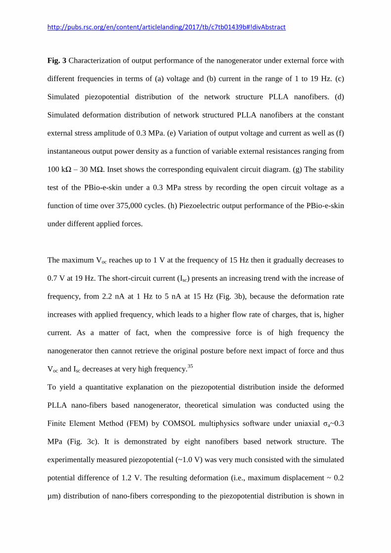
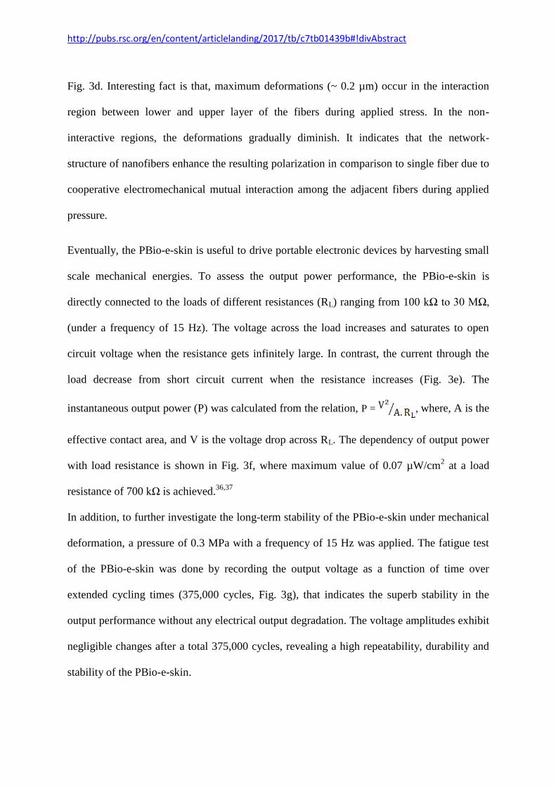
Fig. 3 Characterization of output performance of the nanogenerator under external force with
different frequencies in terms of (a) voltage and (b) current in the range of 1 to 19 Hz. (c)
Simulated piezopotential distribution of the network structure PLLA nanofibers. (d)
Simulated deformation distribution of network structured PLLA nanofibers at the constant
external stress amplitude of 0.3 MPa. (e) Variation of output voltage and current as well as (f)
instantaneous output power density as a function of variable external resistances ranging from
100 kΩ – 30 MΩ. Inset shows the corresponding equivalent circuit diagram. (g) The stability
test of the PBio-e-skin under a 0.3 MPa stress by recording the open circuit voltage as a
function of time over 375,000 cycles. (h) Piezoelectric output performance of the PBio-e-skin
under different applied forces.
The maximum Voc reaches up to 1 V at the frequency of 15 Hz then it gradually decreases to
0.7 V at 19 Hz. The short-circuit current (Isc) presents an increasing trend with the increase of
frequency, from 2.2 nA at 1 Hz to 5 nA at 15 Hz (Fig. 3b), because the deformation rate
increases with applied frequency, which leads to a higher flow rate of charges, that is, higher
current. As a matter of fact, when the compressive force is of high frequency the
nanogenerator then cannot retrieve the original posture before next impact of force and thus
Voc and Isc decreases at very high frequency.35
To yield a quantitative explanation on the piezopotential distribution inside the deformed
PLLA nano-fibers based nanogenerator, theoretical simulation was conducted using the
Finite Element Method (FEM) by COMSOL multiphysics software under uniaxial σa~0.3
MPa (Fig. 3c). It is demonstrated by eight nanofibers based network structure. The
experimentally measured piezopotential (~1.0 V) was very much consisted with the simulated
potential difference of 1.2 V. The resulting deformation (i.e., maximum displacement ~ 0.2
µm) distribution of nano-fibers corresponding to the piezopotential distribution is shown in
http://pubs.rsc.org/en/content/articlelanding/2017/tb/c7tb01439b#!divAbstract
Fig. 3d. Interesting fact is that, maximum deformations (~ 0.2 µm) occur in the interaction
region between lower and upper layer of the fibers during applied stress. In the non-
interactive regions, the deformations gradually diminish. It indicates that the network-
structure of nanofibers enhance the resulting polarization in comparison to single fiber due to
cooperative electromechanical mutual interaction among the adjacent fibers during applied
pressure.
Eventually, the PBio-e-skin is useful to drive portable electronic devices by harvesting small
scale mechanical energies. To assess the output power performance, the PBio-e-skin is
directly connected to the loads of different resistances (RL) ranging from 100 kΩ to 30 MΩ,
(under a frequency of 15 Hz). The voltage across the load increases and saturates to open
circuit voltage when the resistance gets infinitely large. In contrast, the current through the
load decrease from short circuit current when the resistance increases (Fig. 3e). The
instantaneous output power (P) was calculated from the relation, P = , where, A is the
effective contact area, and V is the voltage drop across RL. The dependency of output power
with load resistance is shown in Fig. 3f, where maximum value of 0.07 µW/cm2 at a load
resistance of 700 kΩ is achieved.36,37
In addition, to further investigate the long-term stability of the PBio-e-skin under mechanical
deformation, a pressure of 0.3 MPa with a frequency of 15 Hz was applied. The fatigue test
of the PBio-e-skin was done by recording the output voltage as a function of time over
extended cycling times (375,000 cycles, Fig. 3g), that indicates the superb stability in the
output performance without any electrical output degradation. The voltage amplitudes exhibit
negligible changes after a total 375,000 cycles, revealing a high repeatability, durability and
stability of the PBio-e-skin.
http://pubs.rsc.org/en/content/articlelanding/2017/tb/c7tb01439b#!divAbstract
To evaluate the sensitivity, the output voltages from the PBio-e-skin have been obtained
under different forces. Interestingly, Voc changes linearly with the applied force.
Quantitatively, the sensitivity is defined as, S = , where and are the differences of
Voc and σa respectively. To understand the relationship between applied force and
piezoelectric output voltage, different weights was dropped from a height of 10 cm. The force
from different weight (supporting information, Text S1) corresponds to distinct output
voltage. Fig. 3h shows a nearly linear relationship between the applied force and output
voltage. Interestingly, from the slope of the linear fitted data the sensitivity of the PBio-e-skin
is found to be 22 V/N.
+
-
(b)
(a) C O H
Electrospinning
http://pubs.rsc.org/en/content/articlelanding/2017/tb/c7tb01439b#!divAbstract
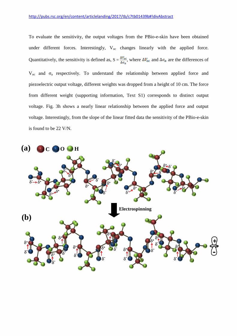
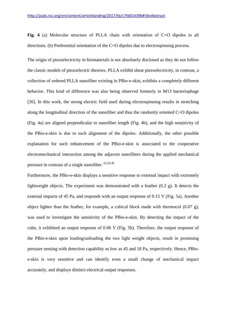
Fig. 4 (a) Molecular structure of PLLA chain with orientation of C=O dipoles in all
directions. (b) Preferential orientation of the C=O dipoles due to electrospinning process.
The origin of piezoelectricity in biomaterials is not absolutely disclosed as they do not follow
the classic models of piezoelectric theories. PLLA exhibit shear piezoelectricity, in contrast, a
collection of ordered PLLA nanofiber existing in PBio-e-skin, exhibits a completely different
behavior. This kind of difference was also being observed formerly in M13 bacteriophage
[26]. In this work, the strong electric field used during electrospinning results in stretching
along the longitudinal direction of the nanofiber and thus the randomly oriented C=O dipoles
(Fig. 4a) are aligned perpendicular to nanofiber length (Fig. 4b), and the high sensitivity of
the PBio-e-skin is due to such alignment of the dipoles. Additionally, the other possible
explanation for such enhancement of the PBio-e-skin is associated to the cooperative
electromechanical interaction among the adjacent nanofibers during the applied mechanical
pressure in contrast of a single nanofiber. 22,26,38
Furthermore, the PBio-e-skin displays a sensitive response to external impact with extremely
lightweight objects. The experiment was demonstrated with a feather (0.2 g). It detects the
external impacts of 45 Pa, and responds with an output response of 0.15 V (Fig. 5a). Another
object lighter than the feather, for example, a cubical block made with thermocol (0.07 g),
was used to investigate the sensitivity of the PBio-e-skin. By detecting the impact of the
cube, it exhibited an output response of 0.06 V (Fig. 5b). Therefore, the output response of
the PBio-e-skin upon loading/unloading the two light weight objects, result in promising
pressure sensing with detection capability as low as 45 and 18 Pa, respectively. Hence, PBio-
e-skin is very sensitive and can identify even a small change of mechanical impact
accurately, and displays distinct electrical output responses.

http://pubs.rsc.org/en/content/articlelanding/2017/tb/c7tb01439b#!divAbstract
Fig. 5 Output voltage response from PBio-e-skin with different applied external pressure for
the detection of a (a) feather (illustrated in the inset) and a (b) cubic thermocol block
(illustrated in the inset). Piezoelectric output (c) current and (d) voltage responses as a
function of area of PBio-e-skin.
To demonstrate the energy harvesting performance and practical application in rough
environment, the PBio-e-skin electrical responses was investigaed by applying compresssive
stress periodically by human finger. Here finger was imparted on PBio-e-skin made with
200 400 600 800 1000 1200
0
100
200
300
400
Cu
rren
t (n
A)
Area of sensor (mm2)
(c)
0 1 2 3 4 5 6
-0.1
0.0
0.1
0.2
0.3
0.4
V
olt
age
(V)
Time (sec)
(a)
200 400 600 800 1000 12000
2
4
6
8
10
12
Volt
age
(V)
Area of sensor (mm2)
(d)
0 1 2 3 4 5 6
-0.05
0.00
0.05
0.10
0.15
Vo
lta
ge
(V)
Time (sec)
(b) 5 cm 5 cm
http://pubs.rsc.org/en/content/articlelanding/2017/tb/c7tb01439b#!divAbstract
different areas. It was observed that the Isc and Voc increases from 30 nA to 400 nA (Fig. 5c)
and 1.25 V to 10 V (Fig. 5d) respectively with an increase in sensor area ranging from 200 to
2200 mm2. The output current and voltage shows almost linear depencency with the area of
total active area of the sensor following the relations of the piezoelectric theory.39
Thus the
current and voltage value can be modulated by changing the active area of PBio-e-skin as per
the power requirement of portable electronic devices.
In addition, the PBio-e-skin was further used for detecting static tactile stimuli. To show
applicabiliy of PBio-e-skin, as skin mountable sports performance monitoring device the
sensor was attatched to the wrist Fig. 6a. The static strain on the PBio-e-skin increases upon
bending of wrist, and recovers its original position after straightining of wrist. The response
of the PBio-e-skin on bending and relaxing of wrist with good quality of sensitivity is
demonstrated in Fig. 6b. This sensory information from skin mountable wearable PBio-e-skin
is beneficial for body movement analysis during sports activities and shows the potential of
using as epidermal device for skin motion monitoring. 40,41
The sensor was further placed near the throat and wrist, not only to measure the blood
pressure but also for measuring vibrations of muscle movements associated with several
human activities. By detecting strain of muscle movement for noninvasively monitoring
human activities, the PBio-e-skin was first attatched arround the throat. The PBio-e-skin
exhibits high sensitivity to the muscle movement of esophagus (the food pipe) and displays
distinct patterns, allowing to differentiate between signals generated by esophagus during
drinking and swallowing (Fig. 6c-d). These results show that for monitoring body motions,
skin strain monitoring is an effective method.
To demonstrate the performance as a stretchable PBio-e-skin for several biomedical
application the PBio-e-skin was attatched to carotid artery and raidal artery of an adult
http://pubs.rsc.org/en/content/articlelanding/2017/tb/c7tb01439b#!divAbstract
human. At first, the sensor is placed on the wrist to allow the measurement of blood pulse
wave (Fig. 6e). Three wave related to radial artery pressure are incident blood pulse wave,
reflected wave from hand and reflected wave from lower body. The acquired real-time
current output response from radial artery pressure is given in Fig. 6f. PBio-e-skin is placed
over carotid artery Fig. 6g and the real-time current output over several pulse period is shown
in Fig. 6h.
(a)
5 10 15 20
0.5
nA
Time (sec)
Cu
rren
t
(c)
0 5 10
1 n
AC
urren
t
Time (sec)
(b)
0 5 10 15 20
50
pA
Cu
rren
t
Time (sec)
(f)
(e)
0 5 10 15 20
20
0 p
A
Time (sec)
Cu
rren
t
(d)
(g)
12.0 12.5 13.0 13.5
50 p
A
PD
PI
Cu
rren
t
Time (sec)
PS
0 5 10 15 20
50
pA
Cu
rren
t
Time (sec)
(h)
http://pubs.rsc.org/en/content/articlelanding/2017/tb/c7tb01439b#!divAbstract
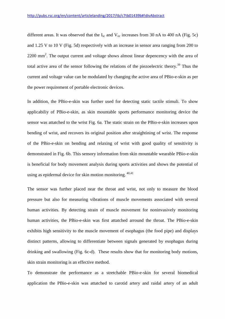
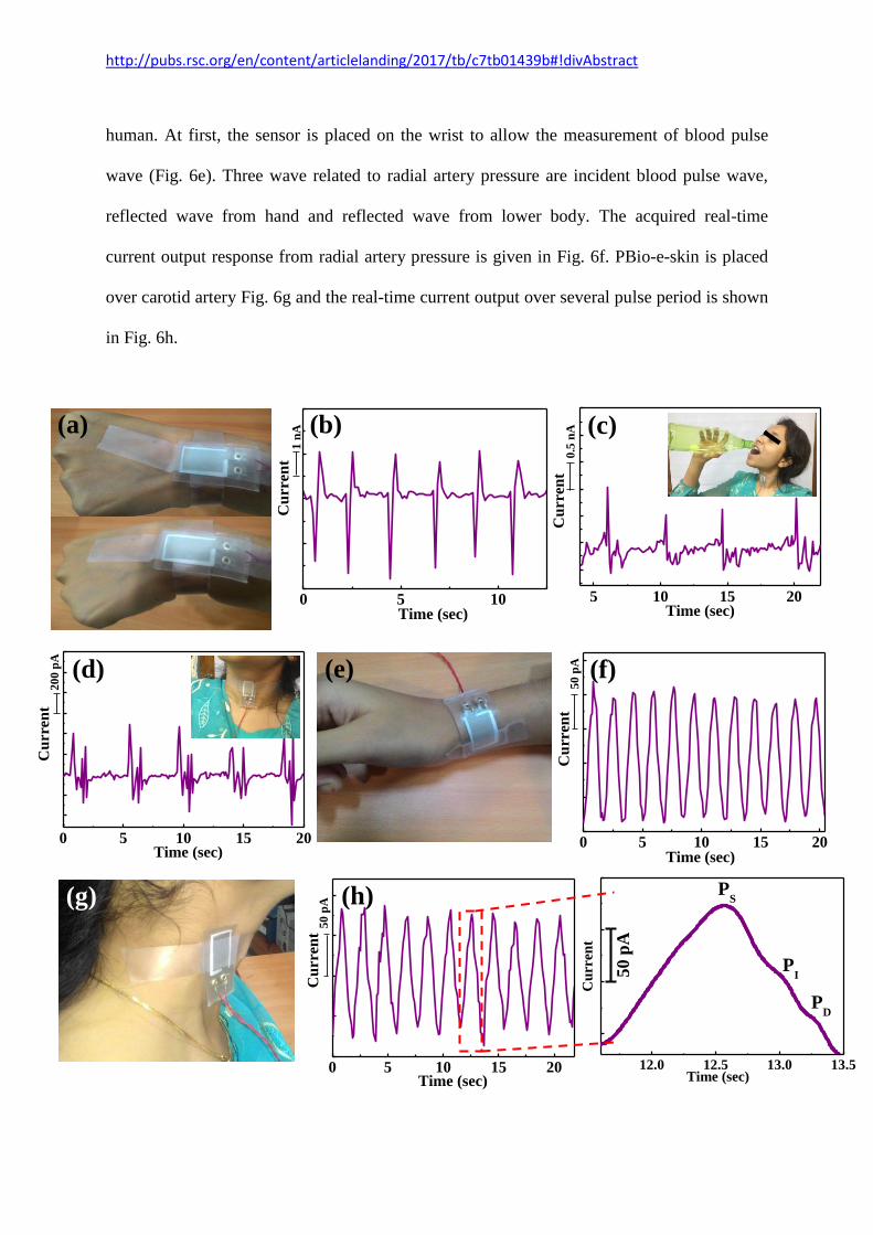
Fig. 6 (a) Photograph of the PBio-e-skin mounted on wrist joint. (b) Detected wrist joint
motion in terms of current output. Monitoring of strain caused by muscle movement for
function of esophagus during (c) drinking and (d) swallowing. (e) Photograph of the PBio-e-
skin placed on a wrist for measuring the pressure associated with flow of blood through near-
surface arteries. (f) Current vs time plot for the PBio-e-skin mounted on the wrist. (g)
Photograph showing PBio-e-skin directly attached to the carotid artery of an adult subject. (h)
The real-time current outputs for the sensor placed over carotid artery. The enlarged view in
the right shows the systolic (PS), point of inflexion (PI) and diastolic peak (PD) of one cycle.
(b)
0 2 4 6Time (s)
Sig
nal
(V)
20
mV
0 2 4 6Time (s)
Sig
nal
(V
)20 m
V
0
10
0
20
0
30
0
40
0
50
0
Frequency (Hz)
00.0
00
00
7
00.9
95
74
7
01.9
91
48
7
02.9
30
00
7
Amplitude
2.90 0.9 1.9
Fre
qu
ency
(H
z)
0
250
500
0 2 4 6Time (s)
Sig
nal
(V
)2
0 m
V
O N P D L
Res
po
nse
(a
.u.)
Hi
Hi
R
esp
on
se (
a.u
.)
Female Voice
Male Voice
(a)
0 5 10 15 20
Res
pon
se (
a.u
.)
Female Voice
PLLA
PLLA
Male Voice
Time (sec)

http://pubs.rsc.org/en/content/articlelanding/2017/tb/c7tb01439b#!divAbstract
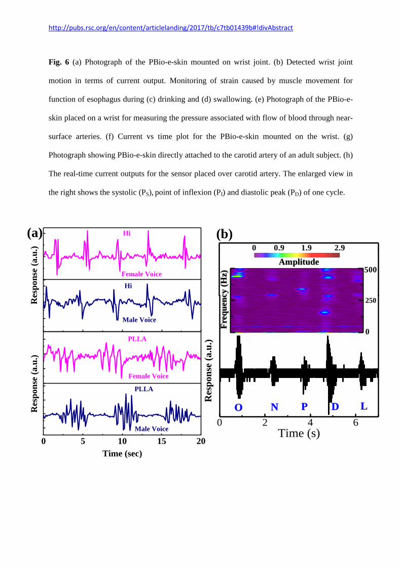
Fig. 7 Wavefront spectrum of female and male voices saying (a) “hi” and “PLLA”. (b)
Output wavefront spectrum observed from PBio-e-skin as different letters (O, N, P, D and L)
are pronounced with FFT signal in the upper part.
Change in current results from variation of pressure related to blood flow. Systolic peak (PS),
point of inflexion (PI) and diastolic peak (PD) can be identified from the enlarged image.
These peaks are known to be composed of three waves which are resulted from pulse wave
ejected from left ventricle, its reflected wave and ejected blood pulse back to left ventricle.42
These types of pulse waveforms provide valuable information for diagnosis and therapy for
cardiovascular diseases.43,44
The skin mounted sensor developed here, proved to be useful as
an arterial pulse wave monitor.
As a speech pattern recognition system, the PBio-e-skin was attached firmly to the human
speaker's neck and pressure difference of muscle movement during speech was recorded. The
sensor exhibits separate patterns when the different words such as “hi” and “PLLA” (Fig. 7a)
were spoken. Both these words were recorded for several cycles to investigate its
repeatability and similar characteristic patterns were obtained. In addition, it can also
differentiate between female and male voice, accurately. For non-invasively monitor the
variation of epidermis deformation and muscle movement throughout the throat during
phonation, alphabets such as ‘O’, ‘N’, ‘P’, ‘D’ and ‘L’ were pronounced. PBio-e-skin can
clearly detect and distinguish between the pronunciation of alphabets by mimicking the vocal
cord vibration and generating output signal waveform (Fig. 7b). The corresponding short time
Fourier transforms (STFT) processed spectrograms of each alphabet (upper part of Fig. 7b)
shows different output profiles. It is evident from the spectrogram that the maximum
amplitude of ‘O’ is in the higher frequency range of 400 Hz, for the letters N’ and ‘P’, the
frequency range is around 250 Hz, and for ‘D’ it is from 100-500 Hz. These results provide
http://pubs.rsc.org/en/content/articlelanding/2017/tb/c7tb01439b#!divAbstract
interesting method for using the PBio-e-skin to monitor muscle movement, voice recognition
and assisting in speech rehabilitation training.
Thus, the PBio-e-skin can be used as multimodal manner for arterial pulse wave, muscle
movement monitoring and as well as voice recognition. This skin strain monitoring might
have future applications on biomedical fields and remote control of human/machine
interfaces.
Conclusion
In summary, a stretchable, skin-mountable, highly sensitive, multifunctional piezoelectric bio
e-skin was developed by using PLLA nanofiber membrane with a longitudinal piezoelectric
coefficient value of 3±1 pm/V. Systematic and elaborate strain tests for sensor and energy
devices based on the fabricated multimodal e-skin showed high sensitivity, good stability,
and reliability with a detection limit of the device as low as 18 Pa. Skin motion detection was
performed byattaching the e-skin on different parts of the body for monitoring human
physiological signals, such as voice recognition, real-time wrist pulse detection etc. The e-
skin, with its ability to conform to bend can be used in different applications in a multitude of
fields, including personal health monitoring, epidermal electronic device, soft robotics,
artificial skins, and human machine interfaces. The good sensing performance of the PBio-e-
skin broadens its application in cost effective wearable electronics for the prevention of
sicknesses, in vivo and in vitro diannostics and the statement of early diseases.
Acknowledgements
This work was supported by the Science and Engineering Research Board (SERB/1759/2014-
http://pubs.rsc.org/en/content/articlelanding/2017/tb/c7tb01439b#!divAbstract
15), Govt. of India. Ayesha Sultana is supported by Maulana Azad National Fellowship (F1-
17.1/2015-16/MANF-2015-17-WES-53885/(SA-III/Website)) from UGC. Md. Mehebub
Alam is supported by UGC-BSR fellowship (Ref. No.P-1/RS/191/14). Authors are also
thankful for instrumental facilities developed by DST, Govt. of India under FIST-II
programme.
References
[1] Morteza Amjadi, Aekachan Pichitpajongkit, Sangjun Lee, Seunghwa Ryu, and Inkyu
Park. Highly Stretchable and Sensitive Strain Sensor Based on Silver Nanowire_Elastomer
Nanocomposite. ACS Nano, 2014, 8, 5154–5163.
[2] Yan Wang , Li Wang , Tingting Yang , Xiao Li , XiaobeiZang , Miao Zhu , Kunlin Wang,
Dehai Wu , and Hongwei Zhu. Wearable and Highly Sensitive Graphene Strain Sensors
forHuman Motion Monitoring. Adv. Funct. Mater.2014, 24, 4666–4670.
[3] Chuan Wang, David Hwang, Zhibin Yu, Kuniharu Takei, Junwoo Park, Teresa Chen,
Biwu Ma and Ali Javey. User-interactive electronic skin for instantaneous pressure
visualization. Nat. Mater. 2013, 12, 899–904.
[4] Xuewen Wang , Yang Gu , Zuoping Xiong , Zheng Cui , and Ting Zhang. Silk-Molded
Flexible, Ultrasensitive, and Highly Stable Electronic Skin for Monitoring Human
Physiological Signals. Adv. Mater. 2014, 26, 1336–1342.
[5] Joseph T. Muth , Daniel M. Vogt , Ryan L. Truby , Yig˘itMengüç , David B. Kolesky,
Robert J. Wood and Jennifer A. Lewis.Embedded 3D Printing of Strain Sensors within
Highly Stretchable Elastomers. Adv. Mater. 2014, 26, 6307–6312.
http://pubs.rsc.org/en/content/articlelanding/2017/tb/c7tb01439b#!divAbstract
[6] R. Chad Webb, Andrew P. Bonifas, Alex Behnaz, Yihui Zhang, Ki Jun Yu, Huanyu
Cheng, Mingxing Shi, Zuguang Bian, Zhuangjian Liu, Yun-Soung Kim, Woon-Hong Yeo,
Jae Suk Park, Jizhou Song, Yuhang Li, Yonggang Huang, Alexander M. Gorbach and John
A. Rogers. Ultrathin conformal devices for precise and continuous thermal characterization
of human skin. Nat. Mater. 2013, 12, 938–944.
[7] Tran Quang Trung and Nae-Eung Lee. Flexible and Stretchable Physical Sensor
Integrated Platforms for Wearable Human-Activity Monitoring and Personal Healthcare.
Adv. Mater. 2016, 28, 4338–4372.
[8] Takeo Yamada, Yuhei Hayamizu, Yuki Yamamoto, Yoshiki Yomogida, Ali Izadi-
Najafabadi, Don N. Futaba and Kenji Hata. A stretchable carbon nanotube strain sensor for
human-motion detection. Nat. Nanotechnol. 2011, 6, 296–301.
[9] Benjamin C-K. Tee, Chao Wang, Ranulfo Allen and Zhenan Bao. An electrically and
mechanically self-healing composite with pressure- and flexion-sensitive properties for
electronic skin applications. Nat. Nanotechnol. 2012, 7, 825–832.
[10] Daniel J. Cohen, Debkishore Mitra, Kevin Peterson and Michel M. Maharbiz. A Highly
Elastic, Capacitive Strain Gauge Based on PercolatingNanotube Networks. Nano Lett. 2012,
12, 1821−1825.
[11] Darren J. Lipomi, Michael Vosgueritchian, Benjamin C-K. Tee, Sondra L.
Hellstrom,Jennifer A. Lee, Courtney H. Fox and ZhenanBao. Skin-like pressure and strain
sensors based on transparent elastic films of carbon nanotubes. Nat. Nanotechnol. 2011, 6,
788–792.
[12] Zhong Lin Wang and Jinhui Song. Piezoelectric Nanogenerators Basedon Zinc Oxide
Nanowire Arrays. Science, 2006, 312, 242–245.
http://pubs.rsc.org/en/content/articlelanding/2017/tb/c7tb01439b#!divAbstract
[13] Xu Xiao, Longyan Yuan, Junwen Zhong, Tianpeng Ding, Yu Liu, Zhixiang Cai,
Yaoguang Rong, Hongwei Han, Jun Zhou and Zhong Lin Wang. High-Strain Sensors Based
on ZnO Nanowire/Polystyrene Hybridized Flexible Films. Adv. Mater. 2011, 23, 5440–5444.
[14] Jin Yang, Jun Chen, Yuanjie Su, Qingshen Jing, Zhaoling Li, Fang Yi, Xiaonan
Wen,Zhaona Wang and Zhong Lin Wang. Eardrum-Inspired Active Sensors for Self-Powered
Cardiovascular System Characterization and Throat-Attached Anti-Interference Voice
Recognition. 2015, 27, 1316–1326.
[15] E. Fukada. PIEZOELECTRICITY OF BIOPOLYMERS. Biorheology, 1995, 32, 593–
609.
[16] J. Kobayashi, T. Asahi, M. Ichiki, A. Oikawa, H. Suzuki. Structural and optical
properties of poly lactic acids. J. Appl. Phys. 1995, 77, 2957–2973.
[17] Y. Ikada, Y. Shikinami Y. Hara, M. Tagawa and E. Fukada. Enhancement of bone
formation by drawn poly(L-lactide). J. Biomed. Mater. Res., 1996, 30, 553–558.
[18] Eiichi Fukada. History and Recent Progress in Piezoelectric Polymers. IEEE Trans.
Ultrason., Ferroelect., Freq. Contro. 2000, 47, 1277–1290.
[19] Eiichi Fukada. New Piezoelectric Polymers. Jpn. J. Appl. Phys., 1998, 37, 2775–2780.
[20] Masamichi Ando, Hideki Kawamura, Hiroaki Kitada, Yasuyuki Sekimoto, Takafumi
Inoue, and Yoshiro Tajitsu. Pressure-Sensitive Touch Panel Based on Piezoelectric Poly(L-
lactic acid) Film. Jpn. J. Appl. Phys., 2013, 52, 09KD17 (1–4).
[21] Masamichi Ando, Hideki Kawamura, Keisuke Kageyama, and Yoshiro Tajitsu. Film
Sensor Device Fabricated by a Piezoelectric Poly(L-lactic acid) Film. Jpn. J. Appl. Phys.,
2012, 51, 09LD14 (1–4).
[22] Sujoy Kumar Ghosh, Dipankar Mandal. Efficient natural piezoelectric nanogenerator:
Electricity generation from fish swim bladder .Nano Energy 28 (2016) 356–365.
http://pubs.rsc.org/en/content/articlelanding/2017/tb/c7tb01439b#!divAbstract
[23] Sujoy Kumar Ghosh and Dipankar Mandal. Bio-assembled, piezoelectric prawn shell
made self-powered wearable sensor for non-invasive physiological signal monitoring.
APPLIED PHYSICS LETTERS 110, 123701(1-5) (2017).
[24] Md. Mehebub Alam and Dipankar Mandal. Native Cellulose Microfiber-Based Hybrid
Piezoelectric Generator for Mechanical Energy Harvesting Utility. ACS Appl. Mater.
Interfaces 2016, 8, 1555−1558.
[25] Sujoy Kumar Ghosh, Prakriti Adhikary, Santanu Jana, Anirban Biswas, Vitor Sencadas,
Sudipto Dutta Gupta, Bipan Tudu, Dipankar Mandal. Electrospun gelatin nanofiber based
self-powered bio-e-skin for health care Monitoring. Nano Energy 36 (2017) 166–175.
[26] Byung Yang Lee, Jinxing Zhang, Chris Zueger, Woo-Jae Chung, So Young Yoo, Eddie
Wang, Joel Meyer, Ramamoorthy Ramesh and Seung-Wuk Lee. Virus-based piezoelectric
energy generation. Nat. Nanotechnol. 7 (2012) 351–356..
[27] Qing Yang Pan, Tasaka Shigeru and Inagaki Norihiro. Ferroelectric behavior in poly-L-
lactic acid.Jpn. J. Appl. Phys., 1996, 35, L1442–L1445.
[28] Sol Jee Lee, ArunAnandPrabu, Kap Jin Kim. Piezoelectric
propertiesofelectrospunpoly(L-lactic acid) nanofiber web. Mater Lett.,2015, 148, 58–62.
[29] T. A. M. Valente, D. M. Silva, P. S. Gomes, M. H. Fernandes, J. D. Santos, and V.
Sencadas. Effect of Sterilization Methods on Electrospun Poly(lactic acid) (PLA) Fiber
Alignment for Biomedical Applications. ACS Appl. Mater. Interfaces. 2016, 8, 3241−3249.
[30] Clarisse Ribeiro, Vitor Sencadas, Carlos Miguel Costa,José Luís Gómez Ribelles and
Senentxu Lanceros-Méndez. Tailoring the morphology and crystallinity of poly(L-lactide
acid) electrospun membranes. Sci. Technol. Adv. Mater., 2011,12, 015001 (1–9).
[31] Nan Jing, Xiaoting Jiang, Qian Wang, Yongjiao Tang and Pudun Zhang. Attenuated
total reflectance/Fourier transforminfrared (ATR/FTIR) mapping coupled with principal
component analysis for the study of in vitro degradation of porous polylactide/hydroxyapatite
http://pubs.rsc.org/en/content/articlelanding/2017/tb/c7tb01439b#!divAbstract
composite material. Anal. Methods, 2014, 6, 5590–5595.
[32] Subrata Maji, Piyush Kanti Sarkar, Leena Aggarwal, Sujoy Kumar Ghosh, Dipankar
Mandal, Goutam Sheet and Somobrata Acharya. Self-oriented b-crystalline phase in the
polyvinylidene fluoride ferroelectric and piezo-sensitive ultrathin Langmuir–Schaefer film.
Phys. Chem. Chem. Phys.,2015, 17, 8159—8165.
[33] Yangjiang Wu, Qingzhao Gu, Guangzhu Ding, Fuqiang Tong, Zhijun Hu and Alain M.
Jonas. Confinement Induced Preferential Orientation of Crystals and Enhancement of
Properties in Ferroelectric Polymer Nanowires. ACS Macro Lett., 2013, 2, 535−538.
[34] Long Gu, Nuanyang Cui, Li Cheng, Qi Xu, SuoBai, Miaomiao Yuan, Weiwei Wu,
Jinmei Liu,Yong Zhao, Fei Ma, Yong Qin and Zhong Lin Wang.Flexible Fiber
Nanogenerator with 209 V Output Voltage Directly Powers a Light-Emitting Diode. Nano
Lett., 2013, 13, 91–94.
[35] Xiao-Sheng Zhang, Meng-Di Han, Ren-Xin Wang, Fu-Yun Zhu, Zhi-Hong Li, Wei
Wang and Hai-Xia Zhang. Frequency- Multiplication High -Output Triboelectric
Nanogenerator for Sustainably Powering Biomedical Microsystems. Nano Lett. 2013, 13,
1168−1172.
[36] Ayesha Sultana, Md. MehebubAlam, Anirban Biswas, Tapas Ranjan Middya and
Dipankar Mandal. Fabrication of wearable semiconducting piezoelectric nanogenerator made
with electrospun-derived zinc sulphide nanorods and poly(vinyl alcohol) nanofibers. Transl.
Mater. Res., 2016, 3, 045001 (1-11).
[37] Ayesha Sultana, Md. Mehebub Alam,Samiran Garain, Tridib Kumar Sinha, Tapas
Ranjan Middya and Dipankar Mandal. An Effective Electrical Throughput from PANI
Supplement ZnS Nanorods and PDMS-Based Flexible Piezoelectric Nanogenerator for
Power up Portable Electronic Devices: An Alternative of MWCNT Filler. ACS Appl. Mater.
Interfaces., 2015, 7, 19091−19097.

http://pubs.rsc.org/en/content/articlelanding/2017/tb/c7tb01439b#!divAbstract
[38] Luana Persano , Canan Dagdeviren , Claudio Maruccio , Laura De Lorenzis , and Dario
Pisignano. Cooperativity in the Enhanced Piezoelectric Response of Polymer Nanowires.
Adv. Mater. 2014, 26, 7574–7580.
[39] Sujoy Kumar Ghosh, Tridib Kumar Sinha, Biswajit Mahanty, Santanu Jana and
Dipankar Mandal. Porous polymer composite membrane based nanogenerator: A realization
of self-powered wireless green energy source for smart electronics applications. J. Appl.
Phys., 2016, 120, 174501 (1-11).
[40] Conor S. Boland, Umar Khan, Claudia Backes, Arlene O’Neill, Joe McCauley, Shane
Duane, Ravi Shanker, Yang Liu, Izabela Jurewicz, Alan B. Dalton and Jonathan N. Coleman.
Sensitive, High-Strain, High-Rate Bodily Motion Sensors Based on Graphene_Rubber
Composites. ACS Nano, 2014, 8, 8819–8830.
[41] Morteza Amjadi, Yong Jin Yoon and Inkyu Park. Ultra-stretchable and skin-mountable
strain sensors using carbon nanotubes–Ecoflex nanocomposites. Nanotechnology, 2015, 26,
375501 (1–11).
[42] Cátia Leitão, Lúcia Bilro, Nélia Alberto, Paulo Antunes, Hugo Lima, Paulo S. André,
RogérioNogueira, and João L. Pinto. Feasibility studies of Bragg probe for noninvasive
carotid pulse waveform assessment. J. Biomed. Opt., 2013, 18, 017006-017006.
[43] Yasser Khan,Aminy E. Ostfeld, Claire M. Lochner, Adrien Pierre, and Ana C.
Arias.Monitoring of Vital Signs with Flexible and WearableMedical Devices. Adv. Mater.
2016, 28, 4373–4395.
[44] Alfredo L. Pauca, Michael F. O’Rourke, Neal D. Kon. Prospective Evaluation of a
Method for EstimatingAscending Aortic Pressure From the Radial Artery Pressure
Waveform. Hypertension 2001, 38, 932–937.
Related Documents


![Shaping neuroplasticity by using powered exoskeletons in ... · for the patient of a real-world setting ambulation [10, 11]. To this end, wearable powered exoskeletons, e.g., the](https://static.cupdf.com/doc/110x72/5f0c28a57e708231d434072f/shaping-neuroplasticity-by-using-powered-exoskeletons-in-for-the-patient-of.jpg)