Human Sensory Neuron-specific Mas-related G Protein-coupled Receptors-X1 Sensitize and Directly Activate Transient Receptor Potential Cation Channel V1 via Distinct Signaling Pathways * Received for publication, August 7, 2012, and in revised form, October 12, 2012 Published, JBC Papers in Press, October 16, 2012, DOI 10.1074/jbc.M112.408617 Hans Jürgen Solinski, Susanna Zierler, Thomas Gudermann, and Andreas Breit 1 From the Walther-Straub-Institut für Pharmakologie und Toxikologie, Ludwig-Maximilians-Universität München 80336 München, Germany Background: MRGPR-X1 are exclusively expressed in primary sensory neurons, provoke the sensation of pain, and are considered as promising targets for pain therapy. Results: MRGPR-X1 sensitize TRPV1 for protons and heat via PKC and directly activate TRPV1 via DAG and PIP 2 . Conclusion: MRGPR-X1 modulate TRPV1 activity via multiple mechanisms. Significance: Interrupting the functional interaction between MRGPR-X1 and TRPV1 is a promising approach to diminish pain. Sensory neuron-specific Mas-related G protein-coupled receptors-X1 (MRGPR-X1) are primate-specific proteins that are exclusively expressed in primary sensory neurons and pro- voke pain in humans. Hence, MRGPR-X1 represent promising targets for future pain therapy, but signaling pathways activated by MRGPR-X1 are poorly understood. The transient receptor potential cation channel V1 (TRPV1) is also expressed in pri- mary sensory neurons and detects painful stimuli such as protons and heat. G q -promoted signaling has been shown to sensitize TRPV1 via protein kinase C (PKC)-dependent phos- phorylation. In addition, recent studies suggested TRPV1 acti- vation via a G q -mediated mechanism involving diacylglycerol (DAG) or phosphatidylinositol-4,5-bisphosphate (PIP 2 ). How- ever, it is not clear if DAG-promoted TRPV1 activation occurs independently from classic TRPV1 activation modes induced by heat and protons. Herein, we analyzed putative functional inter- actions between MRGPR-X1 and TRPV1 in a previously reported F11 cell line stably over-expressing MRGPR-X1. First, we found that MRGPR-X1 sensitized TRPV1 to heat and pro- tons in a PKC-dependent manner. Second, we observed direct MRGPR-X1-mediated TRPV1 activation independent of MRGPR-X1-induced Ca 2 -release and PKC activity or other TRPV1 affecting enzymes such as lipoxygenase, extracellular signal-regulated kinases-1/2, sarcoma, or phosphoinositide 3-kinase. Investigating several TRPV1 mutants, we observed that removal of the TRPV1 binding site for DAG and of the putative PIP 2 sensor decreased MRGPR-X1-induced TRPV1 activation by 71 and 43%, respectively. Therefore, we demon- strate dual functional interactions between MRGPR-X1 and TRPV1, resulting in PKC-dependent TRPV1 sensitization and DAG/PIP 2 -mediated activation. The molecular discrimination between TRPV1 sensitization and activation may help improve the specificity of current pain therapies. Painful stimuli are detected at the peripheral terminals of primary sensory neurons. Thus, proteins expressed in these neurons and involved either in signal perception, propagation, or transmission are potential targets for pain therapy. Of par- ticular interest are proteins that are highly enriched or exclu- sively expressed in primary sensory neurons, because specific modulation of these targets may allow circumvent untoward effects. Recently, Mas-related G protein-coupled receptors-X1 (MRGPR-X1) 2 have been shown to be exclusively expressed in primary sensory neurons and to be activated by bovine adrenal medulla peptide-8-22 (BAM8 –22) originating from proteolytic cleavage of pro-enkephalin by pro-hormone convertases (1, 2). Several studies reported activation of the G q pathway by MRGPR-X1 in overexpression systems (1, 3–5), and a recent study revealed increased pain sensation in 15 healthy volun- teers after BAM8 –22 application (6). In contrast, overexpres- sion of MRGPR-X1 in rat dorsal root ganglia (DRG) neurons resulted in BAM8 –22-mediated inhibition of voltage-gated calcium currents and activation of M-type potassium channels via G i/o proteins believed to blunt pain perception (7). Undoubtedly, MRGPR-X1 are involved in pain perception, but the underlying signaling pathways are still poorly defined. The transient receptor potential cation channel V1 (TRPV1) is a non-selective ion channel that is expressed in primary sen- sory neurons and activated by capsaicin (CAP), heat, or protons (8, 9). Increased TRPV1 activity at the cellular level reliably correlates with enhanced pain perception. Inflammatory medi- ators like bradykinin (BK) potentiate heat- or proton-induced TRPV1 activity via phosphorylation of the channel protein by protein kinase A or C (PKC), calmodulin-dependent kinase II, * This work was supported by Deutsche Forschungsgemeinschaft Grant BR 3346/3-1. 1 To whom correspondence should be addressed. Tel.: 49-89-2180-75755; Fax: 49-89-2180-75721; E-mail: [email protected]. 2 The abbreviations used are: MRGPR-X1, Mas-related G protein-coupled receptors-X1; AUC, area under the curve; BAM, bovine adrenal medulla; BCTC, N-(4-tertiarybutylphenyl)-4-(3-cholorphyridin-2-yl)tetrahydropyr- azine-1(2H)-carbox-amide; BIM-X, bisindolylmaleimide-X; BK, bradykinin; B2R, bradykinin-2 receptor; CAP, capsaicin; DAG, diacylglycerol; DRG, dor- sal root ganglia; GPCR, G protein-coupled receptor; HBS, HEPES-buffered saline; LOX, lipoxygenase; NDGA, nordihydroguaiaretic acid; OAG, 1-oleo- yl-2-acetyl glycerol; PIP 2 , phosphatidylinositol 4,5-bisphosphate; PKD, pro- tein kinase D; RT, room temperature; SRC, sarcoma kinase; TRPV1, transient receptor potential cation channel V1; pF, picofarads; PLC, phospholipase C. THE JOURNAL OF BIOLOGICAL CHEMISTRY VOL. 287, NO. 49, pp. 40956 –40971, November 30, 2012 © 2012 by The American Society for Biochemistry and Molecular Biology, Inc. Published in the U.S.A. 40956 JOURNAL OF BIOLOGICAL CHEMISTRY VOLUME 287 • NUMBER 49 • NOVEMBER 30, 2012 by guest on February 12, 2016 http://www.jbc.org/ Downloaded from

Welcome message from author
This document is posted to help you gain knowledge. Please leave a comment to let me know what you think about it! Share it to your friends and learn new things together.
Transcript
Human Sensory Neuron-specific Mas-related G Protein-coupledReceptors-X1 Sensitize and Directly Activate Transient ReceptorPotential Cation Channel V1 via Distinct Signaling Pathways*
Received for publication, August 7, 2012, and in revised form, October 12, 2012 Published, JBC Papers in Press, October 16, 2012, DOI 10.1074/jbc.M112.408617
Hans Jürgen Solinski, Susanna Zierler, Thomas Gudermann, and Andreas Breit1
From the Walther-Straub-Institut für Pharmakologie und Toxikologie, Ludwig-Maximilians-Universität München80336 München, Germany
Background: MRGPR-X1 are exclusively expressed in primary sensory neurons, provoke the sensation of pain, and areconsidered as promising targets for pain therapy.Results:MRGPR-X1 sensitize TRPV1 for protons and heat via PKC and directly activate TRPV1 via DAG and PIP2.Conclusion:MRGPR-X1 modulate TRPV1 activity via multiple mechanisms.Significance: Interrupting the functional interaction betweenMRGPR-X1 and TRPV1 is a promising approach to diminish pain.
Sensory neuron-specific Mas-related G protein-coupledreceptors-X1 (MRGPR-X1) are primate-specific proteins thatare exclusively expressed in primary sensory neurons and pro-voke pain in humans. Hence, MRGPR-X1 represent promisingtargets for future pain therapy, but signaling pathways activatedby MRGPR-X1 are poorly understood. The transient receptorpotential cation channel V1 (TRPV1) is also expressed in pri-mary sensory neurons and detects painful stimuli such asprotons and heat. Gq-promoted signaling has been shown tosensitize TRPV1 via protein kinase C (PKC)-dependent phos-phorylation. In addition, recent studies suggested TRPV1 acti-vation via a Gq-mediated mechanism involving diacylglycerol(DAG) or phosphatidylinositol-4,5-bisphosphate (PIP2). How-ever, it is not clear if DAG-promoted TRPV1 activation occursindependently fromclassic TRPV1 activationmodes induced byheat and protons.Herein, we analyzed putative functional inter-actions between MRGPR-X1 and TRPV1 in a previouslyreported F11 cell line stably over-expressing MRGPR-X1. First,we found that MRGPR-X1 sensitized TRPV1 to heat and pro-tons in a PKC-dependent manner. Second, we observed directMRGPR-X1-mediated TRPV1 activation independent ofMRGPR-X1-induced Ca2�-release and PKC activity or otherTRPV1 affecting enzymes such as lipoxygenase, extracellularsignal-regulated kinases-1/2, sarcoma, or phosphoinositide3-kinase. Investigating several TRPV1 mutants, we observedthat removal of the TRPV1 binding site for DAG and of theputative PIP2 sensor decreased MRGPR-X1-induced TRPV1activation by 71 and 43%, respectively. Therefore, we demon-strate dual functional interactions between MRGPR-X1 andTRPV1, resulting in PKC-dependent TRPV1 sensitization andDAG/PIP2-mediated activation. The molecular discriminationbetween TRPV1 sensitization and activation may help improvethe specificity of current pain therapies.
Painful stimuli are detected at the peripheral terminals ofprimary sensory neurons. Thus, proteins expressed in theseneurons and involved either in signal perception, propagation,or transmission are potential targets for pain therapy. Of par-ticular interest are proteins that are highly enriched or exclu-sively expressed in primary sensory neurons, because specificmodulation of these targets may allow circumvent untowardeffects. Recently, Mas-related G protein-coupled receptors-X1(MRGPR-X1)2 have been shown to be exclusively expressed inprimary sensory neurons and to be activated by bovine adrenalmedulla peptide-8-22 (BAM8–22) originating fromproteolyticcleavage of pro-enkephalin by pro-hormone convertases (1, 2).Several studies reported activation of the Gq pathway byMRGPR-X1 in overexpression systems (1, 3–5), and a recentstudy revealed increased pain sensation in 15 healthy volun-teers after BAM8–22 application (6). In contrast, overexpres-sion of MRGPR-X1 in rat dorsal root ganglia (DRG) neuronsresulted in BAM8–22-mediated inhibition of voltage-gatedcalcium currents and activation of M-type potassium channelsvia Gi/o proteins believed to blunt pain perception (7).Undoubtedly, MRGPR-X1 are involved in pain perception, butthe underlying signaling pathways are still poorly defined.The transient receptor potential cation channel V1 (TRPV1)
is a non-selective ion channel that is expressed in primary sen-sory neurons and activated by capsaicin (CAP), heat, or protons(8, 9). Increased TRPV1 activity at the cellular level reliablycorrelates with enhanced pain perception. Inflammatorymedi-ators like bradykinin (BK) potentiate heat- or proton-inducedTRPV1 activity via phosphorylation of the channel protein byprotein kinase A or C (PKC), calmodulin-dependent kinase II,
* This work was supported by Deutsche Forschungsgemeinschaft Grant BR3346/3-1.
1 To whom correspondence should be addressed. Tel.: 49-89-2180-75755;Fax: 49-89-2180-75721; E-mail: [email protected].
2 The abbreviations used are: MRGPR-X1, Mas-related G protein-coupledreceptors-X1; AUC, area under the curve; BAM, bovine adrenal medulla;BCTC, N-(4-tertiarybutylphenyl)-4-(3-cholorphyridin-2-yl)tetrahydropyr-azine-1(2H)-carbox-amide; BIM-X, bisindolylmaleimide-X; BK, bradykinin;B2R, bradykinin-2 receptor; CAP, capsaicin; DAG, diacylglycerol; DRG, dor-sal root ganglia; GPCR, G protein-coupled receptor; HBS, HEPES-bufferedsaline; LOX, lipoxygenase; NDGA, nordihydroguaiaretic acid; OAG, 1-oleo-yl-2-acetyl glycerol; PIP2, phosphatidylinositol 4,5-bisphosphate; PKD, pro-tein kinase D; RT, room temperature; SRC, sarcoma kinase; TRPV1, transientreceptor potential cation channel V1; pF, picofarads; PLC, phospholipase C.
THE JOURNAL OF BIOLOGICAL CHEMISTRY VOL. 287, NO. 49, pp. 40956 –40971, November 30, 2012© 2012 by The American Society for Biochemistry and Molecular Biology, Inc. Published in the U.S.A.
40956 JOURNAL OF BIOLOGICAL CHEMISTRY VOLUME 287 • NUMBER 49 • NOVEMBER 30, 2012
by guest on February 12, 2016http://w
ww
.jbc.org/D
ownloaded from

phosphoinositide 3-kinase (PI3K), sarcoma kinase (SRC), orextracellular signal-regulated kinases-1/2 (ERK-1/2) (10–15).However,bonafideactivationofTRPV1causedbyphosphoryla-tion has been questioned (16, 17). In contrast, release of thephospholipase C (PLC) product diacylglycerol (DAG) has beensuggested to enhanceTRPV1 activity via binding to the channelprotein (18) and degradation of the PLC substrate phosphati-dylinositol-4,5-bisphosphate (PIP2) to relieve TRPV1 frominhibitory PIP2 effects, thereby activating the ion channel (17,19). Therefore, GPCR-mediated activation of PLC can modu-late TRPV1 activity via several distinct mechanisms.Because of its dominant role in pain perception and its ver-
satile regulation by G protein-coupled receptors (GPCR),TRPV1 is a likely target of MRGPR-X1-induced signaling.Therefore, we investigated the effects of BAM8–22 on TRPV1activity in F11 cells (rat DRG neurons x murine neuroblastomacells) stably expressing MRGPR-X1. We show that BAM8-22sensitizes TRPV1 to heat and protons in a PKC-dependentmanner. However, BAM8–22/MRGPR-X1 signaling alsoresults inTRPV1 activation at room temperature (RT) and neu-tral pH. TRPV1 activation via MRGPR-X1 is independent ofPKC activity or other TRPV1 modulating enzymes such aslipoxygenase (LOX), PI3K, ERK-1/2, protein kinase D (PKD),and SRC. In contrast, aTRPV1mutant (TRPV1-Y511A) lackingthe binding site for DAG displayed a 71% reduction in BAM8–22-promoted TRPV1 activation, suggesting that DAG produc-tion is involved in MRGPR-X1-dependent TRPV1 activation.Likewise, removal of amino acids 777–820 supposedly encom-passing the TRPV1 sensor for PIP2 (19) decreased BAM8-22-induced activation by 43%, whereas the double mutant(TRPV1-Y511A-�777–820) was fully resistant to MRGPR-X1-mediated channel activation. Thus, we established the TRPV1as an important downstream effecter ofMRGPR-X1-promotedsignaling, which is modulated by PKC- and DAG/PIP2-depen-dentmechanisms. Knowledge about the functional interactionsbetweenMRGPR-X1 andTRPV1will increase our understand-ing of human pain perception and might help to improve ourcurrent pain therapy.
EXPERIMENTAL PROCEDURES
Materials—Ham’s F-12 nutrient mixture, FBS, penicillin/streptomycin, PBS, trypsin/EDTA, G418, and hypoxanthine/aminopterin/thymidine supplement were purchased fromInvitrogen. PromoFectin� was from PromoCell (Heidelberg,Germany). BSA, pluronic F-127, and poly-L-lysine were fromSigma, and fura2-acetoxymethyl ester was obtained from Fluka(Deisenhofen, Germany). Bisindolylmaleimide-X (BIM-X),1-oleoyl-2-acetyl glycerol (OAG), and thapsigargin were fromSigma. DAG kinase inhibitor-2 and U-73122 were from Calbi-ochem. PD-184352, LY-294002, nordihydroguaiaretic acid(NDGA) andCID-755673were fromEnzo life science (Lörrach,Germany), and N-(4-tertiarybutylphenyl)-4-(3-cholorphyri-din-2-yl)-tetrahydropyrazine-1(2H)-carbox-amide (BCTC)was from Biomol (Hamburg, Germany). PP-2 and BAM8–22were purchased from Biotrend (Cologne, Germany), BK wasfrom Sigma, and CAP was obtained from Fluka.Eukaryotic Expression Vectors—The plasmid coding for the
TRPV1-YFP fusion protein (20) and the corresponding point
mutants TRPV1-S800A-YFP (21) or TRPV1-S502A-YFP (22)were kindly provided by Dr. Tim D. Plant (Institute for Phar-macology and Toxicology, Marburg, Germany). The genera-tion of the plasmid coding for the TRPV1-Y511A-YFP was car-ried out using the QuikChange� site-directed mutagenesis kit(Agilent, Waldbronn, Germany) with TRPV1-YFP as templateand appropriate sense and antisense oligonucleotides spanningthe mutated triplet and 25 flanking nucleotides. The deletionconstruct TRPV1-�777–820-YFP was made by PCR usingTRPV1-YFP as the template and the following 5�-phosphory-lated primer: 5�-GGAGGAGCTCAGGGTGCGC-3�, 5�-GGA-TCCCTTAAGCCAGAGG-3�. The double mutant TRPV1-Y511A-�777–820-YFP was also constructed by site-directedmutagenesis with the TRPV1-�777–820-YFP plasmid as tem-plate and the same sense and antisense oligonucleotides as forTRPV1-Y511A-YFP construction. All constructs were verifiedby DNA sequencing.Cell Culture and Transfection—F11 cells stably expressing
MRGPR-X1 were previously described (4) and cultured inHam’s F-12 nutrient mixture supplemented with 15% FBS, 2mM L-glutamine, hypoxanthine/amitriptyline/thymidine sup-plement, G418 (250 �g/ml), penicillin (100 units/ml), andstreptomycin (100 �g/ml). For transient expression, F11-MRGPR-X1 cells were cultured for 24 h and then transfectedwith the appropriate vectors using PromoFectin� according tothe manufacturer’s protocol.Fura2-based Single Cell Ca2� Imaging—48 h before the
experiment, F11-MRGPR-X1 cells were seeded at a density of4–6 � 104 cells on glass coverslips in 6-well plates coated with0.1% poly-L-lysine. After 24 h, cells were transfected with 1 �gof the indicated TRPV1-YFP constructs. The next day cellswere loaded with 5 �M fura2-acetoxymethyl ester in HEPES-buffered saline (HBS, 10 mM HEPES, 5 mM KCl, 1 mM MgCl2,140mMNaCl, 0.1% glucose, and 2mMCaCl2 adjusted to pH 7.4with 1 M NaOH) supplemented with 0.02% pluronic F-127.Coverslips were washed twice withHBS and placed in a record-ing chamber, and YFP fluorescence of single cells was detected(excitation, 485 nm; emission, 510 nm). Afterward, we per-formed Ca2� imaging with the entire cell pool and distin-guished based on the YFP expression Ca2� signals of TRPV1expressing (F11-MRGPR-X1/TRPV1) and non-expressing(F11-MRGPR-X1) cells. This way distinct cell pools weretreated the same way. Of note, fura2 labeling of cells did notinterfere with the detection of YFP-based fluorescence. Aver-age transfection efficacy reached 30–50%, and around 20 cellsper coverslip were measured. Cells were analyzed using a Poly-chrome 5000monochromator (Till-Photonics, Gräfelfing, Ger-many) and an Andor charge-coupled device camera coupled toan inverted microscope (IX71, Olympus, Hamburg, Germany).Ca2� binding to fura2was determined by alternate excitation ofthe cells with 340 or 380 nm every 0.5 s and concomitant detec-tion of fluorescence emission at 520 nm. Fura2 ratios (340/380)were normalized by defining the first ratio (0.5 s) measured as100%. To measure Mn2�-induced quenching of fura2 fluores-cence, fura2 emission was measured after exciting with 360 nmin intervals of 0.25 s. Normalization was carried out by definingthe first ratio (0.25 s)measured as 100%. All inhibitors (concen-trations as indicated) or the corresponding carrier control were
Dual Regulation of TRPV1 Activity by MRGPR-X1
NOVEMBER 30, 2012 • VOLUME 287 • NUMBER 49 JOURNAL OF BIOLOGICAL CHEMISTRY 40957
by guest on February 12, 2016http://w
ww
.jbc.org/D
ownloaded from

preincubated for 30 min and present during the entire mea-surement. For temperature-induced TRPV1 activation, 500 �lof prewarmed HBS (�55 °C) was injected in 500 �l of buffer atRT. This injection led to a sudden temperature increase to�42 °C that declined rapidly to RT within 20 s. F11-MRGPR-X1 cells did not react to this treatment in the absenceof the TRPV1 (data not shown). Proton-induced TRPV1 acti-vation was initiated by injection of 500 �l of HBS with a pH of6.4 (Mn2� quench) or 5.0 (Ca2� imaging) to 500 �l of HBSleading to a decrease in pH from 7.4 to 6.9 or 6.2, respectively.Only TRPV1-expressing cells reacted to this treatment (datanot shown). To quantify ligand-induced calcium signals, thearea between the curve and the base line (100%) of single cellswas determined and is given as area under the curve (AUC). Toquantify actions of effective inhibitors, AUC values of F11-MRGPR-X1/TRPV1 cells were divided by those of F11-MRGPR-X1 cells. Ratios of untreated cells were set to 100%,and effects of chemical inhibitors were calculated as percent-age. Similar, to determine the effects of TRPV1 mutations,AUC values of TRPV1 wild-type or mutant-expressing cellswere divided by those of F11-MRGPR-X1 cells. The ratio ofTRPV1 wild-type cells was set to 100%, and effects of TRPV1mutations were calculated as percentage.Patch Clamp Analysis—Recordings of whole-cell currents
were performed in F11-MRGPR-X1 cells at RT using anEPC-10 amplifier and PatchMaster (HEKA, Lambrecht, Ger-many). Currents were provoked by a voltage ramp protocolspanning from �100 to �100 mV (50-ms duration, 0.5 Hz)starting from a holding potential of 0 mV. All measurementswere corrected for a liquid junction potential of 10 mV. Thestandard extracellular solution was HBS (pH 7.40, 290mosmol), and the intracellular solution contained the follow-ing: 120 mM cesium glutamate, 8 mM NaCl, 1 mM MgCl2, 3.62mM CaCl2, 10 mM HEPES, 2 mM MgATP, 10 mM EGTA, 5 mM
EDTA (pH 7.20, 290 mosmol). Agonists were applied by localsuperfusion using the pressure ejection system MPCU-3(Lorenz, Katlenburg-Lindau, Germany). Whole-cell currentswere extracted at�60mV, normalized to total cell capacitance,and plotted against time. Ligand-induced currents were calcu-lated by subtracting currentsmeasured at time points 60 s fromthose obtained at 150 s. These values were used for quantifica-tion and statistical analysis. IV relationships of representativesingle cells before (60 s) and at the end of ligand application(150 s) are shown.Data Analysis—Data were analyzed using Prism4.0. Statisti-
cal significance of differences was assessed by two-tailed Stu-dent’s t test (between two groups) or by one-way analysis ofvariance and Tukey’s honest significance post-hoc test(between more than two groups).
RESULTS
MRGPR-X1- and B2R-promoted TRPV1 Sensitization in F11Cells—F11 cells (rat DRG neurons x murine neuroblastomacells) endogenously express pain-modulatingGPCR such as thebradykinin-2 receptor (B2R) and are a widely accepted modelsystem to investigate DRG-related signaling pathways (23–35).To explore functional interactions between the MRGPR-X1and the TRPV1, we took advantage of a previously reported F11
cell pool (F11-MRGPR-X1 cells) that stably expressesMRGPR-X1 (4).We transiently expressed a TRPV1-YFP fusionprotein in these cells (F11-MRGPR-X1/TRPV1) and performedfura2 imaging with single cells (for details see “ExperimentalProcedures”). B2R-mediated activation of PLC has been shownto sensitize ligand-induced TRPV1 activity via PKC-dependentphosphorylation (11, 21, 22, 36–39). Therefore, we analyzedBK-mediated sensitization of CAP-induced Mn2� influx intoF11-MRGPR-X1/TRPV1 cells. As expected, prestimulation ofcells with BK significantly increased CAP-promoted Mn2�
influx in a PKC-dependent manner, indicative of TRPV1 sensi-tization (Fig. 1A).Having shown that functional interactions between GPCR
and TRPV1 can be analyzed reliably in F11-MRGPR-X1 cells,we next investigated the effects of BAM8–22 on TRPV1 activ-ity. Similar to BK, the MRGPR-X1-selective agonist signifi-cantly enhanced CAP-inducedMn2� influx into F11-MRGPR-X1/TRPV1 cells (Fig. 1B), indicating TRPV1 sensitization byMRGPR-X1. Pretreatment of cells with the specific PKC inhib-itor BIM-X completely blocked BAM8–22-induced TRPV1sensitization (Fig. 1B). Furthermore, we found that BAM8–22also sensitized TRPV1 to protons (Fig. 1C) or heat (Fig. 1D) in aPKC-dependent manner, indicating that BAM8–22 sensitizedall three modes of TRPV1 activation.PKC-mediated TRPV1 phosphorylation at positions serine
502 or 800 is required forGPCR-mediatedTRPV1 sensitization(22, 40). In line with this notion, BAM8–22 did not sensitizeCAP-inducedMn2�-influx in F11-MRGPR-X1 cells expressingeither theTRPV1-S502A (Fig. 1E) or theTRPV1-S800Amutant(Fig. 1F), further corroborating the notion thatMRGPR-X1 sig-naling sensitizes ligand-induced TRPV1 activity in a PKC-de-pendent manner.MRGPR-X1-induced Direct TRPV1 Activation Is PKC-inde-
pendent—Although TRPV1 sensitization to heat and protonsvia Gq-dependent PKC activation is widely recognized, less isknown about the impact of Gq/PLC signaling on direct TRPV1activation independently from increased temperature or pro-ton concentrations. Thus, we analyzed the consequences ofTRPV1 expression on BAM8–22-induced Mn2� influx at RTand a constant pH of 7.4 in the absence of CAP (Fig. 2,A andB).We observed that BAM8–22-induced Mn2� influx into F11-MRGPR-X1/TRPV1 cells was significantly enhanced whencompared with F11-MRGPR-X1 cells. Because the enhancedBAM8–22-inducedMn2� influx into F11-MRGPR-X1/TRPV1cells was blocked by the specific TRPV1 inhibitor BCTC (41),direct TRPV1 activation by MRGPR-X1 is most likely respon-sible for this effect. Given the important role for PKC inMRGPR-X1-mediated TRPV1 sensitization, we assumed thatPKC would also be involved in TRPV1 activation. However,blockade of PKC activity by BIM-X did not affect BAM8–22-inducedTRPV1 activation (Fig. 2C), suggesting that, in contrastto MRGPR-X1-induced TRPV1 sensitization, direct activationof TRPV1 by MRGPR-X1 is independent from PKC activity.To corroborate this assumption, we measured BAM8–22-
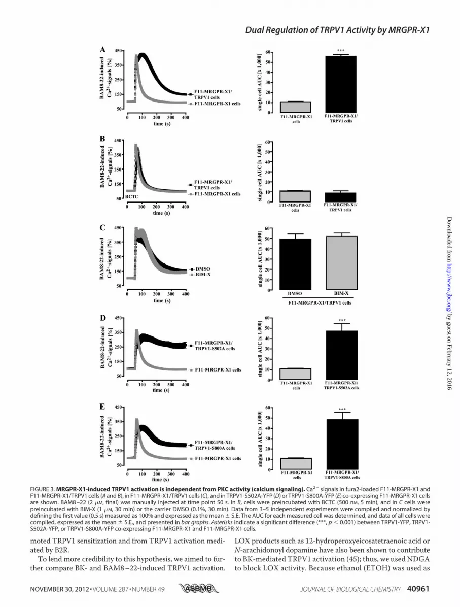
induced Ca2� signals in the presence or absence of TRPV1. Asshown in Fig. 3A, averagedCa2� responses in F11-MRGPR-X1/TRPV1 cells (AUC 55,950 � 1,800) were about 4.1-fold largerthan those obtained in cells solely expressing MRGPR-X1
Dual Regulation of TRPV1 Activity by MRGPR-X1
40958 JOURNAL OF BIOLOGICAL CHEMISTRY VOLUME 287 • NUMBER 49 • NOVEMBER 30, 2012
by guest on February 12, 2016http://w
ww
.jbc.org/D
ownloaded from

(AUC 10,900 � 400) mostly due to prolonged Ca2�-influx viaTRPV1. Accordingly, BCTC abrogated enhanced Ca2� signalsin F11-MRGPR-X1/TRPV1 cells (Fig. 3B), indicating thatdirect activation of TRPV1 by MRGPR-X1 led to enhancedBAM8–22-induced Ca2� influx. To investigate the role of PKCin this process, we either prestimulated F11-MRGPR-X1/TRPV1 cells with BIM-X or monitored BAM8–22-inducedCa
2�
signaling in F11-MRGPR-X1 cells expressing TRPV1mutants deficient of PKC phosphorylation sites. The PKC
inhibitor had no effect on BAM8–22-induced Ca2� signaling(Fig. 3C), and although both mutants were not sensitized byBAM8–22 (Fig. 1, E and F), they were robustly activated byMRGPR-X1 (Fig. 3, D � E), confirming that PKC-mediatedphosphorylation is not involved in MRGPR-X1-inducedTRPV1 activation. In conclusion, these data show thatMRGPR-X1-promoted TRPV1 activation (PKC-independent)ismediated by distinct signaling pathwayswhen comparedwithTRPV1 sensitization (PKC-dependent).
FIGURE 1. MRGPR-X1- or B2R-induced TRPV1 sensitization is dependent on PKC activity. Mn2�-induced quenching of fura2 fluorescence in F11-MRGPR-X1/TRPV1 and F11-MRGPR-X1 cells (A–D) or in F11-MRGPR-X1 and TRPV1-S502A-YFP (E) or TRPV1-S800A-YFP (F) co-expressing F11-MRGPR-X1 cells wasmeasured. CAP (5 nM, final) (A, B, E, and F), HCl (C), or pre-warmed buffer (D) were manually injected at time point 25 s; for details, see “Experimental Procedures.”Cells were preincubated only with BK (100 nM, 5 min, A) or BAM8 –22 (2 �M, 5 min, B–F) and either with the carrier DMSO (0.1%, 30 min, A–D) or with BIM-X (1�M, 30 min, A–D) before TRPV1 activation. Data obtained from 3–5 independent experiments were compiled, normalized by defining the first value (0.25 s)measured as 100%, and expressed as the mean � S.E. In A–D differences between basal and CAP-, proton-, or heat-provoked Mn2�-induced quenching of fura2fluorescence at time point 45 s are shown in bar graphs. Asterisks indicate a significant difference (***, p � 0.001) between basal and stimulated cells; hash signs(###, p � 0.001) indicate a significant difference between stimulated cells treated with either BIM-X or its carrier DMSO.
Dual Regulation of TRPV1 Activity by MRGPR-X1
NOVEMBER 30, 2012 • VOLUME 287 • NUMBER 49 JOURNAL OF BIOLOGICAL CHEMISTRY 40959
by guest on February 12, 2016http://w
ww
.jbc.org/D
ownloaded from

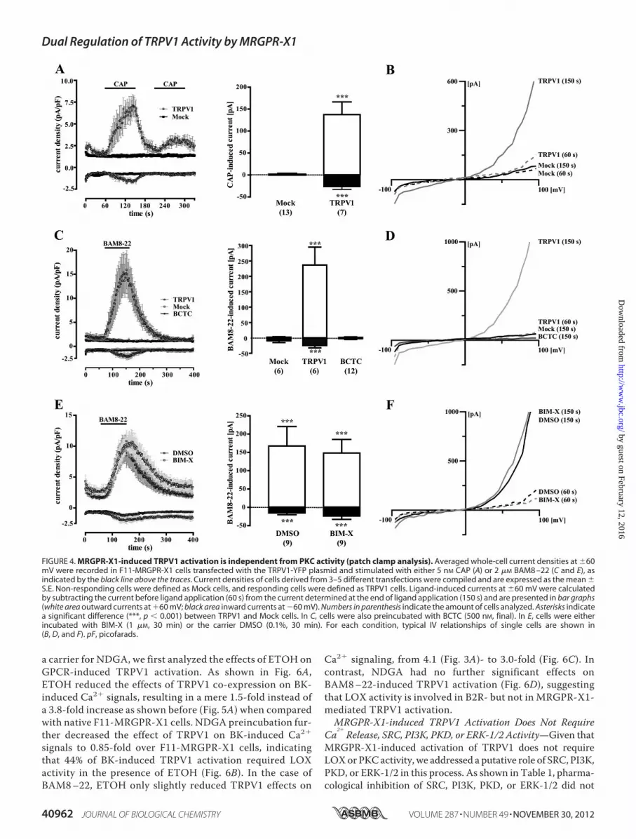
To substantiate our finding of PKC-independent TRPV1activation, we performed whole-cell patch clamp experimentswith TRPV1-YFP-transfected F11-MRGPR-X1 cells. As shownin Fig. 4A, after TRPV1 co-expression, 7 of 20 cells respondedwith inward (�26.6 � 6.4 pA/pF) and outward (137.4 � 28.8pA/pF) currents to stimulation with 5 nM CAP. This ratio ofresponsive (TRPV1) to non-responsive cells (Mock) correlatedwell to the transfection efficacy of �40% observed for theTRPV1-YFP plasmid. Furthermore, CAP-induced currentsobserved after TRPV1-YFP expression in F11-MRGPR-X1 cellsdisplayed characteristic outwardly rectifying IV-relationships(Fig. 4B), indicating that these currents were mediated by theTRPV1 (42–44). Next, we measured whole-cell currents inTRPV1-YFP expressing F11-MRGPR-X1 cells after applicationof BAM8–22. As depicted in Fig. 4C, the MRGPR-X1 agonistinduced inward (�24.6 � 6.2 pA/pF) and outward (236.6 �58.1 pA/pF) currents inTRPV1-YFP-transfected cells thatwereblocked by BCTC and also exhibited a TRPV1-like IV relation-ship (Fig. 4D), indicating that MRGPR-X1 activation inducedTRPV1 currents in F11-MRGPR-X1 cells at RT and neutral pH.To finally assess the role of PKC for MRGPR-X1-inducedTRPV1 activation, we measured BAM8–22-induced currentsin BIM-X pretreated cells (Fig. 4, E and F). Pharmacological
PKC block did not affect BAM8–22-induced currents inTRPV1-expressing F11-MRGPR-X1 compared with DMSO-treated cells. Therefore, in congruence with data shown in Fig.2 and 3, we provide conclusive evidence that direct TRPV1activation via MRGPR-X1 in F11 cells is independent of PKCactivity.B2R-induced TRPV1 Activation Is Dependent on PKC and
LOX Activity—Next, we wondered whether PKC-independentTRPV1 activation is unique for MRGPR-X1 or whether itwould also apply to other established modulators of TRPV1activity, such as BK. As shown in Fig. 5A, BK-induced Ca2�
signals in F11-MRGPR-X1 cells (AUC9900� 700)were similarto those induced by BAM8–22 (Fig. 3A, AUC 10,900 � 400),illustrating that activation of recombinant MRGPR-X1 did notelicit unphysiologically high Ca2� transients compared withendogenously expressed B2R. Of note, BK-induced Ca2� sig-nals were 3.8-fold higher in F11-MRGPR-X1/TRPV1 cells(AUC 47,560 � 3,500) when compared with F11-MRGPR-X1cells (Fig. 5A), indicating direct TRPV1 activation also by B2R.However, blockade of PKC activity by BIM-X significantlyreducedBK-inducedCa2� signals by 53% (Fig. 5C) as comparedwith DMSO-treated cells (Fig. 5B). Thus, MRGPR-X1-inducedTRPV1 activation is clearly distinct from MRGPR-X1-pro-
FIGURE 2. MRGPR-X1-induced TRPV1 activation is independent from PKC activity (manganese influx). Mn2�-induced quenching of fura2 fluorescence inF11-MRGPR-X1 (A) or F11-MRGPR-X1/TRPV1 cells (B and C) were measured. BAM8 –22 (2 �M, final) or HBS (basal) were manually injected at time point 25 s. InA and B cells were preincubated or not with BCTC (500 nM, 5 min), and in C cells were preincubated either with the carrier DMSO (0.1%, 30 min) or with BIM-X(1 �M, 30 min). Data from 3–5 independent experiments were compiled and normalized by defining the first value (0.25 s) measured as 100% and expressedas the mean � S.E. In A and B, differences in Mn2�-induced quenching of fura2 fluorescence before application and at time point 45 s are also shown in the bargraphs. In C, differences between basal (DMSO or BIM-X) and BAM8 –22-provoked Mn2�-induced quenching of fura2 fluorescence at time point 45 s are given.Asterisks indicate a significant difference (***, p � 0.001) between BAM8 –22-stimulated and unstimulated cells; hash signs (###, p � 0.001) between BCTC-treated and untreated cells.
Dual Regulation of TRPV1 Activity by MRGPR-X1
40960 JOURNAL OF BIOLOGICAL CHEMISTRY VOLUME 287 • NUMBER 49 • NOVEMBER 30, 2012
by guest on February 12, 2016http://w
ww
.jbc.org/D
ownloaded from
moted TRPV1 sensitization and from TRPV1 activation medi-ated by B2R.To lend more credibility to this hypothesis, we aimed to fur-
ther compare BK- and BAM8–22-induced TRPV1 activation.
LOX products such as 12-hydroperoxyeicosatetraenoic acid orN-arachidonoyl dopamine have also been shown to contributeto BK-mediated TRPV1 activation (45); thus, we used NDGAto block LOX activity. Because ethanol (ETOH) was used as
FIGURE 3. MRGPR-X1-induced TRPV1 activation is independent from PKC activity (calcium signaling). Ca2� signals in fura2-loaded F11-MRGPR-X1 andF11-MRGPR-X1/TRPV1 cells (A and B), in F11-MRGPR-X1/TRPV1 cells (C), and in TRPV1-S502A-YFP (D) or TRPV1-S800A-YFP (E) co-expressing F11-MRGPR-X1 cellsare shown. BAM8 –22 (2 �M, final) was manually injected at time point 50 s. In B, cells were preincubated with BCTC (500 nM, 5 min), and in C cells werepreincubated with BIM-X (1 �M, 30 min) or the carrier DMSO (0.1%, 30 min). Data from 3–5 independent experiments were compiled and normalized bydefining the first value (0.5 s) measured as 100% and expressed as the mean � S.E. The AUC for each measured cell was determined, and data of all cells werecompiled, expressed as the mean � S.E., and presented in bar graphs. Asterisks indicate a significant difference (***, p � 0.001) between TRPV1-YFP, TRPV1-S502A-YFP, or TRPV1-S800A-YFP co-expressing F11-MRGPR-X1 and F11-MRGPR-X1 cells.
Dual Regulation of TRPV1 Activity by MRGPR-X1
NOVEMBER 30, 2012 • VOLUME 287 • NUMBER 49 JOURNAL OF BIOLOGICAL CHEMISTRY 40961
by guest on February 12, 2016http://w
ww
.jbc.org/D
ownloaded from
a carrier for NDGA, we first analyzed the effects of ETOH onGPCR-induced TRPV1 activation. As shown in Fig. 6A,ETOH reduced the effects of TRPV1 co-expression on BK-induced Ca2� signals, resulting in a mere 1.5-fold instead ofa 3.8-fold increase as shown before (Fig. 5A) when comparedwith native F11-MRGPR-X1 cells. NDGA preincubation fur-ther decreased the effect of TRPV1 on BK-induced Ca2�
signals to 0.85-fold over F11-MRGPR-X1 cells, indicatingthat 44% of BK-induced TRPV1 activation required LOXactivity in the presence of ETOH (Fig. 6B). In the case ofBAM8–22, ETOH only slightly reduced TRPV1 effects on
Ca2� signaling, from 4.1 (Fig. 3A)- to 3.0-fold (Fig. 6C). Incontrast, NDGA had no further significant effects onBAM8–22-induced TRPV1 activation (Fig. 6D), suggestingthat LOX activity is involved in B2R- but not in MRGPR-X1-mediated TRPV1 activation.MRGPR-X1-induced TRPV1 Activation Does Not Require
Ca2�
Release, SRC, PI3K, PKD, or ERK-1/2 Activity—Given thatMRGPR-X1-induced activation of TRPV1 does not requireLOXor PKCactivity, we addressed a putative role of SRC, PI3K,PKD, or ERK-1/2 in this process. As shown in Table 1, pharma-cological inhibition of SRC, PI3K, PKD, or ERK-1/2 did not
FIGURE 4. MRGPR-X1-induced TRPV1 activation is independent from PKC activity (patch clamp analysis). Averaged whole-cell current densities at �60mV were recorded in F11-MRGPR-X1 cells transfected with the TRPV1-YFP plasmid and stimulated with either 5 nM CAP (A) or 2 �M BAM8 –22 (C and E), asindicated by the black line above the traces. Current densities of cells derived from 3–5 different transfections were compiled and are expressed as the mean �S.E. Non-responding cells were defined as Mock cells, and responding cells were defined as TRPV1 cells. Ligand-induced currents at �60 mV were calculatedby subtracting the current before ligand application (60 s) from the current determined at the end of ligand application (150 s) and are presented in bar graphs(white area outward currents at �60 mV; black area inward currents at �60 mV). Numbers in parenthesis indicate the amount of cells analyzed. Asterisks indicatea significant difference (***, p � 0.001) between TRPV1 and Mock cells. In C, cells were also preincubated with BCTC (500 nM, final). In E, cells were eitherincubated with BIM-X (1 �M, 30 min) or the carrier DMSO (0.1%, 30 min). For each condition, typical IV relationships of single cells are shown in(B, D, and F). pF, picofarads.
Dual Regulation of TRPV1 Activity by MRGPR-X1
40962 JOURNAL OF BIOLOGICAL CHEMISTRY VOLUME 287 • NUMBER 49 • NOVEMBER 30, 2012
by guest on February 12, 2016http://w
ww
.jbc.org/D
ownloaded from

significantly affect BAM8–22-induced TRPV1 activation, indi-cating that none of these kinases is involved. Therefore, weturned our attention toward PLC and its metabolites. The PLCinhibitor U-73122 abolished MRGPR-X1-mediated Ca2� sig-nals (Fig. 7A) in the absence or presence of TRPV1. To charac-terize the role of BAM8–22-induced Ca2� release for TRPV1activation, intracellular Ca2� stores were depleted by pretreat-ment of cells with thapsigargin. Emptying of stores precludedBAM8–22-induced Ca2� release in F11-MRGPR-X1 and F11-MRGPR-X1/TRPV1 cells, as illustrated by the loss of the rapidinitial Ca2� peak (Fig. 7B). However, BAM8–22 still elicitedCa
2�
transients (AUC 26,400 � 3,300) in F11-MRGPR-X1/TRPV1 cells, indicative ofMRGPR-X1-dependent TRPV1 acti-vation. Thus, we propose that MRGPR-X1-mediated Ca2�
release is not required for direct TRPV1 activation, therebyexcluding Ca2�-dependent proteins such as calmodulin-de-pendent kinase II as potential signaling intermediates linkingMRGPR-X1 to TRPV1 activation.MRGPR-X1-induced TRPV1 Activation Requires Binding
Sites for DAGand PIP2—Recent reports suggested that the PLCproduct DAG directly binds to and activates TRPV1. However,studies by others did not observe such interactions (18, 46). To
assess the role of DAG in the regulation of TRPV1 activity inF11 cells, we employed a DAG kinase inhibitor to accumulateendogenous DAG. As shown in Fig. 8A, DAG kinase inhibitiongave rise to long-lasting increases in [Ca2�]i in F11-MRGPR-X1/TRPV1, but not in F11-MRGPR-X1 cells, suggesting thatTRPV1 is activated by DAG in F11 cells. Furthermore, OAG, acell-permeable DAG derivative, induced Ca2� signals in F11-MRGPR-X1/TRPV1 but not in F11-MRGPR-X1 cells (Fig. 8B).Because OAG also activates PKCs, we preincubated cells withBIM-X to differentiate between a PKC-mediated and a directOAG effect on TRPV1 activity. A saturating BIM-X concentra-tion partially blocked OAG-induced TRPV1 activation (Fig.8C), indicating that OAG affects TRPV1 activity in a PKC-de-pendent and -independent manner.Recently, it has been shown that tyrosine 511 is involved in
TRPV1 binding to CAP or OAG/DAG (18). However, Tyr-511is not a part of the TRPV1 binding site for protons (47), andproton binding to theTRPV1does not interferewith its bindingto CAP (48). To investigate a role of Tyr-511 for TRPV1 activa-tion by CAP, OAG, protons, BAM8–22, and BK, we replacedtyrosine 511 by an alanine (TRPV1-Y511A-YFP). As expectedfrom previous data, the Y511A switch abrogated CAP-induced
FIGURE 5. B2R-induced TRPV1 activation is dependent on PKC activity. Ca2� signals in fura2-loaded F11-MRGPR-X1 or F11-MRGPR-X1/TRPV1 cells weremonitored. BK (100 nM, final) was manually injected at time point 50 s. In B, cells were preincubated with the carrier DMSO (0.1%, 30 min), and in C cells werepreincubated with BIM-X (1 �M, 30 min). Data from 3–5 independent experiments were compiled, normalized by defining the first value (0.5 s) measured as100%, and expressed as the mean � S.E. The AUC for each measured cell was determined, and data of all cells were compiled and expressed as the mean � S.E.and presented in bar graphs. Asterisks indicate a significant difference (***, p � 0.001) between F11-MRGPR-X1/TRPV1 and F11-MRGPR-X1 cells; hash signsindicate a significant difference (###, p � 0.001) between BIM-X (C)- and DMSO (B)-treated F11-MRGPR-X1/TRPV1 cells.
Dual Regulation of TRPV1 Activity by MRGPR-X1
NOVEMBER 30, 2012 • VOLUME 287 • NUMBER 49 JOURNAL OF BIOLOGICAL CHEMISTRY 40963
by guest on February 12, 2016http://w
ww
.jbc.org/D
ownloaded from

FIGURE 6. LOX activity is required for B2R but not for MRGPR-X1-promoted TRPV1 activation. Ca2� signals in single fura2-loaded F11-MRGPR-X1 orF11-MRGPR-X1/TRPV1 cells were monitored. BK (100 nM, final) (A and B) and BAM8 –22 (2 �M, final) (C and D) were manually injected at time point 50 s. Cellswere preincubated with ETOH (0.1%, 30 min) (A and C) and NDGA (10 �M, 30 min) (B and D). Data from 3–5 independent experiments were compiled,normalized by defining the first ratio (0.5 s) measured as 100%, and expressed as the mean � S.E. The AUC for each measured cell was determined, and dataof all cells were compiled, expressed as the mean � S.E. and presented in bar graphs. Asterisks indicate a significant difference (***, p � 0.001; **, p � 0.01)between F11-MRGPR-X1/TRPV1 and F11-MRGPR-X1 cells; hash signs indicate a significant difference (###, p � 0.001) between ETOH- and NDGA-treatedF11-MRGPR-X1/TRPV1 cells stimulated with BK.
Dual Regulation of TRPV1 Activity by MRGPR-X1
40964 JOURNAL OF BIOLOGICAL CHEMISTRY VOLUME 287 • NUMBER 49 • NOVEMBER 30, 2012
by guest on February 12, 2016http://w
ww
.jbc.org/D
ownloaded from

(Fig. 9A) and drastically reducedOAG-dependent Ca2� signals(Fig. 9B). However, the Y511A mutant and wild-type TRPV1were similarly activated by protons (Fig. 9C), demonstrating thatthe Y511A variant was functionally expressed at the cell surface.Interestingly, enhancement of BAM8–22-induced Ca2� signalsinTRPV1-Y511A expressing F11-MRGPR-X1 cells were signif-icantly reduced when compared with F11-MRGPR-X1/TRPV1cells (Fig. 9D), suggesting that DAG is involved inMRGPR-X1-induced TRPV1 activation. As shown in Fig. 9E, BK-inducedTRPV1 activation was nearly abolished in TRPV1-Y511A-ex-pressing cells, indicating that besides PKC and LOX, DAG isalso involved in B2R-mediated TRPV1 activation. However,although the exchange of tyrosine 511 to alanine diminishedBAM8–22-induced TRPV1 activation, significant activationwas still detectable.ThePLC substrate PIP2 is supposed to inhibit TRPV1 activity
(17). Therefore, PLC-mediated degradation of PIP2 could alsolead to MRGPR-X1-induced TRPV1 activation. Forty-threeamino acid residues in the TRPV1 C terminus have been sug-gested to serve as the PIP2 binding site (19). Thus, we con-structed a mutant that lacks this portion of the TRPV1 protein(TRPV1-�777–820-YFP) and, thus, is not inhibited by PIP2anymore, as indicated by slightly increased basal [Ca2�]i in
TRPV1-�777–820-YFP compared with TRPV1-YFPexpressing F11-MRGPR-X1 cells (data not shown). CAP-in-duced Ca2� signals were indistinguishable between wild-type and TRPV1-�777–820-expressing F11-MRGPR-X1cells (Fig. 10A). In contrast, BAM8–22-induced TRPV1activity was partially inhibited, suggesting that both DAGand PIP2 are involved in MRGPR-X1-mediated TRPV1 acti-vation (Fig. 10B). Notably, BK-induced TRPV1 activationwas abrogated after removal of the PIP2 binding site (Fig.10C), illustrating that, in contrast to the situation withBAM8–22, BK effects on TRPV1 are nearly fully dependenton the PIP2 binding site.
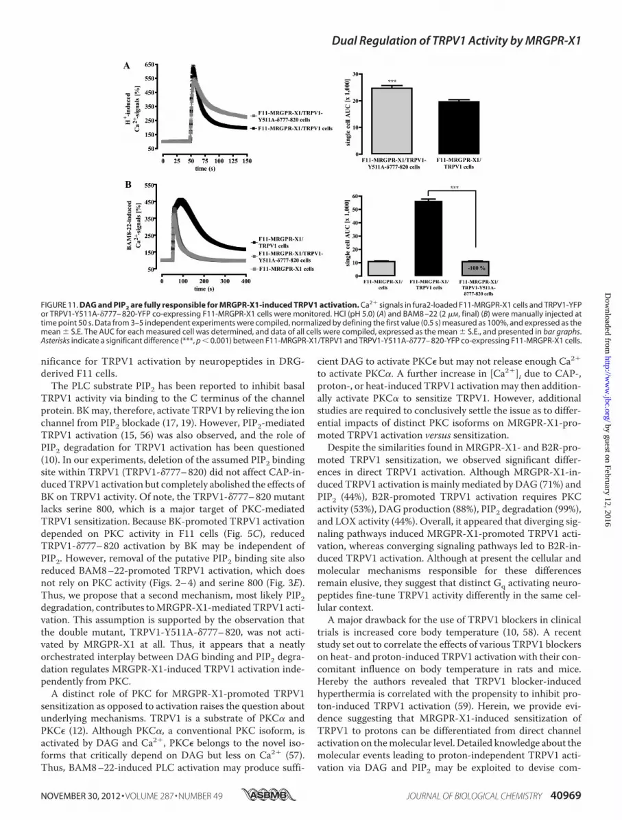
To definitively determine the impact of both DAG and PIP2on MRGPR-X1-mediated TRPV1 activation, we generated adouble mutant that lacks the DAG binding site as well as thereported PIP2 sensor (TRPV1-Y511A-�777–820-YFP). Thisdoublemutant was still activated by protons (Fig. 11A), indicat-ing that these modifications allowed proper folding and mem-brane translocation of a functional active channel protein. Ofnote, BAM8–22 activation was completely abolished (Fig.11B), providing further evidence that both DAG productionand PIP2 degradationmediate BAM8–22-inducedTRPV1 acti-vation in F11-MRGPR-X1 cells.
TABLE 1SRC, PI3K, PKD, or ERK-1/2 activity is not required for MRGPR-X1-promoted TRPV1 activationBAM8–22-induced Ca2� signals in fura2-loaded F11-MRGPR-X1 and F11-MRGPR-X1/TRPV1 cells were measured and quantified by calculating the AUC (�1000) foreach measured cell. Data from 3–5 independent experiments were compiled and expressed as the mean � S.E. To block the activity of various kinases, cells werepreincubated for 30 min with PP-2 (10 �M, SRC), LY-294002 (10 �M, PI3K), CID-755673 (1 �M, PKD), PD-184352 (10 �M, ERK-1/2), or the corresponding carrier controlDMSO (0.1%). No significant difference between DMSO- and inhibitor-treated F11-MRGPR-X1 or F11-MRGPR-X1/TRPV1 cells were observed.
Cells DMSO PP-2 LY-294002 CID-755673 PD-184352
F11-MRGPR-X1 11.7 � 0.9 15.9 � 1.4 14.0 � 1.8 10.7 � 1.0 8.1 � 0.6F11-MRGPR-X1/TRPV1 47.0 � 2.7a 55.2 � 5.6a 56.2 � 8.1a 56.0 � 7.5a 40.8 � 4.1a
a p � 0.001, significant differences between F11-MRGPR-X1 and F11-MRGPR-X1/TRPV1 cells.
FIGURE 7. MRGPR-X1-induced TRPV1 activation is independent from receptor-mediated calcium release. Ca2� signals in fura2-loaded F11-MRGPR-X1 orF11-MRGPR-X1/TRPV1 cells preincubated with U-73122 (10 �M, 30 min) (A) and with thapsigargin (1 �M, 30 min) (B) were monitored. BAM8 –22 (2 �M, final) wasmanually injected at time point 50 s. Data from 3–5 independent experiments were compiled, normalized by defining the first value (0.5 s) measured as 100%,and expressed as the mean � S.E. The AUC for each measured cell was determined, and data of all cells were compiled, expressed as the mean � S.E., andpresented in bar graphs. Asterisks indicate a significant difference (***, p � 0.001) between F11-MRGPR-X1/TRPV1, and F11-MRGPR-X1 cells.
Dual Regulation of TRPV1 Activity by MRGPR-X1
NOVEMBER 30, 2012 • VOLUME 287 • NUMBER 49 JOURNAL OF BIOLOGICAL CHEMISTRY 40965
by guest on February 12, 2016http://w
ww
.jbc.org/D
ownloaded from

DISCUSSIONDue to the exclusive expression in DRG neurons, high
affinity binding to pro-enkephalin cleavage products andtheir role for nociception in humans, MRGPR-X1 are con-sidered as promising novel targets for pain therapy. Of note,MRGPR-X1 are primate-specific receptors (1, 2, 5, 49). Ingeneral, primate-specific genes are predominantly expressedin cells of the reproductive system or in neurons (50), anddisease-causing genes are strongly enriched among primate-specific genes (51), highlighting their significance as thera-peutic targets.However, primate-specific expression is also a major imped-
iment to functional analysis because of lack of suitable in vivomodels and endogenous expression systems. Regarding theMRGPR-X1 subtype, one rodent receptor activated byBAM8–22 was identified in mice and rats (MRGPR-C) (1, 2).However, apart from common high affinity binding to BAM8–22, rodent MRGPR-C and human MRGPR-X1 exhibit funda-mental differences in their affinity to other endogenous pep-
tides, as previously reported (4, 52, 53). Furthermore, incontrast to rodent MRGPR-C, human MRGPR-X1 resist �-ar-restin-dependent, agonist-promoted endocytosis (4), not onlyaffecting signal duration but also directing GPCR-promotedsignaling toward distinct signaling pathways (54). Therefore,we concur with other investigators in that the function ofhuman MRGPR-X1 may not be accurately extrapolated fromstudies with rodent MRGPR-C (5, 7).Herein, we used a recently established F11 cell line (rat DRG
neurons x murine neuroblastoma cells) that stably expressesMRGPR-X1 (4) and analyzed their functional interactions withTRPV1. F11 cells emerged as a useful tool for the analysis ofDRG-related signaling pathways, and several studies pointedout striking similarities between F11 cells and culturedDRGneu-rons (23–35). In this vein,molecularmechanismsunderlyingB2R-dependent signaling to TRPV1 in DRG neurons, i.e. sensitizationby PKC or activation by PIP2 degradation, were also observed inF11-MRGPR-X1 cells. Because Ca2� transients initiated byendogenously expressed B2R and recombinantMRGPR-X1 were
FIGURE 8. DAG/OAG-induced TRPV1 activation in F11 cells. Ca2� signals in fura2-loaded F11-MRGPR-X1 or F11-MRGPR-X1/TRPV1 cells were monitored.DAG kinase (DAGK) inhibitor-2 (10 �M, final) (A) and OAG (100 �M, final) (B and C) were manually injected at time point 50 s. Cells were preincubated with BIM-X(1 �M, 30 min) (A and C) and a separate pool of cells with the carrier DMSO (0.1%, 30 min) (C). Data from 3–5 independent experiments were compiled,normalized by defining the first value (0.5 s) measured as 100%, and expressed as the mean � S.E. The AUC for each measured cell was determined, and dataof all cells were compiled, expressed as the mean � S.E., and presented in bar graphs. Asterisks indicate a significant difference (***, p � 0.001) betweenF11-MRGPR-X1/TRPV1 and F11-MRGPR-X1 cells; hash signs indicate a significant difference (###, p � 0.001) between BIM-X- and DMSO-treated F11-MRGPR-X1/TRPV1 cells.
Dual Regulation of TRPV1 Activity by MRGPR-X1
40966 JOURNAL OF BIOLOGICAL CHEMISTRY VOLUME 287 • NUMBER 49 • NOVEMBER 30, 2012
by guest on February 12, 2016http://w
ww
.jbc.org/D
ownloaded from

comparable,we consider F11-MRGPR-X1cells as a reliablemodelsystem to investigate functional interactions between TRPV1 andhumanMRGPR-X1.Using an established PKC inhibitor and two TRPV1mutants
lacking serine residues required for PKC-mediatedTRPV1 sen-
sitization, we show that MRGPR-X1 sensitize the TRPV1 in aPKC-dependent manner. Thus, we assign the humanMRGPR-X1 to the large group of Gq-coupled receptors thatsensitize nociceptors to the “inflammatory soup” via PKC-de-pendent mechanisms (11, 21, 22, 36–39, 42).
FIGURE 9. MRGPR-X1-induced TRPV1 activation requires the DAG binding site of the TRPV1. Ca2� signals in fura2-loaded F11-MRGPR-X1 cells andTRPV1-YFP or TRPV1-Y511A-YFP co-expressing F11-MRGPR-X1 cells were monitored. CAP (10 nM, final) (A), OAG (100 �M, final) (B), HCl (pH 5.0) (C), BAM8 –22 (2�M, final) (D), and BK (100 nM, final) (E) were manually injected at time point 50 s. In B cells were preincubated with BIM-X (1 �M, 30 min) to avoid the effects ofOAG on TRPV1 activity due to PKC-dependent TRPV1 phosphorylation (see Fig. 8C). Data from 3–5 independent experiments were compiled, normalized bydefining the first value (0.5 s) measured as 100%, and expressed as the mean � S.E. The AUC for each measured cell was determined, and data of all cells werecompiled, expressed as the mean � S.E., and presented in bar graphs. Asterisks (***, p � 0.001) indicate a significant difference between TRPV1-Y511A-YFPco-expressing F11-MRGPR-X1 and F11-MRGPR-X1/TRPV1 cells; hash signs (###, p � 0.001) indicate a significant difference between F11-MRGPR-X1 andTRPV1-Y511A-YFP co-expressing F11-MRGPR-X1 cells.
Dual Regulation of TRPV1 Activity by MRGPR-X1
NOVEMBER 30, 2012 • VOLUME 287 • NUMBER 49 JOURNAL OF BIOLOGICAL CHEMISTRY 40967
by guest on February 12, 2016http://w
ww
.jbc.org/D
ownloaded from

Although TRPV1 sensitization to inflammatory stimuli suchas heat and protons via Gq-induced PKC activation is widelyrecognized, little is known about direct TRPV1 activation as aconsequence of Gq-dependent signaling independent ofincreased temperature and proton concentrations. So far,direct TRPV1 activation has been proposed for muscarinic-3receptors (18) andmetabotropic glutamate-5 receptors (55) viaDAG production and for the B2R via PIP2 degradation (17) orLOX activation (45). Herein, we noted that TRPV1 co-expres-sion in F11-MRGPR-X1 cells profoundly enhanced MRGPR-X1-induced Ca2� signals, Mn2� influx, and cation currents atRT and constant pH, indicative of direct TRPV1 activation.Although PKC activity plays a dominant role in TRPV1 sensi-tization, it does not at all contribute to MRGPR-X1-inducedTRPV1 activation. Because TRPV1 activation by MRGPR-X1was independent fromBAM8–22-inducedCa2� release (Fig. 7)and resistant to chemical inhibition of PI3K, ERK-1/2, PKD,SRC, or LOX (Table 1 and Fig. 6), we postulate a significant rolefor PIP2 and DAG in this process.DAG-promoted channel activation has originally been
described for members of the transient receptor potential cat-ion channel C family but not for TRPV1 when expressed in
Chinese hamster ovary cells (46). However, in rat DRGneuronsOAG-induced calcium signals have been ascribed to increasedTRPV1 activity (18, 55), indicating differential TRPV1 sensitiv-ity to DAG in distinct cell lines. In DRG-derived F11 cells, weobserved that accumulation of endogenous DAG and applica-tion of exogenous OAG induced Ca2� signals solely in F11-MRGPR-X1/TRPV1, but not in F11-MRGPR-X1 cells, indicat-ing that DAG activates TRPV1 in F11 cells. It has been reportedthat tyrosine 511 of TRPV1 is required for OAG-inducedTRPV1 activation (18). In accord with the latter study, OAG-induced Ca2� signals were almost completely abolished inTRPV1-Y511A expressing F11 cells. Because Ca2� signalsinduced by high proton concentrations were unaltered in wild-type and TRPV1-Y511A expressing cells, we took advantage ofthismutant to dissect the role of DAG inGq-dependent TRPV1activation. We observed that removal of the DAG bindingsite reduced MRGPR-X1-mediated TRPV1 activation byabout 70%. Thus, we propose that DAG production is amajor cellular event entailing MRGPR-X1-induced directTRPV1 activation. Because DAG production also had amajor impact on B2R-initiated TRPV1 activation, we assumethat receptor induced DAG accumulation is of general sig-
FIGURE 10. MRGPR-X1-induced TRPV1 activation requires the PIP2 binding site of the TRPV1. Ca2� signals in fura2-loaded F11-MRGPR-X1 cells andTRPV1-YFP or TRPV1-�777– 820-YFP co-expressing F11-MRGPR-X1 cells were monitored. CAP (10 nM, final) (A), BAM8 –22 (2 �M, final) (B), and BK (100 nM, final)(C) were manually injected at time point 50 s. Data from 3–5 independent experiments were compiled, normalized by defining the first value (0.5 s) measuredas 100%, and expressed as the mean � S.E. The AUC for each measured cell was determined, data of all cells compiled, expressed as the mean � S.E. andpresented in bar graphs. Asterisks (***, p � 0.001) indicate a significant difference between TRPV1-�777– 820-YFP co-expressing F11-MRGPR-X1 and F11-MRGPR-X1/TRPV1 cells; hash signs (###, p � 0.001) between F11-MRGPR-X1 and TRPV1-�777– 820-YFP co-expressing F11-MRGPR-X1 cells.
Dual Regulation of TRPV1 Activity by MRGPR-X1
40968 JOURNAL OF BIOLOGICAL CHEMISTRY VOLUME 287 • NUMBER 49 • NOVEMBER 30, 2012
by guest on February 12, 2016http://w
ww
.jbc.org/D
ownloaded from
nificance for TRPV1 activation by neuropeptides in DRG-derived F11 cells.The PLC substrate PIP2 has been reported to inhibit basal
TRPV1 activity via binding to the C terminus of the channelprotein. BKmay, therefore, activate TRPV1 by relieving the ionchannel from PIP2 blockade (17, 19). However, PIP2-mediatedTRPV1 activation (15, 56) was also observed, and the role ofPIP2 degradation for TRPV1 activation has been questioned(10). In our experiments, deletion of the assumed PIP2 bindingsite within TRPV1 (TRPV1-�777–820) did not affect CAP-in-ducedTRPV1 activation but completely abolished the effects ofBK on TRPV1 activity. Of note, the TRPV1-�777–820 mutantlacks serine 800, which is a major target of PKC-mediatedTRPV1 sensitization. Because BK-promoted TRPV1 activationdepended on PKC activity in F11 cells (Fig. 5C), reducedTRPV1-�777–820 activation by BK may be independent ofPIP2. However, removal of the putative PIP2 binding site alsoreduced BAM8–22-promoted TRPV1 activation, which doesnot rely on PKC activity (Figs. 2–4) and serine 800 (Fig. 3E).Thus, we propose that a second mechanism, most likely PIP2degradation, contributes toMRGPR-X1-mediatedTRPV1 acti-vation. This assumption is supported by the observation thatthe double mutant, TRPV1-Y511A-�777–820, was not acti-vated by MRGPR-X1 at all. Thus, it appears that a neatlyorchestrated interplay between DAG binding and PIP2 degra-dation regulates MRGPR-X1-induced TRPV1 activation inde-pendently from PKC.A distinct role of PKC for MRGPR-X1-promoted TRPV1
sensitization as opposed to activation raises the question aboutunderlying mechanisms. TRPV1 is a substrate of PKC� andPKC� (12). Although PKC�, a conventional PKC isoform, isactivated by DAG and Ca2�, PKC� belongs to the novel iso-forms that critically depend on DAG but less on Ca2� (57).Thus, BAM8–22-induced PLC activation may produce suffi-
cient DAG to activate PKC� but may not release enough Ca2�
to activate PKC�. A further increase in [Ca2�]i due to CAP-,proton-, or heat-induced TRPV1 activationmay then addition-ally activate PKC� to sensitize TRPV1. However, additionalstudies are required to conclusively settle the issue as to differ-ential impacts of distinct PKC isoforms on MRGPR-X1-pro-moted TRPV1 activation versus sensitization.Despite the similarities found in MRGPR-X1- and B2R-pro-
moted TRPV1 sensitization, we observed significant differ-ences in direct TRPV1 activation. Although MRGPR-X1-in-duced TRPV1 activation is mainly mediated by DAG (71%) andPIP2 (44%), B2R-promoted TRPV1 activation requires PKCactivity (53%), DAG production (88%), PIP2 degradation (99%),and LOX activity (44%). Overall, it appeared that diverging sig-naling pathways induced MRGPR-X1-promoted TRPV1 acti-vation, whereas converging signaling pathways led to B2R-in-duced TRPV1 activation. Although at present the cellular andmolecular mechanisms responsible for these differencesremain elusive, they suggest that distinct Gq activating neuro-peptides fine-tune TRPV1 activity differently in the same cel-lular context.A major drawback for the use of TRPV1 blockers in clinical
trials is increased core body temperature (10, 58). A recentstudy set out to correlate the effects of various TRPV1 blockerson heat- and proton-induced TRPV1 activation with their con-comitant influence on body temperature in rats and mice.Hereby the authors revealed that TRPV1 blocker-inducedhyperthermia is correlated with the propensity to inhibit pro-ton-induced TRPV1 activation (59). Herein, we provide evi-dence suggesting that MRGPR-X1-induced sensitization ofTRPV1 to protons can be differentiated from direct channelactivation on themolecular level. Detailed knowledge about themolecular events leading to proton-independent TRPV1 acti-vation via DAG and PIP2 may be exploited to devise com-
FIGURE 11. DAG and PIP2 are fully responsible for MRGPR-X1-induced TRPV1 activation. Ca2� signals in fura2-loaded F11-MRGPR-X1 cells and TRPV1-YFPor TRPV1-Y511A-�777– 820-YFP co-expressing F11-MRGPR-X1 cells were monitored. HCl (pH 5.0) (A) and BAM8 –22 (2 �M, final) (B) were manually injected attime point 50 s. Data from 3–5 independent experiments were compiled, normalized by defining the first value (0.5 s) measured as 100%, and expressed as themean � S.E. The AUC for each measured cell was determined, and data of all cells were compiled, expressed as the mean � S.E., and presented in bar graphs.Asterisks indicate a significant difference (***, p � 0.001) between F11-MRGPR-X1/TRPV1 and TRPV1-Y511A-�777– 820-YFP co-expressing F11-MRGPR-X1 cells.
Dual Regulation of TRPV1 Activity by MRGPR-X1
NOVEMBER 30, 2012 • VOLUME 287 • NUMBER 49 JOURNAL OF BIOLOGICAL CHEMISTRY 40969
by guest on February 12, 2016http://w
ww
.jbc.org/D
ownloaded from

pounds specifically targeting TRPV1-related pain withoutaffecting body temperature.In conclusion, we provide a thorough analysis of the func-
tional interactions between MRGPR-X1 and TRPV1. Wedefine a dual modulation of TRPV1 activity by MRGPR-X1leading to PKC-dependent TRPV1 sensitization and DAG/PIP2-mediated direct activation. These findings grant deeperinsight into the molecular events underlying TRPV1-relatedpain syndromes in humans and further highlight the signifi-cance of MRGPR-X1 as putative therapeutic targets.
Acknowledgments—We thank StephanieHunger andRobinMeier forexcellent technical assistance.
REFERENCES1. Lembo, P. M., Grazzini, E., Groblewski, T., O’Donnell, D., Roy, M. O.,
Zhang, J., Hoffert, C., Cao, J., Schmidt, R., Pelletier, M., Labarre, M., Gos-selin, M., Fortin, Y., Banville, D., Shen, S. H., Ström, P., Payza, K., Dray, A.,Walker, P., and Ahmad, S. (2002) Proenkephalin A gene products activatea new family of sensory neuron, specificGPCRs.Nat. Neurosci. 5, 201–209
2. Dong, X., Han, S., Zylka, M. J., Simon, M. I., and Anderson, D. J. (2001) Adiverse family of GPCRs expressed in specific subsets of nociceptive sen-sory neurons. Cell 106, 619–632
3. Breit, A., Gagnidze, K., Devi, L. A., Lagacé, M., and Bouvier, M. (2006)Simultaneous activation of the � opioid receptor (�OR)/sensory neuron-specific receptor-4 (SNSR-4) hetero-oligomer by the mixed bivalent ago-nist bovine adrenal medulla peptide 22 activates SNSR-4 but inhibits �ORsignaling.Mol. Pharmacol. 70, 686–696
4. Solinski, H. J., Boekhoff, I., Bouvier, M., Gudermann, T., and Breit, A.(2010) Sensory neuron-specific MAS-related gene-X1 receptors resist ag-onist-promoted endocytosis.Mol. Pharmacol. 78, 249–259
5. Burstein, E. S., Ott, T. R., Feddock, M., Ma, J. N., Fuhs, S., Wong, S.,Schiffer, H. H., Brann, M. R., and Nash, N. R. (2006) Characterization ofthe Mas-related gene family. Structural and functional conservation ofhuman and rhesus MrgX receptors. Br. J. Pharmacol. 147, 73–82
6. Sikand, P., Dong, X., and LaMotte, R. H. (2011) BAM8–22 peptide pro-duces itch and nociceptive sensations in humans independent of hista-mine release. J. Neurosci. 31, 7563–7567
7. Chen, H., and Ikeda, S. R. (2004)Modulation of ion channels and synaptictransmission by a human sensory neuron-specific G-protein-coupled re-ceptor, SNSR4/mrgX1, heterologously expressed in cultured rat neurons.J. Neurosci. 24, 5044–5053
8. Davis, J. B., Gray, J., Gunthorpe,M. J., Hatcher, J. P., Davey, P. T., Overend,P., Harries, M. H., Latcham, J., Clapham, C., Atkinson, K., Hughes, S. A.,Rance, K., Grau, E., Harper, A. J., Pugh, P. L., Rogers, D. C., Bingham, S.,Randall, A., and Sheardown, S. A. (2000) Vanilloid receptor-1 is essentialfor inflammatory thermal hyperalgesia. Nature 405, 183–187
9. Walker, K. M., Urban, L., Medhurst, S. J., Patel, S., Panesar, M., Fox, A. J.,and McIntyre, P. (2003) The VR1 antagonist capsazepine reverses me-chanical hyperalgesia in models of inflammatory and neuropathic pain.J. Pharmacol. Exp. Ther. 304, 56–62
10. Vay, L., Gu, C., andMcNaughton, P. A. (2012) The thermo-TRP ion chan-nel family. Properties and therapeutic implications. Br. J. Pharmacol. 165,787–801
11. Tominaga, M., Wada, M., and Masu, M. (2001) Potentiation of capsaicinreceptor activity bymetabotropic ATP receptors as a possible mechanismfor ATP-evoked pain and hyperalgesia. Proc. Natl. Acad. Sci. U.S.A. 98,6951–6956
12. Amadesi, S., Cottrell, G. S., Divino, L., Chapman, K., Grady, E. F., Bautista,F., Karanjia, R., Barajas-Lopez, C., Vanner, S., Vergnolle, N., and Bunnett,N. W. (2006) Protease-activated receptor 2 sensitizes TRPV1 by proteinkinase C�- and A-dependentmechanisms in rats andmice. J. Physiol. 575,555–571
13. Huang, J., Zhang, X., and McNaughton, P. A. (2006) Inflammatory pain.The cellular basis of heat hyperalgesia.Curr. Neuropharmacol. 4, 197–206
14. Mandadi, S., Numazaki, M., Tominaga, M., Bhat, M. B., Armati, P. J., andRoufogalis, B. D. (2004) Activation of protein kinase C reverses capsaicin-induced calcium-dependent desensitization of TRPV1 ion channels. CellCalcium 35, 471–478
15. Stein, A. T., Ufret-Vincenty, C.A.,Hua, L., Santana, L. F., andGordon, S. E.(2006) Phosphoinositide 3-kinase binds to TRPV1 and mediates NGF-stimulated TRPV1 trafficking to the plasma membrane. J. Gen. Physiol.128, 509–522
16. Bhave, G., Hu, H. J., Glauner, K. S., Zhu, W., Wang, H., Brasier, D. J.,Oxford,G. S., andGereau, R.W., 4th (2003) Protein kinaseCphosphoryla-tion sensitizes but does not activate the capsaicin receptor transient re-ceptor potential vanilloid 1 (TRPV1). Proc. Natl. Acad. Sci. U.S.A. 100,12480–12485
17. Chuang, H. H., Prescott, E. D., Kong, H., Shields, S., Jordt, S. E., Basbaum,A. I., Chao,M.V., and Julius, D. (2001) Bradykinin and nerve growth factorrelease the capsaicin receptor from PtdIns(4,5)P2-mediated inhibition.Nature 411, 957–962
18. Woo, D. H., Jung, S. J., Zhu, M. H., Park, C. K., Kim, Y. H., Oh, S. B., andLee, C. J. (2008) Direct activation of transient receptor potential vanilloid1(TRPV1) by diacylglycerol (DAG).Mol. Pain 4, 42
19. Prescott, E. D., and Julius, D. (2003) A modular PIP2 binding site as adeterminant of capsaicin receptor sensitivity. Science 300, 1284–1288
20. Hellwig, N., Albrecht, N., Harteneck, C., Schultz, G., and Schaefer, M.(2005) Homo- and heteromeric assembly of TRPV channel subunits.J. Cell Sci. 118, 917–928
21. Kleibeuker, W., Ledeboer, A., Eijkelkamp, N., Watkins, L. R., Maier, S. F.,Zijlstra, J., Heijnen, C. J., and Kavelaars, A. (2007) A role for G protein-coupled receptor kinase 2 in mechanical allodynia. Eur. J. Neurosci. 25,1696–1704
22. Numazaki, M., Tominaga, T., Toyooka, H., and Tominaga, M. (2002) Di-rect phosphorylation of capsaicin receptor VR1 by protein kinase C� andidentification of two target serine residues. J. Biol. Chem. 277,13375–13378
23. Fan, S. F., Shen, K. F., and Crain, S. M. (1993) �- and �-opioid agonists atlow concentrations decrease voltage-dependent K� currents in F11 neu-roblastoma x DRG neuron hybrid cells via cholera toxin-sensitive recep-tors. Brain Res. 605, 214–220
24. Fan, S. F., Shen, K. F., Scheideler, M. A., and Crain, S. M. (1992) F11neuroblastoma x DRG neuron hybrid cells express inhibitory �- and�-opioid receptors which increase voltage-dependent K� currents uponactivation. Brain Res. 590, 329–333
25. Jow, F., He, L., Kramer, A., Hinson, J., Bowlby,M. R., Dunlop, J., andWang,K. (2006) Validation of DRG-like F11 cells for evaluation of KCNQ/M-channel modulators. Assay Drug Dev. Technol. 4, 49–56
26. Francel, P. C., Harris, K., Smith, M., Fishman, M. C., Dawson, G., andMiller, R. J. (1987) Neurochemical characteristics of a novel dorsal rootganglion X neuroblastoma hybrid cell line, F-11. J. Neurochem. 48,1624–1631
27. Jung, H., and Miller, R. J. (2008) Activation of the nuclear factor of acti-vated T-cells (NFAT) mediates up-regulation of CCR2 chemokine recep-tors in dorsal root ganglion (DRG) neurons. A possible mechanism foractivity-dependent transcription inDRGneurons in associationwith neu-ropathic pain.Mol. Cell. Neurosci. 37, 170–177
28. Puttfarcken, P. S., Manelli, A. M., Arneric, S. P., and Donnelly-Roberts,D. L. (1997) Evidence for nicotinic receptors potentially modulating noci-ceptive transmission at the level of the primary sensory neuron. Studieswith F11 cells. J. Neurochem. 69, 930–938
29. McIlvain, H. B., Baudy, A., Sullivan, K., Liu, D., Pong, K., Fennell, M., andDunlop, J. (2006) Pituitary adenylate cyclase-activating peptide (PACAP)induces differentiation in the neuronal F11 cell line through a PKA-de-pendent pathway. Brain Res. 1077, 16–23
30. Boland, L. M., Allen, A. C., and Dingledine, R. (1991) Inhibition by brady-kinin of voltage-activated barium current in a rat dorsal root ganglion cellline. Role of protein kinase C. J. Neurosci. 11, 1140–1149
31. Rothe, K., Solinski, H. J., Boekhoff, I., Gudermann, T., and Breit, A. (2012)Morphine activates the E twenty six-like transcription factor-1/serum re-sponse factor pathway via extracellular signal-regulated kinases 1/2 in F11cells derived from dorsal root ganglia neurons. J. Pharmacol. Exp. Ther.
Dual Regulation of TRPV1 Activity by MRGPR-X1
40970 JOURNAL OF BIOLOGICAL CHEMISTRY VOLUME 287 • NUMBER 49 • NOVEMBER 30, 2012
by guest on February 12, 2016http://w
ww
.jbc.org/D
ownloaded from

342, 41–5232. Li, Y., Ji, A., Weihe, E., and Schäfer, M. K. (2004) Cell-specific expression
and lipopolysaccharide-induced regulation of tumor necrosis factor �(TNF�) and TNF receptors in rat dorsal root ganglion. J. Neurosci. 24,9623–9631
33. Gaudioso, C., Hao, J., Martin-Eauclaire,M. F., Gabriac,M., andDelmas, P.(2012) Menthol pain relief through cumulative inactivation of voltage-gated sodium channels. Pain 153, 473–484
34. Eijkelkamp,N., Heijnen, C. J.,Willemen,H. L., Deumens, R., Joosten, E. A.,Kleibeuker,W., denHartog, I. J., van Velthoven, C. T., Nijboer, C., Nassar,M. A., Dorn, G. W., 2nd, Wood, J. N., and Kavelaars, A. (2010) GRK2. Anovel cell-specific regulator of severity and duration of inflammatory pain.J. Neurosci. 30, 2138–2149
35. Fioravanti, B., De Felice, M., Stucky, C. L., Medler, K. A., Luo, M. C.,Gardell, L. R., Ibrahim, M., Malan, T. P., Jr., Yamamura, H. I., Ossipov,M. H., King, T., Lai, J., Porreca, F., and Vanderah, T. W. (2008) Constitu-tive activity at the cannabinoid CB1 receptor is required for behavioralresponse to noxious chemical stimulation of TRPV1. Antinociceptive ac-tions of CB1 inverse agonists. J. Neurosci. 28, 11593–11602
36. Szallasi, A., Cortright, D.N., Blum,C.A., andEid, S. R. (2007)The vanilloidreceptor TRPV1. 10 years from channel cloning to antagonist proof-of-concept. Nat. Rev. Drug Discov. 6, 357–372
37. Mizumura, K., Sugiura, T., Katanosaka, K., Banik, R. K., and Kozaki, Y.(2009) Excitation and sensitization of nociceptors by bradykinin.What dowe know? Exp. Brain Res. 196, 53–65
38. Cesare, P., Dekker, L. V., Sardini, A., Parker, P. J., and McNaughton, P. A.(1999) Specific involvement of PKC-� in sensitization of the neuronalresponse to painful heat. Neuron 23, 617–624
39. Moriyama, T., Iida, T., Kobayashi, K., Higashi, T., Fukuoka, T., Tsumura,H., Leon, C., Suzuki, N., Inoue, K., Gachet, C., Noguchi, K., and Tominaga,M. (2003) Possible involvement of P2Y2 metabotropic receptors in ATP-induced transient receptor potential vanilloid receptor 1-mediated ther-mal hypersensitivity. J. Neurosci. 23, 6058–6062
40. Vellani, V., Mapplebeck, S., Moriondo, A., Davis, J. B., and McNaughton,P. A. (2001) Protein kinase C activation potentiates gating of the vanilloidreceptor VR1 by capsaicin, protons, heat, and anandamide. J. Physiol. 534,813–825
41. Valenzano, K. J., Grant, E. R., Wu, G., Hachicha, M., Schmid, L., Tafesse,L., Sun, Q., Rotshteyn, Y., Francis, J., Limberis, J., Malik, S., Whittemore,E. R., and Hodges, D. (2003) N-(4-Tertiarybutylphenyl)-4-(3-chloropyri-din-2-yl)tetrahydropyrazine -1(2H)-carbox-amide (BCTC), a novel, orallyeffective vanilloid receptor 1 antagonist with analgesic properties. I. Invitro characterization and pharmacokinetic properties. J. Pharmacol. Exp.Ther. 306, 377–386
42. Plant, T.D., Zöllner, C., Kepura, F.,Mousa, S. S., Eichhorst, J., Schaefer,M.,Furkert, J., Stein, C., andOksche, A. (2007) Endothelin potentiates TRPV1via ETA receptor-mediated activation of protein kinase C.Mol. Pain 3, 35
43. Turner, H., Fleig, A., Stokes, A., Kinet, J. P., and Penner, R. (2003) Discrim-ination of intracellular calcium store subcompartments using TRPV1(transient receptor potential channel, vanilloid subfamily member 1) re-lease channel activity. Biochem. J. 371, 341–350
44. Zygmunt, P. M., Petersson, J., Andersson, D. A., Chuang, H., Sørgård, M.,DiMarzo, V., Julius, D., andHögestätt, E. D. (1999) Vanilloid receptors onsensory nervesmediate the vasodilator action of anandamide.Nature 400,452–457
45. Shin, J., Cho, H., Hwang, S. W., Jung, J., Shin, C. Y., Lee, S. Y., Kim, S. H.,
Lee, M. G., Choi, Y. H., Kim, J., Haber, N. A., Reichling, D. B., Khasar, S.,Levine, J. D., and Oh, U. (2002) Bradykinin-12-lipoxygenase-VR1 signal-ing pathway for inflammatory hyperalgesia. Proc. Natl. Acad. Sci. U.S.A.99, 10150–10155
46. Hofmann, T., Obukhov, A. G., Schaefer, M., Harteneck, C., Gudermann,T., and Schultz, G. (1999) Direct activation of human TRPC6 and TRPC3channels by diacylglycerol. Nature 397, 259–263
47. Ryu, S., Liu, B., Yao, J., Fu, Q., and Qin, F. (2007) Uncoupling protonactivation of vanilloid receptor TRPV1. J. Neurosci. 27, 12797–12807
48. Gavva, N. R., Tamir, R., Klionsky, L., Norman, M. H., Louis, J. C., Wild,K. D., and Treanor, J. J. (2005) Proton activation does not alter antagonistinteraction with the capsaicin-binding pocket of TRPV1.Mol. Pharmacol.68, 1524–1533
49. Choi, S. S., and Lahn, B. T. (2003) Adaptive evolution of MRG, a neuron-specific gene family implicated in nociception. Genome Res. 13,2252–2259
50. Tay, S. K., Blythe, J., and Lipovich, L. (2009) Global discovery of primate-specific genes in the human genome. Proc. Natl. Acad. Sci. U.S.A. 106,12019–12024
51. Hao, L., Ge, X., Wan, H., Hu, S., Lercher, M. J., Yu, J., and Chen, W. H.(2010) Human functional genetic studies are biased against the medicallymost relevant primate-specific genes. BMC Evol. Biol. 10, 316
52. Grazzini, E., Puma, C., Roy, M. O., Yu, X. H., O’Donnell, D., Schmidt, R.,Dautrey, S., Ducharme, J., Perkins, M., Panetta, R., Laird, J. M., Ahmad, S.,and Lembo, P.M. (2004) Sensory neuron-specific receptor activation elic-its central and peripheral nociceptive effects in rats. Proc. Natl. Acad. Sci.U.S.A. 101, 7175–7180
53. Han, S. K., Dong, X., Hwang, J. I., Zylka, M. J., Anderson, D. J., and Simon,M. I. (2002)OrphanG protein-coupled receptorsMrgA1 andMrgC11 aredistinctively activated by RF-amide-related peptides through the G�q/11
pathway. Proc. Natl. Acad. Sci. U.S.A. 99, 14740–1474554. Daaka, Y., Luttrell, L.M., Ahn, S., Della Rocca, G. J., Ferguson, S. S., Caron,
M. G., and Lefkowitz, R. J. (1998) Essential role for G protein-coupledreceptor endocytosis in the activation of mitogen-activated protein ki-nase. J. Biol. Chem. 273, 685–688
55. Kim, Y. H., Park, C. K., Back, S. K., Lee, C. J., Hwang, S. J., Bae, Y. C., Na,H. S., Kim, J. S., Jung, S. J., and Oh, S. B. (2009) Membrane-delimitedcoupling of TRPV1 and mGluR5 on presynaptic terminals of nociceptiveneurons. J. Neurosci. 29, 10000–10009
56. Sowa, N. A., Street, S. E., Vihko, P., and Zylka, M. J. (2010) Prostatic acidphosphatase reduces thermal sensitivity and chronic pain sensitization bydepleting phosphatidylinositol 4,5-bisphosphate. J. Neurosci. 30,10282–10293
57. Poole, A. W., Pula, G., Hers, I., Crosby, D., and Jones, M. L. (2004) PKC-interacting proteins. From function to pharmacology. Trends Pharmacol.Sci. 25, 528–535
58. Gavva, N. R., Bannon, A. W., Surapaneni, S., Hovland, D. N., Jr., Lehto,S. G., Gore, A., Juan, T., Deng, H., Han, B., Klionsky, L., Kuang, R., Le, A.,Tamir, R., Wang, J., Youngblood, B., Zhu, D., Norman, M. H., Magal, E.,Treanor, J. J., and Louis, J. C. (2007) The vanilloid receptor TRPV1 istonically activated in vivo and involved in body temperature regulation.J. Neurosci. 27, 3366–3374
59. Garami, A., Shimansky, Y. P., Pakai, E., Oliveira, D. L., Gavva, N. R., andRomanovsky, A. A. (2010) Contributions of different modes of TRPV1activation to TRPV1 antagonist-induced hyperthermia. J. Neurosci. 30,1435–1440
Dual Regulation of TRPV1 Activity by MRGPR-X1
NOVEMBER 30, 2012 • VOLUME 287 • NUMBER 49 JOURNAL OF BIOLOGICAL CHEMISTRY 40971
by guest on February 12, 2016http://w
ww
.jbc.org/D
ownloaded from

Hans Jürgen Solinski, Susanna Zierler, Thomas Gudermann and Andreas Breitvia Distinct Signaling Pathways
Sensitize and Directly Activate Transient Receptor Potential Cation Channel V1 Human Sensory Neuron-specific Mas-related G Protein-coupled Receptors-X1
doi: 10.1074/jbc.M112.408617 originally published online October 16, 20122012, 287:40956-40971.J. Biol. Chem.
10.1074/jbc.M112.408617Access the most updated version of this article at doi:
Alerts:
When a correction for this article is posted•
When this article is cited•
to choose from all of JBC's e-mail alertsClick here
http://www.jbc.org/content/287/49/40956.full.html#ref-list-1
This article cites 59 references, 30 of which can be accessed free at
by guest on February 12, 2016http://w
ww
.jbc.org/D
ownloaded from
Related Documents