Human rhinovirus infection during naturally occurring COPD exacerbations Sı ˆobha ´n Nicole George, Davinder S. Garcha, Alexander J. Mackay, Anant R.C. Patel, Richa Singh, Raymond J. Sapsford, Gavin C. Donaldson and Jadwiga A. Wedzicha Affiliations: Centre for Respiratory Medicine, University College London, London, UK. Correspondence: S.N. George, University College London, Rowland Hill Street, Hampstead, London, NW3 2PF, UK. E-mail: [email protected] ABSTRACT Human rhinovirus (HRV) infection is an important trigger of exacerbations of chronic obstructive pulmonary disease (COPD) but its role in determining exacerbation frequency phenotype or the time-course of HRV infection in naturally occurring exacerbations is unknown. Sputum samples from 77 patients were analysed by real-time quantitative PCR for both HRV (388 samples), and Streptococcus pneumoniae, Haemophilus influenzae and Moraxella catarrhalis (89 samples). Patients recorded worsening of respiratory symptoms on daily diary cards, from which exacerbations were identified. HRV prevalence and load at exacerbation presentation were significantly higher than in the stable state (prevalence 53.3% versus 17.2%, respectively; p,0.001) but 0% by day 35 post-exacerbation. HRV load was higher in patients with cold symptoms (p50.046) or sore throats (p50.006) than those without. 73% of bacterium-negative but HRV-positive exacerbations were bacterium-positive by day 14. Patients with HRV detected at exacerbation had a higher exacerbation frequency (interquartile range) of 3.01 (2.02–5.30) per year compared with patients without HRV (2.51 (2.00–3.51)) (p50.038). HRV prevalence and load increased at COPD exacerbation, and resolved during recovery. Frequent exacerbators were more likely to experience HRV infection. Secondary bacterial infection is common after HRV infection, and provides a possible mechanism for exacerbation recurrence and a potential target for novel therapies. @ERSpublications Increased HRV prevalence and load are aetiological factors in natural COPD exacerbations and frequent exacerbations http://ow.ly/tlscz This article has supplementary material available from www.erj.ersjournals.com Received: Dec 20 2013 | Accepted after revision: Jan 28 2014 Support statement: This study was funded by the Medical Research Council Patient Cohort Research Initiative G0800570/1. Conflict of interest: Disclosures can be found alongside the online version of this article at www.erj.ersjournals.com Copyright ßERS 2014 ORIGINAL ARTICLE IN PRESS | CORRECTED PROOF Eur Respir J 2014; in press | DOI: 10.1183/09031936.00223113 1 . Published on March 13, 2014 as doi: 10.1183/09031936.00223113 ERJ Express Copyright 2014 by the European Respiratory Society.

Welcome message from author
This document is posted to help you gain knowledge. Please leave a comment to let me know what you think about it! Share it to your friends and learn new things together.
Transcript
Human rhinovirus infection duringnaturally occurring COPD exacerbations
Sıobhan Nicole George, Davinder S. Garcha, Alexander J. Mackay,Anant R.C. Patel, Richa Singh, Raymond J. Sapsford, Gavin C. Donaldson andJadwiga A. Wedzicha
Affiliations: Centre for Respiratory Medicine, University College London, London, UK.
Correspondence: S.N. George, University College London, Rowland Hill Street, Hampstead, London, NW3 2PF,UK. E-mail: [email protected]
ABSTRACT Human rhinovirus (HRV) infection is an important trigger of exacerbations of chronic
obstructive pulmonary disease (COPD) but its role in determining exacerbation frequency phenotype or the
time-course of HRV infection in naturally occurring exacerbations is unknown.
Sputum samples from 77 patients were analysed by real-time quantitative PCR for both HRV (388
samples), and Streptococcus pneumoniae, Haemophilus influenzae and Moraxella catarrhalis (89 samples).
Patients recorded worsening of respiratory symptoms on daily diary cards, from which exacerbations were
identified.
HRV prevalence and load at exacerbation presentation were significantly higher than in the stable state
(prevalence 53.3% versus 17.2%, respectively; p,0.001) but 0% by day 35 post-exacerbation. HRV load was
higher in patients with cold symptoms (p50.046) or sore throats (p50.006) than those without. 73% of
bacterium-negative but HRV-positive exacerbations were bacterium-positive by day 14. Patients with HRV
detected at exacerbation had a higher exacerbation frequency (interquartile range) of 3.01 (2.02–5.30) per
year compared with patients without HRV (2.51 (2.00–3.51)) (p50.038).
HRV prevalence and load increased at COPD exacerbation, and resolved during recovery. Frequent
exacerbators were more likely to experience HRV infection. Secondary bacterial infection is common after
HRV infection, and provides a possible mechanism for exacerbation recurrence and a potential target for
novel therapies.
@ERSpublications
Increased HRV prevalence and load are aetiological factors in natural COPD exacerbations andfrequent exacerbations http://ow.ly/tlscz
This article has supplementary material available from www.erj.ersjournals.com
Received: Dec 20 2013 | Accepted after revision: Jan 28 2014
Support statement: This study was funded by the Medical Research Council Patient Cohort Research Initiative G0800570/1.
Conflict of interest: Disclosures can be found alongside the online version of this article at www.erj.ersjournals.com
Copyright �ERS 2014
ORIGINAL ARTICLEIN PRESS | CORRECTED PROOF
Eur Respir J 2014; in press | DOI: 10.1183/09031936.00223113 1
. Published on March 13, 2014 as doi: 10.1183/09031936.00223113ERJ Express
Copyright 2014 by the European Respiratory Society.
IntroductionChronic obstructive pulmonary disease (COPD) is the third leading cause of death worldwide [1]. It is an
inflammatory condition caused by an abnormal response to particles and noxious gases, mainly cigarette
smoke, in patients with a susceptible genetic background [2]. Episodes of worsening respiratory symptoms
are termed acute exacerbations of COPD. These events are a leading cause of hospital admissions [3], and
are associated with impaired quality of life [4], large healthcare costs [5], faster lung function decline [6]
and higher mortality [7].
Respiratory infections, such as bacteria, viruses or co-infection with both, have been shown to be an
important feature of COPD exacerbation onset, with viruses being detected in two-thirds of exacerbations
[8]. Viral infections are associated with more severe exacerbations in terms of symptoms, resulting in longer
recovery times and greater likelihood of hospitalisation [9]. Human rhinovirus (HRV) is one of the causes
of the common cold and is the major viral pathogen detected in COPD exacerbation [10], having been
identified in up to 60% of virus-associated exacerbations using quantitative PCR (qPCR) [8]. It has been
shown that experimental HRV infection triggers COPD exacerbations [11], although these episodes were
mild events that did not require increased systemic therapy.
COPD exacerbations are complex events that can last for prolonged periods of time. There is little
information on the course of HRV infection during and after naturally occurring COPD exacerbations. This
is surprising, given that HRV infection is an important trigger of exacerbations and that exacerbations are
known to be important events in the natural history of the disease. Information on HRV presence and load
during the onset and recovery of an exacerbation may allow appropriate targeting of therapeutic
interventions, and thus help reduce exacerbation severity. Furthermore, we examined the relationship of
HRV infection with upper airway symptoms, secondary bacterial infection and patient-recorded outcomes
to further expand our knowledge of HRV infection in COPD exacerbation. Some COPD patients are
especially prone to developing exacerbations and the mechanisms underlying this susceptibility are still
unknown [4].
To date, changes in HRV load over the time-course of naturally occurring COPD exacerbations and the
recovery period have not been investigated. We hypothesised that HRV prevalence and load would increase
during naturally occurring COPD exacerbations, and that HRV infection would be associated with greater
symptoms and the development of secondary bacterial infection. For the first time, this study explored
changes in the prevalence and load of HRV in COPD patients using qPCR in the stable state, during
naturally occurring COPD exacerbations and recovery. We also analysed the association of upper airway
symptoms and bacterial infection with changes in HRV load.
MethodsPatient recruitmentThe patients involved in study were all participants in the London COPD Cohort, which is a prospective
study of COPD exacerbations. The 77 patients studied had all presented with one or more acute COPD
exacerbations between January 2008 and December 2011. They had a post-bronchodilator forced expiratory
volume in 1 s (FEV1) ,80% of a normal value predicted from age, height and sex, and FEV1/forced vital
capacity (FVC) ratio of ,0.7 [4, 12]. Patients with a history of any other significant respiratory diseases and
those unable to complete daily diary cards were excluded.
Patients were routinely seen at the research clinic every 3–6 months for sampling of sputum. These visits
were defined as stable-state visits providing there had been no exacerbation onset in the 4 weeks before or
during the 2-week interval after. Once a year, patients underwent a comprehensive review where FEV1 and
FVC were measured with a Vitalograph Gold Standard spirometer (Vitalograph Ltd, Maids Moreton, UK)
and a history was taken of smoking habits (years of smoking and current smoking status).
Definition of exacerbation and exacerbation samplingAll patients were asked to record on daily diary cards any increases in major respiratory symptoms
(dyspnoea, sputum purulence or sputum volume) or minor respiratory symptoms (cold defined as nasal
discharge/congestion, wheeze, sore throat or cough). This daily recording of symptoms was used to
precisely define the onset and recovery of exacerbations as described below. As in our previous work,
exacerbation onset was defined as the first of o2 days in which the patient recorded two or more new or
worsening symptoms, one of which must have been a major symptom. Symptoms were disregarded in
identifying exacerbation onset if recorded continuously in the 5-day period preceding suspected
exacerbation onset. Some exacerbations were identified in the absence of any diary card data, if the
patient had been admitted to hospital for an exacerbation or had seen another physician outside the study
(e.g. if a patient had been on holiday, and been prescribed antibiotics and/or oral corticosteroids).
COPD | S.N. GEORGE ET AL.
DOI: 10.1183/09031936.002231132
Patients were strongly encouraged to contact the study team via a dedicated telephone line when they
experienced any increase in their daily respiratory symptoms. They were seen within 48 h by a physician
from the study team, their symptoms were reviewed and the exacerbation was confirmed according to our
symptomatic definition above. Spontaneously expectorated sputum was collected in a sterile container from
COPD patients. Sputum was collected in the stable state, at exacerbation presentation (ExP), and during
recovery at days 3, 7, 14 and 35 post-ExP. The median time between the onset of the exacerbation and the
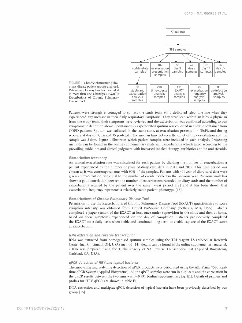
sample was 3 days. Figure 1 illustrates which patient samples were included in each analysis. Processing
methods can be found in the online supplementary material. Exacerbations were treated according to the
prevailing guidelines and clinical judgment with increased inhaled therapy, antibiotics and/or oral steroids.
Exacerbation frequencyAn annual exacerbation rate was calculated for each patient by dividing the number of exacerbations a
patient experienced by the number of years of diary card data in 2011 and 2012. This time period was
chosen as it was contemporaneous with 90% of the samples. Patients with ,1 year of diary card data were
given an exacerbation rate equal to the number of events recalled in the previous year. Previous work has
shown a good correlation between the number of exacerbations recorded on diary cards and the number of
exacerbations recalled by the patient over the same 1-year period [12] and it has been shown that
exacerbation frequency represents a relatively stable patient phenotype [13].
Exacerbations of Chronic Pulmonary Disease ToolPermission to use the Exacerbations of Chronic Pulmonary Disease Tool (EXACT) questionnaire to score
symptom intensity was obtained from United BioSource Company (Bethesda, MD, USA). Patients
completed a paper version of the EXACT at least once under supervision in the clinic and then at home,
based on their symptoms experienced on the day of completion. Patients prospectively completed
the EXACT on a daily basis when stable and continued long-term to enable capture of the EXACT score
at exacerbation.
RNA extraction and reverse transcriptionRNA was extracted from homogenised sputum samples using the TRI reagent LS (Molecular Research
Center Inc., Cincinnati, OH, USA) method [14]; details can be found in the online supplementary material.
cDNA was prepared using the High-Capacity cDNA Reverse Transcription Kit (Applied Biosystems,
Carlsbad, CA, USA).
qPCR detection of HRV and typical bacteriaThermocycling and real-time detection of qPCR products were performed using the ABI Prism 7500 Real-
time qPCR System (Applied Biosystems). All the qPCR samples were run in duplicate and the correlation in
the qPCR results between the two runs was r50.991 (online supplementary fig. E1). Details of primers and
probes for HRV qPCR are shown in table E1.
DNA extraction and multiplex qPCR detection of typical bacteria have been previously described by our
group [15].
89co-infection
analysissamples
58stable and
exacerbationanalysissamples
298time-course
analysissamples
107exacerbationpresentation
samples
58stable-state
samples
58day 3
samples
388 samples
77 patients
69day 7
samples
57day 14
samples
39day 35
samples
171EXACT
analysissamples
73exacerbation
frequencyanalysissamples
FIGURE 1 Chronic obstructive pulm-onary disease patient groups analysed.Patient samples may have been includedin more than one subanalysis. EXACT:Exacerbations of Chronic PulmonaryDisease Tool.
COPD | S.N. GEORGE ET AL.
DOI: 10.1183/09031936.00223113 3
Statistical analysisData were analysed using PASW Statistics v.19 (IBM Corporation, New York, NY, USA). The Kolmogorov–
Smirnov test for normality was applied. Absolute HRV loads were reported as median (interquartile range
(IQR)). For graphical illustrations, log10-transformed data were presented as mean¡SEM. Nonparametric
data were analysed using Mann–Whitney U-tests and Wilcoxon tests for unpaired and paired data,
respectively. Relationships between continuous variables were analysed using Spearman’s rank correlation
in a univariate analysis. A probability value of p,0.05 was considered to be statistically significant.
Exacerbations were considered as separate events when describing HRV time-course because their trigger
(viral or bacterial) could differ within the same individual. In other analysis, where HRV infection was
related to patient characteristics, only the first exacerbation sample was used from each patient to avoid
complications with repeated measures.
Ethical approvalThis study was approved by the Royal Free Research Ethics Committee at the Royal Free NHS Trust
(London, UK) where this work was under-taken (09/H0270/8). All patients involved in the study gave
informed, written consent.
ResultsPatient characteristics77 patients from the London COPD Cohort provided sputum samples for this study (fig. 1). Their baseline
physiological characteristics are reported in table 1. These patients experienced 107 exacerbations, of which
89% were treated with antibiotics and 81% with oral steroids, and 2% led to hospitalisation.
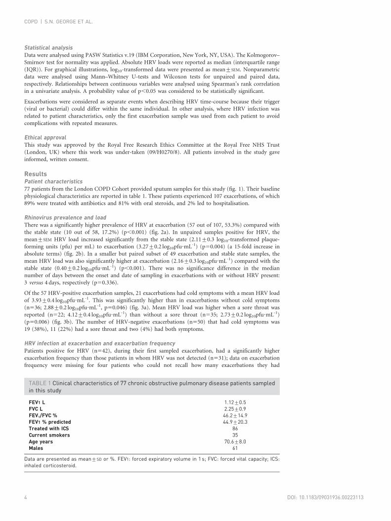
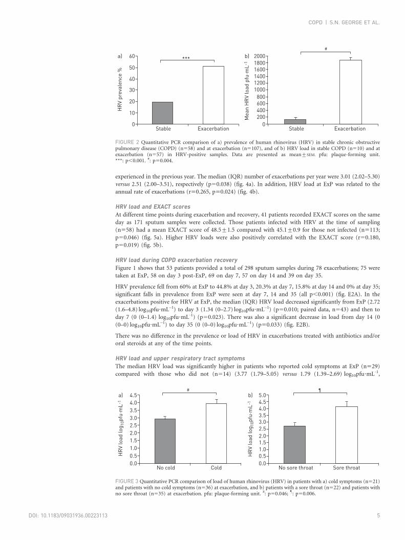
Rhinovirus prevalence and loadThere was a significantly higher prevalence of HRV at exacerbation (57 out of 107, 53.3%) compared with
the stable state (10 out of 58, 17.2%) (p,0.001) (fig. 2a). In unpaired samples positive for HRV, the
mean¡SEM HRV load increased significantly from the stable state (2.11¡0.3 log10-transformed plaque-
forming units (pfu) per mL) to exacerbation (3.27¡0.2 log10pfu?mL-1) (p50.004) (a 15-fold increase in
absolute terms) (fig. 2b). In a smaller but paired subset of 49 exacerbation and stable state samples, the
mean HRV load was also significantly higher at exacerbation (2.16¡0.3 log10pfu?mL-1) compared with the
stable state (0.40¡0.2 log10pfu?mL-1) (p,0.001). There was no significance difference in the median
number of days between the onset and date of sampling in exacerbations with or without HRV present:
3 versus 4 days, respectively (p50.336).
Of the 57 HRV-positive exacerbation samples, 21 exacerbations had cold symptoms with a mean HRV load
of 3.93¡0.4 log10pfu?mL-1. This was significantly higher than in exacerbations without cold symptoms
(n536; 2.88¡0.2 log10pfu?mL-1, p50.046) (fig. 3a). Mean HRV load was higher when a sore throat was
reported (n522; 4.12¡0.4 log10pfu?mL-1) than without a sore throat (n535; 2.73¡0.2 log10pfu?mL-1)
(p50.006) (fig. 3b). The number of HRV-negative exacerbations (n550) that had cold symptoms was
19 (38%), 11 (22%) had a sore throat and two (4%) had both symptoms.
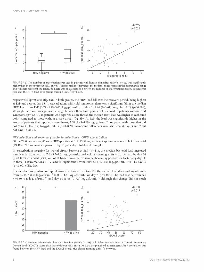
HRV infection at exacerbation and exacerbation frequencyPatients positive for HRV (n542), during their first sampled exacerbation, had a significantly higher
exacerbation frequency than those patients in whom HRV was not detected (n531); data on exacerbation
frequency were missing for four patients who could not recall how many exacerbations they had
TABLE 1 Clinical characteristics of 77 chronic obstructive pulmonary disease patients sampledin this study
FEV1 L 1.12¡0.5FVC L 2.25¡0.9FEV1/FVC % 46.2¡14.9FEV1 % predicted 44.9¡20.3Treated with ICS 86Current smokers 35Age years 70.6¡8.0Males 61
Data are presented as mean¡SD or %. FEV1: forced expiratory volume in 1 s; FVC: forced vital capacity; ICS:inhaled corticosteroid.
COPD | S.N. GEORGE ET AL.
DOI: 10.1183/09031936.002231134
experienced in the previous year. The median (IQR) number of exacerbations per year were 3.01 (2.02–5.30)
versus 2.51 (2.00–3.51), respectively (p50.038) (fig. 4a). In addition, HRV load at ExP was related to the
annual rate of exacerbations (r50.265, p50.024) (fig. 4b).
HRV load and EXACT scoresAt different time points during exacerbation and recovery, 41 patients recorded EXACT scores on the same
day as 171 sputum samples were collected. Those patients infected with HRV at the time of sampling
(n558) had a mean EXACT score of 48.5¡1.5 compared with 45.1¡0.9 for those not infected (n5113;
p50.046) (fig. 5a). Higher HRV loads were also positively correlated with the EXACT score (r50.180,
p50.019) (fig. 5b).
HRV load during COPD exacerbation recoveryFigure 1 shows that 53 patients provided a total of 298 sputum samples during 78 exacerbations; 75 were
taken at ExP, 58 on day 3 post-ExP, 69 on day 7, 57 on day 14 and 39 on day 35.
HRV prevalence fell from 60% at ExP to 44.8% at day 3, 20.3% at day 7, 15.8% at day 14 and 0% at day 35;
significant falls in prevalence from ExP were seen at day 7, 14 and 35 (all p,0.001) (fig. E2A). In the
exacerbations positive for HRV at ExP, the median (IQR) HRV load decreased significantly from ExP (2.72
(1.6–4.8) log10pfu?mL–1) to day 3 (1.34 (0–2.7) log10pfu?mL–1) (p50.010; paired data, n543) and then to
day 7 (0 (0–1.4) log10pfu?mL-1) (p50.023). There was also a significant decrease in load from day 14 (0
(0–0) log10pfu?mL–1) to day 35 (0 (0–0) log10pfu?mL–1) (p50.033) (fig. E2B).
There was no difference in the prevalence or load of HRV in exacerbations treated with antibiotics and/or
oral steroids at any of the time points.
HRV load and upper respiratory tract symptomsThe median HRV load was significantly higher in patients who reported cold symptoms at ExP (n529)
compared with those who did not (n514) (3.77 (1.79–5.05) versus 1.79 (1.39–2.69) log10pfu?mL-1,
50
60a)
30
40
20
10
0H
RV
prev
alen
ce %
ExacerbationStable
*** 200018001600140012001000
800600400200
b)
0
Mea
n H
RV
load
pfu
·mL-
1
ExacerbationStable
#
FIGURE 2 Quantitative PCR comparison of a) prevalence of human rhinovirus (HRV) in stable chronic obstructivepulmonary disease (COPD) (n558) and at exacerbation (n5107), and of b) HRV load in stable COPD (n510) and atexacerbation (n557) in HRV-positive samples. Data are presented as mean¡SEM. pfu: plaque-forming unit.***: p,0.001. #: p50.004.
4.04.5a)
2.52.0
3.03.5
1.51.00.50.0
HR
V lo
ad lo
g 10p
fu·m
L-1
ColdNo cold
#5.04.54.03.53.02.52.01.51.00.5
b)
0.0
HR
V lo
ad lo
g 10p
fu·m
L-1
Sore throatNo sore throat
FIGURE 3 Quantitative PCR comparison of load of human rhinovirus (HRV) in patients with a) cold symptoms (n521)and patients with no cold symptoms (n536) at exacerbation, and b) patients with a sore throat (n522) and patients withno sore throat (n535) at exacerbation. pfu: plaque-forming unit. #: p50.046; ": p50.006.
COPD | S.N. GEORGE ET AL.
DOI: 10.1183/09031936.00223113 5
respectively) (p50.006) (fig. 6a). In both groups, the HRV load fell over the recovery period, being highest
at ExP and zero at day 35. In exacerbations with cold symptoms, there was a significant fall in the median
HRV load from ExP (3.77 (1.79–5.05) log10pfu?mL-1) to day 3 (1.94 (0–3.61) log10pfu?mL-1) (p,0.001),
although there was no significant change between these time points in HRV load in patients without cold
symptoms (p50.517). In patients who reported a sore throat, the median HRV load was higher at each time
point compared to those without a sore throat (fig. 6b). At ExP, the load was significantly higher in the
group of patients that reported a sore throat, 3.38 (2.43–4.99) log10pfu?mL-1 compared with those that did
not (1.67 (1.38–3.19) log10pfu?mL-1) (p50.039). Significant differences were also seen at days 3 and 7 but
not days 14 or 35.
HRV infection and secondary bacterial infection at COPD exacerbationOf the 78 time-courses, 45 were HRV-positive at ExP. Of these, sufficient sputum was available for bacterial
qPCR in 21 time-courses provided by 19 patients, a total of 89 samples.
In exacerbations negative for typical airway bacteria at ExP (n511), the median bacterial load increased
significantly from zero to 7.0 (3.3–7.6) log10-transformed colony-forming units (cfu) per mL by day 14
(p50.002) with eight (73%) out of 11 bacterium-negative samples becoming positive for bacteria by day 14.
In these 11 exacerbations, HRV load fell significantly from ExP (2.7 (1.5–4.9) log10pfu?mL-1) to 0 by day 35
(p,0.001) (fig. 7a).
In exacerbations positive for typical airway bacteria at ExP (n510), the median load decreased significantly
from 6.7 (5.7–8.5) log10cfu?mL-1 to 0 (0–4.4) log10cfu?mL-1 on day 7 (p50.004). The load rose between day
7 (0 (0–4.4) log10cfu?mL-1) and day 14 (5.43 (0–7.0) log10cfu?mL-1) although this change did not reach
8
10#
a)
4
6
2
0
Exac
erba
tions
per
yea
r n
HRV positiveHRV negative
6
8b)
4
2
0
HR
V lo
ad lo
g 10p
fu·m
L-1
Exacerbations n0 2 4 6 8 10 12
r=0.265p=0.024
FIGURE 4 a) The number of exacerbations per year in patients with human rhinovirus (HRV) (n542) was significantlyhigher than in those without HRV (n531). Horizontal lines represent the median, boxes represent the interquartile rangeand whiskers represent the range. b) There was an association between the number of exacerbations had by patients peryear and the HRV load. pfu: plaque-forming unit. #: p50.038.
49
50
51 #a)
46
45
47
48
44
43
EXAC
T sc
ore
HRV positiveHRV negative
6
8
b)
4
2
0
HR
V lo
ad lo
g 10p
fu·m
L-1
EXACT score20 30 40 50 60 70 80
r=0.180p=0.019
FIGURE 5 a) Patients infected with human rhinovirus (HRV) (n558) had higher Exacerbations of Chronic PulmonaryDisease Tool (EXACT) scores than those without HRV (n5113). Data are presented as mean¡SEM. b) A correlation wasfound between the HRV load and the EXACT score. pfu: plaque-forming units. #: p50.046.
COPD | S.N. GEORGE ET AL.
DOI: 10.1183/09031936.002231136
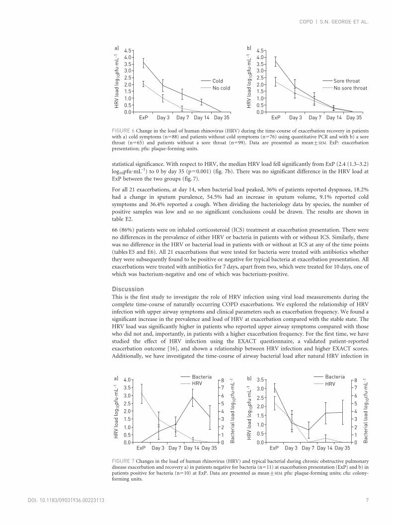
statistical significance. With respect to HRV, the median HRV load fell significantly from ExP (2.4 (1.3–3.2)
log10pfu?mL-1) to 0 by day 35 (p50.001) (fig. 7b). There was no significant difference in the HRV load at
ExP between the two groups (fig. 7).
For all 21 exacerbations, at day 14, when bacterial load peaked, 36% of patients reported dyspnoea, 18.2%
had a change in sputum purulence, 54.5% had an increase in sputum volume, 9.1% reported cold
symptoms and 36.4% reported a cough. When dividing the bacteriology data by species, the number of
positive samples was low and so no significant conclusions could be drawn. The results are shown in
table E2.
66 (86%) patients were on inhaled corticosteroid (ICS) treatment at exacerbation presentation. There were
no differences in the prevalence of either HRV or bacteria in patients with or without ICS. Similarly, there
was no difference in the HRV or bacterial load in patients with or without at ICS at any of the time points
(tables E5 and E6). All 21 exacerbations that were tested for bacteria were treated with antibiotics whether
they were subsequently found to be positive or negative for typical bacteria at exacerbation presentation. All
exacerbations were treated with antibiotics for 7 days, apart from two, which were treated for 10 days, one of
which was bacterium-negative and one of which was bacterium-positive.
DiscussionThis is the first study to investigate the role of HRV infection using viral load measurements during the
complete time-course of naturally occurring COPD exacerbations. We explored the relationship of HRV
infection with upper airway symptoms and clinical parameters such as exacerbation frequency. We found a
significant increase in the prevalence and load of HRV at exacerbation compared with the stable state. The
HRV load was significantly higher in patients who reported upper airway symptoms compared with those
who did not and, importantly, in patients with a higher exacerbation frequency. For the first time, we have
studied the effect of HRV infection using the EXACT questionnaire, a validated patient-reported
exacerbation outcome [16], and shown a relationship between HRV infection and higher EXACT scores.
Additionally, we have investigated the time-course of airway bacterial load after natural HRV infection in
4.5a)
3.5
2.5
1.52.0
3.0
4.0
0.51.0
0.0H
RV
load
log 1
0pfu
·mL-
1ExP Day 3 Day 7 Day 14 Day 35
No coldCold
4.5b)
3.5
2.5
1.52.0
3.0
4.0
0.51.0
0.0
HR
V lo
ad lo
g 10p
fu·m
L-1
ExP Day 3 Day 7 Day 14 Day 35
No sore throatSore throat
FIGURE 6 Change in the load of human rhinovirus (HRV) during the time-course of exacerbation recovery in patientswith a) cold symptoms (n588) and patients without cold symptoms (n576) using quantitative PCR and with b) a sorethroat (n565) and patients without a sore throat (n599). Data are presented as mean¡SEM. ExP: exacerbationpresentation; pfu: plaque-forming units.
4.0a)
3.0
1.52.02.5
3.5
0.51.0
0.0
8
6
345
7
12
0
HR
V lo
ad lo
g 10p
fu·m
L-1
Bac
teri
al lo
ad lo
g 10c
fu·m
L-1
ExP Day 3 Day 7 Day 14 Day 35
BacteriaHRV
3.5b)
2.5
1.5
2.0
3.0
0.5
1.0
0.0
8
6
345
7
12
0
HR
V lo
ad lo
g 10p
fu·m
L-1
Bac
teri
al lo
ad lo
g 10c
fu·m
L-1
ExP Day 3 Day 7 Day 14 Day 35
BacteriaHRV
FIGURE 7 Changes in the load of human rhinovirus (HRV) and typical bacterial during chronic obstructive pulmonarydisease exacerbation and recovery a) in patients negative for bacteria (n511) at exacerbation presentation (ExP) and b) inpatients positive for bacteria (n510) at ExP. Data are presented as mean¡SEM. pfu: plaque-forming units; cfu: colony-forming units.
COPD | S.N. GEORGE ET AL.
DOI: 10.1183/09031936.00223113 7
COPD patients, and shown a relationship between initial HRV infection and secondary bacterial infection at
COPD exacerbation.
HRV has been shown to be associated with exacerbations both in asthma [17] and COPD [8]. In this study, we
found that the prevalence of HRV in sputum was greater at exacerbation (53.3%) than in the stable state
(17.2%) and this is consistent with previous data [9]. Similarly, HRV load, measured by qPCR, was
significantly higher at exacerbation compared with in the stable state, with a 15-fold increase in absolute terms.
HRV infection is the most common cause of the upper airway cold and at exacerbation; patients with cold
symptoms had significantly higher HRV loads than those without. A study of experimental HRV infection
in asthmatics found a similar relationship between HRV load and cold symptoms [18]. In a study of
experimental HRV infection in COPD patients, HRV load was shown to peak at day 5 post-inoculation,
with cold symptoms peaking at day 3 [11]. Thus, our results confirm the association of upper airway
symptoms and HRV infection, and, importantly, emphasise that the presence of HRV may be indicated by
the presence of cold symptoms. It also highlights the importance of early sampling of COPD patients at
exacerbation in order to study their pathophysiology. A unique advantage of the London COPD Cohort is
that we prospectively follow our patients, reviewing them with exacerbation symptoms early after onset,
optimising viral detection and enabling study of the time-course of these events.
Some patients with COPD are particularly susceptible to exacerbations and are termed ‘‘frequent
exacerbators’’ [19]. Our previous work has suggested that these patients are more likely to develop colds
[19] and in this study we detected HRV infection more commonly in frequent exacerbators. For the first
time, we have demonstrated a weak but significant correlation between exacerbation frequency and HRV
loads, suggesting that frequent exacerbators are more susceptible to respiratory viral infection or are less
able to prevent viral replication. A proposed mechanism of increased viral susceptibility is the modulation
of intracellular adhesion molecule (ICAM)-1 on respiratory epithelial cells. ICAM-1 has been shown to be
upregulated in the bronchial mucosa of patients with chronic bronchitis [20], which may lead to increases
in HRV infection in these patients.
The EXACT is a patient-reported outcome measure that allows the standardisation of exacerbation frequency,
severity and duration [16]. Using the EXACT questionnaire, we have demonstrated that patients with HRV at
the time of sampling have higher EXACT scores than those without HRV, suggesting that exacerbations
associated with HRV infection are more severe. EXACT scores could be used to assess exacerbation severity in
future trials of antiviral therapy at COPD exacerbation. However, this scarcely reaches statistical significance
and so future studies or larger study numbers may assist in making definitive conclusions.
A novel aspect of this study is that we assessed HRV load through the whole time-course of a naturally
occurring exacerbation. To our knowledge, there are no previous data on the time-course of HRV infection
in naturally occurring COPD exacerbations and its relationship to upper airway symptoms. We found HRV
prevalence and load decreased from ExP until day 35, when no HRV was detected. There was a significant
fall in the median HRV load from presentation to day 3 and from day 3 to day 7. In our study, HRV load
was highest at presentation, suggesting patients could have become infected a few days before presenting to
the clinic. Colds precede exacerbation symptoms and in an experimental HRV study, HRV load was highest
at day 5 post-inoculation [11], consistent with our findings.
The finding that HRV loads are high at COPD ExP is important. One of the many obstacles to the
development of antiviral therapies for COPD exacerbations is that it is considered that patients are likely to
present with symptoms and be prescribed therapy too late for the antiviral to have an effect. These data,
however, show that HRV load is still high when patients present to the clinic, suggesting that viral
replication may be ongoing and, therefore, antiviral therapy may be effective at this time-point. This
highlights the unmet need for rapid development of antiviral therapies for the treatment of COPD
exacerbations, as patients have high HRV loads even at exacerbation presentation.
We also studied co-infection with HRV and airway bacteria in patients throughout the time-course of the
exacerbation, and found an association between initial HRV infection and secondary bacterial infection. A
small number of studies have considered co-infection, such as the interaction of Haemophilus influenzae and
HRV, showing that H. influenzae load, determined by culture, was markedly increased in the presence of
HRV compared with cases without HRV [21]. Furthermore, MALLIA et al. [22], in a study of COPD patients
experimentally infected with HRV, reported secondary bacterial infection in 60% of subjects after the initial
inoculation, with bacterial load peaking on day 15. In our patients who were negative for bacteria at
exacerbation presentation, 73% of samples were positive by day 14 and the bacterial load increased
significantly by this time-point. This supports the concept of a direct link between HRV infection and
secondary bacterial infection. In patients who were positive for bacteria at presentation, the load decreased
significantly by day 7, although there was a nonsignificant increase by day 14. This increase at day 14 may
COPD | S.N. GEORGE ET AL.
DOI: 10.1183/09031936.002231138
possibly be due to the cessation of antibiotic therapy, although the increase in bacterial load in samples
negative at presentation but positive at day 14, after an HRV infection, suggests that there may be a genuine
link between initial HRV infection and secondary bacterial infection.
The fact that exacerbations that were bacterium-negative at ExP but became positive by day 14 were treated
with antibiotics suggests that in exacerbations where an initial viral infection leads to a secondary bacterial
infection, antiviral therapy may be more effective than antibiotics; however, antibiotics may be more
relevant during the latter part of the exacerbation, when a secondary bacterial infection may develop.
There were very few patients that were not on ICS treatment and, therefore, no clear conclusions can be
made about the effect of this treatment on either viral or bacterial loads.
The COPD patients were still symptomatic at day 14, when bacterial load peaked, and reported dyspnoea,
cough, and changes in sputum volume and purulence, suggesting the increase in airway bacterial infection
at day 14 is of clinical relevance. Patients hospitalised due to exacerbations have more marked lung function
impairment and increased length of stay in the context of viral and bacterial co-infection [23]. Secondary
bacterial infection may also play a part in exacerbation recurrence [13] and further studies are required to
assess the longer term effects of these key findings.
There have been other studies investigating co-infection of viruses and bacteria at COPD exacerbation;
however, they have found lower incidence of co-infection than our study, which may be due to the larger
number of samples collected from patients during each exacerbation [23, 24]. A study in 2006 performed by
PAPI et al. [23], where one sample was taken at each exacerbation, showed viral and bacterial co-infection in
25% of samples. HUTCHINSON et al. [24] collected two samples during exacerbation and reported that 36%
of exacerbations in which a virus was detected at onset developed a secondary bacterial infection over the
following 7 days and overall, 78% of exacerbations in which H. influenzae was isolated in the first 5 days after
onset were preceded by viral symptoms. In our study, we collected multiple samples during exacerbations and
detected a higher incidence of co-infection than the studies mentioned above, which indicates that repeated
sampling over the duration of an exacerbation is more likely to identify the presence of co-infection.
This study was limited to HRV infection and although other viruses have been studied at COPD
exacerbation, HRV has been shown to be the most commonly detected [8]. Similarly, this study was limited
to the commonest typical airway bacteria detected at COPD exacerbation, although it is unlikely that
bacteria other than these three, play a significant role in the aetiology of community exacerbations [25].
In conclusion, this study of quantitative viral loads shows that HRV infection is associated with upper
airway symptoms, typical airway bacteria and patient-reported outcomes in natural COPD exacerbations.
We have also shown that patients with a history of frequent exacerbations are more susceptible to HRV
infection. Secondary bacterial infection is common after HRV infection, and has important implications in
terms of therapy and exacerbation recurrence. Treating respiratory viral infections in patients with COPD may
therefore be key to reducing exacerbation frequency. Thus, there is an unmet need for rapid development of
therapeutic targets for the prevention and treatment of respiratory viral infection in COPD patients.
AcknowledgementsThe authors wish to thank the UK Medical Research Council for funding the London COPD Cohort. We wish to thankN. Bartlett (Imperial College London, London, UK) for his invaluable assistance in the HRV qPCR set-up. We arethankful to all patients of the London COPD Cohort for dedicating their time and energy making this study possible.
References1 Lozano R, Naghavi M, Foreman K, et al. Global and regional mortality from 235 causes of death for 20 age groups
in 1990 and 2010: a systematic analysis for the Global Burden of Disease Study 2010. Lancet 2012; 380: 2095–2128.2 Singanayagam A, Joshi PV, Mallia P, et al. Viruses exacerbating chronic pulmonary disease: the role of immune
modulation. BMC Med 2012; 10: 27.3 Wedzicha JA, Seemungal TAR. COPD exacerbations: defining their cause and prevention. Lancet 2007; 370:
786–796.4 Seemungal TA, Donaldson GC, Paul EA, et al. Effect of exacerbation on quality of life in patients with chronic
obstructive pulmonary disease. Am J Respir Crit Care Med 1998; 157: 1418–1422.5 Pauwels RA, Buist AS, Calverley PM, et al. Global strategy for the diagnosis, management, and prevention of
chronic obstructive pulmonary disease. NHLBI/WHO Global Initiative for Chronic Obstructive Lung Disease(GOLD) Workshop summary. Am J Respir Crit Care Med 2001; 163: 1256–1276.
6 Donaldson GC, Seemungal TAR, Bhowmik A, et al. Relationship between exacerbation frequency and lung functiondecline in chronic obstructive pulmonary disease. Thorax 2002; 57: 847–852.
7 Soler-Cataluna JJ, Martınez-Garcıa MA, Roman Sanchez P, et al. Severe acute exacerbations and mortality inpatients with chronic obstructive pulmonary disease. Thorax 2005; 60: 925–931.
8 Seemungal T, Harper-Owen R, Bhowmik A, et al. Respiratory viruses, symptoms, and inflammatory markersin acute exacerbations and stable chronic obstructive pulmonary disease. Am J Respir Crit Care Med 2001; 164:1618–1623.
COPD | S.N. GEORGE ET AL.
DOI: 10.1183/09031936.00223113 9
9 Seemungal TA, Harper-Owen R, Bhowmik A, et al. Detection of rhinovirus in induced sputum at exacerbation ofchronic obstructive pulmonary disease. Eur Respir J 2000; 16: 677–683.
10 Kurai D, Saraya T, Ishii H, et al. Virus-induced exacerbations in asthma and COPD. Front Microbiol 2013; 4: 293.11 Mallia P, Message SD, Gielen V, et al. Experimental rhinovirus infection as a human model of chronic obstructive
pulmonary disease exacerbation. Am J Respir Crit Care Med 2011; 183: 734–742.12 Quint JK, Donaldson GC, Goldring JJP, et al. Serum IP-10 as a biomarker of human rhinovirus infection at
exacerbation of COPD. Chest 2010; 137: 812–822.13 Hurst JR, Donaldson GC, Quint JK, et al. Temporal clustering of exacerbations in chronic obstructive pulmonary
disease. Am J Respir Crit Care Med 2009; 179: 369–374.14 Chomczynski P, Sacchi N. Single-step method of RNA isolation by acid guanidinium thiocyanate-phenol-
chloroform extraction. Anal Biochem 1987; 162: 156–159.15 Garcha DS, Thurston SJ, Patel ARC, et al. Changes in prevalence and load of airway bacteria using quantitative PCR
in stable and exacerbated COPD. Thorax 2012; 67: 1075–1080.16 Leidy NK, Wilcox TK, Jones PW, et al. Standardizing measurement of chronic obstructive pulmonary disease
exacerbations. Reliability and validity of a patient-reported diary. Am J Respir Crit Care Med 2011; 183: 323–329.17 Friedlander SL, Busse WW. The role of rhinovirus in asthma exacerbations. J Allergy Clin Immunol 2005; 116:
267–273.18 Grunberg K, Timmers MC, de Klerk EP, et al. Experimental rhinovirus 16 infection causes variable airway
obstruction in subjects with atopic asthma. Am J Respir Crit Care Med 1999; 160: 1375–1380.19 Hurst JR, Donaldson GC, Wilkinson TMA, et al. Epidemiological relationships between the common cold and
exacerbation frequency in COPD. Eur Respir J 2005; 26: 846–852.20 Di Stefano A, Maestrelli P, Roggeri A, et al. Upregulation of adhesion molecules in the bronchial mucosa of subjects
with chronic obstructive bronchitis. Am J Respir Crit Care Med 1994; 149: 803–810.21 Wilkinson TMA, Hurst JR, Perera WR, et al. Effect of interactions between lower airway bacterial and rhinoviral
infection in exacerbations of COPD. Chest 2006; 129: 317–324.22 Mallia P, Footitt J, Sotero R, et al. Rhinovirus infection induces degradation of antimicrobial peptides and
secondary bacterial infection in chronic obstructive pulmonary disease. Am J Respir Crit Care Med 2012; 186:1117–1124.
23 Papi A, Bellettato CM, Braccioni F, et al. Infections and airway inflammation in chronic obstructive pulmonarydisease severe exacerbations. Am J Respir Crit Care Med 2006; 173: 1114–1121.
24 Hutchinson AF, Ghimire AK, Thompson MA, et al. A community-based, time-matched, case-control study ofrespiratory viruses and exacerbations of COPD. Respir Med 2007; 101: 2472–2481.
25 White AJ, Gompertz S, Bayley DL, et al. Resolution of bronchial inflammation is related to bacterial eradicationfollowing treatment of exacerbations of chronic bronchitis. Thorax 2003; 58: 680–685.
COPD | S.N. GEORGE ET AL.
DOI: 10.1183/09031936.0022311310
Related Documents