OPEN Human recombinant glutamate oxaloacetate transaminase 1 (GOT1) supplemented with oxaloacetate induces a protective effect after cerebral ischemia M Pe ´ rez-Mato 1 , P Ramos-Cabrer 1 , T Sobrino 1 , M Blanco 1 , A Ruban 2 , D Mirelman 3 , P Menendez 4,5 , J Castillo 1 and F Campos* ,1 Blood glutamate scavenging is a novel and attractive protecting strategy to reduce the excitotoxic effect of extracellular glutamate released during ischemic brain injury. Glutamate oxaloacetate transaminase 1 (GOT1) activation by means of oxaloacetate administration has been used to reduce the glutamate concentration in the blood. However, the protective effect of the administration of the recombinant GOT1 (rGOT1) enzyme has not been yet addressed in cerebral ischemia. The aim of this study was to analyze the protective effect of an effective dose of oxaloacetate and the human rGOT1 alone and in combination with a non-effective dose of oxaloacetate in an animal model of ischemic stroke. Sixty rats were subjected to a transient middle cerebral artery occlusion (MCAO). Infarct volumes were assessed by magnetic resonance imaging (MRI) before treatment administration, and 24 h and 7 days after MCAO. Brain glutamate levels were determined by in vivo MR spectroscopy (MRS) during artery occlusion (80 min) and reperfusion (180 min). GOT activity and serum glutamate concentration were analyzed during the occlusion and reperfusion period. Somatosensory test was performed at baseline and 7 days after MCAO. The three treatments tested induced a reduction in serum and brain glutamate levels, resulting in a reduction in infarct volume and sensorimotor deficit. Protective effect of rGOT1 supplemented with oxaloacetate at 7 days persists even when treatment was delayed until at least 2 h after onset of ischemia. In conclusion, our findings indicate that the combination of human rGOT1 with low doses of oxaloacetate seems to be a successful approach for stroke treatment Cell Death and Disease (2014) 5, e992; doi:10.1038/cddis.2013.507; published online 9 January 2014 Subject Category: Neuroscience Stroke is a leading cause of mortality and morbidity in developed countries, with increasing incidence because of the progressive aging of the population. Pharmacological or mechanical reperfusion therapy is the most effective treat- ment during the acute phase of ischemic stroke and it is associated with good outcome in 50–70% of cases. However, these treatments are only applicable to o10% of patients because of the short therapeutic window. 1 Owing to these therapeutic limitations, the development of new and effective therapies to be used during the acute phase of ischemic stroke is in high demand. After ischemic stroke, there is a rapid but transient elevation of glutamate into the extracellular space followed by a marked increase in intracellular calcium, which provokes a neuronal death through an excitotoxicity mechanism. 2 Consequently, calcium and glutamate antagonists have been widely studied as protective agents in experimental models of cerebral ischemia with encouraging results. Unfortunately, they failed or displayed severe adverse effects when they were tested in clinical trials. 3 Nevertheless, because of the central role of glutamate in the ischemic cascade, the mitigation of gluta- mate excitotoxicity remains one of the most promising strategies for the development of effective treatments to minimize neurological damage following an acute ischemic stroke. In this sense, novel therapeutic approaches should aim at reducing the increased glutamate levels produced early after the acute phase of ischemia. Previous studies have shown that a decrease in blood glutamate concentration leads to a larger natural glutamate gradient between the brain and blood, facilitating the efflux of 1 Department of Neurology, Neurovascular Area, Clinical Neurosciences Research Laboratory, Hospital Clı ´nico Universitario, Health Research Institute of Santiago de Compostela (IDIS), University of Santiago de Compostela, Santiago de Compostela, Spain; 2 Department of Neurobiology, The Weizmann Institute of Science, Rehovot, Israel; 3 Department of Biological Chemistry, Weizmann Institute of Science, Rehovot, Israel; 4 Josep Carreras Leukemia Research Institute, Cell Therapy Program of the University of Barcelona, Barcelona, Spain and 5 Institucio ´ Catalana de Recerca i Estudis Avanc ¸ats (ICREA), Barcelona, Spain *Corresponding author: F Campos, Department of Neurology, Neurovascular Area, Clinical Neurosciences Research Laboratory, Health Research Institute of Santiago de Compostela (IDIS), Hospital Clı ´nico Universitario, University of Santiago de Compostela, c/Travesa da Choupana s/n, 15706 Compostela, Santiago de, Spain. Tel: +34 981 951098; Fax: +34 981 951098; E-mail: [email protected] Received 01.7.13; revised 13.11.13; accepted 18.11.13; Edited by A Verkhratsky Keywords: animal model; cerebral ischemia; GOT; glutamate; neuroprotection; oxaloacetate Abbreviations: ADC, apparent diffusion coefficient; AST, aspartate transaminase; GOT1, glutamate oxaloacetate transaminase 1; MCAO, occlusion of the middle cerebral artery; MRI, magnetic resonance imaging; MRS, magnetic resonance spectroscopy; MTT, thiazolyl blue tetrazolium bromide; RF, radiofrequency; rGOT1, recombinant glutamate oxaloacetate transaminase 1; STAIR, stroke therapy academic industry roundtable; STEAM, stimulated-echo acquisition mode; TBI, traumatic brain injury Citation: Cell Death and Disease (2014) 5, e992; doi:10.1038/cddis.2013.507 & 2014 Macmillan Publishers Limited All rights reserved 2041-4889/14 www.nature.com/cddis

Welcome message from author
This document is posted to help you gain knowledge. Please leave a comment to let me know what you think about it! Share it to your friends and learn new things together.
Transcript

OPEN
Human recombinant glutamate oxaloacetatetransaminase 1 (GOT1) supplemented withoxaloacetate induces a protective effect aftercerebral ischemia
M Perez-Mato1, P Ramos-Cabrer1, T Sobrino1, M Blanco1, A Ruban2, D Mirelman3, P Menendez4,5, J Castillo1 and F Campos*,1
Blood glutamate scavenging is a novel and attractive protecting strategy to reduce the excitotoxic effect of extracellularglutamate released during ischemic brain injury. Glutamate oxaloacetate transaminase 1 (GOT1) activation by means ofoxaloacetate administration has been used to reduce the glutamate concentration in the blood. However, the protective effect ofthe administration of the recombinant GOT1 (rGOT1) enzyme has not been yet addressed in cerebral ischemia. The aim of thisstudy was to analyze the protective effect of an effective dose of oxaloacetate and the human rGOT1 alone and in combinationwith a non-effective dose of oxaloacetate in an animal model of ischemic stroke. Sixty rats were subjected to a transient middlecerebral artery occlusion (MCAO). Infarct volumes were assessed by magnetic resonance imaging (MRI) before treatmentadministration, and 24 h and 7 days after MCAO. Brain glutamate levels were determined by in vivo MR spectroscopy (MRS)during artery occlusion (80min) and reperfusion (180 min). GOT activity and serum glutamate concentration were analyzedduring the occlusion and reperfusion period. Somatosensory test was performed at baseline and 7 days after MCAO. The threetreatments tested induced a reduction in serum and brain glutamate levels, resulting in a reduction in infarct volume andsensorimotor deficit. Protective effect of rGOT1 supplemented with oxaloacetate at 7 days persists even when treatment wasdelayed until at least 2 h after onset of ischemia. In conclusion, our findings indicate that the combination of human rGOT1 withlow doses of oxaloacetate seems to be a successful approach for stroke treatmentCell Death and Disease (2014) 5, e992; doi:10.1038/cddis.2013.507; published online 9 January 2014Subject Category: Neuroscience
Stroke is a leading cause of mortality and morbidity in
developed countries, with increasing incidence because of
the progressive aging of the population. Pharmacological or
mechanical reperfusion therapy is the most effective treat-
ment during the acute phase of ischemic stroke and it is
associated with good outcome in 50–70% of cases. However,
these treatments are only applicable to o10% of patients
because of the short therapeutic window.1 Owing to these
therapeutic limitations, the development of new and effective
therapies to be used during the acute phase of ischemic
stroke is in high demand.
After ischemic stroke, there is a rapid but transient elevation
of glutamate into the extracellular space followed by a marked
increase in intracellular calcium, which provokes a neuronal
death through an excitotoxicity mechanism.2 Consequently,
calcium and glutamate antagonists have been widely studied
as protective agents in experimental models of cerebral
ischemia with encouraging results. Unfortunately, they failed
or displayed severe adverse effects when they were tested in
clinical trials.3 Nevertheless, because of the central role of
glutamate in the ischemic cascade, the mitigation of gluta-
mate excitotoxicity remains one of the most promising
strategies for the development of effective treatments to
minimize neurological damage following an acute ischemic
stroke. In this sense, novel therapeutic approaches should
aim at reducing the increased glutamate levels produced early
after the acute phase of ischemia.
Previous studies have shown that a decrease in blood
glutamate concentration leads to a larger natural glutamate
gradient between the brain and blood, facilitating the efflux of
1Department of Neurology, Neurovascular Area, Clinical Neurosciences Research Laboratory, Hospital Clınico Universitario, Health Research Institute of Santiago deCompostela (IDIS), University of Santiago de Compostela, Santiago de Compostela, Spain; 2Department of Neurobiology, The Weizmann Institute of Science, Rehovot,Israel; 3Department of Biological Chemistry, Weizmann Institute of Science, Rehovot, Israel; 4Josep Carreras Leukemia Research Institute, Cell Therapy Program of theUniversity of Barcelona, Barcelona, Spain and 5Institucio Catalana de Recerca i Estudis Avancats (ICREA), Barcelona, Spain*Corresponding author: F Campos, Department of Neurology, Neurovascular Area, Clinical Neurosciences Research Laboratory, Health Research Institute of Santiagode Compostela (IDIS), Hospital Clınico Universitario, University of Santiago de Compostela, c/Travesa da Choupana s/n, 15706 Compostela, Santiago de, Spain.Tel: +34 981 951098; Fax: +34 981 951098; E-mail: [email protected]
Received 01.7.13; revised 13.11.13; accepted 18.11.13; Edited by A Verkhratsky
Keywords: animal model; cerebral ischemia; GOT; glutamate; neuroprotection; oxaloacetateAbbreviations: ADC, apparent diffusion coefficient; AST, aspartate transaminase; GOT1, glutamate oxaloacetate transaminase 1; MCAO, occlusion of the middlecerebral artery; MRI, magnetic resonance imaging; MRS, magnetic resonance spectroscopy; MTT, thiazolyl blue tetrazolium bromide; RF, radiofrequency; rGOT1,recombinant glutamate oxaloacetate transaminase 1; STAIR, stroke therapy academic industry roundtable; STEAM, stimulated-echo acquisition mode; TBI, traumaticbrain injury
Citation: Cell Death and Disease (2014) 5, e992; doi:10.1038/cddis.2013.507
& 2014 Macmillan Publishers Limited All rights reserved 2041-4889/14
www.nature.com/cddis

excess extracellular brain glutamate into the blood.4
Reduction in glutamate concentration in blood is induced
through the activity of the blood-resident enzyme glutamate
oxaloacetate transaminase 1 (GOT1), which catalyzes the
reversible transformation of oxaloacetate and glutamate
to aspartate and a-ketoglutarate. Thus, the increase in
the co-substrate (oxaloacetate) shifts the equilibrium of
the reaction to the right side, thereby decreasing the
blood glutamate concentration and lowering the brain
glutamate levels (see Teichberg et al.4 and Campos et al.
5
for review).
Based on this mechanism, we6 and others4 have demon-
strated that the intravenous (i.v.) administration of oxaloace-
tate in a rat model of ischemia induced by transient occlusion
of themiddle cerebral artery occlusion (MCAO) decreases the
glutamate concentration in blood and induces a lowering of
infarct volume and edema after ischemia. These effects were
also associated with a significant reduction in the sensor-
imotor deficit. To confirm that the protective effect was
mediated by a decrease in the brain glutamate levels, we
performed magnetic resonance spectroscopy (MRS) in the
infarct region. Spectroscopic analysis revealed that the
increase in brain glutamate seen in control animals after
MCAO was significantly reduced in animals treated with
oxaloacetate.4,6 These experimental results showed that
systemic reduction in glutamate concentration induced by
means of exogenous oxaloacetate administration could be
used as a novel and efficient protective strategy in ischemic
stroke. Importantly, however, the i.v. administration of
humans with oxaloacetate has potential serious limitations
as the required effective dosage of oxaloacetate in humans,
corresponding to that used in rats, might be toxic.7 In a
previous retrospective study with 4400 human patients, we
have found that ischemic patients with good outcome at 3
months after stroke, showed lower glutamate concentration
and higher GOT activity (418U/l) in serum.8,9 Therefore, it is
tentative to postulate that treatments based on the
administration of recombinant GOT1 (rGOT1) alone or
supplemented with low doses of oxaloacetate could
improve the protective effects of oxaloacetate administered
on its own.
The purpose of this study was to analyze the protective
effect of oxaloacetate and human rGOT1 alone and in
combination with a low dose (non-effective dose) of oxaloa-
cetate in the rat model of ischemia induced by transient
MCAO, and to compare their effects with a control group
treated with saline.
Results
Animals included in the study. A total of 119 animals
were used in this study (Table I). Thirty-six animals were
used in the rGOT1 dose–response study, six animals per
group (six groups of treatments). In this study, no animal
deaths were observed within the first 24 h after treatments
administration. In the protective study, 60 animals were
included, six animals/group (four groups in the spectro-
scopic study and other six groups in the non-spectroscopic
study). Eighteen animals were excluded, 16 of them were
not included because of unsuccessful MCA occlusion or
reperfusion and two animals died 24 h after surgery (one
treated with saline and one treated with rGOT1). Finally, five
animals were used as sham-operated control rats (sham)
without MCAO.
Serum glutamate concentration is reduced by oxaloace-
tate and rGOT1 treatments in healthy animals. Adminis-
tration of saline (control group) did not affect (P¼ 0.373) the
basal concentration of glutamate during 24 h after the i.v.
administration (Figure 1a), demonstrating that the surgical
procedure did not interfere with the serum glutamate
concentration. Injection of glutamate (15mM) induced
a significant (Po0.05) increase in serum glutamate
concentration 1 h after administration (Figure 1b). Basal
concentration was normalized 3h after treatment admini-
stration. This effect shows that the administration of glutamate
in healthy animals could be used to simulate the increase in
serum glutamate, a hallmark biochemical parameter after
ischemia.
On the basis of previous reports where rGOT1 has been
used to study other pathologies,10,11 we started with a dose
of 6.44 mg per 100 g to analyze the capacity of the enzyme to
metabolize serum glutamate. Administration of 6.44 mg per
100 g rGOT1 induced a reduction (Po0.01) (Figure 1c)
in serum glutamate concentration with respect to the
maximum increase observed after glutamate injection.
The maximum effect appeared between 2 and 4 h after
administration of treatment. A higher dose of 12.88 mg per
100 g rGOT1 induced a robust reduction (Po0.01) in serum
glutamate concentration (Figure 1d). Similar to the previous
dose, the maximum lowering effect appeared between 2 and
4 h after administration of treatment; however, in this case,
the capacity to reduce serum glutamate concentration was
much higher. Administration of rGOT1 25.76 mg per 100 g
induced a significant (P¼ 0.007) reduction in serum gluta-
mate concentration (Figure 1e). The maximum effect
appeared between 2 and 4 h after administration of the
treatment; however, the capacity of this dose to reduce
serum glutamate concentration was similar to the 12.88 mg
per 100 g dose. These findings showed that 12.88 mg per
100 g seems to be the most appropriate dose of rGOT1 to
achieve a significant reduction in the serum glutamate
concentration in the animals. Of note, serum glutamate
concentrations were normalized 4–6 h after rGOT1 admin-
istration, demonstrating that the effect of the enzymatic
treatment is transient.
In order to determine whether the effect of the enzyme
could be potentiated by oxaloacetate (co-substrate of the
enzymatic reaction), the dose of rGOT1 (12.88 mg per
100 g) was supplemented with a non-effective dose of
oxaloacetate (1.5mg per 100 g). In our previous study
(already published6), we had described that a dose of
1.5mg per 100 g of oxaloacetate did not affect the serum
glutamate levels. Similar results were observed again
in this study (Figure 1f). Administration of rGOT1 12.88 mg
per 100 g with oxaloacetate (1.5mg per 100 g) induced
an immediate and significant (Po0.01) lowering of
serum glutamate concentration, which is the gold
standard approach for reducing increased glutamate
levels after brain ischemia. The decrease in glutamate
Oxaloacetate and GOT1 in cerebral ischemia
M Perez-Mato et al
2
Cell Death and Disease

appeared 30min (with respect to the basal values) after
treatment administration and remained during at least 3 h
(Figure 1g); however, no significant differences with
respect to rGOT1 12.88 mg pet 100 alone (Figure 1d) were
observed.
These results led us to examine whether the protective
efficacy of rGOT1 (12.88 mg per 100 g) or rGOT1 (12.88 mg per
100 g) supplemented with oxaloacetate (1.5mg per 100 g) in
ischemic animals was higher than that observed previously6
with oxaloacetate 3.5mg per 100 g.
Figure 1 Effect of rGOT and oxaloacetate (Oxal) on serum glutamate levels in healthy rats. Glutamate 15mM was injected i.v. 30min prior to treatments. (a) Rats treatedwith saline (control group). (b) Rats treated with glutamate 15mM. (c) Comparison between rats treated with glutamate 15mM (blue line) and rats treated with glutamate 15mMand rGOT1 6.44mg per 100 g (black line). (d) Comparison between rats treated with glutamate 15mM (blue line) and rats treated with glutamate 15mM and rGOT1 12.88mg per100 g (black line). (e) Comparison between rats treated with glutamate (15mM) (blue line) and rats treated with glutamate (15mM) and with rGOT1 (25.76mg per 100 g).(f) Comparison between rats treated with glutamate 15mM (blue line) and rats treated with glutamate (15mM) and oxaloacetate (Oxal) (1.5 mg per 100 g). (g) Comparisonbetween rats treated with glutamate (15mM) (blue line) and rats treated with glutamate (15mM) and rGOT1 (12.88mg per 100 g) plus oxaloacetate (Oxal) (1.5mg per 100 g).Solid arrows denote the injection of glutamate. Dashed arrows indicate the injection of treatments. Serum samples were taken in basal conditions and 1, 2, 4, 6 and 24 h afterglutamate injection. Data are shown as mean±S.E.M. #Po0.05 compared to the basal levels; *Po0.05, **Po0.01 compared with animals treated with glutamate 15mM
Oxaloacetate and GOT1 in cerebral ischemia
M Perez-Mato et al
3
Cell Death and Disease

rGOT1 with or without supplementation of oxaloacetate
induces a protective effect on ischemic animals. As it
was previously shown,6 MCAO induced an increase of 30%
of serum glutamate concentration 1 h after reperfusion
(Figure 2), returning to normal levels 3 h later. In sham-
operated rats in which MCA was not occluded, no changes in
plasma glutamate concentration were detected (data not
shown). The increase in serum glutamate concentration after
cerebral ischemia was inhibited (Po0.05) by each one of the
three treatments; rGOT1 (12.88 mg per 100 g), oxaloacetate
(3.5mg per 100 g) and rGOT1 plus oxaloacetate (12.88 mg
per 100 gþ 1.5mg per 100 g, respectively) (Figure 2). The
small differences observed between the treatments were not
statistically significant.
To confirm that treatment with rGOT1 induced an increase
in systemic GOT1 activity, their levels were measured at
different time points (Figure 3). GOT1 activities in the control
and oxaloacetate groups were not altered after treatment,
whereas rGOT1 treatments induced a significant (Po0.01)
increase in serum GOT activity 1 h after administration, which
returned close to normal levels 24 h after treatment
administration.
Quantitative analysis of MRS revealed a persistent
increase in brain glutamate levels after the occlusion of
MCA in the control group. Glutamate levels in the brain
parenchyma were significantly decreased (Po0.05 relative
to the control animals), with each of the three treatments
tested (Figures 4a and b). To demonstrate that the
reduction in serum and brain glutamate levels observed
after treatment administration leads to a protective effect in
our animal model of cerebral ischemia, infarct volumes
were measured at 24 h and 7 days after ischemia and were
compared with the control group. Figure 5a shows that,
although all treatments were able to induce a significant
(Po0.05) protection against the ischemic damage both 24 h
and 7 days after the onset of ischemia, rGOT1þ
oxaloacetate revealed to be the more effective treatment
(Po0.01, with respect to the control group). Analysis of
infarct volumes adjusted to the ipsilateral hemisphere
showed a similar protective profile (data not shown). Infarct
volumes measured in those animals subjected to the
spectroscopy protocol (Figure 5b) showed the same
protective results for the three treatments. Diffusion images
revealed that all animals subjected to spectroscopy
presented the same ischemic damage before the treatment
administration.
To determine the therapeutic window of rGOT1 treat-
ments, two groups of animals were treated 1 h after
reperfusion (140min after occlusion). Analysis of serum
glutamate concentration in animals treated with rGOT1
(12.88 mg per 100 g) or rGOT1 plus oxaloacetate (12.88 mg
per 100 gþ 1.5mg per 100 g, respectively) showed that
both treatments caused a significant (Po0.05) inhibition of
the increase in glutamate observed in the control group
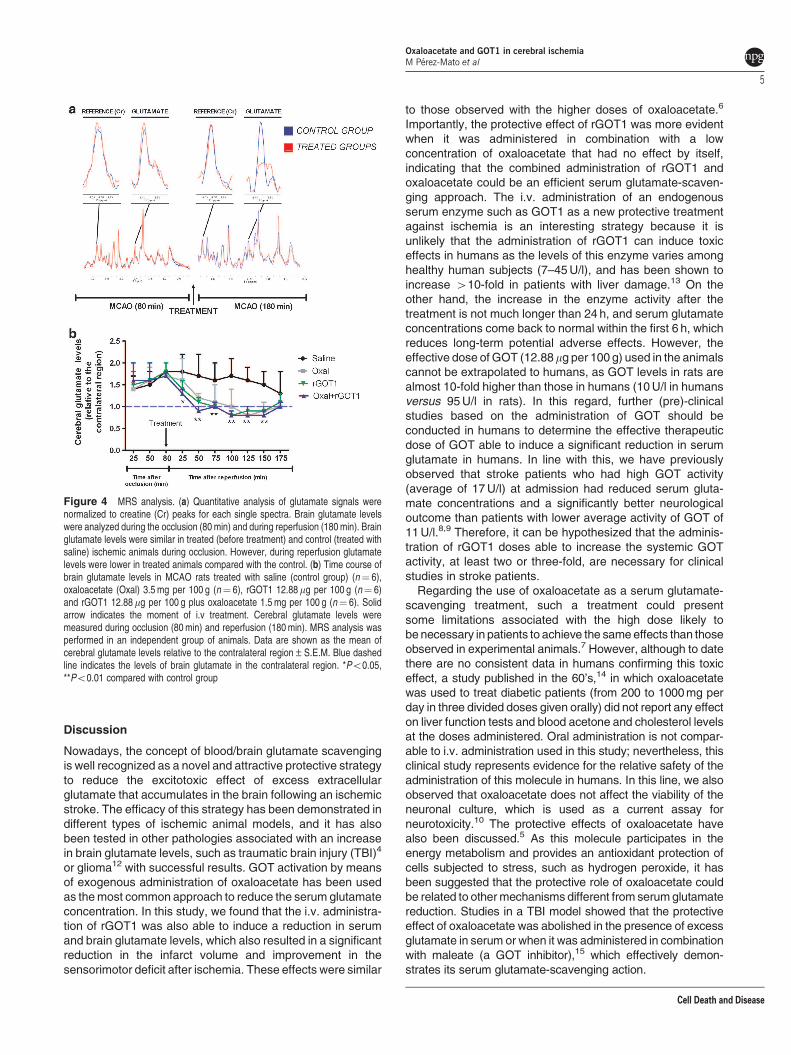
(Figure 6). Analysis of infarct volume determined that both
treatments displayed similar infarct volume reduction at
24 h when administered immediately after reperfusion
(Figures 7a and b), however, while the protective effect
induced for rGOT1 disappears when this treatment is given
1h after reperfusion on day 7; when rGOT1 is administered in
combination with oxaloacetate, the effect persists beyond 1h
after reperfusion (Po0.05, with respect to the control).
Somatosensory test, an important end point assay of drug
screening in stroke, confirmed that reduction in infarct volume
observed with each of the three treatments was associated
with a better neurological outcome measured 7 days after
ischemia, showing the treatment rGOT1þ oxaloacetate to be
a superior effect (Po0.01, with respect to the control)
(Figure 8).
In vitro tests for neurotoxicity of oxaloacetate. To
determine the potential toxicity of oxaloacetate treatment
used in the study, a neuronal culture was exposed to a range
of concentrations of oxaloacetate (100 nM to 1mM) during
48 h. Our analysis showed that, under these experimental
conditions, this compound did not affect the viability of the
neurons (Supplementary Figure I).
Figure 2 Time course of serum glutamate concentration (mM) in MCAO rats.MCAO rats were treated with saline (control group) (n¼ 6), oxaloacetate (Oxal)3.5 mg per 100 g (n¼ 6), rGOT1 12.88mg per 100 g (n¼ 6) and rGOT1 12.88 mgper 100 g plus oxaloacetate 1.5 mg per 100 g (n¼ 6). Solid arrow indicates the timeof treatment injection, administered at the moment of reperfusion (80min afterocclusion). Serum glutamate concentration was measured under basal conditions(before surgery), at the moment of the reperfusion (before treatment administration),and 1, 2 and 3 h after reperfusion. Data are shown as mean±S.E.M. *Po0.05,**Po0.01 compared with control group
Figure 3 Time course of serum GOT activity in MCAO rats. MCAO rats weretreated with saline (control group) (n¼ 6), oxaloacetate (Oxal) 3.5 mg per 100 g(n¼ 6), rGOT1 12.88mg per 100 g (n¼ 6) and rGOT1 12.88mg per 100 g plusoxaloacetate 1.5 mg per 100 g (n¼ 6). Treatments were administered at themoment of reperfusion (80min after occlusion). GOT activity was measured underbasal conditions (before surgery), and 1, 3 and 24 h after reperfusion. Data areshown as percentage compared with basal levels±S.E.M. **Po0.01 with respectto control group
Oxaloacetate and GOT1 in cerebral ischemia
M Perez-Mato et al
4
Cell Death and Disease
Discussion
Nowadays, the concept of blood/brain glutamate scavenging
is well recognized as a novel and attractive protective strategy
to reduce the excitotoxic effect of excess extracellular
glutamate that accumulates in the brain following an ischemic
stroke. The efficacy of this strategy has been demonstrated in
different types of ischemic animal models, and it has also
been tested in other pathologies associated with an increase
in brain glutamate levels, such as traumatic brain injury (TBI)4
or glioma12 with successful results. GOT activation by means
of exogenous administration of oxaloacetate has been used
as themost common approach to reduce the serum glutamate
concentration. In this study, we found that the i.v. administra-
tion of rGOT1 was also able to induce a reduction in serum
and brain glutamate levels, which also resulted in a significant
reduction in the infarct volume and improvement in the
sensorimotor deficit after ischemia. These effects were similar
to those observed with the higher doses of oxaloacetate.6
Importantly, the protective effect of rGOT1 was more evident
when it was administered in combination with a low
concentration of oxaloacetate that had no effect by itself,
indicating that the combined administration of rGOT1 and
oxaloacetate could be an efficient serum glutamate-scaven-
ging approach. The i.v. administration of an endogenous
serum enzyme such as GOT1 as a new protective treatment
against ischemia is an interesting strategy because it is
unlikely that the administration of rGOT1 can induce toxic
effects in humans as the levels of this enzyme varies among
healthy human subjects (7–45U/l), and has been shown to
increase 410-fold in patients with liver damage.13 On the
other hand, the increase in the enzyme activity after the
treatment is not much longer than 24 h, and serum glutamate
concentrations come back to normal within the first 6 h, which
reduces long-term potential adverse effects. However, the
effective dose of GOT (12.88mg per 100 g) used in the animals
cannot be extrapolated to humans, as GOT levels in rats are
almost 10-fold higher than those in humans (10U/l in humans
versus 95U/l in rats). In this regard, further (pre)-clinical
studies based on the administration of GOT should be
conducted in humans to determine the effective therapeutic
dose of GOT able to induce a significant reduction in serum
glutamate in humans. In line with this, we have previously
observed that stroke patients who had high GOT activity
(average of 17U/l) at admission had reduced serum gluta-
mate concentrations and a significantly better neurological
outcome than patients with lower average activity of GOT of
11U/l.8,9 Therefore, it can be hypothesized that the adminis-
tration of rGOT1 doses able to increase the systemic GOT
activity, at least two or three-fold, are necessary for clinical
studies in stroke patients.
Regarding the use of oxaloacetate as a serum glutamate-
scavenging treatment, such a treatment could present
some limitations associated with the high dose likely to
be necessary in patients to achieve the sameeffects than those
observed in experimental animals.7 However, although to date
there are no consistent data in humans confirming this toxic
effect, a study published in the 60’s,14 in which oxaloacetate
was used to treat diabetic patients (from 200 to 1000mg per
day in three divided doses given orally) did not report any effect
on liver function tests and blood acetone and cholesterol levels
at the doses administered. Oral administration is not compar-
able to i.v. administration used in this study; nevertheless, this
clinical study represents evidence for the relative safety of the
administration of this molecule in humans. In this line, we also
observed that oxaloacetate does not affect the viability of the
neuronal culture, which is used as a current assay for
neurotoxicity.10 The protective effects of oxaloacetate have
also been discussed.5 As this molecule participates in the
energy metabolism and provides an antioxidant protection of
cells subjected to stress, such as hydrogen peroxide, it has
been suggested that the protective role of oxaloacetate could
be related to othermechanisms different from serumglutamate
reduction. Studies in a TBI model showed that the protective
effect of oxaloacetate was abolished in the presence of excess
glutamate in serum or when it was administered in combination
with maleate (a GOT inhibitor),15 which effectively demon-
strates its serum glutamate-scavenging action.
Figure 4 MRS analysis. (a) Quantitative analysis of glutamate signals werenormalized to creatine (Cr) peaks for each single spectra. Brain glutamate levelswere analyzed during the occlusion (80min) and during reperfusion (180min). Brainglutamate levels were similar in treated (before treatment) and control (treated withsaline) ischemic animals during occlusion. However, during reperfusion glutamatelevels were lower in treated animals compared with the control. (b) Time course ofbrain glutamate levels in MCAO rats treated with saline (control group) (n¼ 6),oxaloacetate (Oxal) 3.5 mg per 100 g (n¼ 6), rGOT1 12.88mg per 100 g (n¼ 6)and rGOT1 12.88mg per 100 g plus oxaloacetate 1.5 mg per 100 g (n¼ 6). Solidarrow indicates the moment of i.v treatment. Cerebral glutamate levels weremeasured during occlusion (80min) and reperfusion (180min). MRS analysis wasperformed in an independent group of animals. Data are shown as the mean ofcerebral glutamate levels relative to the contralateral region±S.E.M. Blue dashedline indicates the levels of brain glutamate in the contralateral region. *Po0.05,**Po0.01 compared with control group
Oxaloacetate and GOT1 in cerebral ischemia
M Perez-Mato et al
5
Cell Death and Disease

Interestingly, the protective effect of rGOT1 (12.88mg per
100 g) was potentiated when it was administered in combina-
tion with a non-effective low dose of oxaloacetate (1.5mg per
100 g) but not with higher doses of the enzyme (25.76 mg per
100 g). These findings are in agreement with a previous
glioblastoma study reporting that the combination of rGOT1
with low amounts of oxaloacetate together with temozolodine
significantly extends the lifespan of tumor-bearing animals.12
The therapeutic synergistic effect observed between oxaloa-
cetate and GOT can be attributed to the fact that the
endogenous oxaloacetate concentration can become a
limiting factor of the enzymatic reaction when GOT activity
is increased after treatment. Therefore, attending to the rapid
and maintained reduction in serum glutamate concentration,
treatments based on the combined administration of rGOT1
supplemented with a low concentration of oxaloacetate could
be optimal to reach the maximum protective effect in
ischemia. Of note, the low dose of oxaloacetate is sufficient
to increase the glutamate-scavenging activity of rGOT1,
which reduces the potential complications associated with a
high dose of this molecule.
It is crucial to develop treatment approaches capable of
overcoming the narrow therapeutic window in stroke in order to
mitigate the detrimental effects of the excess of brain glutamate
after ischemia, which represents a stumbling-block from a
clinical point of view. Owing to this limitation, only those
treatments (mainly, NMDA antagonists) with protective effects
beyond the first 1–2h after the onset of ischemia have shown
clinical interest.3 In our ischemic experimental model, we have
observed that brain and serum glutamate levels appear to be
increased in the first 2 h after reperfusion. We observed that
lowering of glutamate by means of GOT treatments had
protective effects even when the treatment was delayed until
1 h after reperfusion. Further experiments need to be carried
out to determine the length of the therapeutic window; however,
based on our results, it may be speculated that the therapeutic
window for the administration of rGOT1 could be up to 2h after
the onset of ischemia. As this treatment does not require a prior
computerized tomography scan, it could be given as early as
possible, perhaps even as ambulatory treatment suggesting
potential clinical application. In comparison with previous
glutamate antagonists, another important advantage of
Figure 5 Infarct size assessed by means of MRI in MCAO rats. MCAO rats were treated with saline (control group), oxaloacetate (Oxal) 3.5 mg per 100 g, rGOT1 12.88 mgper 100 g and rGOT1 12.88mg per 100 g plus oxaloacetate 1.5 mg per 100 g. Treatments were administered at the moment of the reperfusion. Infarct size was assessed inischemic rats not subjected to spectrocopy analysis (a) as well as in ischemic rats subjected to spectroscopy analysis (b). Infarct sizes were measured at 24 h and 7 days afterischemia. Only those animals subjected to spectroscopy analysis presented ADC basal volumes (determined before the administration of treatment). Data are shown as themean of infarct volume (mm3)±S.E.M in a, and percentage relative to the basal volume in b. *Po0.05, **Po0.01 with respect to the control group at 24 h; #Po0.05,##Po0.01 as well as on day 7
Figure 6 Time course of serum glutamate concentration (mM) in MCAO rats.MCAO rats were treated with saline (control group) (n¼ 6), rGOT1 12.88 mg per100 g (n¼ 6) and rGOT1 12.88mg per 100 g plus oxaloacetate 1.5 mg per 100 g(n¼ 6) 1 h after reperfusion. Solid arrow indicates the time of the i.v. treatments.Serum glutamate concentration was measured under basal conditions (beforesurgery), at the time of reperfusion (before treatment administration), and 1, 2 and3 h after reperfusion. Data are shown as mean±S.E.M. *Po0.05, **Po0.01 withrespect to the control group
Oxaloacetate and GOT1 in cerebral ischemia
M Perez-Mato et al
6
Cell Death and Disease
serum-scavenging-based treatments is that their effects are
not mediated through the neuronal ionotropic glutamate
receptors, thereby avoiding problems of poor blood–brain
barrier permeability and potential neurotoxic effects.
In conclusion, in this study we show that oxaloacetate
potentiates the protective effect of intravenously administered
rGOT1 after ischemia. This effect is mediated through a
reduction in serum and brain glutamate levels. The protective
effect was observed even when the treatments were initiated
as soon as 2h after the onset of ischemia. The robust
protection shown by the combination of rGOT1 and low doses
of oxaloacetate proposes that this strategy may represent a
valid approach for a successful acute-phase stroke treatment.
Materials and MethodsAnimals. Experimental protocols were approved by the local Animal CareCommittee according to the European Union (EU) rules (86/609/CEE, 2003/65/CEand 2010/63/EU). Male Sprague–Dawley rats (Harlan Laboratories, Barcelona,Spain) with a weight of 250–300 g were used. Rats were watered and fed adlibitum. Anesthesia was induced by inhalation of 5% sevoflurane in a nitrous oxide/oxygen mixture (70/30). Rectal temperature was maintained at 37±0.5 1C byusing a feedback-controlled heating pad. Glucose levels were analyzed beforesurgery (ranging from 180 to 220mg/dl).
Surgical procedures. Transient focal ischemia was induced in rats by usingthe transient MCAO following the surgical procedures previously described.6
In brief, under an operating microscope, the left common, external and internalcarotid arteries were dissected from connective tissue through a midline neckincision. The left external carotid artery and pterygopalatine artery of the internalcarotid artery were separated and ligated by 5–0 silk sutures. A 23-mm segment of3–0 nylon monofilament suture with the tip rounded by heat was inserted into thestump of the left common carotid artery and advanced into the internal carotidartery to 20mm from the bifurcation to occlude the origin of the MCA. The suturewas removed after 80min of occlusion. A laser-Doppler flow probe (tip diameter1 mm) attached to a flowmeter (PeriFlux 5000; Perimed AB, Stockholm, Sweden)was located over the thinned skull in the MCA territory (4 mm lateral to bregma) toobtain a continuous measure of relative cerebral flow during the experiment. Onlyanimals with a cerebral serum flow reduction of over 70% and with reperfusionafter occlusion were included in the study. Experimental procedures wereperformed following five criteria derived from the Stroke Therapy AcademicIndustry Roundtable (STAIR) group guidelines for preclinical evaluation of stroketherapeutics:11,16 (1) cerebral serum flow was measured to confirm the vascularocclusion as an index of the reliability of the ischemic model; (2) animals wererandomly assigned to treatment groups of the study; (3) researchers were blindedto treatment administration; (4) researchers were blinded to treatments duringoutcome assessment; and (5) temperature was controlled during the ischemicperiod.
Experimental protocol. In our previous study, we demonstrated that an i.v.dose of 3.5 mg per 100 g (weight of animal) of oxaloacetate induced a significantreduction in serum glutamate concentration, whereas a dose 1.5 mg per 100 g
Figure 7 Infarct size assessed by means of MRI in MCAO rats. MCAO rats were treated at two different times. (a) saline (control group) (n¼ 6), rGOT1 12.88mg per100 g (rGOT1) (n¼ 6) at the moment of reperfusion, and rGOT1 12.88mg per 100 g (rGOT1þ 1 h) (n¼ 6) 1 h after reperfusion. (b) Saline (control group) (n¼ 6), rGOT112.88mg per 100 g plus oxaloacetate 1.5 mg per 100 g (Oxalþ rGOT1) (n¼ 6) at the time of the reperfusion and rGOT1 12.88 mg per 100 g plus oxaloacetate 1.5 mg per100 g (Oxalþ rGOT1 þ 1 h) (n¼ 6) 1 h after reperfusion. Data are shown as the mean of infarct volume (mm3)±S.E.M. *Po0.05, **Po0.01 compared with control group at24 h; #Po0.05, ##Po0.01 with respect to control group at day 7
Figure 8 Effect of treatments on somatosensory test. Sensorimotor deficitsafter an ischemic insult were assessed using the cylinder test and quantified bylaterality index. þ 1 h denotes that those treatments were administered 1 h afterreperfusion. Other treatments were administered at the moment of the reperfusion.Somatosensory tests were performed 1 day before surgery (baseline) and on day 7after MCAO. Values represent the mean±S.E.M, n¼ 6. *Po0.05, **Po0.01relative to control group
Oxaloacetate and GOT1 in cerebral ischemia
M Perez-Mato et al
7
Cell Death and Disease

(low dose) was found to be ineffective.6 However, as a dose–response study onthe effect of the i.v. administration of rGOT1 on serum glutamate concentrationhas not been yet tested, we divided our study into two steps: (A) a rGOT1 dose–response study and (B) protective study.(A) rGOT1 dose–response study: here, healthy rats were first subjected to an
artificial increase in serum glutamate concentration to determine the effective doseof rGOT1 able to induce a reduction in glutamate concentration (n¼ 6 per dose).The artificial increase in serum glutamate concentration was induced by means ofthe administration of glutamate 15mM (1ml per 300 g weight of the animal). Thisprocedure was used to mimic the increase in serum glutamate observed after anischemic insult. Glutamate 15mM was injected at the beginning of the experiment(after animal anesthesia induction), and 30min later rGOT1 was given. Serumsamples (500ml) were taken (from vein tail) under basal conditions (beforeglutamate injection), and 1, 2, 4, 6 and 24 h after glutamate injection. Treatmentswere administered through the jugular vein in a bolus form. All rGOT1 doses weredissolved in saline (0.9% of NaCl) and the concentration was adjusted to inject 1 mlper animal. pH was adjusted to 7.4. Pure human rGOT1 (5800 U/l, analyzed withReflotron GOT (aspartate transaminase, AST) Test) was provided by ProfessorDavid Mirelman from Weizmann Institute, Israel.(B) Protective study: in this phase, the protective effects of oxaloacetate 3.5 mg
per 100 g (weight of animal) and the optimal doses selected in rGOT1 dose–response studywere tested in an ischemic model. To determine the protective effectof the treatments, brain and serum glutamate levels, serum GOT activity, infarctvolume and functional deficit were measured in the experimental animals.Treatments were administered immediately after reperfusion (80min afterocclusion) or 1 h after reperfusion (140min after occlusion).Brain glutamate levels were measured during occlusion (80min) and during
reperfusion (180min) using the MRS technique. Serum glutamate concentrationwas determined under basal conditions (before surgery), immediately afterreperfusion (80min after occlusion) and 1, 2 and 3 h after reperfusion. GOT activitywas measured under basal conditions (before surgery) and 1, 3 and 24 h afterreperfusion. As it was not technically accurate to measure the serum and brainglutamate levels after reperfusion at the same time in the same animal, treatmentswere tested in two independent groups of animals (n¼ 6 each). One group wasused to measure brain glutamate levels and the other to determine the serumglutamate concentration and GOT activity. Infarct volume was determined duringocclusion (only in those animals subjected to spectroscopy analysis), 24 h and 7days after ischemia. Sensorimotor test was performed under basal conditions (1 daybefore surgery) and 7 days after ischemia.
Serum glutamate analysis. Serum samples were collected in test tubes,centrifuged at 3000 r.p.m. for 7min, serum was removed and immediately frozenand stored at –80 1C. Serum glutamate concentration was determined by meansof Glutamate Assay Kit (Abnova, Taipei City, Taiwan) following the manufacturer’stechnical specifications.
Serum GOT activity analysis. GOT activity was determined by means ofReflotron GOT (AST) Test following the manufacturer’s technical specifications(Roche, Basel, Switzerland).
Magnetic resonance imaging protocol. Infarct size was assessed bymeans of magnetic resonance imaging (MRI). MRI studies were conducted on a9.4-T horizontal bore magnet (Bruker BioSpin, Ettligen, Germany) with 12 cm wideactively shielded gradient coils (440mT/m). Radiofrequency (RF) transmission wasachieved with a birdcage volume redsonator; signal was detected using a four-element arrayed surface coil, positioned over the head of the animal, which wasfixed with a teeth bar, earplugs and adhesive tape. Transmission and receptioncoils were actively decoupled from each other. Gradient-echo pilot scans wereperformed at the beginning of each imaging session for accurate positioning ofthe animal inside the magnet bore. Apparent diffusion coefficient (ADC) mapswere acquired during MCA occlusion (80min after the onset of ischemia) using aspin-echo echo-planar imaging sequence with the following acquisitionparameters: field-of-view 19.2� 19.2mm2, image matrix 128� 128 (in-planeresolution 0.15mm/pixel), 14 consecutive slices of 1mm thickness, repetitiontime¼ 4 s, echo time¼ 30ms and diffusion b values: 0, 100, 300, 600, 800, 1000and 1400 s/mm2. T2-weighted image was acquired 24 h and 7 days after the onsetof ischemia. All images were processed and maps were constructed with ImageJ(Rasband WS, ImageJ, US National Institutes of Health, Bethesda, MD, USA,http://rsb.info.nih. gov/ij/, 1997–2009).
In vivo MRS. MRS was acquired as previously described.17,18 Local shimmingwas performed by manual adjustment of first- and second-order shim coil currentsusing a proton-stimulated-echo acquisition mode (STEAM)-waterline sequence.The field homogeneity in a 3-mm3 voxel typically resulted in signal line widths of10–20Hz for the water signal. Water signal was suppressed by variable power RFpulses with optimized relaxation delays (VAPOR). In vivo 1 H magnetic resonancespectra of the rat brain were acquired by using a STEAM sequence with echotime¼ 3ms, mixing time (TM)¼ 5ms, repetition time¼ 2500ms, 176 averages,cubic voxel¼ 3� 3� 3mm3 (27ml). Spectra were processed using MNOVA 7(Mestrelab Research, Santiago de Compostela, Spain). For the quantitativeanalysis, glutamate signals were normalized to the creatine peak areas for eachsingle spectrum (Supplementary Figure II). MRS was acquired during theocclusion (80min) and during 180min after reperfusion.
Image analysis. All images were processed using ImageJ (Rasband WS,ImageJ, NIH, http://rsb.info.nih.gov/ij). Infarct volumes were determined from(ADC maps) and T2-weighted images by manually selecting areas of reducedADC values or hyperintense T2 signal by a researcher blinded to the animalprotocols.
Sensorimotor test. Sensorimotor deficits after ischemic insult wereassessed using the cylinder test as previously described.19 This test consistsof evaluation of asymmetry of limbs during the exploratory activity. For this test,the animal was put in a cylinder of transparent base of 20 cm diameter. A videocamera is located under this transparent cylinder for recording the verticalexploratory movement of the animal with anterior limbs during 2–10 min,depending on movement grade. For recording analysis, the VirtualDub softwarewas used. Analyzed behaviors were as follows: number of times that the animaltouches the cylinder wall and independent use of each limb in contact with thecylinder wall in each upward movement. Laterality index was calculated(the number of times that the animal touches the cylinder with the right legduring the ascendant movement by the number of times that the animals touch witheach leg). This index is close to 0.5 for healthy animals and tends to be 0 or 1 foranimals that have a preferential use of the left or the right paw, respectively.Somatosensory tests were performed 1 day before MCAO and on day 7 afterischemia during the darkness cycle.
Primary culture of rat cortical neurons. Primary cultures of corticalneurons were performed as described previously.20 Brains were removed fromfetal Sprague–Dawley rats (Harlan Laboratories, Barcelona, Spain) on embryonicday 18, and the cortical area was dissected. Neurons were mechanicallydissociated in incubation medium consisting of 80% Eagle’s minimum essentialmedium (MEM) containing 0.6% glucose, 0.029% glutamine, 16mg/l gentamicin,10% horse serum and 10% fetal calf serum. The dissociated neurons were platedat a density of 100 000 cells per well in poly-lysine-precoated 24-multiwell plates.Medium was replaced twice weekly and studies were performed at in vitro on day 8,when the cultures consisted of Z95% neurons.
Neuronal toxicity MMT assay. After 8 days in culture, neurons wereexposed to different concentrations of oxaloacetate. Cell death was quantified withthiazolyl blue tetrazolium bromide (MTT) (Sigma, St. Louis, MO, USA) 48 h later.Neurons were incubated in 200mg/ml MTT at 37 1C, and after 2 h, the culturemedium was aspirated and cells were lysed in 250ml DMSO. Color intensity wasmeasured at 570 nm. Results were expressed as the percentage of absorbance ofcontrol wells. Vehicle treatment was used in control group and DMSO 2%treatment for control death.
Statistical analysis. Results were expressed as mean±S.E.M. Statisticalanalyses were performed using one-way ANOVA followed by a Bonferroni posthoc analysis to determine between-group differences. A P-value o0.05 wasconsidered as statistically significant. The statistical analysis was conducted usingPASW Statistics 18 for Mac (SPSS Inc., Chicago, CA, USA).
Conflict of Interest
The authors declare no conflict of interest.
Oxaloacetate and GOT1 in cerebral ischemia
M Perez-Mato et al
8
Cell Death and Disease

Acknowledgements. This project has been partially supported by grants fromthe Spanish Ministry of Economy and Competitiveness SAF2011-30517, Xunta deGalicia (Consellerıa Economıa Industria: 10PXIB918282PR; and ConsellerıaEducacion: CN2011/010), Instituto de Salud Carlos III (PI11/00909 and CP12/03121), Spanish Research Network on Cerebrovascular Diseases RETICS-INVICTUS (RD12/0014), Fundacion Mutua Madrilena and by the European Unionprogram FEDER. Furthermore, T. Sobrino and P. Ramos-Cabrer are recipients of aresearch contract from Miguel Servet Program of Instituto de Salud Carlos III.Sponsors did not participate in study design, collection, analysis and interpretationof the data, writing the report or in the decision to submit the paper for publication.P Menendez as an ICREA Research Professor of the Generalitat of Catalunyasupported by ISCIII (PI10/00449 and PI12/03112).
Author contributionsMPM, PRC, TM and JA conduced the experiments. MB, DM, JC and FC analyzedthe data. DM, JC and FC designed the study. MB and PM helped in theinterpretation of the data. All authors participated in the writing and editing of themanuscript. JC and FC were the principal investigators and supervised the project.
1. Tomsick TA, Khatri P, Jovin T, Demaerschalk B, Malisch T, Demchuk A et al. Equipoise
among recanalization strategies. Neurology 2010; 74: 1069–1076.
2. Lipton P. Ischemic cell death in brain neurons. Physiol Rev 1999; 79: 1431–1568.
3. Ginsberg MD. Neuroprotection for ischemic stroke: past present and future.
Neuropharmacology 2008; 55: 363–389.
4. Teichberg VI, Cohen-Kashi-Malina K, Cooper I, Zlotnik A. Homeostasis of glutamate in
brain fluids: an accelerated brain-to-blood efflux of excess glutamate is produced by blood
glutamate scavenging and offers protection from neuropathologies. Neuroscience 2009;
158: 301–308.
5. Campos F, Sobrino T, Ramos-Cabrer P, Castillo J. Oxaloacetate: a novel neuroprotective
for acute ischemic stroke. Int J Biochem Cell Biol 2012; 44: 262–265.
6. Campos F, Sobrino T, Ramos-Cabrer P, Argibay B, Agulla J, Perez-Mato M et al.
Neuroprotection by glutamate oxaloacetate transaminase in ischemic stroke: an
experimental study. J Cereb Blood Flow Metab 2011; 31: 1378–1386.
7. Leibowitz A, Boyko M, Shapira Y, Zlotnik A. Blood glutamate scavenging: insight into
neuroprotection. Int J Mol Sci 2012; 13: 10041–10066.
8. Campos F, Rodriguez-Yanez M, Castellanos M, Arias S, Perez-Mato M, Sobrino T et al.
Blood levels of glutamate oxaloacetate transaminase are more strongly associated with
good outcome in acute ischaemic stroke than glutamate pyruvate transaminase levels.
Clin Sci (Lond) 2011; 121: 11–17.
9. Campos F, Sobrino T, Ramos-Cabrer P, Castellanos M, Blanco M, Rodriguez-Yanez M et al.
High blood glutamate oxaloacetate transaminase levels are associated with good functional
outcome in acute ischemic stroke. J Cereb Blood Flow Metab 2011; 31: 1387–1393.
10. Llorens J, Li AA, Ceccatelli S, Sunol C. Strategies and tools for preventing neurotoxicity:
to test, to predict and how to do it. Neurotoxicology 2012; 33: 796–804.
11. Stroke Therapy Academic Industry Roundtable (STAIR). Recommendations for standards
regarding preclinical neuroprotective and restorative drug development. Stroke 1999; 30:
2752–2758.
12. Ruban A, Berkutzki T, Cooper I, Mohar B, Teichberg VI. Blood glutamate scavengers
prolong the survival of rats and mice with brain-implanted gliomas. Invest New Drugs 2012;
30: 2226–2235.
13. Tian Z, Liu H, Su X, Fang Z, Dong Z, Yu C et al. Role of elevated liver transaminase levels
in the diagnosis of liver injury after blunt abdominal trauma. Exp Ther Med 2012; 4:
255–260.
14. Yoshikawa K. Studies on the anti-diabetic effect of sodium oxaloacetate. Tohoku J Exp
Med 1968; 96: 127–141.
15. Zlotnik A, Gruenbaum SE, Artru AA, Rozet I, Dubilet M, Tkachov S et al. The
neuroprotective effects of oxaloacetate in closed head injury in rats is mediated by its blood
glutamate scavenging activity: evidence from the use of maleate. J Neurosurg Anesthesiol
2009; 21: 235–241.
16. Philip M, Benatar M, Fisher M, Savitz SI. Methodological quality of animal studies of
neuroprotective agents currently in phase II/III acute ischemic stroke trials. Stroke 2009;
40: 577–581.
17. Higuchi T, Graham SH, Fernandez EJ, Rooney WD, Gaspary HL, Weiner MW et al. Effects
of severe global ischemia on N-acetylaspartate and other metabolites in the rat brain.Magn
Reson Med 1997; 37: 851–857.
18. Tkac I, Starcuk Z, Choi IY, Gruetter R. In vivo 1 H NMR spectroscopy of rat brain at 1 ms
echo time. Magn Reson Med 1999; 41: 649–656.
19. Schallert T, Fleming SM, Leasure JL, Tillerson JL, Bland ST. CNS plasticity
and assessment of forelimb sensorimotor outcome in unilateral rat models of stroke,
cortical ablation, parkinsonism and spinal cord injury. Neuropharmacology 2000; 39:
777–787.
20. Moro MA, Fernandez-Tome P, Leza JC, Lorenzo P, Lizasoain I. Neuronal death induced by
SIN-1 in the presence of superoxide dismutase: protection by cyclic GMP. Neuropharma-
cology 1998; 37: 1071–1079.
Cell Death and Disease is an open-access journal
published by Nature Publishing Group. This work is
licensed under a Creative Commons Attribution-NonCommercial-
NoDerivs 3.0 Unported License. To view a copy of this license, visit
http://creativecommons.org/licenses/by-nc-nd/3.0/
Supplementary Information accompanies this paper on Cell Death and Disease website (http://www.nature.com/cddis)
Oxaloacetate and GOT1 in cerebral ischemia
M Perez-Mato et al
9
Cell Death and Disease
Related Documents


![Introduction Figures Results · – Liver enzyme levels (ALT [alanine transaminase], AST [aspartate transaminase], and ALP [alkaline phosphatase]; performed by IDEXX Laboratories).](https://static.cupdf.com/doc/110x72/609a57875bfb030108614738/introduction-figures-results-a-liver-enzyme-levels-alt-alanine-transaminase.jpg)