Human Myeloid Leukemia Cell Lines: A Review 344 Blood. Vol. 56. No. 3 (September), 1980 By H. P. Koeffler and D. W. Golde Several human acute myeloid leukemia cell lines were recently established. These lines provide useful model systems to study the control of differentiation in human myelogenous leukemia and, in a broader framework, the controls of normal myeloid development. The K562 line is composed of undifferentiated blast cells that are rich in glycophorin and may be induced to produce fetal and embryonic hemoglobin in the presence of hemin. The KG-i cell line is composed predominantly of myeloblasts and promyelocytes. A unique characteristic of the KG-i cells is their almost complete dependence on colony-stimulating factor for proliferation in soft-gel culture. The HL-60 is a promyelocytic leukemia cell line. In the presence of DMSO. the cells mature into granulocytes. Both the KG-i and HL-60 cells differentiate into nondividing mononuclear phagocytes when exposed to phorbol esters. Investigations with these cell lines. and selected variants should provide important insights into the cell biology and perhaps therapy of human leukemia. R ESEARCH in human acute myelogenous leu- kemia (AML) has been impeded by the lack of adequate model systems to study. Myeloid leukemia in lower animals does not closely parallel the human disease, and studies on fresh human leukemic cells have been limited in part by the restricted survival of AML cells in vitro. Murine erythroleukemia’ and myelogenous leukemia2’3 cell lines have been estab- lished. These lines have been useful in studying gene expression and the modulation of hematopoietic cell proliferation and differentiation, but they have had more limited applicability in the investigation of the pathophysiology of human AML. A number of cell lines have been established from patients with AML.47 These lines are usually composed of poorly differentiated lymphoblastoid- appearing cells with Epstein-Barr (EB) virus-asso- ciated antigens and lymphocyte cell markers. The cell lines probably arose from EB-virus transformation of non-neoplastic lymphocytes present in the initial culture inoculum. Recently, several human myeloid cell lines have been established that will likely provide the necessary tools permitting a major increase in our knowledge of the regulation of cell growth and differ- entiation in AML.’’#{176} In 1975, Lozzio and Lozzio’ reported the develop- ment of the K562 line from the pleural fluid of a patient with chronic myeloid leukemia in blast crisis. Collins and coworkers9 established a human myeloge- From the Division of Hematology-Oncology, Department of Medicine, UCLA School of Medicine, Los Angeles, and the Veter- ans Administration Hospital, Sepulveda, Calif Supported in part by USPHS Grants CA 15619, CA 15688, CA 16042, and CA 26038 and the Medical Research Service of the Veterans Administration Hospital. H.P.K. is a Scholar of the Leukemia Society of America. Submitted October 10, 1979; accepted April 8, 1980. Address reprint requests to H. P. Koeffler. University of Califor- nia, Los Angeles. Department of Medicine, School of Medicine, Centerfor the Health Sciences, Los Angeles. Calif 91343. ‘0 1 980 by Grune & Stratton, Inc. 0006-4971/80/5603-0002$0l.00/0 nous cell line designated HL-60 from the peripheral blood of a woman with acute promyelocytic leukemia. Active cell growth began in liquid culture supple- mented with conditioned medium from human embryonic lung cells. Later passages no longer required conditioned medium for cell growth. A third myeloid cell line, known as KG-I, was derived in our laboratory from the bone marrow of a man with erythroleukemia.’#{176} After 24 days in liquid culture, the cells began active proliferation. Characteristics of the three myeloid cell lines are summarized in Table I. MORPHOLOGY AND HISTOCHEMISTRY Representative Wright-Giemsa-stained prepara- tions of the three cell lines are shown in Fig. I (A, B, and C). The K562 cell is an undifferentiated blast cell with a diameter of about 20 zm (Fig. I A). The cell has a basophilic cytoplasm containing no granules and there are two or more prominent nucleoli. The K562 cells do not stain with cytochemical reagents normally positive in granulocytes and monocytes. Thus, there is no reaction with peroxidase, Sudan black B, or ASD- chloroacetate esterase stains. Many cells are strongly reactive for acid phosphatase. Most of the HL-60 cells are at the promyelocyte stage of maturation and they contain prominent azurophilic granules (Fig. I B). Myeloblasts, myelo- cytes, and more mature granulocytic forms are occa- sionally present. The HL-60 cells react strongly with cytochemical stains specific for granulocytic cells, includi ng peroxidase, AS D-chloroacetate esterase, and Sudan black B.” The cells do not stain for alkaline phosphatase and they appear not to contain lactofer- nfl. The KG-I line shows considerable pleomorphism with most of the cells at the myeloblast and promyelo- cyte stages, but lO%-20% of the cells are myelocytes or mature granulocytes (Fig. IC). Occasional macro- phages and eosinophils are also present. Clones derived from the parent line show a similar degree of pleomorphism. In later passages (greater than 1 .5 yr For personal use only. on November 17, 2018. by guest www.bloodjournal.org From

Welcome message from author
This document is posted to help you gain knowledge. Please leave a comment to let me know what you think about it! Share it to your friends and learn new things together.
Transcript
Human Myeloid Leukemia Cell Lines: A Review
344 Blood. Vol. 56. No. 3 (September), 1980
By H. P. Koeffler and D. W. Golde
Several human acute myeloid leukemia cell lines were recently established. These lines provide useful model systems to
study the control of differentiation in human myelogenous leukemia and, in a broader framework, the controls of normal
myeloid development. The K562 line is composed of undifferentiated blast cells that are rich in glycophorin and may be
induced to produce fetal and embryonic hemoglobin in the presence of hemin. The KG-i cell line is composed
predominantly of myeloblasts and promyelocytes. A unique characteristic of the KG-i cells is their almost complete
dependence on colony-stimulating factor for proliferation in soft-gel culture. The HL-60 is a promyelocytic leukemia cell
line. In the presence of DMSO. the cells mature into granulocytes. Both the KG-i and HL-60 cells differentiate into
nondividing mononuclear phagocytes when exposed to phorbol esters. Investigations with these cell lines. and selected
variants should provide important insights into the cell biology and perhaps therapy of human leukemia.
R ESEARCH in human acute myelogenous leu-
kemia (AML) has been impeded by the lack of
adequate model systems to study. Myeloid leukemia in
lower animals does not closely parallel the human
disease, and studies on fresh human leukemic cells
have been limited in part by the restricted survival of
AML cells in vitro. Murine erythroleukemia’ and
myelogenous leukemia2’3 cell lines have been estab-
lished. These lines have been useful in studying gene
expression and the modulation of hematopoietic cell
proliferation and differentiation, but they have had
more limited applicability in the investigation of the
pathophysiology of human AML.
A number of cell lines have been established from
patients with AML.47 These lines are usually
composed of poorly differentiated lymphoblastoid-
appearing cells with Epstein-Barr (EB) virus-asso-
ciated antigens and lymphocyte cell markers. The cell
lines probably arose from EB-virus transformation of
non-neoplastic lymphocytes present in the initial
culture inoculum. Recently, several human myeloid
cell lines have been established that will likely provide
the necessary tools permitting a major increase in our
knowledge of the regulation of cell growth and differ-
entiation in AML.’’#{176}
In 1975, Lozzio and Lozzio’ reported the develop-
ment of the K562 line from the pleural fluid of a
patient with chronic myeloid leukemia in blast crisis.
Collins and coworkers9 established a human myeloge-
From the Division of Hematology-Oncology, Department of
Medicine, UCLA School of Medicine, Los Angeles, and the Veter-
ans Administration Hospital, Sepulveda, Calif
Supported in part by USPHS Grants CA 15619, CA 15688, CA
16042, and CA 26038 and the Medical Research Service of the
Veterans Administration Hospital. H.P.K. is a Scholar of the
Leukemia Society of America.
Submitted October 10, 1979; accepted April 8, 1980.
Address reprint requests to H. P. Koeffler. University of Califor-
nia, Los Angeles. Department of Medicine, School of Medicine,
Centerfor the Health Sciences, Los Angeles. Calif 91343.
‘0 1 980 by Grune & Stratton, Inc.
0006-4971/80/5603-0002$0l.00/0
nous cell line designated HL-60 from the peripheral
blood of a woman with acute promyelocytic leukemia.
Active cell growth began in liquid culture supple-
mented with conditioned medium from human
embryonic lung cells. Later passages no longer
required conditioned medium for cell growth. A third
myeloid cell line, known as KG-I, was derived in our
laboratory from the bone marrow of a man with
erythroleukemia.’#{176} After 24 days in liquid culture, the
cells began active proliferation. Characteristics of the
three myeloid cell lines are summarized in Table I.
MORPHOLOGY AND HISTOCHEMISTRY
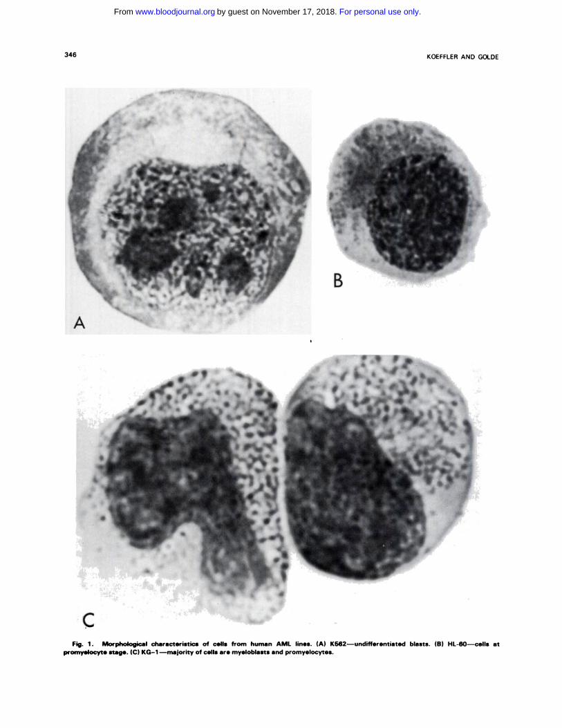
Representative Wright-Giemsa-stained prepara-
tions of the three cell lines are shown in Fig. I (A, B,
and C). The K562 cell is an undifferentiated blast cell
with a diameter of about 20 zm (Fig. I A). The cell has
a basophilic cytoplasm containing no granules and
there are two or more prominent nucleoli. The K562
cells do not stain with cytochemical reagents normally
positive in granulocytes and monocytes. Thus, there is
no reaction with peroxidase, Sudan black B, or ASD-
chloroacetate esterase stains. Many cells are strongly
reactive for acid phosphatase.
Most of the HL-60 cells are at the promyelocyte
stage of maturation and they contain prominent
azurophilic granules (Fig. I B). Myeloblasts, myelo-
cytes, and more mature granulocytic forms are occa-
sionally present. The HL-60 cells react strongly with
cytochemical stains specific for granulocytic cells,
includi ng peroxidase, AS D-chloroacetate esterase,
and Sudan black B.” The cells do not stain for alkaline
phosphatase and they appear not to contain lactofer-
nfl.
The KG-I line shows considerable pleomorphism
with most of the cells at the myeloblast and promyelo-
cyte stages, but lO%-20% of the cells are myelocytes
or mature granulocytes (Fig. IC). Occasional macro-
phages and eosinophils are also present. Clones
derived from the parent line show a similar degree of
pleomorphism. In later passages (greater than 1 .5 yr
For personal use only.on November 17, 2018. by guest www.bloodjournal.orgFrom
HUMAN MYELOID LEUKEMIA AND CELL LINES 345
Table 1 . Properties of Human Myeloi d Leukemia Cell Lines
K562 HL-60 KG-i
Source
Morphology and
histochemistry
Mean doubling time
Chromosome number
Lymphocyte markers
CSF response
Induction of dif-
ferentiation
CML in blast crisis
Undifferentiated blast
cell
1 2 hr
Modal 70 (Ph’ chromosome)
No
No
Erythroid
APL’
Promyelocyte
30-40 hr
Modal 45
No
Yes
Granulocyte or
macrophage
Erythroleukemia
Myeloblast-
promyelocyte
40-50 hr
47
No
Yes
Macrophage
APL. acute promyelocytic leukemia.
in culture), almost all of the cells are morphologically
at the myeloblast and promyelocyte stages. More than
90% of the cells stain strongly for ASD-chloroacetate
esterase, but only I%-2% are peroxidase-positive.’#{176}
The few macrophages present stain heavily for alpha
naphthyl butyrate esterase.
GROWTH KINETICS
All three cell lines grow in suspension culture as
single cells. The KG-I and HL-60 cells have a
doubling line consistent with AML cells in vivo (48-72
hr); K562 has a mean doubling time of I 2 hr.
KARYOTYPE
A prominent feature of the three cell lines is the
marked karyotypic abnormalities. Using banding
techniques, no common karyotypic abnormality is seen
in the three cell types. The K562 cells have nearly 1.5
times the normal number of chromosomes: 70 XXX,
- 13, - 17, +7, +9 (pll), plus small metacentnic
chromosome, Xp-, 3p-, 9p-, t(l5;18) (q21;q23),
r(22), or 22q- . The cells have a small ring chromo-
some r(22) or retain the Philadelphia chromosome
(22q-).’2 Several cytogenetically different clones of
HL-60 are present. The predominant karyotype is:
45x, -5, -8, - 16, -x, plus an A group marker, plus
an acrocentnic D group marker, plus a submetacentnic
E group marker.” The KG-I cells have shown a
consistent karyotype identical to the patient’s
leukemic cells: 47, xy, 7q-, 9p+, l2p+, -20,
+ MAR - 1 (large metacentnic), + MAR - 2 (small
fragment). ‘#{176}
LYMPHOCYTE MARKERS
The cells from all three lines do not have lympho-
cytic characteristics. They have no surface immuno-
globulin, Epstein-Barr virus-associated antigens, nor
do they form rosettes with sheep red cells. Only about
5% of the KG-l and HL-60 cell populations form
rosettes with erythrocytes coated with antibody (EA)
or antibody and complement (EAC). Over 90% of the
K562 cells form EA rosettes, indicating the presence
of Fc receptors. Terminal deoxynucleotidyl transfer-
ase, an enzyme present in immature lymphocytes and
rarely in AML cells, is not detectable in KG-I or
HL-60 cells.
CELL SURFACE ANTIGENS
The Ia-like or DR antigen is usually expressed on
human myeloid leukemia cells. The KG- I cells express
the antigen.’3 The antigen is not present on HL-60,
which is consistent with prior studies showing that at
the promyelocyte stage of development, the antigen is
usually lost.’3 The K562 cells also lack the Ia antigen.
There is evidence that the K562 cells are very early
erythroid precursors and that, perhaps, these cells
have not matured to the stage of Ia antigen expres-
sion.’�’6
As is the case with most hematopoietic cells, the
KG-l and HL-60 cells express HLA-A and HLA-B
antigens. The HL-60 cells are HLA-Al,8, Bl7; and
the KG- I cells express the rare antigens H LA-A30,3 1,
B35.’3 Neither HL-60 nor KG-I cells express acute
lymphocytic leukemia (ALL), granulocyte, or thymo-
cyte antigens. The K562 cell population lacks HLA,
ALL, and thymocyte antigens.
If a specific human acute myelogenous leukemia
cell-associated antibody could be isolated, it would
have great diagnostic and therapeutic potential.
Rabbit antisera have been raised to K562 and KG-l
cells and a primate antiserum was produced by immu-
nization of monkeys with K562.’7’9 The KG-I anti-
serum was produced by pretreating rabbits with anti-
leukocyte serum (ALS) and coating the immunizing
KG-I cells with antilymphocyte serum (ALS).’9 After
absorption with ALL cells, the KG- 1 antiserum
reacted with leukemic cells from 10 of 46 patients with
AML, and 2 of 23 ALL patients. The antiserum had
no activity against normal B or T lymphocytes, granu-
locytes, CML, or CLL cells, or normal granulocyte-
monocyte precursor cells (CFU-C). The K562 anti-
sera react with peripheral blood leukocytes from
For personal use only.on November 17, 2018. by guest www.bloodjournal.orgFrom
346 KOEFFLER AND GOLDE
B
CFig. 1 . Morphological characteristics of cells from human AMI lines. (A) K562-undifferentiated blasts. (B) HL-60-cells at
promyelocyte stage. (C) KG-i -majority of cells are myeloblasts and promyelocytes.
For personal use only.on November 17, 2018. by guest www.bloodjournal.orgFrom
125
100
wU,
+1
U)
Lu
z0-‘I0Q
75
50
25
0 GOl 011 110 10 ‘ ioo
CSF ( ul I ml
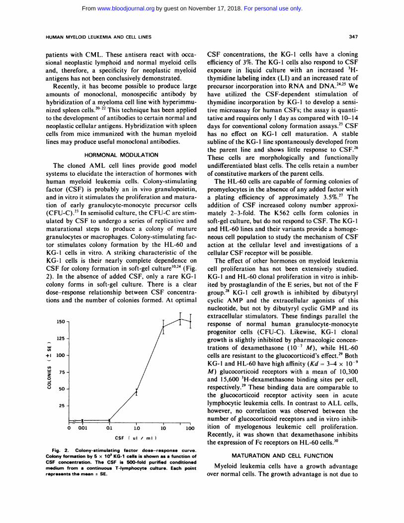
Fig. 2. Colony-stimulating factor dose-response curve.Colony formation by 5 x 10� KG-i cells is shown as a function ofCSF concentration. The CSF is 500-fold purified conditionedmedium from a continuous T-lymphocyte culture. Each point
HUMAN MYELOID LEUKEMIA AND CELL LINES 347
represents the mean ± SE.
patients with CML. These antisera react with occa-
sional neoplastic lymphoid and normal myeloid cells
and, therefore, a specificity for neoplastic myeloid
antigens has not been conclusively demonstrated.
Recently, it has become possible to produce large
amounts of monoclonal, monospecific antibody by
hybridization of a myeloma cell line with hypenimmu-
nized spleen cells.2#{176}22This technique has been applied
to the development of antibodies to certain normal and
neoplastic cellular antigens. Hybridization with spleen
cells from mice immunized with the human myeloid
lines may produce useful monoclonal antibodies.
HORMONAL MODULATION
The cloned AML cell lines provide good model
systems to elucidate the interaction of hormones with
human myeloid leukemia cells. Colony-stimulating
factor (CSF) is probably an in vivo granulopoietin,
and in vitro it stimulates the proliferation and matura-
tion of early granulocyte-monocyte precursor cells
(CFU-C).23 In semisolid culture, the CFU-C are stim-
ulated by CSF to undergo a series of replicative and
maturational steps to produce a colony of mature
granulocytes or macrophages. Colony-stimulating fac-
ton stimulates colony formation by the HL-60 and
KG-I cells in vitro. A striking characteristic of the
KG-l cells is their nearly complete dependence on
CSF for colony formation in soft-gel culture’#{176}’24(Fig.
2). In the absence of added CSF, only a rare KG-i
colony forms in soft-gel culture. There is a clear
dose-response relationship between CSF concentra-
tions and the number of colonies formed. At optimal
1’150
CSF concentrations, the KG-I cells have a cloning
efficiency of 3%. The KG-i cells also respond to CSF
exposure in liquid culture with an increased 3H-
thymidine labeling index (LI) and an increased rate of
precursor incorporation into RNA and DNA.24’25 We
have utilized the CSF-dependent stimulation of
thymidine incorporation by KG- 1 to develop a sensi-
tive microassay for human CSFs; the assay is quanti-
tative and requires only I day as compared with 10-14
days for conventional colony formation assays.25 CSF
has no effect on KG-l cell maturation. A stable
subline of the KG- I line spontaneously developed from
the parent line and shows little response to CSF.26
These cells are morphologically and functionally
undifferentiated blast cells. The cells retain a number
of constitutive markers of the parent cells.
The HL-60 cells are capable of forming colonies of
promyelocytes in the absence of any added factor with
a plating efficiency of approximately 3,5%,27 The
addition of CSF increased colony number approxi-
mately 2-3-fold. The K562 cells form colonies in
soft-gel culture, but do not respond to CSF. The KG-I
and HL-60 lines and their variants provide a homoge-
neous cell population to study the mechanism of CSF
action at the cellular level and investigations of a
cellular CSF receptor will be possible.
The effect of other hormones on myeloid leukemia
cell proliferation has not been extensively studied.
KG-l and HL-60 clonal proliferation in vitro is inhib-
ited by prostaglandin of the E series, but not of the F
group.2’ KG-I cell growth is inhibited by dibutyryl
cyclic AMP and the extracellular agonists of this
nucleotide, but not by dibutyryl cyclic GMP and its
extracellular stimulators. These findings parallel the
response of normal human granulocyte-monocyte
progenitor cells (CFU-C). Likewise, KG-i clonal
growth is slightly inhibited by pharmacologic concen-
trations of dexamethasone (l0� M), while HL-60
cells are resistant to the glucocorticoid’s effect.29 Both
KG-I and HL-60 have high affinity (Kd = 3-4 x i0�
M) glucocorticoid receptors with a mean of 10,300
and I 5,600 3H-dexamethasone binding sites per cell,
respectively.29 These binding data are comparable to
the glucocorticoid receptor activity seen in acute
lymphocytic leukemia cells. In contrast to ALL cells,
however, no correlation was observed between the
number of glucocorticoid receptors and in vitro inhib-
ition of myelogenous leukemic cell proliferation.
Recently, it was shown that dexamethasone inhibits
the expression of Fc receptors on HL-60 cells.3#{176}
MATURATION AND CELL FUNCTION
Myeloid leukemia cells have a growth advantage
over normal cells. The growth advantage is not due to
For personal use only.on November 17, 2018. by guest www.bloodjournal.orgFrom
.�-�
348 KOEFFLER AND GOLDE
A
----- I
the rapid rate of proliferation of the leukemia blast
cells, as these cells usually divide more slowly than
normal cells. The leukemic cells accumulate because
of their inability to mature to functional nondividing
end cells. Since maturation and proliferation are
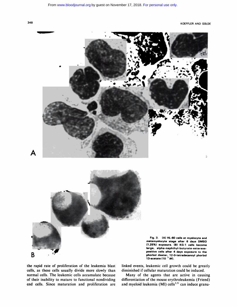
Fig. 3. (A) HL-60 cells at myelocyte and
metamyelocyte stage after 6 days DMSO(1 .25%) exposure. (B) KG-i cells become
large. alpha-naphthyl-butyrate-esterase-positive cells after 4 days exposure to thephorbol diester. 12-0-tetradecanoyl phorbol13-acetate (10 � M).
linked events, leukemic cell growth could be greatly
diminished ifcellular maturation could be induced.
Many of the agents that are active in causing
differentiation of the mouse erythroleukemia (Friend)
and myeloid leukemia (Ml) cells3’3’ can induce granu-
For personal use only.on November 17, 2018. by guest www.bloodjournal.orgFrom
HUMAN MYELOID LEUKEMIA AND CELL LINES 349
locyte maturation of the HL-60 cells, including dime-
thyl sulfoxide, butyric acid, triethylene glycol, N,N-
dimethylformamide, and N ,N ,-dimethylacetamide
(Fig. 3A).32 The mature HL-60 cells respond to
chemotaxins, phagocytose, develop complement recep-
tors, produce superoxide, and reduce NBT dye.33’34
The HL-60 cells do not have alkaline phosphatase or
lactoferrin, and do not appear to develop secondary
cytoplasmic granules.
Both HL-6035 and KG-I36 differentiate into nondi-
viding macrophage-appeaning cells when exposed to
phorbol esters (Fig. 3B). The cells from both lines
become adherent and develop long pseudopodia,
assume the morphological characteristics of macro-
phages, produce NADase, nonspecific acid esterase
and lysozyme, and develop Fc receptors and the ability
to phagocytize Candida albicans and kill Straphylo-
coccus aureus. Thus, these cell lines should be useful
in studying the mechanism of early myeloid differen-
tiation along the granulocytic and mononuclear patho-
cytic pathway.
Recent evidence suggests that some K562 sublines
may be a human erythroleukemia cell line with char-
actenistics similar to the mouse Friend erythroleu-
kemia cell Iine.’�’6’37’3’ The K562 cell membrane
glycoproteins show many similarities to erythrocytes
and, in particular, the cells synthesize glycophorin A,
which is found exclusively in human erythrocytes.’4’38
In the presence of sodium butyrate, some of the cells
become benzidine positive,’5’37 and hemoglobin can be
detected in these cells using a radioimmunoassay.’6
The K562 cells produce fetal and embryonic hemoglo-
bin when grown with 0.1 mM hemin. The K562 cell
appears to be an excellent tool for the study of human
erythroid differentiation and globin gene expression. It
seems clear, however, that there are a number of
strains of K562 and these various strains may have
different properties.
It is curious that human myeloid cell lines are so
difficult to establish when contrasted to the ease with
which lymphoid cell lines are developed. The develop-
ment of B-lymphoblast lines is clearly related to EB-
virus infection, but there also are various T- and
null-lymphoid lines available that are not obviously
virus infected. The difficulty in establishing myeloid
lines as compared to permanent lymphoid cultures
may relate to intrinsic cellular factors or requirements
for as yet unidentified growth factors. Possibly, the use
of somatic hybridization or DNA transformation
using existing myeloid cell lines will permit establish-
ment of new myeloid lines with interesting and useful
features.
The human myelogenous leukemia cell lines may
provide the framework for studies leading to a deeper
understanding of the control of differentiation in
human myeloid leukemia and, in a broader perspec-
tive, the control of normal cellular differentiation.
Future studies with these cell lines and selected van-
ants should shed new light on the cell biology of
human myeloid leukemia and perhaps lead to new
therapeutic modalities. The cells will be useful in the
development of monoclonal antibodies reactive with
myeloid leukemia cells and normal myeloid cells of
different stages of maturation. The lines and their
variants will help elucidate the interaction of CSF
with myeloid cells and provide a convenient tool for
pharmacologic and chemothenapeutic investigation.
The cells should also be of use in studies of the control
of granulocytic and macnophage differentiation. Final-
ly, it is possible that modern molecular biologic tech-
niques may permit the isolation from these cell lines of
genes controlling myeloid differentiation.
REFERENCES
1 . Friend C, Scher W, Holland JG, Sato T: Hemoglobin synthe-
sis in murine virus-induced leukemic cells in vitro: Stimulation of
erythroid differentiation by dimethyl sulfoxide. Proc Natl Acad Sci
USA 68:378, 1971
2. Ichikawa Y: Differentiation of a cell line of myeloid leukemia.
J Cell Physiol 74:223, 1969
3. Sachs L: Control of normal cell differentiation and the pheno-
typic reversion of malignancy in myeloid leukaemia. Nature
274:535, 1978
4. Clarkson B: Formal discussion: On the cellular origins and
distinctive features of cultured cell lines derived from patients with
leukemias and lymphomas. Cancer Res 26:2483, 1967
5. Clarkson B, Strife A, de Harven E: Continuous culture of
seven new cell lines (SK-L1 to 7) from patients with acute leukemia.
Cancer 20:926, 1967
6. Belpomme D, Minowada J, Moore GE: Are some human
lymphoblastoid cell lines established from Ieukemic tissues actually
derived from normal leukocytes? Cancer 30:282, 1972
7. Karpas A, Hayhoc FGJ, Greenberger iS, Barker CR, Cawley
ic, Lowenthal RM, Moloney WC: The establishment and cytologi-
cal, cytochemical and immunological characterisation of human
haemic cell lines: Evidence for heterogeneity. Leukemia Res 1:35,
I977
8. Lozzio CB, Lozzio BB: Human chronic myelogenous leukemia
cell-line with positive Philadelphia chromosome. Blood 45:321,
1975
9. Collins Si, Gallo RC, Gallagher RE: Continuous growth and
differentiation of human myeloid Ieukaemic cells in suspension
culture. Nature 270:347, 1977
10. Koeffier HP, Golde DW: Acute myelogenous leukemia: A
human cell line responsive to colony-stimulating activity. Science
200:1153, 1978
1 1 . Gallagher R, Collins S. Trujillo J, McCredie K, Ahearn M,
Tsai S. Metzgar R, Aulakh G, Ting R, Ruscetti F, Gallo R:
Characterization of the continuous, differentiating myeloid cell line
(HL-60) from a patient with acute promyelocytic leukemia. Blood
54:713, 1979
12. Klein E, Ben-Bassat H, Neumann H, Ralph P. Zeuthen i,
Polliack A, V#{225}nkyF: Properties of the K562 cell line, derived from a
patient with chronic myeloid leukemia. Int i Cancer 18:421, 1976
For personal use only.on November 17, 2018. by guest www.bloodjournal.orgFrom
350 KOEFFLER AND GOLDE
13. Koeffler HP, Billing R, Sparkes RS, Golde DW: Antigens
present on human myeloid leukemia cell lines. Leukemia Res 4:69,
I 980
14. Andersson IC, Nilsson K, Gahmberg CG: K562-A human
erythroleukemic cell line. Int J Cancer 23:143, 1979
15. Andersson LC, Jokinen M, Gahmberg CG: Induction of
erythroid differentiation in the human leukaemia cell line K562.
Nature 278:364, 1979
16. Rutherford TR, Clegg JB, Weatherall Di: K562 human
leukaemic cells synthesize embryonic haemoglobin in response to
haemin. Nature 280:164, 1979
17. Whitson ME, Lozzio CB, Lozzio BB, Wust Ci, Sonoda 1,
Avery B: Cytotoxicity of antisera to a myelogenous leukemia cell
line with the Philadelphia chromosome. J Nail Cancer Inst 56:903,
I 976
18. Lozzio BB, Lozzio CB, Krauss S. Wust Ci, Girardi A:
Leukemia-associated antigens detected by a nonhuman primate
antiserum to a Ph’ + myelogenous leukemia cell line. Blood 50: 1 15,
I977
19. Billing R, Clark B, Koefiler HP, Foon KA, Terasaki P1:
Acute myelogenous leukemia heteroantisera. Clin Immunol Immu-
nopathol July 1980 (in press)
20. Kohler G, Milstein C: Continuous cultures of fused cells
secreting antibody of predefinec specificity. Nature 256:495, 1975
21. Kohler G, Milstein C: Derivation of specific antibody-
producing tissue culture and tumor lines by cell fusion. Eur J
Immunol 6:51 1, 1976
22. Parham P, Bodmer WF: Monoclonal antibody to a human
histocompatibility alloantigen, HLA-A2. Nature 276:397, 1978
23. Golde DW, Cline MJ: Regulation ofgranulopoiesis. N EngI J
Med 291:1388, 1974
24. Koeffier HP, Golde DW: A human acute myelogenous
leukemia cell line, in Golde DW, Cline Mi, Metcalf D, Fox CF
(eds): Hematopoietic Cell Differentiation. New York, Academic,
1978, p 355
25. Koeffler HP, Lusis AJ: Effects of CSF on a human AML cell
line, KG-I: A rapid microassay for human CSF’s. Proc NatI Acad
Sci USA 1980 (in press)
26. Koeffier HP, Billing R, Sparkes RS, Golde DW: An undif-
ferentiated strain derived from the human acute myelogenous
leukemia cell line KG- I . Blood (in press)
27. Gallo R, Ruscetti F, Collins S. Gallagher R: Human myeloid
leukemia cells: Studies on oncornaviral related information and in
vitro growth and differentiation, in Golde DW, Cline Mi, Metcalf
D, Fox CF (eds): Hematopoietic Cell Differentiation. New York,
Academic, 1978, p 335
28. Koeffler HP, Golde DW: Humoral modulation of human
acute myelogenous leukemia cell growth in vitro. Cancer Res
40:1858, 1980
29. Koeffler HP, Golde DW, Lippman ME: Glucocorticoid
sensitivity and receptors in cells of human myelogenous leukemia
lines. Cancer Res 40:563, 1980
30. Crabtree GR, Munck A, Smith KA: Glucocorticoids inhibit
expression of Fc receptors on the human granulocytic cell line
HL-60. Nature 279:338, 1979
31. Tanaka M, Levy J, Terada M, Breslow R, Rifkind RA,
Marks PA: Induction of erythroid differentiation in murine virus
infected erythroleukemia cells by highly polar compounds. Proc
NatI Acad Sci USA 72:1003, 1975
32. Collins Si, Ruscetti FW, Gallagher RE, Gallo RC: Terminal
differentiation of human promyelocytic leukemia cells induced by
dimethyl sulfoxide and other polar compounds. Proc NatI Acad Sci
USA 75:2458, 1978
33. Collins SJ, Ruscetti FW, Gallagher RE, Gallo RC: Normal
functional characteristics of cultured human promyelocytic
leukemia cells (HL-60) after induction of differentiation by dime-
thylsulfoxide. J Exp Med 149:969, 1979
34. Newburger PE, Chovaniec ME, Greenberger JS, Cohen Hi:
Functional changes in human leukemic cell line HL-60. A model for
myeloid differentiation. J Cell Biol 82:315, 1979
35. Rovera G, O’Brien TG, Diamond L: Induction of differentia-
tion in human promyelocytic leukemia cells by tumor promoters.
Science 204:868, 1979
36. Koeffier HP, Bar-Eli M, Territo M: Heterogeneity of human
myeloid leukemia cell response to phorbol diester. BlOOd 54(Suppl
l):174a, 1979
37. Hoffman R, Murnane Mi, Benz Ei Jr. Prohaska R, Floyd V.
Dainiak N, Forget BG, Furthmayr H: Induction of erythropoietic
colonies in a human chronic myelogenous leukemia cell line. Blood
54:1182, 1979
38. Jokinen M, Gahmberg CG, Andersson LC: Biosynthesis of
the major human red cell sialoglycoprotein, glycophorin A, in a
continuous cell line. Nature 279:604, 1979
For personal use only.on November 17, 2018. by guest www.bloodjournal.orgFrom
1980 56: 344-350
HP Koeffler and DW Golde Human myeloid leukemia cell lines: a review
http://www.bloodjournal.org/content/56/3/344.full.htmlUpdated information and services can be found at:
Articles on similar topics can be found in the following Blood collections
http://www.bloodjournal.org/site/misc/rights.xhtml#repub_requestsInformation about reproducing this article in parts or in its entirety may be found online at:
http://www.bloodjournal.org/site/misc/rights.xhtml#reprintsInformation about ordering reprints may be found online at:
http://www.bloodjournal.org/site/subscriptions/index.xhtmlInformation about subscriptions and ASH membership may be found online at:
Copyright 2011 by The American Society of Hematology; all rights reserved.Hematology, 2021 L St, NW, Suite 900, Washington DC 20036.Blood (print ISSN 0006-4971, online ISSN 1528-0020), is published weekly by the American Society of
For personal use only.on November 17, 2018. by guest www.bloodjournal.orgFrom
Related Documents