Human Leukocyte Antigen E Contributes to Protect Tumor Cells from Lysis by Natural Killer Cells 1,2 Elisa Lo Monaco * , Elisa Tremante * , Cristina Cerboni † , Elisa Melucci ‡ , Leonardo Sibilio * ,3 , Alessandra Zingoni † , Maria Rita Nicotra * , Pier Giorgio Natali § and Patrizio Giacomini * *Laboratory of Immunology, Regina Elena Cancer Institute CRS, Rome, Italy; † Department of Molecular Medicine, University “La Sapienza,” Rome, Italy; ‡ Department of Pathology, Regina Elena Cancer Institute, Rome, Italy; § Consorzio Interuniversitario Nazionale per la Bio-Oncologia Laboratories, University “Gabriele d’Annunzio,” Chieti, Italy Abstract The nonclassic class I human leukocyte antigen E (HLA-E) molecule engages the inhibitory NKG2A receptor on sev- eral cytotoxic effectors, including natural killer (NK) cells. Its tissue distribution was claimed to be wider in normal than in neoplastic tissues, and surface HLA-E was undetectable in most tumor cell lines. Herein, these issues were re- investigated taking advantage of HLA-E–specific antibodies, immunohistochemistry, and biochemical methods de- tecting intracellular and surface HLA-E regardless of conformation. Contrary to published evidence, HLA-E was detected in a few normal epithelia and in a large fraction (approximately 1/3) of solid tumors, including those derived from HLA-E–negative/low-normal counterparts. Remarkably, HLA-E was detected in 30 of 30 tumor cell lines repre- sentative of major lymphoid and nonlymphoid lineages, and in 11 of 11, it was surface-expressed, although in a con- formation poorly reactive with commonly used antibodies. Coexpression of HLA-E and HLA class I ligand donors was not required for surface expression but was associated with NKG2A-mediated protection from lysis by the cytotoxic cell line NKL and polyclonal NK cells from healthy donors, as demonstrated by antibody-mediated relief of protection in 10% to 20% of the tested target-effector combinations. NKG2A-mediated protection of additional targets became evident on NK effector blocking with antibodies to activating receptors (DNAM-1, natural cytotoxicity receptors, and NKG2D). Thus, initial evidence that the long-elusive HLA-E molecule is enhanced by malignant transformation and is functional in tumor cells is presented here, although its importance and precise functional role remain to be addressed in the context of a general understanding of the NK ligand–receptor network. Neoplasia (2011) 13, 822–830 Abbreviations: CTL, cytotoxic T lymphocyte; DNAM-1, DNAX accessory molecule 1; E/T, effector-target; HLA, human leukocyte antigen(s); IFN, interferon; ILT, immunoglobulin- like transcript; KIR, killer immunoglobulin-like receptor; NCR, natural cytotoxicity receptor; NHEM, normal human epidermal melanocyte; NK, natural killer; PBMC, peripheral blood mononuclear cell; TcR, T-cell receptor; β 2 m, β 2 -microglobulin Address all correspondence to: Dr. Patrizio Giacomini, Laboratory of Immunology, Regina Elena Cancer Institute CRS, Via delle Messi d’Oro 156, 00158 Rome, Italy. E-mail: [email protected] 1 Work supported by the Italian Ministries of Public Health (C.C., A.Z., and P.G.), University and Research (MIUR) (C.C. and A.Z.), AIRC (C.C., A.Z., P.G.N., and P.G.), and the Center of Excellence (BEMM) Rome, Italy (C.C. and A.Z.). The authors have no conflict of interest. 2 This article refers to supplementary materials, which are designated by Tables W1 to W4 and Figures W1 to W6 and are available online at www.neoplasia.com. 3 Present address: Istituto Biochimico Italiano (IBI) Lorenzini, Via di Fossignano 2, 04011 Aprilia, Italy. Received 6 December 2010; Revised 22 July 2011; Accepted 28 July 2011 Copyright © 2011 Neoplasia Press, Inc. All rights reserved 1522-8002/11/$25.00 DOI 10.1593/neo.101684 www.neoplasia.com Volume 13 Number 9 September 2011 pp. 822–830 822

Welcome message from author
This document is posted to help you gain knowledge. Please leave a comment to let me know what you think about it! Share it to your friends and learn new things together.
Transcript

Volume 13 Number 9 September 2011 pp. 822–830 822
Abbreviatiolike transcrblood monAddress allE-mail: gia1Work supCenter of E2This artic3Present adReceived 6
CopyrightDOI 10.15
www.neoplasia.com
Human Leukocyte Antigen EContributes to Protect TumorCells from Lysis by NaturalKiller Cells1,2
ns: CTL, cytotoxic T lymphocyte; DNAM-1,DNAX accessorymolecule 1; E/T, eipt; KIR, killer immunoglobulin-like receptor; NCR, natural cytotoxicity receptoonuclear cell; TcR, T-cell receptor; β2m, β2-microglobulincorrespondence to: Dr. Patrizio Giacomini, Laboratory of Immunology, Regina [email protected] by the Italian Ministries of Public Health (C.C., A.Z., and P.G.), Universixcellence (BEMM) Rome, Italy (C.C. and A.Z.). The authors have no conflict ole refers to supplementary materials, which are designated by Tables W1 to W4 adress: Istituto Biochimico Italiano (IBI) Lorenzini, Via di Fossignano 2, 04011 ADecember 2010; Revised 22 July 2011; Accepted 28 July 2011
© 2011 Neoplasia Press, Inc. All rights reserved 1522-8002/11/$25.0093/neo.101684
Elisa Lo Monaco*, Elisa Tremante*,Cristina Cerboni†, Elisa Melucci‡,Leonardo Sibilio*,3, Alessandra Zingoni†,Maria Rita Nicotra*, Pier Giorgio Natali§
and Patrizio Giacomini*
*Laboratory of Immunology, Regina Elena Cancer InstituteCRS, Rome, Italy; †Department of Molecular Medicine,University “La Sapienza,” Rome, Italy; ‡Department ofPathology, Regina Elena Cancer Institute, Rome, Italy;§Consorzio Interuniversitario Nazionale per la Bio-OncologiaLaboratories, University “Gabriele d’Annunzio,” Chieti, Italy
AbstractThe nonclassic class I human leukocyte antigen E (HLA-E) molecule engages the inhibitory NKG2A receptor on sev-eral cytotoxic effectors, including natural killer (NK) cells. Its tissue distribution was claimed to be wider in normal thanin neoplastic tissues, and surface HLA-E was undetectable in most tumor cell lines. Herein, these issues were re-investigated taking advantage of HLA-E–specific antibodies, immunohistochemistry, and biochemical methods de-tecting intracellular and surface HLA-E regardless of conformation. Contrary to published evidence, HLA-E wasdetected in a few normal epithelia and in a large fraction (approximately 1/3) of solid tumors, including those derivedfrom HLA-E–negative/low-normal counterparts. Remarkably, HLA-E was detected in 30 of 30 tumor cell lines repre-sentative of major lymphoid and nonlymphoid lineages, and in 11 of 11, it was surface-expressed, although in a con-formation poorly reactive with commonly used antibodies. Coexpression of HLA-E and HLA class I ligand donors wasnot required for surface expression but was associated with NKG2A-mediated protection from lysis by the cytotoxiccell line NKL and polyclonal NK cells from healthy donors, as demonstrated by antibody-mediated relief of protectionin 10% to 20% of the tested target-effector combinations. NKG2A-mediated protection of additional targets becameevident on NK effector blocking with antibodies to activating receptors (DNAM-1, natural cytotoxicity receptors, andNKG2D). Thus, initial evidence that the long-elusive HLA-E molecule is enhanced by malignant transformation and isfunctional in tumor cells is presented here, although its importance and precise functional role remain to be addressedin the context of a general understanding of the NK ligand–receptor network.
Neoplasia (2011) 13, 822–830
ffector-target;HLA, human leukocyte antigen(s); IFN, interferon; ILT, immunoglobulin-r; NHEM, normal human epidermal melanocyte; NK, natural killer; PBMC, peripheral
lena Cancer Institute CRS, Via delle Messi d’Oro 156, 00158 Rome, Italy.
ty and Research (MIUR) (C.C. and A.Z.), AIRC (C.C., A.Z., P.G.N., and P.G.), and thef interest.nd Figures W1 to W6 and are available online at www.neoplasia.com.prilia, Italy.
Neoplasia Vol. 13, No. 9, 2011 HLA-E Expression in Tumors Lo Monaco et al. 823
IntroductionHuman leukocyte antigen E (HLA-E) is a nonclassic class I moleculerecognized by natural killer (NK) cells, CD8 cytotoxic T lymphocytes(CTLs), and a more recently described subset of CD8 effectors withmemory phenotype, called by some authors NK-CTLs [1–4]. NK cellsand certain CTL subsets engage cell surface HLA-E through hetero-dimeric lectin-like receptors, both inhibitory (CD94/NKG2A) andactivating (i.e., CD94/NKG2C). NK-CTLs primarily engage HLA-Ethrough (oligo)clonally rearranged T-cell receptor (TcR) and lyse targetcells, but they may also express NKG2A [1–4].Inhibition through NKG2A is possibly the most thoroughly under-
stood function of HLA-E. It requires the stabilization of the HLA-Eheavy chain through association with its light-chain subunit (β2m)and short peptide ligands cleaved from the signal sequences of “permis-sive” class I alleles (the classic HLA-A, -B, and -C and the nonclassicHLA-G heavy chains), with the aid of class I–dedicated chaperonessuch as TAP and tapasin (reviewed in Rodgers and Cook [5]).Coexpression of HLA-E and permissive alleles, crucial to this mecha-
nism of ligand donation/stabilization, is thought to protect the con-ceptus from a maternal hemiallogeneic response [6] and prevent theinappropriate recognition of somatic self [7], but HLA-Emay also favorimmunoevasion. For instance, some viral genomes encode proteinsacting as surrogate donors of HLA-E ligands [8,9], and ovarian carci-noma cells were shown to express increased levels of the ligand donorHLA-G as a result of interferon γ (IFN-γ) treatment [10]. However,because IFN-γ also upregulates antigen-presenting HLA-A, -B, and -Cmolecules, that is, a full set of major activating T-cell ligands, it is diffi-cult to predict the final outcome (evasion or tumor control) in this andin similar [11] situations.Unfortunately, the critical issue of whether HLA-E levels differ be-
tween normal and neoplastic tissues remains largely not addressed.For instance, immunohistochemistry detected HLA-E at several extra-placental locations, including normal white blood cells, liver, skin, andlung, but the reactive cell types were not specified [12]. Expressionin the skin was subsequently confirmed [11], and HLA-E was also de-tected in certain endothelia but not in the few tested glandular epithelia[13]. As to biochemistry and flow cytometry studies, the commonlyused 3D12 and MEM antibodies detected HLA-E polypeptides inthe soluble extracts and/or on the surface of only 10 of 37 [14] and4 of 31 [15] tumor cell lines. To complicate interpretations, HLA-Etranscripts could be detected in the absence of HLA-E polypeptides[15], and HLA-E polypeptides were detected at an intracellular loca-tion but not on the cell surface [11].On the basis of the available data, one might conclude that HLA-E is
expressed in an undefined, possibly wide, range of normal tissues, butonly in a few tumor cells in culture, either constitutively (seldom) or(possiblymore often) following IFN-γ treatment, providing a weak ratio-nale to investigate its function in tumor cells. Possibly for this reason,there are, to our knowledge, few published studies on this topic [11,16].A more recent study of ours may help to reinterpret some of these
results. In this study [17], it was shown that 3D12 and the MEMantibodies [11,13–15,18] selectively bind a subpopulation of unfoldedHLA-E molecules free of β2m, whereas biochemical approaches,among which the most effective is a reverse biotin labeling method,detect surface HLA-E regardless of conformation.Using the HLA-E–specific [17] MEM-E02 antibody, we report
herein the tissue distribution of HLA-E in normal nonlymphoid tissuesand their malignant counterparts. Through biotin labeling and cyto-toxicity assays, we measure surface HLA-E expression and assess for
the first time the NK-inhibitory function of HLA-E constitutivelyexpressed under the control of its own promoter in untransfected neo-plastic cell lines, in the presence and absence of permissive alleles.The findings reported herein reconcile previous conflicting results,alleviate some theoretical inconsistencies, and bear several implicationsin tumor immunology.
Materials and Methods
ImmunohistochemistryNeoplastic tissues from patients (free from therapy) undergoing
surgery were obtained upon written consent following the recommen-dations of the latest (March 1, 2006) Regina Elena Institute EthicalCommittee Official Guideline. For further details, see SupplementalMaterials and Methods.
Cell LinesThe 221B lymphoblastoid cell line and its transfectants, namely,
221.AEH [19], 221.G1, 221.B15 [20], and 221.B*0702 [21], wereobtained through the courtesy of different investigators and/or the col-laborative efforts of the HLA-G and -E Workshops (see acknowledg-ments). Epstein-Barr virus–immortalized B lymphocytes (EBV-B) andtumor cell lines are listed in the Supplemental Materials and Methodsalong with their HLA-A, -B, and -C typing. Some of these are earlypassage (<10 subcultures) tumor cells previously established [22]. Nor-mal human epidermal melanocytes (NHEMs) were purchased fromLonza (Walkersville, MD). HLA-E genotyping was obtained by directsequencing of genomic DNA, as described [17].
Biochemical MethodsThe mouse monoclonal antibodies MEM-E/02, MEM-E/06,
MEM-E/07, and MEM-E/08 [13,17,18] are all from Exbio, Prague,Czech Republic; 3D12, 4D12 [14,23], W6/32 [24], Namb-1 [25], L31[26], and a polyclonal to ERp57 were used in previous publicationsof ours [22,26–29]. The reverse surface biotin labeling method isdescribed [17].
Flow CytometryTumor cells and peripheral blood mononuclear cells (PBMCs)/
purified NK cells were stained on ice with either fluorochrome-labeledantibodies or with a predetermined optimal (10 μg/ml) concentrationof primary antibody/chimeric immunoglobulin (Ig). In the latter case,primary antibody binding was revealed by fluorescence isothiocyanate–labeled rabbit antibodies to either mouse or human Ig (Dako, Glostrup,Denmark). Isotype-matched control antibodies, or a chimeric Ig ofirrelevant specificity, were included as negative controls. Specificallybound fluorescence was immediately analyzed without fixation by aFACScan flow cytometer (Becton, Dickinson & Co, Mountain View,CA). Antibodies toMICA (159227),MICB (MAB), ULBP1 (170818),ULBP2 (165903), ULBP3 (166510), NKG2A (131411), NKG2C(134591), NKG2D (149810), DNAX accessory molecule 1 (DNAM-1/CD226) (102511), NKp30 (210845), NKp44 (253415), NKp46/CD335 (195314), and recombinant human Fc chimeras to activatingimmune receptors DNAM-1-Fc, NKp30-Fc, NKp44-Fc, and NKp46-Fc were from R&D Systems (Minneapolis, MN). SKII.4 to polyo-virus receptor (CD155) was from Dr Marco Colonna. Antibody toNectin-2 was from BD Pharmingen (San Jose, CA). Antibodies toCD3 (UCHT1) and CD56 (MOC-1) were from Dako. Antibodies

824 HLA-E Expression in Tumors Lo Monaco et al. Neoplasia Vol. 13, No. 9, 2011
to killer Ig-like receptor (KIR) KIR2DL2/DL3 (DX27), KIR3DL1(DX9), and KIR2DL1/DS1 (11PB6) were fromMiltenyi Biotec GmbH(Bergisch Gladbach, Germany). Antibodies to Ig-like transcript (ILT)ILT2 (GHI/75) were from Biolegend (San Diego, CA).
NK Cell–Mediated CytotoxicityThe continuous NK cell line NKL [30] expresses NKG2A and ILT2
[31]. Polyclonal NK cells (typically 60% to 75% CD56+/CD3−, and60% to 80% NKG2A+) were established by culturing healthy donorPBMCs in vitro for 10 to 12 days on feeder layers of RPMI 8866 cells,as described [32]. They were further purified (>98% CD56+/CD3−) bynegative immunomagnetic selection (Miltenyi). Cytotoxicity was mea-sured by a standard 4-hour 51Cr release assay, at the indicated effector-target (E/T) ratios, using as targets 5 × 10³ cells per microplate well intriplicate. Antibodies (10 μg/ml) or Ig fragment antigen binding 2 frag-ments (7.5 μg/ml) to HLA molecules and NK cells were separately in-cubated at room temperature for 15 minutes with target and effectorcells, respectively, before dispensing into the 96-well microplate.
Results
Tissue Distribution of HLA-E in Normal Adult Tissues andNeoplastic Lesions
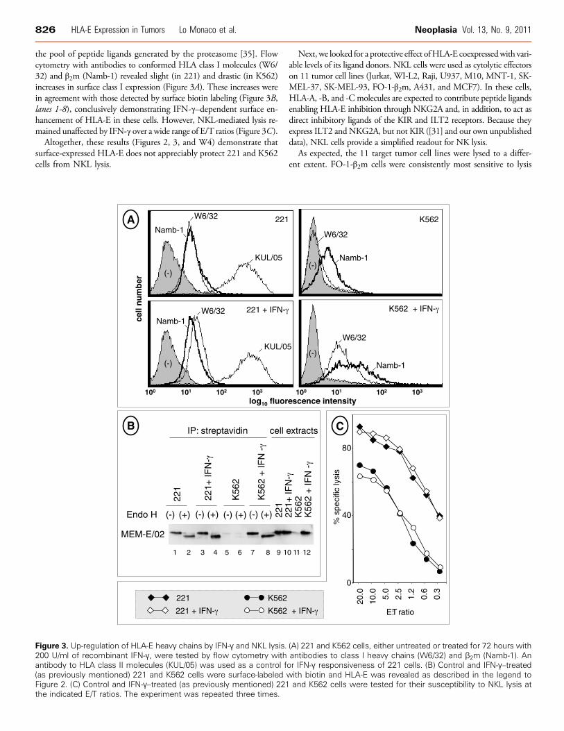
Immunohistochemical staining with MEM-E/02 gave a fine ground-glass pattern sparing the nuclei in a few normal tissues (Figure 1,A-C , andTable W1) and in a much wider spectrum of tumor lesions (Figure 1,D-F ). Frequency and intensities of positive reactions were different indifferent histotypes (Table W2). For instance, ovarian and testiculartumors, non–small cell lung carcinomas, soft tissue tumors (Figure 1D),as well as cutaneous melanoma (Figure 1F ), most of which derive froma tested counterpart in which HLA-E was undetectable, displayed fromintermediate to high frequencies of MEM-E/02 reactivity. Unlike endo-
Figure 1. Immunohistochemical analysis of HLA-E. Normal (A-C) anMEM-E/02 and nuclear counterstained with Mayer hematoxylin. A grcells in the epididymis (A, arrows), in endometrial cells (B, arrows), atumors, variable expression was seen in a case of osteosarcoma (Dmetastatic melanoma (F). Original magnifications, ×160 (A-C, E); ×2
thelia from normal tissues, some tumor endothelia were clearly reactive(Figure 1C). HLA-E down-regulation was exclusively observed in 50%approximately of endometrial carcinoma lesions (Table W2; Figure 1Edisplays a positive case). In summary, one third of the tumor lesions werestained by MEM-E/02, most often because enhancement or de novoappearance associated with malignant transformation.
Widespread Accumulation and Surface Expression of HLA-EPolypeptides in Tumor Cells
Western blot analysis with MEM-E/02 of 24 tumor cell lines fromrepresentative lineages (Figure W1) detected HLA-E polypeptides ofthe correct 42-kDa size at extremely variable levels. All the tumor cellswere clearly positive, including the K562 cell line that was found to ex-press the lowest HLA-E levels, only detectable on long filter exposure(see below).
Next, 33 tumor cell lines were assayed by flow cytometry with theHLA-E–restricted MEM-E/02, MEM-E/06, MEM-E/07, and MEM-E/08 antibodies, and 15 were also tested with the 3D12 and 4D12 anti-bodies. Although four of these cells (U937, Jurkat, Raji, and K562) hadpreviously been shown to differ in 3D12 surface binding [14], all theantibodies including 3D12 and 4D12 gave a dull fluorescence patternin our hands, with differences among antibodies and cell lines beingtoo small to represent a reliable ranking method (representative resultsare shown in Figure W2 and Table W3).
To detect surface HLA-E molecules regardless of antibody reactivity,11 tumor cell lines and two cultures of NHEMs (NHEM1 andNHEM2) were assessed for surface HLA-E expression by reverse bio-tin labeling [17]. Because there are two nonsynonymous HLA-E alleles[33] with high (HLA-E107G) and low (HLA-E107R) surface expression[34], all the cells were HLA-E typed.
As opposed to the similar, dull staining detected by flow cytome-try, biotin labeling (Figure 2A) detected widely different levels of sur-face HLA-E. Cells lacking coexpression of permissive alleles (221,
d neoplastic (D-F) nonlymphoid human tissues were stained withound-glass pattern was detected in the principal but not the basalnd in the vascular endothelium of the myometrium (C, arrows). In), a well-differentiated endometrial carcinoma (E), and an in transit50 (D and F). Scale bars, 100 μm.
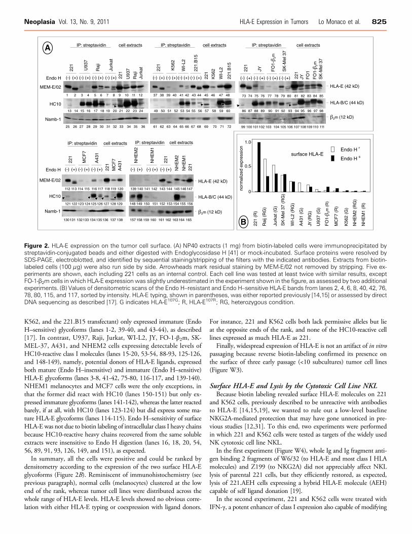
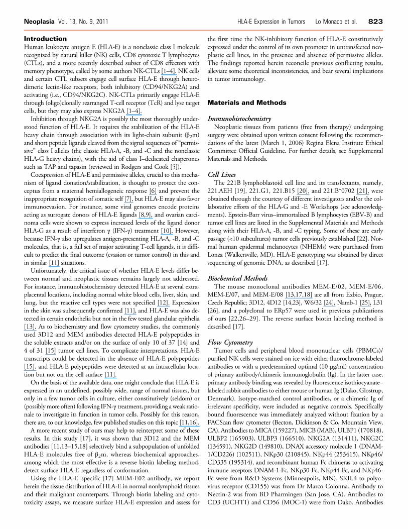
Figure 2. HLA-E expression on the tumor cell surface. (A) NP40 extracts (1 mg) from biotin-labeled cells were immunoprecipitated bystreptavidin-conjugated beads and either digested with Endoglycosidase H [41] or mock-incubated. Surface proteins were resolved bySDS-PAGE, electroblotted, and identified by sequential staining/stripping of the filters with the indicated antibodies. Extracts from biotin-labeled cells (100 μg) were also run side by side. Arrowheads mark residual staining by MEM-E/02 not removed by stripping. Five ex-periments are shown, each including 221 cells as an internal control. Each cell line was tested at least twice with similar results, exceptFO-1-β2m cells in which HLA-E expression was slightly underestimated in the experiment shown in the figure, as assessed by two additionalexperiments. (B) Values of densitometric scans of the Endo H–resistant and Endo H–sensitive HLA-E bands from lanes 2, 4, 6, 8, 40, 42, 76,78, 80, 115, and 117, sorted by intensity. HLA-E typing, shown in parentheses, was either reported previously [14,15] or assessed by directDNA sequencing as described [17]. G indicates HLA-E107G; R, HLA-E107R; RG, heterozygous condition.
Neoplasia Vol. 13, No. 9, 2011 HLA-E Expression in Tumors Lo Monaco et al. 825
K562, and the 221.B15 transfectant) only expressed immature (EndoH–sensitive) glycoforms (lanes 1-2, 39-40, and 43-44), as described[17]. In contrast, U937, Raji, Jurkat, WI-L2, JY, FO-1-β2m, SK-MEL-37, A431, and NHEM2 cells expressing detectable levels ofHC10-reactive class I molecules (lanes 15-20, 53-54, 88-93, 125-126,and 148-149), namely, potential donors of HLA-E ligands, expressedboth mature (Endo H–insensitive) and immature (Endo H–sensitive)HLA-E glycoforms (lanes 3-8, 41-42, 75-80, 116-117, and 139-140).NHEM1 melanocytes and MCF7 cells were the only exceptions, inthat the former did react with HC10 (lanes 150-151) but only ex-pressed immature glycoforms (lanes 141-142), whereas the latter reactedbarely, if at all, with HC10 (lanes 123-124) but did express some ma-ture HLA-E glycoforms (lanes 114-115). Endo H–sensitivity of surfaceHLA-E was not due to biotin labeling of intracellular class I heavy chainsbecause HC10-reactive heavy chains recovered from the same solubleextracts were insensitive to Endo H digestion (lanes 16, 18, 20, 54,56, 89, 91, 93, 126, 149, and 151), as expected.In summary, all the cells were positive and could be ranked by
densitometry according to the expression of the two surface HLA-Eglycoforms (Figure 2B). Reminiscent of immunohistochemistry (seeprevious paragraph), normal cells (melanocytes) clustered at the lowend of the rank, whereas tumor cell lines were distributed across thewhole range of HLA-E levels. HLA-E levels showed no obvious corre-lation with either HLA-E typing or coexpression with ligand donors.
For instance, 221 and K562 cells both lack permissive alleles but lieat the opposite ends of the rank, and none of the HC10-reactive celllines expressed as much HLA-E as 221.
Finally, widespread expression of HLA-E is not an artifact of in vitropassaging because reverse biotin-labeling confirmed its presence onthe surface of three early passage (<10 subcultures) tumor cell lines(Figure W3).
Surface HLA-E and Lysis by the Cytotoxic Cell Line NKLBecause biotin labeling revealed surface HLA-E molecules on 221
and K562 cells, previously described to be unreactive with antibodiesto HLA-E [14,15,19], we wanted to rule out a low-level baselineNKG2A-mediated protection that may have gone unnoticed in pre-vious studies [12,31]. To this end, two experiments were performedin which 221 and K562 cells were tested as targets of the widely usedNK cytotoxic cell line NKL.
In the first experiment (Figure W4), whole Ig and Ig fragment anti-gen binding 2 fragments of W6/32 (to HLA-E and most class I HLAmolecules) and Z199 (to NKG2A) did not appreciably affect NKLlysis of parental 221 cells, but they efficiently restored, as expected,lysis of 221.AEH cells expressing a hybrid HLA-E molecule (AEH)capable of self ligand donation [19].
In the second experiment, 221 and K562 cells were treated withIFN-γ, a potent enhancer of class I expression also capable of modifying
826 HLA-E Expression in Tumors Lo Monaco et al. Neoplasia Vol. 13, No. 9, 2011
the pool of peptide ligands generated by the proteasome [35]. Flowcytometry with antibodies to conformed HLA class I molecules (W6/32) and β2m (Namb-1) revealed slight (in 221) and drastic (in K562)increases in surface class I expression (Figure 3A). These increases werein agreement with those detected by surface biotin labeling (Figure 3B,lanes 1-8), conclusively demonstrating IFN-γ–dependent surface en-hancement of HLA-E in these cells. However, NKL-mediated lysis re-mained unaffected by IFN-γ over a wide range of E/T ratios (Figure 3C).
Altogether, these results (Figures 2, 3, and W4) demonstrate thatsurface-expressed HLA-E does not appreciably protect 221 and K562cells from NKL lysis.
Figure 3. Up-regulation of HLA-E heavy chains by IFN-γ and NKL lysis.200 U/ml of recombinant IFN-γ, were tested by flow cytometry withantibody to HLA class II molecules (KUL/05) was used as a control f(as previously mentioned) 221 and K562 cells were surface-labeledFigure 2. (C) Control and IFN-γ–treated (as previously mentioned) 221the indicated E/T ratios. The experiment was repeated three times.
Next, we looked for a protective effect ofHLA-E coexpressedwith vari-able levels of its ligand donors. NKL cells were used as cytolytic effectorson 11 tumor cell lines (Jurkat, WI-L2, Raji, U937, M10, MNT-1, SK-MEL-37, SK-MEL-93, FO-1-β2m, A431, and MCF7). In these cells,HLA-A, -B, and -C molecules are expected to contribute peptide ligandsenabling HLA-E inhibition through NKG2A and, in addition, to act asdirect inhibitory ligands of the KIR and ILT2 receptors. Because theyexpress ILT2 and NKG2A, but not KIR ([31] and our own unpublisheddata), NKL cells provide a simplified readout for NK lysis.
As expected, the 11 target tumor cell lines were lysed to a differ-ent extent. FO-1-β2m cells were consistently most sensitive to lysis
(A) 221 and K562 cells, either untreated or treated for 72 hours withantibodies to class I heavy chains (W6/32) and β2m (Namb-1). Anor IFN-γ responsiveness of 221 cells. (B) Control and IFN-γ–treatedwith biotin and HLA-E was revealed as described in the legend toand K562 cells were tested for their susceptibility to NKL lysis at

Figure 4. Cytotoxic activity of polyclonal NK cells against tumor cells. (A) CD56+/CD3− effector cells were tested in a 51Cr release assay,in the presence of antibodies to CD56 (C218) and to NKG2A (Z199) on the indicated targets. (B) Summary of cytotoxicity results (at an E/Tratio of 20:1) with NK cells from 6 donors and 10 tumor cell targets in presence of either C218 (−) or Z199 (+). A line connects statis-tically significant lytic enhancements by Z199, that is, differences in average values of triplicates more than three times the SD.
Neoplasia Vol. 13, No. 9, 2011 HLA-E Expression in Tumors Lo Monaco et al. 827
(representative results shown in Figure W5) and displayed the largestrepertoire of activating NK ligands among the five selected cell lines(Table W4). No other obvious correlations were apparent between sus-ceptibility to lysis and expression of NK ligands, including HLA-E.For instance, FO-1-β2m, A431, and U937 cells were lysed to a differ-ent extent (Figure 4) in spite they express similar (intermediate) surfaceHLA-E levels (compared with Figure 2B). As also shown by repre-sentative results in Figure W5, NKL lysis of 7 of 11 cell lines was un-affected by antibodies to ILT2 (GHI/75) and NKG2A (Z199), butGHI/75 enhanced lysis in the remaining 4 cell lines (FO-1-β2m, A431,M10, and SK-Mel 37), 2 of which (FO-1-β2m and A431) becamemore susceptible on incubation with antibodies to NKG2A. We con-clude that a fraction of tumor targets can be demonstrated to be pro-tected through ILT2 and, possibly to a lesser extent, through NKG2A.
Surface HLA-E and Lysis by Polyclonal NK CellsOne may wonder whether the role of the HLA-E/NKG2A axis
might be similarly appreciated using a panel of nonimmortalized effec-tor NK cells. Polyclonal NK cells (>98% CD56+/CD3−) were thenobtained by immunomagnetic sorting from the PBMCs of six differ-ent healthy donors cultured on feeder layers without interleukin 2, asdescribed [32]. Like NKL, these NK cell populations expressed highlevels of inhibitory receptors, not only NKG2A, NKG2D, and ILT2but also KIRs, and in addition they also expressed activating recep-tors such as DNAM-1 and the natural cytotoxicity receptors (NCRs)NKp30, NKp44, and NKp46 (for additional details, see Figure W6).Polyclonal NK cells were used as effectors in a cytotoxicity assay using astargets the immortalized JY B lymphoid line and nine tumor cell lines(Jurkat, WI-L2, Raji, U937, SK-MEL-37, SK-MEL-93, FO-1-β2m,
A431, and MCF7). The protective role of NKG2A from NK lysis wasassessed as previously mentioned in the presence of the NKG2A-specificantibody Z199.
NKG2A-mediated protection (i.e., protection in donor 1:JY anddonor 3:A431 but not in donor 3:MCF7; Figure 4A) was detected ina minority of E/T combinations. A complete synopsis is provided inFigure 4B in which the highest E/T ratios are shown for simplicity.Altogether, NKG2A-mediated protection was observed in 7 (23% ap-proximately) of 27 cases (see connecting bars). As expected, six distinctpatterns were observed, that is, the six polyclonal NK cell populationsdiffered from one another and from NKL cells in their ability to kill agiven target and to display NKG2A-mediated protection. Overall, fourof six NK cell cultures could be demonstrated to be inhibited throughNKG2A with at least one target. Similar experiments performed withbulk NK cells (not submitted to immunomagnetic purification) fromfour donors resulted in 5 (11% approximately) of 44 Z199-mediatedlysis enhancements (not shown).
Functional Role of NKG2A in the Context of ActivatingNK Cell Circuitry
In the attempt to account for the low percentage of E/T combinationsin which the inhibitory role of NKG2A is appreciable, we hypothesizedthat a strong overriding activation might obscure NKG2A-mediatedinhibition. To obtain evidence in favor of this hypothesis, a panel ofthree long-term (FO-1-β2m, SK-MEL-37, and A431) and four earlypassage (Mel20, Br7, End7, and Ova7) tumor cell lines expressing oneor more major NK-activating ligands (Table W4) were tested in cyto-toxicity as targets of polyclonal NK cells from three distinct donors ex-pressing essentially all the cognate activating receptors (Figure W6).

828 HLA-E Expression in Tumors Lo Monaco et al. Neoplasia Vol. 13, No. 9, 2011
Z199-mediated lysis enhancement was assessed not only in the absencebut also in the presence of mixtures of antibodies blocking those activat-ing receptors known to bind the triggering ligands detectable (TableW4)on the target.
Representative results (Figure 5) confirmed that Z199 by itself en-hances NK lysis in a minority (5/21) of E/T combinations, but onblocking of activating receptors and the consequent reduction in NKlysis (observed in 14/21 E/T combinations), antibodies to NKG2Abecome capable of restoring susceptibility to lysis of three additionaltargets. It may be concluded that overriding activation (by DNAM-1and/or NCR and/or NKG2D) may prevent appreciation of the in-hibitory HLA-E/NKG2A axis.
DiscussionConsistent with their primary and specialized role at the fetal-maternalinterface, the expression of several nonclassic class I HLA moleculesis believed to be highly restricted [5,6]. The extensive immunohisto-chemical testing performed herein is consistent with this view and, inaddition, shows that the few normal epithelia and tissues that constitu-
Figure 5. Block of activating receptors reveals NKG2A-mediatedprotection from NK lysis. The indicated tumor cell lines were testedas targets in a 51Cr release assay using CD56+/CD3− cells as ef-fectors, in the presence of the indicated antibodies. Blocking anti-bodies to activating receptors were selected on the basis of theligands expressed by the target: A431 (DNAM-1), End7 (NKG2D +DNAM-1), SK-MEL 37 (DNAM-1), andOva7 (NKG2D+DNAM-1). Dif-ferences in 51Cr release values were statistically significant whenthe average values of triplicates differed from control more thanthree times the SD, that is, at E/T ratios from 20:1 to 2.5:1 in A431:donor 1 as well as End7:donor 1 and at E/T ratios of 20:1 and 10:1 inA431:donor 3. The top two panels display two E/T combinationsin which the inhibitory effect of NKG2A becomes evident only whenactivating receptors are antibody-blocked. End7:donor 1 exemplifiesa case in which NKG2A similarly protects in the presence andabsence of antibody block. In the remaining cases, activating recep-tors and NKG2A cannot be demonstrated to influence susceptibilityto lysis.
tively express HLA-E are from the genitourinary/reproductive tract andthe colonic mucosa. Perhaps, HLA-E contributes to safeguardingsomatic self integrity in these normal tissues.
In contrast, HLA-E expression is wider than previously appreciatedin malignant tissues, one third of which (including those derived froman HLA-E–negative counterpart) are reactive with MEM-E/02 (Fig-ure 1 and Tables W1 and W2). Herein, we present evidence support-ing a functional role of HLA-E expression in tumors.
Widespread Surface Expression of HLA-E in Tumor Cells:Comparison with Previous Studies
Two previous flow cytometry studies with either 4D12 or MEMantibodies detected weak surface expression of HLA-E in a minorityof tumor cell lines [14,15]. In our hands, however, the same anti-bodies resulted in a dull and substantially invariant surface fluorescencepattern on 33 tumor cell lines, 11 of which also tested in the two pre-viously mentioned studies (Figures W2 and 3 and Table W3). Onlyby reverse biotin labeling (Figures 2 and 3) were we able to semi-quantitatively detect a wide range of HLA-E levels. HLA-E was in-variably surface-expressed under the species of two distinct glycoforms.Surprisingly, tumor cells previously [14] ranked by flow cytometryin the order U937 > Jurkat > Raji and K562 (Raji and K562 bothnegative) were ranked by reverse biotin labeling in the order Raji ≅Jurkat >> U937 > K562 (all positive), regardless of the glycoformbeing considered.
A detailed comparison among flow cytometry studies is difficultbecause few staining profiles are published, and/or the results arecharted on the basis of semiquantitative, subjective evaluation scales.Regardless of protocols and interpretations, it may be concluded thatHLA-E molecules, although poorly reactive (at least in our hands)with the available antibodies, are invariably present on the surfaceof tumor cell lines.
HLA-E Contributes to Protect Tumors from NK Cell LysisAs a first step to assess the function of HLA-E on tumor cells, so
far supported by limited evidence [10,16], we focused on its well-known ability to protect from NK cell lysis through NKG2A ex-pressed by the cytotoxic cell line NKL and resting polyclonal NKcells from healthy donors. Specific antibodies to NKG2A relievedprotection in the case of some tumor cell lines coexpressing permis-sive alleles but not in the case of parental 221 (B lymphoblastoid)and K562 (erythromyeloid) cells lacking permissive alleles (Figures 3–5 and W3). We conclude that surface HLA-E molecules are func-tional in at least some tumor cell lines but not in 221 and K562 despitethat these cells do express HLA-E, both constitutively and because ofIFN-γ treatment.
The observation that protection occurred in a minority (10%-20%)of the tested E/T combinations is reminiscent of the bare minority ofHLA-A2–restricted CTL clones to melanoma cells in which NKG2A-mediated inhibition is detectable [16] and is not surprising, given themany differences in lineage, genetic background, and intrinsic suscep-tibility to lysis among tumor targets. Most likely, a redundancy in ac-tivating and inhibitory ligand-receptor interactions masks or quenchesthe specific contribution of HLA-E in certain tumor-NK and tumor-CTL lytic combinations. Experiments (Figure 5) in which the inhibi-tory role of NKG2A could be appreciated only upon block of activatingNK receptors (DNAM-1 and/or NCRs and/or NKG2D) provide evi-dence in support of this interpretation.

Neoplasia Vol. 13, No. 9, 2011 HLA-E Expression in Tumors Lo Monaco et al. 829
A Model for the Regulation and Function of HLA-E in TumorsThere is evidence that HLA-E may favor both tumor escape and
tumor immune surveillance. Tumor escape is supported by a nega-tive prognostic association of HLA-E expression in colorectal andbreast carcinomas [36,37]. A possible mechanism has been suggestedby at least two groups [14,36]. In this model, selective loss of HLA-A,-B, and -C alleles (a phenotype proposed by some to be frequentlyassociated with immune evasion) alleviates competition for β2m, al-lowing ligand donation from the residual class I allele(s) in amounts suf-ficient to stabilize HLA-E, enhance its surface expression, engage theinhibitory NKG2A receptor, and further promote immune escape.However, three lines of evidence argue against the previously men-
tioned model and a purely negative role of HLA-E on survival: 1)although HLA-E may have a low affinity for β2m in defined model sys-tems [34], there is a subset of HLA-E molecules that efficiently bindsβ2m [17]; 2) HLA-E is widely expressed in human tumors, as shownherein; and 3) high HLA-E seems to correlate with good, and not poor,prognosis in melanoma and glioblastoma [38–40].By providing evidence that HLA-E is a widespread tumor recogni-
tion structure enabling NK cell recognition and monitoring, the pres-ent study suggests that HLA-E might represent in at least some tumorsa very efficient checkpoint and failsafe mechanism against all kinds ofHLA losses and not a vulnerable soft spot toward immune evasion intumor mutants that have already accumulated multiple HLA defects.It will be of interest to determine whether HLA-E on the tumor
cell surface engages rearranging and nonrearranging receptors, eitheractivating or inhibitory, expressed on different populations of NK,CTL, and NK-CTL effectors, and whether it modulates immunelysis. The applicable importance of HLA-E in tumor immunology re-mains to be precisely addressed on the clonal level and in the contextof a global appreciation of the immune ligand–receptor network.
AcknowledgmentsThe authors thank Marco Colonna for antibodies to polyovirus re-ceptor; Daniel E. Geraghty for 221.AEH, 221.G1, 3D12 and 4D12;and Michael J. Robertson for NKL cells. Rocco Fraioli and DinaMilana are gratefully acknowledged for skillful technical assistance.Maria Vincenza Sarcone and Paula Franke provided secretarial sup-port and revised the English text.
References[1] Braud VM, Allan DSJ, O’Callaghan CA, Söderström K, D’Andrea A, Ogg GS,
Lazetic S, Young NT, Bell JI, Phillips JH, et al. (1998). HLA-E binds to naturalkiller cell receptors CD94/NKG2A, B and C. Nature 391, 795–799.
[2] Garcia P, Llano M, Heredia AB, Willberg CB, Caparros E, Aparicio P, BraudVM, and Lopez-Botet M (2002). Human T cell receptor–mediated recognitionof HLA-E. Eur J Immunol 32, 936–944.
[3] Pietra G, Romagnani C, Falco M, Vitale M, Castriconi R, Pende D, Millo E,Anfossi S, Biassoni R, Moretta L, et al. (2002). The analysis of the natural killer–like activity of human cytolytic T lymphocytes revealed HLA-E as a novel targetfor TCRα/β–mediated recognition. Eur J Immunol 31, 3687–3693.
[4] Moretta L, Romagnani C, Pietra G, Moretta A, and Mingari MC (2003). NK-CTLs, a novel HLA-E–restricted T-cell subset. Trends Immunol 24, 136–143.
[5] Rodgers J and Cook R (2005). MHC class Ib molecules bridge innate and acquiredimmunity. Nat Rev Immunol 5, 459–471.
[6] Houlihan JM, Biro PA, Harper HM, Jenkinson HJ, and Holmes CH (1995).The human amnion is a site of MHC class Ib expression: evidence for theexpression of HLA-E and HLA-G. J Immunol 154, 5665–5674.
[7] Carosella ED, Paul P, Moreau P, and Rouas-Freiss N (2000). HLA-G and
HLA-E: fundamental and pathophysiological aspects. Immunol Today 21,532–534.
[8] Tomasec P, Braud VM, Rickards C, Powell MB, McSharry BP, Gadola S,Cerundolo V, Borysiewicz LK, McMichael AJ, and Wilkinson GWG (2000).Surface expression of HLA-E, an inhibitor of natural killer cells, enhanced byhuman cytomegalovirus gpUL40. Science 287, 1031–1033.
[9] UlbrechtM,Martinozzi S, Grzeschik M, Hengel H, Ellwart JW, Pla M, andWeissEH (2000). The human cytomegalovirus UL40 gene product contains a ligand forHLA-E and prevents NK cell–mediated lysis. J Immunol 164, 5019–5022.
[10] Malmberg K, Levitsky V, Norell H, de Matos CT, Carlsten M, Schedvins K,Rabbani H, Moretta A, Soderstrom K, Levitskaya J, et al. (2002). IFN-γ protectsshort-term ovarian carcinoma cell lines from CTL lysis via a CD94/NKG2A–dependent mechanism. J Clin Invest 110, 1515–1523.
[11] Derré L, Corvaisier M, Charreau B, Moreau A, Godefroy E, Moreau-Aubry A,Jotereau F, and Gervois N (2006). Expression and release of HLA-E by melanomacells and melanocytes: potential impact on the response of cytotoxic effector cells.J Immunol 177, 3100–3107.
[12] Lee N, Llano M, Carretero M, Ishitani A, Navarro F, Lopez-Botet M, andGeraghty D (1998). HLA-E is a major ligand for the natural killer inhibitoryreceptor CD94/NKG2A. Proc Natl Acad Sci USA 95, 5199–5204.
[13] Coupel S, Moreau A, Hamidou M, Horejsi V, Soulillou JP, and Charreau B(2007). Expression and release of soluble HLA-E is an immunoregulatory featureof endothelial cell activation. Blood 109, 2806–2814.
[14] Marìn R, Ruiz-Cabello F, Pedrinaci S, Mendez R, Jimenez P, Geraghty DE,and Garrido F (2003). Analysis of HLA-E expression in human tumors. Immuno-genetics 54, 767–775.
[15] Palmisano GL, Contardi E, Morabito A, Gargaglione V, Ferrara GB, and PistilloMP (2005). HLA-E surface expression is independent of the availability of HLAclass I signal sequence-derived peptides in human tumor cell lines. Hum Immunol66, 1–12.
[16] Speiser DE, Pittet MJ, Valmori D, Dunbar R, Rimoldi D, Liénard D,MacDonaldHR, Cerottini JC, Cerundolo V, and Romero P (1999). In vivo expression ofnatural killer cell inhibitory receptors by human melanoma-specific cytolyticT lymphocytes. J Exp Med 190, 775–782.
[17] Lo Monaco E, Sibilio L, Melucci E, Tremante E, Suchànek M, Horejsi V,Martayan A, and Giacomini P (2008). HLA-E: strong association with β2mand surface expression in the absence of HLA class I signal sequence-derivedpeptides. J Immunol 181, 5442–5450.
[18] Menier C, Saez B, Horejsi V, Martinozzi S, Krawice-Radanne I, Bruel S,Le Danff C, Reboul M, Hilgert I, Rabreau M, et al. (2003). Characterization ofmonoclonal antibodies recognizing HLA-G or HLA-E: new tools to analyze theexpression of nonclassical HLA class I molecules. Hum Immunol 64, 315–326.
[19] Lee N, Goodlett DR, Ishitani A, Marquardt H, and Geraghty DE (1998).HLA-E surface expression depends on binding of TAP-dependent peptides de-rived from certain HLA class I signal sequences. J Immunol 160, 4951–4960.
[20] Lee N, Malacko AR, Ishitani A, Chen MC, Bajorath J, Marquardt H, andGeraghty DE (1995). The membrane-bound and soluble forms of HLA-G bindidentical sets of endogenous peptides but differ with respect to TAP association.Immunity 3, 591–600.
[21] Litwin V, Gumperz J, Parham P, Phillips JH, and Lanier LL (1993). Specificityof HLA class I antigen recognition by human NK clones: evidence for clonalheterogeneity, protection by self and non–self alleles, and influence of the targetcell type. J Exp Med 178, 1321–1336.
[22] Giacomini P, Giorda E, Fraioli R, Nicotra MR, Vitale N, Setini A, Delfino L,Morabito A, Benevolo M, Venturo I, et al. (1999). Low prevalence of selectivehuman leukocyte antigen (HLA)-A and HLA-B epitope losses in early-passagetumor cell lines. Cancer Res 59, 2657–2667.
[23] Strong RK, Holmes MA, Li P, Braun L, Lee N, and Geraghty DE (2003).HLA-E allelic variants. Correlating differential expression, peptide affinities,crystal structures, and thermal stabilities. J Biol Chem 278, 5082–5090.
[24] Parham P, Barnstable CJ, and Bodmer WF (1979). Use of monoclonalantibody (W6/32) in structural studies of HLA-A,B,C antigens. J Immunol 23,342–349.
[25] Pellegrino MA, Ng AK, Russo C, and Ferrone S (1982). Heterogenous distribu-tion of determinants defined by monoclonal antibodies on HLA-A, -B antigenbearing molecules. Transplantation 34, 18–23.
[26] Setini A, Beretta A, De Santis C, Meneveri R, Martayan A, Mazzilli MC,Appella E, Siccardi AG, Natali PG, and Giacomini P (1996). Distinctive featuresof the α1 domain α helix of HLA-C heavy chains free of β2-microglobulin. HumImmunol 46, 69–81.

830 HLA-E Expression in Tumors Lo Monaco et al. Neoplasia Vol. 13, No. 9, 2011
[27] Giorda E, Sibilio L, Martayan A, Moretti S, Venturo I, Mottolese M, Ferrara GB,Cappellacci S, Eibenschutz L, Catricalà C, et al. (2003). The antigen processingmachinery of human leukocyte antigens: linked patterns of gene expression inneoplastic cells. Cancer Res 63, 4119–4127.
[28] Martayan A, Fraioli R, Giorda E, Setini A, Ciccarelli G, Delfino L, Ferrara GB,and Giacomini P (1999). Biosynthesis of HLA-C heavy chains in melanoma cellswith multiple defects in the expression of HLA-A, -B, -C molecules. Br J Cancer80, 639–649.
[29] Delfino L, Ciccarelli G, Bini D, Morabito A, Pozzi S, Martayan A, Giorda E,Setini A, Fraioli R, Giacomini P, et al. (1999). HLA-A, -B, -C genotyping andexpression in human nonlymphoid tumor cell lines. J Immunother 22, 7–15.
[30] Robertson MJ, Cochran KJ, Cameron C, Le JM, Tantravahi R, and Ritz J (1996).Characterization of a cell line, NKL, derived from an aggressive human naturalkiller cell leukemia. Exp Hematol 24, 406–415.
[31] Navarro F, Llano M, Bellón T, Colonna M, Geraghty DE, and López-Botet M(1999). The ILT2(LIR1) and CD94/NKG2A NK cell receptors respectivelyrecognize HLA-G1 and HLA-E molecules co-expressed on target cells. Eur JImmunol 29, 277–283.
[32] Perussia B, Ramoni C, Anegon I, Cuturi MC, Faust J, and Trinchieri G (1987).Preferential proliferation of natural killer cells among peripheral blood mono-nuclear cells cocultured with B lymphoblastoid cell lines. Nat Immun Cell GrowthRegul 6, 171–188.
[33] Grimsley C, Kawasaki A, Gassner C, Sageshima N, Nose Y, Hatake K, GeraghtyDE, and Ishitani A (2002). Definitive high resolution typing of HLA-E allelicpolymorphisms: identifying potential errors in existing allele data. Tissue Antigens60, 206–212.
[34] Ulbrecht M, Couturier A, Martinozzi S, Pla M, Srivastava R, Peterson PA, andWeiss EH (1999). Cell surface expression of HLA-E: interaction with human β2mand allelic differences. Eur J Immunol 29, 537–547.
[35] Benham AM and Neefjes JJ (1997). Proteasome activity limits the assembly of MHCclass I molecules after IFN-gamma stimulation. J Immunol 159, 5896–5904.
[36] Levy EM, Bianchini M, Von Euw EM, Barrio MM, Bravo AI, Furman D,Domenichini E, Macagno C, Pinsky V, Zucchini C, et al. (2008). Human leuko-cyte antigen-E protein is overexpressed in primary human colorectal cancer. Int JOncol 32, 633–641.
[37] de Kruijf EM, Sajet A, van Nes JG, Natanov R, Putter H, Smit VT, Liefers GJ,van den Elsen PJ, van de Velde CJ, and Kuppen PJ (2010). HLA-E and HLA-Gexpression in classical HLA class I–negative tumors is of prognostic value for clini-cal outcome of early breast cancer patients. J Immunol 185, 7452–7459.
[38] John T, Black MA, Toro TT, Leader D, Gedye CA, Davis ID, Guilford PJ, andCebon JS (2008). Predicting clinical outcome through molecular profiling instage III melanoma. Clin Cancer Res 14, 5173–5180.
[39] Kren L, Slaby O, Muckova K, Lzicarova E, Sova M, Vybihal V, Svoboda T,Fadrus P, Lakomy R, Vanhara P, et al. (2011). Expression of immune-modulatorymolecules HLA-G and HLA-E by tumor cells in glioblastomas: an unexpectedprognostic significance? Neuropathology 31, 129–134.
[40] Mandruzzato S, Callegaro A, Turcatel G, Francescato S, Montesco MC, Chiarion-Sileni V, Mocellin S, Rossi CR, Bicciato S, Wang E, et al. (2006). A gene expres-sion signature associated with survival in metastatic melanoma. J Transl Med 4, 50.
[41] Sibilio L, Martayan A, Setini A, Fraioli R, Fruci D, Shabanowitz J, Hunt DF,and Giacomini P (2005). Impaired assembly results in the accumulation of mul-tiple HLA-C heavy chain folding intermediates. J Immunol 175, 6651–6658.

Supplemental Materials and Methods
ImmunohistochemistrySamples were snap frozen in liquid nitrogen and stored at −80°C.
Four-micrometer cryostat sections were fixed in cold absolute acetonefor 10 minutes and either immediately used for indirect immuno-peroxidase or stored at −20°C up to 6 months with no appreciablechanges in reactivity. Slides were incubated overnight with antibodies(50 μg/ml) at 4°C in a moistened chamber. Control sections were in-cubated with isotype-matched IgG. An indirect avidin-biotin immuno-peroxidase staining was performed with commercially available reagents(VECTASTAIN Elite, Mountain View, CA), and the enzymatic activ-ity was developed using 3-amino-9-ethylcarbazole as the chromogenicsubstrate for 8 minutes. Slices were then rinsed with phosphate-bufferedsaline, counterstained with Mayer hematoxylin, and mounted withbuffered glycerol. At least three nonconsecutive sections from each spec-imen were analyzed. Normal tissues were from at least two patients andwere collected distal to transformed tissues.
Cell LinesJY and BSM are EBV-immortalized cell lines. Tumor cell lines
are listed along with their lineage and HLA-A, -B, and -C typing [1–8]: Molt 4 (T lymphoblastoid; A1, 25; B18, 57; Cw6, 12), Jurkat(T lymphoblastoid; A3; B35), WI-L2 (B lymphoblastoid; A1, 2;B51, 17), Raji (B lymphoblastoid; A3), U937 (myelomonocytic; A3,19; B51, 18; Cw1, 3), HL-60 (myelomonocytic; A10; B57; Cw6),K562 (erythromyeloid; A11, 31; B18, 40; Cw3), Colo 38 (melanoma;A24, 11; B35, 15; Cw3, 4), SK-MEL-37 (melanoma; A2, 11; B15, 55;Cw1), SK-MEL-93 (melanoma; A2, 31; B21, 35; Cw4), FO-1 and itsβ2m transfectant FO-1-β2m (melanoma; A25; B8; Cw7), A549 (carci-noma; A25, 30; B18, 44; Cw12, 16), BT20 (carcinoma; A24; B15;Cw3, 12), Calu-1 (carcinoma; A2, B40, 41; Cw2, 17), End 9 (carci-noma; A24, 25; B15, 52; Cw1, 3), HT-29 (carcinoma; A1, 24; B35,44; Cw4), MCF7 (carcinoma; A2, B18, 44; Cw5), T24 (carcinoma;A1; B18; Cw5), HeLa (carcinoma; A68; B15; Cw16), KJ29 (carci-noma; A2; B27; Cw1), and JAR (choriocarcinoma). Early passagecell lines are melanoma Mel20 (A1, 31; B14, 18), ovarian carcinomaOva7 (A1, 11; B14, 51), endometrial carcinoma End7, and breastcarcinoma Br7.As shown in Table W3 (representative results are shown in Fig-
ure W2), all the antibodies with nominal HLA-E specificity detectedHLA-E on the surface of 221.AEH transfectants overexpressing ahybrid HLA-E heavy chain capable of self (in cis) ligand donation[9], but they failed to detect levels of HLA-E exceeding twice the back-ground in all the other tested cell lines. These include parental 221 cells(expressing HLA-E but lacking HLA-A, -B, and -C and HLA-G), the221.G1 and 221.B7 transfectants coexpressing “permissive” alleles,and four EBV-immortalized, nontransformed B lymphoid cell lines
previously shown to synthesize from intermediate to high levels ofHLA-E [10].
Some of the listed tumor cell lines have previously been testedby others [7,11]: U937 and Jurkat (3D12-positive), Raji (3D12-negative), BT-20 and HT-29 (negative for MEM-E/06, MEM-E/07, and MEM-E/08), Calu 1, A549, and K562 (negative for 3D12and the three MEM antibodies as well), and MCF-7 (negative forthe MEM antibodies except MEM-E/07).
References[1] Sarkar S, Glassy MC, Ferrone S, and Jones OW (1980). Cell cycle and the dif-
ferential expression of HLA-A,B and HLA-DR antigens on human B lymphoidcells. Proc Natl Acad Sci USA 77, 7297–7301.
[2] Hakem R, Le Bouteiller P, Barad M, Trujillo M, Mercier P, Wietzerbin J, andLemonnier FA (1989). IFN-mediated differential regulation of the expression ofHLA-B7 and HLA-A3 class I genes. J Immunol 142, 297–305.
[3] Martayan A, Fiscella M, Setini A, Ciccarelli G, Gambari R, Feriotto G, BerettaA, Siccardi AG, Appella E, and Giacomini P (1997). Conformation and surfaceexpression of free HLA-CW1 heavy chains in the absence of β2-microglobulin.Hum Immunol 53, 23–33.
[4] Martayan A, Fraioli R, Giorda E, Setini A, Ciccarelli G, Delfino L, Ferrara GB,and Giacomini P (1999). Biosynthesis of HLA-C heavy chains in melanoma cellswith multiple defects in the expression of HLA-A, -B, -C molecules. Br J Cancer80, 639–649.
[5] Delfino L, Ciccarelli G, Bini D, Morabito A, Pozzi S, Martayan A, Giorda E,Setini A, Fraioli R, Giacomini P, et al. (1999). HLA-A, -B, -C genotyping andexpression in human nonlymphoid tumor cell lines. J Immunother 22, 7–15.
[6] Giacomini P, Giorda E, Fraioli R, Nicotra MR, Vitale N, Setini A, Delfino L,Morabito A, Benevolo M, Venturo I, et al. (1999). Low prevalence of selectivehuman leukocyte antigen (HLA)-A and HLA-B epitope losses in early-passagetumor cell lines. Cancer Res 59, 2657–2667.
[7] Palmisano GL, Contardi E, Morabito A, Gargaglione V, Ferrara GB, and PistilloMP (2005). HLA-E surface expression is independent of the availability of HLAclass I signal sequence-derived peptides in human tumor cell lines. Hum Immunol66, 1–12.
[8] Adams S, Robbins FM, Chen D,Wagage D, Holbeck SL, Morse HC, Stroncek D,and Marincola FM (2005). HLA class I and II genotype of the NCI-60 cell lines.J Transl Med 3, 1–11.
[9] Lee N, Goodlett DR, Ishitani A, Marquardt H, and Geraghty DE (1998).HLA-E surface expression depends on binding of TAP-dependent peptides derivedfrom certain HLA class I signal sequences. J Immunol 160, 4951–4960.
[10] Lo Monaco E, Sibilio L, Melucci E, Tremante E, Suchànek M, Horejsi V,Martayan A, and Giacomini P (2008). HLA-E: strong association with β2m andsurface expression in the absence of HLA class I signal sequence-derived peptides.J Immunol 181, 5442–5450.
[11] Marìn R, Ruiz-Cabello F, Pedrinaci S, Mendez R, Jimenez P, Geraghty DE, andGarrido F (2003). Analysis of HLA-E expression in human tumors. Immunogenetics54, 767–775.
[12] Fruci D, Ferracuti S, Limongi MZ, Cunsolo V, Giorda E, Fraioli R, Sibilio L,Carroll O, Hattori A, van Endert PM, et al. (2006). Expression of endoplasmic re-ticulum aminopeptidases in EBV-B cell lines from healthy donors and in leukemia/lymphoma, carcinoma, and melanoma cell lines. J Immunol 176, 4869–4879.
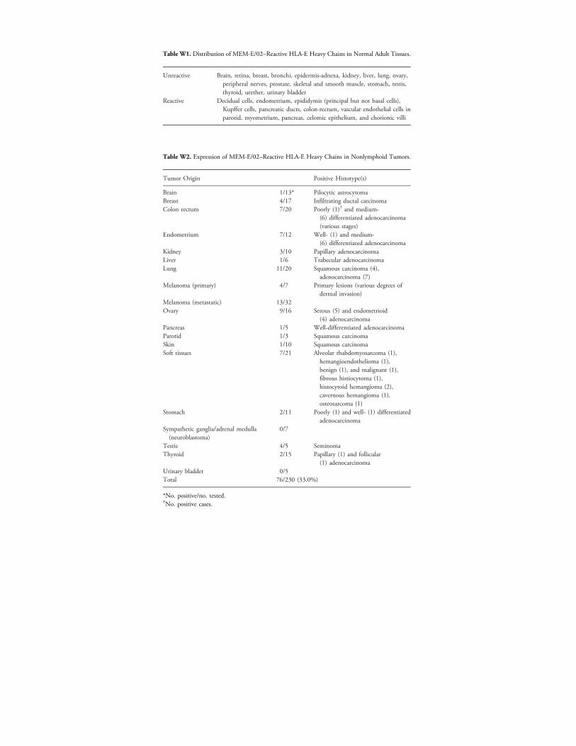
Table W1. Distribution of MEM-E/02–Reactive HLA-E Heavy Chains in Normal Adult Tissues.
Unreactive
Brain, retina, breast, bronchi, epidermis-adnexa, kidney, liver, lung, ovary,peripheral nerves, prostate, skeletal and smooth muscle, stomach, testis,thyroid, urether, urinary bladderReactive
Decidual cells, endometrium, epididymis (principal but not basal cells),Kupffer cells, pancreatic ducts, colon-rectum, vascular endothelial cells inparotid, myometrium, pancreas, celomic epithelium, and chorionic villiTable W2. Expression of MEM-E/02–Reactive HLA-E Heavy Chains in Nonlymphoid Tumors.
Tumor Origin
Positive Histotype(s)Brain
1/13* Pilocytic astrocytoma Breast 4/17 Infiltrating ductal carcinoma Colon rectum 7/20 Poorly (1)† and medium-(6) differentiated adenocarcinoma(various stages)
Endometrium
7/12 Well- (1) and medium-(6) differentiated adenocarcinomaKidney
3/10 Papillary adenocarcinoma Liver 1/6 Trabecular adenocarcinoma Lung 11/20 Squamous carcinoma (4),adenocarcinoma (7)
Melanoma (primary) 4/7 Primary lesions (various degrees ofdermal invasion)
Melanoma (metastatic) 13/32 Ovary 9/16 Serous (5) and endometrioid(4) adenocarcinoma
Pancreas 1/5 Well-differentiated adenocarcinoma Parotid 1/3 Squamous carcinoma Skin 1/10 Squamous carcinoma Soft tissues 7/21 Alveolar rhabdomyosarcoma (1),hemangioendothelioma (1),benign (1), and malignant (1),fibrous histiocytoma (1),histocytoid hemangioma (2),cavernous hemangioma (1),osteosarcoma (1)
Stomach
2/11 Poorly (1) and well- (1) differentiatedadenocarcinomaSympathetic ganglia/adrenal medulla(neuroblastoma)
0/7
Testis
4/5 Seminoma Thyroid 2/15 Papillary (1) and follicular(1) adenocarcinoma
Urinary bladder 0/5 Total 76/230 (33.0%)*No. positive/no. tested.†No. positive cases.

Figure W1. Western blot analysis of neoplastic cell lines. Equal amounts (100 μg per lane) of soluble extracts from the indicated celllines were run on a SDS-PAGE (10% acrylamide) slab under reducing conditions and electroblotted. Two strips of the filter were stainedwith MEM-E/02 and a polyclonal antibody to ERp57, respectively. HLA-E accumulation was largely independent from lineage and levelsof coexpressed HLA-A, -B, and -C molecules, assessed in a previous study on the same panel [12].
Figure W2. Flow cytometry analysis of the reactivity of MEM-E/02, MEM-E/06, MEM-E/07, MEM-E/08, 3D12 and 4D12. Cell lines (for acomplete list and a synopsis see Table W3) were tested for surface reactivity with the MEM antibodies, 3D12, 4D12, and W6/32, as acontrol. Bound fluorescence was analyzed on a FACScan (Becton, Dickinson & Co). Staining with an isotype-matched (IgG1), irrelevantantibody is shown (−). Each cell line was tested three to five times.

Table W3. Flow Cytometry Analysis of HLA-E Cell Surface Expression with the MEM Antibodies,3D12 and 4D12*.
Cell Lines
Figure W3. HLAcell lines. NP40performed as deantibodies. Thenot removed by
-E expressiextracts froscribed in Farrowheadstripping.
W6/32
on on sm surfigure 2marks
MEM-E
urface of early passace biotin-labeling ewere blotted with thresidual staining by
3D12
age tuxperime indicMEM-
4D12
/02
/06 /07 /08221 and transfectants
221 30† 7 3 3 4 2 2 221.AEH 213 NT‡ 20 31 38 25 11 221.G1 206 4 4 4 3 4 4 221.B7 73 0 5 1 1 2 2EBV-B cells
BSM 252 5 1 2 2 NT NT LG2 179 5 2 5 5 NT NT MGAR 364 4 4 4 7 NT NT JY 97 2 2 1 2 2 2Lymphoblastoid cells
Molt 4 61 3 2 2 1 NT NT Jurkat 124 3 4 3 2 2 2 WI-L2 149 1 2 0 0 NT NT U937 292 1 3 3 1 4 2 Raji 88 3 2 2 1 3 2 K562 17 1 1 1 1 NT NTMelanoma cells
Colo 38 75 0 0 1 2 NT NT FO-1 3 2 0 2 1 NT NT FO-1-β2m 85 0 1 1 1 0 0 HO-1 41 1 1 1 1 0 0 IR8 62 0 1 0 1 NT NT M10 42 0 1 0 0 1 1 MNT-1 141 0 0 0 2 NT NT SK-MEL-37 56 0 0 1 1 5 2 SK-MEL-93 182 1 1 1 1 NT NTCarcinoma cells
A431 81 0 1 2 2 1 1 A549 111 2 2 1 0 NT NT BT20 14 0 2 0 0 NT NT Calu-1 93 1 2 2 3 0 5 End 9 9 1 0 0 1 NT NT H494 44 1 2 2 1 NT NT HT-29 32 8 1 2 2 NT NT JAR 6 1 3 3 2 NT NT KJ29-β2m 179 2 2 5 4 NT NT MCF7 10 0 2 1 4 0 0 T24 31 1 3 1 2 NT NT*Representative results of this analysis are shown in Figure W2.†All the values are expressed as specific mean fluorescence intensity (MFI) after background sub-traction. Background values (invariably <9) were calculated by staining in parallel with irrelevant anti-bodies of the IgG1 and IgG2a isotypes. SpecificMFI values represent themean of three to five separateflow cytometry determinations.‡Not tested.
morentsatedE/02

Figure W4. Cytotoxic activity of NKL effectors against 221 targetcells. 221 and 221.AEH target cells were tested in parallel, at theindicated E/T ratios, for their susceptibility to NK lysis in a conven-tional 4-hour 51Cr release assay, using the same seed of NKL cellsas effectors, in the absence and presence of the indicated anti-bodies or F(ab)2 fragments. A representative experiment is shownof six that were performed.
Figure W5. Cytotoxic activity of NKL cells against tumor cells. NKLcells were tested for their ability to lyse the indicated targets ina 51Cr release assay, in the presence of the indicated antibodies(C218 to CD56, GHI/75 to ILT2, and Z199 to NKG2A). Lytic enhance-ments by GHI/75 and Z199 were statistically significant when theaverage values of triplicates differed from control more than threetimes the SD, that is, at E/T ratios from 20:1 to 2.5:1 in FO-1-β2mand at E/T ratios of 20:1 and 10:1 in A431. All the cell lines weretested three times.
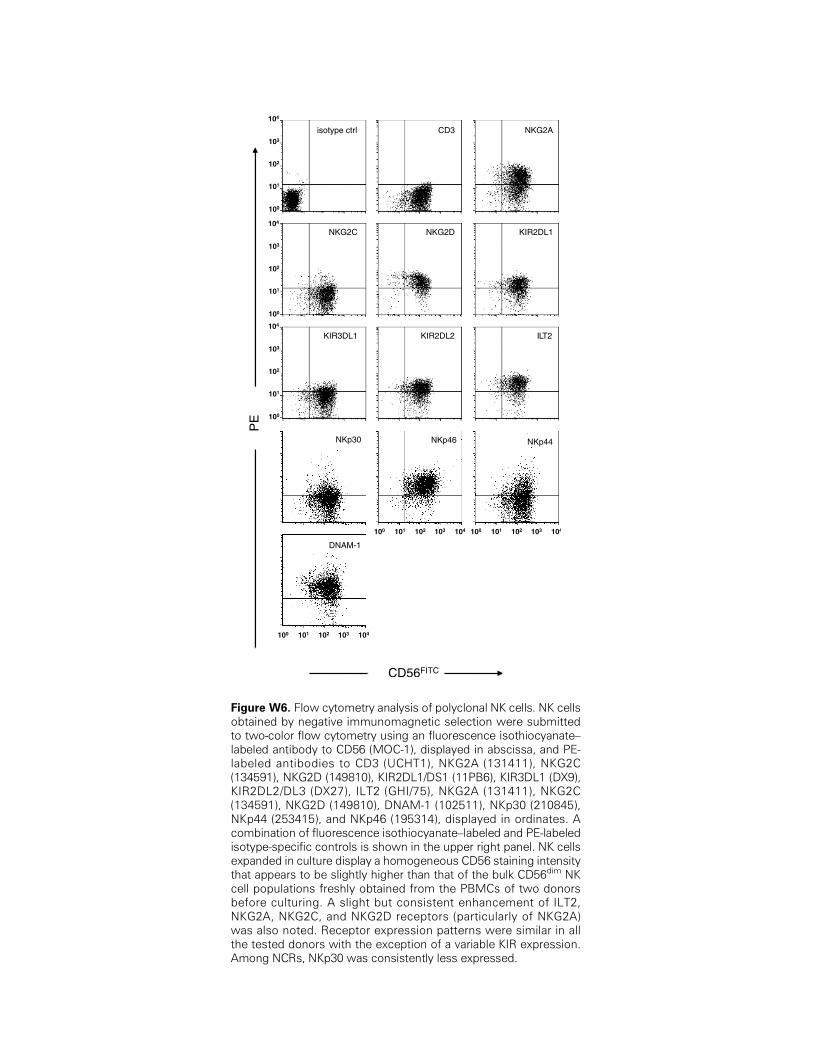
Figure W6. Flow cytometry analysis of polyclonal NK cells. NK cellsobtained by negative immunomagnetic selection were submittedto two-color flow cytometry using an fluorescence isothiocyanate–labeled antibody to CD56 (MOC-1), displayed in abscissa, and PE-labeled antibodies to CD3 (UCHT1), NKG2A (131411), NKG2C(134591), NKG2D (149810), KIR2DL1/DS1 (11PB6), KIR3DL1 (DX9),KIR2DL2/DL3 (DX27), ILT2 (GHI/75), NKG2A (131411), NKG2C(134591), NKG2D (149810), DNAM-1 (102511), NKp30 (210845),NKp44 (253415), and NKp46 (195314), displayed in ordinates. Acombination of fluorescence isothiocyanate–labeled and PE-labeledisotype-specific controls is shown in the upper right panel. NK cellsexpanded in culture display a homogeneous CD56 staining intensitythat appears to be slightly higher than that of the bulk CD56dim NKcell populations freshly obtained from the PBMCs of two donorsbefore culturing. A slight but consistent enhancement of ILT2,NKG2A, NKG2C, and NKG2D receptors (particularly of NKG2A)was also noted. Receptor expression patterns were similar in allthe tested donors with the exception of a variable KIR expression.Among NCRs, NKp30 was consistently less expressed.
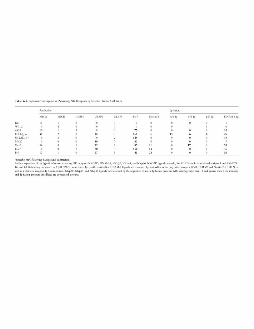
Table W4. Expression* of Ligands of Activating NK Receptors by Selected Tumor Cell Lines.
Antibodies
Ig-fusionMICA
MICB ULBP1 ULBP2 ULBP3 PVR Nectin-2 p30-Ig p44-Ig p46-Ig DNAM-1-IgRaji
11 1 0 0 0 0 0 0 0 0 1 WI-L2 0 4 0 0 0 0 0 0 1 1 0 A431 10 1 3 8 0 75 0 0 0 0 10 FO-1-β2m 41 2 0 10 0 161 6 15 6 6 25 SK-MEL-37 0 3 0 0 2 145 0 0 0 0 19 Mel20 0 0 0 19 0 52 0 0 0 0 7 Ova7 14 0 1 22 2 85 11 0 17 0 55 End7 0 0 2 30 6 150 14 0 0 0 36 Br7 12 1 0 27 5 43 22 0 0 0 30*Specific MFI following background subtraction.Surface expression of the ligands of major activating NK receptors NKG2D, DNAM-1, NKp30, NKp44, and NKp46. NKG2D ligands, namely, the MHC class I chain-related antigen A and B (MICA/B), and UL16 binding proteins 1 to 3 (ULBP1-3), were tested by specific antibodies. DNAM-1 ligands were assessed by antibodies to the polyovirus receptor (PVR, CD155) and Nectin-2 (CD112), aswell as a chimeric receptor-Ig fusion protein. NKp30, NKp44, and NKp46 ligands were assessed by the respective chimeric Ig-fusion proteins. MFI values greater than 11 and greater than 5 for antibodyand Ig-fusion proteins (boldface) are considered positive.
Related Documents


