Venkatachari et al. Virology Journal 2010, 7:119 http://www.virologyj.com/content/7/1/119 Open Access RESEARCH © 2010 Venkatachari et al; licensee BioMed Central Ltd. This is an Open Access article distributed under the terms of the Creative Com- mons Attribution License (http://creativecommons.org/licenses/by/2.0), which permits unrestricted use, distribution, and reproduc- tion in any medium, provided the original work is properly cited. Research Human immunodeficiency virus type 1 Vpr: oligomerization is an essential feature for its incorporation into virus particles Narasimhan J Venkatachari 1 , Leah A Walker 1 , Oznur Tastan 2 , Thien Le 1 , Timothy M Dempsey 1 , Yaming Li 1 , Naveena Yanamala 3 , Alagarsamy Srinivasan 4 , Judith Klein-Seetharaman 2,3 , Ronald C Montelaro 5 and Velpandi Ayyavoo* 1 Abstract HIV-1 Vpr, a nonstructural viral protein associated with virus particles, has a positive role in the efficient transport of PIC into the nucleus of non-dividing target cells and enhances virus replication in primary T cells. Vpr is a 96 amino acid protein and the structure by NMR shows three helical domains. Vpr has been shown to exist as dimers and higher order oligomers. Considering the multifunctional nature of Vpr, the contribution of distinct helical domains to the dimer/ oligomer structure of Vpr and the relevance of this feature to its functions are not clear. To address this, we have utilized molecular modeling approaches to identify putative models of oligomerization. The predicted interface residues were subjected to site-directed mutagenesis and evaluated their role in intermolecular interaction and virion incorporation. The interaction between Vpr molecules was monitored by Bimolecular Fluorescence complementation (BiFC) method. The results show that Vpr forms oligomers in live cells and residues in helical domains play critical roles in oligomerization. Interestingly, Vpr molecules defective in oligomerization also fail to incorporate into the virus particles. Based on the data, we suggest that oligomerization of Vpr is essential for virion incorporation property and may also have a role in the events associated with virus infection. Background HIV-1 vpr gene encodes a protein of 96 amino acids with a predicted molecular weight of 14 kDa, which is con- served in both HIV and SIV [1]. Vpr is packaged into assembling virions by binding to the p6 domain of viral p55 Gag precursor protein. The presence of a functional Vpr is necessary for the efficient translocation of the pre- integration complex (PIC) into the nucleus and subse- quent infection of primary monocytes/macrophages and other non-dividing cells [2-4]. Analysis of HIV-1 acces- sory genes (including vpr) in long-term non-progressors and asymptomatic patients suggests that defects in acces- sory genes are related to non-progressive status [5,6]. In this regard, the presence of defective or mutated vpr qua- sispecies has been shown to be associated with long-term non-progressive mothers [6-8]. Though vpr is selected against in tissue culture, selection for an intact Vpr occurs in vivo [9,10]. This finding suggests that vpr is required for optimal virus production and pathogenesis in vivo [11]. These observations clearly indicate the importance of Vpr in viral pathogenesis and disease pro- gression. HIV-1 Vpr is known to oligomerize both in vitro and in vivo [12,13]. This has been demonstrated by using cells in which Vpr was expressed either in the context of transfec- tion of plasmid DNAs or through virus infection. Similar observations have also been reported with the purified Vpr protein generated using the prokaryotic expression system. Vpr has been shown to exist as dimers, trimers, tetramers and higher order multimers [13]. In general, protein oligomerization is thought to be an advantageous feature for the stability of the protein, interaction/binding with other proteins, allosteric control and the establish- ment of higher-order complexity [14]. HIV-1 Vpr, a non- structural protein, is incorporated into the virus particles and possesses several characteristic features that are * Correspondence: [email protected] 1 Department of Infectious Diseases and Microbiology, Graduate School of Public Health, University of Pittsburgh, Pittsburgh, PA, USA Full list of author information is available at the end of the article

Welcome message from author
This document is posted to help you gain knowledge. Please leave a comment to let me know what you think about it! Share it to your friends and learn new things together.
Transcript

Venkatachari et al. Virology Journal 2010, 7:119http://www.virologyj.com/content/7/1/119
Open AccessR E S E A R C H
ResearchHuman immunodeficiency virus type 1 Vpr: oligomerization is an essential feature for its incorporation into virus particlesNarasimhan J Venkatachari1, Leah A Walker1, Oznur Tastan2, Thien Le1, Timothy M Dempsey1, Yaming Li1, Naveena Yanamala3, Alagarsamy Srinivasan4, Judith Klein-Seetharaman2,3, Ronald C Montelaro5 and Velpandi Ayyavoo*1
AbstractHIV-1 Vpr, a nonstructural viral protein associated with virus particles, has a positive role in the efficient transport of PIC into the nucleus of non-dividing target cells and enhances virus replication in primary T cells. Vpr is a 96 amino acid protein and the structure by NMR shows three helical domains. Vpr has been shown to exist as dimers and higher order oligomers. Considering the multifunctional nature of Vpr, the contribution of distinct helical domains to the dimer/oligomer structure of Vpr and the relevance of this feature to its functions are not clear. To address this, we have utilized molecular modeling approaches to identify putative models of oligomerization. The predicted interface residues were subjected to site-directed mutagenesis and evaluated their role in intermolecular interaction and virion incorporation. The interaction between Vpr molecules was monitored by Bimolecular Fluorescence complementation (BiFC) method. The results show that Vpr forms oligomers in live cells and residues in helical domains play critical roles in oligomerization. Interestingly, Vpr molecules defective in oligomerization also fail to incorporate into the virus particles. Based on the data, we suggest that oligomerization of Vpr is essential for virion incorporation property and may also have a role in the events associated with virus infection.
BackgroundHIV-1 vpr gene encodes a protein of 96 amino acids witha predicted molecular weight of 14 kDa, which is con-served in both HIV and SIV [1]. Vpr is packaged intoassembling virions by binding to the p6 domain of viralp55Gag precursor protein. The presence of a functionalVpr is necessary for the efficient translocation of the pre-integration complex (PIC) into the nucleus and subse-quent infection of primary monocytes/macrophages andother non-dividing cells [2-4]. Analysis of HIV-1 acces-sory genes (including vpr) in long-term non-progressorsand asymptomatic patients suggests that defects in acces-sory genes are related to non-progressive status [5,6]. Inthis regard, the presence of defective or mutated vpr qua-sispecies has been shown to be associated with long-termnon-progressive mothers [6-8]. Though vpr is selected
against in tissue culture, selection for an intact Vproccurs in vivo [9,10]. This finding suggests that vpr isrequired for optimal virus production and pathogenesisin vivo [11]. These observations clearly indicate theimportance of Vpr in viral pathogenesis and disease pro-gression.
HIV-1 Vpr is known to oligomerize both in vitro and invivo [12,13]. This has been demonstrated by using cells inwhich Vpr was expressed either in the context of transfec-tion of plasmid DNAs or through virus infection. Similarobservations have also been reported with the purifiedVpr protein generated using the prokaryotic expressionsystem. Vpr has been shown to exist as dimers, trimers,tetramers and higher order multimers [13]. In general,protein oligomerization is thought to be an advantageousfeature for the stability of the protein, interaction/bindingwith other proteins, allosteric control and the establish-ment of higher-order complexity [14]. HIV-1 Vpr, a non-structural protein, is incorporated into the virus particlesand possesses several characteristic features that are
* Correspondence: [email protected] Department of Infectious Diseases and Microbiology, Graduate School of Public Health, University of Pittsburgh, Pittsburgh, PA, USAFull list of author information is available at the end of the article
© 2010 Venkatachari et al; licensee BioMed Central Ltd. This is an Open Access article distributed under the terms of the Creative Com-mons Attribution License (http://creativecommons.org/licenses/by/2.0), which permits unrestricted use, distribution, and reproduc-tion in any medium, provided the original work is properly cited.

Venkatachari et al. Virology Journal 2010, 7:119http://www.virologyj.com/content/7/1/119
Page 2 of 11
known to play important roles in HIV-1 replication anddisease progression. Vpr interacts with both viral and cel-lular host proteins, which are essential for Vpr-mediatedfunctions. For instance, Vpr interacts with Gag-p6 andpackages in the virus particles and virion-incorporatedVpr is known to positively regulate infection of non-dividing cells and enhance virus production in T cells[4,11,15,16]. However, it is not clear whether oligomer-ization of Vpr is required for virion incorporation and/orfor its interactions with cellular proteins.
Vpr also has a well-defined role in apoptosis, cell cyclearrest and dysregulation of immune functions [17-19].Many of the Vpr functions are carried out by virion-asso-ciated Vpr similar to de novo synthesized Vpr, suggestingthat incorporation of Vpr into virus particles is an impor-tant event in HIV-1 biology. While the structure of Vprbased on X-ray crystallography is not yet available, bio-chemical analysis and NMR studies suggest that Vpr iscomposed of three alpha helices connected by loops[13,20-22]. Site-directed mutagenesis studies targetingsingle residues in Vpr indicated that amino acids in the Nterminal region including the helical domains are essen-tial for stability and virion incorporation and a regioncomprising the Helix III and the C terminal region deter-mines the nuclear transport of Vpr [23-26]. With respectto oligomerization, it has been suggested that a leucine-zipper type mechanism is likely involving helix III basedon the analysis of a peptide corresponding to the C-ter-minal region by NMR [27]. However, the structure ofhelix III in the peptide is different from that observed inthe full-length protein [20]. Furthermore, mutagenesisstudies have implicated additional amino acids in thehydrophobic core of the protein [12] in addition to adirect role for residue 44 in oligomerization through dele-tion. Thus, the roles of specific domains and residuesinvolved in oligomerization are not yet defined.
To gain a better understanding of Vpr oligomerizationand its role in virion association, we have utilized Bimo-lecular Fluorescence complementation (BiFC) analysis.This involves a chimeric protein strategy in which HIV-1Vpr is fused to either N- or C-terminus fragment of theVenus protein. Upon expression and formation of dimersin live cells the Venus will emit fluorescence that can bedetected by microscopy and flow cytometry. Such anapproach combined with specific alterations in Vpr basedon NMR structure and modeling have allowed us to eval-uate the domains and residues essential for Vpr oligomer-ization and its relevance in virion incorporation. Theresults from the studies presented here indicate that Vprmolecules with distinct mutations in helical domains I, IIand III dysregulate Vpr oligomerization and alter the abil-ity of Vpr to incorporate into virus particles.
Materials and MethodsCells and plasmidHeLa, and 293T cells were grown in DMEM supple-mented with 10% FCS, 1% glutamine and 1% penicillin-streptomycin. HeLa cells were obtained from NIH, AIDSreagent program. Proviral construct pNL43ΔVpr wasgenerated by mutating the start codon of Vpr and verifiedby sequencing and westernblot analysis. Vpr expressionplasmids were generated as described before [28]. All themutant constructs were sequenced to verify the integrityof the mutations. For BiFC assays, sequences encodingthe amino (residues 1 to 173, VN) or carboxyl (residues155 to 238, VC) fragments of Venus fluorescence protein(template generously provided by Dr. Ronald Montelero,University of Pittsburgh) were fused to the N terminus ofHIV-1 Vpr via a six-alanine linker and HA-tag for detec-tion. All plasmids were isolated using a QIAGEN Max-iprep kit (QIAGEN, Valencia, CA), and the specificmutations were confirmed by DNA sequencing.
Vpr dimer interface using docking modelStructural dimer models of Vpr were generated usingClusPro [29,30] and RosettaDock software [31] based onthe full length monomer NMR structure of Vpr (PDBid:1m8l) [20]. First an approximate orientation of thedimer was obtained using the ClusPro server. The homo-multimeric docking option and DOT method [32,33]starting with 25000 initial conformations were employed.Of the resulting 10 best docking conformations, twomodels comprising antiparallel and parallel conformationbased on best fit to the experimental data were selected.Each of these conformations was used as input into theRosetta Dock server as the starting conformations forrefinement via local docking of [31]. The hexamer modelwas generated using Cluspro server based on the mono-mer structure that comprised residues 15-78. The flexibleN and C-termini were excluded since their inclusionresulted in unfeasible structural models where N and Ctermini formed the interface. All models are availableupon request.
Expression of Vpr molecules by immunoblotHEK293T cells were cotransfected with Vpr expressionplasmid using L Lipofectamine. Forty-eight hours post-transfection, Cells were washed twice with PBS and lysedin RIPA buffer containing 50 mM Tris (pH 7.5), 150 mMNaCl, 1% Triton X-100, 1 mM sodium orthovanadate, 10mM sodium fluoride, 1.0 mM phenylmethylsulfonyl fluo-ride, 0.05% deoxycholate, 10% sodium dodecyl sulfate,aprotinin (0.07 trypsin inhibitor unit/ml), and the pro-tease inhibitors leupeptin, chymostatin, and pepstatin (1μg/ml; Sigma). Cell lysates were clarified by centrifuga-tion, and total cell lysates (50 μg) were separated on a 12%
Venkatachari et al. Virology Journal 2010, 7:119http://www.virologyj.com/content/7/1/119
Page 3 of 11
sodium dodecyl sulfate-polyacrylamide gel (SDS-PAGE)electrophoresis gel, transferred, and immunoblotted withanti-HIV-1 p24 for Gag and anti-HA for Vpr. The blotswere developed using an ECL kit (Amersham Biosci-ences, Piscataway, NJ).
BiFC Flow cytometryThirty-six hours post transfection 293T cells werewashed with PBS and fixed in 3.7% formaldehyde at roomtemperature for 15 minutes, and washed and resus-pended in 200 μl of FACS buffer. Samples were analyzedusing Epics-XL (Beckman Coulter, Miami, FL) with mini-mum of 40000 gated events acquired for each sample, andpercent BiFC positive cells was calculated using Flow Josoftware.
ImmunofluorescenceHeLa cells were used to perform all immunofluorescenceand microscopy assays. Thirty-six hours post transfec-tion, cells were washed with PBS and fixed in 3.7% form-aldehyde at room temperature for 10 minutes, andwashed and permeabilized with 0.5% Triton X-100 for anadditional 10 minutes. After washing 3 times with PBS,cells were blocked with 5% BSA at room temperature for1 hour followed by incubation with primary antibody(HA or Vpr; 1:200 dilution) for 1 hour at room tempera-ture and incubated with rabbit anti-mouse or mouse anti-rabbit IgG Rhodamine (RRX) (1:400; Jackson ImmunoRe-search, West Grove, PA) for 1 hour at room temperature.Cells were mounted with VECTASHIELD mountingmedia containing DAPI (Vector Laboratories, Burl-ingame, CA). Immunofluorescence analysis was per-formed using a fluorescence microscope with NikonSPOT camera (Fryer, Huntley, IL) and images were pro-cessed using MetaMorph software (Universal ImagingCorporation, Downington, PA).
Vpr-Gag interaction by BiFC and virion incorporationOligomerization of Vpr in live cells was evaluated by BiFCmethods. HeLa cells were seeded in 6-well plates withglass slides and transfected with combinations of Vprwt,Vpr mutants and Gag expression plasmids with theirrespective VN or VC combinations or with control plas-mids using Lipofectamine. Forty hours post-transfection,cells were washed with PBS and fixed with 2% paraform-aldehyde. Cells were mounted with VECTASHIELD con-taining DAPI (Vector Laboratories, CA) and fluorescencewas detected using Nikon inverted fluorescence micro-scope with appropriate filters. The remaining cells in theplates were analyzed by flow cytometry followed by cellquest software to measure the percent fluorescent posi-tive cells. Ability to Vpr mutants to interact with Gag andpackage into virus particles was assessed by monitoringthe presence of Vpr in virus particles. Briefly, cells were
cotransfected with pGag and pVpr mutants or vectorplasmid as described before. Forty hours post transfec-tion, supernatant was collected, spun at 2000 rpm toremove cell debris and passed through 0.22 μM filter toremove aggregates and cell debris. Filtered supernatantwas ultracentrifuged to pellet the virus particles and usedin SDS-PAGE followed by immunoblot with anti-Gag(p24) and anti-Vpr antibody. Similarly, cells were pelletedand cell lysates were used to detect the presence andexpression of Gag and Vpr in transfected culture.
ResultsConstruction and characterization of chimeric Vpr for oligomerization studies using BiFC analysisThe ability of Vpr to oligomerize was demonstrated bybiochemical methods using bacterially produced orchemically synthesized Vpr protein and/or peptides [15].A recent study has shown that Vpr forms dimers and oli-gomers in relevant eukaryotic cells by using fluorescencespectroscopy and imaging analysis [12]. These observa-tions have prompted us to evaluate the requirement ofspecific domains/residues of Vpr in the oligomerizationof the molecules. It is important to note that, althougholigomerization in live cells has been reported previously,the assays used for this purpose were mostly qualitative innature. We have selected Bimolecular Fluorescence Com-plementation (BiFC) system based on Venus as a reporterprotein to quantify the interactions between Vpr mono-mers. Chimeric proteins containing Vpr and N- or C- ter-minus fragments of the Venus reporter are to result inreconstitution of a functional Venus protein with fluores-cence. Since the assay does not distinguish between dim-ers and oligomers containing more than two Vprproteins, we will use the term oligomerization to describethe contact between at least two Vpr proteins throughoutthe manuscript. Briefly, Vpr from NL43 (which will bereferred as Vprwt) was cloned downstream of its N termi-nus (1-173 aa) or C terminus (155-238) of Venus fluores-cent protein as described [34]. The schematicrepresentation of the constructs is presented in Fig. 1A.The recombinant plasmids were assessed for expressionof the correctly sized protein products by transient trans-fection in HEK293T cells followed by immunoblot assay.The results shown in Fig. 1B indicate that a chimeric(Venus-Vpr) protein was expressed in cells transfectedwith the respective plasmids. The chimeric protein ofexpected size was detected by antibody against Vpr. Thesteady state expression levels of chimeric proteins weresimilar to that of wild type Vpr. Next, we also analyzedthe subcellular localization pattern of the chimeric pro-teins in comparison to the untagged wild type Vpr byusing Vpr specific antibody. Such an analysis showed thatfusion of N- and C-terminal fragment of Venus reporterdid not alter Vpr localization (Fig. 1C). Together these

Venkatachari et al. Virology Journal 2010, 7:119http://www.virologyj.com/content/7/1/119
Page 4 of 11
results indicate that fusion of chimeric molecules (VCand VN) did not alter the expression or subcellular distri-bution of Vpr.
To monitor oligomerization in live cells, HeLa cellswere cotransfected with equal amounts of Venus-C Vprand Venus-N Vpr plasmids or each plasmid with back-bone Venus C or N-terminal fragment containing vectoras a control. Vpr oligomerization was monitored thirty-six hours posttransfection by flow cytometry (Fig. 2A)and by fluorescence microscopy (Fig. 2B). Results indi-cate that cells transfected with both VC-Vpr and VN-Vprexhibit a positive signal (26% of BiFC positive cells) that isdetected by both approaches, whereas VC and VN vector,VC-Vpr or VN-Vpr with a vector control plasmid did notshow any signal. Similar results were observed in Jurkatcells transfected with Vpr BiFC plasmids (data not
shown). To ascertain the specificity of Vpr-Vpr interac-tions, we have also carried out a competition experimentin which vector backbone or untagged Vpr is expressedalong with chimeric VC-Vpr and VN-Vpr. As expected,the inclusion of untagged Vpr has resulted in a dimin-ished BiFC signal in a dose dependent manner (Fig. 2C).Considering the BiFC positive cells in wells transfectedwith VC-Vpr and VN-Vpr plasmids against the vectorbackbone as 100%, we assessed the BiFC positive cells in apool of cells transfected with a combination of three plas-mids including pVpr. The addition of pVpr at a ratio of 1:1(2.5:2.5 μg of DNA) to cells cotransfected with VC-Vprand VN-Vpr did not reduce BiFC positive cells, whereasat a ratio of 1:2.5 there is a 40% reduction and it wasreduced to less than 20% BiFC positive cells at 1:10 ratio.As increasing amount of untagged Vpr plasmid competed
Figure 1 Construction and characterization of Vpr plasmids for oligomerization studies in live cells using BiFC analysis. (A) Schematic rep-resentation of Vprwt fused with Venus-N terminal or Venus-C-terminal fragments. (B) Expression of Venus-C Vprwt and Venus-N Vprwt was assessed in HEK293T cells by transient transfection. HEK293T cells were transfected with Venus-N-Vprwt or Venus C-Vprwt expression plasmids or Vector control plasmid, and assessed by Western blot. (C) Subcellular localization pattern of Vprwt or Venus-Vprwt fusion proteins was assessed in HeLa cells by tran-sient transfection. HeLa cells were transfected with Vprwt or Venus C - Vprwt or Venus N - Vprwt expression plasmids or Vector control plasmid, and as-sessed by Immunofluorescence for subcellular localization pattern using Vpr-specific antibody. Figure represents one of five independent experiments (n = 5) with similar results.

Venkatachari et al. Virology Journal 2010, 7:119http://www.virologyj.com/content/7/1/119
Page 5 of 11
with VC and VN chimeras, percent positive BiFC cellswas reduced. Cell viability was monitored in these cul-tures during the experimental period and no significantcell death was observed suggesting that the observedreduction in BiFC signal is due to the competition ofuntagged Vpr and not due to Vpr-induced apoptosis (datanot shown). Together these results indicate that oli-gomerization of Vpr is specific and this technique wouldallow us to study the interaction in live cells more effi-ciently, especially in HIV-1 target cells.
Structure-based prediction of the Vpr dimer and oligomeric interfacesWe employed computational approaches to derivehypotheses regarding putative in vivo dimer interfaces.
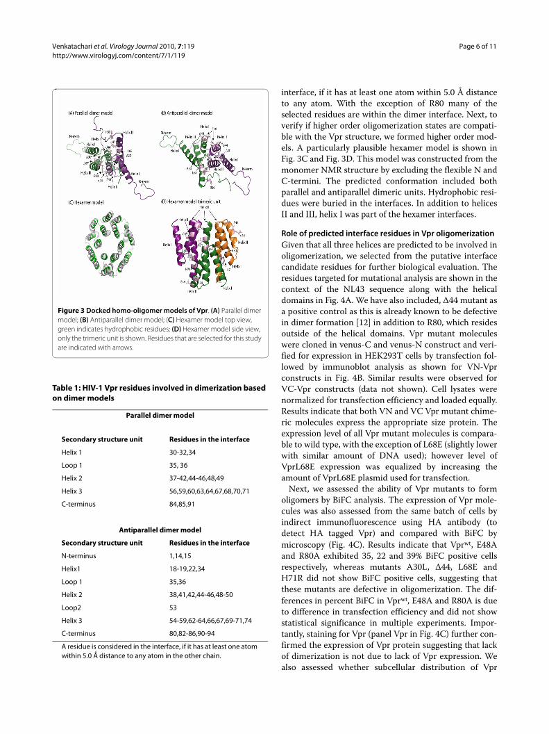
Using the available full-length Vpr NMR protein struc-ture (pdbid:1M8L), we built docking models using Clus-pro [29,35] and Rosetta softwares [31]. The highly rankedmodels included both antiparallel and parallel orienta-tions. In the majority of models, the interface comprisedmainly residues from helices II and III. Fig. 3A and 3Bshow models with parallel and antiparallel orientations,respectively. Helix I faces away from the dimer interfacein the models shown in Fig. 3. This leaves the possibilityfor helix I to mediate higher order oligomerization. Sucha role is feasible because helix I has high coiled coil pro-pensity (data not shown). The list of the interface residuesin the dimer models together with the secondary struc-ture is listed in Table 1. A residue is considered in the
Figure 2 Visualization of Vpr oligomerization by (A) Flow cytometry or (B) Fluorescent microscopy. (A) Quantitative analysis by flow cytometry of Venus fragment complementation in HeLa cells transfected with VC-Vpr and VN-Vpr or with control plasmid. Thirty-six hours posttransfection, cells were harvested and analyzed by flow cytometry to determine the percentage of cells positive for BiFC fluorescence. Results represent the means of five independent experiments. (B) Subcellular localization of the BiFC complex: HeLa cells grown on glass coverslips were cotransfected with VC and VN plasmids, VN-Vprwt and VC-Vprwt or VN- Vprwt or VC- Vprwt with control plasmid pairs using Lipofectamine. At 36 hours post-transfection, cells were fixed, stained with DAPI and imaged at 60× magnification. (C) To confirm the specificity of VC and VN based oligomerization, VC-Vpr (0.5 ug) and VN-Vpr (0.5 ug) plasmids were cotransfected with increasing concentrations of empty vector or Vpr expression plasmid. Thirty-six hours post transfection cells were fixed and assessed by flow cytometry. BiFC positive cells (%) were calculated in each treatment compared with the control. Figure repre-sents one of five independent experiments (n = 5) with similar results.
Venkatachari et al. Virology Journal 2010, 7:119http://www.virologyj.com/content/7/1/119
Page 6 of 11
interface, if it has at least one atom within 5.0 Å distanceto any atom. With the exception of R80 many of theselected residues are within the dimer interface. Next, toverify if higher order oligomerization states are compati-ble with the Vpr structure, we formed higher order mod-els. A particularly plausible hexamer model is shown inFig. 3C and Fig. 3D. This model was constructed from themonomer NMR structure by excluding the flexible N andC-termini. The predicted conformation included bothparallel and antiparallel dimeric units. Hydrophobic resi-dues were buried in the interfaces. In addition to helicesII and III, helix I was part of the hexamer interfaces.
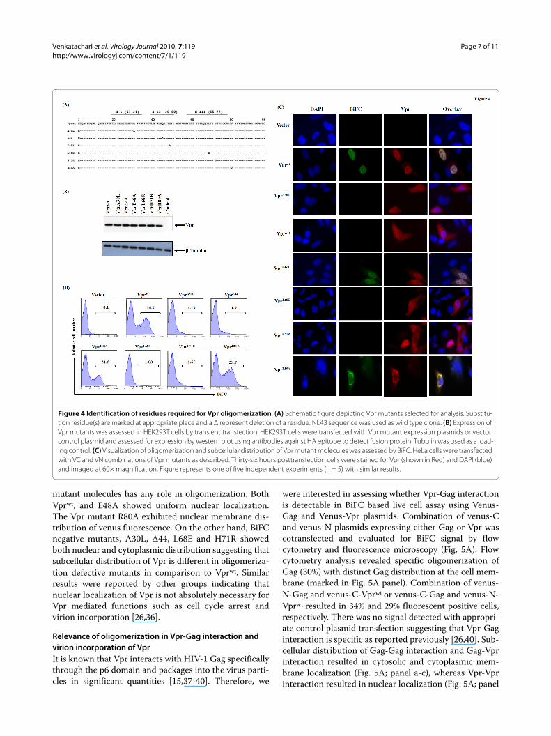
Role of predicted interface residues in Vpr oligomerizationGiven that all three helices are predicted to be involved inoligomerization, we selected from the putative interfacecandidate residues for further biological evaluation. Theresidues targeted for mutational analysis are shown in thecontext of the NL43 sequence along with the helicaldomains in Fig. 4A. We have also included, Δ44 mutant asa positive control as this is already known to be defectivein dimer formation [12] in addition to R80, which residesoutside of the helical domains. Vpr mutant moleculeswere cloned in venus-C and venus-N construct and veri-fied for expression in HEK293T cells by transfection fol-lowed by immunoblot analysis as shown for VN-Vprconstructs in Fig. 4B. Similar results were observed forVC-Vpr constructs (data not shown). Cell lysates werenormalized for transfection efficiency and loaded equally.Results indicate that both VN and VC Vpr mutant chime-ric molecules express the appropriate size protein. Theexpression level of all Vpr mutant molecules is compara-ble to wild type, with the exception of L68E (slightly lowerwith similar amount of DNA used); however level ofVprL68E expression was equalized by increasing theamount of VprL68E plasmid used for transfection.
Next, we assessed the ability of Vpr mutants to formoligomers by BiFC analysis. The expression of Vpr mole-cules was also assessed from the same batch of cells byindirect immunofluorescence using HA antibody (todetect HA tagged Vpr) and compared with BiFC bymicroscopy (Fig. 4C). Results indicate that Vprwt, E48Aand R80A exhibited 35, 22 and 39% BiFC positive cellsrespectively, whereas mutants A30L, Δ44, L68E andH71R did not show BiFC positive cells, suggesting thatthese mutants are defective in oligomerization. The dif-ferences in percent BiFC in Vprwt, E48A and R80A is dueto difference in transfection efficiency and did not showstatistical significance in multiple experiments. Impor-tantly, staining for Vpr (panel Vpr in Fig. 4C) further con-firmed the expression of Vpr protein suggesting that lackof dimerization is not due to lack of Vpr expression. Wealso assessed whether subcellular distribution of Vpr
Figure 3 Docked homo-oligomer models of Vpr. (A) Parallel dimer model; (B) Antiparallel dimer model; (C) Hexamer model top view, green indicates hydrophobic residues; (D) Hexamer model side view, only the trimeric unit is shown. Residues that are selected for this study are indicated with arrows.
Table 1: HIV-1 Vpr residues involved in dimerization based on dimer models
Parallel dimer model
Secondary structure unit Residues in the interface
Helix 1 30-32,34
Loop 1 35, 36
Helix 2 37-42,44-46,48,49
Helix 3 56,59,60,63,64,67,68,70,71
C-terminus 84,85,91
Antiparallel dimer model
Secondary structure unit Residues in the interface
N-terminus 1,14,15
Helix1 18-19,22,34
Loop 1 35,36
Helix 2 38,41,42,44-46,48-50
Loop2 53
Helix 3 54-59,62-64,66,67,69-71,74
C-terminus 80,82-86,90-94
A residue is considered in the interface, if it has at least one atom within 5.0 Ǻ distance to any atom in the other chain.
Venkatachari et al. Virology Journal 2010, 7:119http://www.virologyj.com/content/7/1/119
Page 7 of 11
mutant molecules has any role in oligomerization. BothVprwt, and E48A showed uniform nuclear localization.The Vpr mutant R80A exhibited nuclear membrane dis-tribution of venus fluorescence. On the other hand, BiFCnegative mutants, A30L, Δ44, L68E and H71R showedboth nuclear and cytoplasmic distribution suggesting thatsubcellular distribution of Vpr is different in oligomeriza-tion defective mutants in comparison to Vprwt. Similarresults were reported by other groups indicating thatnuclear localization of Vpr is not absolutely necessary forVpr mediated functions such as cell cycle arrest andvirion incorporation [26,36].
Relevance of oligomerization in Vpr-Gag interaction and virion incorporation of VprIt is known that Vpr interacts with HIV-1 Gag specificallythrough the p6 domain and packages into the virus parti-cles in significant quantities [15,37-40]. Therefore, we
were interested in assessing whether Vpr-Gag interactionis detectable in BiFC based live cell assay using Venus-Gag and Venus-Vpr plasmids. Combination of venus-Cand venus-N plasmids expressing either Gag or Vpr wascotransfected and evaluated for BiFC signal by flowcytometry and fluorescence microscopy (Fig. 5A). Flowcytometry analysis revealed specific oligomerization ofGag (30%) with distinct Gag distribution at the cell mem-brane (marked in Fig. 5A panel). Combination of venus-N-Gag and venus-C-Vprwt or venus-C-Gag and venus-N-Vprwt resulted in 34% and 29% fluorescent positive cells,respectively. There was no signal detected with appropri-ate control plasmid transfection suggesting that Vpr-Gaginteraction is specific as reported previously [26,40]. Sub-cellular distribution of Gag-Gag interaction and Gag-Vprinteraction resulted in cytosolic and cytoplasmic mem-brane localization (Fig. 5A; panel a-c), whereas Vpr-Vprinteraction resulted in nuclear localization (Fig. 5A; panel
Figure 4 Identification of residues required for Vpr oligomerization. (A) Schematic figure depicting Vpr mutants selected for analysis. Substitu-tion residue(s) are marked at appropriate place and a Δ represent deletion of a residue. NL43 sequence was used as wild type clone. (B) Expression of Vpr mutants was assessed in HEK293T cells by transient transfection. HEK293T cells were transfected with Vpr mutant expression plasmids or vector control plasmid and assessed for expression by western blot using antibodies against HA epitope to detect fusion protein. Tubulin was used as a load-ing control. (C) Visualization of oligomerization and subcellular distribution of Vpr mutant molecules was assessed by BiFC. HeLa cells were transfected with VC and VN combinations of Vpr mutants as described. Thirty-six hours posttransfection cells were stained for Vpr (shown in Red) and DAPI (blue) and imaged at 60× magnification. Figure represents one of five independent experiments (n = 5) with similar results.

Venkatachari et al. Virology Journal 2010, 7:119http://www.virologyj.com/content/7/1/119
Page 8 of 11
d). Together, the fluorescence microscopic analyses revealthat Vpr interacts with Gag specifically at the cytoplasmicmembrane as well as in the cytoplasm and this interac-tion results in differential localization of Vpr correspond-ing to virion incorporation and virus assembly.
Next, we assessed the ability of Vpr mutants to incorpo-rate into virus particles. HEK293T cells were cotrans-fected with pNL43Δvpr proviral plasmid and Vprexpression plasmids or vector control. Vpr amount in celllysate was quantitated by loading protein normalized fortransfection efficiency and amount of Vpr in virus parti-cles was quantitated by loading viral lysates normalizedusing Gag-p24 antigen, respectively (Fig. 5B). Presence ofVpr in virus particles was noted only in Vprwt, E48A andR80A, whereas, A30L, Δ44, L68E and H71R did not showvirion associated Vpr. However, all mutants showed equalamount of p24-Gag in the same sample. These resultssuggest that mutants A30L, Δ44, L68E and H71R aredefective in virion incorporation and they did not alter/interfere with the virus production in the cells contrans-fected with NL43Δvpr proviral DNA and Vpr encodingplasmid. To further confirm that the lack of Vpr in virusparticles is not due to lack of Vpr expression in thesecells, cell lysates from the same samples were assessed forVpr and p24 levels (data not shown). The results indicatethat comparable levels of Vpr and p24 were observed in
all cell lysates (Fig. 4B). Together theses studies indicatethat Vpr oligomerization is an essential feature for virionincorporation.
To further confirm that Vpr mutants that are defectivein virion incorporation lack the ability to interact withGag, Vpr mutants were cotransfected with VN-Gag orVC-Gag with appropriate controls and assessed for BiFC(Table 2). Results indicate that VC/VN chimeric Vprwt,E48A and R80A plasmids in combination with VC/VN-Gag showed measurable BiFC positive cells, whereasA30L, Δ44, L68E and H71R are negative for BiFC com-pared to positive control. Collectively, these studies indi-cate that Vpr mutants that are defective in virionincorporation are defective in Gag interaction and virionincorporation.
DiscussionHIV-1 and 2 are members of lentivirus family of retrovi-ruses and are grouped as complex retroviruses. Theunique feature of this group in comparison to simple ret-roviruses is that the viral genome codes for several pro-teins in addition to the core structural proteins. In thisregard, HIV-1 is known to code for six auxiliary proteins(Vif, Vpr, Tat, Rev, Vpu and Nef) besides the structuralproteins. Previous studies have demonstrated that auxil-iary proteins play an essential role in HIV-1 replicationand pathogenesis [41]. Our laboratory has been inter-
Figure 5 Role of Vpr oligomerization in incorporation of Vpr into virus particles. (A) Visualization of Vprwt and Gag interaction in HeLa cells by BiFC. HeLa cells were transfected with VN-Vprwt and VC-Gag ex-pression plasmids. Thirty-six hours post transfection, cells were fixed, stained with DAPI and analyzed for presence and pattern of BiFC signal at 60× magnification. (B) Incorporation of Vpr mutant molecules into virus particles. Vpr plasmids were cotransfected with pNL43ΔVpr pro-viral plasmid in HEK293T cells. Forty-eight hours post-transfection, su-pernatant was collected, filtered, lysed and subjected to SDS-PAGE electrophoresis and evaluated for the presence viral proteins p24-Gag and Vpr using p24-specific and Vpr specific antibodies by western blot. Figure represents one of five independent experiments (n = 5) with similar results.
Table 2: Percent BiFC positive cells in wells transfected with a combination of Vpr and Gag expression plasmids.
Vpr plasmids VC-Gag VN-Gag
VC-Vprwt 0 31
VN-Vprwt 27 0
VC-VprA30L 0 0
VN-VprA30L 0 0
VC-VprΔ44 0 0
VN-VprΔ44 0 0
VC-VprE48A 0 29
VN-VprE48E 28 0
VC-VprL68E 0 0
VN-VprL68E 0 0
VC-VprH71R 0 0
VN-VprH71R 0 0
VC-VprR80A 0 33
VN-VprR80A 27 0
293T cells were cotransfected with combinations of VC and VN-chimeric Vpr and Gag expression plasmids as described in methods. Thirty-six hours post transfection cells were fixed and assessed by flow cytometry. BiFC positive (%) cells are presented in the respective combination. Results represent one of three independent experiments with similar results.

Venkatachari et al. Virology Journal 2010, 7:119http://www.virologyj.com/content/7/1/119
Page 9 of 11
ested for several years in evaluating the contribution ofauxiliary proteins including Vpr. In this study, we haveanalyzed the requirement of sequences in Vpr essentialfor oligomerization feature of Vpr and its relevance to thefunctions of Vpr. Specifically, Vpr shares this feature withother auxiliary proteins such as Vif, Rev, Vpu, and Nef.
HIV-1 Vpr is a small oligomeric protein that plays animportant role in HIV pathogenesis [5,17,19,23,42]. Theunderlying reasons for the selection of Vpr for the presentstudies are the following: (i) Vpr is a virion associatedprotein; (ii) Vpr plays a critical role for the replication ofvirus in macrophages and positively regulates viral repli-cation in T cells; (iii) Vpr is a transcriptional activator ofHIV-1 and heterologous cellular genes; (iv) Vpr inhibitsproliferation of cells at G2/M phase; (v) Vpr inducesapoptosis in diverse cell types including T cells and neu-rons; (vi) Vpr exhibits immune suppressive effects. Fur-ther, studies from non human primates and analysis ofviral genes in long term non progressors suggest a corre-lation between defective Vpr and delayed progression ofthe disease [5,43,44]. More importantly, several Vpr-mediated functions are known to be induced by both cell-associated and virion-associated Vpr [17,18,45]. Togetherthese studies point out the biological significance ofvirion associated factors and its role in early events asso-ciated with virus infection. Therefore, understanding therole of oligomerization in Vpr functions and disease pro-gression may provide useful information for the develop-ment of therapeutics against HIV-1 targeting Vpr.
The oligomerization feature of Vpr was evaluated byusing a complementation system based on Venus proteinas a reporter. A strategy involving the generation of chi-meric Venus-Vpr protein has allowed us to monitor oli-gomerization in live cells. This system has the sensitivityto detect Vpr-Vpr and Vpr-Gag interactions. Studies onthe oligomerization of Vpr, analyzed by site-specificmutagenesis, identified some residues located in specificdomains of Vpr. However, there is no information avail-able regarding the dimer interface structure of Vpr. Toaddress this, we have utilized the available NMR struc-ture and modeling approaches to identify the residuesthat form putative dimer and oligomer interfaces of Vpr.Deletion of residue at position 44, which is predomi-nantly glutamine (Q), is known to be oligomerizationdefective and was used as a control in these experiments.We reasoned that moderate replacements, in particularthose mostly affecting size, should show significant bio-logical effects if these residues form part of the interfaces.Thus, for A30 and L68 we chose L and E replacements,respectively, conserving hydrophobicity but changing thesize of the position. On the other hand, at position H71,we chose R, maintaining the positive charge. Mutant Vprmolecules were generated containing alterations in theselected residues. The results regarding the expression
and steady state level of Vpr indicated that mutants lack-ing the ability to oligomerize exhibit a pattern similar tothat of wild type Vpr. This observation suggests thatmonomeric Vpr molecules are stable in cells, which is inagreement with an earlier report [36].
To interpret the mutagenesis data in structural terms,we created structural models of the Vpr dimer and oli-gomers through molecular docking based on the availablefull length monomer structure [20]. Our models areshown in Figure 3, highlighting the positions of the resi-dues that resulted in oligomerization defective propertiesof Vpr in vivo. The docked models revealed propensitiesfor both parallel and antiparallel orientations of themonomers within a dimer indicating that both of the con-formations are plausible. Regardless of orientation, heli-ces II and III constitute the dimer interface in themajority of the models. In the parallel orientation, resi-dues 30, 44, 68, 71, all of which fail to oligomerize whenmutated, even with the conservative substitutions cho-sen, were part of the interface; while in the antiparallelorientation residues 44, 68 and 71 contributed to thebinding energy in the interface. Further supporting ourpredicted interfaces, recent studies of Vpr mutants I60Aand I67A indicated that these residues play a major rolein trafficking the Vpr to the nuclear rim [12]. Both ofthese residues are part of the interface in the predictedparallel conformation, whereas I60 is part of the antipar-allel model. In agreement with the experimental resultspresented here suggest that both of these two dimer con-formations are likely to occur in vivo. The parallel orien-tation fits the experimental data better, but furtherstudies will be needed to fully differentiate between thetwo models. Furthermore, assays are needed that canclearly distinguish dimers from higher order oligomers.Based on the combined modeling and experimentalresults, we propose that dimerization primarily involveshelices II and III, while oligomerization includes helix Ialso. The fact that the Vpr mutation A30L reported hereand the recently studied nuclear rim localization defec-tive mutant L23F [12] are located in helix I supports theproposed role of helix I in oligomerization. We predictthat because helix I in the dimer models faces away fromthe dimer interface, it may play a pivotal role in mediatinghigher order Vpr-Vpr interactions. We therefore builtmodels for higher order multimeric forms, and a hexamermodel (Fig. 3C and Fig. 3D) exhibited an interface whereall the three helices participated in interaction interfaces.In this hexamer model, both parallel and antiparalleldimeric units were present and the hydrophobic residuesfaced to interior of the helices (Fig. 3D).
HIV-1 Vpr is one of the non-structural proteins that ispackaged in significant quantities in virus particles.Virion-associated Vpr is present in the infected cells priorto de novo synthesis and is known to cause the host cellu-

Venkatachari et al. Virology Journal 2010, 7:119http://www.virologyj.com/content/7/1/119
Page 10 of 11
lar dysfunctions during early infection [17,19,42]. Studieshave indicated that the p6 domain of Gag is critical forthe incorporation of Vpr into virus particles [15,39,40,46].However, it is not clear whether Vpr oligomerization is aprerequisite for virion incorporation. As expected, chi-meric Venus containing wild type Vpr and chimericVenus containing Gag resulted in the reconstitution ofVenus with fluorescence suggesting an interactionbetween these two proteins. On the other hand, chimericVenus containing mutant Vpr failed to interact with Gag.Vpr mutants that showed oligomerization negative phe-notype also failed to incorporate into virus particles. Sev-eral studies have reported that virus particle containsbetween 14-275 molecules of Vpr in comparison toapproximately 2500-2750 molecules of Gag proteindepending on the system and methods used [38,47]. Thissuggests that low amount of Vpr to Gag may be due to theinteraction restricted to the specific configuration of Gag.Several studies evaluated Vpr-Gag interaction andreported that helical domain I (residues E25 and A30),P35 and helical domain III (isoleucine-leucine residues)in Vpr are required for interaction with Gag, thus virionincorporation [26,39,48]. Our results on Vpr-Gag interac-tion using BiFC (Table 2) are in agreement with thesestudies and further supports the utility of BiFC assay forevaluating the interactions of Vpr with interacting part-ner proteins. Oligomerization defective mutants, A30L,Δ44, L68E and H71R lack the ability to incorporate intothe virus particles, suggesting that Vpr oligomerizationmight be directly linked to virion associated Vpr func-tions, pathogenesis and disease progression. A veryrecent publication further confirms our findings that Vproligomerization is required for interaction with Gag andoligomerization deficient mutants of Gag interacted withVpr [49].
An understanding of HIV-1 Vpr functions and its prop-erties, in our view, is likely to shed light on the mecha-nisms involved in Vpr incorporation into the virusparticle and how oligomerization feature influencesinfection of non dividing target cells. Although Vpr is notessential for virus replication in in vitro studies usingestablished cell lines, it is well established that virion-associated Vpr play a major role in macrophage infectionby aiding the transport of PIC into the nucleus [50,51].More importantly, virion-associated Vpr is known tomediate several host cellular events and immune evasivefunctions that are very similar to de novo synthesized Vpr[18,52,53]. This further bolsters the significance of virion-associated proteins present both in infectious and nonin-fectious particles and their role in HIV-1 pathogenesis.These studies further support the idea of developingpotential therapeutic agents including small moleculesagainst Vpr-Vpr interaction, Vpr-Gag interaction, virion
incorporation and virion associated Vpr induced host celldysregulation to combat HIV-1 infection.
Competing interestsThe authors declare that they have no competing interests.
Authors' contributionsNJV, OT, AS, JKS and VA conceived and designed the experiments, NJV, LAW, OTand YL performed the experiments, NJV, LAW, OT, AR, NY, AS, JKS, RCM and VAanalyzed the data, NJV, LAW, OT, TL, TMD, AR and NY contributed reagents/materials/analysis tools, NJV, OT, AS, JKS and VA wrote the paper. All authorshave read and approved the final manuscript.
AcknowledgementsThis work was supported in part by the grant GM082251 from the NIAID, NIH.
Author Details1Department of Infectious Diseases and Microbiology, Graduate School of Public Health, University of Pittsburgh, Pittsburgh, PA, USA, 2School of Computer Science, Carnegie Mellon University, Pittsburgh, PA, USA, 3Department of Structural Biology, University of Pittsburgh, Pittsburgh, PA, USA, 4NanoBio Diagnostics, West Chester, PA, USA and 5Vaccine Research Center, University of Pittsburgh, Pittsburgh, PA, USA
References1. Cohen EA, Dehni G, Sodroski JG, Haseltine WA: Human
immunodeficiency virus vpr product is a virion-associated regulatory protein. J Virol 1990, 64:3097-3099.
2. Connor RI, Chen BK, Choe S, Landau NR: Vpr is required for efficient replication of human immunodeficiency virus type-1 in mononuclear phagocytes. Virology 1995, 206:935-944.
3. Freed EO, Englund G, Martin MA: Role of the basic domain of human immunodeficiency virus type 1 matrix in macrophage infection. J Virol 1995, 69:3949-3954.
4. Subbramanian RA, Kessous-Elbaz A, Lodge R, Forget J, Yao XJ, Bergeron D, Cohen EA: Human immunodeficiency virus type 1 Vpr is a positive regulator of viral transcription and infectivity in primary human macrophages. J Exp Med 1998, 187:1103-1111.
5. Lum JJ, Cohen OJ, Nie Z, Weaver JG, Gomez TS, Yao XJ, Lynch D, Pilon AA, Hawley N, Kim JE, et al.: Vpr R77Q is associated with long-term nonprogressive HIV infection and impaired induction of apoptosis. J Clin Invest 2003, 111:1547-1554.
6. Wang B, Ge YC, Palasanthiran P, Xiang SH, Ziegler J, Dwyer DE, Randle C, Dowton D, Cunningham A, Saksena NK: Gene defects clustered at the C-terminus of the vpr gene of HIV-1 in long-term nonprogressing mother and child pair: in vivo evolution of vpr quasispecies in blood and plasma. Virology 1996, 223:224-232.
7. Yedavalli VR, Ahmad N: Low conservation of functional domains of HIV type 1 vif and vpr genes in infected mothers correlates with lack of vertical transmission. AIDS Res Hum Retroviruses 2001, 17:911-923.
8. Yedavalli VR, Chappey C, Ahmad N: Maintenance of an intact human immunodeficiency virus type 1 vpr gene following mother-to-infant transmission. J Virol 1998, 72:6937-6943.
9. Michael NL, Chang G, d'Arcy LA, Ehrenberg PK, Mariani R, Busch MP, Birx DL, Schwartz DH: Defective accessory genes in a human immunodeficiency virus type 1-infected long-term survivor lacking recoverable virus. J Virol 1995, 69:4228-4236.
10. Saksena NK, Wang B, Ge YC, Xiang SH, Dwyer DE, Cunningham AL: Coinfection and genetic recombination between HIV-1 strains: possible biological implications in Australia and South East Asia. Ann Acad Med Singapore 1997, 26:121-127.
11. Goh WC, Rogel ME, Kinsey CM, Michael SF, Fultz PN, Nowak MA, Hahn BH, Emerman M: HIV-1 Vpr increases viral expression by manipulation of the cell cycle: a mechanism for selection of Vpr in vivo. Nat Med 1998, 4:65-71.
12. Fritz JV, Didier P, Clamme JP, Schaub E, Muriaux D, Cabanne C, Morellet N, Bouaziz S, Darlix JL, Mely Y, de Rocquigny H: Direct Vpr-Vpr interaction in
Received: 28 February 2010 Accepted: 7 June 2010 Published: 7 June 2010This article is available from: http://www.virologyj.com/content/7/1/119© 2010 Venkatachari et al; licensee BioMed Central Ltd. This is an Open Access article distributed under the terms of the Creative Commons Attribution License (http://creativecommons.org/licenses/by/2.0), which permits unrestricted use, distribution, and reproduction in any medium, provided the original work is properly cited.Virology Journal 2010, 7:119
http://www.ncbi.nlm.nih.gov/entrez/query.fcgi?cmd=Retrieve&db=PubMed&dopt=Abstract&list_uids=2139896
http://www.ncbi.nlm.nih.gov/entrez/query.fcgi?cmd=Retrieve&db=PubMed&dopt=Abstract&list_uids=7531918
http://www.ncbi.nlm.nih.gov/entrez/query.fcgi?cmd=Retrieve&db=PubMed&dopt=Abstract&list_uids=7745752
http://www.ncbi.nlm.nih.gov/entrez/query.fcgi?cmd=Retrieve&db=PubMed&dopt=Abstract&list_uids=9529326
http://www.ncbi.nlm.nih.gov/entrez/query.fcgi?cmd=Retrieve&db=PubMed&dopt=Abstract&list_uids=8806556
http://www.ncbi.nlm.nih.gov/entrez/query.fcgi?cmd=Retrieve&db=PubMed&dopt=Abstract&list_uids=9658150
http://www.ncbi.nlm.nih.gov/entrez/query.fcgi?cmd=Retrieve&db=PubMed&dopt=Abstract&list_uids=7769682
http://www.ncbi.nlm.nih.gov/entrez/query.fcgi?cmd=Retrieve&db=PubMed&dopt=Abstract&list_uids=9140590
Venkatachari et al. Virology Journal 2010, 7:119http://www.virologyj.com/content/7/1/119
Page 11 of 11
cells monitored by two photon fluorescence correlation spectroscopy and fluorescence lifetime imaging. Retrovirology 2008, 5:87.
13. Zhao LJ, Wang L, Mukherjee S, Narayan O: Biochemical mechanism of HIV-1 Vpr function. Oligomerization mediated by the N-terminal domain. J Biol Chem 1994, 269:32131-32137.
14. Ali MH, Imperiali B: Protein oligomerization: how and why. Bioorg Med Chem 2005, 13:5013-5020.
15. Jenkins Y, Pornillos O, Rich RL, Myszka DG, Sundquist WI, Malim MH: Biochemical analyses of the interactions between human immunodeficiency virus type 1 Vpr and p6(Gag). J Virol 2001, 75:10537-10542.
16. Poon B, Chen IS: Human immunodeficiency virus type 1 (HIV-1) Vpr enhances expression from unintegrated HIV-1 DNA. J Virol 2003, 77:3962-3972.
17. Tungaturthi PK, Sawaya BE, Singh SP, Tomkowicz B, Ayyavoo V, Khalili K, Collman RG, Amini S, Srinivasan A: Role of HIV-1 Vpr in AIDS pathogenesis: relevance and implications of intravirion, intracellular and free Vpr. Biomed Pharmacother 2003, 57:20-24.
18. Majumder B, Venkatachari NJ, Srinivasan A, Ayyavoo V: HIV-1 mediated immune pathogenesis: spotlight on the role of viral protein R (Vpr). Curr HIV Res 2009, 7:169-177.
19. Andersen JL, Planelles V: The role of Vpr in HIV-1 pathogenesis. Curr HIV Res 2005, 3:43-51.
20. Morellet N, Bouaziz S, Petitjean P, Roques BP: NMR structure of the HIV-1 regulatory protein VPR. J Mol Biol 2003, 327:215-227.
21. Wecker K, Morellet N, Bouaziz S, Roques BP: NMR structure of the HIV-1 regulatory protein Vpr in H2O/trifluoroethanol. Comparison with the Vpr N-terminal (1-51) and C-terminal (52-96) domains. Eur J Biochem 2002, 269:3779-3788.
22. Singh SP, Tomkowicz B, Lai D, Cartas M, Mahalingam S, Kalyanaraman VS, Murali R, Srinivasan A: Functional role of residues corresponding to helical domain II (amino acids 35 to 46) of human immunodeficiency virus type 1 Vpr. J Virol 2000, 74:10650-10657.
23. Mahalingam S, Ayyavoo V, Patel M, Kieber-Emmons T, Weiner DB: Nuclear import, virion incorporation, and cell cycle arrest/differentiation are mediated by distinct functional domains of human immunodeficiency virus type 1 Vpr. J Virol 1997, 71:6339-6347.
24. Mahalingam S, Patel M, Collman RG, Srinivasan A: The carboxy-terminal domain is essential for stability and not for virion incorporation of HIV-1 Vpr into virus particles. Virology 1995, 214:647-652.
25. Mahalingam S, Collman RG, Patel M, Monken CE, Srinivasan A: Functional analysis of HIV-1 Vpr: identification of determinants essential for subcellular localization. Virology 1995, 212:331-339.
26. Votteler J, Studtrucker N, Sorgel S, Munch J, Rucker E, Kirchhoff F, Schick B, Henklein P, Fossen T, Bruns K, et al.: Proline 35 of human immunodeficiency virus type 1 (HIV-1) Vpr regulates the integrity of the N-terminal helix and the incorporation of Vpr into virus particles and supports the replication of R5-tropic HIV-1 in human lymphoid tissue ex vivo. J Virol 2007, 81:9572-9576.
27. Bourbigot S, Beltz H, Denis J, Morellet N, Roques BP, Mely Y, Bouaziz S: The C-terminal domain of the HIV-1 regulatory protein Vpr adopts an antiparallel dimeric structure in solution via its leucine-zipper-like domain. Biochem J 2005, 387:333-341.
28. Thotala D, Schafer EA, Tungaturthi PK, Majumder B, Janket ML, Wagner M, Srinivasan A, Watkins S, Ayyavoo V: Structure-functional analysis of human immunodeficiency virus type 1 (HIV-1) Vpr: role of leucine residues on Vpr-mediated transactivation and virus replication. Virology 2004, 328:89-100.
29. Comeau SR, Gatchell DW, Vajda S, Camacho CJ: ClusPro: a fully automated algorithm for protein-protein docking. Nucleic Acids Res 2004, 32:W96-99.
30. Comeau SR, Vajda S, Camacho CJ: Performance of the first protein docking server ClusPro in CAPRI rounds 3-5. Proteins 2005, 60:239-244.
31. Lyskov S, Gray JJ: The RosettaDock server for local protein-protein docking. Nucleic Acids Res 2008, 36:W233-238.
32. Mandell JG, Roberts VA, Pique ME, Kotlovyi V, Mitchell JC, Nelson E, Tsigelny I, Ten Eyck LF: Protein docking using continuum electrostatics and geometric fit. Protein Eng 2001, 14:105-113.
33. Ten Eyck LF, Mandell J, Roberts VA, Pique M.E: Surveying molecular interactions with DOT. Proceedings of the 1995 ACM/IEEE Supercomputing Conference ACM Press, New York 1995.
34. Jin J, Sturgeon T, Chen C, Watkins SC, Weisz OA, Montelaro RC: Distinct intracellular trafficking of equine infectious anemia virus and human immunodeficiency virus type 1 Gag during viral assembly and budding revealed by bimolecular fluorescence complementation assays. J Virol 2007, 81:11226-11235.
35. Comeau SR, Camacho CJ: Predicting oligomeric assemblies: N-mers a primer. J Struct Biol 2005, 150:233-244.
36. Bolton DL, Lenardo MJ: Vpr cytopathicity independent of G2/M cell cycle arrest in human immunodeficiency virus type 1-infected CD4+ T cells. J Virol 2007, 81:8878-8890.
37. Accola MA, Ohagen A, Gottlinger HG: Isolation of human immunodeficiency virus type 1 cores: retention of Vpr in the absence of p6(gag). J Virol 2000, 74:6198-6202.
38. Singh SP, Tungaturthi P, Cartas M, Tomkowicz B, Rizvi TA, Khan SA, Kalyanaraman VS, Srinivasan A: Virion-associated HIV-1 Vpr: variable amount in virus particles derived from cells upon virus infection or proviral DNA transfection. Virology 2001, 283:78-83.
39. Bachand F, Yao XJ, Hrimech M, Rougeau N, Cohen EA: Incorporation of Vpr into human immunodeficiency virus type 1 requires a direct interaction with the p6 domain of the p55 gag precursor. J Biol Chem 1999, 274:9083-9091.
40. Paxton W, Connor RI, Landau NR: Incorporation of Vpr into human immunodeficiency virus type 1 virions: requirement for the p6 region of gag and mutational analysis. J Virol 1993, 67:7229-7237.
41. Malim MH, Emerman M: HIV-1 accessory proteins--ensuring viral survival in a hostile environment. Cell Host Microbe 2008, 3:388-398.
42. Le Rouzic E, Benichou S: The Vpr protein from HIV-1: distinct roles along the viral life cycle. Retrovirology 2005, 2:11.
43. Mologni D, Citterio P, Menzaghi B, Zanone Poma B, Riva C, Broggini V, Sinicco A, Milazzo L, Adorni F, Rusconi S, et al.: Vpr and HIV-1 disease progression: R77Q mutation is associated with long-term control of HIV-1 infection in different groups of patients. Aids 2006, 20:567-574.
44. Lang SM, Weeger M, Stahl-Hennig C, Coulibaly C, Hunsmann G, Muller J, Muller-Hermelink H, Fuchs D, Wachter H, Daniel MM, et al.: Importance of vpr for infection of rhesus monkeys with simian immunodeficiency virus. J Virol 1993, 67:902-912.
45. Cui J, Tungaturthi PK, Ayyavoo V, Ghafouri M, Ariga H, Khalili K, Srinivasan A, Amini S, Sawaya BE: The role of Vpr in the regulation of HIV-1 gene expression. Cell Cycle 2006, 5:2626-2638.
46. Zhu H, Jian H, Zhao LJ: Identification of the 15FRFG domain in HIV-1 Gag p6 essential for Vpr packaging into the virion. Retrovirology 2004, 1:26.
47. Muller B, Tessmer U, Schubert U, Krausslich HG: Human immunodeficiency virus type 1 Vpr protein is incorporated into the virion in significantly smaller amounts than gag and is phosphorylated in infected cells. J Virol 2000, 74:9727-9731.
48. Yao XJ, Subbramanian RA, Rougeau N, Boisvert F, Bergeron D, Cohen EA: Mutagenic analysis of human immunodeficiency virus type 1 Vpr: role of a predicted N-terminal alpha-helical structure in Vpr nuclear localization and virion incorporation. J Virol 1995, 69:7032-7044.
49. Fritz JV, Dujardin D, Godet J, Didier P, De Mey J, Darlix JL, Mely Y, de Rocquigny H: HIV-1 Vpr oligomerization but not that of Gag directs the interaction between Vpr and Gag. J Virol 84:1585-1596.
50. Nitahara-Kasahara Y, Kamata M, Yamamoto T, Zhang X, Miyamoto Y, Muneta K, Iijima S, Yoneda Y, Tsunetsugu-Yokota Y, Aida Y: Novel nuclear import of Vpr promoted by importin alpha is crucial for human immunodeficiency virus type 1 replication in macrophages. J Virol 2007, 81:5284-5293.
51. Sherman MP, Greene WC: Slipping through the door: HIV entry into the nucleus. Microbes Infect 2002, 4:67-73.
52. Poon B, Grovit-Ferbas K, Stewart SA, Chen IS: Cell cycle arrest by Vpr in HIV-1 virions and insensitivity to antiretroviral agents. Science 1998, 281:266-269.
53. Arokium H, Kamata M, Chen I: Virion-associated Vpr of human immunodeficiency virus type 1 triggers activation of apoptotic events and enhances fas-induced apoptosis in human T cells. J Virol 2009, 83:11283-11297.
doi: 10.1186/1743-422X-7-119Cite this article as: Venkatachari et al., Human immunodeficiency virus type 1 Vpr: oligomerization is an essential feature for its incorporation into virus particles Virology Journal 2010, 7:119
http://www.ncbi.nlm.nih.gov/entrez/query.fcgi?cmd=Retrieve&db=PubMed&dopt=Abstract&list_uids=7798208
http://www.ncbi.nlm.nih.gov/entrez/query.fcgi?cmd=Retrieve&db=PubMed&dopt=Abstract&list_uids=9261351
http://www.ncbi.nlm.nih.gov/entrez/query.fcgi?cmd=Retrieve&db=PubMed&dopt=Abstract&list_uids=8553570
http://www.ncbi.nlm.nih.gov/entrez/query.fcgi?cmd=Retrieve&db=PubMed&dopt=Abstract&list_uids=7571402
http://www.ncbi.nlm.nih.gov/entrez/query.fcgi?cmd=Retrieve&db=PubMed&dopt=Abstract&list_uids=8230445
http://www.ncbi.nlm.nih.gov/entrez/query.fcgi?cmd=Retrieve&db=PubMed&dopt=Abstract&list_uids=8380472
http://www.ncbi.nlm.nih.gov/entrez/query.fcgi?cmd=Retrieve&db=PubMed&dopt=Abstract&list_uids=7474123
Related Documents