Human Immunodeficiency Virus-Associated Plasmablastic Lymphoma Poor Prognosis in the Era of Highly Active Antiretroviral Therapy Jorge J. Castillo, MD 1 ; Michael Furman, MD 1 ; Brady E. Beltra ´n, MD 2 ; Michele Bibas, MD 3 ; Mark Bower, MD 4 ; Weina Chen, MD 5 ; Jose ´ L. Dı ´ez-Martı ´n, MD 6 ; Jane J. Liu, MD 7 ; Roberto N. Miranda, MD 8 ; Silvia Montoto, MD 9 ; Nahid M. Nanaji, MD 10 ; Jose ´-Toma ´s Navarro, MD 11 ; Adam C. Seegmiller, MD 12 ; and Julie M. Vose, MD 13 BACKGROUND: Plasmablastic lymphoma (PBL) is a rare and aggressive B-cell lymphoma strongly associated with human immunode- ficiency virus (HIV) infection. The authors conducted a multi-institutional, retrospective study to describe characteristics and deter- mine prognostic factors in HIV-associated PBL. METHODS: For this study, the investigators included consecutive, HIV-positive patients diagnosed between the years 2000 and 2010 whose tumors had a plasmablastic morphology, were cluster of differentiation 20 (CD20)-negative, and expressed markers of plasmacytic differentiation. RESULTS: Fifty patients from 13 institutions were eval- uated. The median age was 43 years, and there was a male predominance. The median count of cells that were positive for CD4 (a glycoprotein expressed on the surface of T-helper cells, monocytes, macrophages, and dendritic cells) was 206 cells/mm 3 . At presen- tation, 90% of patients had extranodal involvement, 69% presented with advanced stage disease, and 27% had oral involvement. Rearrangements of v-myc myelocytomatosis viral oncogene homolog (MYC) were detected in 41% of the tested patients. Eighty-five percent of patients received chemotherapy, with 63% receiving cyclophosphamide, doxorubicin, vincristine, and prednisone and 37% receiving more intensive regimens. The complete response (CR) rate was 66%. The median overall survival (OS) was 11 months regard- less of the intensity of chemotherapy. In the survival analysis, an Eastern Cooperative Oncology Group performance status 2, advanced stage, and MYC rearrangements were associated significantly with a worse outcome, whereas attaining a CR with chemo- therapy was associated with a better outcome. CONCLUSIONS: The prognosis of PBL in HIV-infected individuals remains poor in the highly active antiretroviral therapy era. Intensive chemotherapy regimens do not seem to increase survival in patients with HIV-associ- ated PBL. Cancer 2012;118:5270-7. V C 2012 American Cancer Society. KEYWORDS: human immunodeficiency virus, acquired immunodeficiency syndrome, plasmablastic, highly active antiretroviral therapy, chemotherapy. INTRODUCTION In 1997, Delecluse and colleagues presented a case series of 16 patients with plasmablastic lymphoma (PBL), an aggressive subtype of diffuse large B-cell lymphoma (DLBCL) with distinct clinicopathologic characteristics. 1 In that case series, the large majority of patients had human immunodeficiency virus (HIV) infection and presented with involvement of the oral cavity. PBL has been included in the World Health Organization (WHO) classification as 1 of the lymphomas observed more commonly in HIV-infected individuals. 2 It is believed that the cell of origin of PBL is a postgerminal center B-lymphocyte or plasmablast. 3 Hence, the malignant cells in PBL usually do not express cluster of differentiation 20 (CD20) (B-lymphocyte antigen) but do express markers of DOI: 10.1002/cncr.27551, Received: December 8, 2011; Revised: February 28, 2012; Accepted: February 29, 2012, Published online April 17, 2012 in Wiley Online Library (wileyonlinelibrary.com) Corresponding author: Jorge J. Castillo, MD, 164 Summit Avenue, Providence, RI 02906; Fax: (401) 793-7132; [email protected] Presented at the 53rd Annual Meeting of the American Society of Hematology; December 10-13, 2011; San Diego, CA. Dr. Beltra ´n thanks Drs. Rocio Rea ´tegui and Domingo Morales from the Department of Oncology and Pathology at Edgardo Rebagliati Martins National Hospital. Dr. Bibas thanks Dr. Andrea Antinori, Chief of the Clinical Research Department at Lazzaro Spallanzani National Institute for Infections Diseases. Dr. Diez-Martin thanks Pascual Balsalobre, Transplant Coordinator at Gregorio Maran ˜o ´ n General University Hospital. Dr. Liu thanks Dr. Ling Zhang, hematopathologist at Moffitt Cancer Center. Dr. Navarro thanks Drs. Jose ´-Maria Ribera, Jose ´-Luis Mate, and Gustavo Tapia at Germans Trias i Pujol Hospital. Dr. Vose thanks Martin Bast, Lead Coordinator of the Lymphoma Study Group at the University of Nebraska Medical Center. 1 The Warren Alpert Medical School of Brown University, Division of Hematology and Oncology, The Miriam Hospital, Providence, Rhode Island; 2 Department of Oncology and Radiotherapy, Edgardo Rebagliati Martins National Hospital, Lima, Peru; 3 Department of Clinical Research, Hematology, Lazzaro Spallanzani National Institute for Infections Diseases, Rome, Italy; 4 Department of Medical Oncology, Chelsea and Westminster Hospital, London, United Kingdom; 5 Ameri- path/Quest Diagnostics, North Texas, Texas; 6 Department of Hematology, Gregorio Maran ˜o ´ n General University Hospital, Madrid, Spain; 7 Department of Hematol- ogy and Oncology, H. Lee Moffitt Cancer Center and Research Institute, Tampa, Florida; 8 Department of Hematopathology, The University of Texas M. D. Anderson Cancer Center, Houston, Texas; 9 Center for Hemato-Oncology, Barts Cancer Institute, Queen Mary University of London, London, United Kingdom; 10 Department of Pathology, University of Maryland Medical Center, Baltimore, Maryland; 11 Department of Hematology, Germans Trias i Pujol University Hospital, Barcelona, Spain; 12 Department of Pathology, Vanderbilt University Medical Center, Nashville, Tennessee; 13 Division of Hematology and Oncology, University of Nebraska Medical Center, Omaha, Nebraska 5270 Cancer November 1, 2012 Original Article

Welcome message from author
This document is posted to help you gain knowledge. Please leave a comment to let me know what you think about it! Share it to your friends and learn new things together.
Transcript
Human Immunodeficiency Virus-Associated Plasmablastic
Lymphoma
Poor Prognosis in the Era of Highly Active Antiretroviral Therapy
Jorge J. Castillo, MD1; Michael Furman, MD1; Brady E. Beltran, MD2; Michele Bibas, MD3; Mark Bower, MD4;
Weina Chen, MD5; Jose L. Dıez-Martın, MD6; Jane J. Liu, MD7; Roberto N. Miranda, MD8; Silvia Montoto, MD9;
Nahid M. Nanaji, MD10; Jose-Tomas Navarro, MD11; Adam C. Seegmiller, MD12; and Julie M. Vose, MD13
BACKGROUND: Plasmablastic lymphoma (PBL) is a rare and aggressive B-cell lymphoma strongly associated with human immunode-
ficiency virus (HIV) infection. The authors conducted a multi-institutional, retrospective study to describe characteristics and deter-
mine prognostic factors in HIV-associated PBL. METHODS: For this study, the investigators included consecutive, HIV-positive
patients diagnosed between the years 2000 and 2010 whose tumors had a plasmablastic morphology, were cluster of differentiation
20 (CD20)-negative, and expressed markers of plasmacytic differentiation. RESULTS: Fifty patients from 13 institutions were eval-
uated. The median age was 43 years, and there was a male predominance. The median count of cells that were positive for CD4 (a
glycoprotein expressed on the surface of T-helper cells, monocytes, macrophages, and dendritic cells) was 206 cells/mm3. At presen-
tation, 90% of patients had extranodal involvement, 69% presented with advanced stage disease, and 27% had oral involvement.
Rearrangements of v-myc myelocytomatosis viral oncogene homolog (MYC) were detected in 41% of the tested patients. Eighty-five
percent of patients received chemotherapy, with 63% receiving cyclophosphamide, doxorubicin, vincristine, and prednisone and 37%
receiving more intensive regimens. The complete response (CR) rate was 66%. The median overall survival (OS) was 11 months regard-
less of the intensity of chemotherapy. In the survival analysis, an Eastern Cooperative Oncology Group performance status �2,
advanced stage, and MYC rearrangements were associated significantly with a worse outcome, whereas attaining a CR with chemo-
therapy was associated with a better outcome. CONCLUSIONS: The prognosis of PBL in HIV-infected individuals remains poor in the
highly active antiretroviral therapy era. Intensive chemotherapy regimens do not seem to increase survival in patients with HIV-associ-
ated PBL. Cancer 2012;118:5270-7.VC 2012 American Cancer Society.
KEYWORDS: human immunodeficiency virus, acquired immunodeficiency syndrome, plasmablastic, highly active antiretroviral
therapy, chemotherapy.
INTRODUCTIONIn 1997, Delecluse and colleagues presented a case series of 16 patients with plasmablastic lymphoma (PBL), an aggressivesubtype of diffuse large B-cell lymphoma (DLBCL) with distinct clinicopathologic characteristics.1 In that case series, thelarge majority of patients had human immunodeficiency virus (HIV) infection and presented with involvement of the oralcavity. PBL has been included in the World Health Organization (WHO) classification as 1 of the lymphomas observedmore commonly in HIV-infected individuals.2
It is believed that the cell of origin of PBL is a postgerminal center B-lymphocyte or plasmablast.3 Hence, the malignantcells in PBL usually do not express cluster of differentiation 20 (CD20) (B-lymphocyte antigen) but do express markers of
DOI: 10.1002/cncr.27551, Received: December 8, 2011; Revised: February 28, 2012; Accepted: February 29, 2012, Published online April 17, 2012 in Wiley
Online Library (wileyonlinelibrary.com)
Corresponding author: Jorge J. Castillo, MD, 164 Summit Avenue, Providence, RI 02906; Fax: (401) 793-7132; [email protected]
Presented at the 53rd Annual Meeting of the American Society of Hematology; December 10-13, 2011; San Diego, CA.
Dr. Beltran thanks Drs. Rocio Reategui and Domingo Morales from the Department of Oncology and Pathology at Edgardo Rebagliati Martins National Hospital.
Dr. Bibas thanks Dr. Andrea Antinori, Chief of the Clinical Research Department at Lazzaro Spallanzani National Institute for Infections Diseases. Dr. Diez-Martin
thanks Pascual Balsalobre, Transplant Coordinator at Gregorio Maranon General University Hospital. Dr. Liu thanks Dr. Ling Zhang, hematopathologist at Moffitt
Cancer Center. Dr. Navarro thanks Drs. Jose-Maria Ribera, Jose-Luis Mate, and Gustavo Tapia at Germans Trias i Pujol Hospital. Dr. Vose thanks Martin Bast, Lead
Coordinator of the Lymphoma Study Group at the University of Nebraska Medical Center.
1The Warren Alpert Medical School of Brown University, Division of Hematology and Oncology, The Miriam Hospital, Providence, Rhode Island; 2Department of
Oncology and Radiotherapy, Edgardo Rebagliati Martins National Hospital, Lima, Peru; 3Department of Clinical Research, Hematology, Lazzaro Spallanzani
National Institute for Infections Diseases, Rome, Italy; 4Department of Medical Oncology, Chelsea and Westminster Hospital, London, United Kingdom; 5Ameri-
path/Quest Diagnostics, North Texas, Texas; 6Department of Hematology, Gregorio Maranon General University Hospital, Madrid, Spain; 7Department of Hematol-
ogy and Oncology, H. Lee Moffitt Cancer Center and Research Institute, Tampa, Florida; 8Department of Hematopathology, The University of Texas M. D.
Anderson Cancer Center, Houston, Texas; 9Center for Hemato-Oncology, Barts Cancer Institute, Queen Mary University of London, London, United Kingdom;10Department of Pathology, University of Maryland Medical Center, Baltimore, Maryland; 11Department of Hematology, Germans Trias i Pujol University Hospital,
Barcelona, Spain; 12Department of Pathology, Vanderbilt University Medical Center, Nashville, Tennessee; 13Division of Hematology and Oncology, University of
Nebraska Medical Center, Omaha, Nebraska
5270 Cancer November 1, 2012
Original Article

plasmacytic differentiation, such as CD38 (cyclic adenosinediphosphate ribose hydrolase), CD138 (syndecan-1), ormultiple myeloma oncogene 1/interferon regulatory factor4 (MUM1/IRF4), akin to plasma cell myeloma (PCM).4
However, based on genomic profiling, PBL appears to bemore in line with DLBCL.5 Case reports and small case se-ries have demonstrated that HIV-associated PBL is associ-ated with an aggressive clinical course and poor survivalrates with standard therapies.6,7 PBL, hence, poses a chal-lenge for hematopathologists and oncologists alike.We con-ducted a multi-institutional, retrospective study to evaluatethe characteristics and determine prognostic factors inpatients with a pathologic diagnosis of HIV-associated PBLin the era of highly active antiretroviral therapy (HAART).
MATERIALS AND METHODS
Case Selection
Institutions in the United States, Europe, and SouthAmerica submitted patient-level data on consecutiveHIV-positive individuals with a pathologic diagnosis ofPBL. This study has been approved by the institutionalreview board at each of the participating centers. For thisstudy, all patients were required to have plasmablasticmorphology, be CD20 negative, and express at least 1plasmacytic marker (ie, CD38, CD138, and/or MUM1/IRF4). All cases were diagnosed between the years 2000and 2011 and were reviewed by 2 hematopathologists atthe center of initial diagnosis. Cases of primary cutaneousand primary brain PBL and cases of PBL diagnosed inHIV-negative individuals were not included. Threepatients have been previously reported.8 Clinical dataincluded age, sex, performance status according to theEastern Cooperative Oncology Group (ECOG) scale,years of HIV infection before PBL diagnosis, count ofcells positive for CD4 (a glycoprotein expressed on thesurface of T-helper cells, monocytes, macrophages, anddendritic cells), HIV viral load, receipt of HAART,opportunistic infections, the presence of B symptoms, thenumber and location of extralymph node sites, diseasestage, lactate dehydrogenase (LDH) levels, age-adjustedInternational Prognostic Index (aaIPI) score, chemothera-peutic regimen, receipt of radiotherapy, response andmethod of response assessment, final outcome, progres-sion-free survival (PFS), overall survival (OS), and causeof death. Laboratory data included hemoglobin level,white blood cell (WBC) count, absolute lymphocyte(ALC) and platelet counts, and the presence of a mono-clonal spike by serum protein electrophoresis. Pathologicdata included immunohistochemical expression of CD45(protein tyrosine phosphatase receptor, type C), CD20,
CD4, CD8 (transmembrane glycoprotein, a coreceptorfor the T-cell receptor), CD56 (neural cell adhesion mole-cule), CD38, CD138, MUM1/IRF4, B-cell lymphoma 2(BCL2), BCL6, anaplastic lymphoma kinase (ALK),Epstein Barr virus (EBV) latent membrane protein-1(LMP1), human herpesvirus 8 (HHV8) latency-associ-ated nuclear antigen (LANA), and Ki67. Molecular stud-ies included the detection of EBV-encoded RNA (EBER)by in situ hybridization, HHV8 by polymerase chain reac-tion, and immunoglobulin heavy-chain gene and v-mycmyelocytomatosis viral oncogene homolog (MYC) rear-rangements by standard cytogenetic, fluorescent in situhybridization or polymerase chain reaction studies.
Statistical Analysis
Continuous and categorical variables are presented usingdescriptive statistics. Response to therapy was assessed usingthe revised response criteria whenever possible.9 PFS wasdefined as the time between diagnosis and progression,death, or last follow-up. OS was defined as the timebetween diagnosis and death or last follow-up. Univariatesurvival analyses were performed using the Kaplan-Meiermethod and the log-rank test. P values < .05 were consid-ered statistically significant. All calculations and graphswere obtained using the statistical softwareMedCalc (Med-Calc Software, Mariakerke, Belgium).
RESULTS
Patient Characteristics
Of 53 patients who were identified in 13 institutions, 50were included in the current analysis. Two patients whowere CD20-positive and 1 patient who had primary braininvolvement were excluded. Twenty-four patients (48%)were from Europe, 23 patients (46%) were from the UnitedStates, and 3 patients (6%) were from South America. Themedian age was 43 years (range, 19-66 years). There was apredominance of men (4:1). The median CD4-positivecount was 206 cells/mm3 (range, 5-683 cells/mm3), and themedian viral load at presentation was 261,560 copies/mL(range, from undetectable to 4.7 million copies/mL). PBLwas the initial presentation of HIV infection in 29% ofpatients (n ¼ 13). The median duration between HIVinfection and PBL diagnoses was 8.9 years (range, 0-26years). Twenty-one patients (43%) were receiving HAARTat the time of PBL diagnosis. Selected clinical characteristicsare listed in Table 1. The most common extralymph nodesites of involvement were oral cavity (n ¼ 12; 24%), liver/spleen (n ¼ 8; 16%), gastrointestinal tract (n ¼ 7; 14%),central nervous system (n ¼ 7; 14%), lungs (n ¼ 6; 12%),bone/muscle (n ¼ 5; 10%), skin (n ¼ 3; 6%), and gonads
HIV-Positive Plasmablastic Lymphoma/Castillo et al
Cancer November 1, 2012 5271

(n¼ 3; 6%). The following laboratory were reported: 39%(n ¼ 16) had a WBC count <4.0/lL, 51% (n ¼ 21) hadan ALC <1.0/lL, 34% (n ¼ 14) had hemoglobin level<10 g/dL, and 32% (n ¼ 13) had a platelet count <150/lL. Pathologic characteristics are listed in Table 2. Thepathologic profile of a patient with HIV-associated PBL isprovided in Figure 1.
Therapy and Outcome
Chemotherapy was received by 85% of patients (n¼ 40),with 63% (n ¼ 25) receiving cyclophosphamide, doxoru-bicin, vincristine, and prednisone (CHOP) and 37% (n¼ 15) receiving more intensive regimens (8 patientsreceived infusional etoposide, doxorubicin, vincristine,cyclophosphamide, and prednisone [EPOCH]; 5 patientsreceived hyperfractionated cyclophosphamide, vincris-tine, doxorubicin, and dexamethasone [hyper-CVAD]; 1patient received bortezomib, dexamethasone, thalido-mide, cisplatin, doxorubicin, cyclophosphamide, and eto-poside [VDT-PACE]; and 1 patient received combined
Table 1. Clinical Characteristics, Treatment, and Outcome of50 Patients With Human Immunodeficiency Virus-PositivePlasmablastic Lymphoma
Patient Characteristic(No. of Patients WithAvailable Data)
No. ofPatients
Percentage
Age (n ¼ 50), y>40 36 72
£40 14 28
Sex (n ¼ 50)Male 39 78
Female 11 22
CD4þ count (n ¼ 48), cells/mm3
£200 28 58
>200 20 42
HAART before PBL diagnosis (n ¼ 49)Yes 21 43
No 28 57
HAART with PBL diagnosis (n ¼ 49)Yes 40 82
No 9 18
B symptoms (n ¼ 42)Absent 12 29
Present 30 71
ECOG performance status (n ¼ 34)0-1 15 44
‡2 19 56
LDH levels (n ¼ 40)Normal 10 25
Elevated 30 75
Clinical stage (n ¼ 48)I and II 15 31
III and IV 33 69
No. of extra lymph node sites (n ¼ 48)0-1 28 58
>1 20 42
Age-adjusted IPI score (n ¼ 40)Low/low-intermediate 10 25
High/high-intermediate 30 75
Lymphoma therapy (n ¼ 40)CHOP/CHOP-like 25 63
Other regimens 15 37
Response to therapy (n ¼ 38)Complete 25 66
Partial 2 5
None 11 29
Outcome (n ¼ 48)Alive 15 31
Dead 33 69
Cause of death (n ¼ 33)Lymphoma 24 73
Infection 8 24
Abbreviations: CD4þ, positive for cluster of differentiation 4 (a glycoprotein
expressed on the surface of T-helper cells, monocytes, macrophages, and den-
dritic cells); CHOP, cyclophosphamide, doxorubicin, vincristine and prednisone;
ECOG, Eastern Cooperative Oncology Group; HAART, highly active antiretroviral
therapy; IPI, International Prognostic Index; LDH, lactate dehydrogenase; PBL,
plasmablastic lymphoma.
Table 2. Pathologic Characteristics of 50 Patients With aDiagnosis of Human Immunodeficiency Virus-PositivePlasmablastic Lymphoma
Marker No. ofPatientsTested
No. Positivefor Marker (%)
CD45 31 20 (65)
CD20 50 0 (0)
CD4 17 1 (6)
CD8 11 0 (0)
CD56 25 9 (36)
CD38 9 8 (89)
CD138 30 26 (87)
MUM1/IRF4 25 25 (100)
BCL2 27 6 (22)
BCL6 26 1 (4)
ALK 14 0 (0)
Ki67 >80% 34 28 (82)
EBV LMP1 14 9 (64)
EBER 37 35 (95)
HHV8 LANA 28 0 (0)
HHV8 PCR 5 0 (0)
MYC rearrangement 21 9 (41)
Abbreviations: ALK, anaplastic lymphoma kinase; BCL2, B-cell lymphoma 2;
BCL6, B-cell lymphoma 6; CD138, cluster of differentiation 138 (syndecan-1);
CD20, cluster of differentiation 20 (B-lymphocyte antigen); CD38, cluster of
differentiation 38 (cyclic adenosine diphosphate ribose hydrolase); CD4, clus-
ter of differentiation 4 (glycoprotein expressed on the surface of T-helper cells,
monocytes, macrophages, and dendritic cells); CD45, cluster of differentiation
45 (protein tyrosine phosphatase receptor, type C); CD56, cluster of differen-
tiation 56 (neural cell adhesion molecule); CD8, cluster of differentiation 8
(transmembrane glycoprotein, a coreceptor for the T-cell receptor); EBER,
Epstein-Barr virus-encoded RNA; EBV, Epstein Barr virus; HHV8, human her-
pesvirus 8; LANA, latency-associated nuclear antigen; LMP1, latent mem-
brane protein-1; MUM1/IRF4, multiple myeloma oncogene 1/interferon
regulatory factor 4; MYC, v-myc myelocytomatosis viral oncogene homolog;
PCR: polymerase chain reaction.
Original Article
5272 Cancer November 1, 2012

Figure 1. The pathologic profile of a patient with human immunodeficiency virus-associated plasmablastic lymphoma is illus-trated. H&E indicates hematoxylin and eosin staining; CD20, cluster of differentiation 20 (B-lymphocyte antigen); BCL6, B-cellchronic lymphocytic leukemia/lymphoma 6; CD138, cluster of differentiation 138 (syndecan-1); Ki67, KI-67 antigen (identified bythe monoclonal antibody Ki-67); EBER, Epstein-Barr virus-encoded RNA.
HIV-Positive Plasmablastic Lymphoma/Castillo et al
Cancer November 1, 2012 5273

cyclophosphamide, vincristine, doxorubicin, and metho-trexate plus ifosfamide, mesna, etoposide, and cytarabine[the Magrath regimen]). Radiotherapy was received by 6patients (13%), mainly for palliative purposes, because 5patients had stage IV disease. A minority of patients (n ¼4; 10%) underwent autologous hematopoietic stem celltransplantation (HSCT), including 1 patient as part offront-line treatment and 3 patients in the relapsed setting.Response to therapy was assessed by positron emission to-mography/computed tomography scans in 9 patients(23%) and by computed tomography scans in 30 patients(77%). A complete response (CR) was obtained in 66%of patients (n ¼ 25), 5% of patients (n ¼ 2) had a partialresponse, and 29% of patients (n ¼ 11) had no response.Two patients died before response could be assessed andwere not evaluable for response. Patients received a me-
dian of 2 lines of treatment (range, 1-5 lines of treatment).After a median follow-up of 48 months, 69% of patients(n¼ 33) had died.
Survival Analysis and Prognostic Factors
The median PFS and OS after diagnosis were 6 monthsand 11 months, respectively, and the estimated 5-yearPFS and OS rates were 23% and 24%, respectively (Fig.2). Among the patients who received chemotherapy,obtaining a CR was associated with a median OS of 48months compared with 3 months for patients whoobtained less than a CR (P < .001). In the survival analy-ses, an ECOG performance status �2, advanced stage,andMYC rearrangement were associated with shorter me-dian PFS and OS rates. A high/high-intermediate aaIPIscore was associated with worse PFS and OS than a low/
Figure 2. These charts illustrate the median (A) progression-free survival and (B) overall survival of 50 patients with plasmablas-tic lymphoma.
Figure 3. (A) Progression-free survival and (B) overall survival are illustrated according to age-adjusted International PrognosticIndex (IPI) scores in 40 patients with plasmablastic lymphoma.
Original Article
5274 Cancer November 1, 2012
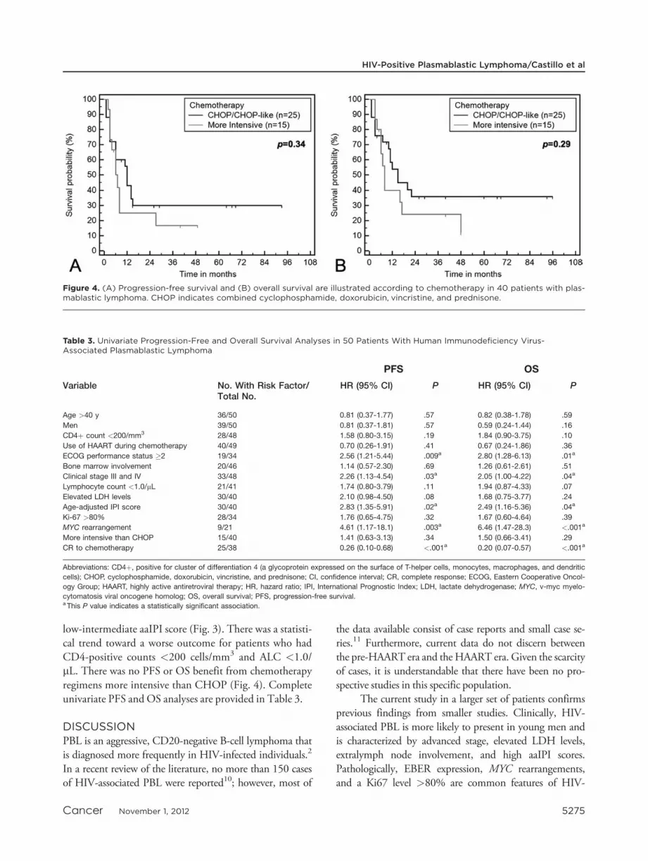
low-intermediate aaIPI score (Fig. 3). There was a statisti-cal trend toward a worse outcome for patients who hadCD4-positive counts <200 cells/mm3 and ALC <1.0/lL. There was no PFS or OS benefit from chemotherapyregimens more intensive than CHOP (Fig. 4). Completeunivariate PFS and OS analyses are provided in Table 3.
DISCUSSIONPBL is an aggressive, CD20-negative B-cell lymphoma thatis diagnosed more frequently in HIV-infected individuals.2
In a recent review of the literature, no more than 150 casesof HIV-associated PBL were reported10; however, most of
the data available consist of case reports and small case se-ries.11 Furthermore, current data do not discern betweenthe pre-HAART era and theHAART era. Given the scarcityof cases, it is understandable that there have been no pro-spective studies in this specific population.
The current study in a larger set of patients confirmsprevious findings from smaller studies. Clinically, HIV-associated PBL is more likely to present in young men andis characterized by advanced stage, elevated LDH levels,extralymph node involvement, and high aaIPI scores.Pathologically, EBER expression, MYC rearrangements,and a Ki67 level >80% are common features of HIV-
Figure 4. (A) Progression-free survival and (B) overall survival are illustrated according to chemotherapy in 40 patients with plas-mablastic lymphoma. CHOP indicates combined cyclophosphamide, doxorubicin, vincristine, and prednisone.
Table 3. Univariate Progression-Free and Overall Survival Analyses in 50 Patients With Human Immunodeficiency Virus-Associated Plasmablastic Lymphoma
PFS OS
Variable No. With Risk Factor/Total No.
HR (95% CI) P HR (95% CI) P
Age >40 y 36/50 0.81 (0.37-1.77) .57 0.82 (0.38-1.78) .59
Men 39/50 0.81 (0.37-1.81) .57 0.59 (0.24-1.44) .16
CD4þ count <200/mm3 28/48 1.58 (0.80-3.15) .19 1.84 (0.90-3.75) .10
Use of HAART during chemotherapy 40/49 0.70 (0.26-1.91) .41 0.67 (0.24-1.86) .36
ECOG performance status �2 19/34 2.56 (1.21-5.44) .009a 2.80 (1.28-6.13) .01a
Bone marrow involvement 20/46 1.14 (0.57-2.30) .69 1.26 (0.61-2.61) .51
Clinical stage III and IV 33/48 2.26 (1.13-4.54) .03a 2.05 (1.00-4.22) .04a
Lymphocyte count <1.0/lL 21/41 1.74 (0.80-3.79) .11 1.94 (0.87-4.33) .07
Elevated LDH levels 30/40 2.10 (0.98-4.50) .08 1.68 (0.75-3.77) .24
Age-adjusted IPI score 30/40 2.83 (1.35-5.91) .02a 2.49 (1.16-5.36) .04a
Ki-67 >80% 28/34 1.76 (0.65-4.75) .32 1.67 (0.60-4.64) .39
MYC rearrangement 9/21 4.61 (1.17-18.1) .003a 6.46 (1.47-28.3) <.001a
More intensive than CHOP 15/40 1.41 (0.63-3.13) .34 1.50 (0.66-3.41) .29
CR to chemotherapy 25/38 0.26 (0.10-0.68) <.001a 0.20 (0.07-0.57) <.001a
Abbreviations: CD4þ, positive for cluster of differentiation 4 (a glycoprotein expressed on the surface of T-helper cells, monocytes, macrophages, and dendritic
cells); CHOP, cyclophosphamide, doxorubicin, vincristine, and prednisone; CI, confidence interval; CR, complete response; ECOG, Eastern Cooperative Oncol-
ogy Group; HAART, highly active antiretroviral therapy; HR, hazard ratio; IPI, International Prognostic Index; LDH, lactate dehydrogenase; MYC, v-myc myelo-
cytomatosis viral oncogene homolog; OS, overall survival; PFS, progression-free survival.a This P value indicates a statistically significant association.
HIV-Positive Plasmablastic Lymphoma/Castillo et al
Cancer November 1, 2012 5275

associated PBL. With regard to therapy and outcome, mostpatients in our study received CHOP or more aggressiveregimens; and, despite a 69%CR rate to chemotherapy, themedian PFS and OS remained poor at 6 and 11 months,respectively. Not surprisingly, obtaining a CR to chemo-therapy was associated with longer OS in patients withHIV-associated PBL. Finally, the most common cause ofdeath in these patients was progression of the lymphoma.
Our study also provides new information on thebiology, clinical behavior, pathology, therapy, and prog-nosis of HIV-associated PBL in the HAART era. First, themedian CD4-positive count at the time of PBL diagnosiswas 206 cells/mm3. This is likely a reflection of the higherproportion of patients on HAART at presentation in ourcohort. This is a new finding, because other, more com-mon HIV-associated lymphomas, such as DLBCL, pri-mary central nervous system lymphoma, and primaryeffusion lymphoma, classically present with lower CD40-positive counts. Conversely, the risk of the more aggres-sive Burkitt lymphoma does not appear to be associatedwith such severe immunosuppression.12 The reasonsbehind these patterns are yet to be fully explained.
Second, MYC rearrangements were detected in 41%of the patients (n ¼ 9 of 22 patients) in our cohort, and itspresence was associated with worse PFS and OS. Our studyconfirms thatMYC rearrangements are frequent in PBL.8,13
The number of patients tested in our study, however, wassmall, and these findings should be taken with caution. It isnoteworthy thatMYC rearrangements have been associatedwith a worse prognosis in other aggressive B-cell lympho-mas,14,15 although most of the lymphomas associated withMYC rearrangements have had a germinal center pheno-type.15 In the case of PBL, the cell of origin is a plasmablast,a postgerminal center B-lymphocyte that has undergoneclass-switching recombination and hypersomatic mutationbut has not fully developed into a plasma cell.3 Further stud-ies are needed to understand the mechanism of acquisitionand maintenance of MYC rearrangements in postgerminalcenter lymphomas.
Third, there was a difference between the originalstudy by Delecluse and colleagues,1 who reported 100%oral cavity involvement, and ours, in which we observed24% oral cavity involvement. This proportion is similarto a recent report in patients with HIV-negative PBL inwhich 21% presented with oral cavity involvement.16
Potential explanations for this discordance include thepossibility that none of the patients on the study by Dele-cluse et al were receiving HAART, suggesting thatHAART may have changed the scope of the disease orthat, because of an increased clinical suspicion, more PBL
cases that otherwise would have gone undiagnosed or mis-diagnosed were identified. Currently, the mechanisms areunclear by which PBL would have a tropism for the oralcavity of HIV-positive individuals.
Fourth, in our survival analysis, an ECOG perform-ance status �2 and advanced clinical stage were staticallysignificant, adverse prognostic factors for PFS and OS. Thishas been reported previously in patients with HIV-associ-ated aggressive B-cell lymphomas but provides additionaldata supporting use of the aaIPI score as a prognostic tool inpatients with PBL. CD4-positive counts <200 cells/mm3
and ALC <1.0/lL had a trend toward significance, likelyreflecting the potential role of the immune system in theprognosis of patients withHIV-associated PBL. Lymphope-nia has been variously associated with a worse prognosis inother systemic non-Hodgkin lymphomas,17-19 but its prog-nostic role in PBL should be evaluated more extensively.Although the receipt of HAART did not seem to confer asurvival advantage in our cohort, CRs to HAART havebeen reported in patients with HIV-associated PBL,20 andthe initiation of HAART should be advised.
A most important finding here is that patients with adiagnosis of HIV-associated PBL have a poor prognosisregardless of the therapy received. In patients with aggressivelymphoid malignancies, the lack of CD20 expression,21 aplasmablastic morphology,22 and the presence ofMYC rear-rangements14 identify tumors that are refractory to chemo-therapy and confer a worse outcome. In our cohort, themedian OS of the 25 patients who received CHOP orCHOP-like regimens did not differ significantly comparedwith the 15 patients who received more intensive regimens,such as hyper-CVAD or the Magrath regimen. This sup-ports findings from a previous literature review in which nosurvival benefit was reported from regimens that were moreintensive than CHOP.23 It is understandable that, given thepoor results observed with CHOP-like regimens and thepresence ofMYC rearrangements in patients with PBL, theNational Comprehensive Cancer Network would recom-mend more intensive regimens.24 Although the results fromour study do not support such a recommendation, futureprospective studies are needed to address the effect of thera-peutic regimens on survival.
There are several potential therapeutic options forpatients with PBL. Recently, a small case series in HIV-negative PBL suggested that autologous HSCT in firstremission was associated with an improved survival insuch patients.25 In our study, 1 patient underwent autolo-gous HSCT in first CR, and 3 patients underwent autolo-gous HSCT in the relapsed setting. At the time of thisreport, none of the patients who underwent
Original Article
5276 Cancer November 1, 2012

transplantation remained alive. Other options includeborrowing agents from the antimyeloma armamentarium.A few case reports have suggested that the proteasome in-hibitor bortezomib (Velcade; Millennium Pharmaceuti-cals, Cambridge, Mass) may be active in PBL.26,27 Inaddition, recent data indicate specific activity of bortezo-mib in patients with postgerminal center DLBCL.28
Immunomodulatory agents like lenalidomide (Revlimid;Celgene, Cambridge, Mass) also have demonstrated activ-ity in PBL, although only at the level of case reports.26
In conclusion, in the HAART era, patients withHIV-associated PBL have a poor prognosis. The survivalof patients with PBL does not appear to improve withmore intensive chemotherapeutic regimens. Although theresults are preliminary, it appears that MYC rearrange-ments are frequent in HIV-associated PBL and confer aworse prognosis. Because novel therapies are needed totreat these patients, we believe the current article shouldincrease awareness of this rare lymphoma. Multi-institu-tional collaboration is warranted.
FUNDING SOURCESDr. Montoto was kindly supported by grants from the OliviaWalduck Family and the Mark Ridgwell Family trusts. Dr. Nav-arro was supported in part by grant RD06/0020/1056 from theCancer Cooperative Cancer Research Network (RTICC) at Car-los III Institute.
CONFLICT OF INTEREST DISCLOSURESThe authors made no disclosures.
REFERENCES1. Delecluse HJ, Anagnostopoulos I, Dallenbach F, et al. Plasmablastic
lymphomas of the oral cavity: a new entity associated with the humanimmunodeficiency virus infection. Blood. 1997;89:1413-1420.
2. Stein H, Harris N, Campo E. Plasmablastic lymphoma. In: Swer-dlow S, Campo E, Harris N, et al, eds. WHO Classification ofTumours of the Haematopoietic and Lymphoid Tissues. 4th ed.Lyon, France: IARC; 2008:256-257.
3. Castillo JJ, Reagan JL. Plasmablastic lymphoma: a systematicreview. ScientificWorldJournal. 2011;11:687-696.
4. Vega F, Chang CC, Medeiros LJ, et al. Plasmablastic lymphomasand plasmablastic plasma cell myelomas have nearly identical immu-nophenotypic profiles. Mod Pathol. 2005;18:806-815.
5. Chang CC, Zhou X, Taylor JJ, et al. Genomic profiling of plasma-blastic lymphoma using array comparative genomic hybridization(aCGH): revealing significant overlapping genomic lesions with diffuselarge B-cell lymphoma [serial online]. J Hematol Oncol. 2009;2:47.
6. Colomo L, Loong F, Rives S, et al. Diffuse large B-cell lymphomaswith plasmablastic differentiation represent a heterogeneous groupof disease entities. Am J Surg Pathol. 2004;28:736-747.
7. Teruya-Feldstein J, Chiao E, Filippa DA, et al. CD20-negativelarge-cell lymphoma with plasmablastic features: a clinically heterog-enous spectrum in both HIV-positive and -negative patients. AnnOncol. 2004;15:1673-1679.
8. Bogusz AM, Seegmiller AC, Garcia R, et al. Plasmablastic lympho-mas with MYC/IgH rearrangement: report of 3 cases and review ofthe literature. Am J Clin Pathol. 2009;132:597-605.
9. Cheson BD, Pfistner B, Juweid ME, et al. Revised response criteriafor malignant lymphoma. J Clin Oncol. 2007;25:579-586.
10. Castillo JJ, Winer ES, Stachurski D, et al. Clinical and pathologicaldifferences between human immunodeficiency virus-positive andhuman immunodeficiency virus-negative patients with plasmablasticlymphoma. Leuk Lymphoma. 2010;51:2047-2053.
11. Castillo J, Pantanowitz L, Dezube BJ. HIV-associated plasmablasticlymphoma: lessons learned from 112 published cases. Am J Hema-tol. 2008;83:804-809.
12. Biggar RJ, Chaturvedi AK, Goedert JJ, et al. AIDS-related cancerand severity of immunosuppression in persons with AIDS. J NatlCancer Inst. 2007;99:962-972.
13. Valera A, Balague O, Colomo L, et al. IG/MYC rearrangements arethe main cytogenetic alteration in plasmablastic lymphomas. Am JSurg Pathol. 2010;34:1686-1694.
14. Barrans S, Crouch S, Smith A, et al. Rearrangement of MYC isassociated with poor prognosis in patients with diffuse large B-celllymphoma treated in the era of rituximab. J Clin Oncol.2010;28:3360-3365.
15. Li S, Lin P, Fayad LE, et al. B-cell lymphomas with MYC/8q24rearrangements and IGH@BCL2/t(14;18)(q32;q21): an aggressivedisease with heterogeneous histology, germinal center B-cell immu-nophenotype and poor outcome. Mod Pathol. 2011;25:145-156.
16. Castillo JJ, Winer ES, Stachurski D, et al. HIV-negative plasmablas-tic lymphoma: not in the mouth. Clin Lymphoma Myeloma Leuk.2011;11:185-189.
17. Castillo JJ, Morales D, Quinones P, et al. Lymphopenia as a prog-nostic factor in patients with peripheral T-cell lymphoma, unspeci-fied. Leuk Lymphoma. 2010;51:1822-1828.
18. Ray-Coquard I, Cropet C, Van Glabbeke M, et al. Lymphopenia asa prognostic factor for overall survival in advanced carcinomas, sar-comas, and lymphomas. Cancer Res. 2009;69:5383-5391.
19. Talaulikar D, Choudhury A, Shadbolt B, et al. Lymphocytopenia asa prognostic marker for diffuse large B cell lymphomas. Leuk Lym-phoma. 2008;49:959-964.
20. Armstrong R, Bradrick J, Liu YC. Spontaneous regression of anHIV-associated plasmablastic lymphoma in the oral cavity: a casereport. J Oral Maxillofac Surg. 2007;65:1361-1364.
21. Johnson NA, Boyle M, Bashashati A, et al. Diffuse large B-cell lym-phoma: reduced CD20 expression is associated with an inferior sur-vival. Blood. 2009;113:3773-3780.
22. Rajkumar SV, Fonseca R, Lacy MQ, et al. Plasmablastic morphol-ogy is an independent predictor of poor survival after autologousstem-cell transplantation for multiple myeloma. J Clin Oncol.1999;17:1551-1557.
23. Castillo JJ, Winer ES, Stachurski D, et al. Prognostic factors inchemotherapy-treated patients with HIV-associated plasmablasticlymphoma. Oncologist. 2010;15:293-299.
24. National Comprehensive Cancer Network (NCCN). NCCNGuidelines, Version 3.2011: AIDS-Related B-Cell Lymphoma.AIDS-3. Available at: http://www.nccn.org. Accessed October 31,2011.
25. Liu JJ, Zhang L, Ayala E, et al. Human immunodeficiency virus(HIV)-negative plasmablastic lymphoma: a single institutional expe-rience and literature review. Leuk Res. 2011;35:1571-1577.
26. Bibas M, Grisetti S, Alba L, et al. Patient with HIV-associated plas-mablastic lymphoma responding to bortezomib alone and in combi-nation with dexamethasone, gemcitabine, oxaliplatin, cytarabine,and pegfilgrastim chemotherapy and lenalidomide alone. J ClinOncol. 2010;28:e704-e708.
27. Bose P, Thompson C, Gandhi D, et al. AIDS-related plasmablasticlymphoma with dramatic, early response to bortezomib. Eur J Hae-matol. 2009;82:490-492.
28. Dunleavy K, Pittaluga S, Czuczman MS, et al. Differential efficacyof bortezomib plus chemotherapy within molecular subtypes of dif-fuse large B-cell lymphoma. Blood. 2009;113:6069-6076.
HIV-Positive Plasmablastic Lymphoma/Castillo et al
Cancer November 1, 2012 5277
Related Documents