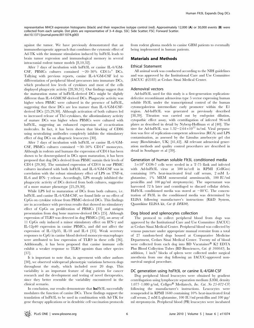
Human Flt3L Generates Dendritic Cells from Canine Peripheral Blood Precursors: Implications for a Dog Glioma Clinical Trial Weidong Xiong 1. , Marianela Candolfi 1. , Chunyan Liu 1 , A. K. M. Ghulam Muhammad 1 , Kader Yagiz 1 , Mariana Puntel 1 , Peter F. Moore 2 , Julie Avalos 3 , John D. Young 3 , Dorothy Khan 3 , Randy Donelson 4 , G. Elizabeth Pluhar 5 , John R. Ohlfest 4,6 , Kolja Wawrowsky 7 , Pedro R. Lowenstein 1 , Maria G. Castro 1 * 1 Gene Therapeutics Research Institute, Cedars-Sinai Medical Center and Department of Medicine and Department of Molecular and Medical Pharmacology, David Geffen School of Medicine, University of California Los Angeles, Los Angeles, California, United States of America, 2 Pathology, Microbiology, and Immunology, School of Veterinary Medicine, University of California Davis, Davis, California, United States of America, 3 Department of Comparative Medicine, Cedars-Sinai Medical Center, Los Angeles, California, United States of America, 4 Department of Pediatrics, University of Minnesota, Minneapolis, Minnesota, United States of America, 5 Department of Small Animal Clinical Sciences, University of Minnesota, St. Paul, Minnesota, United States of America, 6 Department of Neurosurgery, University of Minnesota, Minneapolis, Minnesota, United States of America, 7 Department of Medicine, Cedars-Sinai Medical Center and David Geffen School of Medicine, University of California Los Angeles, Los Angeles, California, United States of America Abstract Background: Glioblastoma multiforme (GBM) is the most common primary brain tumor in adults and carries a dismal prognosis. We have developed a conditional cytotoxic/immunotherapeutic approach using adenoviral vectors (Ads) encoding the immunostimulatory cytokine, human soluble fms-like tyrosine kinase 3 ligand (hsFlt3L) and the conditional cytotoxic molecule, i.e., Herpes Simplex Type 1- thymide kinase (TK). This therapy triggers an anti-tumor immune response that leads to tumor regression and anti-tumor immunological memory in intracranial rodent cancer models. We aim to test the efficacy of this immunotherapy in dogs bearing spontaneous GBM. In view of the controversy regarding the effect of human cytokines on dog immune cells, and considering that the efficacy of this treatment depends on hsFlt3L-stimulated dendritic cells (DCs), in the present work we tested the ability of Ad-encoded hsFlt3L to generate DCs from dog peripheral blood and compared its effects with canine IL-4 and GM-CSF. Methodology/Principal Findings: Our results demonstrate that hsFlT3L expressed form an Ad vector, generated DCs from peripheral blood cultures with very similar morphological and phenotypic characteristics to canine IL-4 and GM-CSF- cultured DCs. These include phagocytic activity and expression of CD11c, MHCII, CD80 and CD14. Maturation of DCs cultured under both conditions resulted in increased secretion of IL-6, TNF-a and IFN-c. Importantly, hsFlt3L-derived antigen presenting cells showed allostimulatory potential highlighting their ability to present antigen to T cells and elicit their proliferation. Conclusions/Significance: These results demonstrate that hsFlt3L induces the proliferation of canine DCs and support its use in upcoming clinical trials for canine GBM. Our data further support the translation of hsFlt3L to be used for dendritic cells’ vaccination and gene therapeutic approaches from rodent models to canine patients and its future implementation in human clinical trials. Citation: Xiong W, Candolfi M, Liu C, Muhammad AKMG, Yagiz K, et al. (2010) Human Flt3L Generates Dendritic Cells from Canine Peripheral Blood Precursors: Implications for a Dog Glioma Clinical Trial. PLoS ONE 5(6): e11074. doi:10.1371/journal.pone.0011074 Editor: Maciej Lesniak, The University of Chicago, United States of America Received May 12, 2010; Accepted May 19, 2010; Published June 11, 2010 This is an open-access article distributed under the terms of the Creative Commons Public Domain declaration which stipulates that, once placed in the public domain, this work may be freely reproduced, distributed, transmitted, modified, built upon, or otherwise used by anyone for any lawful purpose. Funding: This work is supported by National Institutes of Health/National Institute of Neurological Disorders & Stroke (NIH/NINDS) Grant 1R21-NS054143, 1UO1 NS052465 and 1RO1-NS057711 to M.G.C.; NIH/NINDS Grants 1 RO1 NS 054193, and RO1 NS061107 to P.R.L.; The Bram and Elaine Goldsmith and the Medallions Group Endowed Chairs in Gene Therapeutics Research to P.R.L. and M.G.C., respectively, The Linda Tallen & David Paul Kane Foundation Annual Fellowship and the Board of Governors at CSMC. M.C. is supported by NIH/NINDS 1F32NS058156.01. The funders had no role in study design, data collection and analysis, decision to publish, or preparation of the manuscript. Competing Interests: The authors have declared that no competing interests exist. * E-mail: [email protected] . These authors contributed equally to this work. Introduction Glioblastoma multiforme (GBM) is the most common and aggressive primary brain tumor in adults. GBM accounts for 35% of all primary brain tumor cases and 80% of malignant brain tumors (www.cbtrus.org). The standard of care for treatment of GBM, which consists of surgical resection, followed by radiation therapy and chemotherapy with temozolomide, leads to a median survival of 6–18 months [1]. Thus, novel approaches are being developed for the treatment of this devastating disease [2–6]. Our previous studies have shown that a combined conditional cytotoxic-immunotherapy approach using adenoviral vectors PLoS ONE | www.plosone.org 1 June 2010 | Volume 5 | Issue 6 | e11074

Welcome message from author
This document is posted to help you gain knowledge. Please leave a comment to let me know what you think about it! Share it to your friends and learn new things together.
Transcript
Human Flt3L Generates Dendritic Cells from CaninePeripheral Blood Precursors: Implications for a DogGlioma Clinical TrialWeidong Xiong1., Marianela Candolfi1., Chunyan Liu1, A. K. M. Ghulam Muhammad1, Kader Yagiz1,
Mariana Puntel1, Peter F. Moore2, Julie Avalos3, John D. Young3, Dorothy Khan3, Randy Donelson4, G.
Elizabeth Pluhar5, John R. Ohlfest4,6, Kolja Wawrowsky7, Pedro R. Lowenstein1, Maria G. Castro1*
1 Gene Therapeutics Research Institute, Cedars-Sinai Medical Center and Department of Medicine and Department of Molecular and Medical Pharmacology, David Geffen
School of Medicine, University of California Los Angeles, Los Angeles, California, United States of America, 2 Pathology, Microbiology, and Immunology, School of
Veterinary Medicine, University of California Davis, Davis, California, United States of America, 3 Department of Comparative Medicine, Cedars-Sinai Medical Center, Los
Angeles, California, United States of America, 4 Department of Pediatrics, University of Minnesota, Minneapolis, Minnesota, United States of America, 5 Department of
Small Animal Clinical Sciences, University of Minnesota, St. Paul, Minnesota, United States of America, 6 Department of Neurosurgery, University of Minnesota,
Minneapolis, Minnesota, United States of America, 7 Department of Medicine, Cedars-Sinai Medical Center and David Geffen School of Medicine, University of California
Los Angeles, Los Angeles, California, United States of America
Abstract
Background: Glioblastoma multiforme (GBM) is the most common primary brain tumor in adults and carries a dismalprognosis. We have developed a conditional cytotoxic/immunotherapeutic approach using adenoviral vectors (Ads)encoding the immunostimulatory cytokine, human soluble fms-like tyrosine kinase 3 ligand (hsFlt3L) and the conditionalcytotoxic molecule, i.e., Herpes Simplex Type 1- thymide kinase (TK). This therapy triggers an anti-tumor immune responsethat leads to tumor regression and anti-tumor immunological memory in intracranial rodent cancer models. We aim to testthe efficacy of this immunotherapy in dogs bearing spontaneous GBM. In view of the controversy regarding the effect ofhuman cytokines on dog immune cells, and considering that the efficacy of this treatment depends on hsFlt3L-stimulateddendritic cells (DCs), in the present work we tested the ability of Ad-encoded hsFlt3L to generate DCs from dog peripheralblood and compared its effects with canine IL-4 and GM-CSF.
Methodology/Principal Findings: Our results demonstrate that hsFlT3L expressed form an Ad vector, generated DCs fromperipheral blood cultures with very similar morphological and phenotypic characteristics to canine IL-4 and GM-CSF-cultured DCs. These include phagocytic activity and expression of CD11c, MHCII, CD80 and CD14. Maturation of DCscultured under both conditions resulted in increased secretion of IL-6, TNF-a and IFN-c. Importantly, hsFlt3L-derived antigenpresenting cells showed allostimulatory potential highlighting their ability to present antigen to T cells and elicit theirproliferation.
Conclusions/Significance: These results demonstrate that hsFlt3L induces the proliferation of canine DCs and support itsuse in upcoming clinical trials for canine GBM. Our data further support the translation of hsFlt3L to be used for dendriticcells’ vaccination and gene therapeutic approaches from rodent models to canine patients and its future implementation inhuman clinical trials.
Citation: Xiong W, Candolfi M, Liu C, Muhammad AKMG, Yagiz K, et al. (2010) Human Flt3L Generates Dendritic Cells from Canine Peripheral Blood Precursors:Implications for a Dog Glioma Clinical Trial. PLoS ONE 5(6): e11074. doi:10.1371/journal.pone.0011074
Editor: Maciej Lesniak, The University of Chicago, United States of America
Received May 12, 2010; Accepted May 19, 2010; Published June 11, 2010
This is an open-access article distributed under the terms of the Creative Commons Public Domain declaration which stipulates that, once placed in the publicdomain, this work may be freely reproduced, distributed, transmitted, modified, built upon, or otherwise used by anyone for any lawful purpose.
Funding: This work is supported by National Institutes of Health/National Institute of Neurological Disorders & Stroke (NIH/NINDS) Grant 1R21-NS054143, 1UO1NS052465 and 1RO1-NS057711 to M.G.C.; NIH/NINDS Grants 1 RO1 NS 054193, and RO1 NS061107 to P.R.L.; The Bram and Elaine Goldsmith and the MedallionsGroup Endowed Chairs in Gene Therapeutics Research to P.R.L. and M.G.C., respectively, The Linda Tallen & David Paul Kane Foundation Annual Fellowship andthe Board of Governors at CSMC. M.C. is supported by NIH/NINDS 1F32NS058156.01. The funders had no role in study design, data collection and analysis,decision to publish, or preparation of the manuscript.
Competing Interests: The authors have declared that no competing interests exist.
* E-mail: [email protected]
. These authors contributed equally to this work.
Introduction
Glioblastoma multiforme (GBM) is the most common and
aggressive primary brain tumor in adults. GBM accounts for 35%
of all primary brain tumor cases and 80% of malignant brain
tumors (www.cbtrus.org). The standard of care for treatment of
GBM, which consists of surgical resection, followed by radiation
therapy and chemotherapy with temozolomide, leads to a median
survival of 6–18 months [1]. Thus, novel approaches are being
developed for the treatment of this devastating disease [2–6]. Our
previous studies have shown that a combined conditional
cytotoxic-immunotherapy approach using adenoviral vectors
PLoS ONE | www.plosone.org 1 June 2010 | Volume 5 | Issue 6 | e11074
(Ad) that express the immunostimulatory cytokine human soluble
fms-like tyrosine kinase 3 ligand (hsFlt3L, Ad-hsFlt3L), and the
conditionally cytotoxic Herpes Simplex Type 1- thymide kinase
(TK, Ad-TK) induces tumor regression, long-term survival, and
immunological memory in rats and mice bearing large intracranial
syngeneic glioblastomas or intracranial melanomas [7–10].
Intratumoral expression of Ad-hsFlt3L recruits dendritic cells into
the brain parenchyma [11], improving tumor antigen presenta-
tion, while Ad-TK exerts a cytotoxic effect exclusively in
proliferating GBM cells in the presence of ganciclovir (GCV),
leading to the release of tumor antigens and inflammatory
molecules from dying tumor cells [7,8,10].
Spontaneous canine GBM constitutes an attractive animal
model for testing novel therapies for GBM. GBM is the most
common primary brain tumor in dogs, specially in certain breeds,
such as Boston terriers and Boxers [11–16]. Spontaneous GBM
in dogs exhibit the same histopathological features, clinical signs
and standard treatment as the human disease [17–20]. A clinical
trial in canine GBM patients would provide very useful data: i)
due to the infiltrative nature of spontaneous GBM in dogs, they
will allow testing the efficacy of novel therapies, and their toxicity
to the normal brain, ii) due to the large size of the dog brain, they
would be useful for assessing the doses and volumes needed in
order to optimize treatment protocols, iii) the fact that dogs
undergo similar standard care for GBM allows performing
clinical trials that mimic more closely the clinical scenario, in
which new therapies are implemented in conjunction with
standard of care, iv) the individual variability of outbreed dogs
better mimics the clinical scenario. Thus we aim to test the
efficacy of Ad-hsFlt3L and Ad-TK in dog patients bearing
spontaneous GBM. To this end we and others have previously
demonstrated the feasibility of delivering therapeutic transgenes
to dog GBM cells in vitro and dog brain cells in vivo upon
intracranial injection of gene therapy vectors, such as type 5
adenoviral vectors [21–23] and adeno-associated viral vectors
[24]. Also, we have shown that TK expression exerts a powerful
cytotoxic effect on dog GBM cells [21,22]. However, the
functionality of human soluble Flt3L on dog dendritic cells
(DCs) has been controversial [25–27]. Considering that the
efficacy of this treatment depends on an anti-tumor immune
response triggered by dendritic cells upon Flt3L stimulation
[10], and as a prelude for a clinical trial in canine GBM patients,
we aimed to evaluate whether hsFlt3L expressed by the
therapeutic Ad-hsFlt3L would exert a trophic effect on dog
dendritic cells.
It has been previously demonstrated that DCs can be
successfully obtained from dog peripheral blood mononuclear
cells (PBMC) using canine IL-4 and GM-CSF, in vitro [28]. In
order to evaluate the ability of hsFlt3L to generate DCs from dog
PBMC we compared it with canine IL-4 and GM-CSF. Our
results demonstrate that hsFLT3L is capable of generating DCs
from PBMC cultures with very similar characteristics to canine IL-
4 and GM-CSF-cultured DCs. Thus, indicating that hsFlt3L is
able to modulate the function of dog dendritic cells. We found that
dendritic cells generated using hsFlt3L express monocytic and
dendritic cell markers, they are capable of phagocytosis and
overexpression of activation markers and pro-inflammatory
cytokines upon maturation. Also, they are effective at antigen
presentation to T cells. Thus, this report supports the use hsFlt3L
encoded within an Ad in combination with Ad-TK (plus GCV) in
dog GBM patients. Our data will be critical for translating the use
of hsFlt3L in both dendritic cells’ vaccination approaches and also
in gene therapeutic strategies from rodents to canine patients and
ultimately to human patients.
Results
Morphological features of DCs cultured with hsFlt3L orcanine IL-4 and GM-CSF
Adherent dog peripheral blood leucocytes were cultured with
hsFlt3L derived from Ad-hsFlt3L conditioned medium or with
canine IL-4 and GM-CSF. Although the gross appearance of both
cultures was similar and adherent cellular aggregates were readily
observed after 4–6 days of culture in both conditions, the amount
of cells outside the aggregates was higher in the presence of canine-
IL-4 and GM-CSF (Fig. 1). After 1 week, cultured cells that
differentiated into non-adherent cells exhibiting thin cytoplasmic
processes were observed in hsFlt3L as well as in IL-4+GM-CSF
cultures (Inset Fig. 1). At day 7–9 floating cells were collected and
cellular aggregates were dislodged for flow cytometric analysis of
immune cell markers or to activate the immature DCs. The
average yield of cells collected from these cultures (n = 27 dogs)
was 456104 (67.56104) and 1016104 (6146104) for hsFlt3L and
canine-IL-4/GM-CSF cultures, respectively (Fig. 1C).
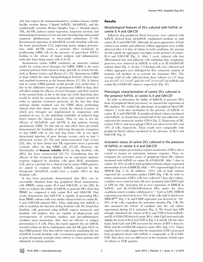
Phenotypic characterization of canine DCs cultured inthe presence hsFlt3L or canine IL-4 and GM-CSF
In order to determine the ability of hdFlt3L to generate DCs
from of peripheral blood precursors, we assessed the expression of
DC markers. We studied the phenotype of peripheral blood DC
cultures 1 week after incubation in the presence of hsFlt3L or
canine IL-4 and GM-CSF. In peripheral blood cultures incubated
with hsFlt3L, we found that around half of the non-adherent cells
expressed the monocytic marker CD14 (Fig. 2). Expression of DC
marker CD11c and macrophage CD18 was observed in 20% and
10% of cells, respectively. These results were comparable with
peripheral blood cultures incubated in the presence of IL-4 and
GM-CSF (Fig. 2).
Activation status of canine DCs cultured in the presenceof hsFlt3L or canine IL-4 and GM-CSF
Optimal antigen presentation requires maturation of DCs; this is
crucial to mount an anti-tumor immune response. Thus, we
evaluated the activation status of peripheral blood DC cultures
incubated with hsFlt3L or canine IL-4/GM-CSF. After 7 days in
culture 40–50% of cells in both peripheral blood cultures expressed
intermediate levels of MHCII, while ,10% expressed high levels of
MHCII (Fig. 3 A). In addition ,65% cells in both cultures
expressed the co-activation marker CD80 (Fig. 3 B). In order to
induce maturation of DCs, cells were collected 7 days after culture,
cytokines were removed and cells were incubated with ODN CpG
or LPS for 24h. Activation led to over expression of MHCII in
hsFlt3L- and IL-4/GM-CSF-derived DCs under the three
conditions tested (cytokine withdrawal +/2 CpG or LPS). MHCII
expression was observed in over 70% of the cells, half of which were
MHCIIhigh (Fig. 3 A) and CD80 expression was detected in ,80–
90% of the cells, regardless the activation stimulus (Fig. 3 B). We
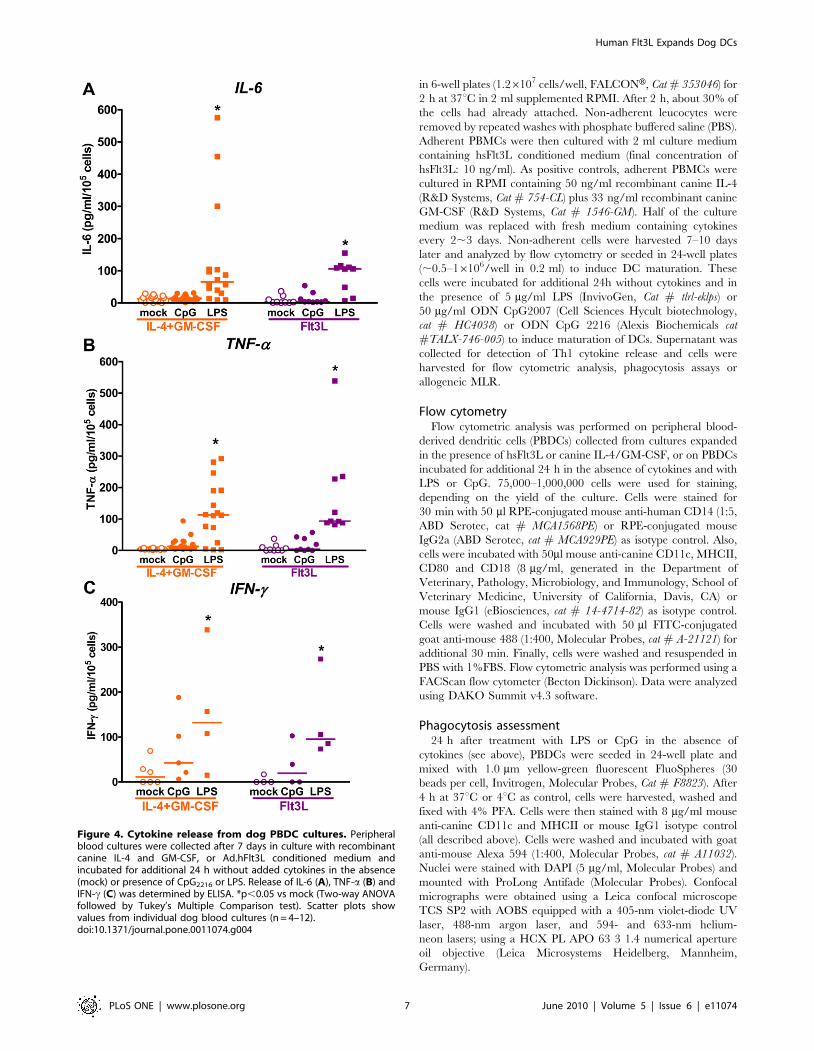
also measured the release of cytokines into the cell culture
supernatant during 24 h activation (Fig. 4). We found that LPS
strongly stimulated the release of IL-6 and TNF-a from hsFlt3L-
and IL-4/GM-CSF-derived canine DCs, while CpG increased only
slightly the levels of IL-6 and TNF-a (Fig. 4 A and B). On the other
hand, both CpG and LPS increased the release of IFN-c from hs-
Flt3L and IL-4/GM-CSF-cultured canine DCs (Fig. 4 C). Taken
together these results suggest that the maturation of DCs generated
from peripheral blood with hsFlt3L is comparable to IL-4/GM-
CSF. Also, there were no differences in the sensitivity of both types
of cultures to TLR agonists.
Human Flt3L Expands Dog DCs
PLoS ONE | www.plosone.org 2 June 2010 | Volume 5 | Issue 6 | e11074

Phagocytic activity of canine DCs cultured with hsFlt3L orcanine IL-4 and GM-CSF
Considering that tumor antigen uptake by DCs is a crucial
event in the initiation of the anti-tumor immune response induced
by the combined Ad-hsFlt3L+Ad-TK therapy [10], we evaluated
the phagocytic activity of peripheral blood-derived dendritic cells
(PBDCs) generated with hsFlt3L. We compared the phagocytic
activity of hsFlt3L- or IL-4/GM-CSF-derived PBDCs to take up
yellow green fluorescence beads. After 2h incubation with beads,
cells were fixed and stained by immunofluorescence with anti-
MHCII or anti-CD11c antibodies. MHCII+ cells and CD11c+
DCs containing beads in their cytoplasm were readily detected in
hsFlt3L and IL-4/GM-CSF cultures analyzed by confocal
microscopy (Fig. 5A). We quantified the phagocytic activity of
PBDCs after activation with CpG or LPS. We found that the basal
percentage of CD11c+ DCs with phagocytic activity was higher in
cultures derived from hsFlt3L than canine IL-4/GM-CSF (Fig. 5
B). Also, the phagocytic activity of both hsFlt3L- and IL-4/GM-
CSF-derived DCs was reduced upon activation with CpG or LPS
(Fig. 5 B).
Figure 1. Morphology of peripheral blood-derived dog dendritic cells cultured with canine IL-4+GM-CSF or Ad.hFlt3L conditionedmedium. White cells from dog peripheral blood were purified using Lymphocyte Separation Media and cultured in the presence of recombinantcanine IL-4 (50 ng/ml) and GM-CSF (33 ng/ml) or Ad.hsFlt3L conditioned medium (10 ng/ml). The images show the appearance of the culture after 7days in culture. Low magnification images (206) show typical spheres formed in culture supplemented with canine IL-4+GM-CSF (A) or hsFlt3L (B).Insets show higher magnification images of cells that exhibit typical long cytoplasmic processes characteristic of dendritic cell morphology. Scalebars: 10 mm. C, After 7–9 days in culture non-adherent cells were collected and counted. The graph shows the yield of cells per ml of blood obtainedfrom 27 dogs after culture with hsFlt3L or canine-IL-4/GM-CSF.doi:10.1371/journal.pone.0011074.g001
Human Flt3L Expands Dog DCs
PLoS ONE | www.plosone.org 3 June 2010 | Volume 5 | Issue 6 | e11074
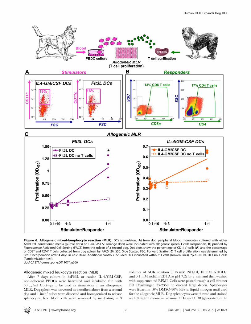
Human soluble Flt3L enhances the capacity of DCs toinduce proliferation of allogeneic T cells
We observed that hsFlt3L can generate DCs from peripheral
blood precursors; activation stimulates the release of Th1 cytokines
and reduces the phagocytic activity of DCs. Finally, since the
ability to prime and stimulate T cell proliferation is an important
assessment of the functionality of DCs [25], we performed a mixed
leucocyte reaction (MLR) to determine the proliferation of
allogeneic lymphocytes induced by mature DCs. DCs were
generated from dog peripheral blood, cultured with hsFlt3L or
canine IL-4/GM-CSF for 1 week, and activated for additional 24h
with CpG (Fig. 6 A). Splenocytes were collected from a second dog
and CD4+and CD8a+ T cells were purified by flow cytometry cell
sorting after staining with specific antibodies (Fig. 6 B). Dog T cells
were co-cultured with increasing numbers of allogeneic mature
DCs for 7 days. T cell proliferation was assessed by incorporation
of BrdU. As controls, DCs were incubated in the absence of T
cells. We found that increasing numbers of hsFlt3L-derived
PBDCs exerted a stimulatory effect on the proliferation of
allogeneic T cells (Fig. 6 C). Although at a lesser extent, canine
IL-4/GM-CSF-derived dog DCs also induced allogeneic T cell
proliferation. These results indicate that hsFlt3L exerts a potent
trophic effect on dog DCs, which in turn efficiently activate
proliferation of allogeneic naı̈ve T cells.
Discussion
We show that human Flt3L expressed from an adenoviral
vector generates dendritic cells from dog peripheral blood
precursors. Although previous reports have shown that maturation
of DCs can be achieved by culturing peripheral blood leucocytes
with T cell conditioned media [29] and canine cytokines
[28,30,31], the ability of human cytokines to exert trophic effects
on canine immune cells has been a matter of debate. Weber et al.
reported no activity of human TNF-a in the growth of canine DCs
and little cross-reaction of human Flt3L with canine immune cells
[25]. However, Hagglund et al. demonstrated that while the
combination of human GM-CSF, human TNF-a and human IL-4
failed to stimulate proliferation of canine DCs, replacement of
human IL-4 by human Flt3L in the cytokine cocktail led to
generation of DCs in vitro from dog bone marrow progenitors [26].
Moreover, dogs that received daily subcutaneous injections of
human recombinant Flt3L for 10 days exhibited increased
numbers of leucocytes, mainly monocytes, in peripheral blood
and circulating DCs displayed strong allostimulatory potential
[27]. Our results show that hsFlt3L exerts a trophic effect on dog
DCs that is comparable to the canine cytokine cocktail comprising
of IL-4 and GM-CSF. These findings support the rationale of
administering an Ad vector encoding hsFlt3L to dog patients
bearing spontaneous gliomas to mount an immune response
Figure 2. Phenotypic characterization of dog peripheral blood DC cultures. Peripheral blood cultures were collected after 7 days ofculture with recombinant canine IL-4 and GM-CSF or Ad.hFlt3L conditioned medium. Immune cell populations in the culture were identified usingantibodies against CD14 (monocytes), CD11c (dendritic cells) and CD18 (macrophages). Numbers in representative dot plots indicate thepercentage of positive cells. Approximately 12,000 events were collected from each sample. Dot plots are representative of 4 dogs. SSC: SideScatter; FSC: Forward Scatter.doi:10.1371/journal.pone.0011074.g002
Human Flt3L Expands Dog DCs
PLoS ONE | www.plosone.org 4 June 2010 | Volume 5 | Issue 6 | e11074
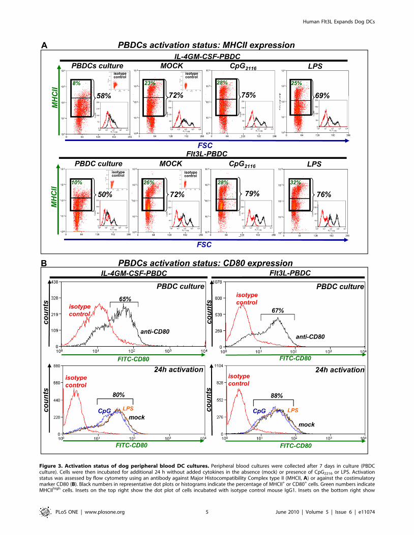
Figure 3. Activation status of dog peripheral blood DC cultures. Peripheral blood cultures were collected after 7 days in culture (PBDCculture). Cells were then incubated for additional 24 h without added cytokines in the absence (mock) or presence of CpG2216 or LPS. Activationstatus was assessed by flow cytometry using an antibody against Major Histocompatibility Complex type II (MHCII, A) or against the costimulatorymarker CD80 (B). Black numbers in representative dot plots or histograms indicate the percentage of MHCII+ or CD80+ cells. Green numbers indicateMHCIIhigh cells. Insets on the top right show the dot plot of cells incubated with isotype control mouse IgG1. Insets on the bottom right show
Human Flt3L Expands Dog DCs
PLoS ONE | www.plosone.org 5 June 2010 | Volume 5 | Issue 6 | e11074
against the tumor. We have previously demonstrated that an
immunotherapeutic approach that combines the cytotoxic effect of
Ad-TK with the immune stimulation induced by hsFlt3L leads to
brain tumor regression and immunological memory in several
intracranial rodent tumor models [8,10,32].
After 7 days of incubation with hsFlt3L or canine IL-4/GM-
CSF, PBMCs cultures contained ,20–30% CD11c+ DCs.
Tallying with previous reports, canine IL-4/GM-CSF led to
differentiation of peripheral blood precursors into immature DCs,
which produced low levels of cytokines and most of the cells
displayed phagocytic activity [28,30,31]. Our findings suggest that
the maturation status of hsFlt3L-derived DCs might be slightly
different than IL-4-GMCSF-derived DCs. Phagocytic activity was
higher when PBMC were cultured in the presence of hsFlt3L,
suggesting that these DCs are less mature than IL-4/GM-CSF-
derived DCs [25,29,30]. Although activation of both cultures led
to increased release of Th1-cytokines, the allostimulatory activity
of mature DCs was higher when PBMCs were cultured with
hsFlt3L, suggesting an enhanced expression of co-activation
molecules. In fact, it has been shown that blocking of CD86
using neutralizing antibodies completely inhibits the stimulatory
effect of dog DCs on allogeneic leucocytes [30].
After 7 days of incubation with hsFlt3L or canine IL-4/GM-
CSF, PBMCs cultures contained ,30–50% CD14+ monocytes.
Although in rodents and humans the expression of CD14 has been
shown to be downregulated in DCs upon maturation, it has been
proposed that dog DCs derived from PBMC sustain their levels of
CD14 [29,30]. The sustained expression of CD14 in our PBMC
cultures incubated with both hsFlt3L and IL-4/GM-CSF was in
correlation with the robust stimulatory effect of LPS on TNF-a,
IL-6 and IFN- c release. Accordingly, LPS strongly inhibited the
phagocytic activity of DCs derived from both cultures, suggestive
of a more mature phenotype [25,29,30].
While LPS led to maturation of DCs from both cultures, i.e.
hsFlt3L and canine IL-4/GM-CSF, we found little or no effect of
CpGs on cytokine release from PBMC-derived DCs. This findings
are in accordance with previous results that showed no stimulatory
effect of CpGs on proliferation of PBMCs [33] and antigen
presentation from dog bone marrow-derived DCs [25]. Although
expression of TLR9 was detected in dog PBMCs [34], an array of
11 CpGs only induced a minor stimulatory effect on IFN-c and
IL-12p40 expression in canine PBMCs, and did not affect the
expression of IL-12p35, IL-18 and IL-4 [35]. Weak secretory
responses to CpG in canine blood derived monocyte-macrophages
were attributed to low expression of TLR9 in these cells [36].
Additionally, it has been proposed that canine immune cells
exhibit a weaker response to TLR9 agonists than other species
[37].
It is important to note that, in agreement with other authors
[30], we observed widespread phenotypic variations between dogs
throughout the study, which included over 20 dogs. This
variability is an important feature of dog patients for cancer
research and the development and testing of novel therapeutics,
since they better resemble the variability encountered in the
clinical scenario.
In conclusion, our results demonstrate that hsFlt3L successfully
modulates the function of canine DCs. These findings support the
translation of hsFlt3L to be used in combination with Ad-TK for
gene therapy applications or in dendritic cell vaccination protocols
from rodent glioma models to canine GBM patients to eventually
being implemented in human patients.
Materials and Methods
Ethical StatementAll animal work was conducted according to the NIH guidelines
and was approved by the Institutional Care and Use Committee
(IACUC #2183) at Cedars Sinai Medical Center.
Adenoviral vectorsAd-hsFlt3L used for this study is a first-generation replication-
defective recombinant adenovirus type 5 vector expressing human
soluble Flt3L under the transcriptional control of the human
cytomegalovirus intermediate early promoter within the E1
region. Ad-hsFlt3L was generated as previously described
[38,39]. Titration was carried out by end-point dilution,
cytopathic effect assay, with centrifugation of infected 96-well
plates as described in detail by Nyberg-Hoffman et al [40]. The
titer for Ad-hsFlt3L was 1.32,2.6461012 iu/ml. Viral prepara-
tion was free of replication-competent adenovirus (RCA) and LPS
contamination, as assessed by the Limulus amebocyte gel clot
assay (Biowhittaker, UK) [41,42]. All relevant adenoviral gener-
ation methods and quality control procedures are described in
detail in Southgate et al [39].
Generation of human soluble Flt3L conditioned media56106 COS-7 cells were seeded in a T-75 flask and infected
with Ad-hsFlt3L virus at 500 iu/cell in RPMI (CellGro)
containing 10% heat-inactivated fetal calf serum, 2 mM L-
glutamine, 1% MEM nonessential amminoacids, 100 IU/ml
penicillin and 100 mg/ml streptomycin). The supernatant was
harvested 72 h later and centrifuged to discard cellular debris.
HsFlt3L conditioned media was stored at 280uC. The concen-
tration of Flt3L in the conditioned media was determined by
ELISA following manufacturer’s instructions (R&D System,
Quantikine ELISA kit, Cat # DFK00).
Dog blood and splenocytes collectionThe protocol to collect peripheral blood from dogs was
approved by the Institutional Care and Use Committee (IACUC)
at Cedars Sinai Medical Center. Peripheral blood was collected by
venous puncture under appropriate manual restraint from a total
of 27 random-bred dogs housed at Comparative Medicine
Department, Cedars Sinai Medical Center. Twenty ml of blood
were collected from each dog into BD VacutainerH K2 EDTA
Plus Blood Collection Tubes (BD Biosciences, Cat. # 366643). In
addition, 1 inch3 blocks of spleen were collected under surgical
anesthesia from one dog following an IACUC-approved non-
survival surgical procedure.
DC generation using hsFlt3L or canine IL-4/GM-CSFDog peripheral blood leucocytes were obtained by gradient
centrifugation using lymphocyte separation medium (LSM, density
1.077–1.080 g/ml, CellgroH Mediatech, Inc. Cat. No 25-072-CV)
following the manufacturer’s instructions. Leucocytes were
resuspended in RPMI 1640 containing 10% heat-inactivated fetal
calf serum, 2 mM L-glutamine, 100 IU/ml penicillin and 100 mg/
ml streptomycin. Peripheral blood (PB) leucocytes were incubated
representative MHCII expression histograms (black) and their respective isotype control (red). Approximately 12,000 (A) or 30,000 events (B) werecollected from each sample. Dot plots are representative of 3–4 dogs. SSC: Side Scatter; FSC: Forward Scatter.doi:10.1371/journal.pone.0011074.g003
Human Flt3L Expands Dog DCs
PLoS ONE | www.plosone.org 6 June 2010 | Volume 5 | Issue 6 | e11074
in 6-well plates (1.26107 cells/well, FALCONH, Cat # 353046) for
2 h at 37uC in 2 ml supplemented RPMI. After 2 h, about 30% of
the cells had already attached. Non-adherent leucocytes were
removed by repeated washes with phosphate buffered saline (PBS).
Adherent PBMCs were then cultured with 2 ml culture medium
containing hsFlt3L conditioned medium (final concentration of
hsFlt3L: 10 ng/ml). As positive controls, adherent PBMCs were
cultured in RPMI containing 50 ng/ml recombinant canine IL-4
(R&D Systems, Cat # 754-CL) plus 33 ng/ml recombinant canine
GM-CSF (R&D Systems, Cat # 1546-GM). Half of the culture
medium was replaced with fresh medium containing cytokines
every 2,3 days. Non-adherent cells were harvested 7–10 days
later and analyzed by flow cytometry or seeded in 24-well plates
(,0.5–16106/well in 0.2 ml) to induce DC maturation. These
cells were incubated for additional 24h without cytokines and in
the presence of 5 mg/ml LPS (InvivoGen, Cat # tlrl-eklps) or
50 mg/ml ODN CpG2007 (Cell Sciences Hycult biotechnology,
cat # HC4038) or ODN CpG 2216 (Alexis Biochemicals cat
#TALX-746-005) to induce maturation of DCs. Supernatant was
collected for detection of Th1 cytokine release and cells were
harvested for flow cytometric analysis, phagocytosis assays or
allogeneic MLR.
Flow cytometryFlow cytometric analysis was performed on peripheral blood-
derived dendritic cells (PBDCs) collected from cultures expanded
in the presence of hsFlt3L or canine IL-4/GM-CSF, or on PBDCs
incubated for additional 24 h in the absence of cytokines and with
LPS or CpG. 75,000–1,000,000 cells were used for staining,
depending on the yield of the culture. Cells were stained for
30 min with 50 ml RPE-conjugated mouse anti-human CD14 (1:5,
ABD Serotec, cat # MCA1568PE) or RPE-conjugated mouse
IgG2a (ABD Serotec, cat # MCA929PE) as isotype control. Also,
cells were incubated with 50ml mouse anti-canine CD11c, MHCII,
CD80 and CD18 (8 mg/ml, generated in the Department of
Veterinary, Pathology, Microbiology, and Immunology, School of
Veterinary Medicine, University of California, Davis, CA) or
mouse IgG1 (eBiosciences, cat # 14-4714-82) as isotype control.
Cells were washed and incubated with 50 ml FITC-conjugated
goat anti-mouse 488 (1:400, Molecular Probes, cat # A-21121) for
additional 30 min. Finally, cells were washed and resuspended in
PBS with 1%FBS. Flow cytometric analysis was performed using a
FACScan flow cytometer (Becton Dickinson). Data were analyzed
using DAKO Summit v4.3 software.
Phagocytosis assessment24 h after treatment with LPS or CpG in the absence of
cytokines (see above), PBDCs were seeded in 24-well plate and
mixed with 1.0 mm yellow-green fluorescent FluoSpheres (30
beads per cell, Invitrogen, Molecular Probes, Cat # F8823). After
4 h at 37uC or 4uC as control, cells were harvested, washed and
fixed with 4% PFA. Cells were then stained with 8 mg/ml mouse
anti-canine CD11c and MHCII or mouse IgG1 isotype control
(all described above). Cells were washed and incubated with goat
anti-mouse Alexa 594 (1:400, Molecular Probes, cat # A11032).
Nuclei were stained with DAPI (5 mg/ml, Molecular Probes) and
mounted with ProLong Antifade (Molecular Probes). Confocal
micrographs were obtained using a Leica confocal microscope
TCS SP2 with AOBS equipped with a 405-nm violet-diode UV
laser, 488-nm argon laser, and 594- and 633-nm helium-
neon lasers; using a HCX PL APO 63 3 1.4 numerical aperture
oil objective (Leica Microsystems Heidelberg, Mannheim,
Germany).
Figure 4. Cytokine release from dog PBDC cultures. Peripheralblood cultures were collected after 7 days in culture with recombinantcanine IL-4 and GM-CSF, or Ad.hFlt3L conditioned medium andincubated for additional 24 h without added cytokines in the absence(mock) or presence of CpG2216 or LPS. Release of IL-6 (A), TNF-a (B) andIFN-c (C) was determined by ELISA. *p,0.05 vs mock (Two-way ANOVAfollowed by Tukey’s Multiple Comparison test). Scatter plots showvalues from individual dog blood cultures (n = 4–12).doi:10.1371/journal.pone.0011074.g004
Human Flt3L Expands Dog DCs
PLoS ONE | www.plosone.org 7 June 2010 | Volume 5 | Issue 6 | e11074
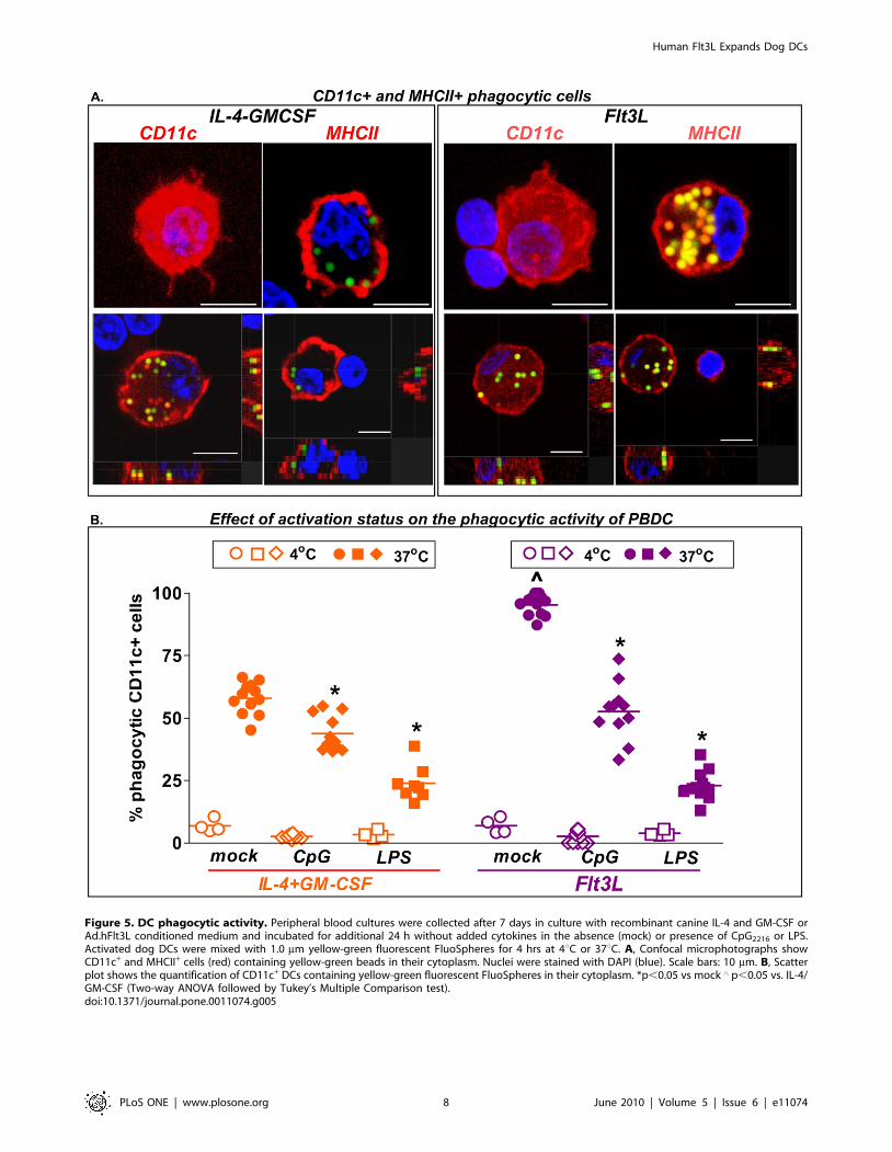
Figure 5. DC phagocytic activity. Peripheral blood cultures were collected after 7 days in culture with recombinant canine IL-4 and GM-CSF orAd.hFlt3L conditioned medium and incubated for additional 24 h without added cytokines in the absence (mock) or presence of CpG2216 or LPS.Activated dog DCs were mixed with 1.0 mm yellow-green fluorescent FluoSpheres for 4 hrs at 4uC or 37uC. A, Confocal microphotographs showCD11c+ and MHCII+ cells (red) containing yellow-green beads in their cytoplasm. Nuclei were stained with DAPI (blue). Scale bars: 10 mm. B, Scatterplot shows the quantification of CD11c+ DCs containing yellow-green fluorescent FluoSpheres in their cytoplasm. *p,0.05 vs mock ‘ p,0.05 vs. IL-4/GM-CSF (Two-way ANOVA followed by Tukey’s Multiple Comparison test).doi:10.1371/journal.pone.0011074.g005
Human Flt3L Expands Dog DCs
PLoS ONE | www.plosone.org 8 June 2010 | Volume 5 | Issue 6 | e11074
Allogeneic mixed leukocyte reaction (MLR)After 7 days culture in hsFlt3L or canine IL-4/GM-CSF,
non-adherent PBDCs were harvested and incubated 6 h with
50 mg/ml CpG2007 to be used as stimulators in an alloegeneic
MLR. Dog spleen was harvested as described above from a second
dog and 1 inch3 cubes were dissected and homogenized to release
splenocytes. Red blood cells were removed by incubating in 3
volumes of ACK solution (0.15 mM NH4Cl, 10 mM KHCO3,
and 0.1 mM sodium EDTA at pH 7.2) for 2 min and then washed
with supplemented RPMI. Cells were passed trough a cell strainer
BD Pharmingen 35-2350) to discard large debris. Splenocytes
were frozen in 10% DMSO-90% FBS in liquid nitrogen until used
for the allogeneic MLR. Dog splenocytes were thawed and stained
with 8 mg/ml mouse anti-canine CD4 and CD8 (generated in the
Figure 6. Allogeneic mixed-lymphocyte reaction (MLR). DCs (stimulators, A) from dog peripheral blood monocytes cultured with eitherAd.hFlt3L conditioned media (purple dots) or IL-4+GM-CSF (orange dots) were incubated with allogeneic spleen T cells (responders, B) purified byFluorescence Activated Cell Sorting (FACS) from the spleen of a second dog. Dot plots show the percentage of CD11c+-cells (A) and the percentageof CD8+ and CD4+ T cells collected from dog spleen by FACS (B). SSC: Side Scatter; FSC: Forward Scatter. C, T cell proliferation was determined byBrdU incorporation after 4 days in co-culture. Additional controls included DCs incubated without T cells (broken lines). *p,0.05 vs. DCs no T cells(Randomization test).doi:10.1371/journal.pone.0011074.g006
Human Flt3L Expands Dog DCs
PLoS ONE | www.plosone.org 9 June 2010 | Volume 5 | Issue 6 | e11074
Department of Veterinary, Pathology, Microbiology, and Immu-
nology, School of Veterinary Medicine, University of California,
Davis, CA). Cells were washed and incubated with FITC-
conjugated goat anti-mouse 488 (1:400) for additional 30 min.
CD4+ and C8+ T cells were then sorted by FACS (Becton
Dickinson). 50,000 total responder T cells (50% CD4 and 50%
CD8) were incubated in flat bottom 96-well plates (BECTON
DICKINSON cat #353072) with increasing numbers of allogeneic
stimulator DCs (0, 1:100, 1:30, 1:10, 1:3, 1:1) in 100 ml
supplemented RPMI at 37uC. DCs were incubated in the absence
of T cells as controls. Four days later 10 ml BrdU labeling solution
(1:100) was added to the media for additional 18 h and BrdU
incorporation into all genomic cellular DNA strands was assessed
by ELISA following manufacturer’s instructions (Roche-Applied-
Science, Cat # 11 647 229 001). Absorbance of the samples was
measured without stop solution in an ELISA reader (Spectramax
Plus, Molecular Devices) at 370 nm.
ELISARelease of IL-6, TNF-a and IFN-c was determined by ELISA
following the manufacturer’s protocol (R&D system, Quantikine
ELISA kit cat# CA6000, CATA00 and EL781, respectively).
Absorbance was read on a 96-well plate reader (Spectramax
Plus, Molecular Devices) at 450 nm and 570 nm to subtract
background.
Statistical AnalysisNCSS Statistical and power analysis software, (Kaysville, Utah,
USA) was employed to perform the statistical analysis. MLR
curves were analyzed using randomization test. The phagocytosis
and ELISA data were analyzed by ANOVA. When data failed
normality or Levene’s test for Variance Homogeneity, they were
log-transformed. Differences between groups were considered
significant at p,0.05.
Author Contributions
Conceived and designed the experiments: PRL MGC. Performed the
experiments: WdX MC CL AGM KY MP PFM JA JDY DK RD JRO
GEP KW. Analyzed the data: WdX MC CL AGM KY MP JA JDY DK
RD JRO GEP KW PRL MGC. Contributed reagents/materials/analysis
tools: PFM JA JDY DK RD JRO GEP. Wrote the paper: WdX MC PRL
MGC.
References
1. Hegi ME, Diserens AC, Gorlia T, Hamou MF, de Tribolet N, et al. (2005)
MGMT gene silencing and benefit from temozolomide in glioblastoma.
N Engl J Med 352: 997–1003.
2. Martin V, Liu D, Gomez-Manzano C (2009) Encountering and advancing
through antiangiogenesis therapy for gliomas. Curr Pharm Des 15: 353–364.
3. Barth RF (2009) Boron neutron capture therapy at the crossroads: challengesand opportunities. Appl Radiat Isot 67: S3–6.
4. Parker JN, Bauer DF, Cody JJ, Markert JM (2009) Oncolytic viral therapy of
malignant glioma. Neurotherapeutics 6: 558–569.
5. Asadi-Moghaddam K, Chiocca EA (2009) Gene- and viral-based therapies forbrain tumors. Neurotherapeutics 6: 547–557.
6. Candolfi M, Kroeger KM, Muhammad AK, Yagiz K, Farrokhi C, et al. (2009)
Gene therapy for brain cancer: combination therapies provide enhanced efficacyand safety. Curr Gene Ther 9: 409–421.
7. Candolfi M, Yagiz K, Foulad D, Alzadeh GE, Tesarfreund M, et al. (2009)
Release of HMGB1 in response to proapoptotic glioma killing strategies: efficacyand neurotoxicity. Clin Cancer Res 15: 4401–4414.
8. Muhammad AKMG, Candolfi M, King GD, Yagiz K, Foulad D, et al. (2009)
Antiglioma Immunological Memory in Response to Conditional Cytotoxic/Immune-Stimulatory Gene Therapy: Humoral and Cellular Immunity Lead to
Tumor Regression. Clinical Cancer Research 15: 6113–6127.
9. Ali S, King GD, Curtin JF, Candolfi M, Xiong W, et al. (2005) Combinedimmunostimulation and conditional cytotoxic gene therapy provide long-term
survival in a large glioma model. Cancer Res 65: 7194–7204.
10. Curtin J, Liu N, Candolfi M, Xiong W, Assi A, et al. (2009) HMGB1 mediatesendogenous TLR2 activation and brain tumor regression. PLoS Medicine 6:
e10.
11. Curtin JF, King GD, Barcia C, Liu C, Hubert FX, et al. (2006) Fms-like tyrosinekinase 3 ligand recruits plasmacytoid dendritic cells to the brain. J Immunol 176:
3566–3577.
12. Heidner GL, Kornegay JN, Page RL, Dodge RK, Thrall DE (1991) Analysis ofsurvival in a retrospective study of 86 dogs with brain tumors. J Vet Intern Med
5: 219–226.
13. Foster ES, Carrillo JM, Patnaik AK (1988) Clinical signs of tumors affecting therostral cerebrum in 43 dogs. J Vet Intern Med 2: 71–74.
14. LeCouteur RA (1999) Current concepts in the diagnosis and treatment of brain
tumours in dogs and cats. J Small Anim Pract 40: 411–416.
15. Stonehewer J, Mackin AJ, Tasker S, Simpson JW, Mayhew IG (2000) Idiopathicphenobarbital-responsive hypersialosis in the dog: an unusual form of limbic
epilepsy? J Small Anim Pract 41: 416–421.
16. Stoica G, Kim HT, Hall DG, Coates JR (2004) Morphology, immunohisto-chemistry, and genetic alterations in dog astrocytomas. Vet Pathol 41: 10–19.
17. Candolfi M, Curtin JF, Nichols WS, Muhammad AG, King GD, et al. (2007)
Intracranial glioblastoma models in preclinical neuro-oncology: neuropatholog-ical characterization and tumor progression. J Neurooncol 85: 133–148.
18. Lipsitz D, Higgins RJ, Kortz GD, Dickinson PJ, Bollen AW, et al. (2003)
Glioblastoma multiforme: clinical findings, magnetic resonance imaging, andpathology in five dogs. Vet Pathol 40: 659–669.
19. Orrison WW, Jr., Rose DF, Hart BL, Maclin EL, Sanders JA, et al. (1992)
Noninvasive preoperative cortical localization by magnetic source imaging.AJNR Am J Neuroradiol 13: 1124–1128.
20. Braund K (2003) Neoplasia of the Nervous System. In: Braund K, ed. Clinical
Neurology in Small Animals - Localization, Diagnosis and Treatment.
IthacaNew York: International Veterinary Information Service (www.ivis.org).pp B02270203.
21. Candolfi M, Kroeger KM, Pluhar GE, Bergeron J, Puntel M, et al. (2007)
Adenoviral-mediated gene transfer into the canine brain in vivo. Neurosurgery60: 167–177; discussion 178.
22. Candolfi M, Pluhar GE, Kroeger K, Puntel M, Curtin J, et al. (2007)
Optimization of adenoviral vector-mediated transgene expression in the canine
brain in vivo, and in canine glioma cells in vitro. Neuro Oncol 9: 245–258.
23. Oh S, Pluhar GE, McNeil EA, Kroeger KM, Liu C, et al. (2007) Efficacy ofnonviral gene transfer in the canine brain. J Neurosurg 107: 136–144.
24. Ciron C, Desmaris N, Colle MA, Raoul S, Joussemet B, et al. (2006) Gene
therapy of the brain in the dog model of Hurler’s syndrome. Ann Neurol 60:
204–213.
25. Weber M, Lange C, Gunther W, Franz M, Kremmer E, et al. (2003) Minorhistocompatibility antigens on canine hemopoietic progenitor cells. J Immunol
170: 5861–5868.
26. Hagglund HG, McSweeney PA, Mathioudakis G, Bruno B, Georges GE, et al.(2000) Ex vivo expansion of canine dendritic cells from CD34+ bone marrow
progenitor cells. Transplantation 70: 1437–1442.
27. Mielcarek M, Kucera KA, Nash R, Torok-Storb B, McKenna HJ (2007)
Identification and characterization of canine dendritic cells generated in vivo.Biol Blood Marrow Transplant 13: 1286–1293.
28. Ibisch C, Pradal G, Bach JM, Lieubeau B (2005) Functional canine dendritic
cells can be generated in vitro from peripheral blood mononuclear cells and
contain a cytoplasmic ultrastructural marker. J Immunol Methods 298:175–182.
29. Wijewardana V, Sugiura K, Oichi T, Fujimoto M, Akazawa T, et al. (2006)
Generation of canine dendritic cells from peripheral blood monocytes withoutusing purified cytokines. Vet Immunol Immunopathol 114: 37–48.
30. Bonnefont-Rebeix C, de Carvalho CM, Bernaud J, Chabanne L, Marchal T,
et al. (2006) CD86 molecule is a specific marker for canine monocyte-derived
dendritic cells. Vet Immunol Immunopathol 109: 167–176.
31. Catchpole B, Stell AJ, Dobson JM (2002) Generation of blood-derived dendriticcells in dogs with oral malignant melanoma. J Comp Pathol 126: 238–241.
32. King GD, Muhammad AKM, Curtin JF, Barcia C, Puntel M, et al. (2008) Flt3L
and TK gene therapy eradicate multifocal glioma in a syngeneic glioblastoma
model. Neuro Oncol 10: 19–31.
33. Wernette CM, Smith BF, Barksdale ZL, Hecker R, Baker HJ (2002) CpGoligodeoxynucleotides stimulate canine and feline immune cell proliferation. Vet
Immunol Immunopathol 84: 223–236.
34. Hashimoto M, Asahina Y, Sano J, Kano R, Moritomo T, et al. (2005) Cloningof canine toll-like receptor 9 and its expression in dog tissues. Vet Immunol
Immunopathol 106: 159–163.
35. Kurata K, Iwata A, Masuda K, Sakaguchi M, Ohno K, et al. (2004)
Identification of CpG oligodeoxynucleotide sequences that induce IFN-gammaproduction in canine peripheral blood mononuclear cells. Vet Immunol
Immunopathol 102: 441–450.
36. House AK, Gregory SP, Catchpole B (2008) Pattern-recognition receptor
mRNA expression and function in canine monocyte/macrophages andrelevance to canine anal furunuclosis. Vet Immunol Immunopathol 124:
230–240.
37. Im Hof M, Williamson L, Summerfield A, Balmer V, Dutoit V, et al. (2008)Effect of synthetic agonists of toll-like receptor 9 on canine lymphocyte
Human Flt3L Expands Dog DCs
PLoS ONE | www.plosone.org 10 June 2010 | Volume 5 | Issue 6 | e11074

proliferation and cytokine production in vitro. Vet Immunol Immunopathol
124: 120–131.38. Ali S, Curtin JF, Zirger JM, Xiong W, King GD, et al. (2004) Inflammatory and
anti-glioma effects of an adenovirus expressing human soluble Fms-like tyrosine
kinase 3 ligand (hsFlt3L): treatment with hsFlt3L inhibits intracranial gliomaprogression. Mol Ther 10: 1071–1084.
39. Southgate T, Kroeger KM, Liu C, Lowenstein PR, Castro MG (2008) Genetransfer into neural cells in vitro using adenoviral vectors. Curr Protoc Neurosci
Chapter 4: Unit 4 23.
40. Nyberg-Hoffman C, Aguilar-Cordova E (1999) Instability of adenoviral vectors
during transport and its implication for clinical studies. Nat Med 5: 955–957.
41. Dion LD, Fang J, Garver RI, Jr. (1996) Supernatant rescue assay vs. polymerase
chain reaction for detection of wild type adenovirus-contaminating recombinant
adenovirus stocks. J Virol Methods 56: 99–107.
42. Cotten M, Baker A, Saltik M, Wagner E, Buschle M (1994) Lipopolysaccharide
is a frequent contaminant of plasmid DNA preparations and can be toxic to
primary human cells in the presence of adenovirus. Gene Ther 1: 239–246.
Human Flt3L Expands Dog DCs
PLoS ONE | www.plosone.org 11 June 2010 | Volume 5 | Issue 6 | e11074
Related Documents