Human enteroviruses are not the cause of neurological impairments in children at the Korle-Bu Teaching Hospital Prudence Tettey 1 , Ebenezer Badoe 2 , Theophilus Adiku 1 , Eva Obodai 3 , John Kofi Odoom 3,& 1 Department of Microbiology, University of Ghana Medical School, Korle-Bu, Accra, Ghana, 2 Department of Child Health, University of Ghana Medical School, Korle-Bu, Accra, Ghana, 3 Department of Virology, Noguchi Memorial Institute for Medical Research, University of Ghana, Legon, Accra, Ghana & Corresponding author: John Kofi Odoom, Department of Virology, Noguchi Memorial Institute for Medical Research, University of Ghana, Legon, Accra, Ghana Key words: Convulsion, neurological, Human enterovirus, arbovirus, bacteriological, parasitological Received: 21/08/2013 - Accepted: 03/07/2014 - Published: 21/07/2014 Abstract Introduction: Convulsions associated with fever and acute onset of unknown aetiology with case fatalities have become a long observed medical condition at the Child Health Department of the Korle-Bu Teaching Hospital. Children admitted to the department with seizures of undetermined origin and fever has been a source of diagnostic confusion. Studies from the Asia Pacific region suggest a link with non-polio enteroviruses. The aim of the study was to investigate the association between non-polio enterovirus and acute encephalopathy causing neurological morbidity in children. Methods: One hundred and fifty cerebrospinal fluid (CSF), throat swab and serum samples were collected from participants at the Child Health Department of the Korle-Bu Teaching Hospital for virus isolation and characterization. Samples were cultured on cells and positive culture assayed by microneutralisation. Direct PCR as well as multiplex PCR were used to detect other viral agents present. Results: Enterovirus isolation rate was approximately 0.67 %. Intratypic differentiation by molecular characterization identified a poliovirus from vaccine origin. Further screening by real-time RT-PCR identified the virus as normal Sabin and not vaccine-derive poliovirus. No arbovirus was however detected. Conclusion: Non- polio enteroviruses and chikugunya virus were found not to be the etiologic agent responsible for the convulsion with neurologic morbidity observed in the Ghanaian children. Investigation for other viral agents is recommended. Pan African Medical Journal. 2014; 18:232 doi:10.11604/pamj.2014.18.232.3253 This article is available online at: http://www.panafrican-med-journal.com/content/article/18/232/full/ © John Kofi Odoom et al. The Pan African Medical Journal - ISSN 1937-8688. This is an Open Access article distributed under the terms of the Creative Commons Attribution License (http://creativecommons.org/licenses/by/2.0), which permits unrestricted use, distribution, and reproduction in any medium, provided the original work is properly cited. Pan African Medical Journal – ISSN: 1937- 8688 (www.panafrican-med-journal.com) Published in partnership with the African Field Epidemiology Network (AFENET). (www.afenet.net) Research Open Access

Welcome message from author
This document is posted to help you gain knowledge. Please leave a comment to let me know what you think about it! Share it to your friends and learn new things together.
Transcript

Page number not for citation purposes 1
Human enteroviruses are not the cause of neurological impairments in children at the
Korle-Bu Teaching Hospital
Prudence Tettey1, Ebenezer Badoe2, Theophilus Adiku1, Eva Obodai3, John Kofi Odoom3,&
1Department of Microbiology, University of Ghana Medical School, Korle-Bu, Accra, Ghana,2Department of Child Health, University of Ghana Medical
School, Korle-Bu, Accra, Ghana,3Department of Virology, Noguchi Memorial Institute for Medical Research, University of Ghana, Legon, Accra,
Ghana
&Corresponding author: John Kofi Odoom, Department of Virology, Noguchi Memorial Institute for Medical Research, University of Ghana, Legon,
Accra, Ghana
Key words: Convulsion, neurological, Human enterovirus, arbovirus, bacteriological, parasitological
Received: 21/08/2013 - Accepted: 03/07/2014 - Published: 21/07/2014
Abstract
Introduction: Convulsions associated with fever and acute onset of unknown aetiology with case fatalities have become a long observed medical
condition at the Child Health Department of the Korle-Bu Teaching Hospital. Children admitted to the department with seizures of undetermined
origin and fever has been a source of diagnostic confusion. Studies from the Asia Pacific region suggest a link with non-polio enteroviruses. The
aim of the study was to investigate the association between non-polio enterovirus and acute encephalopathy causing neurological morbidity in
children. Methods: One hundred and fifty cerebrospinal fluid (CSF), throat swab and serum samples were collected from participants at the Child
Health Department of the Korle-Bu Teaching Hospital for virus isolation and characterization. Samples were cultured on cells and positive culture
assayed by microneutralisation. Direct PCR as well as multiplex PCR were used to detect other viral agents present. Results: Enterovirus isolation
rate was approximately 0.67 %. Intratypic differentiation by molecular characterization identified a poliovirus from vaccine origin. Further screening
by real-time RT-PCR identified the virus as normal Sabin and not vaccine-derive poliovirus. No arbovirus was however detected. Conclusion: Non-
polio enteroviruses and chikugunya virus were found not to be the etiologic agent responsible for the convulsion with neurologic morbidity
observed in the Ghanaian children. Investigation for other viral agents is recommended.
Pan African Medical Journal. 2014; 18:232 doi:10.11604/pamj.2014.18.232.3253
This article is available online at: http://www.panafrican-med-journal.com/content/article/18/232/full/
© John Kofi Odoom et al. The Pan African Medical Journal - ISSN 1937-8688. This is an Open Access article distributed under the terms of the Creative Commons Attribution License (http://creativecommons.org/licenses/by/2.0), which permits unrestricted use, distribution, and reproduction in any medium, provided the original
work is properly cited.
Pan African Medical Journal – ISSN: 1937- 8688 (www.panafrican-med-journal.com) Published in partnership with the African Field Epidemiology Network (AFENET). (www.afenet.net)
Research
Open Access
Page number not for citation purposes 2
Introduction
Convulsion with acute onset in general is a common cause of
admission in paediatric emergency wards and risk for neurological,
cognitive impairment and epilepsy. Early diagnosis for the etiological
cause and immediate clinical management is crucial to the survival
of the child [1-4]. Clinical and experimental data suggest that
prolonged seizures can have immediate and long-term adverse
consequences on the immature and developing brain [5]. It is
estimated that about 4% to 6% of all children will have a seizure in
the first 16 years of life [6]. The incidence is predominant in
children under the age of 3 years, with a declining frequency in
older children [7]. Epidemiologic investigations have revealed that
approximately 150,000 children will sustain a first-time, unprovoked
seizure each year, and of those, 30,000 will develop epilepsy5 with
the highest risk being among children with prior condition of
neurodevelopmental abnormality and family history of afebrile
seizures [6,8]. The incidence of convulsions in developing countries
including Ghana is higher than developed countries because of high
infection rates [9-12]. Convulsion denotes a clinical symptom of an
underlying pathologic condition with many possible causes.
Convulsion may be caused by genetic and metabolic factors, fever,
head injury, excessive alcohol intake, ischaemic stroke, intracranial
haemorrhage, use of illicit drugs, meningitis, encephalitis and
infection with parasites, bacteria or viruses [9,13,14]. Some of the
viruses implicated in cases of convulsion include Human herpesvirus
6 [15,16], influenza A [17,18], Chikungunya virus [19,20] and
Human enterovirus 71 [21,22].
At the Child Health Department of the Korle-Bu Teaching Hospital
(KBTH), sporadic cases of convulsion with associated fever of
unknown aetiology have become a long observed medical condition
with diagnostic confusion. Routine parasitological and bacteriological
investigations conducted have been inconclusive and no further
investigations have been able to establish the cause of the disease.
Cases include children between the ages of one day and twelve
years with presentations of convulsion and fever, with occasional
rashes. Many of the patients without any history of neurological
problem were found to have developed temporal or permanent
neurological impairment. Isolation of the causative agent would help
to curtail unnecessary investigations, rationalise treatment, improve
reliability of prognosis and prevent overuse of antimicrobial agents
with consequent antimicrobial resistance.
In the Asian-Pacific regions, this manifestation of childhood
convulsion associated with fever and neurological complication
observed at the Child Health Department of the KBTH is usually
associated with the non-polio enterovirus known as Human
enterovirus 71 (HEV71) [23-27]. Currently, there is very little
literature supporting the circulation of HEV71 in Africa which include
the isolation of HEV71- like virus from children with acute flaccid
paralysis in Central Africa Republic [28] and two small institutional
outbreaks of HEV71 infection in HIV orphanages in Nairobi, Kenya
[29]. However, the circulation of other enteroviruses is prevalent,
which include poliovirus, Coxsackievirus, echovirus, hepatitis A virus
and enterovirus 70 (5) [30,31]. Although HEV71 is yet to be isolated
in Ghana, migration, travel, tourism and pilgrimage of Muslims from
Ghana to HEV71 endemic regions, may get infected and become a
source of infection for others.
Epidemics of viral infections causing central nervous system effects
are continuously being reported from around the world and
clinicians are challenged to be abreast with local epidemiology. This
study therefore aimed to investigate whether non-polio enterovirus
was the etiological cause of the neurological disorders observed in
the children.
Methods
Study population: The study population comprised children
between the ages of one day and twelve years old admitted to the
hospital having clinical diagnosis of convulsion associated with fever
and rash. Convulsions includes seizures lasting for at least half an
hour, or convulsions followed by coma lasting two hours or more or
convulsions followed by paralysis or other neurological signs not
previously present and lasting 24 hours or more and convulsions
that presented as encephalitis [32-34]. Only children with fever,
convulsion, skin rash, herpangina, viral meningitis, viral encephalitis
and other neurological manifestations were considered for the
study.
Virus isolation: Viruses in stool specimens were isolated on Hep-
2C (derived from human carcinoma cells) and RD
(Rhabdomyosarcoma) cell lines in accordance with standard
protocols (WHO,2004). Briefly, Hep-2C and RD cells were seeded in
tissue culture tubes with growth medium (Eagle's MEM
supplemented with 10% FCS) 48 hr prior to inoculation.
Page number not for citation purposes 3
Suspensions of faecal samples pre-treated with chloroform was
inoculated on serum-free medium and incubated at 37oC and
observed daily for the characteristic enterovirus cytopathic effect
(CPE). The tubes with CPE up to 75% and above were harvested
and kept at -20oC while those negative after 5 days of incubation
were re-passaged (blind passage) on the same cell line and if it
remained negative after 5 days was considered negative
Microneutralization and real time PCR: Identification of isolates
on Hep-2C and RD cells lines was carried out by microneutralisation
technique using polyclonal antisera raised in horse against
Coxsackie and echoviruses prepared by the National Institute of
Public Health and the Environment (RIVM), Netherlands.
Microneutralisation assay with HEV71 antiserum was carried out on
the isolates that were untypable with the antiseraum pools. Samples
that showed neutralization in the wells with polio pool antisera
selected for Real-Time Reverse Transcriptase polymerase Chain
Reaction (rRT-PCR).
RNA extraction and PCR: Viral RNA was extracted by QIAamp
Viral Mini Spin Protocol (Second edition, December 2005) according
to manufacturer's instruction. Reverse Transcriptase Polymerase
Chan Reaction (RT-PCR) with Pan-EV primers MD90 (5’- ATT GTC
ACC ATA AGC AGC CA-3’) and anti-sense MD91 (5’ - CCT CCG GCC
CCT GAA TGC GGC TAA T -3’) were used to amplify the VP1 region
under the following conditions RT: 97°C for 3 minutes, PCR: 95°C
for 45 seconds, 55°C for 45 seconds, and 70°C for 45 seconds and
cooled to 4°C. The products were observed on 1% agarose gel
electrophoresis.
Data analysis: Data analysis was done using SPSS version 19. The
analysis involves frequency distribution of responses and cross
tabulation of variables. The analyzed information was presented
using tables, graphs, charts and other diagrams that depicted the
pattern of findings.
Ethical issues: Ethical approval was obtained from the Ethical
Committee of the College of Health Sciences, and Research
Committee of the University of Ghana Medical School (UGMS).
Informed consent was sought from parent/guardian of subjects
before the commencement of the study. All ethical considerations
were adhered to. Data collected from the study was handled
anonymously and confidentially. Samples had only the identification
numbers of the subjects to ensure anonymity. We protected the
confidentiality of patients through use of codes.
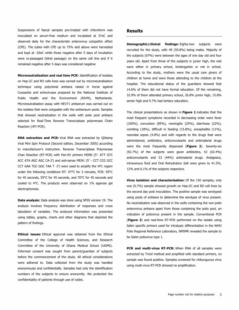
Results
Demographic/clinical findings: Eighty-two subjects were
recruited for the study, with 49 (59.8%) being males. Majority of
the subjects (67%) were between the ages of one day old and four
years old. Apart from three of the subjects in junior high, the rest
were either in primary school, kindergarten or not in school.
According to the study, mothers were the usual care givers of
children at home and were those attending to the children at the
hospital. The educational status of the guardians showed that
14.6% of them did not have formal education. Of the remaining,
32.9% of them attended primary school, 26.8% junior high, 15.8%
senior high and 9.7% had tertiary education.
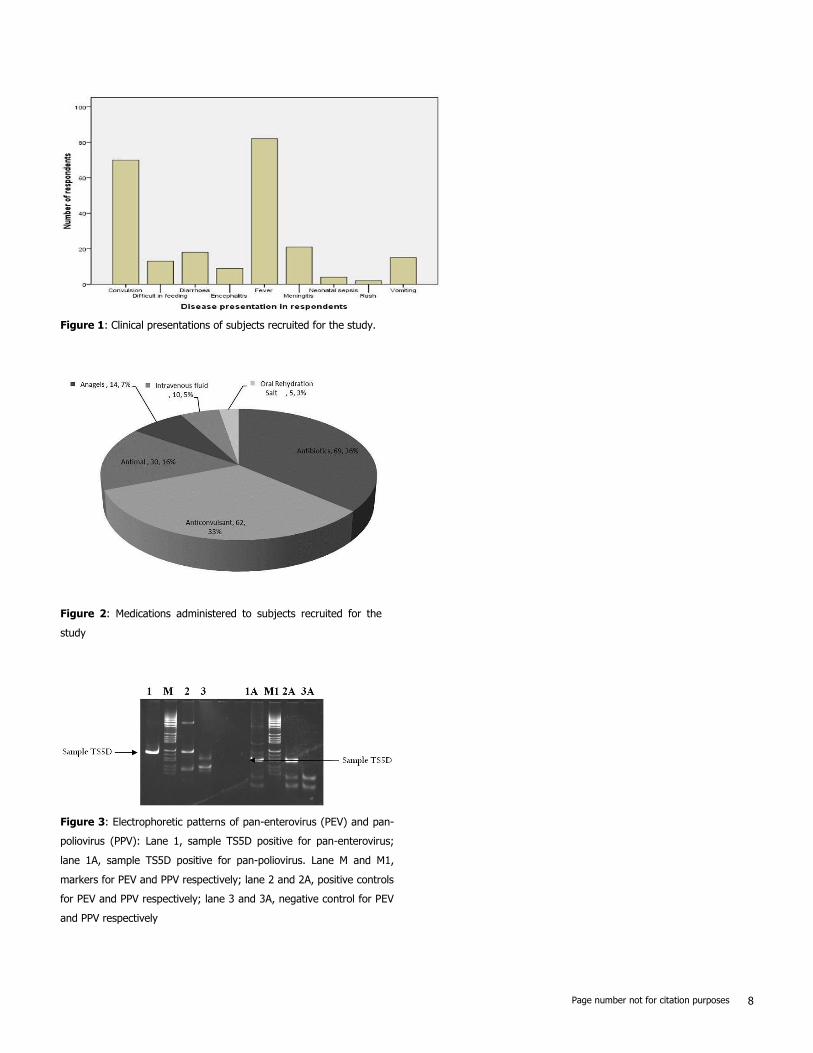
The clinical presentations as shown in Figure 1 indicates that the
most frequent symptoms recorded in decreasing order were fever
(100%), convulsion (85%), meningitis (25%), diarrhoea (22%),
vomiting (18%), difficult in feeding (15.8%), encephalitis (11%),
neonatal sepsis (4.8%) and with regards to the drugs that were
administered, antibiotics, anticonvulsants and antimalarial drugs
were the most frequently dispensed (Figure 2). Seventy-six
(92.7%) of the subjects were given antibiotics, 52 (63.4%)
anticonvulsants and 53 (44%) antimalarial drugs. Analgesics,
intravenous fluid and Oral Rehydration Salt were given to 41.5%,
12% and 6.1% of the subjects respective.
Virus isolation and characterisation: Of the 150 samples, only
one (0.7%) sample showed growth on Hep-2C and RD cell lines by
the second day post inoculation. The positive sample was serotyped
using pools of antisera to determine the serotype of virus present.
No neutralization was observed in the wells containing the non-polio
enterovirus antisera apart from those containing the polio pool, an
indication of poliovirus present in the sample. Conventional PCR
(Figure 3) and real-time RT-PCR performed on the isolate using
Sabin specific primers used for intratypic differentiation in the WHO
Polio Regional Reference Laboratory, NMIMR revealed the sample to
be Sabin poliovirus type 1.
PCR and multi-virus RT-PCR: When RNA of all samples were
extracted by Trizol method and amplified with standard primers, no
sample was found positive. Samples screened for chikungunya virus
using multi-virus RT-PCR showed no amplification.
Page number not for citation purposes 4
Discussion
The etiological cause of convulsion with acute onset in children in
admission at the Korle-Bu Teaching Hospital was investigated for
viral involvement such as non-polio enteroviruses and chikugunya
virus using virus isolation and polymerase chain reaction techniques.
The results as shown by the study did not link the viruses to the
cause of the convulsions. Patients were managed with
anticonvulsants, antibiotics, antimalarial and antivirals which were
combined with Paracetamol, intravenous fluid (IVF) and oral
rehydration salt (ORS) depending on the clinical presentation. The
anticonvulsants were administered to manage clinically severe
convulsion cases while antibiotics and antimalarial drugs were
prescribed due to the febrile condition which may have either
bacterial or parasitological origin.
Routine bacteriological and parasitological laboratory tests
requested by the physicians to isolate the aetiological agent failed to
detect neither bacteria nor any parasite. The failure of the
laboratory to isolate the aetiological agent compelled these
emergency physicians to continue the symptomatic management. In
spite of this necessity, the practise of treating patients
symptomatically is not always recommended because it may mask
the presence of the underlying etiology which will then be forgotten
or treated with great delay. Symptomatic treatment with
antimicrobial agent can lead to antimicrobial resistance [35,36]. The
absence of parasites and bacteria during the laboratory investigation
did not justify the extensive usage of the antimicrobial agents. While
the hospital emergency department denotes a place of initiating
empiric antimicrobial therapy as a form of medical intervention and
also a site of extensive use of antimicrobial agents, emergency
clinicians and all other clinicians must be fully conscious of the fact
that inappropriate use and over prescribing of antimicrobial agents
accelerates the development of antimicrobial resistance [37,38].
Antimicrobial resistance, as it is already known, inflates the patient's
budget, prolonging stay in hospital and also pressurizes drug
manufacturing companies to make available new drugs that these
agents would be susceptible to [39,40].
To many emergency room physicians, the threat of antimicrobial
resistance has not sunk in yet. It has been apparent through this
study that emergency department physician's fundamental and
principal concern in an emergency situation is how possible he could
resuscitate his patient and that, the issue of the impact of
antimicrobial use on the prevalence of resistance was not a crucial
consideration at that moment. Many physicians and patients do not
see antimicrobial resistance as a reason to abstain from its use
[41,42]. Emergency department physicians may therefore not be
different from other physicians in their frequent prescription of
antimicrobials for conditions that do not appear to profit from their
use. This raises a general concern if the principle of prudent use of
antimicrobials is being adhered to.
The findings from this study could not establish non-polio
enterovirus or chikugunya association with the etiological cause of
the manifestation. All molecular virological assays to determine the
aetiology after RNA extraction from 150 samples yielded only one
positive for poliovirus. Further characterization identified the
poliovirus as a Sabin 1 poliovirus. The virus was obtained from the
throat swab of a one month old baby with the presentations of
fever, convulsion and fast breathing. These symptoms are not
characteristic of poliovirus infection. Since Sabin 1 poliovirus is a
component of the oral polio vaccine (OPV), the child might have
acquired the virus from either polio vaccination during the recent
national polio immunization days, contact with immunized person or
from the environment. The oral polio vaccine (OPV) apart from
seeding the gut of the recipient could also immunize children in
contact with the faeces of the OPV recipient [43,44]. It is also a way
of gaining natural immunity when a person has not received the
OPV. The OPV is unstable and can revert to neurovirulence in some
instances.
Further investigation to determine whether the Sabin 1 poliovirus
isolated was a normal Sabin or a VDPV have shown that the virus is
a normal Sabin 1 poliovirus and not VDPV. It was very expedient to
have screened for the presence of VDPV because of its latest
implication in cases of acute flaccid paralysis in many parts of the
world. Screening for Chikungunya virus using the multi-virus real
time PCR did not also establish the aetiological agent. In West-
Africa, similar clinical presentations of fever, convulsion,
maculopapular rash and meningoencephalitis have been recorded to
be caused by Chikungunya virus (CHIKV). Since its discovery in
Tanganyika (Tanzania), Africa, in 1952, Chikungunya virus
outbreaks have occurred sporadically in Africa, West Africa (Senegal
and Nigeria), South Asia, and Southeast Asia with recent outbreaks
spreading the disease over a wider range.
The study limitations include our inability to collect stool samples or
anal swap since enteroviruses are readily present in stool. Secondly
Page number not for citation purposes 5
we did not investigate other DNA viruses with similar presentation.
The etiological agent may be a DNA virus while the study was
geared towards the detection of RNA virus. In the literature, other
viruses associated with such manifestation are influenza,
parainfluenza virus, herpes virus 6, Herpes virus 7 and RSV. Similar
manifestations must be investigated for the presence of these
viruses. Solving this puzzle will save the Government a lot of money
that is spent on antibiotics, antimalarial and other drugs which in
future will lead to drug resistance in the children.
Conclusion
The findings from this study indicate that the aetiological agent for
the observed convulsions in the children was neither due to non-
polio enteroviruses nor chikugunya. We recommend that other viral
agents capable of causing convulsion in children be investigated.
The use of drugs to manage convulsions should be minimized and
efforts should be made toward identifying the aetiological agent.
Competing interests
Competing interests The authors declare no competing
interests. Disclaimer: The findings and conclusions in this paper
are those of the authors and do not necessarily represent the views
of any of their affiliated Research Institutions.
Authors’ contributions
Prudence Tettey participated in design, implementation of study
protocol, writing of the manuscript, performed the laboratory work
and contributed to data analysis. Ebenezer Badoe supervised the
collection of the samples and contributed to the data analysis. Dr
Adiku supervised the implementation of study protocol and the
manuscript writing. John Kofi Odoom participated and supervised
the design and implementation of study protocol, the manuscript
writing.
Acknowledgments
We are very grateful to Ishmael Dzigbodi, Michael Alale, Gifty
Mawuli and Miriam Eshun of the Virology Department of NMIMR for
technical support. We are also very grateful to the staff of University
of Ghana Medical School, Department Of Microbiology, University Of
Ghana, Legon, Ghana.
Figures
Figure 1: Clinical presentations of subjects recruited for the study.
Figure 2: Medications administered to subjects recruited for the
study
Figure 3: Electrophoretic patterns of pan-enterovirus (PEV) and
pan-poliovirus (PPV): Lane 1, sample TS5D positive for pan-
enterovirus; lane 1A, sample TS5D positive for pan-poliovirus. Lane
M and M1, markers for PEV and PPV respectively; lane 2 and 2A,
positive controls for PEV and PPV respectively; lane 3 and 3A,
negative control for PEV and PPV respectively
References
1. Sasidaran K, Singhi S et Singhi P. Management of acute seizure
and status epilepticus in pediatric emergency. Indian J Pediatr.
2012; 79 (4): 510-7. PubMed | Google Scholar
2. Abend NS, Huh JW, Helfaer MA and Dlugos DJ. Anticonvulsant
medications in the pediatric emergency room and intensive
care unit. Pediatr Emerg Care. 2008; 24 (10): 705-
18. PubMed | Google Scholar
3. Berg AT et Shinnar S. Complex febrile seizures. Epilepsia.
1996; 37 (2): 126-33. PubMed | Google Scholar
4. Verity CM, Butler NR et Golding J. Febrile convulsions in a
national cohort followed up from birth; I--Prevalence and
recurrence in the first five years of life. Br Med J (Clin Res Ed).
1985; 290(6478): 1307-10. PubMed | Google Scholar

Page number not for citation purposes 6
5. Hubert P, Parain D and Vallee L. Management of convulsive
status epilepticus in infants and children. Rev Neurol (Paris).
2009; 165 (4): 390-7. PubMed | Google Scholar
6. McAbee GN and Wark JE. A practical approach to
uncomplicated seizures in children. Am Fam Physician. 2000;
62 (5): 1109-16. PubMed | Google Scholar
7. Vining EP. Pediatric seizures. Emerg Med Clin North Am. 1994;
12 (4): 973-88. PubMed | Google Scholar
8. Hauser WA. The prevalence and incidence of convulsive
disorders in children. Epilepsia. 1994; 35 (Suppl 2): S1-
6.PubMed | Google Scholar
9. Idro R et al. The incidence, aetiology and outcome of acute
seizures in children admitted to a rural Kenyan district hospital.
BMC Pediatr. 2008; 8: 5. PubMed | Google Scholar
10. Owusu-Ofori A, Agbenyega T, Ansong D and Scheld WM.
Routine lumbar puncture in children with febrile seizures in
Ghana: should it continue? Int J Infect Dis. 2004; 8(6): 353-
61. PubMed | Google Scholar
11. Akpede GO, Abiodun PO and Sykes RM. Pattern of infections in
children under-six years old presenting with convulsions
associated with fever of acute onset in a children's emergency
room in Benin City, Nigeria. J Trop Pediatr. 1993; 39(1): 11-
5. PubMed | Google Scholar
12. Birbeck GL. Seizures in rural Zambia. Epilepsia. 2000; 41(3):
277-81. PubMed | Google Scholar
13. Pohlmann-Eden B, Beghi E, Camfield C and Camfield P. The
first seizure and its management in adults and children. BMJ.
2006; 332(7535): 339-42. PubMed | Google Scholar
14. Wolf SM and McGoldrick PE. Recognition and management of
pediatric seizures. Pediatr Ann. 2006; 35(5): 332-
44.PubMed | Google Scholar
15. Jee SH et al. Risk of recurrent seizures after a primary human
herpesvirus 6-induced febrile seizure. Pediatr Infect Dis J.
1998; 17(1): 43-8. PubMed | Google Scholar
16. Ward KN, Andrews NJ, Verity CM, Miller E and Ross EM.
Human herpesviruses-6 and -7 each cause significant
neurological morbidity in Britain and Ireland. Arch Dis Child.
2005; 90: 619-23. PubMed | Google Scholar
17. Chung B and Wong V. Relationship between five common
viruses and febrile seizure in children. Archives of disease in
childhood. 2007; 92(7): 589-593. PubMed | Google Scholar
18. Kwong KL, Lam SY, Que TL and Wong SN. Influenza A and
febrile seizures in childhood. Pediatr Neurol. 2006; 35(6): 395-
9. PubMed | Google Scholar
19. Ng KW et al. Clinical features and epidemiology of chikungunya
infection in Singapore. Singapore Med J. 2009; 50(8): 785-
90. PubMed | Google Scholar
20. Lewthwaite P et al. Chikungunya virus and central nervous
system infections in children, India. Emerg Infect Dis. 2009;
15(2): 329-31. PubMed | Google Scholar
21. McMinn P, Stratov I, Nagarajan L and Davis S. Neurological
manifestations of enterovirus 71 infection in children during an
outbreak of hand, foot, and mouth disease in Western
Australia. Clin Infect Dis. 2001; 32(2): 236-
42. PubMed | Google Scholar
22. Palacios G and Oberste MS. Enteroviruses as agents of
emerging infectious diseases. J Neurovirol. 2005; 11 (5): 424-
33.PubMed | Google Scholar
23. Cardosa MJ et al. Molecular epidemiology of human enterovirus
71 strains and recent outbreaks in the Asia-Pacific region:
comparative analysis of the VP1 and VP4 genes. Emerg Infect
Dis. 2003; 9(4): 461-8. PubMed | Google Scholar
24. AbuBakar S et al. Enterovirus 71 outbreak, Brunei. Emerg
Infect Dis. 2009; 15(1): 79-82. PubMed | Google Scholar
25. McMinn PC. An overview of the evolution of enterovirus 71 and
its clinical and public health significance. FEMS Microbiol Rev.
2002; 26(1): 91-107. PubMed | Google Scholar
Page number not for citation purposes 7
26. Chang LY, Huang YC and Lin TY. Fulminant neurogenic
pulmonary oedema with hand, foot, and mouth disease.
Lancet. 1998; 352(9125): 367-8. PubMed | Google Scholar
27. Mizuta K et al. Frequent importation of enterovirus 71 from
surrounding countries into the local community of Yamagata,
Japan, between 1998 and 2003. J Clin Microbiol. 2005; 43(5):
6171-5. PubMed | Google Scholar
28. Bessaud M et al. Molecular characterization of human
enteroviruses in the Central African Republic: uncovering wide
diversity and identification of a new human enterovirus A71
genogroup. J Clin Microbiol. 2012; 50(5): 1650-
8. PubMed |Google Scholar
29. Chakraborty R et al. An epidemic of enterovirus 71 infection
among HIV-1-infected orphans in Nairobi. AIDS. 2004; 18 (14):
1968-70. PubMed | Google Scholar
30. Otatume S and Addy P. Ecology of enteroviruses in tropics; I-
Circulation of enteroviruses in healthy infants in tropical urban
area. Jpn J Microbiol. 1975; 19(3): 201-9. PubMed | Google
Scholar
31. Silva PA et al. Molecular characterization of enteric viral agents
from children in northern region of Ghana. J Med Virol. 2008;
80(10): 1790-8. PubMed | Google Scholar
32. Patel AD and Vidaurre J. Complex febrile seizures: a practical
guide to evaluation and treatment. J Child Neurol. 2013; 28(6):
759-64. PubMed | Google Scholar
33. DiMario FJ. Children presenting with complex febrile seizures
do not routinely need computed tomography scanning in the
emergency department. Pediatrics. 2006; 117 (2): 528-
530. PubMed | Google Scholar
34. Freedman SB and Powell EC. Pediatric seizures and their
management in the emergency department. Clinical Pediatric
Emergency Medicine. 2003; 4(3): 195-206. PubMed | Google
Scholar
35. Bisht R, Katiyar A, Singh R and Mittal P. Antibiotic resistance-A
global issue of concern. Asian Journal of Pharmaceutical and
Clinical Research. 2009; 2 (2): 34-39. PubMed | Google
Scholar
36. Chung CH. Emergency physician's dilemma in antibiotic
prescribing. Hong Kong j emerg med. Apr 2005; 12(2): 70-
76.PubMed | Google Scholar
37. Harbarth S and Samore MH. Antimicrobial resistance
determinants and future control. Emerg Infect Dis. 2005;
11(6): 794-801. PubMed | Google Scholar
38. Use IA. Appropriate antimicrobial prescribing: approaches that
limit antibiotic resistance. Am Fam Physician. 2001; 64 (6):
999-1005. PubMed | Google Scholar
39. McGowan JE Jr. Economic impact of antimicrobial resistance.
Emerg Infect Dis. 2001; 7(2): 286-92. PubMed | Google
Scholar
40. Cosgrove SE and Carmeli Y. The impact of antimicrobial
resistance on health and economic outcomes. Clin Infect Dis.
2003; 36(11): 1433-7. PubMed | Google Scholar
41. Simpson SA, Wood F and Butler CC. General practitioners'
perceptions of antimicrobial resistance: a qualitative study. J
Antimicrob Chemother. 2007; 59(2): 292-
6. PubMed | Google Scholar
42. Le Conte P, Potel G and Baron D. Emergency antibiotic
prescription in the emergency room. Reanimation Urgences.
2001; 10 (7): 673-678. PubMed | Google Scholar
43. Sabin AB. Oral poliovirus vaccine: history of its development
and use and current challenge to eliminate poliomyelitis from
the world. J Infect Dis. 1985; 151 (3): 420-
36. PubMed | Google Scholar
44. MacLennan C and MacLennan J. What threat from persistent
vaccine-related poliovirus? Lancet. 2005; 366 (9483): 351-
3.PubMed | Google Scholar
Page number not for citation purposes 8
Figure 1: Clinical presentations of subjects recruited for the study.
Figure 2: Medications administered to subjects recruited for the
study
Figure 3: Electrophoretic patterns of pan-enterovirus (PEV) and pan-
poliovirus (PPV): Lane 1, sample TS5D positive for pan-enterovirus;
lane 1A, sample TS5D positive for pan-poliovirus. Lane M and M1,
markers for PEV and PPV respectively; lane 2 and 2A, positive controls
for PEV and PPV respectively; lane 3 and 3A, negative control for PEV
and PPV respectively
Related Documents