2011;71:3098-3109. Published OnlineFirst March 10, 2011. Cancer Res Gianluca Civenni, Anne Walter, Nikita Kobert, et al. Long-term Growth with Metastasis Establish Tumor Heterogeneity and Human CD271-Positive Melanoma Stem Cells Associated Updated Version 10.1158/0008-5472.CAN-10-3997 doi: Access the most recent version of this article at: Material Supplementary html http://cancerres.aacrjournals.org/content/suppl/2011/03/10/0008-5472.CAN-10-3997.DC1. Access the most recent supplemental material at: Cited Articles http://cancerres.aacrjournals.org/content/71/8/3098.full.html#ref-list-1 This article cites 36 articles, 11 of which you can access for free at: E-mail alerts related to this article or journal. Sign up to receive free email-alerts Subscriptions Reprints and . [email protected] Publications Department at To order reprints of this article or to subscribe to the journal, contact the AACR Permissions . [email protected] Department at To request permission to re-use all or part of this article, contact the AACR Publications American Association for Cancer Research Copyright © 2011 on April 13, 2011 cancerres.aacrjournals.org Downloaded from Published OnlineFirst March 10, 2011; DOI:10.1158/0008-5472.CAN-10-3997

Welcome message from author
This document is posted to help you gain knowledge. Please leave a comment to let me know what you think about it! Share it to your friends and learn new things together.
Transcript
2011;71:3098-3109. Published OnlineFirst March 10, 2011.Cancer Res Gianluca Civenni, Anne Walter, Nikita Kobert, et al. Long-term Growthwith Metastasis Establish Tumor Heterogeneity and Human CD271-Positive Melanoma Stem Cells Associated
Updated Version 10.1158/0008-5472.CAN-10-3997doi:
Access the most recent version of this article at:
MaterialSupplementary
htmlhttp://cancerres.aacrjournals.org/content/suppl/2011/03/10/0008-5472.CAN-10-3997.DC1.Access the most recent supplemental material at:
Cited Articles http://cancerres.aacrjournals.org/content/71/8/3098.full.html#ref-list-1
This article cites 36 articles, 11 of which you can access for free at:
E-mail alerts related to this article or journal.Sign up to receive free email-alerts
SubscriptionsReprints and
[email protected] Department atTo order reprints of this article or to subscribe to the journal, contact the AACR
To request permission to re-use all or part of this article, contact the AACR Publications
American Association for Cancer Research Copyright © 2011 on April 13, 2011cancerres.aacrjournals.orgDownloaded from
Published OnlineFirst March 10, 2011; DOI:10.1158/0008-5472.CAN-10-3997
Tumor and Stem Cell Biology
Human CD271-Positive Melanoma Stem Cells Associatedwith Metastasis Establish Tumor Heterogeneity andLong-term Growth
Gianluca Civenni1, Anne Walter3, Nikita Kobert4, Daniela Mihic-Probst5, Marie Zipser4, Benedetta Belloni4,Burkhardt Seifert2, Holger Moch5, Reinhard Dummer4, Maries van den Broek3, and Lukas Sommer1
AbstractHuman melanoma is composed of distinct cell types reminiscent of neural crest derivatives and contains
multipotent cells that express the neural crest stem cell markers CD271(p75NTR) and Sox10. When isolated fromsolid tumors by using a method that leaves intact cell surface epitopes, CD271-positive, but not CD271-negative,cells formed tumors on transplantation into nude or nonobese diabetic/severe combined immunodeficient(NOD/SCID) mice. These tumors fully mirrored the heterogeneity of the parental melanoma and could bepassaged more than 5 times. In contrast, in more immunocompromised NOD/SCID/IL2rgnull mice, or in naturalkiller cell–depleted nude or NOD/SCID mice, both CD271-positive and CD271-negative tumor cell fractionsestablished tumors. However, tumors resulting from either fraction did not phenocopy the parental tumors, andtumors derived from the CD271-negative cell fraction could not be passaged multiple times. Together, ourfindings identify CD271-positive cells as melanoma stem cells. Our observation that a relatively high frequency ofCD271/Sox10-positive cells correlates with higher metastatic potential and worse prognosis furthersupports that CD271-positive cells within human melanoma represent genuine cancer stem cells. CancerRes; 71(8); 3098–109. �2011 AACR.
Introduction
Tumors usually are heterogenic and comprise cells withdifferent capacities to proliferate and differentiate. It has beenproposed that this cellular heterogeneity depends on thepresence of so-called cancer stem cells, which are definedas cells that can induce de novo tumor formation, self-renew invivo, and reestablish the cellular composition of the parentaltumor (1). Although the cancer stem cell concept is acceptedfor several types of tumors (2), data for human melanoma, themost aggressive skin cancer, are conflicting (3). For example,melanoma cells exhibiting stem cell properties in vitro dis-played increased tumorigenicity as compared with melanomacells lacking self-renewal potential on xenotransplantationinto immunodeficient nude or nonobese diabetic/severe com-bined immunodeficient (NOD/SCID) mice (4). Later studiessuggested that the expression of markers such as CD20,CD133, and MDR1 are associated with melanoma stem cells
(4–6), but a strict correlation between marker expression, self-renewal in vitro and in vivo, multilineage differentiation, andhigh tumorigenicity remains to be established (7). Morerecently, ABCB5 has been identified as a marker of mela-noma-initiating cells capable of self-renewal and differentia-tion and associated with clinical melanoma progression inhuman patients (8). Similarly, in fully immunocompromisedmouse models, including NOD/SCID/IL2rgnull (NSG) mice,melanoma cells expressing the neurotrophin receptorCD271 (p75NTR) had a higher tumor-initiation capacity thanCD271-negative cells, although the negative fraction was alsoable to generate tumors in these mouse models (9).
In contrast, other recent studies reported a generally highfrequency of tumorigenic melanoma cells when NSG micewere used as recipients (10, 11). In addition, these studies didnot find a correlation between the capacity to form a tumorand the expression of previously published markers, includingCD271 and ABCB5, thus questioning the concept of cancerstem cells (10, 11).
Because cancer stem cells by definition must be able toreproduce the full heterogeneity of the parental tumor and togrow continuously even after multiple passages, we embarkedto compare those aspects for the tumors arising in NOD/SCID,nude, and NSG mice after transplantation of CD271-positiveversus -negative melanoma cell fractions. To this end, the cellswere isolated from human melanoma samples applying anenzymatic tumor digestion protocol that does not harm sur-face epitopes. Using this gentle method of cell fraction pre-paration, we reveal that CD271-positive melanoma cells meet
Authors' Affiliations: 1Cell and Developmental Biology, Institute of Anat-omy and 2Biostatistics Unit ISPM, University of Zurich; Departments of3Oncology, 4Dermatology, and 5Pathology, Institute of Surgical Pathology,University Hospital Zurich, Zurich, Switzerland
Note: Supplementary data for this article are available at Cancer ResearchOnline (http://cancerres.aacrjournals.org/).
Corresponding Author: Lukas Sommer, University of Zurich, Winterthur-erstrasse 190, Zurich 8057, Switzerland. Phone: 41-44-6355350; Fax: 41-44-6356895; E-mail: [email protected]
doi: 10.1158/0008-5472.CAN-10-3997
�2011 American Association for Cancer Research.
CancerResearch
Cancer Res; 71(8) April 15, 20113098
American Association for Cancer Research Copyright © 2011 on April 13, 2011cancerres.aacrjournals.orgDownloaded from
Published OnlineFirst March 10, 2011; DOI:10.1158/0008-5472.CAN-10-3997
the definition of melanoma stem cells. Intriguingly, the inci-dence of such cells in patient biopsies is associated with poorprognosis.
Materials and Methods
Tissue microarray analysisAll analyses involving human melanoma tissue were carried
out in accordance with the ethical committee in cantonZurich. The tissue microarray used here comprised 54 primarymelanomas, 141 melanoma metastases, and 53 melanoma celllines and was generated as described (12, 13). Coexpression ofCD271 and Sox10 was identified by double immunostaining(Sox10, 1:800, R&D, MBA2864; CD271, 1:50, Miltenyi Biotec,130-091-883). The staining procedure was done as previouslydescribed (12). The frequency of cells that express CD271 plusSox10 is presented as mean � SD and median with inter-quartile range (IQR). Values were compared by using theMann–Whitney U test with exact P values. Tumor-specificsurvival was analyzed by using Kaplan–Meier curves andcompared between groups by log-rank test. SPSS 15 software(SPSS Inc.) was used for statistical analyses. Two-tailed Pvalues of 0.05 or less were considered statistically significant.
MiceBALB/c-Swiss nude (CAnN.Cg-Foxn1nu/Crl, Nude) and NSG
(NOD.Cg-Prkdcscid Il2rgtm1Wjl/S, NSG) mice were pur-chased from Charles River. NOD/SCID (NOD.CB17/JHliHsd-Prkdcscid, N/S) mice were obtained from Harlan Laboratories.Mice were housed under standard conditions with free accessto water and food. Experiments were carried out with male orfemale mice of 6 to 10 weeks of age in accordance with theSwiss federal and cantonal laws on animal protection.
Tumor cell isolation and xenotransplantationAll patients enrolled in the study were treated at the
Dermatology Department of the University Hospital of Zurich.The study was approved by the ethics committee of cantonZurich and all patients gave informed consent. Immediatelyafter surgical resection, the solid, metastatic lesions weredissociated into single-cell suspensions by using Hank's buf-fered salt solution (without Ca2þ and Mg2þ; Invitrogen) con-taining collagenase III (1 mg/mL; Worthington Biochem) anddispase (0.5 mg/mL; Roche). Incubation at 37�C for 1 hourwith concurrent mincing allowed complete digestion. In somecases, xenografts and cell lines were dissociated as indicatedearlier in the text, followed by an additional digestion withtrypsin (0.05% trypsin-EGTA; Gibco) for 5 minutes at 37�C,exactly as described (10, 11). The resulting cell suspension wasfiltered through 40-mm nylon mesh and single cells wereharvested. Tumor xenografts (1 cm2) were harvested fromeuthanized mice and dissociated as described earlier in thetext. A 1,000-bulk tumor, either in vitro cultured or as fluor-escence-activated cell sorting (FACS)-sorted cells, was resus-pended in Matrigel matrix (BD Biosciences) 1:1 diluted withRPMI-1640 (Invitrogen), and 200 mL were injected s.c. in theflank of mice with a 1-mL syringe with a 25-gauge hypodermicneedle.
ImmunofluorescenceParaffin-embedded, 4-mm formalin fixed tissue sections
were deparaffinized in xylene and rehydrated. Followingheat-induced epitope retrieval by using 10 mmol/L trisodiumcitrate buffer at pH 6.0 and a Microwave Histoprocessor(Milestone), the sections were incubated overnight at 4�Cas described with antibodies for the following markers: Sox10(1:800; R&D, MAB2864), CD271 (1:200; Alomone Labs, ANT-007), MelanA (1:50; Abcam, Ab785), S100P (1:400; DAKO,Z0611), HMB45 (1:100; DAKO, M0634), microphthalmia-asso-ciated transcription factor (MITF; 1:100; Acris, DM4625),neuron-specific class III beta-tubulin (TuJ1; 1:200; Sigma,T8660), neurofilament 160 (NF; 1:200; Sigma, N5264), neu-ron-specific enolase (NSE; 1:100; Abcam, Ab53025), Peripherin(1:100; Abcam, ab4666), smooth muscle actin (SMA; 1:400;Sigma, A2547), and green fluorescent protein (GFP; 1:500;Abcam, Ab290). Sections were subsequently incubated withfollowing secondary antibodies for 1 hour at room tempera-ture: Cy3-conjugated goat antimouse (1:500; Jackson Immu-noResearch Laboratories), Cy3-conjugated goat antirabbit(1:500; Jackson ImmunoResearch Laboratories), Alexa 488-conjugated goat antimouse (1:500; Invitrogen). All slides werecounterstained with Hoechst 33342 (Invitrogen). In addition,sections were stained with hematoxylin and eosin (H&E).Stainings on cultured cells were done as described (14).
Quantification of positive cells was done by ImageJ software(NIH). Three images from each biopsy were taken randomly.Images were imported into ImageJ, converted to 8-bit formats,and subjected to automatic threshold. The "Analyze Particles"function was used to count positive cells.
Flow cytometry and FACSAll steps for flow cytometry were carried out in RPMI-1640
supplemented with 10% fetal calf serum (Invitrogen), 5% Pen/Strep (Invitrogen), and 2 mmol/L EDTA. For sorting oranalysis, single cell tumor samples were stained withantihuman CD271 (Miltenyi Biotec; FITC-conjugated: 130-091-917, or APC-conjugated: 130-091-884), antihuman CD44(1:100; BD Pharmingen, 555477), or anti-ABCB5 antibody(1:200; clone 3C2-1D12, a gift from Markus Frank). Sampleswere measured with FACScanto II (BD Biosciences) andanalyzed with Diva software (BD Biosciences). Sorting wasdone with FACSAria (BD Biosciences), and the purity of sortedfractions was 95% to 99% of CD271-positive cells in the CD271-enriched fraction and less than 0.5% of CD271-positive cells inthe CD271-depleted fraction.
Natural killer cell depletionMice were injected i.p. with 50 mL polyclonal rabbit anti-
asialo GM1 antibodies (Wako Chemicals, catalogue no. 986-10001) in 150 mL PBS every 5 days starting at the day ofmelanoma cell engraftment, which resulted in a greater than95% depletion of CD3� DX5þ natural killer (NK) cells for theduration of the experiment asmeasuredby flow cytometry (15).
Sphere cultureSphere cultures were established as previously described for
neural stem cells (16). Briefly, single cells were plated in flasks
Human Melanoma Contains CD271-Positive Melanoma Stem Cells
www.aacrjournals.org Cancer Res; 71(8) April 15, 2011 3099
American Association for Cancer Research Copyright © 2011 on April 13, 2011cancerres.aacrjournals.orgDownloaded from
Published OnlineFirst March 10, 2011; DOI:10.1158/0008-5472.CAN-10-3997
(Nunc) coated with Poly(2-hydroxyethylmethacrylate)(Poly-Hema; Sigma) at a density of 20,000 viable cells/mLin DMEM-F12 1:1 media (Gibco) containing 1� B-27 supple-ment (Invitrogen), 20 ng/mL FGF2 (PeproTech), 10 ng/mLEGF (PeproTech), and 5% Pen/Strep (Invitrogen). The result-ing spheres were collected after 7 to 10 days by gentlecentrifugation (800 rpm), dissociated by using PBS containing2 mmol/L EDTA, and replated into Poly-Hema–coated cultureflasks. To ensure plating of single, viable cells, the dissociatedsphere cells were passed through a 40 mmnylonmesh followedby Trypan blue examination. To assess the relative spherenumbers over passage, spheres were grown for 7 days,counted, dissociated, and replated under the same conditions.
Melanoma cell linesCell lines M990115 and M010817 were previously described
(17). The cell line M070302 was established from surplusmaterial from a xenograft (patient 2481) generated in nudemice. Cell lines were grown in sphere culture condition asdescribe earlier in the text.
Transduction of melanoma with GFP-expressinglentivirus
The GFP-expressing lentivirus was produced as described(18). Lentivirus-containing supernatants were collected 48hours after transfection, filtered through a 0.22-mm mem-brane, and transferred to target cells (see the following text).We xenografted a fresh human melanoma into nude mice,processed the xenograft into single cells and cultured themunder sphere conditions. Cells were transduced with GFP-lentivirus at the eighth passage. After 1 week, resulting GFP-tagged cells were checked by FACS and injected s.c. into nudemice. The GFP-tagged xenografts were collected after 2months, dissociated into single cells, and GFP-positive cellswere purified by FACS sorting.
Generation of GFP-tagged clonesAfter in vitro transduction,GPF-expressing cellswere stained
for CD271 and single-cell sorting was done on a BD FACSAriasorting system. Fibronectin-coated (Sigma) 96-well flat-bottomtissue culture plates (Nunc) were used for 1 cell per well sortinginto 200 mL of medium (RPMI-1640 supplemented with 10%FCS, 2mmol/Lglutamine,5%Pen/Strep)perwell. Eachwellwaschecked with a fluorescence microscope, and only wells con-taining a single cell were processed further. After 2 weeks inculture, clones were stained for CD271 expression and 3selected clones were processed as follows. The clone wasdivided into 2 aliquots; 1 aliquot was cultured for 4 hours fixed,and stained fordifferentmarkers asdescribedearlier in the text,and the other aliquotwas xenografted into nude andNSGmice.
Results
Xenotransplantation into highly immunocompromisedmouse models fails to phenocopy the cellularheterogeneity of parental human melanoma samples
Because melanoma derives from the neural crest cell line-age, we expect putative melanoma stem cells to exhibit
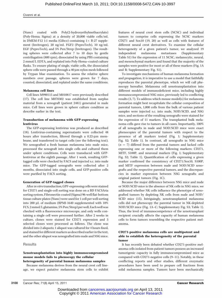
features of neural crest stem cells (NCSC) and individualtumors to comprise cells expressing the NCSC markersCD271 and Sox10 (19, 20) as well as cells with features ofdifferent neural crest derivatives. To examine the cellularheterogeneity of a given patient's tumor, we analyzed 19independent melanoma metastases (SupplementaryTable S1) for the expression of 11 NCSC, melanocytic, neural,and mesenchymal markers and found that the majority of thesamples were positive for most or all of these markers (Fig. 1Aand B; Supplementary Fig. S1).
To investigate mechanisms of human melanoma formationand propagation, it is imperative to use a model that faithfullyreproduces the parental phenotype, which we will call phe-nocopy hereafter. Melanoma cell xenotransplantation intodifferent models of immunodeficient mice, including highlyimmunocompromised NSG mice, previously led to conflictingresults (3, 7). To address which mouse model(s) for melanomaformation might best recapitulate the cellular composition ofparental tumors, 1,000 cells from the bulk of various patientsamples were injected s.c. into nude, NOD/SCID, and NSGmice, and sections of the resulting xenografts were stained forthe expression of 11 markers. The transplanted bulk mela-noma cells gave rise to tumors in all cases. Importantly, 100%of all xenografts in nude and NOD/SCID mice were exactphenocopies of the parental tumors with respect to thepresence of all markers tested (Fig. 1; SupplementaryFig. S2; Table 1). In contrast, all xenografts in NSG mice(n ¼ 7) differed from the parental tumors and lacked cellsexpressing one or more of the following markers: CD271,MITF, S100P, and neuronal markers (Fig. 1; SupplementaryFig. S2; Table 1). Quantification of cells expressing a givenmarker confirmed the consistency of CD271/Sox10, S100P,and MITF expression between nude and NOD/SCID xeno-grafts and the respective parental tumors, and the discrepan-cies in marker expression between NSG xenografts andoriginal patient tumors (Fig. 1C).
Because the major difference between NSG mice and nudeor NOD/SCID mice is the absence of NK cells in NSG mice, weaddressed whether NK cells influence the phenotype of xeno-grafted tumors by depleting NK cells from nude and NOD/SCID mice (15). Intriguingly, xenotransplanted melanomacells did not phenocopy the parental tumor in NK-depletedNOD/SCID mice (Fig. 1A–C; Supplementary Fig. S3; Table 1).Thus, the level of immunocompetence of the xenotransplantrecipient crucially affects the capacity of human melanomacells to form tumors resembling the respective patient mel-anoma.
CD271-positive melanoma cells are multipotent andable to establish the heterogeneity of the parentaltumor
It has recently been debated whether CD271-positive mel-anoma cells isolated from patient tumors possess an increasedtumorigenic capacity in fully immunocompromised mice ascompared with CD271-negative cells (9–11). Notably, in theseconflicting reports and other studies, different enzymaticprocedures have been used to prepare cell fractions fromsolid melanoma samples. Tumors have been mechanically
Civenni et al.
Cancer Res; 71(8) April 15, 2011 Cancer Research3100
American Association for Cancer Research Copyright © 2011 on April 13, 2011cancerres.aacrjournals.orgDownloaded from
Published OnlineFirst March 10, 2011; DOI:10.1158/0008-5472.CAN-10-3997
dissociated followed by enzymatic digestion with collagenase(8), a mix of collagenase and dispase (9), or collagenase andtrypsin (10, 11, 21). Therefore, we addressed whether thesedifferent tumor digestion protocols might affect detectabilityof surface markers used to directly isolate putative tumori-genic melanoma cell populations. Strikingly, the percentage ofretrievable CD271- and ABCB5-positive cells was consistentlyreduced in conditions that included a trypsin incubation step(11), whereas CD44 was not affected (Supplementary Fig. S4).These results show that a strong proteolytic activity canselectively damage surface protein epitopes and thus leadto reduced detectability by the corresponding antibodies. Thisunavoidably results in underestimation of surface marker
expression in humanmelanoma cells and in negative fractionsbeing contaminated by cells actually expressing the selectedmarker. For this reason, in this study human biopsies andxenografts were dissociated in the absence of trypsin activity.
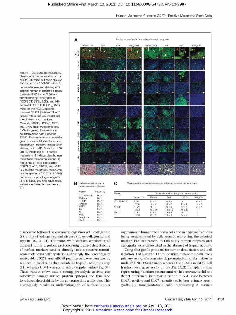
Using this gentle protocol for tumor dissociation and cellisolation, FACS-sorted CD271-positive melanoma cells fromprimary xenografts consistently promoted tumor formation innude and NOD/SCID mice, whereas the CD271-negative cellfraction never gave rise to tumors (Fig. 2A; 32 transplantationsrepresenting 7 distinct patient tumors). In contrast, we did notdetect differences in tumor initiation in NSG mice betweenCD271-positive and CD271-negative cells from primary xeno-grafts (12 transplantations each, representing 3 distinct
Figure 1. Xenografted melanomaphenocopy the parental tumor inNOD/SCIDmice, but not in NSGorNK-depleted NOD/SCID mice. A,immunofluorescent staining of 2original human melanoma tissues(patients 31651 and 3298) andcorresponding xenografts inNOD/SCID (N/S), NSG, and NK-depleted NOD/SCID (N/S_GM1)mice for the NCSC-specificmarkers CD271 (red) and Sox10(green; white arrows, insets) andthe differentiation markersMelanA, S100P, HMB45, MITF,TuJ1, NF, NSE, Peripherin, andSMA (in green). Tissues werecounterstained with Hoechst33342. Expression or absence of agiven marker is labeled by þ or �,respectively. Bottom, tissues afterstaining with H&E. Scale bar, 100mm. B, incidence of 11 testedmarkers in 19 independent humanmetastatic melanoma lesions. C,frequency of cells expressingCD271/Sox10, S100P, and MITFin 2 human metastatic melanomatissues (patients 31651 and 3298)and in corresponding xenograftsin N/S, NSG, and N/S_GM1 mice.Values are presented as mean �SD.
A
B C
Human Melanoma Contains CD271-Positive Melanoma Stem Cells
www.aacrjournals.org Cancer Res; 71(8) April 15, 2011 3101
American Association for Cancer Research Copyright © 2011 on April 13, 2011cancerres.aacrjournals.orgDownloaded from
Published OnlineFirst March 10, 2011; DOI:10.1158/0008-5472.CAN-10-3997
patient tumors; Fig. 2A). Strikingly, in NK-depleted nude orNOD/SCID mice both CD271-positive and CD271-negativecells initiated tumor formation. Hence, the tumorigenic poten-tial of melanoma cell subpopulations is influenced by thepresence of NK cells in xenograft recipients.
Because a cancer stem cell by definition must display thedifferentiation capacity reflecting its parental tumor, weinvestigated the heterogeneity of different successfully xeno-grafted tumors in NOD/SCID, nude, and NSG mice and of thecorresponding parental tumor. The cellular composition of allnude and NOD/SCID xenografts derived from CD271-positivecells (tumors representing 7 distinct patients) was analogousto the corresponding parental tumors (Fig. 2B; SupplementaryFig. S5). In contrast, in NSG mice neither CD271-positive norCD271-negative cells were able to fully phenocopy the cellularheterogeneity of the corresponding parental tumors withrespect to all 11 markers tested (Fig. 2B; SupplementaryFig. S5; Table 2). Of importance, both the CD271-positiveand -negative cell fractions failed to consistently generatexenografts expressing crucial melanoma markers such asMITF and S100P or neuronal markers present in the parentaltumors. Moreover, none of the tumors produced by CD271-negative cells in NSG mice comprised CD271-positive cells,despite the presence of such cells in the parental tumors(Fig. 2B; Supplementary Fig. S5; Table 2; 12 transplantationsrepresenting 3 patients’ tumors). In summary, faithful phe-nocopies of original patient tumors were only achieved ontransplantation of CD271-positive melanoma cells into nudeand NOD/SCID mice, but not into NSG mice.
The fact that CD271-positive cells generate the full hetero-geneity of human melanoma on xenotransplantation intoNOD/SCID or nude mice suggests that these cells are multi-potent. To specifically address this issue, we infected mela-noma cells from a xenograft of patient sample 2481 with aGFP-expressing lentivirus, followed by FACS of GFP/CD271double-positive cells and plating of those cells at clonal
density. Although 19 � 4% of the CD271-positive cells gaverise to clones consisting of more than 10 cells after 2 weeks inculture, such clones were not observed in the CD271-negativecell fraction (Fig. 2C). Three clones derived from GFP/CD271double-positive founder cells were selected and subjected todifferentiation assays in cell culture, revealing the emergenceof multiple cell types, such as cells expressing CD271/Sox10,neuronal and melanocytic markers, and SMA (Fig. 2D). Thus,the CD271-positive melanoma cell fraction contains a popula-tion that is clonogenic and multipotent in cell culture, similarto NCSCs.
To address the multipotency of CD271-positive melanomacells in vivo, cells from the 3 selected clones were grafted s.c.into nude and NSGmice, and the xenografts were analyzed forexpression of the 11 markers as described before. In nudemice, the 3 clones were able to establish a cellular hierarchywith all 11 markers present in the xenografts. In particular,10.2 � 2.0% of the xenograft cells derived from a CD271-positive cell expressed CD271. Moreover, although mostsmooth muscle cells present in the xenografts were hostderived, all nude xenografts comprised cells double positivefor SMA and GFP, revealing that some smooth muscle cells inthe tumor originated from CD271-positive melanoma cells. Incontrast, neither the NCSC markers CD271/Sox10 nor S100P,MITF, and neuronal traits were consistently expressed inxenografts derived from clones transplanted into NSG mice.Moreover, we did not find any GFP/SMA double-positive cellsin the NSG grafts, suggesting that the NSG host environmenteither suppresses the in vivo multipotency of CD271-positivecells or supports the growth of melanoma cells with restricteddevelopmental capacities (Fig. 2D; Supplementary Fig. S6).
CD271-positive melanoma cells have self-renewalcapacity and sustain long-term tumor growth in vivo
To address whether CD271-positive cells have self-renewalcapacity, in addition to being multipotent (Fig. 2B–D), we
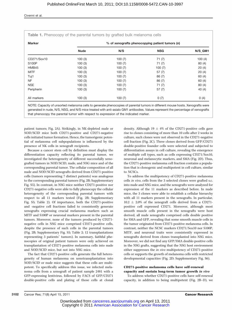
Table 1. Phenocopy of the parental tumors by grafted bulk melanoma cells
Marker % of xenografts phenocopying patient tumors (n)
Nude N/S NSG N/S_GM1
CD271/Sox10 100 (3) 100 (7) 71 (7) 100 (4)S100P 100 (3) 100 (7) 71 (7) 80 (4)HMB45 100 (3) 100 (7) 100 (7) 20 (4)MITF 100 (3) 100 (7) 57 (7) 20 (4)Tuj1 100 (3) 100 (7) 86 (7) 60 (4)NF 100 (3) 100 (7) 86 (7) 60 (4)NSE 100 (3) 100 (7) 71 (7) 80 (4)Peripherin 100 (3) 100 (7) 57 (7) 40 (4)
All markers 100 (3) 100 (7) 0 (7) 0 (4)
NOTE: Capacity of unsorted melanoma cells to generate phenocopies of parental tumors in different mouse hosts. Xenografts weregenerated in nude, N/S, NSG, and N/S mice treated with anti-asialo GM1 antibodies. Values represent the percentage of xenograftsthat phenocopy the parental tumor with respect to expression of the indicated marker.
Civenni et al.
Cancer Res; 71(8) April 15, 2011 Cancer Research3102
American Association for Cancer Research Copyright © 2011 on April 13, 2011cancerres.aacrjournals.orgDownloaded from
Published OnlineFirst March 10, 2011; DOI:10.1158/0008-5472.CAN-10-3997
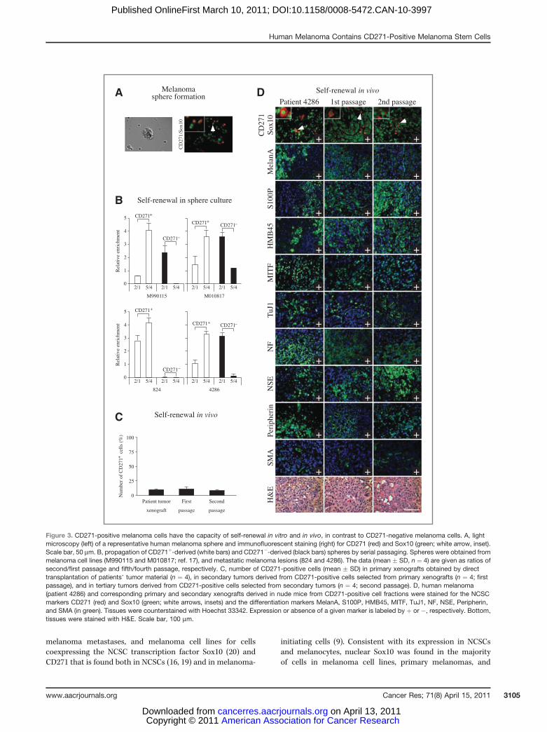
carried out sphere assays under conditions that are known tosupport sphere formation and self-renewal of normal skin-derived NCSCs (16). CD271-positive cells isolated from freshmelanoma tissue or from melanoma cell lines readily gener-
ated spheres that contained CD271/Sox10-positive cells andthat could be propagated by serial passaging, whereas the fewspheroid aggregates generated by the CD271-depleted mela-noma cell fraction displayed a reduced passaging capacity
Clone forming capacityA C
DB
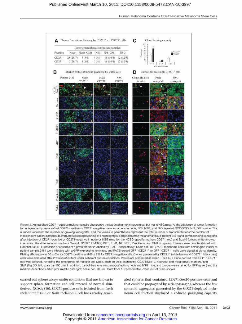
Figure 2. Xenografted CD271-positive melanoma cells phenocopy the parental tumor in nude mice, but not in NSG mice. A, the efficiency of tumor formationfor independently xenografted CD271-positive or CD271-negative melanoma cells in nude, N/S, NSG, and NK-depleted NOD/SCID (N/S_GM1) mice. Thenumbers represent the number of growing xenografts, and the values in parentheses represent the total number of transplantations/the number ofindependent patient samples. B, immunofluorescent staining of a representative original humanmelanoma tissue (patient 2481) and corresponding xenograftsafter injection of CD271-positive or CD271-negative in nude or NSG mice for the NCSC-specific markers CD271 (red) and Sox10 (green; white arrows,insets) and the differentiation markers MelanA, S100P, HMB45, MITF, TuJ1, NF, NSE, Peripherin, and SMA (in green). Tissues were counterstained withHoechst 33342. Expression or absence of a given marker is labeled by þ or �, respectively. Scale bar, 100 mm. C, melanoma cells from a xenograft (nude) ofpatient sample 2481 were infected with a GFP-expressing lentivirus, and FACS-sorted GFPþ/CD271þ or GFPþ/CD271� cells were plated at clonal density.Plating efficiency was 56� 6% for CD271-positive and 69� 7% for CD271-negative cells. Clones generated by CD271þ (white bars) and CD271� (black bars)cells were evaluated after 2 weeks of culture under adherent culture conditions. Values are presented as mean � SD. D, a clone derived from GFPþ/CD271þ
cell was cultured, revealing the emergence of multiple cell types, such as cells expressing CD271/Sox10, neuronal and melanocytic markers, andSMA (Fig. 3D, left; scale bar 100 mm). In addition, part of the clone was xenografted into nude and NSGmice, and tumors were stained for GFP (green) and themarkers described earlier (red; middle and right; scale bar, 50 mm). Data from 1 representative clone out of 3 are shown.
Human Melanoma Contains CD271-Positive Melanoma Stem Cells
www.aacrjournals.org Cancer Res; 71(8) April 15, 2011 3103
American Association for Cancer Research Copyright © 2011 on April 13, 2011cancerres.aacrjournals.orgDownloaded from
Published OnlineFirst March 10, 2011; DOI:10.1158/0008-5472.CAN-10-3997
(Fig. 3A and B). Determining the ratio between numbers ofsecondary over primary and quinternary over quaternaryspheres showed that CD271-positive, but not CD271-negative,cells have an extensive self-renewal potential and expand theirstem cell activity over time (Fig. 3B).
To confirm the self-renewal potential of CD271-positivemelanoma cells in vivo, we isolated CD271-positive andCD271-negative cells from primary xenografts generated bybulk tumor cells in nude mice and retransplanted these cellfractions. We found that only CD271-positive cells were able togenerate secondary tumors in nude mice (Fig. 3C and D;Supplementary Fig. S7, "first passage"; n ¼ 4 for each biopsy).Likewise, CD271-positive cells reisolated from secondaryxenografts consistently generated tertiary tumors, unlikeCD271-negative cells (Fig. 3C and D; Supplementary Fig. S7,"second passage"). To test whether the cellular heterogeneityoriginating from CD271-positive cells is a stable trait in vivo,we analyzed first and second passage xenografts by immuno-fluorescence. Intriguingly, not only the cellular heterogeneitywith respect to neural crest-specific differentiation markers(Fig. 3D; Supplementary Fig. S7) but also the frequency ofCD271-positive cells was maintained in xenografts directlygenerated from unsegregated patients’melanoma cells and intumors obtained by serial in vivo passaging of CD271-positivecells (Fig. 3C; percentage of CD271-positive cells in primaryxenograft, first passage, and second passage, respectively: 10� 1%, 11 � 3%, 8 � 2%; n ¼ 4). Hence, CD271-positivemelanoma stem cells faithfully phenocopy the parental tumorin nude mice, even after multiple passages in vivo.
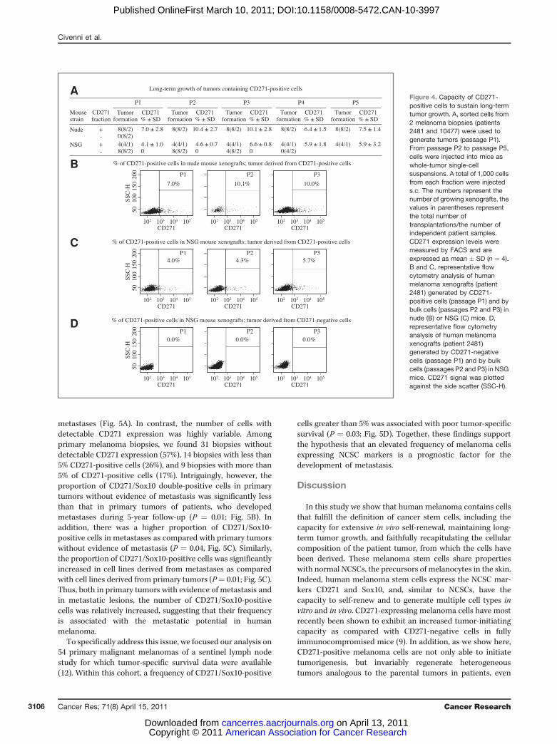
Our data suggest that the presence of CD271-expressingmelanoma stem cells is a prerequisite for long-term tumorpropagation. Therefore, although CD271-negative melanomacells can generate tumors in NSG, NK-depleted NOD/SCID, orNK-depleted nude mice (Fig. 2; refs. 9, 10), we expected thatsuch tumors could not be maintained in vivo over a prolongedtime period. To test this, we serially passaged tumors gener-
ated in NSG mice from CD271-positive or CD271-negativecells, using tumors produced by CD271-positive melanomacells in nude mice as positive control. In both nude and NSGmice, tumors originating from CD271-positive cells allowedpropagation in vivo over 5 passages, in accordance with theearlier-described in vivo self-renewal capacity of CD271-posi-tive cells in nude mice (Fig. 4). In striking contrast, tumorsderived from CD271-negative cells in NSG mice exhaustedwith time and could only be propagated in vivo for 3 to 4passages (Fig. 4). Quantification of CD271-expressing cells inconsecutively produced NSG xenografts revealed that thecapacity for tumor propagation over several passages wasassociated with the presence of CD271-positive melanomastem cells in each xenograft (Fig. 4A–C). CD271-negative cells,however, were unable to produce CD271-positive cells, evenafter xenograft passaging (Fig. 4A and D). It should be noted,however, that although CD271-positive cells generate tumorsin NSG mice that can be serially passaged, those tumors didnot phenocopy the parental tumor (Fig. 2).
Thus, NSG mice, NK-depleted NOD/SCID mice, or NK-depleted nude mice provided a host environment permissivefor tumor growth by CD271-negative cells, whereas such cellswere unable to initiate tumorigenesis in NOD/SCID or nudemice. In contrast, CD271-positive melanoma stem cells weretumorigenic in all mouse models tested. In any case, only innude or NOD/SCIDmice CD271-positive melanoma stem cellsproduced tumors that completely phenocopied the parentalmelanoma even on serial xenotransplantation, whereas theyfailed to do so in NSG mice.
Expression of the NCSC markers CD271/Sox10correlates with metastatic potential and poor prognosisof melanoma
To verify our findings with a large number of patientsamples, we stained tissue microarrays containing more than200 different melanoma biopsies of primary melanomas,
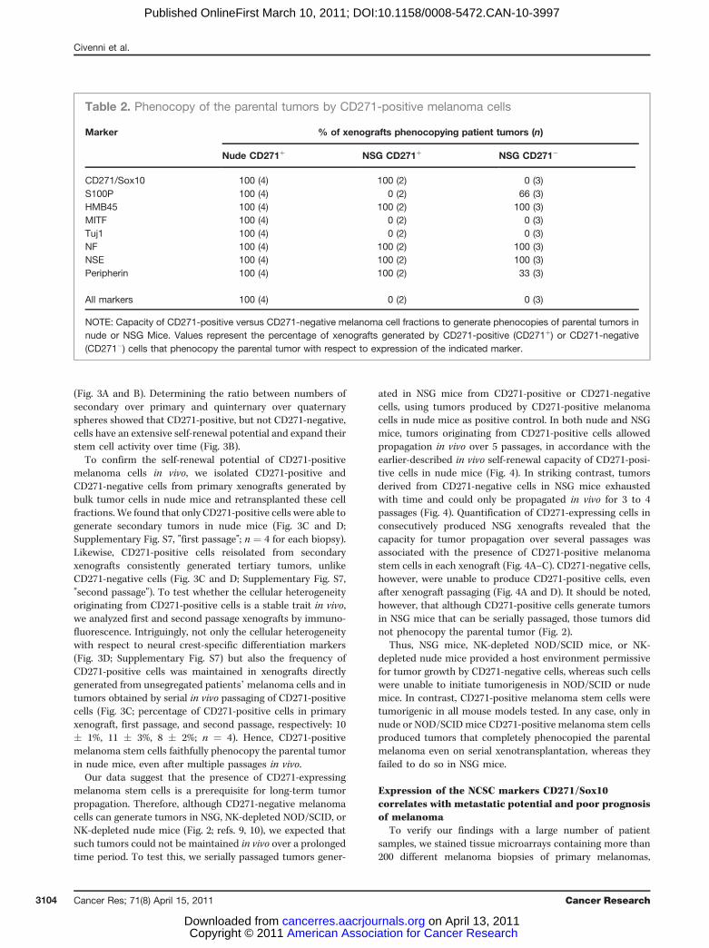
Table 2. Phenocopy of the parental tumors by CD271-positive melanoma cells
Marker % of xenografts phenocopying patient tumors (n)
Nude CD271þ NSG CD271þ NSG CD271�
CD271/Sox10 100 (4) 100 (2) 0 (3)S100P 100 (4) 0 (2) 66 (3)HMB45 100 (4) 100 (2) 100 (3)MITF 100 (4) 0 (2) 0 (3)Tuj1 100 (4) 0 (2) 0 (3)NF 100 (4) 100 (2) 100 (3)NSE 100 (4) 100 (2) 100 (3)Peripherin 100 (4) 100 (2) 33 (3)
All markers 100 (4) 0 (2) 0 (3)
NOTE: Capacity of CD271-positive versus CD271-negative melanoma cell fractions to generate phenocopies of parental tumors innude or NSG Mice. Values represent the percentage of xenografts generated by CD271-positive (CD271þ) or CD271-negative(CD271�) cells that phenocopy the parental tumor with respect to expression of the indicated marker.
Civenni et al.
Cancer Res; 71(8) April 15, 2011 Cancer Research3104
American Association for Cancer Research Copyright © 2011 on April 13, 2011cancerres.aacrjournals.orgDownloaded from
Published OnlineFirst March 10, 2011; DOI:10.1158/0008-5472.CAN-10-3997
melanoma metastases, and melanoma cell lines for cellscoexpressing the NCSC transcription factor Sox10 (20) andCD271 that is found both in NCSCs (16, 19) and in melanoma-
initiating cells (9). Consistent with its expression in NCSCsand melanocytes, nuclear Sox10 was found in the majorityof cells in melanoma cell lines, primary melanomas, and
A
B
C
D
Figure 3. CD271-positive melanoma cells have the capacity of self-renewal in vitro and in vivo, in contrast to CD271-negative melanoma cells. A, lightmicroscopy (left) of a representative human melanoma sphere and immunofluorescent staining (right) for CD271 (red) and Sox10 (green; white arrow, inset).Scale bar, 50 mm. B, propagation of CD271þ-derived (white bars) and CD271�-derived (black bars) spheres by serial passaging. Spheres were obtained frommelanoma cell lines (M990115 and M010817; ref. 17), and metastatic melanoma lesions (824 and 4286). The data (mean � SD, n ¼ 4) are given as ratios ofsecond/first passage and fifth/fourth passage, respectively. C, number of CD271-positive cells (mean � SD) in primary xenografts obtained by directtransplantation of patients’ tumor material (n ¼ 4), in secondary tumors derived from CD271-positive cells selected from primary xenografts (n ¼ 4; firstpassage), and in tertiary tumors derived from CD271-positive cells selected from secondary tumors (n ¼ 4; second passage). D, human melanoma(patient 4286) and corresponding primary and secondary xenografts derived in nude mice from CD271-positive cell fractions were stained for the NCSCmarkers CD271 (red) and Sox10 (green; white arrows, insets) and the differentiation markers MelanA, S100P, HMB45, MITF, TuJ1, NF, NSE, Peripherin,and SMA (in green). Tissues were counterstained with Hoechst 33342. Expression or absence of a given marker is labeled by þ or �, respectively. Bottom,tissues were stained with H&E. Scale bar, 100 mm.
Human Melanoma Contains CD271-Positive Melanoma Stem Cells
www.aacrjournals.org Cancer Res; 71(8) April 15, 2011 3105
American Association for Cancer Research Copyright © 2011 on April 13, 2011cancerres.aacrjournals.orgDownloaded from
Published OnlineFirst March 10, 2011; DOI:10.1158/0008-5472.CAN-10-3997
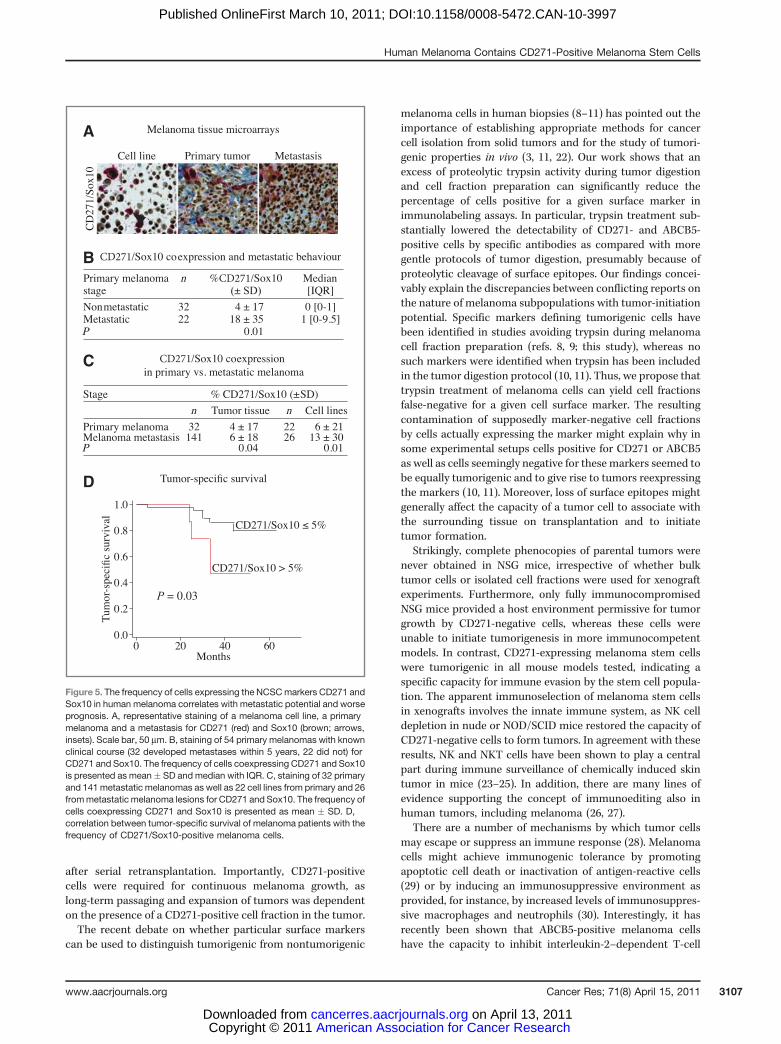
metastases (Fig. 5A). In contrast, the number of cells withdetectable CD271 expression was highly variable. Amongprimary melanoma biopsies, we found 31 biopsies withoutdetectable CD271 expression (57%), 14 biopsies with less than5% CD271-positive cells (26%), and 9 biopsies with more than5% of CD271-positive cells (17%). Intriguingly, however, theproportion of CD271/Sox10 double-positive cells in primarytumors without evidence of metastasis was significantly lessthan that in primary tumors of patients, who developedmetastases during 5-year follow-up (P ¼ 0.01; Fig. 5B). Inaddition, there was a higher proportion of CD271/Sox10-positive cells in metastases as compared with primary tumorswithout evidence of metastasis (P ¼ 0.04, Fig. 5C). Similarly,the proportion of CD271/Sox10-positive cells was significantlyincreased in cell lines derived from metastases as comparedwith cell lines derived from primary tumors (P¼ 0.01; Fig. 5C).Thus, both in primary tumors with evidence of metastasis andin metastatic lesions, the number of CD271/Sox10-positivecells was relatively increased, suggesting that their frequencyis associated with the metastatic potential in humanmelanoma.
To specifically address this issue, we focused our analysis on54 primary malignant melanomas of a sentinel lymph nodestudy for which tumor-specific survival data were available(12). Within this cohort, a frequency of CD271/Sox10-positive
cells greater than 5% was associated with poor tumor-specificsurvival (P ¼ 0.03; Fig. 5D). Together, these findings supportthe hypothesis that an elevated frequency of melanoma cellsexpressing NCSC markers is a prognostic factor for thedevelopment of metastasis.
Discussion
In this study we show that human melanoma contains cellsthat fulfill the definition of cancer stem cells, including thecapacity for extensive in vivo self-renewal, maintaining long-term tumor growth, and faithfully recapitulating the cellularcomposition of the patient tumor, from which the cells havebeen derived. These melanoma stem cells share propertieswith normal NCSCs, the precursors of melanocytes in the skin.Indeed, human melanoma stem cells express the NCSC mar-kers CD271 and Sox10, and, similar to NCSCs, have thecapacity to self-renew and to generate multiple cell types invitro and in vivo. CD271-expressing melanoma cells have mostrecently been shown to exhibit an increased tumor-initiatingcapacity as compared with CD271-negative cells in fullyimmunocompromised mice (9). In addition, as we show here,CD271-positive melanoma cells are not only able to initiatetumorigenesis, but invariably regenerate heterogeneoustumors analogous to the parental tumors in patients, even
A
B
C
D
Figure 4. Capacity of CD271-positive cells to sustain long-termtumor growth. A, sorted cells from2 melanoma biopsies (patients2481 and 10477) were used togenerate tumors (passage P1).From passage P2 to passage P5,cells were injected into mice aswhole-tumor single-cellsuspensions. A total of 1,000 cellsfrom each fraction were injecteds.c. The numbers represent thenumber of growing xenografts, thevalues in parentheses representthe total number oftransplantations/the number ofindependent patient samples.CD271 expression levels weremeasured by FACS and areexpressed as mean � SD (n ¼ 4).B and C, representative flowcytometry analysis of humanmelanoma xenografts (patient2481) generated by CD271-positive cells (passage P1) and bybulk cells (passages P2 and P3) innude (B) or NSG (C) mice. D,representative flow cytometryanalysis of human melanomaxenografts (patient 2481)generated by CD271-negativecells (passage P1) and by bulkcells (passages P2 and P3) in NSGmice. CD271 signal was plottedagainst the side scatter (SSC-H).
Civenni et al.
Cancer Res; 71(8) April 15, 2011 Cancer Research3106
American Association for Cancer Research Copyright © 2011 on April 13, 2011cancerres.aacrjournals.orgDownloaded from
Published OnlineFirst March 10, 2011; DOI:10.1158/0008-5472.CAN-10-3997
after serial retransplantation. Importantly, CD271-positivecells were required for continuous melanoma growth, aslong-term passaging and expansion of tumors was dependenton the presence of a CD271-positive cell fraction in the tumor.The recent debate on whether particular surface markers
can be used to distinguish tumorigenic from nontumorigenic
melanoma cells in human biopsies (8–11) has pointed out theimportance of establishing appropriate methods for cancercell isolation from solid tumors and for the study of tumori-genic properties in vivo (3, 11, 22). Our work shows that anexcess of proteolytic trypsin activity during tumor digestionand cell fraction preparation can significantly reduce thepercentage of cells positive for a given surface marker inimmunolabeling assays. In particular, trypsin treatment sub-stantially lowered the detectability of CD271- and ABCB5-positive cells by specific antibodies as compared with moregentle protocols of tumor digestion, presumably because ofproteolytic cleavage of surface epitopes. Our findings concei-vably explain the discrepancies between conflicting reports onthe nature of melanoma subpopulations with tumor-initiationpotential. Specific markers defining tumorigenic cells havebeen identified in studies avoiding trypsin during melanomacell fraction preparation (refs. 8, 9; this study), whereas nosuch markers were identified when trypsin has been includedin the tumor digestion protocol (10, 11). Thus, we propose thattrypsin treatment of melanoma cells can yield cell fractionsfalse-negative for a given cell surface marker. The resultingcontamination of supposedly marker-negative cell fractionsby cells actually expressing the marker might explain why insome experimental setups cells positive for CD271 or ABCB5as well as cells seemingly negative for these markers seemed tobe equally tumorigenic and to give rise to tumors reexpressingthe markers (10, 11). Moreover, loss of surface epitopes mightgenerally affect the capacity of a tumor cell to associate withthe surrounding tissue on transplantation and to initiatetumor formation.
Strikingly, complete phenocopies of parental tumors werenever obtained in NSG mice, irrespective of whether bulktumor cells or isolated cell fractions were used for xenograftexperiments. Furthermore, only fully immunocompromisedNSG mice provided a host environment permissive for tumorgrowth by CD271-negative cells, whereas these cells wereunable to initiate tumorigenesis in more immunocompetentmodels. In contrast, CD271-expressing melanoma stem cellswere tumorigenic in all mouse models tested, indicating aspecific capacity for immune evasion by the stem cell popula-tion. The apparent immunoselection of melanoma stem cellsin xenografts involves the innate immune system, as NK celldepletion in nude or NOD/SCID mice restored the capacity ofCD271-negative cells to form tumors. In agreement with theseresults, NK and NKT cells have been shown to play a centralpart during immune surveillance of chemically induced skintumor in mice (23–25). In addition, there are many lines ofevidence supporting the concept of immunoediting also inhuman tumors, including melanoma (26, 27).
There are a number of mechanisms by which tumor cellsmay escape or suppress an immune response (28). Melanomacells might achieve immunogenic tolerance by promotingapoptotic cell death or inactivation of antigen-reactive cells(29) or by inducing an immunosuppressive environment asprovided, for instance, by increased levels of immunosuppres-sive macrophages and neutrophils (30). Interestingly, it hasrecently been shown that ABCB5-positive melanoma cellshave the capacity to inhibit interleukin-2–dependent T-cell
CD271
CD271
CD271
A
B
C
D
Figure 5. The frequency of cells expressing the NCSCmarkers CD271 andSox10 in human melanoma correlates with metastatic potential and worseprognosis. A, representative staining of a melanoma cell line, a primarymelanoma and a metastasis for CD271 (red) and Sox10 (brown; arrows,insets). Scale bar, 50 mm. B, staining of 54 primary melanomas with knownclinical course (32 developed metastases within 5 years, 22 did not) forCD271 and Sox10. The frequency of cells coexpressing CD271 and Sox10is presented as mean� SD and median with IQR. C, staining of 32 primaryand 141metastatic melanomas as well as 22 cell lines from primary and 26frommetastatic melanoma lesions for CD271 and Sox10. The frequency ofcells coexpressing CD271 and Sox10 is presented as mean � SD. D,correlation between tumor-specific survival of melanoma patients with thefrequency of CD271/Sox10-positive melanoma cells.
Human Melanoma Contains CD271-Positive Melanoma Stem Cells
www.aacrjournals.org Cancer Res; 71(8) April 15, 2011 3107
American Association for Cancer Research Copyright © 2011 on April 13, 2011cancerres.aacrjournals.orgDownloaded from
Published OnlineFirst March 10, 2011; DOI:10.1158/0008-5472.CAN-10-3997
activation and to induce tolerization by regulatory T cells (31).Reduced expression levels of specific tumor antigens, such asMelan-A/MART-1, tyrosinase, and gp-100, might presentanother mechanism for immune evasion by melanoma cells.Notably, CD271- and ABCB5-positive melanoma cells expresslow levels of melanoma-associated antigens such as MART-1,supporting the idea that melanoma cells expressing mela-noma stem cell markers escape the immune system attack bythe host (refs. 9, 29, 31; G. Civenni and L. Sommer, unpublisheddata). Of note, we identified a subpopulation of CD271-posi-tive melanoma cells that also express ABCB5 (G. Civenni andL. Sommer, unpublished data), although the functional impli-cation of this finding remains to be addressed. All together,these results suggest that in patients, melanoma stem cellsmight be able to evade or modulate the immune response,allowing these cells to promote tumorigenic growth and toprovide resistance to immunotherapy. The hypothesis thatthese processes are relevant in patients is further supported byour data that only in mice with a certain level of immuno-competence, CD271-positive melanoma cells generatedtumors fully phenocopying the original patient melanoma.However, because xenotransplantation models cannot accu-rately recapitulate the immune response induced by cancer inhuman patients, the interaction between cancer cell popula-tions and the immune system should be addressed by usingsyngeneic melanoma mouse models, in which components ofthe anticancer immune system can be manipulated duringtumor initiation and progression.
Our data are consistent with the idea that CD271-positivemelanoma cells play a crucial role in tumor formation inhuman patients. This is further supported by our clinical dataobtained with an extensive tissue array of melanoma samples.These show an association between the proportion of CD271/Sox10-positive cells in primary melanoma and metastaticdisease as well as poor tumor-specific survival. Hence, highnumbers of melanoma stem cells expressing NCSC markersmight influence aggressiveness and the metastatic behavior ofmalignant melanoma. This conceivably reflects intrinsic,NCSC-like features of melanoma stem cells, given that normalNCSCs have the capacity to extensively migrate throughembryonic tissue before differentiation into melanocytesand other cell types. In support of this, increased levels ofCD271 expression in melanoma have been associated withenhanced invasive potential in culture (32). Interestingly,CD271/Sox10 expression in metastatic primary melanomawas higher than that in metastatic lesions, which could beexplained by different patient groups. Whereas all primarymelanomas (metastatic versus nonmetastatic) were primarytumors without chemotherapy or vaccination therapy, tissueof the metastases was obtained from patients after adjuvanttherapies. Possibly, this therapy might result in a lower pre-valence of cells with CD271/Sox10 expression in the metas-tases. Alternatively, the site of primary metastatic tumorformation might offer a microenvironment more favorablefor stem cells than provided by distant metastases. In any case,however, the proportion of CD271/Sox10-positive cells canpotentially be used as a predictor of metastases and conse-quently poor tumor-specific survival. This in turn suggests
that monitoring melanoma cells with NCSC-like features inprimary melanoma might be of great prognostic relevance.
Several biopsies of primary, mostly nonmetastatic mela-noma did not contain CD271/Sox10-positive cells accordingto our TMA analysis. CD271/Sox10-positive cells might there-fore be involved in the formation of metastatic melanoma,whereas less aggressive tumors might originate from othertypes of melanoma-initiating cells. By extrapolation onecould argue that tumor aggressiveness depends on whetherthe key oncogenic mutation occurred in a normal stem cell orin a more restricted progenitor cell (33). However, althoughthe melanoma stem cells identified in this study displaysimilar marker expression and potential as normal NCSCsin the adult skin (16, 34), melanoma-initiating cells could alsoarise by dedifferentiation of more mature melanocytic cells(35). Similarly, we cannot exclude that depending on thecontext or the oncogenotype, CD271/Sox10-melanoma stemcells emerge from other tumor cells, for instance on a processreminiscent of an epithelial-to-mesenchymal transition dur-ing metastasis or by epigenetic modification (36, 37). Intrigu-ingly, however, at least in the conditions chosen for this study,CD271 expression was a stable trait of human melanoma-initiating cells, and we did not find any evidence for CD271-positive cells originating from the CD271-negative tumor cellfraction even after prolonged incubation in vivo. Moreover,CD271-negative cells did not acquire properties of actualmelanoma stem cells, in that they could neither reproducethe cellular heterogeneity of parental tumors nor sustainlong-term tumor formation.
In conclusion, the identification of human melanoma stemcells required for continuous tumor growth points to potentialculprits of tumor formation in patients. In the future, it mightbe possible to establish specific treatments that reduce tumor-igenesis by elimination of these cells or by targeting "stem-ness" in melanoma (1). Thus, efforts should be made todevelop drugs able to promote differentiation of melanomastem cells, to selectively kill these cells, or to specifically blocktheir self-renewal and expansion.
Disclosure of Potential Conflict of Interest
No potential conflicts of interest were disclosed.
Acknowledgments
We thank M. Storz, P. Frei, and S. Behnke for assistance and Dr. M. Frank forproviding the antibody to ABCB5. We would also like to thank the technical staffof the Central Flow Cytometry Laboratory of the ETH and University of Zurichfor their assistance.
Grant Support
This work was supported by the Swiss Cancer League, the Swiss NationalScience Foundation, the Vontobel Foundation, UBS Wealth Management andthe Swiss NCCR "Neural Plasticity and Repair."
The costs of publication of this article were defrayed in part by the paymentof page charges. This article must therefore be hereby marked advertisement inaccordance with 18 U.S.C. Section 1734 solely to indicate this fact.
Received November 3, 2010; revised January 31, 2011; accepted February 14,2011; published OnlineFirst March 10, 2011.
Civenni et al.
Cancer Res; 71(8) April 15, 2011 Cancer Research3108
American Association for Cancer Research Copyright © 2011 on April 13, 2011cancerres.aacrjournals.orgDownloaded from
Published OnlineFirst March 10, 2011; DOI:10.1158/0008-5472.CAN-10-3997
References1. Ward RJ, Dirks PB. Cancer stem cells: at the headwaters of tumor
development. Annu Rev Pathol 2007;2:175–89.2. Cho RW, Clarke MF. Recent advances in cancer stem cells. Curr Opin
Genet Dev 2008;18:48–53.3. Dirks P. Cancer stem cells: Invitation to a second round. Nature
2010;466:40–1.4. Fang D, Nguyen TK, Leishear K, Finko R, Kulp AN, Hotz S, et al. A
tumorigenic subpopulation with stem cell properties in melanomas.Cancer Res 2005;65:9328–37.
5. Monzani E, Facchetti F, Galmozzi E, Corsini E, Benetti A, Cavazzin C,et al. Melanoma contains CD133 and ABCG2 positive cells withenhanced tumourigenic potential. Eur J Cancer 2007;43:935–46.
6. Keshet GI, Goldstein I, Itzhaki O, Cesarkas K, Shenhav L, YakirevitchA, et al. MDR1 expression identifies human melanoma stem cells.Biochem Biophys Res Commun 2008;368:930–6.
7. Zabierowski SE, Herlyn M. Melanoma stem cells: the dark seed ofmelanoma. J Clin Oncol 2008;26:2890–4.
8. Schatton T, Murphy GF, Frank NY, Yamaura K, Waaga-Gasser AM,Gasser M, et al. Identification of cells initiating human melanomas.Nature 2008;451:345–9.
9. Boiko AD, Razorenova OV, van de Rijn M, Swetter SM, Johnson DL,Ly DP, et al. Human melanoma-initiating cells express neural crestnerve growth factor receptor CD271. Nature 2010;466:133–7.
10. Quintana E, Shackleton M, Sabel MS, Fullen DR, Johnson TM,Morrison SJ. Efficient tumour formation by single human melanomacells. Nature 2008;456:593–8.
11. Quintana E, Shackleton M, Foster HR, Fullen DR, Sabel MS, JohnsonTM, et al. Phenotypic heterogeneity among tumorigenic melanomacells from patients that is reversible and not hierarchically organized.Cancer Cell 2010;18:510–23.
12. Mihic-Probst D, Kuster A, Kilgus S, Bode-Lesniewska B, Ingold-Heppner B, Leung C, et al. Consistent expression of the stem cellrenewal factor BMI-1 in primary and metastatic melanoma. Int JCancer 2007;121:1764–70.
13. Reschke M, Mihic-Probst D, van der Horst EH, Knyazev P, Wild PJ,Hutterer M, et al. HER3 is a determinant for poor prognosis inmelanoma. Clin Cancer Res 2008;14:5188–97.
14. Kl�eber M, Lee HY, Wurdak H, Buchstaller J, Riccomagno MM, IttnerLM, et al. Neural crest stem cell maintenance by combinatorial Wntand BMP signaling. J Cell Biol 2005;169:309–20.
15. Yoshino H, Ueda T, Kawahata M, Kobayashi K, Ebihara Y, Manabe A,et al. Natural killer cell depletion by anti-asialo GM1 antiserum treat-ment enhances human hematopoietic stem cell engraftment in NOD/Shi-scid mice. Bone Marrow Transplant 2000;26:1211–6.
16. Wong CE, Paratore C, Dours-Zimmermann MT, Rochat A, Pietri T,Suter U, et al. Neural crest-derived cells with stem cell features can betraced back to multiple lineages in the adult skin. J Cell Biol2006;175:1005–15.
17. Hoek KS, Schlegel NC, Brafford P, Sucker A, Ugurel S, Kumar R, et al.Metastatic potential of melanomas defined by specific gene expres-sion profiles with no BRAF signature. Pigment Cell Res 2006;19:290–302.
18. Follenzi A, Naldini L. Generation of HIV-1 derived lentiviral vectors.Methods Enzymol 2002;346:454–65.
19. Stemple DL, Anderson DJ. Isolation of a stem cell for neurons and gliafrom the mammalian neural crest. Cell 1992;71:973–85.
20. Paratore C, Goerich DE, Suter U, Wegner M, Sommer L. Survival andglial fate acquisition of neural crest cells are regulated by an interplaybetween the transcription factor Sox10 and extrinsic combinatorialsignaling. Development 2001;128:3949–61.
21. Held MA, Curley DP, Dankort D, McMahon M, Muthusamy V, Bosen-berg MW. Characterization of melanoma cells capable of propagatingtumors from a single cell. Cancer Res 2010;70:388–97.
22. Shackleton M, Quintana E, Fearon ER, Morrison SJ. Heterogeneity incancer: cancer stem cells versus clonal evolution. Cell 2009;138:822–9.
23. Smyth MJ, Thia KY, Street SE, Cretney E, Trapani JA, Taniguchi M,et al. Differential tumor surveillance by natural killer (NK) andNKT cells.J Exp Med 2000;191:661–8.
24. Smyth MJ, Godfrey DI. NKT cells and tumor immunity–a double-edged sword. Nat Immunol 2000;1:459–60.
25. Smyth MJ, Crowe NY, Godfrey DI. NK cells and NKT cells collaboratein host protection frommethylcholanthrene-induced fibrosarcoma. IntImmunol 2001;13:459–63.
26. MacKie RM, Reid R, Junor B. Fatal melanoma transferred in a donatedkidney 16 years after melanoma surgery. N Engl J Med 2003;348:567–8.
27. Sengupta N, MacFie TS, MacDonald TT, Pennington D, Silver AR.Cancer immunoediting and "spontaneous" tumor regression. PatholRes Pract 2010;206:1–8.
28. Mapara MY, Sykes M. Tolerance and cancer: mechanisms of tumorevasion and strategies for breaking tolerance. J Clin Oncol2004;22:1136–51.
29. Schatton T, FrankMH. Antitumor immunity and cancer stem cells. AnnN Y Acad Sci 2009;1176:154–69.
30. De Santo C, Arscott R, Booth S, Karydis I, Jones M, Asher R, et al.Invariant NKT cells modulate the suppressive activity of IL-10-secret-ing neutrophils differentiated with serum amyloid A. Nat Immunol2010;11:1039–46.
31. Schatton T, Sch€utte U, Frank NY, Zhan Q, Hoerning A, Robles SC,et al. Modulation of T-cell activation by malignant melanoma initiatingcells. Cancer Res 2010;70:697–708.
32. Walch ET, Albino AP, Marchetti D. Correlation of overexpression of thelow-affinity p75 neurotrophin receptor with augmented invasion andheparanase production in human malignant melanoma cells. Int JCancer 1999;82:112–20.
33. Curtis SJ, Sinkevicius KW, Li D, Lau AN, Roach RR, Zamponi R, et al.Primary tumor genotype is an important determinant in identificationof lung cancer propagating cells. Cell Stem Cell 2010;7:127–33.
34. Shakhova O, Sommer L. Neural crest-derived stem cells. In: Stem-Book [Internet]. Cambridge (MA): Harvard Stem Cell Institute; 2010.
35. Real C, Glavieux-Pardanaud C, Le Douarin NM, Dupin E. Clonallycultured differentiated pigment cells can dedifferentiate and generatemultipotent progenitors with self-renewing potential. Dev Biol2006;300:656–69.
36. Mani SA, Guo W, Liao MJ, Eaton EN, Ayyanan A, Zhou AY, et al. Theepithelial-mesenchymal transition generates cells with properties ofstem cells. Cell 2008;133:704–15.
37. Roesch A, Fukunaga-Kalabis M, Schmidt EC, Zabierowski SE, Braf-ford PA, Vultur A, et al. A temporarily distinct subpopulation of slow-cycling melanoma cells is required for continuous tumor growth. Cell2010;141:583–94.
Human Melanoma Contains CD271-Positive Melanoma Stem Cells
www.aacrjournals.org Cancer Res; 71(8) April 15, 2011 3109
American Association for Cancer Research Copyright © 2011 on April 13, 2011cancerres.aacrjournals.orgDownloaded from
Published OnlineFirst March 10, 2011; DOI:10.1158/0008-5472.CAN-10-3997
Related Documents