REVIEW Open Access Human babesiosis, an emerging tick-borne disease in the People’ s Republic of China Xia Zhou 1,2 , Shang Xia 1 , Ji-Lei Huang 2 , Ernest Tambo 1,3 , Hong-Xiang Zhuge 2* and Xiao-Nong Zhou 1* Abstract Babesiosis is a typical zoonotic, emerging disease caused by a tick-borne intraerythrocytic protozoan of Babesia spp. that also can be transmitted by blood transfusion. Babesiosis imposes an increasing public-health threat. We reviewed and mapped epidemiological studies on Babesia in vectors and/or rodents in the People’s Republic of China (P.R. China) and found that B. microti was the predominant species detected in the investigated regions such as Heilongjiang, Zhejiang, Fujian provinces and Taiwan island. We reviewed a series of sporadic human babesiosis cases collected from 1940’s to 2013, in Yunnan, Inner Mongolia, Taiwan and Zhejiang and other regions including a main endemic area of malaria on the China-Myanmar border areas in P.R. China. Clinical manifestations of human babesiosis were also reviewed. Human babesiosis may have previously been overlooked in P.R. China due to a lack of medical awareness and the limitation of clinical diagnostic methods. Keywords: Human babesiosis, Babesia, Emerging disease, P.R. China Review Babesiosis is an intraerythrocytic parasitic and zoonotic disease, caused by Babesia spp. in humans and animals worldwide. Babesia microti–like organisms have been reported to cause illness in Japan, People's Republic of China (P.R. China) and other Asia-Pacific regions. In USA, most documented cases are caused by B. microti, and in Europe by B. divergens [1]. Babesiosis is also gain- ing increasing attention as a potential emerging tick- borne zoonosis which can also be transmitted by blood transfusion [2-5]. P.R. China is located in both northern and eastern hemispheres and the distinctive characteris- tic of China’ s climate is its variety. The northern region has a subarctic climate, whereas the southern area is dominated by tropical weather, and the climate variabil- ity would undoubtedly affect the vector-borne diseases [6,7]. During the last decade new tick-borne infections have emerged and the incidence rate has been rising steadily in several regions of China [8-15]. The latest research indicated that there is co-prevalence of B. microti infections and malaria on the China-Myanmar border areas in Yunnan province in south of P.R. China [16]. Recent studies of cytokine activation and erythro- cyte cytoadherence in babesiosis and malaria have exploited these similarities to provide new insights into malaria pathobiology [17]. Meanwhile, the latest infor- mation on human babesiosis in P.R. China was exam- ined and the primary question we wanted to address was whether this zoonosis has been sporadic or more wide- spread than previously appreciated. Our review high- lights the challenges of medical awareness of the disease, diagnostic tools and techniques in microbial detection methods, and recommends innovative strategic mea- sures for prevention and control of human babesiosis. Clinical features, pathogenensis, and diagnosis of babesiosis and malaria Both Plasmodium and Babesia species are intraerythro- cytic protozoans and elicit similar inflammatory re- sponses [17] with similarities in clinical manifestations including headaches, fever, chills, nausea, vomiting, my- algia, altered mental status, disseminated intravascular coagulation, anaemia with hypotension, respiratory dis- tress, hepatomegaly and renal insufficiency. All symp- toms are common to both diseases [18]. Traditionally, * Correspondence: [email protected]; [email protected] 2 Department of Parasitology, Medical College of Soochow University, No.199 Renai Road, Suzhou 215123, People’s Republic of China 1 National Institute of Parasitic Diseases, Chinese Center for Disease Control and Prevention, WHO Collaborating Centre for Malaria, Schistosomiasis and Filariasis; Key Laboratory of Parasite & Vector Biology, Ministry of Health, Shanghai 200025, People’s Republic of China Full list of author information is available at the end of the article © 2014 Zhou et al.; licensee BioMed Central Ltd. This is an Open Access article distributed under the terms of the Creative Commons Attribution License (http://creativecommons.org/licenses/by/2.0), which permits unrestricted use, distribution, and reproduction in any medium, provided the original work is properly credited. The Creative Commons Public Domain Dedication waiver (http://creativecommons.org/publicdomain/zero/1.0/) applies to the data made available in this article, unless otherwise stated. Zhou et al. Parasites & Vectors 2014, 7:509 http://www.parasitesandvectors.com/content/7/1/509

Welcome message from author
This document is posted to help you gain knowledge. Please leave a comment to let me know what you think about it! Share it to your friends and learn new things together.
Transcript
Zhou et al. Parasites & Vectors 2014, 7:509http://www.parasitesandvectors.com/content/7/1/509
REVIEW Open Access
Human babesiosis, an emerging tick-borne diseasein the People’s Republic of ChinaXia Zhou1,2, Shang Xia1, Ji-Lei Huang2, Ernest Tambo1,3, Hong-Xiang Zhuge2* and Xiao-Nong Zhou1*
Abstract
Babesiosis is a typical zoonotic, emerging disease caused by a tick-borne intraerythrocytic protozoan of Babesia spp.that also can be transmitted by blood transfusion. Babesiosis imposes an increasing public-health threat. Wereviewed and mapped epidemiological studies on Babesia in vectors and/or rodents in the People’s Republic ofChina (P.R. China) and found that B. microti was the predominant species detected in the investigated regions suchas Heilongjiang, Zhejiang, Fujian provinces and Taiwan island. We reviewed a series of sporadic human babesiosiscases collected from 1940’s to 2013, in Yunnan, Inner Mongolia, Taiwan and Zhejiang and other regions including amain endemic area of malaria on the China-Myanmar border areas in P.R. China. Clinical manifestations of humanbabesiosis were also reviewed. Human babesiosis may have previously been overlooked in P.R. China due to a lackof medical awareness and the limitation of clinical diagnostic methods.
Keywords: Human babesiosis, Babesia, Emerging disease, P.R. China
ReviewBabesiosis is an intraerythrocytic parasitic and zoonoticdisease, caused by Babesia spp. in humans and animalsworldwide. Babesia microti–like organisms have beenreported to cause illness in Japan, People's Republic ofChina (P.R. China) and other Asia-Pacific regions. InUSA, most documented cases are caused by B. microti,and in Europe by B. divergens [1]. Babesiosis is also gain-ing increasing attention as a potential emerging tick-borne zoonosis which can also be transmitted by bloodtransfusion [2-5]. P.R. China is located in both northernand eastern hemispheres and the distinctive characteris-tic of China’s climate is its variety. The northern regionhas a subarctic climate, whereas the southern area isdominated by tropical weather, and the climate variabil-ity would undoubtedly affect the vector-borne diseases[6,7]. During the last decade new tick-borne infectionshave emerged and the incidence rate has been risingsteadily in several regions of China [8-15]. The latest
* Correspondence: [email protected]; [email protected] of Parasitology, Medical College of Soochow University, No.199Renai Road, Suzhou 215123, People’s Republic of China1National Institute of Parasitic Diseases, Chinese Center for Disease Controland Prevention, WHO Collaborating Centre for Malaria, Schistosomiasis andFilariasis; Key Laboratory of Parasite & Vector Biology, Ministry of Health,Shanghai 200025, People’s Republic of ChinaFull list of author information is available at the end of the article
© 2014 Zhou et al.; licensee BioMed Central LCommons Attribution License (http://creativecreproduction in any medium, provided the orDedication waiver (http://creativecommons.orunless otherwise stated.
research indicated that there is co-prevalence of B.microti infections and malaria on the China-Myanmarborder areas in Yunnan province in south of P.R. China[16]. Recent studies of cytokine activation and erythro-cyte cytoadherence in babesiosis and malaria haveexploited these similarities to provide new insights intomalaria pathobiology [17]. Meanwhile, the latest infor-mation on human babesiosis in P.R. China was exam-ined and the primary question we wanted to address waswhether this zoonosis has been sporadic or more wide-spread than previously appreciated. Our review high-lights the challenges of medical awareness of the disease,diagnostic tools and techniques in microbial detectionmethods, and recommends innovative strategic mea-sures for prevention and control of human babesiosis.
Clinical features, pathogenensis, and diagnosis ofbabesiosis and malariaBoth Plasmodium and Babesia species are intraerythro-cytic protozoans and elicit similar inflammatory re-sponses [17] with similarities in clinical manifestationsincluding headaches, fever, chills, nausea, vomiting, my-algia, altered mental status, disseminated intravascularcoagulation, anaemia with hypotension, respiratory dis-tress, hepatomegaly and renal insufficiency. All symp-toms are common to both diseases [18]. Traditionally,
td. This is an Open Access article distributed under the terms of the Creativeommons.org/licenses/by/2.0), which permits unrestricted use, distribution, andiginal work is properly credited. The Creative Commons Public Domaing/publicdomain/zero/1.0/) applies to the data made available in this article,
Zhou et al. Parasites & Vectors 2014, 7:509 Page 2 of 10http://www.parasitesandvectors.com/content/7/1/509
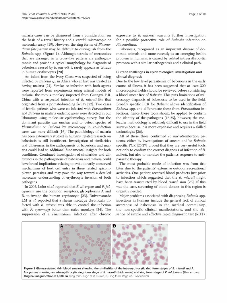
malaria cases can be diagnosed from a consideration onthe basis of a travel history and a careful microscopic ormolecular assay [19]. However, the ring forms of Plasmo-dium falciparum may be difficult to distinguish from theBabesia spp. (Figure 1). Although tetrads of merozoitesthat are arranged in a cross-like pattern are pathogno-monic and provide a typical morphology for diagnosis ofbabesiosis caused by B. microti, it rarely appears as tetradsin human erythrocytes [20].An infant from the Ivory Coast was suspected of being
infected by Babesia sp. in Africa who at first was treated ashaving malaria [21]. Similar co-infection with both agentswere reported from experiments using animal models ofmalaria, the rhesus monkey imported from Guangxi, P.R.China with a suspected infection of B. microti-like thatoriginated from a primate-breeding facility [22]. Two casesof febrile patients who were co-infected with Plasmodiumand Babesia in malaria endemic areas were detected in ourlaboratory using molecular epidemiology survey, but thedominant parasite was unclear and to detect species ofPlasmodium or Babesia by microscopy in co-infectioncases was more difficult [16]. The pathobiology of malariahas been extensively studied in humans; related research onbabesiosis is still insufficient. Investigation of similaritiesand differences in the pathogenesis of babesiosis and mal-aria could lead to additional fundamental insights for bothconditions. Continued investigation of similarities and dif-ferences in the pathogenesis of babesiosis and malaria couldhave broad implications relating to evolutionarily conservedmechanisms of host cell entry in these related apicom-plexan parasites and may pave the way toward a detailedmolecular understanding of erythrocyte invasion of bothpathogens.In 2005, Lobo et al. reported that B. divergens and P. fal-
ciparum use the common receptors, glycophorins A andB, to invade the human erythrocyte [23]. DuivenvoordeLM et al. reported that a rhesus macaque chronically in-fected with B. microti was able to control the infectionwith P. cynomolgi better than naïve monkeys [24]. Thesuppression of a Plasmodium infection after chronic
Figure 1 Giemsa-stained thin blood smears showing the similarities ofalciparum, showing an intraerythrocytic ring form stage of B. microtiOriginal magnification × 1,000. (A. Ring form stage of B. microti, B. Ring f
exposure to B. microti warrants further investigationfor a possible protective role of Babesia infection onPlasmodium.Babesiosis, recognized as an important disease of do-
mestic animals and more recently as an emerging healthproblem in humans, is caused by related intraerythrocyticprotozoa with a similar pathogenesis and a clinical path.
Current challenges in epidemiological investigation andclinical diagnosisDue to the low level parasitemia of babesiosis in the earlycourse of illness, it has been suggested that at least 300microscopical fields should be reviewed before consideringa blood smear free of Babesia. This puts limitations of mi-croscopy diagnosis of babesiosis to be used in the field.Broadly specific PCR for Babesia allows identification ofBabesia spp. and differentiate these from Plasmodium in-fections, hence these tools should be applied to confirmthe identity of the pathogens [16,25], however, the mo-lecular methodology is relatively difficult to use in the fieldsurveys because it is more expensive and requires a skilledtechnologist [26].All of these three confirmed B. microti-infection pa-
tients, either by investigations of smears and/or Babesiaspecific PCR [25,27] proved that they are very useful toolsnot only to confirm the correct diagnosis of infection of B.microti, but also to monitor the patient’s response to anti-parasitic therapy.The most probable mode of infection was from tick
bites due to the patients’ extensive outdoor recreationalactivities. One patient received blood products just priorto infection which suggested that the B. microti mighthave been transmitted by blood transfusion [28]. If thiswas the case, screening of blood donors in this region isurgently needed.Major problems associated with diagnosing Babesia spp.
infections in humans include the general lack of clinicalawareness of babesiosis in the medical community,the non-specific clinical manifestations, and the ab-sence of simple and effective rapid diagnostic test (RDT).
f the intraerythrocytic ring form stages of B. microti and P.(thick arrow) and ring form stage of P. falciparum (thin arrow);orm stage of P. falciparum).
Zhou et al. Parasites & Vectors 2014, 7:509 Page 3 of 10http://www.parasitesandvectors.com/content/7/1/509
Convenient, well-evaluated diagnostic tools such as sero-logical tests or molecular biological assays designed forrapid and reliable detection of such pathogens are not yetreadily available to most routine diagnostic labora-tories [4]. Additionally, conventional laboratory test-results in clinical cases of human babesiosis may benon-specific, such as high levels of transaminases,alkaline phosphatases, unconjugated bilirubin, andlactic dehydrogenase in addition to normochromia,normocytic anaemia, and thrombocytopenia. Occa-sionally, leucopenia may also be present, probablyowing to a tumour necrosis factor (TNF)-mediated im-mune response similar to that seen in severe cases ofmalaria [29].On the immunological diagnosis of babesiosis, indirect
immunofluorescence assay (IFA) is regarded as the stand-ard assay for the detection of Babesia antibody [30]. An-other immunological assay by immunoblot for detectionof B. microti antibody is also available [31]. Luo Y. et al.[32] identified a novel secreted antigen designated as B.microti secreted antigen 1 (BmSA1) and found BmSA1could be a promising and universal target for the serodiag-nosis of human babesiosis and for an epidemiological sur-vey. Ooka H. et al. [33] applied an ELISA using rBmP94/CT for diagnosis of B. microti infection, and it demon-strated high sensitivity and specificity when tested withthe sera from mice experimentally infected with B. microtiand other species of Babesia. These results indicated thatBmSA1 and BmP94/CT could be potential markers forsurveillance of human babesiosis caused by B.microti.However, to date, serological studies on population tickexposure to Babesia have not been conducted and thediagnostic value of these antigens to human babesiosis hasnot been evaluated. In New England (Country) where thedisease is enzootic, to evaluate acute babesiosis cases, animmunofluorescent assay (IFA) was carried out. The sensi-tivity of the IFA was 91%, the specificity was 99%, the posi-tive predictive value was 86%, and the negative was 99%.So IFA should be a sensitive, specific routine clinical diag-nosis of acute and convalescent babesiosis [34]. The latestsurvey on seroprevalence of blood donors was carried outin Babesia endemic areas of the Northeast and UpperMidwest in the USA. The presence of antibodies againstB.microti was tested by using an IFA. The results showedthat 2% (42/2150) of the donors were positive and one pa-tient was confirmed to have an ongoing infection of Babe-sia by positive PCR (1/42) [35]. The ELISA survey on thefebrile cases were applied by diagnostic antigen of BmSA1expression and purified in our group to conduct theprevalence of the human babesiosis. The results showedthat BmSA1 have cross-reactivity with malaria cases inChina. The former seroprevalence survey in Babesiaendemic areas of the Northeast and Upper Midwestin the USA showed that most of the sero-positive cases
(41/42) were negative in the molecular survey. Thus,clinical diagnosis of human babesiosis can be furthercomplicated by persistent low parasitemia or asymp-tomatic latent infections, particularly in malaria andbabesiosis syndemic areas.
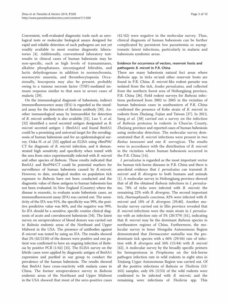
Evidence for occurrence of vectors, reservoir hosts andpathogenic B. microti in P.R. ChinaThere are many babesiosis natural foci areas whereBabesia spp. in ticks or/and other reservoir hosts arefound in P.R. China. B. microti-like rodent parasite wasisolated from the tick, Ixodes persulcatus, and collectedfrom the northern forest area of Heilongjiang province,P.R. China [36]. Field rodent surveys for Babesia infec-tions performed from 2002 to 2005 in the vicinities ofhuman babesiosis cases in southeastern of P.R. Chinaconfirmed the presence of Kobe strain of B. microti inrodents from Zhejiang, Fujian and Taiwan [37]. In 2012,Jiang et al. [38] carried out a survey on the infectionof Babesia protozoa in rodents in Chun’an County,Zhejiang province and reported cases of human babesiosisusing molecular detection. The molecular survey dem-onstrated that B. microti infections were present in twoRattus tanezumi and one R. norvegicus. The resultswere in accordance with the distribution of B. microtiin the vicinities where human babesiosis occurred inthe P.R. China [14].I. persulcatus is regarded as the most important vector
for human tick-borne diseases in P.R. China and there isanecdotal evidence that I. persulcatus can transmit B.microti and B. divergens to both humans and animals[1]. A molecular survey in Heilongjiang province showedthat of all the obtained Babesia infections in I. persulca-tus, 78% of ticks were infected with B. microti; theremaining 22% with B. divergens. The second importanttick, Haemaphysalis concinna, 82% were infected with B.microti and 18% of B. divergens [39,40]. Another mo-lecular survey carried out in Jilin province revealed thatB. microti-infections were the main strain in I. persulca-tus with an infection rate of 5% (20/379) [41], indicatingthat B. microti may be the dominant Babesia species innortheastern regions of China. Furthermore, the mo-lecular survey in Inner Mongolia Autonomous Regiondemonstrated that Dermacentor nuttallia was the pre-dominant tick species with a 66% (29/44) rate of infec-tion with B. divergens and 34% (15/44) with B. microti[42]. A molecular survey by the broadly specific primersfor hemoprotozoa in Piroplasma on the tick-bornepathogen infection rate in wild rodents in eight sites inXinjiang Uygur Autonomous Region was carried out. Ofall the positive infections of Babesia or Theileria (53/165) samples, only 6% (3/53) of the wild rodents wereconfirmed to be infected with B. microti, and theremaining were infections of Theileria spp. This
Zhou et al. Parasites & Vectors 2014, 7:509 Page 4 of 10http://www.parasitesandvectors.com/content/7/1/509
indicates that B. microti-infections were not the predom-inant species in Xinjiang Uygur Autonomous Region[43]. In 2008, B. microti-like parasites were identifiedin one rhesus monkey (Macaca mulatta) importedfrom Guangxi Zhuang Autonomous Region and the sus-pected infection of B. microti originated from a primate-breeding facility [22]. This provided anecdoctal evidenceof the presence of natural foci of B. mciroti in GuangxiZhuang Autonomous Region. B. microti was also detectedfrom I. persulcatus in Beijing and the partial sequences of18S RNA gene of B. microti was submitted to NCBIGenBank (accession no. JX962781). Recently, a total of 33rodents (22 Apodemus agrarius and 11 R. norvegicus) werecaptured from Xinyang City, Henan province. Inoculationexperiments were carried out in mice and indicated thatone of the rodents was co-infected with A. phagocytophi-lum and B. microti [44]. Thus, the detection and isolationof both pathogens from the single rodent in Henanprovince further highlighted the possibility of co-infection in human beings and should alert publichealth. In summary, previous epidemiological studieson Babesia infections in vectors or rodents by univer-sal primers for Babesia spp. has shown that the occur-rence of pathogenic Babesia spp. exists widely in P.R.China. Specific PCR confirmed that B. microti infec-tions were the predominant species in southeastern and
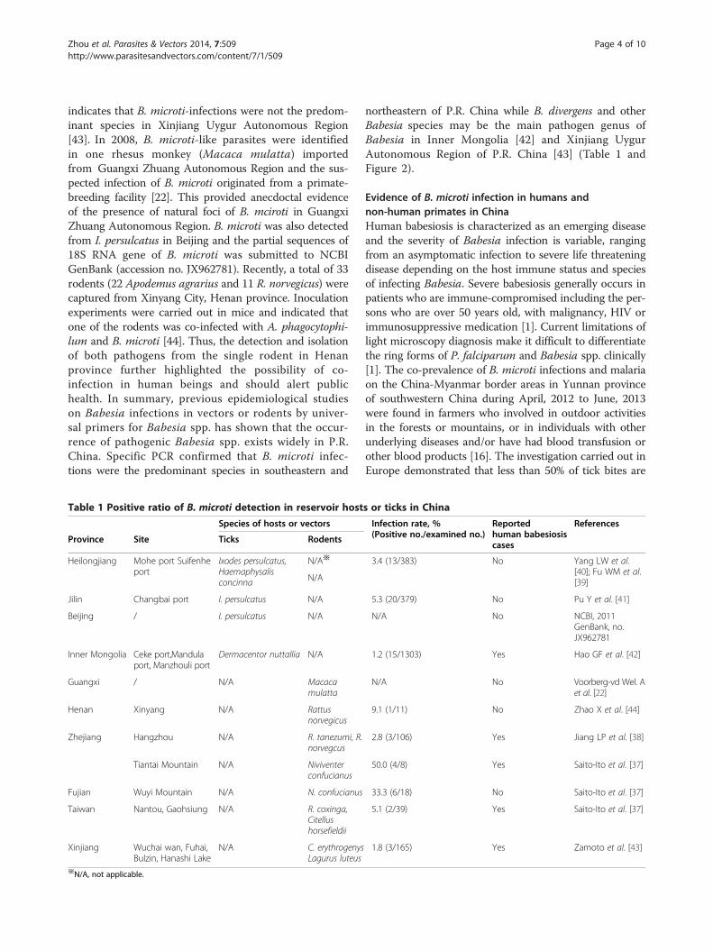
Table 1 Positive ratio of B. microti detection in reservoir host
Species of hosts or vectors
Province Site Ticks Rodents
Heilongjiang Mohe port Suifenheport
Ixodes persulcatus,Haemaphysalisconcinna
N/A※
N/A
Jilin Changbai port I. persulcatus N/A
Beijing / I. persulcatus N/A
Inner Mongolia Ceke port,Mandulaport, Manzhouli port
Dermacentor nuttallia N/A
Guangxi / N/A Macacamulatta
Henan Xinyang N/A Rattusnorvegicus
Zhejiang Hangzhou N/A R. tanezumi, R.norvegcus
Tiantai Mountain N/A Niviventerconfucianus
Fujian Wuyi Mountain N/A N. confucianus
Taiwan Nantou, Gaohsiung N/A R. coxinga,Citellushorsefieldii
Xinjiang Wuchai wan, Fuhai,Bulzin, Hanashi Lake
N/A C. erythrogenysLagurus luteus
※N/A, not applicable.
northeastern of P.R. China while B. divergens and otherBabesia species may be the main pathogen genus ofBabesia in Inner Mongolia [42] and Xinjiang UygurAutonomous Region of P.R. China [43] (Table 1 andFigure 2).
Evidence of B. microti infection in humans andnon-human primates in ChinaHuman babesiosis is characterized as an emerging diseaseand the severity of Babesia infection is variable, rangingfrom an asymptomatic infection to severe life threateningdisease depending on the host immune status and speciesof infecting Babesia. Severe babesiosis generally occurs inpatients who are immune-compromised including the per-sons who are over 50 years old, with malignancy, HIV orimmunosuppressive medication [1]. Current limitations oflight microscopy diagnosis make it difficult to differentiatethe ring forms of P. falciparum and Babesia spp. clinically[1]. The co-prevalence of B. microti infections and malariaon the China-Myanmar border areas in Yunnan provinceof southwestern China during April, 2012 to June, 2013were found in farmers who involved in outdoor activitiesin the forests or mountains, or in individuals with otherunderlying diseases and/or have had blood transfusion orother blood products [16]. The investigation carried out inEurope demonstrated that less than 50% of tick bites are
s or ticks in China
Infection rate, %(Positive no./examined no.)
Reportedhuman babesiosiscases
References
3.4 (13/383) No Yang LW et al.[40]; Fu WM et al.[39]
5.3 (20/379) No Pu Y et al. [41]
N/A No NCBI, 2011GenBank, no.JX962781
1.2 (15/1303) Yes Hao GF et al. [42]
N/A No Voorberg-vd Wel. Aet al. [22]
9.1 (1/11) No Zhao X et al. [44]
2.8 (3/106) Yes Jiang LP et al. [38]
50.0 (4/8) Yes Saito-Ito et al. [37]
33.3 (6/18) No Saito-Ito et al. [37]
5.1 (2/39) Yes Saito-Ito et al. [37]
1.8 (3/165) Yes Zamoto et al. [43]
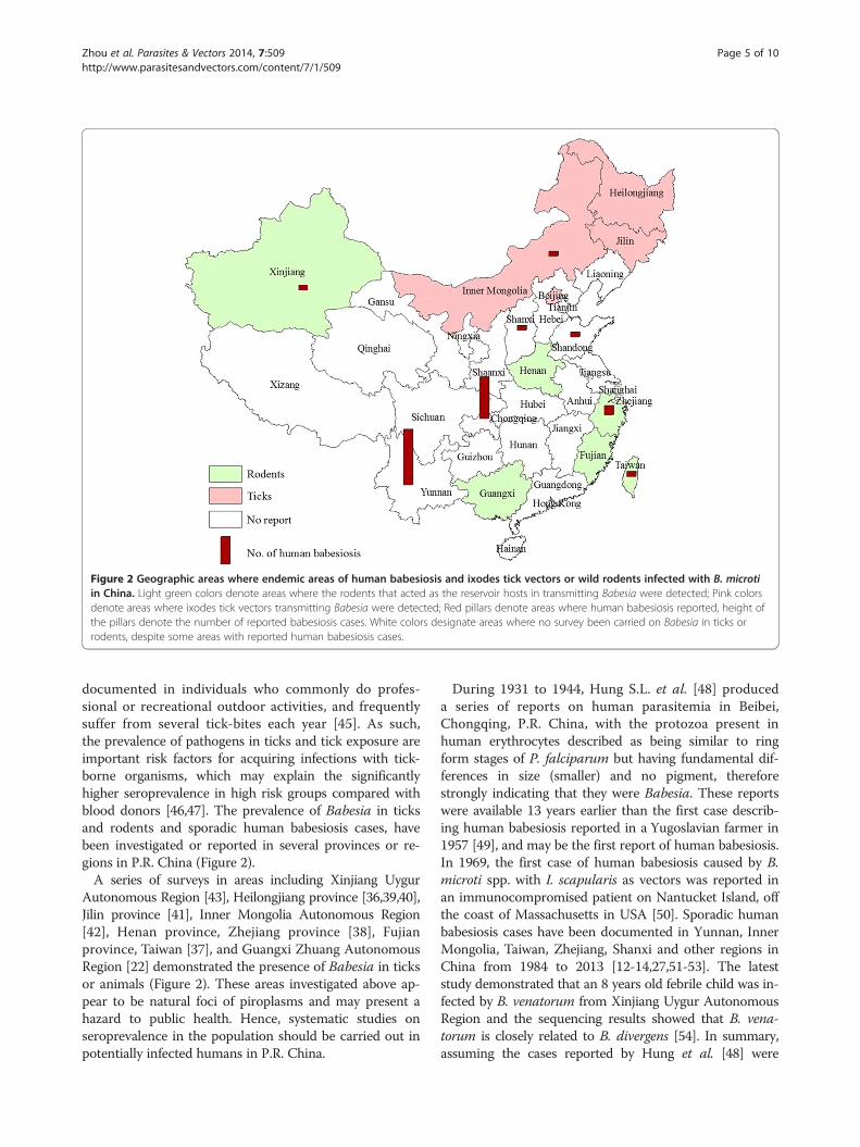
Figure 2 Geographic areas where endemic areas of human babesiosis and ixodes tick vectors or wild rodents infected with B. microtiin China. Light green colors denote areas where the rodents that acted as the reservoir hosts in transmitting Babesia were detected; Pink colorsdenote areas where ixodes tick vectors transmitting Babesia were detected; Red pillars denote areas where human babesiosis reported, height ofthe pillars denote the number of reported babesiosis cases. White colors designate areas where no survey been carried on Babesia in ticks orrodents, despite some areas with reported human babesiosis cases.
Zhou et al. Parasites & Vectors 2014, 7:509 Page 5 of 10http://www.parasitesandvectors.com/content/7/1/509
documented in individuals who commonly do profes-sional or recreational outdoor activities, and frequentlysuffer from several tick-bites each year [45]. As such,the prevalence of pathogens in ticks and tick exposure areimportant risk factors for acquiring infections with tick-borne organisms, which may explain the significantlyhigher seroprevalence in high risk groups compared withblood donors [46,47]. The prevalence of Babesia in ticksand rodents and sporadic human babesiosis cases, havebeen investigated or reported in several provinces or re-gions in P.R. China (Figure 2).A series of surveys in areas including Xinjiang Uygur
Autonomous Region [43], Heilongjiang province [36,39,40],Jilin province [41], Inner Mongolia Autonomous Region[42], Henan province, Zhejiang province [38], Fujianprovince, Taiwan [37], and Guangxi Zhuang AutonomousRegion [22] demonstrated the presence of Babesia in ticksor animals (Figure 2). These areas investigated above ap-pear to be natural foci of piroplasms and may present ahazard to public health. Hence, systematic studies onseroprevalence in the population should be carried out inpotentially infected humans in P.R. China.
During 1931 to 1944, Hung S.L. et al. [48] produceda series of reports on human parasitemia in Beibei,Chongqing, P.R. China, with the protozoa present inhuman erythrocytes described as being similar to ringform stages of P. falciparum but having fundamental dif-ferences in size (smaller) and no pigment, thereforestrongly indicating that they were Babesia. These reportswere available 13 years earlier than the first case describ-ing human babesiosis reported in a Yugoslavian farmer in1957 [49], and may be the first report of human babesiosis.In 1969, the first case of human babesiosis caused by B.microti spp. with I. scapularis as vectors was reported inan immunocompromised patient on Nantucket Island, offthe coast of Massachusetts in USA [50]. Sporadic humanbabesiosis cases have been documented in Yunnan, InnerMongolia, Taiwan, Zhejiang, Shanxi and other regions inChina from 1984 to 2013 [12-14,27,51-53]. The lateststudy demonstrated that an 8 years old febrile child was in-fected by B. venatorum from Xinjiang Uygur AutonomousRegion and the sequencing results showed that B. vena-torum is closely related to B. divergens [54]. In summary,assuming the cases reported by Hung et al. [48] were
Zhou et al. Parasites & Vectors 2014, 7:509 Page 6 of 10http://www.parasitesandvectors.com/content/7/1/509
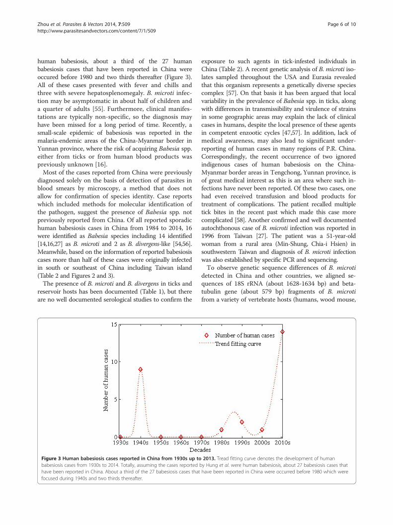
human babesiosis, about a third of the 27 humanbabesiosis cases that have been reported in China wereoccured before 1980 and two thirds thereafter (Figure 3).All of these cases presented with fever and chills andthree with severe hepatosplenomegaly. B. microti infec-tion may be asymptomatic in about half of children anda quarter of adults [55]. Furthermore, clinical manifes-tations are typically non-specific, so the diagnosis mayhave been missed for a long period of time. Recently, asmall-scale epidemic of babesiosis was reported in themalaria-endemic areas of the China-Myanmar border inYunnan province, where the risk of acquiring Babesia spp.either from ticks or from human blood products waspreviously unknown [16].Most of the cases reported from China were previously
diagnosed solely on the basis of detection of parasites inblood smears by microscopy, a method that does notallow for confirmation of species identity. Case reportswhich included methods for molecular identification ofthe pathogen, suggest the presence of Babesia spp. notpreviously reported from China. Of all reported sporadichuman babesiosis cases in China from 1984 to 2014, 16were identified as Babesia species including 14 identified[14,16,27] as B. microti and 2 as B. divergens-like [54,56].Meanwhile, based on the information of reported babesiosiscases more than half of these cases were originally infectedin south or southeast of China including Taiwan island(Table 2 and Figures 2 and 3).The presence of B. microti and B. divergens in ticks and
reservoir hosts has been documented (Table 1), but thereare no well documented serological studies to confirm the
Figure 3 Human babesiosis cases reported in China from 1930s up tobabesiosis cases from 1930s to 2014. Totally, assuming the cases reported bhave been reported in China. About a third of the 27 babesiosis cases thatfocused during 1940s and two thirds thereafter.
exposure to such agents in tick-infested individuals inChina (Table 2). A recent genetic analysis of B. microti iso-lates sampled throughout the USA and Eurasia revealedthat this organism represents a genetically diverse speciescomplex [57]. On that basis it has been argued that localvariability in the prevalence of Babesia spp. in ticks, alongwith differences in transmissibility and virulence of strainsin some geographic areas may explain the lack of clinicalcases in humans, despite the local presence of these agentsin competent enzootic cycles [47,57]. In addition, lack ofmedical awareness, may also lead to significant under-reporting of human cases in many regions of P.R. China.Correspondingly, the recent occurrence of two ignoredindigenous cases of human babesiosis on the China-Myanmar border areas in Tengchong, Yunnan province, isof great medical interest as this is an area where such in-fections have never been reported. Of these two cases, onehad even received transfusion and blood products fortreatment of complications. The patient recalled multipletick bites in the recent past which made this case morecomplicated [58]. Another confirmed and well documentedautochthonous case of B. microti infection was reported in1996 from Taiwan [27]. The patient was a 51-year-oldwoman from a rural area (Min-Shung, Chia-i Hsien) insouthwestern Taiwan and diagnosis of B. microti infectionwas also established by specific PCR and sequencing.To observe genetic sequence differences of B. microti
detected in China and other countries, we aligned se-quences of 18S rRNA (about 1628-1634 bp) and beta-tubulin gene (about 579 bp) fragments of B. microtifrom a variety of vertebrate hosts (humans, wood mouse,
2013. Tread fitting curve denotes the development of humany Hung et al. were human babesiosis, about 27 babesiosis cases thathave been reported in China were occurred before 1980 which were
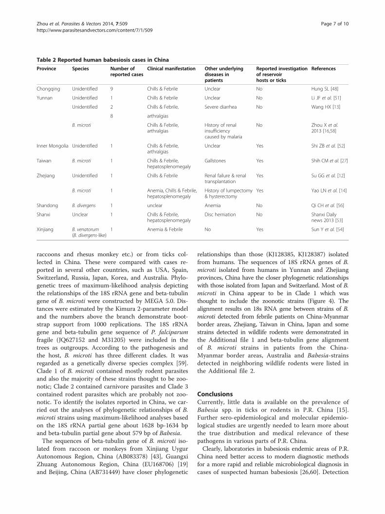
Table 2 Reported human babesiosis cases in China
Province Species Number ofreported cases
Clinical manifestation Other underlyingdiseases inpatients
Reported investigationof reservoirhosts or ticks
References
Chongqing Unidentified 9 Chills & Febrile Unclear No Hung SL [48]
Yunnan Unidentified 1 Chills & Febrile Unclear No Li JF et al. [51]
Unidentified 2 Chills & Febrile, Severe diarrhea No Wang HX [13]
8 arthralgias
B. microti Chills & Febrile,arthralgias
History of renalinsufficiencycaused by malaria
No Zhou X et al.2013 [16,58]
Inner Mongolia Unidentified 1 Chills & Febrile,arthralgias
Unclear Yes Shi ZB et al. [52]
Taiwan B. microti 1 Chills & Febrile,hepatosplenomegaly
Gallstones Yes Shih CM et al. [27]
Zhejiang Unidentified 1 Chills & Febrile Renal failure & renaltransplantation
Yes Su GG et al. [12]
B. microti 1 Anemia, Chills & Febrile,hepatosplenomegaly
History of lumpectomy& hysterectomy
Yes Yao LN et al. [14]
Shandong B. divergens 1 unclear Anemia No Qi CH et al. [56]
Shanxi Unclear 1 Chills & Febrile,hepatosplenomegaly
Disc herniation No Shanxi Dailynews 2013 [53]
Xinjiang B. venatorum(B. divergens-like)
1 Anemia & Febrile No Yes Sun Y et al. [54]
Zhou et al. Parasites & Vectors 2014, 7:509 Page 7 of 10http://www.parasitesandvectors.com/content/7/1/509
raccoons and rhesus monkey etc.) or from ticks col-lected in China. These were compared with cases re-ported in several other countries, such as USA, Spain,Switzerland, Russia, Japan, Korea, and Australia. Phylo-genetic trees of maximum-likelihood analysis depictingthe relationships of the 18S rRNA gene and beta-tubulingene of B. microti were constructed by MEGA 5.0. Dis-tances were estimated by the Kimura 2-parameter modeland the numbers above the branch demonstrate boot-strap support from 1000 replications. The 18S rRNAgene and beta-tubulin gene sequence of P. falciparumfragile (JQ627152 and M31205) were included in thetrees as outgroups. According to the pathogenesis andthe host, B. microti has three different clades. It wasregarded as a genetically diverse species complex [59].Clade 1 of B. microti contained mostly rodent parasitesand also the majority of these strains thought to be zoo-notic; Clade 2 contained carnivore parasites and Clade 3contained rodent parasites which are probably not zoo-notic. To identify the isolates reported in China, we car-ried out the analyses of phylogenetic relationships of B.microti strains using maximum-likelihood analyses basedon the 18S rRNA partial gene about 1628 bp-1634 bpand beta-tubulin partial gene about 579 bp of Babesia.The sequences of beta-tubulin gene of B. microti iso-
lated from raccoon or monkeys from Xinjiang UygurAutonomous Region, China (AB083378) [43], GuangxiZhuang Autonomous Region, China (EU168706) [19]and Beijing, China (AB731449) have closer phylogenetic
relationships than those (KJ128385, KJ128387) isolatedfrom humans. The sequences of 18S rRNA genes of B.microti isolated from humans in Yunnan and Zhejiangprovinces, China have the closer phylogenetic relationshipswith those isolated from Japan and Switzerland. Most of B.microti in China appear to be in Clade 1 which wasthought to include the zoonotic strains (Figure 4). Thealignment results on 18s RNA gene between strains of B.microti detected from febrile patients on China-Myanmarborder areas, Zhejiang, Taiwan in China, Japan and somestrains detected in wildlife rodents were demonstrated inthe Additional file 1 and beta-tubulin gene alignmentof B. microti strains in patients from the China-Myanmar border areas, Australia and Babesia-strainsdetected in neighboring wildlife rodents were listed inthe Additional file 2.
ConclusionsCurrently, little data is available on the prevalence ofBabesia spp. in ticks or rodents in P.R. China [15].Further sero-epidemiological and molecular epidemio-logical studies are urgently needed to learn more aboutthe true distribution and medical relevance of thesepathogens in various parts of P.R. China.Clearly, laboratories in babesiosis endemic areas of P.R.
China need better access to modern diagnostic methodsfor a more rapid and reliable microbiological diagnosis incases of suspected human babesiosis [26,60]. Detection
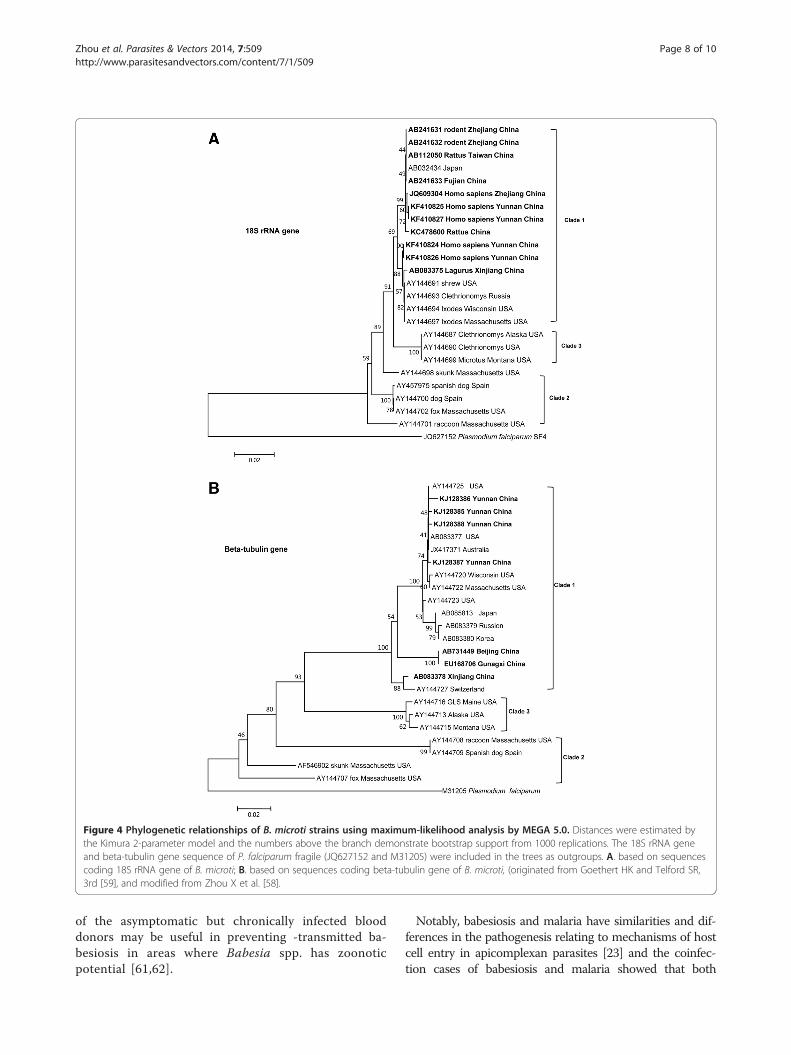
Figure 4 Phylogenetic relationships of B. microti strains using maximum-likelihood analysis by MEGA 5.0. Distances were estimated bythe Kimura 2-parameter model and the numbers above the branch demonstrate bootstrap support from 1000 replications. The 18S rRNA geneand beta-tubulin gene sequence of P. falciparum fragile (JQ627152 and M31205) were included in the trees as outgroups. A. based on sequencescoding 18S rRNA gene of B. microti; B. based on sequences coding beta-tubulin gene of B. microti, (originated from Goethert HK and Telford SR,3rd [59], and modified from Zhou X et al. [58].
Zhou et al. Parasites & Vectors 2014, 7:509 Page 8 of 10http://www.parasitesandvectors.com/content/7/1/509
of the asymptomatic but chronically infected blooddonors may be useful in preventing -transmitted ba-besiosis in areas where Babesia spp. has zoonoticpotential [61,62].
Notably, babesiosis and malaria have similarities and dif-ferences in the pathogenesis relating to mechanisms of hostcell entry in apicomplexan parasites [23] and the coinfec-tion cases of babesiosis and malaria showed that both
Zhou et al. Parasites & Vectors 2014, 7:509 Page 9 of 10http://www.parasitesandvectors.com/content/7/1/509
diseases appear to be endemic on the China-Myanmarborder [42,63,64]. This research approach should pave theway towards successfully controlling both pathogens basedon the molecular analysis of erythrocyte invasion.
Additional files
Additional file 1: 18s RNA gene alignment results between strainsof B. microti detected from febrile patients on China-Myanmarborder areas babesiosis Zhejiang in P.R. China, Japan and somestrains detected in wildlife rodents.
Additional file 2: Beta-tubulin gene alignment of B. microti strainsin patients from the China-Myanmar border areas, Australia andBabesia-strains detected in neighboring wildlife rodents.
Competing interestThe authors declared that they have no competing interests.
Authors’ contributionsXZ and XNZ conceived the study, collected the data and analyzed it, anddrafted the manuscript. SX, JLH and ET conceived the project and providedtechnical support for data collection and analysis. HXZG and XNZ conceivedthe study and revised the manuscript. All authors read and approved thefinal manuscript.
AcknowledgmentThis project was supported by National Natural Science Foundation of China(grant No. 81273192). Zhou XN was supported by Special Fund for HealthResearch in the Public Interest (grant No. 201202019) and the National S&TMajor Program (grant No. 2012ZX10004220).We would express our sincere gratitude to Dr. Li Mei from the malariadepartment of National Institute of Parasitic Diseases, China CDC for providethe Giemsa stain slides of P. falciparum in Figure 2B.We would express our sincere gratitude to Dr. Merritt Christine fromWisconsin, USA for helping to review the manuscript and editing thelanguage.
Author details1National Institute of Parasitic Diseases, Chinese Center for Disease Controland Prevention, WHO Collaborating Centre for Malaria, Schistosomiasis andFilariasis; Key Laboratory of Parasite & Vector Biology, Ministry of Health,Shanghai 200025, People’s Republic of China. 2Department of Parasitology,Medical College of Soochow University, No.199 Renai Road, Suzhou 215123,People’s Republic of China. 3Biochemistry Department, Centers forSustainable Malaria Control, Faculty of Natural & Agricultural Sciences,University of Pretoria, Pretoria, South Africa.
Received: 1 February 2014 Accepted: 28 October 2014
References1. Vannier E, Krause PJ: Human babesiosis. N Engl J Med 2012, 366(25):2397–2407.2. Homer MJ, Aguilar-Delfin I, Telford SR 3rd, Krause PJ, Persing DH: Babesiosis.
Clin Microbiol Rev 2000, 13(3):451–469.3. Hunfeld KP, Hildebrandt A, Gray JS: Babesiosis: recent insights into an
ancient disease. Int J Parasitol 2008, 38(11):1219–1237.4. Lobo CA, Cursino-Santos JR, Alhassan A, Rodrigues M: Babesia: an emerging
infectious threat in transfusion medicine. PLoS Pathog 2013, 9(7):e1003387.5. Herwaldt BL, Linden JV, Bosserman E, Young C, Olkowska D, Wilson M:
Transfusion-associated babesiosis in the United States: a description ofcases. Ann Intern Med 2011, 155(8):509–519.
6. Bi Y, Yu W, Hu W, Lin H, Guo Y, Zhou XN, Tong S: Impact of climatevariability on Plasmodium vivax and Plasmodium falciparum malaria inYunnan Province, China. Parasit Vectors 2013, 6:357.
7. Li T, Yang Z, Wang M: Temperature, relative humidity and sunshine maybe the effective predictors for occurrence of malaria in Guangzhou,southern China, 2006–2012. Parasit Vectors 2013, 6:155.
8. Chen XP, Cong ML, Li MH, Kang YJ, Feng YM, Plyusnin A, Xu J, Zhang YZ:Infection and pathogenesis of Huaiyangshan virus (a novel tick-bornbunyavirus) in laboratory rodents. J Gen Virol 2012, 93(Pt6):1288–1293.
9. Zhang YZ, Zhou DJ, Xiong Y, Chen XP, He YW, Sun Q, Yu B, Li J, Dai YA,Tian JH, Qin XC, Jin D, Cui Z, Luo XL, Li W, Lu S, Wang W, Peng JS, Guo WP,Li MH, Li ZJ, Zhang S, Chen C, Wang Y, De Jong MD, Xu J: Hemorrhagicfever caused by a novel tick-borne bunyavirus in Huaiyangshan, China.Chin J Epidemiol 2011, 32(3):209–220 (in Chinese).
10. Xu B, Liu L, Huang X, Ma H, Zhang Y, Du Y, Wang P, Tang X, Wang H, KangK, Zhang SQ, Zhao GH, Wu WL, Yang YH, Chen HM, MuF CWJ:Metagenomic analysis of fever, thrombocytopenia and leukopeniasyndrome (FTLS) in Henan Province, China: discovery of a newbunyavirus. PLoS Pathog 2011, 7(11):e1002369.
11. Si BY, Jiang T, Zhang Y, Deng YQ, Huo QB, Zheng YC, Qin ED, Qin CF, ZhuQY: Complete genome sequence analysis of tick-borne encephalitisviruses isolated in northeastern China. Arch Virol 2011, 156(8):1485–1488.
12. Su GG, Zhao NF, Ye YX: A babesia case report. Chin J of Zoon 2002,18:112–155 (in Chinese).
13. Wang HX: One case of babesiosis in Kunming. Int J Med Parasit Dis 2012,39:190–192 (in Chinese).
14. Yao LN, Ruan W, Zeng CY, Li ZH, Zhang X, Lei YL, Lu QY, Che HL: Pathogenidentification and clinical diagnosis for one case infected with Babesia.Chin J Parasitol Parasit Dis 2012, 30(2):118–121 (in Chinese).
15. Wu XB, Na RH, Wei SS, Zhu JS, Peng HJ: Distribution of tick-borne diseasesin China. Parasit Vectors 2013, 6:119.
16. Zhou X, Li SG, Chen SB, Wang JZ, Xu B, Zhou HJ, Ge HX, Chen JH, Hu W:Co-infections with Babesia microti and Plasmodium parasites along theChina-Myanmar border. Infect Dis Poverty 2013, 2(1):24.
17. Krause PJ, Daily J, Telford SR, Vannier E, Lantos P, Spielman A: Sharedfeatures in the pathobiology of babesiosis and malaria. Trends Parasitol2007, 23(12):605–610.
18. Clark IA, Jacobson LS: Do babesiosis and malaria share a common diseaseprocess? Ann Trop Med Parasitol 1998, 92(4):483–488.
19. Rodgers SE, Mather TN: Human Babesia microti incidence and Ixodesscapularis distribution, Rhode Island, 1998–2004. Emerg Infect Dis 2007,13(4):633–635.
20. Vannier E, Gewurz BE, Krause PJ: Human babesiosis. Infect Dis Clin North Am2008, 22(3):469–488. viii-ix.
21. Vermeil C, Menut J, Miegeville M, Cruziat J, Julienne F, Morin O, Roger AP,Marjolet M, Bouillard C: Babesiasis, pediatric malaria: does confusion existin Africa? Bull Soc Pathol Exot Filiales 1983, 76(5 Pt 2):797–804.
22. Voorberg-vd Wel A, Kocken CH, Zeeman AM, Thomas AW: Detection ofnew Babesia microti-like parasites in a rhesus monkey (Macaca mulatta)with a suppressed Plasmodium cynomolgi infection. Am J Trop Med Hyg2008, 78(4):643–645.
23. Lobo CA: Babesia divergens and Plasmodium falciparum use commonreceptors, glycophorins A and B, to invade the human red blood cell.Infect Immun 2005, 73(1):649–651.
24. van Duivenvoorde LM, Voorberg-van der Wel A, van der Werff NM,Braskamp G, Remarque EJ, Kondova I, Kocken CH, Thomas AW: Suppressionof Plasmodium cynomolgi in rhesus macaques by coinfection withBabesia microti. Infect Immun 2010, 78(3):1032–1039.
25. Persing DH, Mathiesen D, Marshall WF, Telford SR, Spielman A, ThomfordJW, Conrad PA: Detection of Babesia microti by polymerase chainreaction. J Clinl Microbiol 1992, 30(8):2097–2103.
26. Chen JH, Wang H, Chen JX, Bergquist R, Tanner M, Utzinger J, Zhou XN:Frontiers of parasitology research in the People’s Republic of China:infection, diagnosis, protection and surveillance. Parasit Vectors 2012, 5:221.
27. Shih CM, Liu LP, Chung WC, Ong SJ, Wang CC: Human babesiosis inTaiwan: asymptomatic infection with a Babesia microti-like organism in aTaiwanese woman. J Clin Microbiol 1997, 35(2):450–454.
28. Rosenblatt JE: Laboratory diagnosis of infections due to blood and tissueparasites. Clin Infect Dis 2009, 49(7):1103–1108.
29. Mylonakis E: When to suspect and how to monitor babesiosis.Am Fam Physician 2001, 63(10):1969–1974.
30. Krause PJ, Telford SR 3rd, Ryan R, Conrad PA, Wilson M, Thomford JW,Spielman A: Diagnosis of babesiosis: evaluation of a serologic test for thedetection of Babesia microti antibody. J Infect Dis 1994, 169(4):923–926.
31. Ryan R, Krause PJ, Radolf J, Freeman K, Spielman A, Lenz R, Levin A:Diagnosis of babesiosis using an immunoblot serologic test.Clin Diagn Lab Immunol 2001, 8(6):1177–1180.
Zhou et al. Parasites & Vectors 2014, 7:509 Page 10 of 10http://www.parasitesandvectors.com/content/7/1/509
32. Luo Y, Jia H, Terkawi MA, Goo YK, Kawano S, Ooka H, Li Y, Yu L, Cao S,Yamagishi J, Fujisaki K, Nishikawa Y, Saito-Ito A, Igarashi I, Xuan X: Identificationand characterization of a novel secreted antigen 1 of Babesia microti andevaluation of its potential use in enzyme-linked immunosorbent assay andimmunochromatographic test. Parasitol Int 2011, 60(2):119–125.
33. Ooka H, Terkawi MA, Goo YK, Luo Y, Li Y, Yamagishi J, Nishikawa Y, IgarashiI, Xuan X: Babesia microti: molecular and antigenic characterizations of anovel 94-kDa protein (BmP94). Exp Parasitol 2011, 127(1):287–293.
34. Krause PJ, Ryan R, Telford S 3rd, Persing D, Spielman A: Efficacy ofimmunoglobulin M serodiagnostic test for rapid diagnosis of acutebabesiosis. J Clin Microbiol 1996, 34(8):2014–2016.
35. Tonnetti L, Thorp AM, Deisting B, Bachowski G, Johnson ST, Wey AR,Hodges JS, Leiby DA, Mair D: Babesia microti seroprevalence in Minnesotablood donors. Transfusion 2013, 53(8):1698–1705.
36. Sun Y, Liu G, Yang L, Xu R, Cao W: Babesia microti-like rodent parasitesisolated from Ixodes persulcatus (Acari: Ixodidae) in Heilongjiang Province,China. Vet Parasitol 2008, 156(3–4):333–339.
37. Saito-Ito A, Takada N, Ishiguro F, Fujita H, Yano Y, Ma XH, Chen ER:Detection of Kobe-type Babesia microti associated with Japanese humanbabesiosis in field rodents in central Taiwan and southeastern mainlandChina. Parasitology 2008, 135(6):691–699.
38. Jiang LP, Zhang L, QH B, QY L, SY C, Xu BX: Babesia DNA segmentsdetected from rodents in Zhejiang province. Chin J Vector Biol Control2012, 23:303–305 (in Chinese).
39. Fu WM, He H, Hu MX, Cui Y, Yang L: Babesia microti-like rodent parasitesisolated from Ixodes persulcatus (Acari: Ixodidae) at China -Russia ports ofHeilongjiang Province. Chin Front Heal Quarant 2010, 33:99–104(in Chinese).
40. Yang LW, Hou Y, Li M, Wen ZQ, Yang J, Fan D: Study of tick-bornepathogens at Heilongjiang port. Chin Front Heal Quarant 2007, 30:77–82(in Chinese).
41. Pu Y, Li J, Song XH, Li BG, Ma CZ, Li BX, Yang HN: Detection and analysison important ticks and tick-borne pathogen at Jilin port areas, China.Acta Parasitol Med Entomol Sin 2009, 16(4):228–232 (in Chinese).
42. Hao GF, Li H, Sun Y, Ge RP, Qiao GQ, Li B, Tian WZ, Shi NX, Yang XY:Detection of tick and tick-borne pathogen in some ports of InnerMongolia. Chin J Epidemiol 2009, 30(4):365–367 (in Chinese).
43. Zamoto A, Tsuji M, Wei Q, Cho SH, Shin EH, Kim TS, Leonova GN, HagiwaraK, Asakawa M, Kariwa H, Takashima I, Ishihara C: Epizootiologic survey forBabesia microti among small wild mammals in northeastern Eurasia anda geographic diversity in the beta-tubulin gene sequences. J Vet Med Sci2004, 66(7):785–792.
44. Zhao XG, Li H, Sun Y, Zhang YY, Jiang JF, Liu W, Cao WC: Dual infectionwith Anaplasma phagocytophilum and Babesia microti in a Rattusnorvegicus. China. Ticks Tick Borne Dis 2013, 4(5):399–402 (in Chinese).
45. Wilske B, Fingerle V: Diagnosis of Lyme borreliosis. How to corroboratesuspected borreliosis. MMW Fortschr Med 2000, 142(15):28–31.
46. Hunfeld KP, Lambert A, Kampen H, Albert S, Epe C, Brade V, Tenter AM:Seroprevalence of Babesia infections in humans exposed to ticks inmidwestern Germany. J Clin Microbiol 2002, 40(7):2431–2436.
47. Hunfeld KP, Brade V: Zoonotic Babesia: possibly emerging pathogens to beconsidered for tick-infested humans in Central Europe. Int J Med Microbiol2004, 293(Suppl 37):93–103.
48. Hung SL: Notes on a species of malaria parasite finding from Bei-Bei.Nat Med J Chin (Chongqing ed) 1944, 19:571–573 (in Chinese).
49. Skrabalo Z, Deanovic Z: Piroplasmosis in man; report of a case. Doc MedGeogr Trop 1957, 9(1):11–16.
50. Western KA, Benson GD, Gleason NN, Healy GR, Schultz MG: Babesiosis in aMassachusetts resident. N Engl J Med 1970, 283(16):854–856.
51. Li JF, Meng DB, Wang QF: The discovery of human babesiasis. Chin J VetMed 1984, 6:19–20 (in Chinese).
52. Shi ZB, Li ZZ, Gao QR, Ding YM: One case of human babesia infection.Chin J Parasitol Parasit Dis 1996, 14:240 (in Chinese).
53. Shanxi Daily News: Men suffering from a rare babesiosis swelled such aspregnant women. 2013, http://www.sxdaily.com.cn/n/2013/0715/c349-5174146/html (in Chinese).
54. Sun Y, Li SG, Jiang JF, Wang X, Zhang Y, Wang H, Cao WC: Babesiavenatorum infection in child. Emerg Infect Dis 2014, 20(5):896–897.
55. Krause PJ, McKay K, Gadbaw J, Christianson D, Closter L, Lepore T, TelfordSR 3rd, Sikand V, Ryan R, Persing D, Radolf JD, Spielman A: Increasing
health burden of human babesiosis in endemic sites. Am J Trop Med Hyg2003, 68(4):431–436.
56. Qi C, Zhou D, Liu J, Cheng Z, Zhang L, Wang L, Wang Z, Yang D, Wang S, Chai T:Detection of Babesia divergens using molecular methods in anemic patientsin Shandong Province, China. Parasitol Res 2011, 109(1):241–245.
57. Telford SR 3rd, Goethert HK: Emerging tick-borne infections:rediscovered and better characterized, or truly ‘new’? Parasitology 2004,129(Suppl):S301–S327.
58. Zhou X, Li SG, Wang JZ, Huang JL, Zhou HJ, Chen JH, Zhou XN: Emergenceof human babesiosis along the border of China with Myanmar:detection by PCR and confirmation by sequencing. Emerg Microb Infect2014, 3:e55.
59. Goethert HK, Telford SR 3rd: What is Babesia microti? Parasitology 2003,127(Pt 4):301–309.
60. Chen Z, Liu Q, Jiao FC, Xu BL, Zhou XN: Detection of piroplasms infectionin sheep, dogs and hedgehogs in Central China. Infect Dis Poverty 2014,3:18.
61. Tambo E, Ai L, Zhou X, Chen JH, Hu W, Bergquist R, Guo JG, Utzinger J,Tanner M, Zhou XN: Surveillance-response systems: the key toelimination of tropical diseases. Infect Dis Poverty 2014, 3:17.
62. Zhou XN: Prioritizing research for "One health - One world".Infect Dis Poverty 2012, 1:1.
63. Tambo E, Adedeji AA, Huang F, Chen JH, Zhou SS, Tang LH: Scaling upimpact of malaria control programmes: a tale of events in Sub-SaharanAfrica and People’s Republic of China. Infect Dis Poverty 2012, 1:7.
64. Bi Y, Tong S: Poverty and malaria in the Yunnan Province, China. InfectDis Poverty 2014, 3:32.
doi:10.1186/s13071-014-0509-3Cite this article as: Zhou et al.: Human babesiosis, an emerging tick-bornedisease in the People’s Republic of China. Parasites & Vectors 2014 7:509.
Submit your next manuscript to BioMed Centraland take full advantage of:
• Convenient online submission
• Thorough peer review
• No space constraints or color figure charges
• Immediate publication on acceptance
• Inclusion in PubMed, CAS, Scopus and Google Scholar
• Research which is freely available for redistribution
Submit your manuscript at www.biomedcentral.com/submit
Related Documents