Human Anatomy, 3rd edition Prentice Hall, © 2001 Chapter 16

Human Anatomy, 3rd edition Prentice Hall, © 2001 Chapter 16.
Dec 18, 2015
Welcome message from author
This document is posted to help you gain knowledge. Please leave a comment to let me know what you think about it! Share it to your friends and learn new things together.
Transcript

Human Anatomy, 3rd editionPrentice Hall, © 2001
Chapter 16
Human Anatomy, 3rd editionPrentice Hall, © 2001
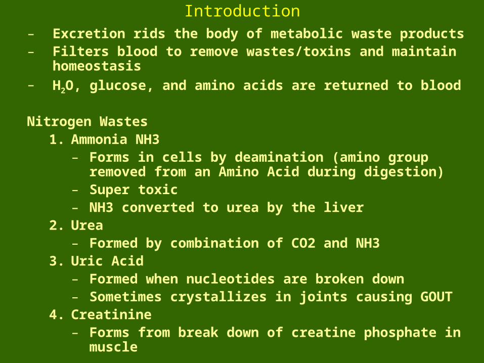
Introduction– Excretion rids the body of metabolic waste products– Filters blood to remove wastes/toxins and maintain homeostasis– H2O, glucose, and amino acids are returned to blood
Nitrogen Wastes1. Ammonia NH3
– Forms in cells by deamination (amino group removed from an Amino Acid during digestion)
– Super toxic– NH3 converted to urea by the liver
2. Urea– Formed by combination of CO2 and NH3
3. Uric Acid– Formed when nucleotides are broken down– Sometimes crystallizes in joints causing GOUT
4. Creatinine– Forms from break down of creatine phosphate in muscle
Human Anatomy, 3rd editionPrentice Hall, © 2001
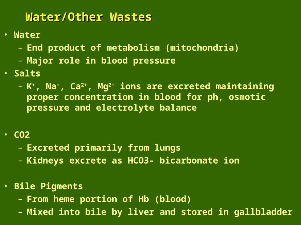
Water/Other WastesWater/Other Wastes
• Water
– End product of metabolism (mitochondria)
– Major role in blood pressure
• Salts
– K+, Na+, Ca2+, Mg2+ ions are excreted maintaining proper concentration in blood for ph, osmotic pressure and electrolyte balance
• CO2
– Excreted primarily from lungs
– Kidneys excrete as HCO3- bicarbonate ion
• Bile Pigments
– From heme portion of Hb (blood)
– Mixed into bile by liver and stored in gallbladder
Human Anatomy, 3rd editionPrentice Hall, © 2001
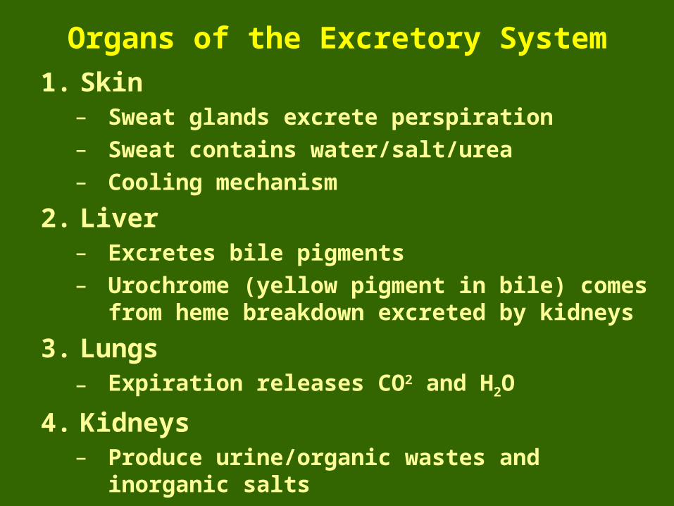
Organs of the Excretory System
1. Skin– Sweat glands excrete perspiration
– Sweat contains water/salt/urea
– Cooling mechanism
2. Liver– Excretes bile pigments
– Urochrome (yellow pigment in bile) comes from heme breakdown excreted by kidneys
3. Lungs– Expiration releases CO2 and H2O
4. Kidneys– Produce urine/organic wastes and inorganic salts
Human Anatomy, 3rd editionPrentice Hall, © 2001
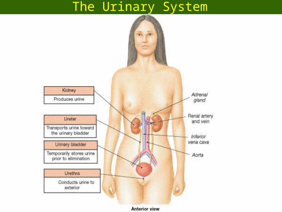
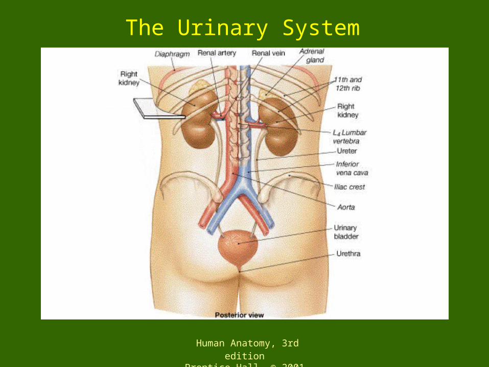
The Urinary System
Human Anatomy, 3rd editionPrentice Hall, © 2001
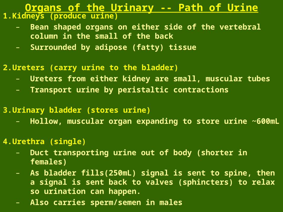
1. Kidneys (produce urine)
– Bean shaped organs on either side of the vertebral column in the small of the back
– Surrounded by adipose (fatty) tissue
2. Ureters (carry urine to the bladder)
– Ureters from either kidney are small, muscular tubes
– Transport urine by peristaltic contractions
3. Urinary bladder (stores urine)
– Hollow, muscular organ expanding to store urine ~600mL
4. Urethra (single)
– Duct transporting urine out of body (shorter in females)
– As bladder fills(250mL) signal is sent to spine, then a signal is sent back to valves (sphincters) to relax so urination can happen.
– Also carries sperm/semen in males
Organs of the Urinary -- Path of Urine
Human Anatomy, 3rd editionPrentice Hall, © 2001
The Urinary System

Human Anatomy, 3rd editionPrentice Hall, © 2001
Kidneys – Interior Gross Anatomy
• On the concave hilium side / depression is where renal blood vessels and the ureter enter
1. Renal capsule
– Connective tissue
2. Renal artery
– Blood (oxygenated) to the kidney
3. Renal vein
– Receives blood from kidney
4. Ureter
– Drains urine
Human Anatomy, 3rd editionPrentice Hall, © 2001
Internal Structure of the Kidney
Human Anatomy, 3rd editionPrentice Hall, © 2001
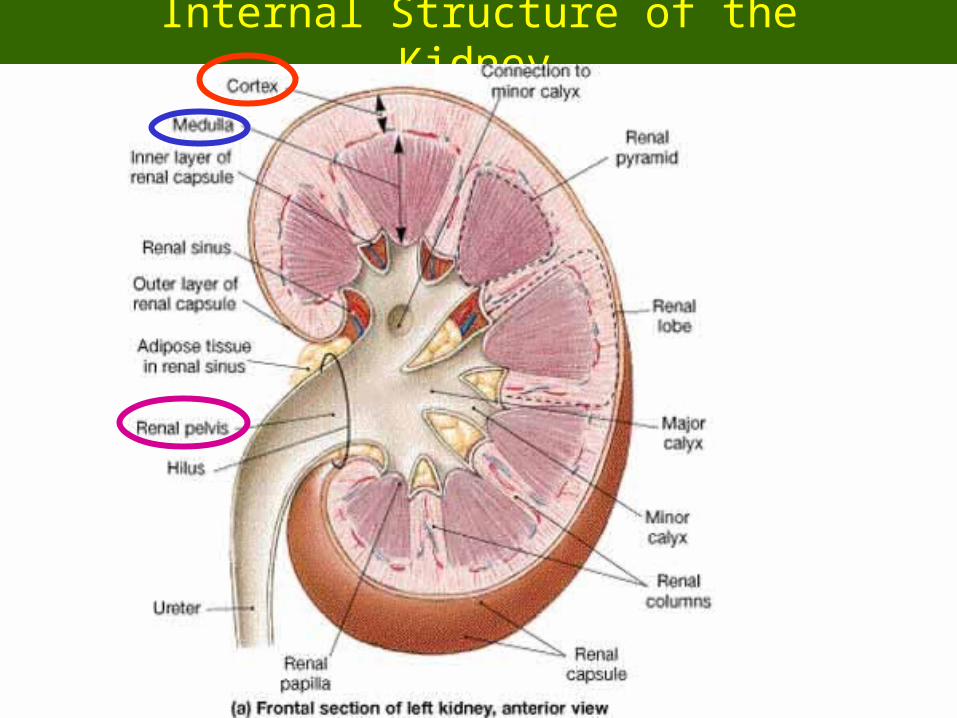
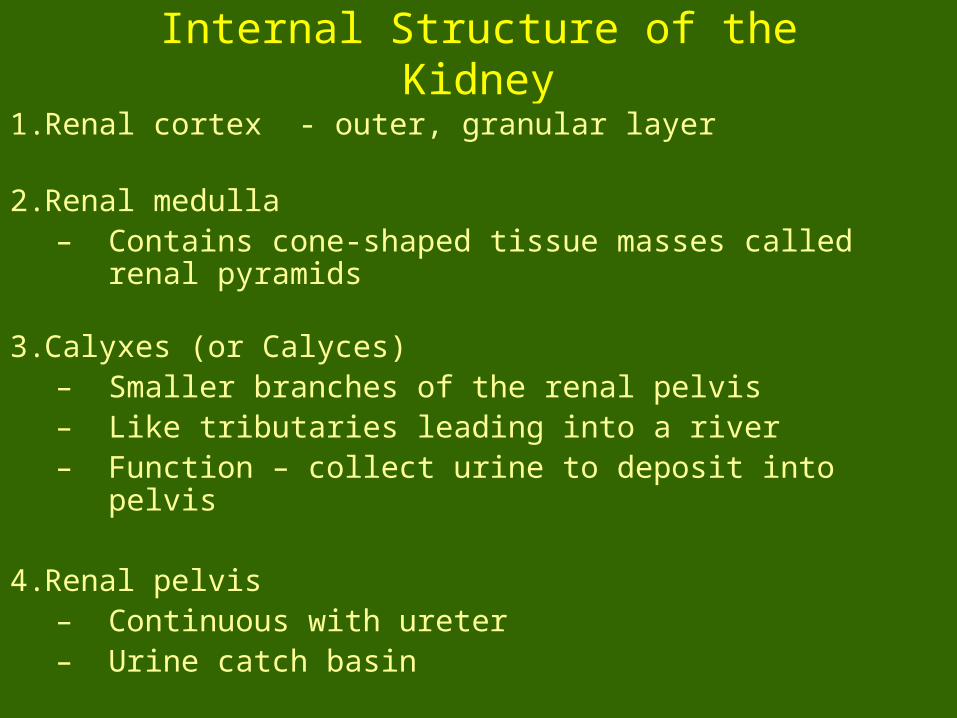
Internal Structure of the Kidney
1. Renal cortex - outer, granular layer
2. Renal medulla – Contains cone-shaped tissue masses called renal pyramids
3. Calyxes (or Calyces)– Smaller branches of the renal pelvis– Like tributaries leading into a river– Function – collect urine to deposit into pelvis
4. Renal pelvis– Continuous with ureter– Urine catch basin

Human Anatomy, 3rd editionPrentice Hall, © 2001
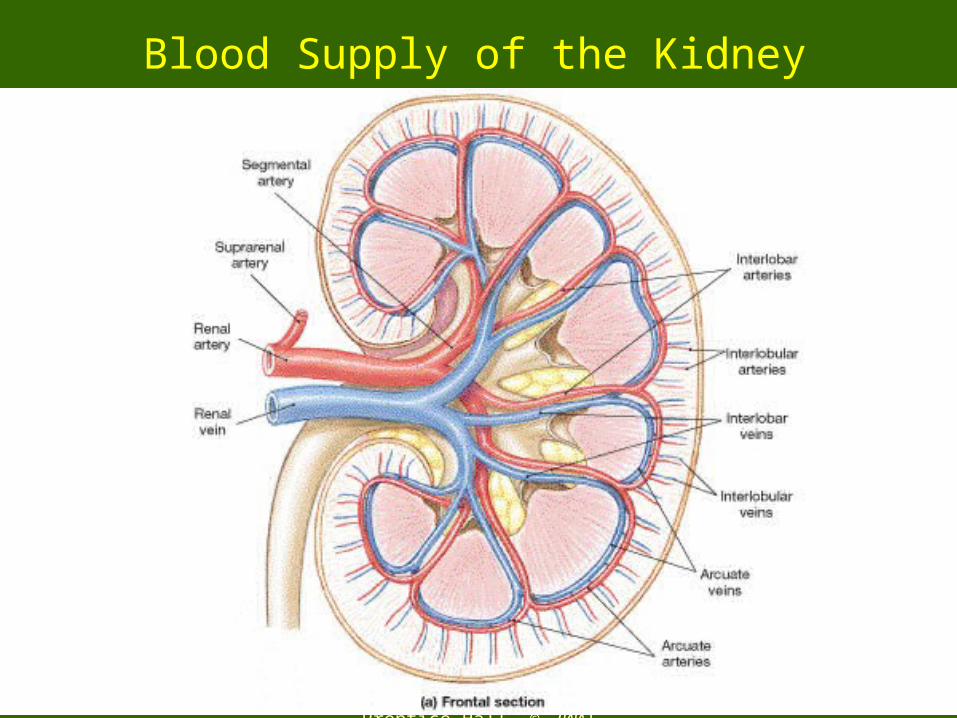
Blood Supply of the Kidney
– 100% of our blood of the body passes through the kidneys every 24 hours.
– Renal artery branches inside the kidney• Supplies the pyramids and the cortex
– Venous blood leaves the cortex and medulla• Small veins join the renal vein
Human Anatomy, 3rd editionPrentice Hall, © 2001
Blood Supply of the Kidney
Human Anatomy, 3rd editionPrentice Hall, © 2001
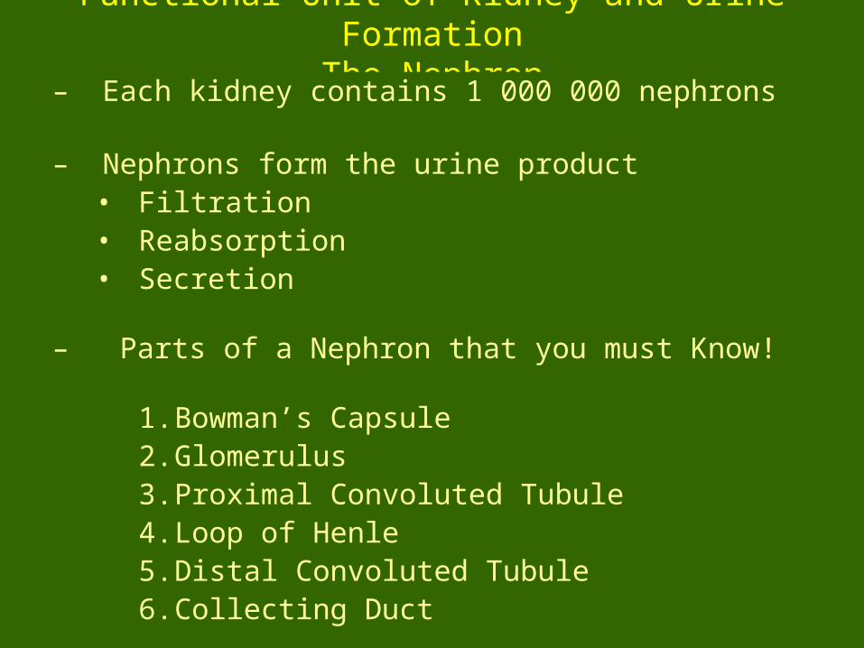
Functional Unit of Kidney and Urine FormationThe Nephron
– Each kidney contains 1 000 000 nephrons
– Nephrons form the urine product• Filtration• Reabsorption• Secretion
– Parts of a Nephron that you must Know!
1. Bowman’s Capsule2. Glomerulus3. Proximal Convoluted Tubule4. Loop of Henle5. Distal Convoluted Tubule6. Collecting Duct

Human Anatomy, 3rd editionPrentice Hall, © 2001
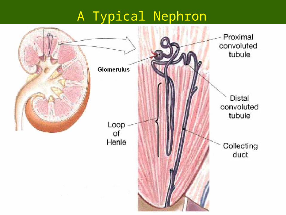
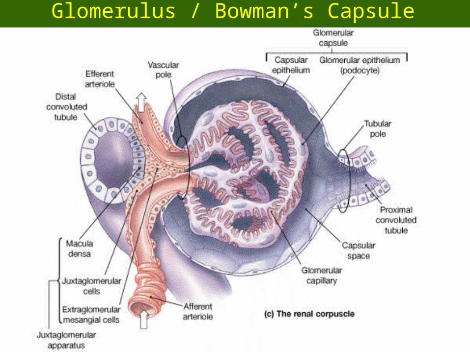
1.1. GlomerulusGlomerulus – a knot of capillaries leading to the renal tubule- 70 mm Hg pressure squeezes wastes out of blood vessels- squeezes out glucose, water, salts, AA’s but not blood cells
2.2. Renal tubule (about 2 inches long)Renal tubule (about 2 inches long)
a) Bowman’s capsule ---> [close to surface ie. Cortex]- surrounds the glomerulus
b) Proximal convoluted tubule --> [in Cortex]- reabsorption of nutrients like NaCl and water
c) Henle’s Loop – making a “u-turn -->[deep in Medulla brine bath] - reabsorption of nutrients like NaCl and water
d) Distal convoluted tubule ---> [in Cortex] – has many mitochondria for active transport-
e) Collecting duct ---> [in Medulla] - collects final waste products to send to bladder
Human Anatomy, 3rd editionPrentice Hall, © 2001
A Typical Nephron
Human Anatomy, 3rd editionPrentice Hall, © 2001

Human Anatomy, 3rd editionPrentice Hall, © 2001
Look at all the Nephrons
Human Anatomy, 3rd editionPrentice Hall, © 2001
Glomerulus / Bowman’s Capsule

Human Anatomy, 3rd editionPrentice Hall, © 2001
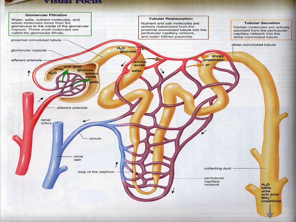
Urine Formation1. High Pressure Filtration
– Blood in afferent arteriole is under high pressure– Blood entering Glomerulus (filter) split into 2 components
A) Filterable = substances pass from the blood to Bowman’s Capsule
= water, nitogenous waste, salts, nutrients (glucose, AA’s)
B) Nonfilterable = large molecules that can’t pass into Bowman’s Capsule = RBC’s, Platlets, WBC’s, Proteins
– Filtered blood leaves the glomerulus by the efferent arteriole
2. Selective Reabsorption– Filtrate contains some useful substances to return to the blood– Selective reabsorption can be passive (no E) or active (req’s E)– Most “returns”occur in the proximal convoluted tubule
Human Anatomy, 3rd editionPrentice Hall, © 2001
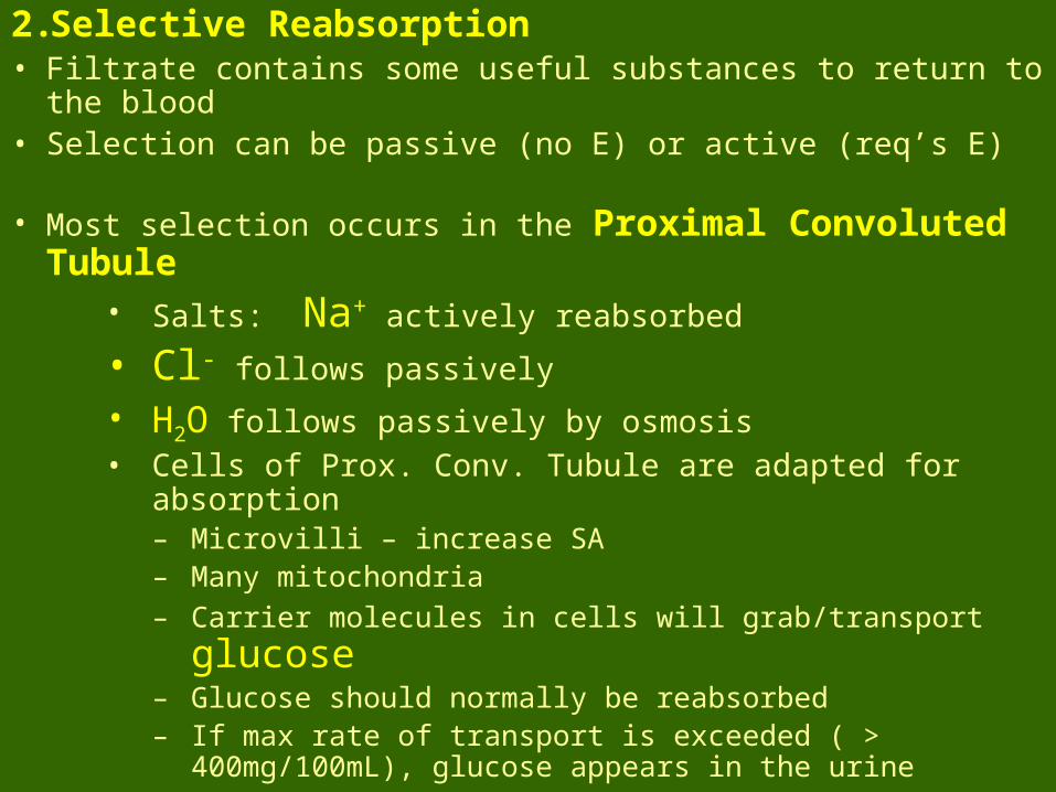
2. Selective Reabsorption• Filtrate contains some useful substances to return to the blood• Selection can be passive (no E) or active (req’s E)
• Most selection occurs in the Proximal Convoluted Tubule• Salts: Na+ actively reabsorbed
• Cl- follows passively
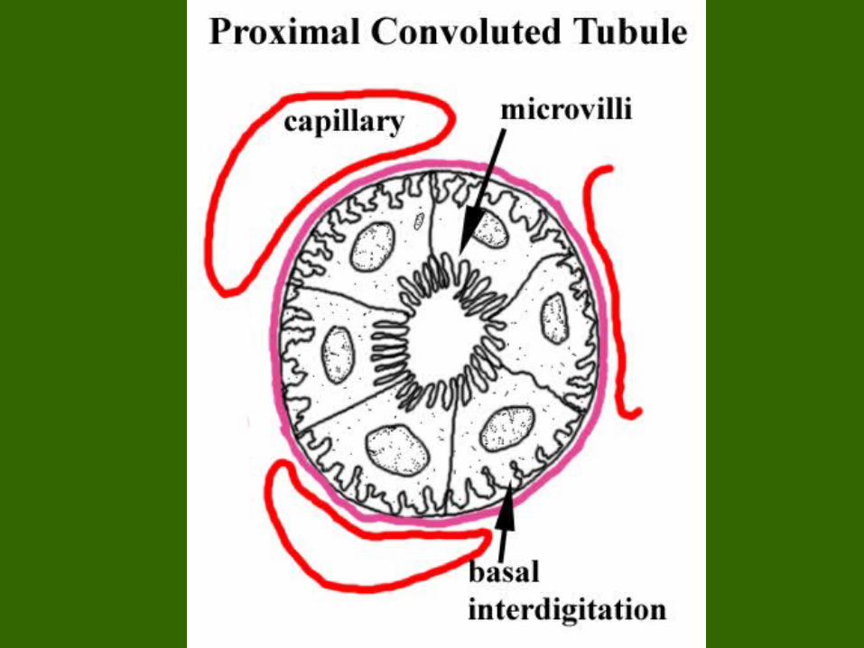
• H2O follows passively by osmosis• Cells of Prox. Conv. Tubule are adapted for absorption
– Microvilli – increase SA– Many mitochondria
– Carrier molecules in cells will grab/transport glucose– Glucose should normally be reabsorbed– If max rate of transport is exceeded ( > 400mg/100mL), glucose
appears in the urine
Human Anatomy, 3rd editionPrentice Hall, © 2001

Human Anatomy, 3rd editionPrentice Hall, © 2001
3. Tubular Secretion• Active Transport used to pump some substances missed by the
glomerulus straight into the Distal Convoluted Tubule (DCT)
1. H+ and K+ ions
2. Creatinine, NH33. Drugs (ex. Penicillin)4. Destined for urine
Reabsorbing Water – a terrible thing to lose• Kidneys maintain the water-salt balance • So, they maintain Blood Pressure• Most of the H20 and Salts are reabsored at the Proximal Convoluted
Tubule (PCT)• However, much is also absorbed by the Loop of Henle
Human Anatomy, 3rd editionPrentice Hall, © 2001
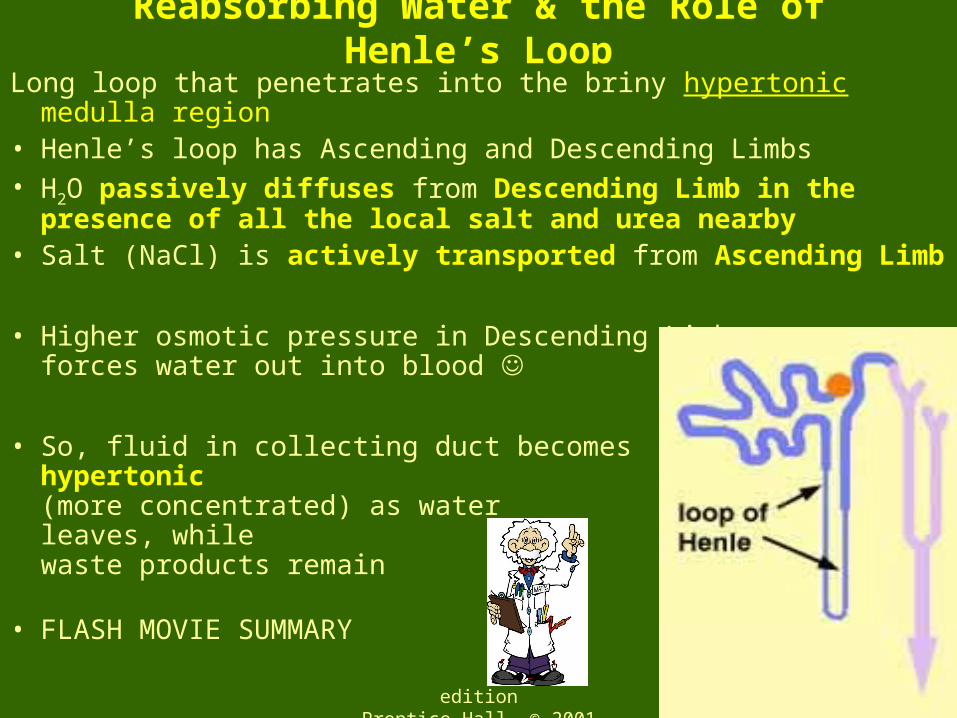
Reabsorbing Water & the Role of Henle’s Loop
Long loop that penetrates into the briny hypertonic medulla region• Henle’s loop has Ascending and Descending Limbs• H2O passively diffuses from Descending Limb in the presence of
all the local salt and urea nearby• Salt (NaCl) is actively transported from Ascending Limb
• Higher osmotic pressure in Descending Limb forces water out into blood
• So, fluid in collecting duct becomes hypertonic(more concentrated) as water leaves, while waste products remain
• FLASH MOVIE SUMMARY

Human Anatomy, 3rd editionPrentice Hall, © 2001

Human Anatomy, 3rd editionPrentice Hall, © 2001
Control of Blood Composition by Kidneys• Maintain blood pH (7.35 – 7.45) / Salt balance is critical to homeostasishomeostasis
• If blood is acidic, H+ is excreted with NH3 & Na+ / HCO3- are reabsorbed• If blood is basic, fewer H+ are excreted and less Na/HCO3- are reabsorbed
• Regulated by hormones1. ADH (Antiduretic Hormone)
- normally we pass water (diuresis = water loss)
- ADH stops this. It’s secreted by posterior pituitary gland- ADH increases water reabsorption
- makes Henle’s Loop /Collecting Duct more permeable to H20- alcohol interferes with ADH & reabsorption
---> this increases urination
2. Aldosterone secreted by the adrenal glands
– increases sodium reabsorption and therefore water absorption
- important way to increase B.P. ---> consider blood loss from injury

Human Anatomy, 3rd editionPrentice Hall, © 2001
Kidneys as BP Devices– Kidneys will secrete Aldosterone to bring up BP– So, kidneys are blood pressure monitors
BP Sensor that turns on Aldosterone = Juxtaglomerular ApparatusJuxtaglomerular Apparatus
- An interface between DCT and Afferent Arteriole1. Cells in J.A. sense
pressure change in blood
2. Afferent Arteriole instructed to release
RENIN (enzyme to blood)
3. Renin splits Angiotensin blood protein from
Angiotensin I II
4. Adrenal cortex stimulated to release Aldosterone to incr. BP
Related Documents


