Human amygdala stimulation effects on emotion physiology and emotional experience Cory S. Inman, Emory University Kelly R. Bijanki, Emory University David I. Bass, Emory University Robert Gross, Emory University Stephan Hamann, Emory University Jon T. Willie, Emory University Journal Title: Neuropsychologia Publisher: Elsevier | 2018-01-01 Type of Work: Article | Post-print: After Peer Review Publisher DOI: 10.1016/j.neuropsychologia.2018.03.019 Permanent URL: https://pid.emory.edu/ark:/25593/v4cf2 Final published version: http://dx.doi.org/10.1016/j.neuropsychologia.2018.03.019 Copyright information: © 2018 Elsevier Ltd This is an Open Access work distributed under the terms of the Creative Commons Attribution-NonCommercial-NoDerivatives 4.0 International License (http://creativecommons.org/licenses/by-nc-nd/4.0/). Accessed February 18, 2022 2:02 AM EST

Welcome message from author
This document is posted to help you gain knowledge. Please leave a comment to let me know what you think about it! Share it to your friends and learn new things together.
Transcript
Human amygdala stimulation effects on emotionphysiology and emotional experienceCory S. Inman, Emory UniversityKelly R. Bijanki, Emory UniversityDavid I. Bass, Emory UniversityRobert Gross, Emory UniversityStephan Hamann, Emory UniversityJon T. Willie, Emory University
Journal Title: NeuropsychologiaPublisher: Elsevier | 2018-01-01Type of Work: Article | Post-print: After Peer ReviewPublisher DOI: 10.1016/j.neuropsychologia.2018.03.019Permanent URL: https://pid.emory.edu/ark:/25593/v4cf2
Final published version:http://dx.doi.org/10.1016/j.neuropsychologia.2018.03.019
Copyright information:© 2018 Elsevier LtdThis is an Open Access work distributed under the terms of the CreativeCommons Attribution-NonCommercial-NoDerivatives 4.0 International License(http://creativecommons.org/licenses/by-nc-nd/4.0/).
Accessed February 18, 2022 2:02 AM EST
Human amygdala stimulation effects on emotion physiology and emotional experience
Cory S. Inman1, Kelly R. Bijanki1, David I. Bass2, Robert E. Gross1,3,4, Stephan Hamann6,*, and Jon T. Willie1,*
1Department of Neurosurgery, Emory University School of Medicine, 1365 Clifton Road, Atlanta, GA 30322
2Graduate Program in Neuroscience, Emory University, 1462 Clifton Road, Atlanta, GA 30322
3Department of Neurology, Emory University School of Medicine, 1365 Clifton Road, Atlanta, GA 30322
4Coulter Department of Biomedical Engineering, Georgia Institute of Technology
5Emory University School of Medicine, 1760 Haygood Dr., Atlanta, GA 30322
6Department of Psychology, Emory University, 36 Eagle Row, Atlanta, GA 30322
Abstract
The amygdala is a key structure mediating emotional processing. Few studies have used direct
electrical stimulation of the amygdala in humans to examine stimulation-elicited physiological and
emotional responses, and the nature of such effects remains unclear. Determining the effects of
electrical stimulation of the amygdala has important theoretical implications for current discrete
and dimensional neurobiological theories of emotion, which differ substantially in their
predictions about the emotional effects of such stimulation. To examine the effects of amygdala
stimulation on physiological and subjective emotional responses we examined epilepsy patients
undergoing intracranial EEG monitoring in which depth electrodes were implanted unilaterally or
bilaterally in the amygdala. Nine subjects underwent both sham and acute monopolar electrical
stimulation at various parameters in electrode contacts located in amygdala and within lateral
temporal cortex control locations. Stimulation was applied at either 50 Hz or 130 Hz, while
amplitudes were increased stepwise from 1-12 V, with subjects blinded to stimulation condition.
Electrodermal activity (EDA), heart rate (HR), and respiratory rate (RR) were simultaneously
Corresponding Author: Jon T. Willie, [email protected], Department of Neurological Surgery, Emory University School of Medicine, 1365 Clifton Road, Suite B6200, Atlanta, GA 30322.*Equal Contributions
Publisher's Disclaimer: This is a PDF file of an unedited manuscript that has been accepted for publication. As a service to our customers we are providing this early version of the manuscript. The manuscript will undergo copyediting, typesetting, and review of the resulting proof before it is published in its final citable form. Please note that during the production process errors may be discovered which could affect the content, and all legal disclaimers that apply to the journal pertain.
Author ContributionsC.S.I., K.R.B., D.I.B, S.H., and J.T.W. contributed to the study design. C.S.I., K.R.B., D.I.B., R.F., and J.T.W. contributed to the data collection. J.T.W. and R.E.G. conducted the surgeries. C.S.I., K.R.B., and J.T.W. contributed to the electrode localization. C.S.I. and K.R.B. performed the behavioral, psychophysiological, and statistical analyses. K.R.B. designed the mixed linear models. C.S.I, K.R.B., S.H., and J.T.W. contributed to the interpretation. C.S.I., K.R.B., S.H., and J.T.W. wrote the manuscript. C.S.I., J.T.W., K.R.B., D.I.B., R.E.G., and S.H. edited the manuscript. All authors discussed and commented on the manuscript.
HHS Public AccessAuthor manuscriptNeuropsychologia. Author manuscript; available in PMC 2019 September 15.A
uthor Manuscript
Author M
anuscriptA
uthor Manuscript
Author M
anuscript
recorded and subjective emotional response was probed after each stimulation period. Amygdala
stimulation (but not lateral control or sham stimulation) elicited immediate and substantial dose-
dependent increases in EDA and decelerations of HR, generally without affecting RR. Stimulation
elicited subjective emotional responses only rarely, and did not elicit clinical seizures in any
subject. These physiological results parallel stimulation findings with animals and are consistent
with orienting/defensive responses observed with aversive visual stimuli in humans. In summary,
these findings suggest that acute amygdala stimulation in humans can be safe and can reliably
elicit changes in emotion physiology without significantly affecting subjective emotional
experience, providing a useful approach for investigation of amygdala-mediated modulatory
effects on cognition.
Keywords
Amygdala; Brain Stimulation; Autonomic Reactivity; Psychophysiology; Electrodermal Activity; Heart Rate; Emotional Experience
The amygdala is an almond-shaped brain structure in the medial temporal lobe that is
implicated in a wide range of emotional functions. The amygdala is comprised of multiple
subnuclei that differ in their function and projections, yielding extensive modulatory
connections to widespread brain regions (Adolphs et al., 1995; Pessoa, 2008; Pessoa and
Adolphs, 2010; Ledoux, 2000; Burgdorf and Panksepp, 2006). The role of the amygdala in
mediating emotional responses has attracted particular attention with respect to subjective
responses (e.g., fear) and physiological affective changes in autonomic nervous system
(ANS) activity (Mangina and Beuzeron-Mangina, 1996; Asahina et al., 2003; Guillory and
Bujarski, 2014). Considerable evidence from nonhuman animal, human neuroimaging, and
lesion studies have shown that the amygdala modulates ANS activity in the context of the
processing of affectively salient stimuli (Adolphs et al., 1994; Ledoux, 2000; Bradley et al.,
2001; Critchley, 2002; Critchley, 2005; Feinstein, Adolphs, Damasio, & Tranel, 2011).
Amygdala lesions have been observed to blunt normal physiological and subjective
emotional responses to some types of emotionally salient stimuli, particularly fear-related
responses to aversive stimuli (Adolphs et al., 1997; Ledoux, 2017; Davis and Whalen, 2001),
though the amygdala’s role in emotion likely extends beyond aversive stimuli and fear to
encompass positive emotion, reward, and aspects of social processing (Adolphs, 2010;
Anderson & Adolphs, 2014; Hamann et al., 1999).
An important current debate in the cognitive neuroscience of emotion concerns the neural
representation of emotion in the human brain. Key in this debate is whether individual brain
regions have specific emotion functions, or alternately, whether emotions are an emergent
property of processing that is distributed across multiple brain regions and networks.
Reflecting this debate, current neurobiological theories of emotion propose markedly
different functions of the amygdala in mediating emotional responses (Anderson & Adolphs,
2014; Vytal and Hamann, 2010; Barrett and Satpute, 2013; Lindquist et al., 2012; Ekman,
1992; Bradley et al., 2001; Barrett et al., 2007). One theoretical issue concerns whether the
amygdala specifically mediates the basic emotion of fear, or alternately, whether the
amygdala’s role is more general and intrinsic to brain networks mediating multiple emotions
Inman et al. Page 2
Neuropsychologia. Author manuscript; available in PMC 2019 September 15.
Author M
anuscriptA
uthor Manuscript
Author M
anuscriptA
uthor Manuscript
(Hamann, 2012; Lindquist et al., 2012; Pessoa, 2010). A closely related issue is the extent to
which the amygdala may mediate the emotional dimensions of intensity (arousal) or valence
(degree of pleasantness or unpleasantness) that have been proposed by influential
psychological dimensional theories of emotion (Barrett and Satpute, 2013; Hamann, 2012;
Bradley et al., 2001; Pessoa, 2010).
Although human neuroimaging and neuropsychological lesion studies have provided
important evidence regarding the neural representation of emotion in the human brain and
the role of the amygdala, both approaches have important limitations (Adolphs, 2016; Vytal
and Hamann, 2010; Barrett and Satpute, 2013). Neuroimaging can provide correlational
evidence suggesting the amygdala’s role in emotion, but such evidence cannot be used to
infer a causal or necessary role of the amygdala. Neuropsychological studies of patients with
brain lesions incorporating the amygdala have reported variable impairments in the
subjective experience of fear in some patients with bilateral amygdala lesions (Feinstein et
al., 2011; Adolphs et al., 1999). However, although studies of patients with bilateral lesions
have contributed greatly to understanding of amygdala function, such patients are rare and
thus these studies have limitations related to small sample sizes, individual differences in
post-lesion neural reorganization, developmental differences, and other factors. In addition,
human lesions caused by degenerative, ischemic, or post-surgical processes are typically not
restricted to the amygdala, although a few patients with bilateral lesions restricted to the
amygdala have been extensively studied (Adolphs et al., 1999; Adolphs, 2016).
By contrast, acute electrical brain stimulation of the amygdala and other brain regions may
provide more direct causal evidence for specific emotional functions (Burgdorf et al., 2000;
Panksepp, 1982), complementing the correlational evidence obtained using methods such as
functional neuroimaging and electrophysiological recording (Rutishauser et al., 2015).
Discrete focal brain stimulation can produce reliable, temporally precise changes in
electrophysiological brain states, measures of peripheral physiology, and subjective mental
state in awake human subjects, lending causal evidence to evaluate the predictions of current
neurobiological theories of emotion (Mayberg et al., 2005; Selimbeyoglu and Parvizi, 2010;
Guillory and Bujarski, 2014). In the current study, we examined both subjective and
psychophysiological responses to direct electrical stimulation of the amygdala. While direct
amygdala stimulation has been reported in a few studies to elicit subjective emotional
responses and changes in emotional physiology, the relationships among stimulation dosage
and subjective and physiological measures of emotion have not been determined (Lanteaume
et al., 2007; Meletti et al., 2006; Bijanki et al., 2014; Smith et al., 2006).
Several studies have examined the subjective emotional responses associated with
stimulation of several different brain regions (Guillory and Bujarski, 2014). In contrast, the
current study focused specifically on the amygdala, a region which has been investigated
less frequently and systematically (Halgren et al., 1978; Halgren et al., 1992; Mangina and
Beuzeron-Mangina, 1996; Meletti et al., 2006; Lanteaume et al., 2007). Four studies have
used stimulation mapping of the amygdala and other brain structures in epilepsy patients
undergoing intracranial electroencephalographic (iEEG) monitoring to examine the effects
of electrical stimulation on subjective emotional experience (Lanteaume et al., 2007; Meletti
et al., 2006; Bijanki et al., 2014; Smith et al., 2006). Electrical stimulation of the amygdala
Inman et al. Page 3
Neuropsychologia. Author manuscript; available in PMC 2019 September 15.
Author M
anuscriptA
uthor Manuscript
Author M
anuscriptA
uthor Manuscript
typically elicits subjective responses relatively rarely (Meletti et al., 2006). For example,
Meletti and colleagues reported evoked subjective emotional responses in only 12% of
amygdala stimulation trials. With respect to the nature of elicited subjective responses,
stimulation to the amygdala can induce both positive and negative emotional experiences
(e.g., elation or fear), although negative emotional responses are more frequently observed
(Bijanki et al., 2014; Lanteaume et al., 2007; Meletti et al., 2006; Smith et al., 2006). Across
stimulation of various regions in the left or right hemisphere, Smith et al. (2006) found that
right-hemisphere stimulation produced more dysphoric responses than left-hemisphere
stimulation. Similarly, Lanteaume et al. reported that right amygdala stimulation elicited
only negative responses (e.g., fear and sadness), whereas left amygdala stimulation elicited
both negative and positive responses. In addition, Lanteaume et al. found that amygdala
stimulation elicited a larger electrodermal response (EDA) when a subjective emotional
experience was also elicited, relative to stimulation conditions in which no emotional
experience was elicited. In contrast, Bijanki et al. (2014) reported a significant positive
response with relatively high-amplitude, intermittent stimulation (15 V, 5 s On, 5 s Off) to
the right amygdala in a patient with severe comorbid depression. Taken together, these
studies establish that direct amygdala stimulation can elicit changes in emotion physiology
(EDA only) and subjective emotional experience. However, the extent to which these
autonomic effects (EDA, heart rate, or respiration) are dependent upon the dose of
stimulation (i.e., stimulation amplitude) and the presence or absence of a subjective
emotional response, has yet to be established.
Importantly, emotional responses involve changes in three primary domains: neural,
behavioral/subjective, and physiological (Bradley and Lang, 2000a; Bradley and Lang,
2000b; Lang, 1995; Lang et al., 1997; Hamann, 2001). For example, encountering a
poisonous snake elicits brain activation related to processing threat, behavioral responses
such as recoiling or freezing, and physiological changes in autonomic nervous system
activity, such as changes in heart rate and skin conductance. The extent to which changes in
each affective domain are mediated independently by the amygdala remains to be fully
characterized. Accordingly, in the current study we examined how direct electrical
stimulation of the amygdala influenced concurrent physiological and behavioral/subjective
responses, relative to sham stimulation and stimulation to a positive control region (i.e., the
lateral temporal cortex). In addition, the present study examined changes in autonomic
reactivity to varying parameters of direct electrical stimulation to the human amygdala
(stimulation amplitude, hemisphere, and frequency). Specifically, we measured changes in
electrodermal activity (EDA), heart rate (HR), and respiration rate (RR) while delivering
long (30 s), intermittent electrical stimulation trains to the human amygdala. We also
examined whether there was a relationship between these autonomic changes and the
subjective experience of emotion. We hypothesized that even below subjective thresholds,
stimulation would produce an amplitude- and location-dependent increase in autonomic
arousal when stimulating the amygdala, shown by increased SCR, decreased heart rate, and
respiration consistent with orienting and defensive responses observed with aversive visual
stimuli in humans (Bradley et al., 2001; Lang & Bradley, 2010) and previous studies
strongly implicating the amygdala in emotional arousal processes (Adolphs, 2010;
Anderson, 2003; Lang & Bradley, 2010).
Inman et al. Page 4
Neuropsychologia. Author manuscript; available in PMC 2019 September 15.
Author M
anuscriptA
uthor Manuscript
Author M
anuscriptA
uthor Manuscript
Methods
2.1 Subjects
Nine patients undergoing intracranial EEG monitoring for drug-resistant epilepsy were
recruited to participate in the study (4 female; all right-handed; M(SD)age=36(10); see Table
1 for detailed patient demographics and epilepsy information). All patients gave written
informed consent to a study protocol approved by the Emory University Institutional Review
Board. In all 6 of 9 subjects in which functional MRI was obtained, left-hemisphere
language dominance was confirmed. Standard stereotactic EEG depth electrode arrays (Ad-
Tech; 0.86 mm diameter, 2 mm length platinum-coated contacts, spaced along 5 mm
intervals) were implanted into the brain parenchyma by either of two neurosurgeons (J.T.W.,
R.E.G.) for the sole purposes of clinical seizure investigation. One patient was excluded
from all further autonomic analysis due to poor psychophysiology recordings (S9). All 9
analyzed patients had stimulated electrode contacts localized within the amygdala (Figure
1). On average, patients were monitored in the Emory University Hospital Epilepsy
Monitoring Unit for 2 weeks. Study inclusion required subjects to be English speaking
adults (>18 years, regardless of gender, race or ethnicity), implanted with intracranial depth
electrodes, including those localized to the left and/or right amygdala. All patients had low
average to average full scale intelligence quotients and were able to converse and respond to
questions about their subjective feelings. Subjects also had to be able to understand an
informed consent (comprehend potential risks and benefits), and give written and verbal
informed consent to all experiments.
2.2 Stimulation Electrode Localization
High-resolution pre-surgical and post-implantation anatomical scans were gathered on each
patient (T1 MPRAGE, Siemens 1.5T, TR = 1900, TE = 3.5). We applied the following
method to localize the precise position of the stimulating electrode contacts relative to the
BLA using custom MATLAB scripts (Fig. 1). Preoperative and postoperative T1 images
were aligned with an automated linear co-registration using the FLIRT module of FSL
(Jenkinson and Smith, 2001), which was then corrected for postsurgical tissue displacement
and deformation using a manually-guided nonlinear thin-plate spline warping (Bookstein,
1989; Rohr et al., 2001). Control points for the nonlinear warping were selected at locations
where the anatomical correspondence between pre- and post-operative images could be
unambiguously identified in a side-by-side visual comparison, with emphasis on features
bounding the electrode location as closely as imaging artifacts allowed. Amygdala nuclei
were then projected into the space of the preoperative image through nonlinear warping of
deformable meshes representing the structures of interest. The meshes were obtained by
digitizing a stereotactic atlas of the human brain (Mai et al., 2008; Oya et al., 2009) and
were projected into the pre-operative image space by first aligning the outer boundary of the
atlas-derived amygdala with an amygdala boundary surface obtained through automated
subcortical segmentation (FSL FIRST; Patenaude et al., 2011), with manual adjustment of
the latter to improve accuracy, when necessary. These respective surfaces then provided
control points for a nonlinear thin-plate spline warping, which allowed the atlas-derived
meshes to be projected into the space of the subject’s preoperative image. Locations of
stimulating and recording electrodes in the amygdala were further verified retrospectively in
Inman et al. Page 5
Neuropsychologia. Author manuscript; available in PMC 2019 September 15.
Author M
anuscriptA
uthor Manuscript
Author M
anuscriptA
uthor Manuscript
each patient, by automated co-registration of each postoperative structural brain T1 MRI and
head CT images with each preoperative brain MRI using a stereotactic neurosurgical
planning computer workstation (ROSA Surgical Planning Software, MedTech Surgical, Inc.,
New York, NY, USA). A neurosurgeon (J.T.W.) directly compared contact locations to
standard MRI and tissue sections atlases of the human brain (Duvernoy, 2005; Mai et al.,
2008). Prior prospective BLA localizations were consistent with this direct retrospective
method.
2.3 Procedure
2.3.1. Stimulation Experiment—Amygdala stimulation was alternated with stimulation
at lateral temporal lobe (i.e. middle temporal gyrus) control locations and a sham, no-
stimulation condition presented unpredictably interspersed between trials of escalating
stimulation amplitude, with the patient blinded to stimulation condition. The patient was
asked to sit quietly and minimize movement throughout testing. Respiration, heart rate, and
intracranial EEG were continuously monitored by a neurologist or neurosurgeon. No after-
discharges or seizure-like activity were detected during any stimulation periods included in
the analysis.
Stimulation amplitude (V, Volts) and frequency (Hz, Hertz) were manipulated within a block
of either amygdala or lateral temporal cortex control stimulation. A block was constituted by
stimulation within one region, for one set of amplitudes and frequency, with randomly
interspersed sham control trials. Stimulation was delivered using a voltage-controlled
clinical neurostimulator typically used to stimulate percutaneous leads for treatment of
movement and pain disorders (Model 3628 Dual Screen, Medtronic, Inc., Minneapolis, MN,
USA) that utilized a bidirectional pulse pair designed to avoid thermal and electrolytic
damage. Each trial consisted of stimulation for 30 seconds on and at least 30 seconds off.
These stimulation and inter-trial interval durations were selected to maximize the number of
parameters that could be tested in a given session (~90 min), while allowing adequate time
between each stimulation trial for autonomic measures (described below) to return to
baseline, as determined by real time data interpretation by a researcher (C.S.I), before
beginning the next trial. After each stimulation, patients were asked to report any emotional
or physiological sensation and the 30 second intertrial interval began at least 15 seconds
after the patient had stopped talking to allow for physiological measures to return to a steady
baseline.
Frequency was set to either 50 or 130 Hz with a pulse width 300 or 90 μsec, respectively.
Pulse width was adjusted with frequency to maintain approximate equivalence in the charge
density delivered between the two frequencies. Initially in early subjects, 50 and 130 Hz
were tested, but after finding these frequencies to be indistinguishable, we performed
stimulation only at 50 Hz thereafter. Stimulation amplitude varied in a stepwise fashion in
either 1 or 2 V increments from 1 to 12 V, with subjects blinded to stimulation condition. It
was considered desirable to escalate voltages in a stepwise manner (interspersed with sham
stimulation) in the design of the study to a) determine the lowest dosage of any individual
subjective responses that might confound subsequent trials by carry-over effects and to b)
Inman et al. Page 6
Neuropsychologia. Author manuscript; available in PMC 2019 September 15.
Author M
anuscriptA
uthor Manuscript
Author M
anuscriptA
uthor Manuscript
minimize the risk that eliciting afterdischarges or seizures (although neither were detected in
this study) at higher dosages would preclude data collection.
The clinician delivering the stimulation stated audibly “On” and “Off” at the onset and offset
of each stimulation trial, regardless of active stimulation, positive control (lateral temporal
cortex), or sham condition (see Supplementary Tables 1 and 2 for average trial counts per
condition). To summarize, participants received blocked stimulation including either a)
active amygdala stimulation interleaved with sham or b) active positive control stimulation
targeting non-limbic control site (lateral temporal lobe neocortex) interleaved with sham.
Stimulation was delivered on a voltage-controlled basis in stepwise ascending order. Sham
trials were interspersed unpredictably with actual stimulation trials to blinded subjects
(Figure 2). The research procedure session was completely independent of any clinical
mapping procedures (e.g. cortical function and/or seizure network mapping), and the only
communication in the room was the announcement of the beginning and end of trials
referred to aloud as an ascending condition number (e.g. “Condition 1 on”, Condition 1 off”,
“Condition 2 on”, “Condition 2 off”, etc.), with the associated stimulation target (i.e.
amygdala or lateral temporal lobe) and parameters (actual versus sham stimulation) being
predetermined by the investigators and unknown to the subject. The patient was asked to
comment on any subjective experience after each and every trial, including sham conditions.
While we did not directly measure impedance for every stimulation electrode, we did
determine the impedance range of depth electrode contacts to be between 800 and 1000
Ohms in a patient by measuring multiple contacts (localized to gray matter by post-implant
MRI) along different electrode arrays using the impedance measurement function of a
clinical radiofrequency lesion generator (G4, Cosman Medical, Inc. Burlington, MA)
connected by alligator clips to externalized electrode tails. These measurements are
consistent with the reported impedance range of Ad-Tech reduced diameter depth electrode
contacts that provide valid measurements of iEEG activity. Using this impedance
(resistance) range, the study voltage range of 1-12 V corresponds to expected currents of
1-1.25 mA for 1 V and 12-15 mA for 12 V (overall range ~1-15 mA), according to Ohm’s
Law.
2.3.2. Measures of autonomic response—Electrodermal activity (EDA),
electrocardiogram (ECG), and respiration (RSP) were recorded while behavior was
monitored and videotaped. Data were recorded and analyzed using Acqknowledge 4.2 with a
MP150 amplifier and wireless Bionomadix transceivers (Biopac, Inc., Goleta, CA, USA).
Heart rate (HR) and respiration rate (RR) were calculated from the ECG and RSP data,
respectively. EDA leads were placed on the left and right palms for each patient to minimize
movement and dropout artifacts that are often found when recording from the fingers. For
each subject, whichever hand recording was of the highest quality (the least occurrence of
signal dropout artifacts) was selected for analysis throughout the entire session, with the
lower quality recording being completely omitted from further analysis. EDA amplitude
(natural log (ln) transformed to correct for known skew in EDA distributions; Boucsein et
al., 2012) was calculated as the magnitude of the trough-to-peak change in EDA from
stimulation onset to offset and in two 15-second intervals during stimulation. All changes in
EDA amplitude were z-scored within each patient to adjust for overall levels of EDA
Inman et al. Page 7
Neuropsychologia. Author manuscript; available in PMC 2019 September 15.
Author M
anuscriptA
uthor Manuscript
Author M
anuscriptA
uthor Manuscript
reactivity across patients. The ECG leads were placed just under the patient’s right collar
bone (cathode), just below the patient’s lowest left rib (anode), and on the patients left collar
bone (ground) in a lead III configuration. All dropout artifacts were corrected with linear
interpolation from the last physiological signal to the next physiological signal. Heart rate
was calculated using a sliding window assessment of the ECG R-waves (R-to-R interval).
The respiratory data were collected using a chest band with force transducer. All artifacts
were manually smoothed using the Acqknowledge software by linearly interpolating across
the nonphysiological recording. For respiration rate analyses, after apparent movement
artifacts were corrected with linear interpolation, the respiration signal was first resampled
from 1000 samples/s to 62.5 samples/s, then digitally band pass filtered between 0.5 Hz and
1 Hz with a Finite Impulse Response function (FIR) to remove slow and fast variations in
the signal that were likely non-respiratory. The respiration rate calculation, used the positive
peaks of the respiration signal and calculated the time between these peaks to generate an
ongoing change in respiration rate over time in accordance with the hardware/software
manufacturer’s instructions (Biopac, Inc; https://www.biopac.com/knowledge-base/
respiration-recording/). Average changes in HR and RR were calculated after subtracting a
15 second pre-stimulus baseline for the 30-second stimulation periods and in 15-second
windows across the stimulation periods.
2.4 Statistical analyses
Mixed linear models with random and fixed effects were used to estimate the effects of
stimulation voltage, location (amygdala, lateral temporal control), and hemisphere (left,
right) on three outcome measures of autonomic arousal. Outcome variables included 1)
natural log-transformed (ln) maximum of EDA amplitude across 30 seconds of stimulation,
2) change in average HR across 30 seconds of stimulation compared to pre-stimulus
baseline, and 3) change in average RR across 30 seconds of stimulation compared to pre-
stimulus baseline. EDA amplitude was very strongly skewed, and therefore was ln-
transformed to better meet the model assumptions of normality and to allow for greater
interpretability of the parameter estimates than other normalization methods (z-scoring or
square root transformation). The overall model design was as follows:
Yi = β0 + β1Volts + β2Location + β3Hemisphere + γ01 + εi
Yi is a vector of the outcome responses for the ith subject and i = 1 to 7, where β0 represents
the model intercept, β1 represents stimulation volts (continuous variable, fixed effect), β2
represents location of stimulation (indicator variable, fixed effect), β3 represents hemisphere
(indicator variables, fixed effects), and γ01 indicates a random intercept modeled for each
participant. Modeling was performed with SAS 9.4 using the proc mixed procedure. The two
patients that had subjective responses to higher voltages of amygdala stimulation (S8 and
S9) were excluded from the models because they had notably different subjective responses
to the stimulation.
Inman et al. Page 8
Neuropsychologia. Author manuscript; available in PMC 2019 September 15.
Author M
anuscriptA
uthor Manuscript
Author M
anuscriptA
uthor Manuscript
Results
Linear mixed models were fit to a data set collected from 7 participants who completed an
average of 45 trials each (range 22-98) across a stimulation range of 0 to 12 volt amplitudes.
84% of trials were stimulated below 9 V; there were 6 trials at 11 volts and 13 trials at 12
volts. Testing also included 72 single-blinded sham trials interleaved with active stimulation
conditions. 150 active trials took place in the amygdala, and 91 trials at positive control
locations (lateral temporal cortex), for a total of 241 total active stimulations. Of those, 101
were to the left hemisphere, 127 to the right hemisphere, and 13 were bilateral stimulations
(including both amygdala trials and positive control trials). 201 trials were completed at 50
Hz, 40 trials were completed at 130 Hz. Differences in frequency (50 Hz vs. 130 Hz) did not
contribute significantly to differences in autonomic responses, and thus frequency was not
included as a parameter in the final models. Typical raw traces from several stimulation and
sham condition trials an exemplar patient are presented in Figure 3 (S1). From visual
inspection of the raw data of this exemplar and all other patients, nearly immediate (within 3
seconds) changes in EDA and HR are apparent.
3.1 Electrodermal Activity (EDA)
The mixed linear model to predict EDA amplitude measures included the following
predictors: stimulation voltage, location (amygdala, positive control), and hemispheric
laterality (left, right). Bilateral stimulation was excluded from the model due to an
inadequate number of trials relative to other conditions. Voltage was a significant linear
predictor of the change in EDA amplitude (p = 0.002; see Table 2 for full model statistics);
every 1 V increase of amygdala stimulation is predicted to evoke a 1.06 microsiemen (μS)
change in skin conductance, controlling for effects of other predictors. Given this
observation, this model would predict that 12 volts of stimulation would lead to a 12.77 μS
increase in EDA amplitude. The application of stimulation to the amygdala as opposed to
sham also significantly predicted increased EDA amplitude (p = .01), while positive control
location stimulation was not a significant predictor of change in EDA amplitude in this
model relative to sham trials (p = .12). Using post-hoc contrasts to compare left versus right
amygdala stimulation the model would predict that left amygdala stimulation would produce
a 2.01 μS greater increase in EDA than right amygdala stimulation. See Figure 4 for plots of
the amplitude-dependent effects of amygdala stimulation on change in EDA per patient.
3.2 Heart Rate (HR)
The mixed linear model to predict a change in HR included the following predictors:
stimulation voltage, location (amygdala only), hemisphere (left, right). Bilateral stimulation
was excluded from the model due to an inadequate number of trials relative to other
conditions. The impact of positive control stimulation upon heart rate did not significantly
differ from sham stimulation, so the model includes sham and positive control as a pooled
comparison group. Voltage was a significant predictor of the change in HR (p = 0.002; see
Table 2 for full model statistics), with every 1 V increase of amygdala stimulation leading to
a −0.24 beat per minute (BPM) decrease in HR, holding all other predictors constant. Given
this observation, this model would predict that 12 volts of stimulation would lead to a −2.8
BPM decrease in HR. Stimulation to the amygdala as opposed to the positive control and
Inman et al. Page 9
Neuropsychologia. Author manuscript; available in PMC 2019 September 15.
Author M
anuscriptA
uthor Manuscript
Author M
anuscriptA
uthor Manuscript
sham, significantly predicted a decrease in HR (p = .0003). Using post-hoc contrasts to
compare left versus right amygdala stimulation, we found that left amygdala stimulation
would be predicted to elicit a −2.18 BPM greater deceleration in heart rate than right
amygdala stimulation. See Figure 5 for plots of the amplitude-dependent effects of amygdala
stimulation on change in HR per patient.
3.3 Respiration Rate (RR)
The mixed linear model to predict changes in RR included the following predictors:
stimulation voltage, location (amygdala only), and hemisphere (left, right). Bilateral
stimulation was excluded from the model due to an inadequate number of trials relative to
other conditions. The impact of positive control stimulation upon respiration rate did not
significantly differ from sham stimulation, so the model includes sham and positive control
as a pooled comparison group. Voltage was a significant predictor of the change in RR (p =
0.004; see Table 2 for full model statistics), with every 1 V increase of amygdala stimulation
leading to a −0.21 breaths per minute (BrPM) decrease in RR, holding all other predictors
constant. Given this observation, this model would predict that 12 volts of stimulation would
lead to a −2.5 (BrPM) decrease in RR. Amygdala stimulation was a non-significant predictor
of a change in RR relative to sham and positive control trials (p = .08). For contrasts based
on the hemisphere of stimulation, left and right hemisphere stimulation were non-significant
predictors of change in RR relative to sham trials (p = 0.19 and p= 0.73, respectively). When
directly contrasting left versus right amygdala stimulation, our model predicts a −0.021
BrPM difference between left and right amygdala stimulation where left stimulation
produces a stronger suppression in respiratory rate. See Figure 6 for plots of the amplitude-
dependent effects of amygdala stimulation on change in respiration per patient.
3.4 Emotional and autonomic changes in patients with subjective emotional responses
Only two of the nine patients (subjects 8 and 9) had amplitude-dependent subjective
emotional responses during stimulation. Both had stimulated electrodes that were within the
basolateral amygdala complex and resembled those of several other subjects that did not
report subjective experiences. Subject 8 was excluded from the mixed linear models because
of apparent differences in his autonomic response to stimulation above their subjective
emotional response threshold, while subject 9 was excluded from any psychophysiological
modeling analyses due to insufficient autonomic recordings. Similar to most subjects
reported in this study, subject 9 reported no subjective affective responses with unilateral
stimulation to the left or right amygdala. With bilateral stimulation, subject 9 experienced an
olfactory sensation in which he smelled an unfamiliar “burnt peppermint” odor between 4-8
V and with bilateral stimulation at 10 V, two instances of a spontaneous brief laughter and
when asked to describe this initially he used descriptors like ‘joy’ and ‘weird’ (130 Hz, 300
μS pulse width) and one instance of an anxious sensation in which he described a mild
unpleasant sense that everyone that had been in the room with him now felt unfamiliar (50
Hz, 300 μS pulse width). The patient denied that these experiences were anything like his
typical auras, and no after-discharges or other seizure-like activity were observed.
Subject 8 had both a subjective response to right and left amygdala stimulation and sufficient
autonomic recordings. He described a much more vivid and reliable subjective experience of
Inman et al. Page 10
Neuropsychologia. Author manuscript; available in PMC 2019 September 15.
Author M
anuscriptA
uthor Manuscript
Author M
anuscriptA
uthor Manuscript
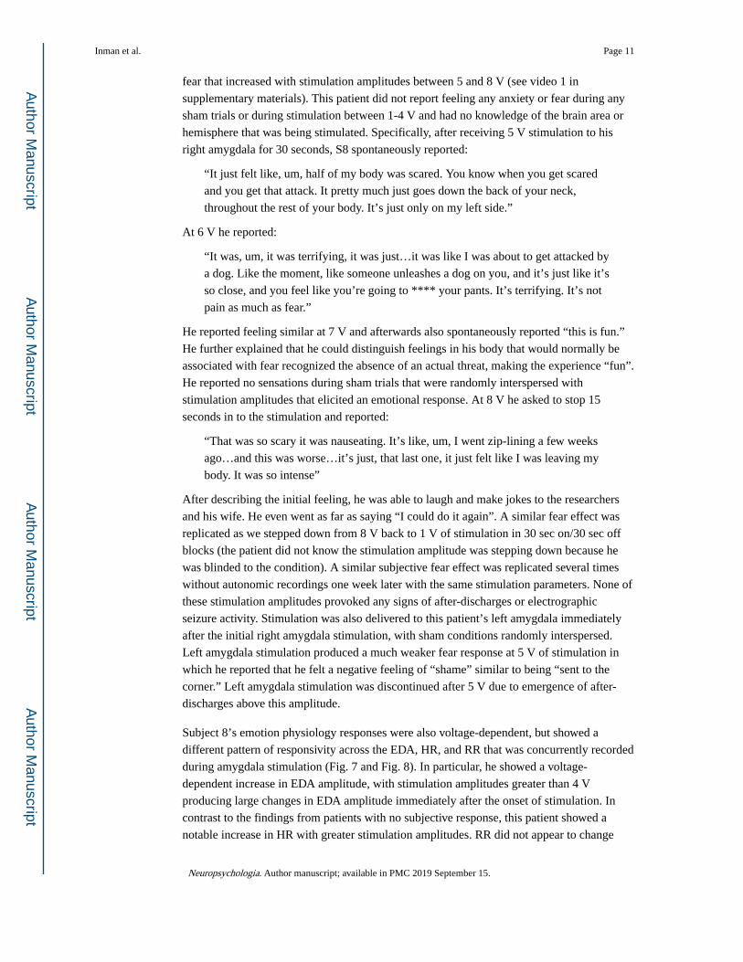
fear that increased with stimulation amplitudes between 5 and 8 V (see video 1 in
supplementary materials). This patient did not report feeling any anxiety or fear during any
sham trials or during stimulation between 1-4 V and had no knowledge of the brain area or
hemisphere that was being stimulated. Specifically, after receiving 5 V stimulation to his
right amygdala for 30 seconds, S8 spontaneously reported:
“It just felt like, um, half of my body was scared. You know when you get scared
and you get that attack. It pretty much just goes down the back of your neck,
throughout the rest of your body. It’s just only on my left side.”
At 6 V he reported:
“It was, um, it was terrifying, it was just…it was like I was about to get attacked by
a dog. Like the moment, like someone unleashes a dog on you, and it’s just like it’s
so close, and you feel like you’re going to **** your pants. It’s terrifying. It’s not
pain as much as fear.”
He reported feeling similar at 7 V and afterwards also spontaneously reported “this is fun.”
He further explained that he could distinguish feelings in his body that would normally be
associated with fear recognized the absence of an actual threat, making the experience “fun”.
He reported no sensations during sham trials that were randomly interspersed with
stimulation amplitudes that elicited an emotional response. At 8 V he asked to stop 15
seconds in to the stimulation and reported:
“That was so scary it was nauseating. It’s like, um, I went zip-lining a few weeks
ago…and this was worse…it’s just, that last one, it just felt like I was leaving my
body. It was so intense”
After describing the initial feeling, he was able to laugh and make jokes to the researchers
and his wife. He even went as far as saying “I could do it again”. A similar fear effect was
replicated as we stepped down from 8 V back to 1 V of stimulation in 30 sec on/30 sec off
blocks (the patient did not know the stimulation amplitude was stepping down because he
was blinded to the condition). A similar subjective fear effect was replicated several times
without autonomic recordings one week later with the same stimulation parameters. None of
these stimulation amplitudes provoked any signs of after-discharges or electrographic
seizure activity. Stimulation was also delivered to this patient’s left amygdala immediately
after the initial right amygdala stimulation, with sham conditions randomly interspersed.
Left amygdala stimulation produced a much weaker fear response at 5 V of stimulation in
which he reported that he felt a negative feeling of “shame” similar to being “sent to the
corner.” Left amygdala stimulation was discontinued after 5 V due to emergence of after-
discharges above this amplitude.
Subject 8’s emotion physiology responses were also voltage-dependent, but showed a
different pattern of responsivity across the EDA, HR, and RR that was concurrently recorded
during amygdala stimulation (Fig. 7 and Fig. 8). In particular, he showed a voltage-
dependent increase in EDA amplitude, with stimulation amplitudes greater than 4 V
producing large changes in EDA amplitude immediately after the onset of stimulation. In
contrast to the findings from patients with no subjective response, this patient showed a
notable increase in HR with greater stimulation amplitudes. RR did not appear to change
Inman et al. Page 11
Neuropsychologia. Author manuscript; available in PMC 2019 September 15.
Author M
anuscriptA
uthor Manuscript
Author M
anuscriptA
uthor Manuscript
significantly during stimulation trials. As in all of the other patients, sham trials and control
location stimulation did not produce any significant change in EDA, HR, or RR.
Discussion
The current study’s examination of the effects of human amygdala stimulation on
physiological and subjective emotional responses yielded three primary findings. First, we
found that amygdala stimulation strongly modulated autonomic activity without eliciting
concurrent subjective emotional responses in most patients. Second, we found that these
effects were specific to the amygdala relative to lateral temporal lobe stimulation, indicating
that the effects were not a general effect of brain or temporal lobe stimulation. Further, we
found that the effects of amygdala stimulation were immediate and dose-dependent:
stepwise increases in voltage elicited linear, dose-dependent increases in EDA amplitude and
decreases in HR within seconds of the stimulation onset. Third, we found that only in a
minority of patients, amygdala stimulation provoked subjective emotional responses. In one
patient, these subjective emotional responses were strongly negative in valence whereas the
other patient’s responses were primarily negative with a few positively-valenced responses.
In our most extensively documented patient with subjective emotional responses, fear
responses were accompanied by dose-dependent increases in heart rate rather than
decreases, which was a markedly different pattern of physiological response than what was
observed in the patients who did not show subjective emotional responses. The fact that
amygdala stimulation elicited physiological changes in the absence of after-discharges or
seizure-like activity suggests that acute amygdala stimulation may be used carefully in
humans to study autonomic and behavioral-physiological responses without confounding
effects of seizures.
A key novel finding of the current study was that amygdala stimulation can elicit substantial
changes in autonomic physiology without necessarily eliciting concurrent subjective
emotional responses. Indeed, although the majority of patients (7 of 9) reported no
subjective emotional responses when queried, relatively large stimulation-induced changes
in SCR and HR were still observed in each of these patients. These findings strongly suggest
that stimulation-induced physiological changes are not a consequence of concurrent
subjective emotional responses (Lanteaume et al., 2007), and that they are dissociable from
subjective responses. These results also highlight the importance of measuring both
physiological and subjective emotional responses to brain stimulation to fully assess
responses in different domains of emotional response. These findings also raise the question
of why these patients did not report any subjective emotional responses despite having
substantial physiological responses. One possibility is that the patients were either unaware
of their physiological responses, perhaps because these responses were below the threshold
of interoceptive awareness, or because attention was not focused on interoceptive stimuli. It
seems unlikely that the patients were aware of these physiological responses, but interpreted
or appraised them as non-emotional (e.g., somatic) as no somatic changes were
spontaneously reported.
Notably, we did not observe significant changes in respiratory rate and observed no episodes
of outright apnea or respiratory arrest under our experimental conditions. It has long been
Inman et al. Page 12
Neuropsychologia. Author manuscript; available in PMC 2019 September 15.
Author M
anuscriptA
uthor Manuscript
Author M
anuscriptA
uthor Manuscript
observed that direct electrical stimulation in the region of the temporal pole, uncus,
amygdala, orbitofrontal region, and anterior insula can cause respiratory depression in
animals (Spencer, 1894; Kaada, 1951) and also an awake patient using 30-60 Hz sine wave
pulses at 1-4 V (Kaada and Jasper, 1952). In the latter study, respiratory arrest could still be
elicited by stimulating the insula and temporal lobe white matter, even after complete
resection of the ipsilateral medial temporal lobe including amygdala. A more recent study of
direct amygdala stimulation using depth electrodes and 50 Hz stimulation at high amplitudes
(15-20 V) likewise reported reproducible respiratory arrests in 3 human subjects (Dlouhy et
al., 2015). Our study, by contrast, examined autonomic changes induced by amygdala
stimulation in a larger sample size at lower, possibly more physiological, stimulation
amplitudes. We observed dose-response effects on HR and SCR, but not RR, at low
stimulation doses. Further studies are required, but it seems unlikely that the amygdala
represents an “apnea center”. High amplitude stimulation of the amygdala and nearby
structures (possibly spreading to hypothalamus and brainstem, which are rarely directly
stimulated in humans) may engage a wider integrated autonomic/visceral emotional motor
outflow network.
In previous studies of amygdala stimulation effects, this relationship between physiological
and subjective emotional responses was either not assessed or was assessed in a way that
precluded valid statistical analysis. In the one study that did examine this relationship,
statistical evidence bearing directly on this issue was not provided (Lanteaume et al., 2007).
Specifically, although the frequency with which amygdala stimulation elicited a significant
SCR with vs. without subjective responses was assessed (i.e., 100% of subjective response
trials and 43% or 12/28 trials with no subjective response), this comparison was not tested
statistically, possibly because the small number of total stimulations that did not elicit a
subjective emotional response (28 stimulations) precluded valid statistical comparisons.
Moreover, the mean SCR magnitude reported for these 28 stimulation trials in was close to
zero, with error bars indicating the standard error of the mean overlapped with zero (Fig. 4
in Lanteaume et al., 2007), consistent with the interpretation that the mean SCRs for
stimulation trials that did not elicit subjective responses did not differ significantly from zero
(the relevant statistical test was not reported). Thus, this prior study did not provide evidence
that significant SCR responses to amygdala stimulation can occur in the absence of
subjective emotional responses (Lanteaume et al., 2007).
Another important finding of the current study was that the stimulation-induced
physiological changes were dose-dependent and were associated specifically with amygdala
stimulation and not observed with either sham stimulation or stimulation of the positive
control site in the lateral temporal cortex. Including a control stimulation site that was
consistent across patients in the current study allowed us to more conclusively establish the
amygdala as the proximal location of the stimulation effects, in contrast to previous studies
which did not include a control stimulation site (e.g., Lanteaume et al., 2007), in which
general effects of temporal lobe stimulation could not be ruled out. Although there was inter-
subject variability in response patterns, testing responses across multiple stimulation voltage
levels also allowed the shape of the dose-dependent response profile to be assessed for the
first time, confirming that the relationship between stimulation voltage and ANS changes
was approximately linear.
Inman et al. Page 13
Neuropsychologia. Author manuscript; available in PMC 2019 September 15.
Author M
anuscriptA
uthor Manuscript
Author M
anuscriptA
uthor Manuscript
In line with most prior studies of amygdala stimulation in humans, we found that subjective
emotional responses were relatively infrequent, observed in only 2 of the 9 patients studied
at relatively high-doses of stimulation (> 5 V; Halgren et al., 1978; Halgren et al., 1992;
Mangina and Beuzeron-Mangina, 1996; Meletti et al., 2006). Though infrequent, these
subjective emotional responses could be highly intense, particularly in the case of subject 8,
who reported intense feelings of fear and anxiety. In the two patients who reported
subjective emotional responses, the valence of their subjective response was most often
affectively negative and most commonly was reported as being experienced as fear or
anxiety, though a few more ambiguous positive and negative emotions were also reported.
These findings parallel most previous amygdala stimulation studies in that reported
subjective emotional responses have been typically predominantly aversive, with fear and
anxiety the most commonly reported emotions (Meletti et al., 2006; Smith et al., 2006;
Lanteume et al., 2007). Notably, we elicited negatively-valenced emotional responses from
both left (“guilt”) and right (“fear”) amygdala stimulations, with the left possibly being the
less intense subjectively negative experience for the patient. A few studies have reported
positive subjective emotional responses following left-hemisphere amygdala stimulation
(Meletti et al., 2006; Lanteume et al., 2007) and one case study (Bijanki et al., 2014)
reported a significant positive response with high-amplitude, intermittent stimulation (15 V;
50 Hz; 5 s On, 5 s Off) to the right amygdala in a patient with severe comorbid depression.
Bilateral stimulation at 10 V in subject 9 was the only instance in which amygdala
stimulation in the present study elicited a positive ‘joyful’, but ‘weird’ response, suggesting
that perhaps activation of a broader emotion-related network connected to the amygdala
using higher-amplitude stimulation is needed to elicit subjectively experienced positive
emotions.
Future studies should use more sensitive measures of emotional experience, such as affective
bias tasks (Bijanki et al., 2014), to supplement the subjective report of the patients. Finally,
all subjective emotional responses were transient, subsiding after the offset of stimulation
and subjects often reflected on the experience as ‘interesting’, ‘weird’, or ‘fun’, even in the
case of stimulation-induced fearful or anxious emotions. In addition, subjects reported that
their emotional responses did not seem to be triggered by their immediate environment, such
as stimuli in the room. Future studies should examine the extent to which amygdala
stimulation, both below and above thresholds for subjective awareness, modulates emotional
responses to external stimuli and contexts.
The subjective emotional responses in patient 8 were accompanied by dose-dependent
increases rather than decreases in heart rate, a markedly different pattern of physiological
response than what was observed in the patients who did not experience subjective
emotional responses during amygdala stimulation. It is difficult to know which specific
factors may account for the differences in subject 8’s response to amygdala stimulation.
Speculatively, the location of his right amygdala stimulation appears closer to the central
nucleus than all but one other patient (subject 6, in whom a subjective response was absent),
potentially resulting in stronger stimulation of the central nucleus, which has direct efferent
connections to subcortical structures controlling the ANS, such as the hypothalamus
(Critchley & Harrison, 2013). However, the factors determining whether subjective
Inman et al. Page 14
Neuropsychologia. Author manuscript; available in PMC 2019 September 15.
Author M
anuscriptA
uthor Manuscript
Author M
anuscriptA
uthor Manuscript
emotional responses are observed or not will require further study to provide a more definite
answer to this question.
What implications do these findings have for current debates about the neural representation
of emotion in the human brain? With regard to dimensional and discrete basic emotion
theories, the current results indicate that amygdala stimulation elicits responses that have
both dimensional and discrete aspects. The robust increases in SCR, a well-established
physiological measure of sympathetic ANS activation, are characteristic of a sympathetic
arousal response and fit with a large body of research implicating the amygdala in emotional
arousal processes (Adolphs, 2010; Anderson, 2003; Lang & Bradley, 2010; Critchley, 2002;
Critchley, 2005). The pattern of SCR increases and heart rate decreases we observed is
consistent with a defensive cascade response characteristic of responses to negatively-
valenced stimuli (Bradley and Lang, 2000a; Lang, 1995; Lang et al., 1997). However, this
response pattern is also generally consistent with the known functional connectivity of the
amygdala to regions mediating physiological responses and therefore does not
unambiguously reflect a negatively-valenced emotional response. In contrast, the observed
subjective emotional responses were primarily negative in valence, consistent with prior
literature that suggests that the amygdala has a preferential (but not exclusive) role in
mediating negatively-valenced emotion (Anderson & Adolphs, 2014; Hamann, 2012; Zald,
2003).
Paralleling prior reports, the observed subjective emotional responses were predominantly
self-reported as being fear or anxiety, rather than other basic emotions such as disgust, anger,
sadness, or happiness. Subject 9’s responses were not clearly related to fear or anxiety, but
rather an unpleasant feeling of unfamiliarity and occasionally a ‘weird joyful’ feeling. These
subjective responses suggest that amygdala stimulation elicits a response profile that is
predominantly biased towards eliciting a specific basic emotion state (fear and anxiety) but
is not exclusively specialized for fear or anxiety. This response profile is consistent with
other evidence from neuroimaging and human lesion studies that suggest that processing of
basic emotions (and emotional dimensions such as arousal and valence) is preferentially
associated with some brain regions (such as the amygdala) more than other regions.
Individual brain regions typically participate in processing multiple emotions rather than
being dedicated to processing individual basic emotions (Anderson & Adolphs, 2014;
Hamann, 2012; Pessoa, 2010). That is, the subjective experience of emotion appears to be an
emergent property of complex brain networks rather than a product of activity within a
single brain structure.
This study has additional important implications for the role of the amygdala in the
modulation of both emotion physiology and emotional experience. By measuring several
psychophysiological measures of ANS activity concurrently with amygdala stimulation, this
study provides novel evidence that amygdala stimulation produces physiological responses
predicted by theoretical views that propose that aversive stimuli and threats trigger a
defensive motivation response and an ensuing cascade of physiological responses that
change with the imminence and severity of threat (Bradley et al., 2001). Defensive
motivation responses are characterized as a series of autonomic and somatic reflexive
behaviors that prepare an organism for defensive behavior and facilitate processing of the
Inman et al. Page 15
Neuropsychologia. Author manuscript; available in PMC 2019 September 15.
Author M
anuscriptA
uthor Manuscript
Author M
anuscriptA
uthor Manuscript
threat context (Bradley et al., 2001). These defensive motivation responses are mediated by
projections from the basolateral nuclei of the amygdala (BLA), the most frequent sub-
nuclear amygdala location in the present study’s stimulation electrodes (Fig. 1; Critchley &
Harrison, 2013). Responses initiated by activation of projections from the BLA are both
behavioral and physiological. Behavioral responses to BLA activation include both freezing
and active flight (Fanselow, 1994). Previously reported physiological responses to BLA
activation include acute increases in electrodermal activity, bradycardia or slowing of the
heart rate (Kapp, Frysinger, Gallagher, & Haselton, 1979; Kapp et al., 1982), increased
blood pressure (LeDoux, 1990), and potentiation of the startle response (Davis, 2000).
In the current study we found that, within the range of parameters tested, increasing
amplitudes of amygdala stimulation reliably increased electrodermal activity, significantly
slowed heart rate, and was associated with marginal and non-significant decreases in
respiration rate. The defensive cascade model (Bradley and Lang, 2000a; Lang, 1995; Lang
et al., 1997) suggests that this pattern of increased skin conductance and heart rate
deceleration reflects an orienting response, in which perceptual processing is facilitated in
the early stages of defense upon exposure to a threat, but prior to overt action. This pattern
of coactivation of sympathetic and parasympathetic systems (Cacioppo & Berntson, 1994;
Cacioppo, Gardner, & Berntson, 1999) can be provoked by the perception of unpleasant
pictures in humans (Lang et al., 1997). These findings suggest that direct electrical
stimulation to the amygdala may provoke a state of defensive readiness (i.e. orienting
response) in the human autonomic nervous system even in the absence of subjective
emotional responses.
In infrequent cases, higher amplitudes of amygdala stimulation (>4 volts) produced
subjective emotional responses consistent with negative defensive behavioral responses
typically elicited by exposure to threat, such as fear and anxiety. Physiological responses
during amygdala stimulation trials that evoked an emotional response were more indicative
of later stages of the defensive motivation cascade during which electrodermal activity
increases even further and heart rate transitions from deceleration to acceleration. Notably,
these overt negative fear responses and their concurrent physiological responses were
consistently provoked immediately after the onset of amygdala stimulation greater than 5 V.
Specifically, the patient’s skin conductance amplitude and heart rate acutely increased with
stimulation amplitudes that elicited an emotional response. In addition, initially after
receiving lower amplitudes of stimulation in a blinded fashion to the right amygdala, 5 V of
stimulation prompted the patient to spontaneously report that the left side of his body “felt
scared”. This finding suggests that there may have been perceptible contralateral autonomic
effects of right hemisphere amygdala stimulation on the left side of the patient’s body.
During subsequent testing a week later, these effects were replicated, but no evidence of
specific lateralized physiological changes was detected (i.e., piloerection only on the left
extremities could not be observed). Consistent with previous studies (Smith et al., 2006;
Lanteaume et al., 2007), this patient also had a much more prominent fear response with
right amygdala stimulation than left amygdala stimulation. In addition, higher amplitudes of
right amygdala stimulation produced an increase in heart rate, while similar amplitudes of
left amygdala stimulation produced a deceleration in heart rate, consistent with the responses
seen in patients that had no subjective emotional experience. Taken together, these findings
Inman et al. Page 16
Neuropsychologia. Author manuscript; available in PMC 2019 September 15.
Author M
anuscriptA
uthor Manuscript
Author M
anuscriptA
uthor Manuscript
suggest that direct electrical amygdala stimulation can provoke a subjective fear or anxiety
response at higher amplitudes of stimulation and drive the ANS to more closely resemble an
animal in an overt defensive state.
The current study has a number of limitations. First, as in any study examining the effects of
direct electrical stimulation in patients undergoing intracranial monitoring, our study
necessarily consisted of patients with intractable epilepsy, rather than neurotypical patients.
Second, given the vast number of possible combinations of stimulation amplitude, duration,
frequency, location, and hemisphere, some parameter combinations were not adequately
sampled here (e.g., bilateral stimulation, longer durations, and a wider range of frequencies).
Further, some patients only received a single trial for higher voltage stimulation conditions
due to time constraints. By automating stimulation delivery and adjusting stimulation
durations, future studies may be able to more efficiently sample a broader range of
stimulation parameter combinations across multiple instances of each stimulation condition.
In addition, stimulation of the amygdala for longer than the 30 second stimulation blocks
used in the current study may reveal different patterns of change in measures of autonomic
physiology and subjective emotional responses at longer time scales. Future studies should
stimulate for longer durations to determine whether prolonged amygdala stimulation at
higher amplitudes produces more frequent subjective emotional responses and examine the
modulatory effect of amygdala stimulation on the subjective and physiological response to
emotionally evocative stimuli. Further, the stimulator in the present study delivered constant
voltage rather than constant current. However, we measured impedances across multiple
contacts in electrode arrays in a patient to be relatively consistent, and thus calculated that
voltages of 1-12 V correspond to a current range of ~1-15 mA. Importantly, although
impedance (and therefore current) could vary modestly at different contacts, the impedance
at a given contact is expected to remain constant across the experimental session, allowing
dose-response assessments of stimulation effects at that contact in a subject to be validly
recorded. Future extensions of the present work should examine the psychophysiological
changes associated with current-controlled amygdala stimulation to ensure full control over
stimulation amplitude, especially among patients. Finally, our method of assessing
subjective emotional responses may not have been sufficiently sensitive to exhaustively
probe whether patients were experiencing subtle changes in emotional experience. For
instance, although we asked patients to report any change in emotional experience, they may
have elected to not report subtle or transient emotional responses. Future studies will also be
aided by the use of a double-blind design, above and beyond the present study’s patient-
blinded design, when examining the effects of stimulation on physiology and emotional
experience.
The present study suggests several fruitful future directions to pursue in expanding
understanding of the amygdala’s causal role in modulating emotional experience, autonomic
physiology, and activity in other functionally connected brain regions. First, future studies
should simultaneously measure other informative indices of ANS activity, such as blood
pressure, heart rate variability, pupillometry, and cardiac output. Additional measures of
ANS activity can potentially provide a more complete picture of the sympathetic and
parasympathetic influences of direct electrical stimulation to the human amygdala. Second,
because prior studies suggest that the emotional responses elicited by stimulation to the
Inman et al. Page 17
Neuropsychologia. Author manuscript; available in PMC 2019 September 15.
Author M
anuscriptA
uthor Manuscript
Author M
anuscriptA
uthor Manuscript
amygdala region should depend on the location and volume of tissue activated (including
both grey and white matter structures), future studies should further investigate the
physiological and subjective effects of precisely stimulating each of the major amygdala
nuclei. More precise electrical field modeling of the volume of tissue activated in nucleated
structures will also be needed to fully examine this issue (Butson et al., 2007). In addition,
future studies should use diffusion tensor imaging on patients to examine the spatial
distribution of electrophysiological effects of stimulation in regions connected to the
amygdala (Riva-Posse et al., 2014). Mapping the precise location of each stimulation
electrode and their volume of tissue activated based on stimulation dose relative to subnuclei
of the amygdala and specific white matter tracts may help to explain inter-subject variability
in dose responses in future studies. Further, future studies may be able to test the
physiological and neural effects of acute amygdala stimulation using concurrent measures of
local field potentials or functional MRI Finally, although lower stimulation amplitudes did
not elicit physiological responses in the present study, there is evidence that at even lower
stimulation amplitudes (<2 V), amygdala stimulation can have beneficial effects on
cognitive processes such as memory (Inman et al., 2017). Future studies should use more
sensitive measures of neural and autonomic processing to examine the potential impact of
lower stimulation amplitudes on central and peripheral nervous system activity.
In conclusion, the current study found that amygdala stimulation in humans can have a
strong, dose-dependent impact upon ANS activity in the absence of concurrent subjective
emotional responses. When subjective emotional responses elicited by amygdala stimulation
were reported, they were primarily negative in valence, and were most commonly described
as fear or anxiety, consistent with a response profile for the amygdala that preferentially but
not exclusively involves fear and anxiety. These results parallel previous stimulation findings
with animals and humans and are consistent with responses predicted by theoretical views
positing a defensive cascade of physiological responses to aversive situations. In general,
stimulation-induced amygdala responses further validate the proposal that the amygdala is
involved in multiple emotions but has a preferential involvement in processing emotional
arousal as well as fear and anxiety. Further investigation of the effects of direct electrical
stimulation to the human amygdala should consider the factors determining whether
subjective emotional responses are elicited, and the factors that account for the variable
nature of such responses. The results of the current study provide novel evidence that the
human amygdala plays a causal role in the modulation of both emotion physiology and
emotional experience.
Supplementary Material
Refer to Web version on PubMed Central for supplementary material.
Acknowledgments
We would like to thank the patients, physicians, and staff of the Emory University Hospital Epilepsy Monitoring Unit for their contributions to this project. We also thank Nigel Pedersen for helpful conversation and providing useful historical brain stimulation context. KRB was supported in part by career development awards from the American Foundation for Suicide Prevention and the NIH (KL2TR000455). JTW was supported in part by career development awards from the Sleep Research Society Foundation and the Neurosurgery Research Education Fund.
Inman et al. Page 18
Neuropsychologia. Author manuscript; available in PMC 2019 September 15.
Author M
anuscriptA
uthor Manuscript
Author M
anuscriptA
uthor Manuscript
References
Adolphs R, Tranel D, Damasio H, Damasio A. Impaired recognition of emotion in facial expressions following bilateral damage to the human amygdala. Nature. 1994; 372:669–72. [PubMed: 7990957]
Adolphs R, Tranel D, Damasio H, Damasio AR. Fear and the human amygdala. Journal of Neuroscience. 1995; 15(9):5879–5891. [PubMed: 7666173]
Adolphs R, Tranel D, Hamann S, Young AW, Calder AJ, et al. Recognition of facial emotion in nine individuals with amygdala damage. Neuropsychologia. 1999; 37:1111–1117. [PubMed: 10509833]
Adolphs R. What does the amygdala contribute to social cognition? Annals of the New York Academy of Sciences. 2010; 1191:42–61. DOI: 10.1111/j.1749-6632.2010.05445 [PubMed: 20392275]
Adolphs R. Human Lesion Studies in the 21st Century. Neuron. 2016; 90(6):1151–1153. DOI: 10.1016/j.neuron.2016.05.014 [PubMed: 27311080]
Anderson AK, Christoff K, Stappen I, Panitz D, Ghahremani DG, Glover G, Sobel N. Dissociated neural representations of intensity and valence in human olfaction. Nature Neuroscience. 2003; 6(2):196. [PubMed: 12536208]
Anderson DJ, Adolphs R. A Framework for Studying Emotions across Species. Cell. 2014; 157(1):187–200. DOI: 10.1016/j.cell.2014.03.003 [PubMed: 24679535]
Asahina M, Suzuki A, Mori M, Kanesaka T, Hattori T. Emotional sweating response in a patient with bilateral amygdala damage. International Journal of Psychophysiology. 2003; 47(1):87–93. DOI: 10.1016/S0167-8760(02)00123-X [PubMed: 12543449]
Barrett LF, Satpute AB. Large-scale brain networks in affective and social neuroscience: towards an integrative functional architecture of the brain. Current Opinion in Neurobiology. 2013; 23(3):361–372. DOI: 10.1016/j.conb.2012.12.012 [PubMed: 23352202]
Barrett LF, Mesquita B, Ochsner KN, Gross JJ. The Experience of Emotion. Annual Review of Psychology. 2007; 55(1):373–403. DOI: 10.1146/annurev.psych.58.110405.085709
Bijanki KR, Kovach CK, McCormick LM, Kawasaki H, Dlouhy BJ, Feinstein J, Howard MA III. Case Report: Stimulation of the Right Amygdala Induces Transient Changes in Affective Bias. Brain Stimulation. 2014; 7(5):690–693. DOI: 10.1016/j.brs.2014.05.005 [PubMed: 24972588]
Bookstein FL. Principal warps: Thin-plate splines and the decomposition of deformations. Pattern Analysis and Machine Intelligence, IEEE Transactions on. 1989; 11:567–585.
Boucsein W, Fowles DC, Grimnes S, Ben-Shakhar G, Roth WT, Dawson ME, Filion DL. Publication recommendations for electrodermal measurements. Psychophysiology. 2012; 49:1017–1034. [PubMed: 22680988]
Bradley MM, Lang PJ. Emotion and motivation. Handbook of psychophysiology. 2000a; 2:602–642.
Bradley MM, Lang PJ. Measuring emotion: Behavior, feeling, and physiology. Cognitive neuroscience of emotion. 2000b; 25:49–59.
Bradley MM, Codispoti M, Cuthbert BN, Lang PJ. Emotion and motivation I: Defensive and appetitive reactions in picture processing. Emotion. 2001; 1(3):276–298. DOI: 10.1037/A528-3542.L3.276 [PubMed: 12934687]
Burgdorf J, Knutson B, Panksepp J. Anticipation of rewarding electrical brain stimulation evokes ultrasonic vocalization in rats. Behavioral Neuroscience. 2000; 114(2):320–327. DOI: 10.1037/0735-7044.114.2.320 [PubMed: 10832793]
Burgdorf J, Panksepp J. The neurobiology of positive emotions. Neuroscience & Biobehavioral Reviews. 2006; 30(2):173–187. DOI: 10.1016/j.neubiorev.2005.06.001 [PubMed: 16099508]
Butson CR, Cooper SE, Henderson JM, McIntyre CC. Patient-specific analysis of the volume of tissue activated during deep brain stimulation. Neuroimage. 2007; 34(2):661–670. DOI: 10.1016/j.neuroimage.2006.09.034 [PubMed: 17113789]
Cacioppo JT, Berntson GG. Relationship between attitudes and evaluative space: A critical review, with emphasis on the separability of positive and negative substrates. Psychological Bulletin. 1994; 115(3):401–423. DOI: 10.1037/0033-2909.115.3.401
Cacioppo JT, Gardner WL, Berntson GG. The affect system has parallel and integrative processing components: Form follows function. Journal of Personality and Social Psychology. 1999; 76:839–855.
Inman et al. Page 19
Neuropsychologia. Author manuscript; available in PMC 2019 September 15.
Author M
anuscriptA
uthor Manuscript
Author M
anuscriptA
uthor Manuscript
Critchley HD. Electrodermal Responses: What Happens in the Brain. The Neuroscientist. 2002; 5(2):132–142. DOI: 10.1177/107385840200800209
Critchley HD. Neural mechanisms of autonomic, affective, and cognitive integration. The Journal of Comparative Neurology. 2005; 493(1):154–166. DOI: 10.1002/cne.20749 [PubMed: 16254997]
Critchley HD, Harrison NA. Visceral influences on brain and behavior. Neuron. 2013; 77(4):624–638. [PubMed: 23439117]
Davis M. The role of the amygdala in conditioned and unconditioned fear and anxiety. In: Aggleton JP, editorThe amygdala. Vol. 2. Oxford, England: Oxford University Press; 2000. 213–287.
Davis M, Whalen PJ. The amygdala: vigilance and emotion. Molecular Psychiatry; New York. 2001; 6(1):13–34. DOI: 10.1038/sj.mp.4000812
Dlouhy BJ, Gehlbach BK, Kreple CJ, Kawasaki H, Oya H, Buzza C, Richerson GB. Breathing Inhibited When Seizures Spread to the Amygdala and upon Amygdala Stimulation. Journal of Neuroscience. 2015; 35(28):10281–10289. DOI: 10.1523/JNEUROSCI.0888-15.2015 [PubMed: 26180203]
Duvernoy HM. The Human Hippocampus: Functional Anatomy, Vascularization and Serial Sections with MRI. Springer Science & Business Media; 2005.
Ekman P. An argument for basic emotions. Cognition and Emotion. 1992; 6(3-4):169–200. DOI: 10.1080/02699939208411068
Feinstein JS, Adolphs R, Damasio AR, Tranel D. The human amygdala and the induction and experience of fear. Current Biology. 2011; 21(1):34–38. DOI: 10.1016/j.cub.2010.11.042 [PubMed: 21167712]
Guillory SA, Bujarski KA. Exploring emotions using invasive methods: review of 60 years of human intracranial electrophysiology. Social Cognitive and Affective Neuroscience. 2014; 9(12):1880.doi: 10.1093/scan/nsu002 [PubMed: 24509492]
Hamann S. Cognitive and neural mechanisms of emotional memory. TICS. 2001; 5(9):394–400.
Hamann S. Mapping discrete and dimensional emotions onto the brain: controversies and consensus. TICS. 2012; 16(9):458–466. DOI: 10.1016/j.tics.2012.07.006
Halgren E, Walter RD, Cherlow DG, Crandall PH. Mental phenomena evoked by electrical stimulation of the human hippocampal formation and amygdala. Brain. 1978; 101:83–117. [PubMed: 638728]
Halgren E. The amygdala: Neurobiological aspects of emotion, memory, and mental dysfunction. New York, NY, US: Wiley-Liss; 1992. Emotional neurophysiology of the amygdala within the context of human cognition; 191–228.
Inman CS, Manns JR, Bijanki KR, Bass DI, Hamann S, Drane DL, Fasano R, Kovach CK, Gross RE, Willie JT. Direct electrical stimulation of the amygdala enhances declarative memory in humans. Proceedings of the National Academy of Sciences USA. 2017
Jenkinson M, Smith S. A global optimization method for robust affine registration of brain images. Medical image analysis. 2001; 5:143–156. [PubMed: 11516708]
Kaada BR. Stomato-motor, autonomic and electrocorticographic responses to electrical stimulation of rhinencephalic and other structures in primates, cat, and dog; a study of responses from the limbic, subcallosal, orbito-insular, piriform and temporal cortex, hippocampus-fornix and amygdala. Acta Physiologica Scandinavica Supplementum. 1951; 24(83):1–262.
Kaada BR, Jasper H. Respiratory responses to stimulation of temporal pole, insula, and hippocampal and limbic gyri in man. A M A Archives of Neurology and Psychiatry. 1952; 68(5):609–619. [PubMed: 12984874]
Lang PJ. The emotion probe: Studies of motivation and attention. American Psychologist. 1995; 50(5):372–385. DOI: 10.1037/0003-066X.50.5.372 [PubMed: 7762889]
Lang PJ, Bradley MM, Cuthbert BN. Motivated attention: Affect, activation, and action. In: Lang PJ, Simons RF, Balaban MT, editorsAttention and orienting: Sensory and motivational processes. Hillsdale, NJ: Erlbaum; 1997. 97–135.
Lang PJ, Bradley MM. Emotion and the motivational brain. Biological Psychology. 2010; 84:437–450. [PubMed: 19879918]
Lanteaume L, et al. Emotion induction after direct intracerebral stimulations of human amygdala. Cerebral Cortex. 2007; 17(6):1307–1313. [PubMed: 16880223]
Inman et al. Page 20
Neuropsychologia. Author manuscript; available in PMC 2019 September 15.
Author M
anuscriptA
uthor Manuscript
Author M
anuscriptA
uthor Manuscript
LeDoux JE. Information flow from sensation to emotion plasticity in the neural computation of stimulus value. In: Gabriel M, Moore J, editorsLearning and computational neuroscience: Foundations of adaptive networks. Cambridge, MA: Bradford Books/MIT Press; 1990. 3–52.
LeDoux JE. Emotion Circuits in the Brain. Annual Review of Neuroscience. 2000; 23(1):155–184. DOI: 10.1146/annurev.neuro.23.L155
LeDoux JE. Semantics, Surplus Meaning, and the Science of Fear. Trends in Cognitive Sciences. 2017; 21(5):303–306. DOI: 10.1016/j.tics.2017.02.004 [PubMed: 28318937]
Lindquist KA, Barrett LF. A functional architecture of the human brain: emerging insights from the science of emotion; Trends in Cognitive Sciences. 2012. 1–8.
Mai JK, Paxinos G, Voss T. Atlas of the Human Brain. 3rd. Academic Press; 2008.
Mangina CA, Beuzeron-Mangina JH. Direct electrical stimulation of specific human brain structures and bilateral electrodermal activity. International Journal of Psychophysiology. 1996; 22(1-2):1–8. DOI: 10.1016/0167-8760(96)00022-0 [PubMed: 8799763]
Mayberg HS, Lozano AM, Voon V, McNeely HE, Seminowicz D, Hamani C, Kennedy SH. Deep Brain Stimulation for Treatment-Resistant Depression. Neuron. 2005; 45(5):651–660. DOI: 10.1016/j.neuron.2005.02.014 [PubMed: 15748841]
Meletti S, et al. Emotions induced by intracerebral electrical stimulation of the temporal lobe. Epilepsia. 2006; 47.s5:47–51.
Oya H, Kawasaki H, Dahdaleh NS, Wemmie JA, Howard MA III. Stereotactic atlas-based depth electrode localization in the human amygdala. Stereotactic and functional neurosurgery. 2009; 87:219–228. [PubMed: 19556831]
Panksepp J. Toward a general psychobiological theory of emotions. Behavioral and Brain Sciences. 1982; 5(3):407–422. DOI: 10.1017/S0140525X00012759
Patenaude B, Smith SM, Kennedy DN, Jenkinson M. A Bayesian model of shape and appearance for subcortical brain segmentation. NeuroImage. 2011; 56:907–922. [PubMed: 21352927]
Pessoa L. On the relationship between emotion and cognition. Nature Reviews Neuroscience. 2008; 9(2):148–158. DOI: 10.1038/nrn2317 [PubMed: 18209732]
Pessoa L, Adolphs R. Emotion processing and the amygdala: from a “low road” to “many roads” of evaluating biological significance. Nature Reviews Neuroscience. 2010; 11(11):773–783. DOI: 10.1038/nrn2920 [PubMed: 20959860]
Pessoa L. Emotion and cognition and the amygdala: from “what is it?” to “what’s to be done? Neuropsychologia. 2010; 48(12):3416–3429. [PubMed: 20619280]
Riva-Posse P, Choi KS, Holtzheimer PE, McIntyre CC, Gross RE, Chaturvedi A, Mayberg HS. Defining Critical White Matter Pathways Mediating Successful Subcallosal Cingulate Deep Brain Stimulation for Treatment-Resistant Depression. Biological Psychiatry. 2014; 76(12):963–969. [PubMed: 24832866]
Rohr K, et al. Landmark-based elastic registration using approximating thin-plate splines. IEEE Transactions on medical imaging. 2001; 20(6):526–534. [PubMed: 11437112]
Rutishauser U, Mamelak AN, Adolphs R. The primate amygdala in social perception – insights from electrophysiological recordings and stimulation. Trends in Neurosciences. 2015; 38(5):295–306. DOI: 10.1016/j.tins.2015.03.001 [PubMed: 25847686]
Selimbeyoglu A, Parvizi J. Electrical stimulation of the human brain: perceptual and behavioral phenomena reported in the old and new literature. Frontiers in Human Neuroscience. 2010; 4doi: 10.3389/fnhum.2010.00046
Smith JR, Lee GP, Fountas K, King DW, Jenkins PD. Intracranial stimulation study of lateralization of affect. Epilepsy & Behavior. 2006; 8(3):534–541. DOI: 10.1016/j.yebeh.2005.12.014 [PubMed: 16546450]
Spencer WG. The Effect Produced upon Respiration by Faradic Excitation of the Cerebrum in the Monkey, Dog, Cat, and Rabbit. Philosophical Transactions of the Royal Society of London B. 1894; 185:609–657.
Vytal K, Hamann S. Neuroimaging support for discrete neural correlates of basic emotions: a voxel-based meta-analysis. Journal of Cognitive Neuroscience. 2010; 22(12):2864–2885. DOI: 10.1162/jocn.2009.21366 [PubMed: 19929758]
Inman et al. Page 21
Neuropsychologia. Author manuscript; available in PMC 2019 September 15.
Author M
anuscriptA
uthor Manuscript
Author M
anuscriptA
uthor Manuscript
Zald DH. The human amygdala and the emotional evaluation of sensory stimuli. Brain Research Reviews. 2003; 41:88–123. [PubMed: 12505650]
Inman et al. Page 22
Neuropsychologia. Author manuscript; available in PMC 2019 September 15.
Author M
anuscriptA
uthor Manuscript
Author M
anuscriptA
uthor Manuscript
Highlights
• Increasing amplitudes of amygdala stimulation elicited dose-dependent
increases in electrodermal activity and decreases in heart rate.
• In one patient, amygdala stimulation elicited subjective experiences of fear
and anxiety, accompanied by increased heart rate.
• Amygdala stimulation reliably elicits changes in autonomic activity in a dose-
dependent and safe manner, and elicits subjective emotional experiences only
infrequently.
Inman et al. Page 23
Neuropsychologia. Author manuscript; available in PMC 2019 September 15.
Author M
anuscriptA
uthor Manuscript
Author M
anuscriptA
uthor Manuscript
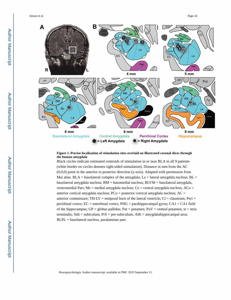
Figure 1. Precise localization of stimulation sites overlaid on illustrated coronal slices through the human amygdalaBlack circles indicate estimated centroids of stimulation in or near BLA in all 9 patients
(white border on circles denotes right-sided stimulation). Distance in mm from the AC
(0,0,0) point in the anterior to posterior direction (y-axis). Adapted with permission from
Mai atlas. BLA = basolateral complex of the amygdala; La = lateral amygdala nucleus; BL =
basolateral amygdala nucleus; BM = basomedial nucleus; BLVM = basolateral amygdala,
ventromedial Part; Me = medial amygdala nucleus; Ce = central amygdala nucleus; ACo =
anterior cortical amygdala nucleus; PCo = posterior cortical amygdala nucleus; AC =
anterior commissure; TH-LV = temporal horn of the lateral ventricle; Cl = claustrum; Peri =
perirhinal cortex; EC = entorhinal cortex; PHG = parahippocampal gyrus; CA1 = CA1 field
of the hippocampus; GP = globus pallidus; Put = putamen; PuV = ventral putamen; st = stria
terminalis; Sub = subiculum; PrS = pre-subiculum; AHi = amygdalahippocampal area;
BLPL = basolateral nucleus, paralaminar part.
Inman et al. Page 24
Neuropsychologia. Author manuscript; available in PMC 2019 September 15.
Author M
anuscriptA
uthor Manuscript
Author M
anuscriptA
uthor Manuscript
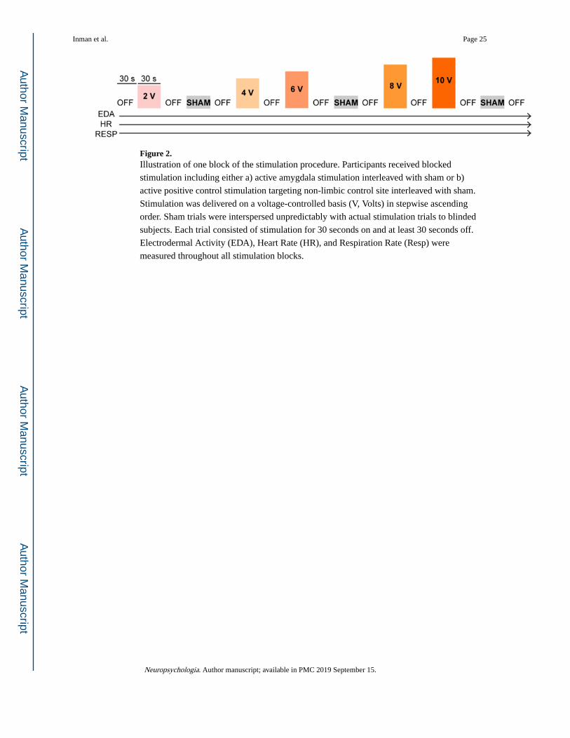
Figure 2. Illustration of one block of the stimulation procedure. Participants received blocked
stimulation including either a) active amygdala stimulation interleaved with sham or b)
active positive control stimulation targeting non-limbic control site interleaved with sham.
Stimulation was delivered on a voltage-controlled basis (V, Volts) in stepwise ascending
order. Sham trials were interspersed unpredictably with actual stimulation trials to blinded
subjects. Each trial consisted of stimulation for 30 seconds on and at least 30 seconds off.
Electrodermal Activity (EDA), Heart Rate (HR), and Respiration Rate (Resp) were
measured throughout all stimulation blocks.
Inman et al. Page 25
Neuropsychologia. Author manuscript; available in PMC 2019 September 15.
Author M
anuscriptA
uthor Manuscript
Author M
anuscriptA
uthor Manuscript
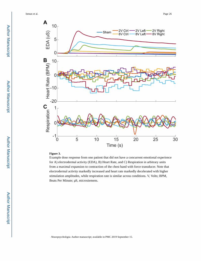
Figure 3. Example dose response from one patient that did not have a concurrent emotional experience
for A) electrodermal activity (EDA), B) Heart Rate, and C) Respiration in arbitrary units
from a maximal expansion to contraction of the chest band with force transducer. Note that
electrodermal activity markedly increased and heart rate markedly decelerated with higher
stimulation amplitudes, while respiration rate is similar across conditions. V, Volts; BPM,
Beats Per Minute; μS, microsiemens.
Inman et al. Page 26
Neuropsychologia. Author manuscript; available in PMC 2019 September 15.
Author M
anuscriptA
uthor Manuscript
Author M
anuscriptA
uthor Manuscript
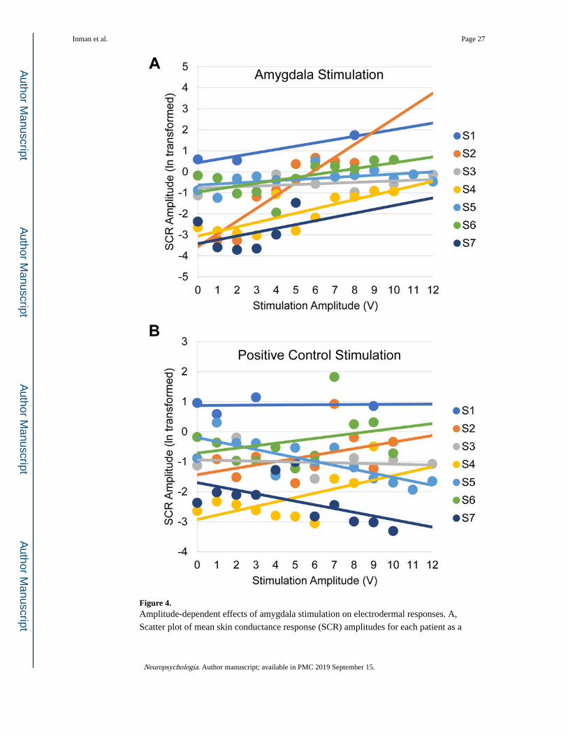
Figure 4. Amplitude-dependent effects of amygdala stimulation on electrodermal responses. A,
Scatter plot of mean skin conductance response (SCR) amplitudes for each patient as a
Inman et al. Page 27
Neuropsychologia. Author manuscript; available in PMC 2019 September 15.
Author M
anuscriptA
uthor Manuscript
Author M
anuscriptA
uthor Manuscript
function of amygdala stimulation voltage (dots) with a patient-wise linear trend line.
Stimulation-related changes in EDA is indicated by SCR amplitude. Note the consistency of
increases in SCR amplitude with increases in stimulation voltage. B, Scatter plot of mean
SCR amplitudes for each patient as a function of positive control (lateral temporal cortex)
stimulation voltage (dots) with a patient-wise linear trend line. Note the inconsistency of
changes in SCR amplitude with increases in stimulation voltage. SCR, Skin Conductance
Response; μS, microseimens.
Inman et al. Page 28
Neuropsychologia. Author manuscript; available in PMC 2019 September 15.
Author M
anuscriptA
uthor Manuscript
Author M
anuscriptA
uthor Manuscript

Figure 5. Amplitude-dependent effects of amygdala stimulation on change in heart rate. A, Scatter
plot of mean change in heart rate for each patient as a function of amygdala stimulation
voltage (dots) with a patient-wise linear trend line. Note the consistency of decreases in
Inman et al. Page 29
Neuropsychologia. Author manuscript; available in PMC 2019 September 15.
Author M
anuscriptA
uthor Manuscript
Author M
anuscriptA
uthor Manuscript
heart rate with increases in stimulation voltage. B, Scatter plot of mean change in heart rate
for each patient as a function of positive control (lateral temporal cortex) stimulation voltage
(dots) with a patient-wise linear trend line. Note the inconsistency of changes in heart rate
with increases in stimulation voltage. BPM, Beats Per Minute; V, Volts.
Inman et al. Page 30
Neuropsychologia. Author manuscript; available in PMC 2019 September 15.
Author M
anuscriptA
uthor Manuscript
Author M
anuscriptA
uthor Manuscript
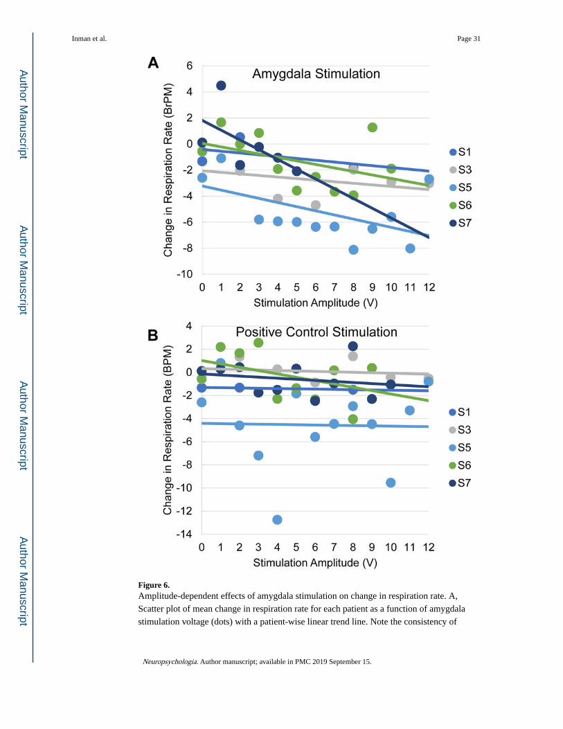
Figure 6. Amplitude-dependent effects of amygdala stimulation on change in respiration rate. A,
Scatter plot of mean change in respiration rate for each patient as a function of amygdala
stimulation voltage (dots) with a patient-wise linear trend line. Note the consistency of
Inman et al. Page 31
Neuropsychologia. Author manuscript; available in PMC 2019 September 15.
Author M
anuscriptA
uthor Manuscript
Author M
anuscriptA
uthor Manuscript
decreases in respiration rate with increases in stimulation voltage. B, Scatter plot of mean
change in respiration rate for each patient as a function of positive control (lateral temporal
cortex) stimulation voltage (dots) with a patient-wise linear trend line. Note the
inconsistency of changes in respiration rate with increases in stimulation voltage. BrPM,
Breaths Per Minute.
Inman et al. Page 32
Neuropsychologia. Author manuscript; available in PMC 2019 September 15.
Author M
anuscriptA
uthor Manuscript
Author M
anuscriptA
uthor Manuscript
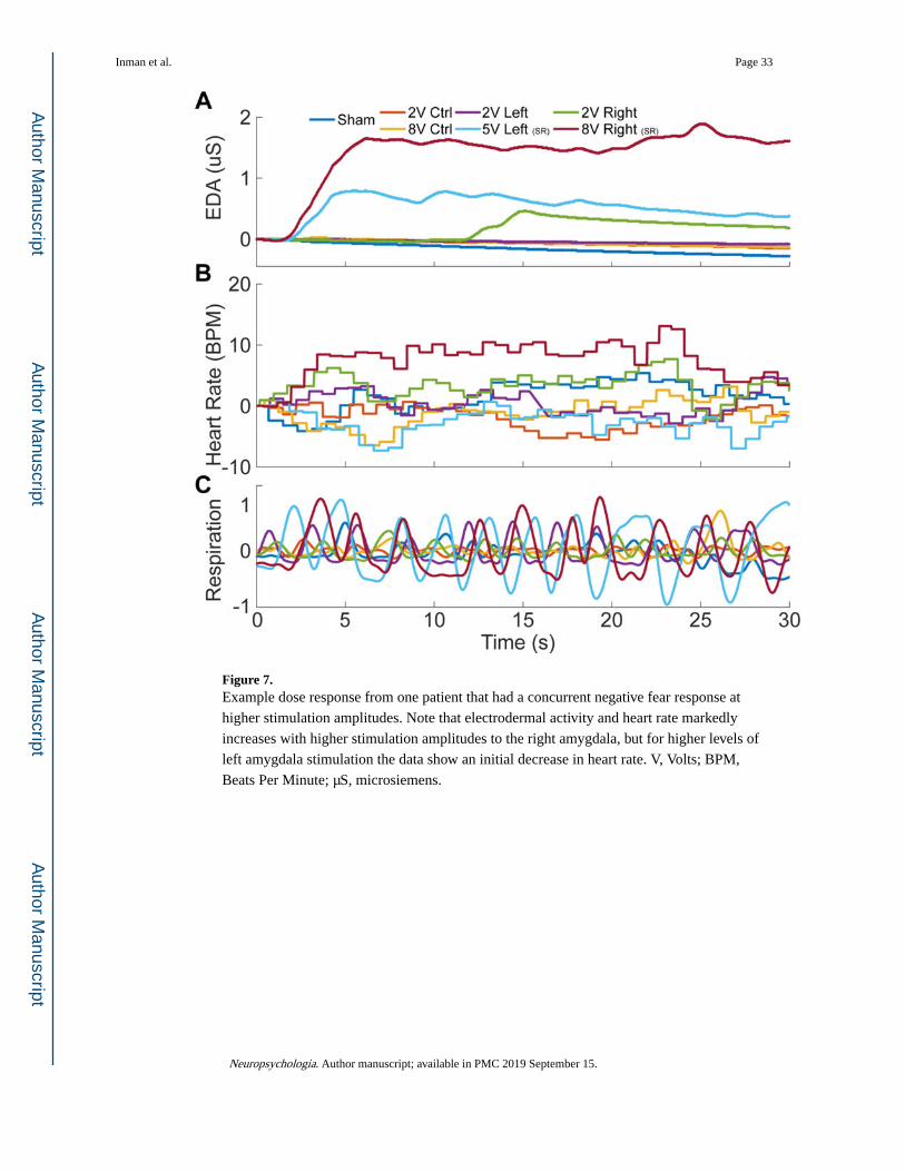
Figure 7. Example dose response from one patient that had a concurrent negative fear response at
higher stimulation amplitudes. Note that electrodermal activity and heart rate markedly
increases with higher stimulation amplitudes to the right amygdala, but for higher levels of
left amygdala stimulation the data show an initial decrease in heart rate. V, Volts; BPM,
Beats Per Minute; μS, microsiemens.
Inman et al. Page 33
Neuropsychologia. Author manuscript; available in PMC 2019 September 15.
Author M
anuscriptA
uthor Manuscript
Author M
anuscriptA
uthor Manuscript
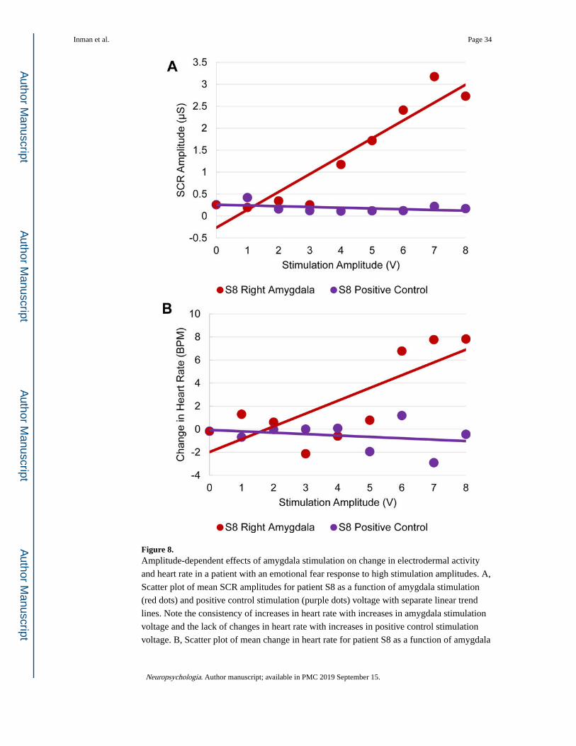
Figure 8. Amplitude-dependent effects of amygdala stimulation on change in electrodermal activity
and heart rate in a patient with an emotional fear response to high stimulation amplitudes. A,
Scatter plot of mean SCR amplitudes for patient S8 as a function of amygdala stimulation
(red dots) and positive control stimulation (purple dots) voltage with separate linear trend
lines. Note the consistency of increases in heart rate with increases in amygdala stimulation
voltage and the lack of changes in heart rate with increases in positive control stimulation
voltage. B, Scatter plot of mean change in heart rate for patient S8 as a function of amygdala
Inman et al. Page 34
Neuropsychologia. Author manuscript; available in PMC 2019 September 15.
Author M
anuscriptA
uthor Manuscript
Author M
anuscriptA
uthor Manuscript

stimulation (red dots) and positive control stimulation (purple dots) voltage with separate
linear trend lines. Note the consistency of decreases in heart rate with increases in amygdala
stimulation voltage and the lack of changes in heart rate with increases in positive control
stimulation voltage.
Inman et al. Page 35
Neuropsychologia. Author manuscript; available in PMC 2019 September 15.
Author M
anuscriptA
uthor Manuscript
Author M
anuscriptA
uthor Manuscript

Author M
anuscriptA
uthor Manuscript
Author M
anuscriptA
uthor Manuscript
Inman et al. Page 36
Tab
le 1
Dem
ogra
phic
s an
d E
pile
psy
Tabl
e. L
, Lef
t; R
, Rig
ht; S
z, S
eizu
re; M
, Mal
e; F
, Fem
ale.
IC
M, I
ntra
cran
ial M
onito
ring
; SL
AH
, Ste
reot
actic
Las
er
Am
ygda
lohi
ppoc
ampe
ctom
y; *
Subj
ect
Sex
Age
Stim
ulat
ion
Hem
isph
ere
Pri
or R
esec
tion
Pos
t-IC
M S
urge
ryO
utco
me
1F
36L
& R
Non
eFu
rthe
r IC
MM
ultif
ocal
Sz
2F
45L
Non
eR
SL
AH
Sz F
ree
3M
30L
R F
ront
al R
esec
tion
R F
ront
al L
aser
Abl
atio
nSz
Fre
e
4F
36L
R T
empo
ral L
obec
tom
yR
Inf
erio
-Tem
pora
l Res
ectio
nSz
Fre
e
5M
47R
Non
eL
Inf
erio
r Te
mpo
ro-P
arie
tal R
esec
tion
Not
Sz
Free
6F
23R
Non
eN
one
Mul
tifoc
al S
z
7M
44R
Non
eN
one
Sz F
ree
8M
26L
& R
R L
ater
al T
empo
ral A
stro
cyto
ma
R L
ater
al T
empo
ral L
aser
Abl
atio
nSz
Fre
e
9M
46R
Non
eR
Ant
erio
r Te
mpo
ral L
obec
tom
yN
ot S
z Fr
ee
Neuropsychologia. Author manuscript; available in PMC 2019 September 15.
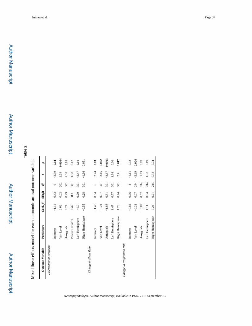
Author M
anuscriptA
uthor Manuscript
Author M
anuscriptA
uthor Manuscript
Inman et al. Page 37
Tab
le 2
Mix
ed li
near
eff
ects
mod
el f
or e
ach
auto
nom
ic a
rous
al o
utco
me
vari
able
.
Out
com
e V
aria
ble
Pre
dict
ors
Coe
f. β
SE(β
)df
tp
Ele
ctro
derm
al R
espo
nse
Inte
rcep
t−
1.12
0.43
6−
2.59
0.04
Vol
t Lev
el0.
060.
0230
13.
590.
0004
Am
ygda
la0.
740.
2930
12.
520.
01
Posi
tive
Con
trol
0.47
0.3
301
1.58
0.12
Lef
t Hem
isph
ere
−0.
70.
2930
1−
2.47
0.01
Rig
ht H
emis
pher
e−
0.55
0.28
301
−1.
960.
051
Cha
nge
in H
eart
Rat
e
Inte
rcep
t−
1.48
0.54
6−
2.74
0.03
Vol
t Lev
el−
0.24
0.07
301
−3.
150.
002
Am
ygda
la−
1.86
0.51
301
−3.
670.
0003
Lef
t Hem
isph
ere
1.47
0.77
301
1.91
0.06
Rig
ht H
emis
pher
e1.
790.
7430
12.
40.
017
Cha
nge
in R
espi
ratio
n R
ate
Inte
rcep
t−
0.84
0.76
4−
1.11
0.33
Vol
t Lev
el−
0.21
0.07
244
−2.
890.
004
Am
ygda
la−
0.89
0.52
244
−1.
730.
09
Lef
t Hem
isph
ere
1.11
0.84
244
1.32
0.19
Rig
ht H
emis
pher
e0.
240.
7124
40.
330.
74
Neuropsychologia. Author manuscript; available in PMC 2019 September 15.
Related Documents