Human Adipose-Derived Mesenchymal Stem Cells as a New Model of Spinal and Bulbar Muscular Atrophy Marta Dossena 1. , Gloria Bedini 1. , Paola Rusmini 2 , Elisa Giorgetti 2,3 , Alessandra Canazza 1 , Valentina Tosetti 1 , Ettore Salsano 4 , Anna Sagnelli 4 , Caterina Mariotti 5 , Cinzia Gellera 5 , Stefania Elena Navone 1 , Giovanni Marfia 1 , Giulio Alessandri 1 , Fabio Corsi 6 , Eugenio Agostino Parati 1 , Davide Pareyson "4 *, Angelo Poletti "2 * 1 Cellular Neurobiology Laboratory, Unit of Cerebrovascular Disease, Fondazione IRCCS Istituto Neurologico Carlo Besta, Milan, Italy, 2 Dipartimento di Scienze Farmacologiche e Biomolecolari, Centro Interdipartimentale sulle Malattie Neurodegenerative, Universita ` degli Studi di Milano, Milan, Italy, 3 Department of Pathology, University of Michigan, Ann Arbor, Michigan, 48109, United States of America, 4 Clinic of Central and Peripheral Degenerative Neuropathies Unit, Department of Clinical Neurosciences, Fondazione IRCCS Istituto Neurologico Carlo Besta, Milan, Italy, 5 Unit of Genetics of Neurodegenerative and Metabolic Diseases, Department of Diagnostic and Applied Technology, Fondazione IRCCS Istituto Neurologico Carlo Besta, Milan, Italy, 6 Surgery Division, Department of Clinical Sciences, University of Milan, ‘‘Luigi Sacco’’ Hospital, Milan, Italy Abstract Spinal and bulbar muscular atrophy (SBMA) or Kennedy’s disease is an X-linked CAG/polyglutamine expansion motoneuron disease, in which an elongated polyglutamine tract (polyQ) in the N-terminal androgen receptor (ARpolyQ) confers toxicity to this protein. Typical markers of SBMA disease are ARpolyQ intranuclear inclusions. These are generated after the ARpolyQ binds to its endogenous ligands, which promotes AR release from chaperones, activation and nuclear translocation, but also cell toxicity. The SBMA mouse models developed so far, and used in preclinical studies, all contain an expanded CAG repeat significantly longer than that of SBMA patients. Here, we propose the use of SBMA patients adipose-derived mesenchymal stem cells (MSCs) as a new human in vitro model to study ARpolyQ toxicity. These cells have the advantage to express only ARpolyQ, and not the wild type AR allele. Therefore, we isolated and characterized adipose-derived MSCs from three SBMA patients (ADSC from Kennedy’s patients, ADSCK) and three control volunteers (ADSCs). We found that both ADSCs and ADSCKs express mesenchymal antigens, even if only ADSCs can differentiate into the three typical cell lineages (adipocytes, chondrocytes and osteocytes), whereas ADSCKs, from SBMA patients, showed a lower growth potential and differentiated only into adipocyte. Moreover, analysing AR expression on our mesenchymal cultures we found lower levels in all ADSCKs than ADSCs, possibly related to negative pressures exerted by toxic ARpolyQ in ADSCKs. In addition, with proteasome inhibition the ARpolyQ levels increased specifically in ADSCKs, inducing the formation of HSP70 and ubiquitin positive nuclear ARpolyQ inclusions. Considering all of this evidence, SBMA patients adipose-derived MSCs cultures should be considered an innovative in vitro human model to understand the molecular mechanisms of ARpolyQ toxicity and to test novel therapeutic approaches in SBMA. Citation: Dossena M, Bedini G, Rusmini P, Giorgetti E, Canazza A, et al. (2014) Human Adipose-Derived Mesenchymal Stem Cells as a New Model of Spinal and Bulbar Muscular Atrophy. PLoS ONE 9(11): e112746. doi:10.1371/journal.pone.0112746 Editor: Udai Pandey, Children’s Hospital of Pittsburgh, University of Pittsburgh Medical Center, United States of America Received August 7, 2014; Accepted October 13, 2014; Published November 13, 2014 Copyright: ß 2014 Dossena et al. This is an open-access article distributed under the terms of the Creative Commons Attribution License, which permits unrestricted use, distribution, and reproduction in any medium, provided the original author and source are credited. Data Availability: The authors confirm that all data underlying the findings are fully available without restriction. All relevant data are within the paper. Funding: This work was funded by Regione Lombardia (to D.P., A.P., E.A.P.); AriSLA Foundation Italy (ALS_HSPB8 to A.P), Telethon - Italy (GGP14039 to A.P.); Italian Ministry of Labour, Health and Social Affairs (Convenzione Fondazione Mondino/UNIMI to A.P.); Universita ` degli Studi di Milano (to A.P.); Fondazione CARIPLO (2008-2307 to A.P.); Fondation Thierry Latran, France (to A.P.), Association Franc ¸aise contre les Myopathies (to A.P.) The funders had no role in study design, data collection and analysis, decision to publish, or preparation of the manuscript. Competing Interests: The authors have read the journal’s policy and the authors of this manuscript have the following competing interests: DP received research grants to his Institution from Telethon Italy, Regione Lombardia, ACMTRete (patients’ association), CMTA (patients’ association); DP received travel grants from Pfizer Italy and Kedrion. AP received research grants to his Institution by: Regione Lombardia, AriSLA Foundation Italy, Telethon - Italy, Fondazione CARIPLO, Fondation Thierry Latran France, Association Franc ¸aise contre les Myopathies. This does not alter the authors’ adherence to PLOS ONE policies on sharing data and materials. * Email: [email protected] (DP); [email protected] (AP) . These authors contributed equally to this work. " These authors also contributed equally to this work. Introduction Spinal and bulbar muscular atrophy (SBMA) or Kennedy’s disease, an X-linked disorder affecting adult males, is character- ized by wasting and weakness of facial, bulbar and limb muscles associated with motoneuron degeneration in brainstem and spinal cord. Mild sensory signs occur related to abnormalities of dorsal root ganglia neurons [1]. Muscle atrophy results from both denervation and direct involvement of muscle cells [2]. Signs of androgen insensitivity (gynecomastia, hypogonadism, and reduced fertility) can be also observed. No treatment or cure for SBMA is available. SBMA is linked to a CAG repeat expansion in the androgen receptor (AR) gene, which is translated into an elongated PLOS ONE | www.plosone.org 1 November 2014 | Volume 9 | Issue 11 | e112746

Welcome message from author
This document is posted to help you gain knowledge. Please leave a comment to let me know what you think about it! Share it to your friends and learn new things together.
Transcript
Human Adipose-Derived Mesenchymal Stem Cells as aNew Model of Spinal and Bulbar Muscular AtrophyMarta Dossena1., Gloria Bedini1., Paola Rusmini2, Elisa Giorgetti2,3, Alessandra Canazza1,
Valentina Tosetti1, Ettore Salsano4, Anna Sagnelli4, Caterina Mariotti5, Cinzia Gellera5, Stefania
Elena Navone1, Giovanni Marfia1, Giulio Alessandri1, Fabio Corsi6, Eugenio Agostino Parati1,
Davide Pareyson"4*, Angelo Poletti"2*
1Cellular Neurobiology Laboratory, Unit of Cerebrovascular Disease, Fondazione IRCCS Istituto Neurologico Carlo Besta, Milan, Italy, 2Dipartimento di Scienze
Farmacologiche e Biomolecolari, Centro Interdipartimentale sulle Malattie Neurodegenerative, Universita degli Studi di Milano, Milan, Italy, 3Department of Pathology,
University of Michigan, Ann Arbor, Michigan, 48109, United States of America, 4Clinic of Central and Peripheral Degenerative Neuropathies Unit, Department of Clinical
Neurosciences, Fondazione IRCCS Istituto Neurologico Carlo Besta, Milan, Italy, 5Unit of Genetics of Neurodegenerative and Metabolic Diseases, Department of
Diagnostic and Applied Technology, Fondazione IRCCS Istituto Neurologico Carlo Besta, Milan, Italy, 6 Surgery Division, Department of Clinical Sciences, University of
Milan, ‘‘Luigi Sacco’’ Hospital, Milan, Italy
Abstract
Spinal and bulbar muscular atrophy (SBMA) or Kennedy’s disease is an X-linked CAG/polyglutamine expansion motoneurondisease, in which an elongated polyglutamine tract (polyQ) in the N-terminal androgen receptor (ARpolyQ) confers toxicityto this protein. Typical markers of SBMA disease are ARpolyQ intranuclear inclusions. These are generated after the ARpolyQbinds to its endogenous ligands, which promotes AR release from chaperones, activation and nuclear translocation, but alsocell toxicity. The SBMA mouse models developed so far, and used in preclinical studies, all contain an expanded CAG repeatsignificantly longer than that of SBMA patients. Here, we propose the use of SBMA patients adipose-derived mesenchymalstem cells (MSCs) as a new human in vitro model to study ARpolyQ toxicity. These cells have the advantage to express onlyARpolyQ, and not the wild type AR allele. Therefore, we isolated and characterized adipose-derived MSCs from three SBMApatients (ADSC from Kennedy’s patients, ADSCK) and three control volunteers (ADSCs). We found that both ADSCs andADSCKs express mesenchymal antigens, even if only ADSCs can differentiate into the three typical cell lineages (adipocytes,chondrocytes and osteocytes), whereas ADSCKs, from SBMA patients, showed a lower growth potential and differentiatedonly into adipocyte. Moreover, analysing AR expression on our mesenchymal cultures we found lower levels in all ADSCKsthan ADSCs, possibly related to negative pressures exerted by toxic ARpolyQ in ADSCKs. In addition, with proteasomeinhibition the ARpolyQ levels increased specifically in ADSCKs, inducing the formation of HSP70 and ubiquitin positivenuclear ARpolyQ inclusions. Considering all of this evidence, SBMA patients adipose-derived MSCs cultures should beconsidered an innovative in vitro human model to understand the molecular mechanisms of ARpolyQ toxicity and to testnovel therapeutic approaches in SBMA.
Citation: Dossena M, Bedini G, Rusmini P, Giorgetti E, Canazza A, et al. (2014) Human Adipose-Derived Mesenchymal Stem Cells as a New Model of Spinal andBulbar Muscular Atrophy. PLoS ONE 9(11): e112746. doi:10.1371/journal.pone.0112746
Editor: Udai Pandey, Children’s Hospital of Pittsburgh, University of Pittsburgh Medical Center, United States of America
Received August 7, 2014; Accepted October 13, 2014; Published November 13, 2014
Copyright: � 2014 Dossena et al. This is an open-access article distributed under the terms of the Creative Commons Attribution License, which permitsunrestricted use, distribution, and reproduction in any medium, provided the original author and source are credited.
Data Availability: The authors confirm that all data underlying the findings are fully available without restriction. All relevant data are within the paper.
Funding: This work was funded by Regione Lombardia (to D.P., A.P., E.A.P.); AriSLA Foundation Italy (ALS_HSPB8 to A.P), Telethon - Italy (GGP14039 to A.P.);Italian Ministry of Labour, Health and Social Affairs (Convenzione Fondazione Mondino/UNIMI to A.P.); Universita degli Studi di Milano (to A.P.); FondazioneCARIPLO (2008-2307 to A.P.); Fondation Thierry Latran, France (to A.P.), Association Francaise contre les Myopathies (to A.P.) The funders had no role in studydesign, data collection and analysis, decision to publish, or preparation of the manuscript.
Competing Interests: The authors have read the journal’s policy and the authors of this manuscript have the following competing interests: DP receivedresearch grants to his Institution from Telethon Italy, Regione Lombardia, ACMTRete (patients’ association), CMTA (patients’ association); DP received travel grantsfrom Pfizer Italy and Kedrion. AP received research grants to his Institution by: Regione Lombardia, AriSLA Foundation Italy, Telethon - Italy, Fondazione CARIPLO,Fondation Thierry Latran France, Association Francaise contre les Myopathies. This does not alter the authors’ adherence to PLOS ONE policies on sharing dataand materials.
* Email: [email protected] (DP); [email protected] (AP)
. These authors contributed equally to this work.
" These authors also contributed equally to this work.
Introduction
Spinal and bulbar muscular atrophy (SBMA) or Kennedy’s
disease, an X-linked disorder affecting adult males, is character-
ized by wasting and weakness of facial, bulbar and limb muscles
associated with motoneuron degeneration in brainstem and spinal
cord. Mild sensory signs occur related to abnormalities of dorsal
root ganglia neurons [1]. Muscle atrophy results from both
denervation and direct involvement of muscle cells [2]. Signs of
androgen insensitivity (gynecomastia, hypogonadism, and reduced
fertility) can be also observed. No treatment or cure for SBMA is
available.
SBMA is linked to a CAG repeat expansion in the androgen
receptor (AR) gene, which is translated into an elongated
PLOS ONE | www.plosone.org 1 November 2014 | Volume 9 | Issue 11 | e112746
polyglutamine tract (polyQ) in the AR protein (ARpolyQ) [3]. The
ARpolyQ alters AR behaviour, conferring neurotoxicity respon-
sible for motoneuron death [3–5]. In fact, the polyQ induces AR
misfolding and its aggregation into cytoplasmic and nuclear
inclusions. This is triggered by testosterone and dihydrotestoster-
one, which activate AR [6–8] inducing the AR nuclear
neurotoxicity [9,10].
Different SBMA mouse models have been developed and used
in preclinical studies until now, which demonstrated the promi-
nent role of androgens in symptoms appearance, disease
progression and death. These mice have been generated using a
CAG repeat of a size markedly higher than that found in the
human disease [10–16]. In addition, in most mouse models the
AR transgene expression is driven by constitutive promoters (such
as actin or prion promoters), with the only exception of a knock-in
SBMA mouse model, in which ARpolyQ expression is driven by
endogenous promoter to maintain normal AR synthesis and
localization. Alternative SBMA mice models have been developed
using a human AR promoter by using either YAC or BAC
constructs to insert the entire human AR gene. Despite of being
under the control of an ‘‘exogenous’’ promoter, and the possible
differences in transcriptional regulation between species, these
mice should also mimic the tissue distribution of the AR protein
found in human [12,13,17]. However, the use of longer AR CAG
repeats dramatically accelerates the disease phenotype in these
SBMA animal models, which instead is normally characterized by
a very slow progression rate in patients. This aspect has not been
taken into account in all murine models [18]. Therefore, it is
important to develop a new model closer to human pathological
condition to test innovative drug treatments designed to reduce
cytotoxic aggregates.
Induced pluripotent stem cells (iPSCs) have been recently
developed from SBMA patients. Their relevant value is to be cells
of human origin that can be successfully differentiated toward a
motoneuronal phenotype, to produce reliable cell models that
mimic disease in this particular cell type affected in SBMA [19].
However, muscle tissue is another target of ARpolyQ toxicity,
and, to the best of our knowledge, all attempts to generate muscle
cells from iPSCs failed so far. In addition, iPSCs are produced by
genetic transformation of fibroblasts, using four oncogenic or
differentiating agents that may impact on cell behavior. Thus,
other cell models of human origin may be of value to complement
the data obtained in iPSCs.
Mesenchymal stem cells (MSCs), originally identified in bone
marrow stroma, can be isolated from different tissues (e.g.:
umbilical cord blood, adipose tissue), expanded and differentiated
ex vivo into multiple cell types [20]. Moreover, compared to
iPSCs, MSCs are not retro-induced with genes involved in
oncogenic cell transformation.
Adipose tissue is an abundant, accessible source of adipose-
derived MSCs (ADSCs) [20], which contains a population of
mesenchymal stem cells with no tumorigenic or telomerase
activities [21,22], with marked neuro-immunomodulatory prop-
erties and the capability to migrate to sites of injury, thereby
conferring them as a possible contributor in tissue repair. AR is
highly expressed in adipose tissue [21,23], where androgens
modulate ADSCs commitment to pre-adipocytes [21,24]. More-
over, ADSCs from SBMA patients present the advantage that they
express only the ARpolyQ, and not the wild type allele. Hence, we
evaluated the potential of ADSCs to be used as novel human
SBMA in vitro models to better understand ARpolyQ-toxicity
with the overall goal of finding novel future therapeutic
approaches.
We found that AR is highly expressed in normal ADSCs while it
was reduced in SBMA ADSCs. Testosterone induced AR nuclear
translocation and, in a limited SBMA cellular population, AR
nuclear inclusions after proteasome inhibition. Therefore, ADSCs
could be considered as an innovative SBMA human model useful
for clarifying molecular mechanisms underlying SBMA patho-
physiology.
Materials and Methods
Cell isolation and cultureThe study was approved by the local institutional review board
of the Fondazione IRCCS Istituto Neurologico ‘‘C. Besta’’ (Milan,
Italy). Informed written consent was obtained from all volunteers
and SBMA patients. The study conformed with the 2013 WMA
Declaration of Helsinki.
Specimens of fat from periumbilical regions of three male
controls undergoing surgery for ventriculoperitoneal shunt (ADSC
samples) (aged 55, 69, 73 years; specimen CAG repeat
number = 22, 23, 24) and three SBMA patients (ADSC Kennedy,
ADSCK, samples) (aged 49, 57, 76 years; leukocyte CAG repeat
number = 46, 44, 44), were mechanically dissociated, washed in
PBS 1X (EuroClone, Milan, Italy) and centrifuged at 13006g for
10 min; the upper phase was plated into T75-cm2 flasks, allowed
to dry and then Stem Cells Medium was added [SCM: DMEM-
F12 with 10% Fetal Bovine Serum (Gibco, Grand Island, NY,
USA), 1% penicillin/streptomycin solution (Sigma-Aldrich, Basel,
Switzerland)] [25]. Cells were seeded in T75-cm2 flasks at 16104
cells/cm2, and passed weekly, for expansion or freezing proce-
dures. Freezing was performed in FBS with 10% of dimethyl
sulfoxide (DMSO) (–80uC freezer for 24 hours, then stored in
liquid nitrogen). After de-freezing by quickly thawing at 37uC, cellswere plated in T75-cm2 flasks with SCM for 24 hours, and grown
in fresh SCM. For the experiments, cells were used before passage
nine. Cell viability was assessed by Trypan Blue dye exclusion
assay (EuroClone).
Growth curveCell growth was analysed by direct cell counts and by
calculation of cumulative population doublings at each passage
(three to nine) with the formula:
log10 (harvested cells/seeded cells)/log10 (2) [26].
Cells (26105) were seeded in a T25-cm2 flask with 3.5 ml of
SCM. After 4 days of culture, cells were harvested, counted and
re-seeded for next passage growth.
Flow cytometry analysisFlow cytometry (FC) was performed to evaluate the mesenchy-
mal phenotype: CD105 (AbDSerotec, Raleigh, NC, USA), CD90
(Millipore Temecula, CA, USA), CD73, CD14, CD19, CD31,
CD34, CD45, HLA-DR (BD Pharmingen, San Jose, CA, USA).
Briefly, 105 cells/tube were stained with fluorochrome conjugated
monoclonal antibodies for 30 min at 4uC in the dark. After
centrifugation at 13006g for 10 min and a PBS wash, cells were
fixed with 4% paraformaldehyde (Sigma Aldrich). Fluorescence-
activated cell sorting was performed with Cell Quest software (BD
Pharmingen). Non-viable cells were excluded according to the side
scatter vs. forward scatter parameters, and 5,000 events were
acquired for each sample.
Multipotency characterization of ADSCsADSCs and ADSCKs were tested for their ability to differen-
tiate into adipocytes, chondrocytes and osteocytes using Human
Mesenchymal Stem Cell Functional Identification Kit (R&D
Mesenchymal Stem Cells and SBMA
PLOS ONE | www.plosone.org 2 November 2014 | Volume 9 | Issue 11 | e112746
Systems), according to the manufacturer instructions. Adipogenic
differentiation was performed starting from 2.16104 cells/cm2 in
SCM up to confluence, when SCM was replaced with Adipogenic
Differentiation Medium (and changed every 3–4 days). After 21
days, cells were fixed with 4% paraformaldehyde for 1 hour and
visualized by Oil Red O Staining (Sigma Aldrich).
Chondrogenic differentiation was performed with 2.56105 cells
seeded in 15 ml conical tubes in Chondrogenic Differentiation
Medium (replaced every 2–3 days); after 21 days, chondrocyte
pellet was fixed with 4% paraformaldehyde for 20 min and
immunostained for aggrecan (R&D System).
Osteogenic differentiation was done starting form 4.26103
cells/cm2 in SCM up to 70–80% confluence, when SCM was
replaced with Osteogenic Differentiation Medium (changed every
3–4 days). After 21 days, cells were fixed 10 min with 70% ethanol
and processed for Alizarian Red Staining (Sigma Aldrich). Images
were obtained at 20X magnification, using Nikon Eclipse TE300
equipped with the Axiovision device camera (Zeiss Instr.,
Oberkochen, Germany). Images were processed using Axiovision
release 4.6.3 (Zeiss Instr., Oberkochen, Germany).
Immunofluorescence analysisCells (4000 cell/mL) plated on glass coverslips in 24-well
multiwell plates in SCM with 10% charcoal stripped-FBS (CS-
FBS; basal condition to eliminate endogenous steroids) were grown
to confluence in absence (–T) or presence (+T) of 10 nM of
testosterone (T) for 48 hours, with or without a proteasome
inhibitor (MG132, 10 mM; Sigma-Aldrich, St Louis, MO, USA)
for 24 hours. Cells were fixed using 4% paraformaldehyde and AR
detected using an anti-AR rabbit antibody (D6F11, Cell Signaling
Technology, Inc., Danvers, MA, USA) 1:200 in milk followed by
Alexa 488 anti-rabbit (Molecular Probes), 1:1,000 in milk. Double
immunofluorescence (IF) analyses were done with H280 anti-AR
(H280) (Santa Cruz Biotech, SantaCruz, CA, USA) 1:100 in milk
with a) mouse monoclonal Hsp70 (sc-24, Santa Cruz Biotech) or b)
mouse monoclonal anti-Ubiquitin (sc-8017, Santa Cruz Biotech)
1:100 in milk, followed by Alexa 488 anti-rabbit (to visualize AR)
or Alexa 594 anti-mouse (Molecular Probes, to visualize Hsp70 or
Ubiquitin) (1:1,000 in milk).
Nuclei were visualized with DAPI. Images were obtained at
63X magnification, using an Axiovert 200 microscope (Zeiss Instr.,
Oberkochen, Germany) with Photometric Cool-Snap CCD
camera (Ropper Scientific, Trenton, NJ, USA). Images were
processed using Metamorph software version 7.7.7.0 (Universal
Imaging, Downingtown, PA, USA).
Western blot analysisWestern blot analysis (WB) was performed as previously
described [27]. Cells were grown in SCM with 10% CS-FBS in
the absence (–T) or in the presence (+T) of 10 nM of testosterone
(T) for 48 hours, then harvested, centrifuged 10 min at 13006g.
Pellets were resuspended in 150 ml RIPA buffer containing
protease inhibitors cocktail (Sigma-Aldrich) homogenized using
slight sonication and total protein concentration determined with
bicinchoninic acid kit (BCA assay, Thermo Scientific Pierce, IL,
USA). WB was performed using 10% SDS-PAGE with 15 mg totalproteins. Electrotransfer on nitrocellulose membrane was done
with Transblot Turbo Transfer System (Bio-Rad). Membranes
were treated with 5% nonfat dried milk powder (Euroclone, Italy)
in Tween-TBS (TBS-T, 20 mM TrisHCl, pH 7.5, 0.5 M NaCl,
0.05% Tween-20) for 1 hour and incubated with the following
primary antibodies: (a) rabbit polyclonal AR-H280 (Santa Cruz,
1:1,000) to detect wtAR and ARpolyQ; (b) mouse monoclonal
anti-a-tubulin (Sigma Aldrich, 1:3,000). The following secondary
peroxidase-conjugated antibodies were used: goat anti-rabbit to
identify the anti-AR (sc-2004, Santa Cruz, dilution 1:5,000) and
goat anti-mouse to identify the anti-a-tubulin (sc-2005, Santa
Cruz, dilution 1:5,000). The immunoreactivity was visualized with
enhanced chemiluminescence detection kit (Amersham ECL
Prime Western Blotting Detection Reagent). WB images were
obtained with ChemiDoc XRS System (Bio-Rad).
mRNA expression analysisFor real-time PCR (RT-qPCR), cells were plated into T75-cm2
flasks in SMC+10% FBS. Total RNA was isolated with
TRIreagent protocol (Sigma-Aldrich) as previously described
[27]. Total RNA (1 mg), treated with DNAse, was reverse-
transcribed into cDNA using High-Capacity cDNA Archive Kit
(Applied Biosystems).
RT-qPCR for human AR and GAPDH mRNAs were designed
using the program Primer Express 3 and synthesized by
MWGBiotech (Ebersberg, Germany) with the following sequences:
hARforward: 59-ATCCCAGTCCCACTTGTGTC-39; hARre-
verse: 59-GGTCTTCTGGGGTGGAAAGT-39; hGAPDHfor-
ward: 59-GAAGGTGAAGGTCGGAGTC-39, hGAPDHreverse:
59-TTGATGGCAACAATATCCACTT-39. Primer efficiencies
was close to 100% for both target and reference gene. RT-qPCR
was performed using CFX 96 Real Time System (Bio-Rad) in a
10 ml total volume with iTaq SYBR Green Supermix (Bio-Rad),
and 500 nmol primers, in the following conditions: 94uC for
10 min, 35 cycles at 94uC for 15 s and 60uC for 1 min. Melting
curve analysis was always performed at the end of each PCR assay
to control specificity. Data were expressed as Ct values and used
for relative quantification of targets with DDCt calculation.
Potential bias, due to averaging data transformed through the
equation 22DDCt to give N-fold changes in gene expression, were
excluded performing all statistics with DCt values, and hAR values
normalized with hGAPDH values.
Statistical analysisStatistical analysis was performed using one-way analysis of
variance (ANOVA) for group comparisons followed by Bonferroni
post hoc test, using PRISM software (GraphPad, San Diego, CA,
USA). Data were expressed as mean6SD or mean6SEM of three
independent samples. P,0.05 was considered statistically signif-
icant.
Results
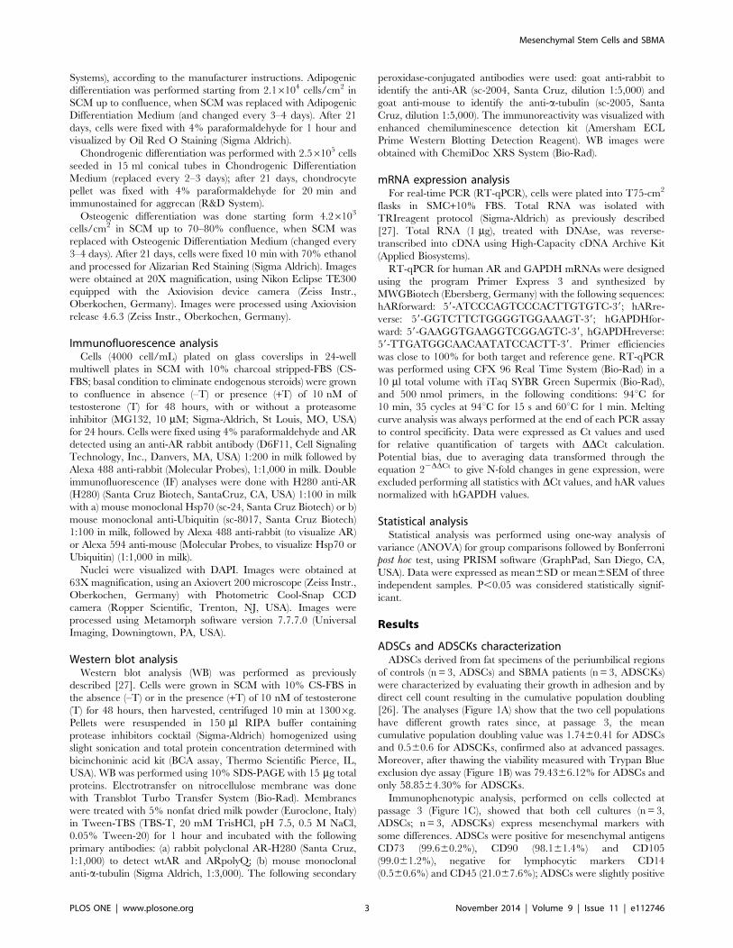
ADSCs and ADSCKs characterizationADSCs derived from fat specimens of the periumbilical regions
of controls (n = 3, ADSCs) and SBMA patients (n = 3, ADSCKs)
were characterized by evaluating their growth in adhesion and by
direct cell count resulting in the cumulative population doubling
[26]. The analyses (Figure 1A) show that the two cell populations
have different growth rates since, at passage 3, the mean
cumulative population doubling value was 1.7460.41 for ADSCs
and 0.560.6 for ADSCKs, confirmed also at advanced passages.
Moreover, after thawing the viability measured with Trypan Blue
exclusion dye assay (Figure 1B) was 79.4366.12% for ADSCs and
only 58.8564.30% for ADSCKs.
Immunophenotypic analysis, performed on cells collected at
passage 3 (Figure 1C), showed that both cell cultures (n = 3,
ADSCs; n= 3, ADSCKs) express mesenchymal markers with
some differences. ADSCs were positive for mesenchymal antigens
CD73 (99.660.2%), CD90 (98.161.4%) and CD105
(99.061.2%), negative for lymphocytic markers CD14
(0.560.6%) and CD45 (21.067.6%); ADSCs were slightly positive
Mesenchymal Stem Cells and SBMA
PLOS ONE | www.plosone.org 3 November 2014 | Volume 9 | Issue 11 | e112746
for early hematopoietic marker CD34 (32.8626.3%) and negative
for CD19 (2.562.2%), for endothelial typical marker CD31
(4.864.1%), and for immunological marker HLA-DR
(10.6610.9%). ADSCKs were positive for CD73 (97.261.4%),
CD90 (80.168.6%) and CD105 (97.661.3%) and negative for
CD14 (0.160.0%), CD45 (15.665.0%), CD34 (6.064.1%), CD19
(0.360.1%), CD31 (4.262.6%); only the immunological marker
HLA-DR was slightly higher than expected (48.4629.9%).
Immunophenotypic characterization performed after repeated
passages (passage 8), showed that all the three different cultures of
ADSCs preserved mesenchymal markers expression CD73
(99.260.5%), CD90 (98.961.2%) and CD105 (98.662.1%),
remaining negative for lymphocytic (CD14 0.460.2%; CD45
5.263.3%), early hematopoietic (CD34 19.0613.7%; CD19
2.261.91%), endothelial (CD31 3.964.0%) and immunological
(HLA-DR 9.068.7%) markers. The three different cultures of
ADSCKs were not analysed because of their reduced viability at
high passages (see Figure 1A).
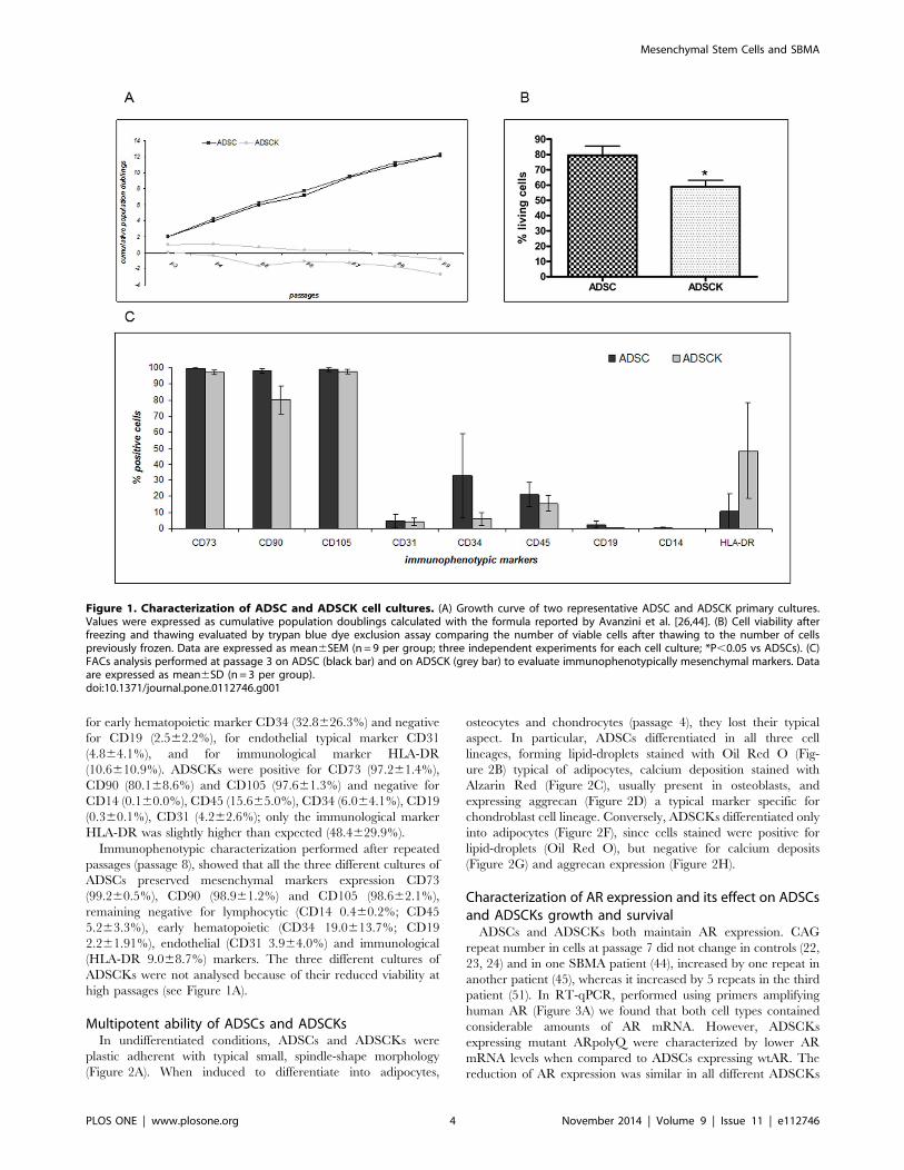
Multipotent ability of ADSCs and ADSCKsIn undifferentiated conditions, ADSCs and ADSCKs were
plastic adherent with typical small, spindle-shape morphology
(Figure 2A). When induced to differentiate into adipocytes,
osteocytes and chondrocytes (passage 4), they lost their typical
aspect. In particular, ADSCs differentiated in all three cell
lineages, forming lipid-droplets stained with Oil Red O (Fig-
ure 2B) typical of adipocytes, calcium deposition stained with
Alzarin Red (Figure 2C), usually present in osteoblasts, and
expressing aggrecan (Figure 2D) a typical marker specific for
chondroblast cell lineage. Conversely, ADSCKs differentiated only
into adipocytes (Figure 2F), since cells stained were positive for
lipid-droplets (Oil Red O), but negative for calcium deposits
(Figure 2G) and aggrecan expression (Figure 2H).
Characterization of AR expression and its effect on ADSCsand ADSCKs growth and survivalADSCs and ADSCKs both maintain AR expression. CAG
repeat number in cells at passage 7 did not change in controls (22,
23, 24) and in one SBMA patient (44), increased by one repeat in
another patient (45), whereas it increased by 5 repeats in the third
patient (51). In RT-qPCR, performed using primers amplifying
human AR (Figure 3A) we found that both cell types contained
considerable amounts of AR mRNA. However, ADSCKs
expressing mutant ARpolyQ were characterized by lower AR
mRNA levels when compared to ADSCs expressing wtAR. The
reduction of AR expression was similar in all different ADSCKs
Figure 1. Characterization of ADSC and ADSCK cell cultures. (A) Growth curve of two representative ADSC and ADSCK primary cultures.Values were expressed as cumulative population doublings calculated with the formula reported by Avanzini et al. [26,44]. (B) Cell viability afterfreezing and thawing evaluated by trypan blue dye exclusion assay comparing the number of viable cells after thawing to the number of cellspreviously frozen. Data are expressed as mean6SEM (n= 9 per group; three independent experiments for each cell culture; *P,0.05 vs ADSCs). (C)FACs analysis performed at passage 3 on ADSC (black bar) and on ADSCK (grey bar) to evaluate immunophenotypically mesenchymal markers. Dataare expressed as mean6SD (n = 3 per group).doi:10.1371/journal.pone.0112746.g001
Mesenchymal Stem Cells and SBMA
PLOS ONE | www.plosone.org 4 November 2014 | Volume 9 | Issue 11 | e112746
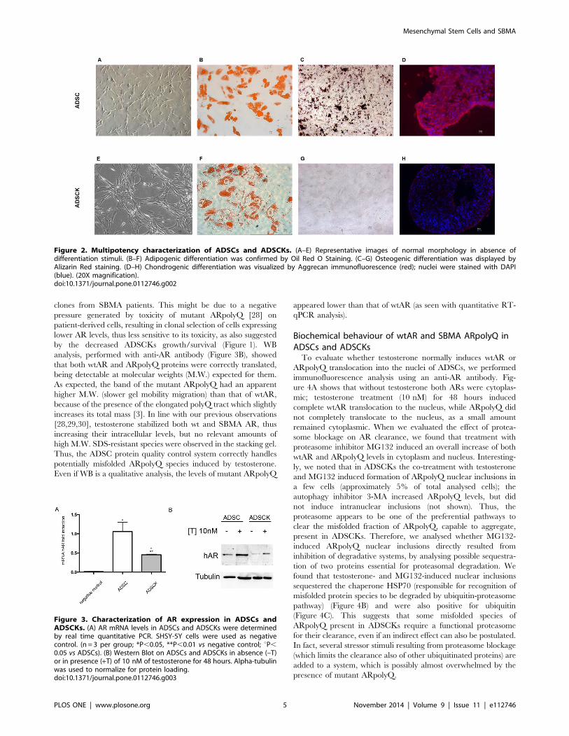
clones from SBMA patients. This might be due to a negative
pressure generated by toxicity of mutant ARpolyQ [28] on
patient-derived cells, resulting in clonal selection of cells expressing
lower AR levels, thus less sensitive to its toxicity, as also suggested
by the decreased ADSCKs growth/survival (Figure 1). WB
analysis, performed with anti-AR antibody (Figure 3B), showed
that both wtAR and ARpolyQ proteins were correctly translated,
being detectable at molecular weights (M.W.) expected for them.
As expected, the band of the mutant ARpolyQ had an apparent
higher M.W. (slower gel mobility migration) than that of wtAR,
because of the presence of the elongated polyQ tract which slightly
increases its total mass [3]. In line with our previous observations
[28,29,30], testosterone stabilized both wt and SBMA AR, thus
increasing their intracellular levels, but no relevant amounts of
high M.W. SDS-resistant species were observed in the stacking gel.
Thus, the ADSC protein quality control system correctly handles
potentially misfolded ARpolyQ species induced by testosterone.
Even if WB is a qualitative analysis, the levels of mutant ARpolyQ
appeared lower than that of wtAR (as seen with quantitative RT-
qPCR analysis).
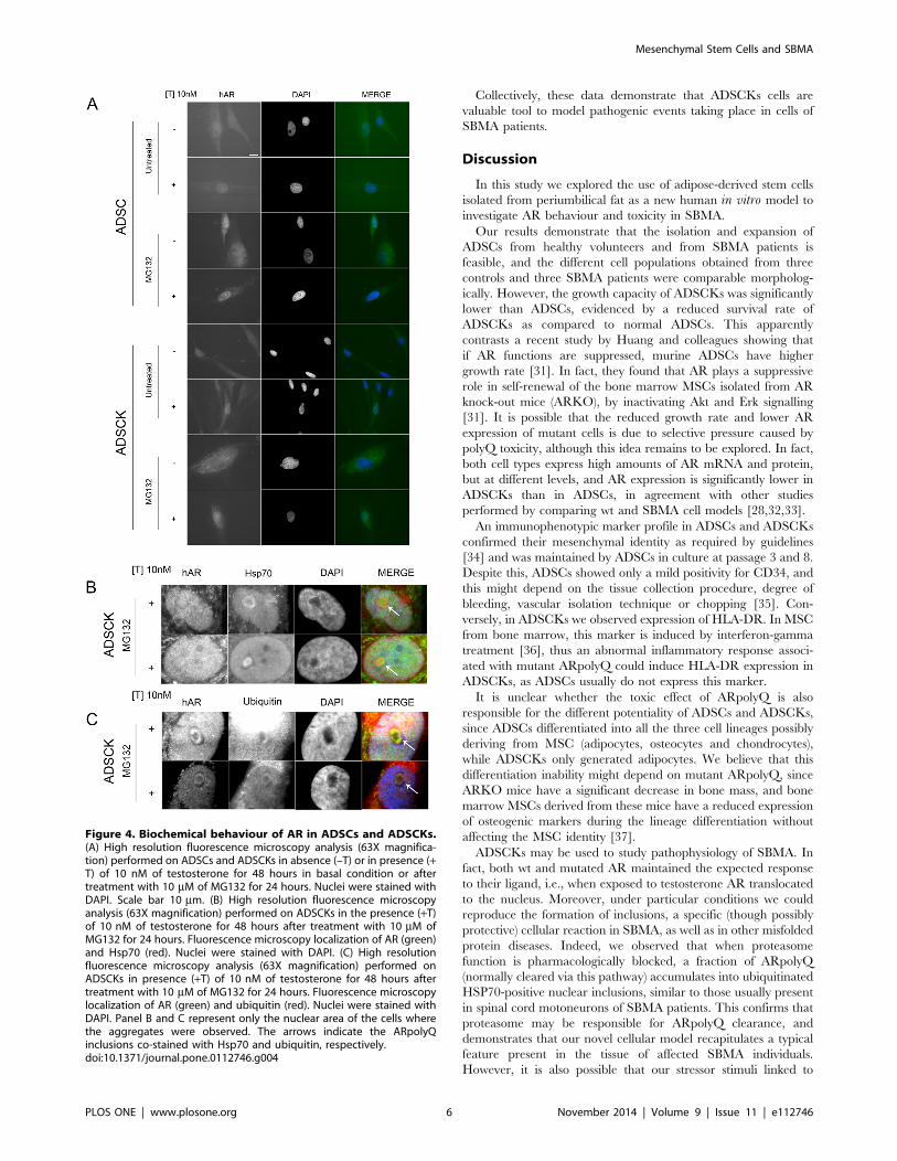
Biochemical behaviour of wtAR and SBMA ARpolyQ inADSCs and ADSCKsTo evaluate whether testosterone normally induces wtAR or
ARpolyQ translocation into the nuclei of ADSCs, we performed
immunofluorescence analysis using an anti-AR antibody. Fig-
ure 4A shows that without testosterone both ARs were cytoplas-
mic; testosterone treatment (10 nM) for 48 hours induced
complete wtAR translocation to the nucleus, while ARpolyQ did
not completely translocate to the nucleus, as a small amount
remained cytoplasmic. When we evaluated the effect of protea-
some blockage on AR clearance, we found that treatment with
proteasome inhibitor MG132 induced an overall increase of both
wtAR and ARpolyQ levels in cytoplasm and nucleus. Interesting-
ly, we noted that in ADSCKs the co-treatment with testosterone
and MG132 induced formation of ARpolyQ nuclear inclusions in
a few cells (approximately 5% of total analysed cells); the
autophagy inhibitor 3-MA increased ARpolyQ levels, but did
not induce intranuclear inclusions (not shown). Thus, the
proteasome appears to be one of the preferential pathways to
clear the misfolded fraction of ARpolyQ, capable to aggregate,
present in ADSCKs. Therefore, we analysed whether MG132-
induced ARpolyQ nuclear inclusions directly resulted from
inhibition of degradative systems, by analysing possible sequestra-
tion of two proteins essential for proteasomal degradation. We
found that testosterone- and MG132-induced nuclear inclusions
sequestered the chaperone HSP70 (responsible for recognition of
misfolded protein species to be degraded by ubiquitin-proteasome
pathway) (Figure 4B) and were also positive for ubiquitin
(Figure 4C). This suggests that some misfolded species of
ARpolyQ present in ADSCKs require a functional proteasome
for their clearance, even if an indirect effect can also be postulated.
In fact, several stressor stimuli resulting from proteasome blockage
(which limits the clearance also of other ubiquitinated proteins) are
added to a system, which is possibly almost overwhelmed by the
presence of mutant ARpolyQ.
Figure 2. Multipotency characterization of ADSCs and ADSCKs. (A–E) Representative images of normal morphology in absence ofdifferentiation stimuli. (B–F) Adipogenic differentiation was confirmed by Oil Red O Staining. (C–G) Osteogenic differentiation was displayed byAlizarin Red staining. (D–H) Chondrogenic differentiation was visualized by Aggrecan immunofluorescence (red); nuclei were stained with DAPI(blue). (20X magnification).doi:10.1371/journal.pone.0112746.g002
Figure 3. Characterization of AR expression in ADSCs andADSCKs. (A) AR mRNA levels in ADSCs and ADSCKs were determinedby real time quantitative PCR. SHSY-5Y cells were used as negativecontrol. (n = 3 per group; *P,0.05, **P,0.01 vs negative control; uP,0.05 vs ADSCs). (B) Western Blot on ADSCs and ADSCKs in absence (–T)or in presence (+T) of 10 nM of testosterone for 48 hours. Alpha-tubulinwas used to normalize for protein loading.doi:10.1371/journal.pone.0112746.g003
Mesenchymal Stem Cells and SBMA
PLOS ONE | www.plosone.org 5 November 2014 | Volume 9 | Issue 11 | e112746
Collectively, these data demonstrate that ADSCKs cells are
valuable tool to model pathogenic events taking place in cells of
SBMA patients.
Discussion
In this study we explored the use of adipose-derived stem cells
isolated from periumbilical fat as a new human in vitro model to
investigate AR behaviour and toxicity in SBMA.
Our results demonstrate that the isolation and expansion of
ADSCs from healthy volunteers and from SBMA patients is
feasible, and the different cell populations obtained from three
controls and three SBMA patients were comparable morpholog-
ically. However, the growth capacity of ADSCKs was significantly
lower than ADSCs, evidenced by a reduced survival rate of
ADSCKs as compared to normal ADSCs. This apparently
contrasts a recent study by Huang and colleagues showing that
if AR functions are suppressed, murine ADSCs have higher
growth rate [31]. In fact, they found that AR plays a suppressive
role in self-renewal of the bone marrow MSCs isolated from AR
knock-out mice (ARKO), by inactivating Akt and Erk signalling
[31]. It is possible that the reduced growth rate and lower AR
expression of mutant cells is due to selective pressure caused by
polyQ toxicity, although this idea remains to be explored. In fact,
both cell types express high amounts of AR mRNA and protein,
but at different levels, and AR expression is significantly lower in
ADSCKs than in ADSCs, in agreement with other studies
performed by comparing wt and SBMA cell models [28,32,33].
An immunophenotypic marker profile in ADSCs and ADSCKs
confirmed their mesenchymal identity as required by guidelines
[34] and was maintained by ADSCs in culture at passage 3 and 8.
Despite this, ADSCs showed only a mild positivity for CD34, and
this might depend on the tissue collection procedure, degree of
bleeding, vascular isolation technique or chopping [35]. Con-
versely, in ADSCKs we observed expression of HLA-DR. In MSC
from bone marrow, this marker is induced by interferon-gamma
treatment [36], thus an abnormal inflammatory response associ-
ated with mutant ARpolyQ could induce HLA-DR expression in
ADSCKs, as ADSCs usually do not express this marker.
It is unclear whether the toxic effect of ARpolyQ is also
responsible for the different potentiality of ADSCs and ADSCKs,
since ADSCs differentiated into all the three cell lineages possibly
deriving from MSC (adipocytes, osteocytes and chondrocytes),
while ADSCKs only generated adipocytes. We believe that this
differentiation inability might depend on mutant ARpolyQ, since
ARKO mice have a significant decrease in bone mass, and bone
marrow MSCs derived from these mice have a reduced expression
of osteogenic markers during the lineage differentiation without
affecting the MSC identity [37].
ADSCKs may be used to study pathophysiology of SBMA. In
fact, both wt and mutated AR maintained the expected response
to their ligand, i.e., when exposed to testosterone AR translocated
to the nucleus. Moreover, under particular conditions we could
reproduce the formation of inclusions, a specific (though possibly
protective) cellular reaction in SBMA, as well as in other misfolded
protein diseases. Indeed, we observed that when proteasome
function is pharmacologically blocked, a fraction of ARpolyQ
(normally cleared via this pathway) accumulates into ubiquitinated
HSP70-positive nuclear inclusions, similar to those usually present
in spinal cord motoneurons of SBMA patients. This confirms that
proteasome may be responsible for ARpolyQ clearance, and
demonstrates that our novel cellular model recapitulates a typical
feature present in the tissue of affected SBMA individuals.
However, it is also possible that our stressor stimuli linked to
Figure 4. Biochemical behaviour of AR in ADSCs and ADSCKs.(A) High resolution fluorescence microscopy analysis (63X magnifica-tion) performed on ADSCs and ADSCKs in absence (–T) or in presence (+T) of 10 nM of testosterone for 48 hours in basal condition or aftertreatment with 10 mM of MG132 for 24 hours. Nuclei were stained withDAPI. Scale bar 10 mm. (B) High resolution fluorescence microscopyanalysis (63X magnification) performed on ADSCKs in the presence (+T)of 10 nM of testosterone for 48 hours after treatment with 10 mM ofMG132 for 24 hours. Fluorescence microscopy localization of AR (green)and Hsp70 (red). Nuclei were stained with DAPI. (C) High resolutionfluorescence microscopy analysis (63X magnification) performed onADSCKs in presence (+T) of 10 nM of testosterone for 48 hours aftertreatment with 10 mM of MG132 for 24 hours. Fluorescence microscopylocalization of AR (green) and ubiquitin (red). Nuclei were stained withDAPI. Panel B and C represent only the nuclear area of the cells wherethe aggregates were observed. The arrows indicate the ARpolyQinclusions co-stained with Hsp70 and ubiquitin, respectively.doi:10.1371/journal.pone.0112746.g004
Mesenchymal Stem Cells and SBMA
PLOS ONE | www.plosone.org 6 November 2014 | Volume 9 | Issue 11 | e112746
proteasome blockage, which also impairs the clearance of other
ubiquitinated proteins, when added to a system which is possibly
almost overwhelmed by the presence of mutant ARpolyQ, may
limit the clearance of all the misfolded species including ARpolyQ.
Interestingly, SBMA patient-derived iPSCs have been recently
used to study some molecular alteration occurring as a
consequence of ARpolyQ expression. The great advantage of
these SBMA iPSCs is to be of human origin, and with a great
potential to be differentiated to ‘‘bona fide’’ motoneurons [19].
Using the iPSCs models, Grunseich and coll. showed that
testosterone activated ARpolyQ induced an increase of acetylated
alpha-tubulin and reduced HDAC6 [19], with consequently a
reduction of the perinuclear accumulation of lysosomes.
A limitation of the iPSCs could be that, as far as we know, there
are no established procedures to differentiate them into muscle cell
types. In our view, this is relevant, since muscle cells have been
recently recognized as targets of ARpolyQ toxicity [17,38,39].
However, to the best of our knowledge, all attempts to generate
muscle cells from iPSCs failed so far. In addition, iPSCs are
produced by genetic transformation of fibroblasts, using four
oncogenic or differentiating agents that may impact on cell
behavior. Thus, other cell models of human origin may be of value
to complement the data obtained in iPSCs. Conversely, despite the
fact that ADSCKs are difficult to maintain in culture for many
passages, there are studies demonstrating the possibility to
differentiate ADSC into muscle cell lines [40–43]. It must be
taken into account also that, compared to iPSCs, ADSCs have the
advantages of no retro transduction and manipulation needing,
and thus do not express exogenous genes.
In conclusion, ADSCs represent a potential novel model of
patient-derived cell populations useful to study the SBMA disease
mechanism. Although these cells are not differentiable in
motoneurons, at present, ADSCKs express AR and mimic some
pathogenic SBMA mechanisms. Moreover, ADSCKs can be easily
obtained with minimally invasive approach. Therefore, they have
an interesting and still unexplored potential in studying disease
mechanisms, and in designing and testing therapeutic approaches
in SBMA and other disorders.
Acknowledgments
We are grateful to Dr. Kenneth Fischbeck for helpful reading of the
manuscript. Funding: Regione Lombardia (to D.P., A.P., E.A.P.); AriSLA
Foundation Italy (ALS_HSPB8 to A.P), Telethon - Italy (GGP14039 to
A.P.); Italian Ministry of Labour, Health and Social Affairs (Convenzione
Fondazione Mondino/UNIMI to A.P.); Universita degli Studi di Milano
(to A.P.); Fondazione CARIPLO (2008-2307 to A.P.); Fondation Thierry
Latran, France (to A.P.), Association Francaise contre les Myopathies (to
A.P.).
Author Contributions
Conceived and designed the experiments: MD GB PR ES SEN GM GA
EAP DP AP. Performed the experiments: MD GB PR EG AC VT SEN
GM FC. Analyzed the data: MD GB PR EG VT ES AS CMCG SEN GM
GA EAP DP AP. Contributed reagents/materials/analysis tools: CG FC
EAP AP. Contributed to the writing of the manuscript: MD GB PR AS DP
AP.
References
1. Fischbeck KH (1997) Kennedy disease. J Inher Metab Dis 20: 152–158.
2. Jordan CL, Lieberman AP (2008) Spinal and bulbar muscular atrophy: a
motoneuron or muscle disease? Curr Opin Pharmacol 8: 752–758.
3. La Spada AR, Wilson EM, Lubahn DB, Harding AE, Fischbeck KH (1991)
Androgen receptor gene mutations in X-linked spinal and bulbar muscular
atrophy. Nature 352: 77–79.
4. Poletti A (2004) The polyglutamine tract of androgen receptor: from functions to
dysfunctions in motor neurons. Front Neuroendocrinol 25: 1–26.
5. Grunseich C, Kats IR, Bott LC, Rinaldi C, Kokkinis A, et al. (2014) Early onset
and novel features in a spinal and bulbar muscular atrophy patient with a 68
CAG repeat. Neuromuscul Disord. 70: 12–20.
6. Poletti A, Rampoldi A, Piccioni F, Volpi S, Simeoni S, et al. (2001) 5Alpha-
reductase type 2 and androgen receptor expression in gonadotropin releasing
hormone GT1-1 cells. J Neuroendocrinol 13: 353–357.
7. Pozzi P, Bendotti C, Simeoni S, Piccioni F, Guerini V, et al. (2003) Androgen 5-
alpha-reductase type 2 is highly expressed and active in rat spinal cord motor
neurones. J Neuroendocrinol 15: 882–887.
8. Grunseich C, Rinaldi C, Fischbeck KH (2014) Spinal and bulbar muscular
atrophy: pathogenesis and clinical management. Oral Dis 20: 6–9.
9. Katsuno M, Adachi H, Doyu M, Minamiyama M, Sang C, et al. (2003)
Leuprorelin rescues polyglutamine-dependent phenotypes in a transgenic mouse
model of spinal and bulbar muscular atrophy. Nat Med 9: 768–773.
10. Katsuno M, Adachi H, Kume A, Li M, Nakagomi Y, et al. (2002) Testosterone
reduction prevents phenotypic expression in a transgenic mouse model of spinal
and bulbar muscular atrophy. Neuron 35: 843–854.
11. Adachi H, Kume A, Li M, Nakagomi Y, Niwa H, et al. (2001) Transgenic mice
with an expanded CAG repeat controlled by the human AR promoter show
polyglutamine nuclear inclusions and neuronal dysfunction without neuronal cell
death. Hum Mol Genet 10: 1039–1048.
12. La Spada AR, Peterson KR, Meadows SA, McClain ME, Jeng G, et al. (1998)
Androgen receptor YAC transgenic mice carrying CAG 45 alleles show
trinucleotide repeat instability. Human Mol Genet 7: 959–967.
13. Sopher BL, Thomas PS Jr, LaFevre-Bernt MA, Holm IE, Wilke SA, et al. (2004)
Androgen receptor YAC transgenic mice recapitulate SBMA motor neurono-
pathy and implicate VEGF164 in the motor neuron degeneration. Neuron 41:
687–699.
14. Abel A, Walcott J, Woods J, Duda J, Merry DE (2001) Expression of expanded
repeat androgen receptor produces neurologic disease in transgenic mice. Hum
Mol Genet 10: 107–116.
15. Yu Z, Dadgar N, Albertelli M, Scheller A, Albin RL, et al. (2006) Abnormalities
of germ cell maturation and sertoli cell cytoskeleton in androgen receptor 113
CAG knock-in mice reveal toxic effects of the mutant protein. The American
journal of pathology 168: 195–204.
16. Chevalier-Larsen ES, O’Brien CJ, Wang H, Jenkins SC, Holder L, et al. (2004)
Castration restores function and neurofilament alterations of aged symptomatic
males in a transgenic mouse model of spinal and bulbar muscular atrophy.
J Neurosci 24: 4778–4786.
17. Cortes CJ, Ling SC, Guo LT, Hung G, Tsunemi T, et al. (2014) Muscle
expression of mutant androgen receptor accounts for systemic and motor neuron
disease phenotypes in spinal and bulbar muscular atrophy. Neuron 82: 295–307.
18. Rocchi A, Pennuto M (2013) New routes to therapy for spinal and bulbar
muscular atrophy. J Mol Neurosci 50: 514–523.
19. Grunseich C, Zukosky K, Kats IR, Ghosh L, Harmison GG, et al. (2014) Stem
cell-derived motor neurons from spinal and bulbar muscular atrophy patients.
Neurobiol Dis 70: 12–20.
20. Pittenger MF, Mackay AM, Beck SC, Jaiswal RK, Douglas R, et al. (1999)
Multilineage potential of adult human mesenchymal stem cells. Science 284:
143–147.
21. Casteilla L, Dani C (2006) Adipose tissue-derived cells: from physiology to
regenerative medicine. Diabetes Metab 32: 393–401.
22. Ogura F, Wakao S, Kuroda Y, Tsuchiyama K, Bagheri M, et al. (2014) Human
adipose tissue possesses a unique population of pluripotent stem cells with non-
tumorigenic and low telomerase activities: potential implications in regenerative
medicine. Stem Cells Dev. 23: 717–728.
23. Ibrahim MM (2010) Subcutaneous and visceral adipose tissue: structural and
functional differences. Obes Rev 11: 11–18.
24. Chazenbalk G, Singh P, Irge D, Shah A, Abbott DH, et al. (2013) Androgens
inhibit adipogenesis during human adipose stem cell commitment to
preadipocyte formation. Steroids 78: 920–926.
25. Alessandri G, Pagano S, Bez A, Benetti A, Pozzi S, et al. (2004) Isolation and
culture of human muscle-derived stem cells able to differentiate into myogenic
and neurogenic cell lineages. Lancet 364: 1872–1883.
26. Avanzini MA, Bernardo ME, Cometa AM, Perotti C, Zaffaroni N, et al. (2009)
Generation of mesenchymal stromal cells in the presence of platelet lysate: a
phenotypic and functional comparison of umbilical cord blood- and bone
marrow-derived progenitors. Haematologica 94: 1649–1660.
27. Rusmini P, Crippa V, Giorgetti E, Boncoraglio A, Cristofani R, et al. (2013)
Clearance of the mutant androgen receptor in motoneuronal models of spinal
and bulbar muscular atrophy. Neurobiol Aging 34: 2585–2603.
28. Simeoni S, Mancini MA, Stenoien DL, Marcelli M, Weigel NL, et al. (2000)
Motoneuronal cell death is not correlated with aggregate formation of androgen
receptors containing an elongated polyglutamine tract. Hum Mol Genet 9: 133–
144.
Mesenchymal Stem Cells and SBMA
PLOS ONE | www.plosone.org 7 November 2014 | Volume 9 | Issue 11 | e112746
29. Piccioni F, Pinton P, Simeoni S, Pozzi P, Fascio U, et al. (2002) Androgen
receptor with elongated polyglutamine tract forms aggregates that alter axonaltrafficking and mitochondrial distribution in motor neuronal processes. Faseb J
16: 1418–1420.
30. Rusmini P, Sau D, Crippa V, Palazzolo I, Simonini F, et al. (2007) Aggregationand proteasome: the case of elongated polyglutamine aggregation in spinal and
bulbar muscular atrophy. Neurobiol Aging 28: 1099–1111.31. Huang CK, Tsai MY, Luo J, Kang HY, Lee SO, et al. (2013) Suppression of
androgen receptor enhances the self-renewal of mesenchymal stem cells through
elevated expression of EGFR. Biochim Biophys Acta 1833: 1222–1234.32. Brooks BP, Merry DE, Paulson HL, Lieberman AP, Kolson DL, et al. (1998) A
cell culture model for androgen effects in motor neurons. J Neurochem 70:1054–1060.
33. Brooks BP, Paulson HL, Merry DE, Salazar-Grueso EF, Brinkmann AO, et al.(1997) Characterization of an expanded glutamine repeat androgen receptor in a
neuronal cell culture system. Neurobiol Dis 3: 313–323.
34. Dominici M, Le Blanc K, Mueller I, Slaper-Cortenbach I, Marini F, et al. (2006)Minimal criteria for defining multipotent mesenchymal stromal cells. The
International Society for Cellular Therapy position statement. Cytotherapy 8:315–317.
35. Bourin P, Bunnell BA, Casteilla L, Dominici M, Katz AJ, et al. (2013) Stromal
cells from the adipose tissue-derived stromal vascular fraction and cultureexpanded adipose tissue-derived stromal/stem cells: a joint statement of the
International Federation for Adipose Therapeutics and Science (IFATS) and theInternational Society for Cellular Therapy (ISCT). Cytotherapy 15: 641–648.
36. Le Blanc K, Tammik C, Rosendahl K, Zetterberg E, Ringden O (2003) HLAexpression and immunologic properties of differentiated and undifferentiated
mesenchymal stem cells. Exp Hematol 31: 890–896.
37. Tsai MY, Shyr CR, Kang HY, Chang YC, Weng PL, et al. (2011) The reduced
trabecular bone mass of adult ARKO male mice results from the decreased
osteogenic differentiation of bone marrow stroma cells. Biochem Biophys Res
Commun 411: 477–482.
38. Chua JP, Reddy SL, Merry DE, Adachi H, Katsuno M, et al. (2014)
Transcriptional activation of TFEB/ZKSCAN3 target genes underlies enhanced
autophagy in spinobulbar muscular atrophy. Hum Mol Genet 23: 1376–1386.
39. Lieberman AP, Yu Z, Murray S, Peralta R, Low A, et al. (2014) Peripheral
androgen receptor gene suppression rescues disease in mouse models of spinal
and bulbar muscular atrophy. Cell Rep 7: 774–784.
40. Desiderio V, De Francesco F, Schiraldi C, De Rosa A, La Gatta A, et al. (2013)
Human Ng2+ adipose stem cells loaded in vivo on a new crosslinked hyaluronic
acid-Lys scaffold fabricate a skeletal muscle tissue. J Cell Physiol 228: 1762–
1773.
41. de la Garza-Rodea AS, van der Velde-van Dijke I, Boersma H, Goncalves MA,
van Bekkum DW, et al. (2012) Myogenic properties of human mesenchymal
stem cells derived from three different sources. Cell Transplant 21: 153–173.
42. Marra KG, Brayfield CA, Rubin JP (2011) Adipose stem cell differentiation into
smooth muscle cells. Methods Mol Biol 702: 261–268.
43. Wang C, Yin S, Cen L, Liu Q, Liu W, et al. (2010) Differentiation of adipose-
derived stem cells into contractile smooth muscle cells induced by transforming
growth factor-beta1 and bone morphogenetic protein-4. Tissue Eng Part A 16:
1201–1213.
44. Kotaja N, Karvonen U, Janne OA, Palvimo JJ (2002) The nuclear receptor
interaction domain of GRIP1 is modulated by covalent attachment of SUMO-1.
J Biol Chem 277: 30283–30288.
Mesenchymal Stem Cells and SBMA
PLOS ONE | www.plosone.org 8 November 2014 | Volume 9 | Issue 11 | e112746
Related Documents