electronic reprint ISSN: 2053-2296 journals.iucr.org/c HUG and SQUEEZE: using CRYSTALS to incorporate resonant scattering in the SQUEEZE structure-factor contributions to determine absolute structure Richard I. Cooper, Howard D. Flack and David J. Watkin Acta Cryst. (2017). C73, 845–853 IUCr Journals CRYSTALLOGRAPHY JOURNALS ONLINE Copyright c International Union of Crystallography Author(s) of this paper may load this reprint on their own web site or institutional repository provided that this cover page is retained. Republication of this article or its storage in electronic databases other than as specified above is not permitted without prior permission in writing from the IUCr. For further information see http://journals.iucr.org/services/authorrights.html Acta Cryst. (2017). C73, 845–853 Richard I. Cooper et al. · HUG and SQUEEZE

Welcome message from author
This document is posted to help you gain knowledge. Please leave a comment to let me know what you think about it! Share it to your friends and learn new things together.
Transcript

electronic reprint
ISSN: 2053-2296
journals.iucr.org/c
HUG and SQUEEZE: using CRYSTALS to incorporate resonantscattering in the SQUEEZE structure-factor contributions todetermine absolute structure
Richard I. Cooper, Howard D. Flack and David J. Watkin
Acta Cryst. (2017). C73, 845–853
IUCr JournalsCRYSTALLOGRAPHY JOURNALS ONLINE
Copyright c© International Union of Crystallography
Author(s) of this paper may load this reprint on their own web site or institutional repository provided thatthis cover page is retained. Republication of this article or its storage in electronic databases other than asspecified above is not permitted without prior permission in writing from the IUCr.
For further information see http://journals.iucr.org/services/authorrights.html
Acta Cryst. (2017). C73, 845–853 Richard I. Cooper et al. · HUG and SQUEEZE

feature articles
Acta Cryst. (2017). C73, 845–853 https://doi.org/10.1107/S2053229617013304 845
Received 17 August 2017
Accepted 25 September 2017
Edited by P. Raithby, University of Bath, UK
‡ Deceased 2 February 2017.
§ RIC & DJW are grateful to HDF for his
inspiration and encouragement. We have tried
to preserve his contributions to this manuscript
unaltered where possible; any errors or omis-
sions are our own.
Keywords: disorder; resonsant scattering; abso-
lute structure.
HUG and SQUEEZE: using CRYSTALS to incorpo-rate resonant scattering in the SQUEEZE structure-factor contributions to determine absolute structure
Richard I. Cooper,a* Howard D. Flackbठand David J. Watkina
aChemical Crystallography, University of Oxford, 12 Manseld Road, Oxford, Oxfordshire OX1 3TA, England, andbChimie minerale, analytique et appliquee, University of Geneva, Geneva, Switzerland. *Correspondence e-mail:
The resonant-scattering contributions to single-crystal X-ray diffraction data
enable the absolute structure of crystalline materials to be determined. Crystal
structures can be determined even if they contain considerably disordered
regions because a correction is available via a discrete Fourier transform of the
residual electron density to approximate the X-ray scattering from the
disordered region. However, the corrected model cannot normally account for
resonant scattering from atoms in the disordered region. Straightforward
determination of absolute structure from crystals where the strongly resonantly
scattering atoms are not resolved has therefore not been possible. Using an
approximate resonant-scattering correction to the X-ray scattering from the
disordered regions, we have developed and tested a procedure (HUG) to
recover the absolute structure using conventional Flack x refinement or other
post-refinement determination methods. Results show that in favourable cases
the HUG method works well and the absolute structure can be correctly
determined. It offers no useful improvement in cases where the original
correction for the disordered region scattering density is problematic, for
example, when a large fraction of the scattering density in the crystal is
disordered, or when voids are not occupied equally by the disordered species.
Crucially, however, if the approach does not work for a given structure, the
statistics for the absolute structure measures are not improved, meaning it is
unlikely to lead to misassignment of absolute structure.
1. Background
The refinement of crystal structures usually requires the
structural parameters to be adjusted by the method of least
squares to minimize the differences between |Fo| and |Fc| (or Io
and Ic, where I are squared structure amplitudes, |F2|).
Cases exist where this procedure is complicated by the fact
that part of the structure cannot easily be modelled by clearly
defined individual atoms. This situation may exist in extended
lattice structures with voids which contain independent mole-
cules (host and guest structures), or in discrete molecule
structures in which the lattice is stabilized by the inclusion of
solvent molecules or ions necessary to preserve charge balance.
If these subsidiary molecules are not spatially constrained
by the surrounding lattice, they may have freedom to move
even in the solid state, or have the possibility of occupying
alternative positions and orientations. Often this ambiguity
can be modelled by large anisotropic atomic displacement
factors (ADPs) or by the superposition of displaced partially
occupied images of the molecule. In unfavourable cases, the
average scattering density in the cavity cannot reasonably be
modelled by independent atoms. This situation has been
ISSN 2053-2296
# 2017 International Union of Crystallography
electronic reprint
addressed by replacing the atomic model of the contents of the
cavity by the discrete Fourier transform of the electron density
in the cavity computed from the observed structure ampli-
tudes and phases obtained from the atomic model of the
resolved part of the structure (van der Sluis & Spek, 1990;
Spek, 2015).
1.1. The SQUEEZE procedure
The structure factor can be computed either as the Fourier
transform of the continuous periodic electron density in the
crystal:
Fh ¼ZV
�ðxÞe2�iðhxÞdV; ð1Þ
or as the summation of the contributions from individual
‘atoms’:
Fh ¼Xj
fje2�iðhxjÞ: ð2Þ
SQUEEZE defines a region of the unit cell, V, in which the
disordered part of the crystal structure is located. No atomic
model of V is available. The content of V is represented by a
real electron density, �V(x):
�VðxÞ ¼ �ðxÞ; if x 2 V
0; otherwise:
�ð3Þ
The SQUEEZE procedure then uses the hybrid structure-
factor expression:
Fh ¼Xj
fje2�iðhxjÞ þ
ZV
�VðxÞe2�iðhxÞdV: ð4Þ
The first term is a summation over the resolved atoms as in
equation 2. The integral in the second term is evaluated for
x 2 V, which contains unresolved electron density. The
resulting expression accounts for scattering from both
resolved atoms and unresolved electron density.
The integral can be replaced by a summation over a suitable
resolution grid of electron density:
Fh ¼Xj
fje2�iðhxjÞ þ
Xx
�VðxÞe2�iðhxÞ: ð5Þ
1.2. Resonant scattering
If a material is in a noncentrosymmetric space group and it
contains one or more atoms with significant resonant scat-
tering for the wavelength in use, it may be possible to deter-
mine the absolute structure of the sample using Flack’s
interpretation of the observed Bijvoet differences (Flack,
1983). Representing |F|2h by I+ and |F|2�h by I�:
Iþo � Iþc ¼ ð1 � xÞ Iþs þ xI�s ; ð6Þwhere the subscript s indicates a quantity computed from the
atomic model with the Flack parameter x set to zero (i.e. a
nontwinned single crystal), c a quantity computed from an
inversion-twinned model (i.e. Flack parameter not necessarily
zero) and o an observed quantity (Cooper et al., 2016). The
Flack(x) may be determined either during the least-squares
refinement, or by post-refinement methods (Parsons et al.,
2013). The success of this procedure depends upon the quality
of the data and upon the absolute structure resolving power of
the material, conveniently estimated by Friedif (Flack &
Shmueli, 2007). The magnitude of Friedif is increased in the
presence of atoms with large resonant scattering factors, even
if these atoms are not part of an enantiomerically pure host
material. This means that the possibility of reliably deter-
mining the absolute structure of an all-light-atom structure
can be increased if the material crystallizes with a suitable
molecule of solvation.
In the case that the solvent molecule is highly disordered, it
may not be possible to model it with discrete atoms, so that the
only way to complete the analysis is to SQUEEZE the solvent
region, which in the standard implementation makes no
allowance for a resonant contribution from the solvent to the
computed structure amplitudes. This means that a conven-
tionally SQUEEZEd solvent cannot be used to help in the
determination of absolute structure, as demonstrated at the
start of x3 below.
2. Methods
Fourier transformation of �V(x) leads to its structure factor,
F(�V(x))h, which is added to the structure factor of the atomic
model for the ordered part of the unit cell.
The structure factor is a complex number having both
magnitude and phase, and may be written as Fh = Ah + iBh,
where Ah and Bh are the real and imaginary parts of Fh,
respectively, and i =ffiffiffiffiffiffi�1
p.
2.1. The HUG procedure – enhancing SQUEEZE to includeresonant scattering
If the disordered volume contains atoms with strong resonant
scattering, how might one proceed to incorporate this resonant
scattering contribution to �V(x)?Method 1 Construct a model in which the resonant scat-
tering contribution of V is distributed uniformly over V. Then
�V(x) can be modified to become the complex �0V(x):
�0VðxÞ ¼ �VðxÞ þ cþ id; ð7Þ
in which c and d are constants to be chosen or determined in
some way.
The inconvenience of this simple model is that the resonant-
scattering contribution is distributed widely over V and its
contribution in reciprocal space will diminish more rapidly as a
function of sin�/� than with an atomic model. Such consid-
eration leads to:
Method 2 Construct a model in which the resonant scat-
tering contribution of V is assumed to be proportional to �V(x)
at each point x. Then �V(x) can be modified to become the
complex �0V(x):
�0VðxÞ ¼ �VðxÞ þ c�VðxÞ þ id�VðxÞ ¼ ð1 þ cþ idÞ�VðxÞ: ð8Þ
In this way, the major part of the resonant-scattering contri-
bution will be located at the positions of high electron density
feature articles
846 Richard I. Cooper et al. � HUG and SQUEEZE Acta Cryst. (2017). C73, 845–853
electronic reprint
in �V(x). The advantage of this model is that it is ‘more atomic’
than that of Method 1, a positive attribute intended to imply
exactly the same as the ‘large f’, usually associated with
heavier elements, and whose ghosts would leave more
miasma1 in the difference density.
If Fh is the Fourier transform of �(x), then the Fourier
transform, F 0h, of �0
V(x) is given by:
F 0h ¼ ð1 þ cÞFh þ idFh
¼ ½ð1 þ cÞAh � dBh� þ i½ð1 þ cÞBh þ dAh�;ð9Þ
where c and d set the ratios of the real and imaginary parts of
the resonant-scattering contribution from the electron density
in the disordered region of the crystal.
A reasonable first approximation for c and d is to assume
that the regions of high electron density in the unresolved
volume are those of highest resonant scattering. One may
even take the step of assuming that the resonant-scattering
contribution is proportional to electron density, so that:
c ¼P
f 0solPfsol
and d ¼P
f 00solPfsol
; ð10Þ
where the summations are over the expected atoms in the
solvent. Note that equation 10 might use f 0sol instead of fsol in
the denominator, to avoid overcorrection for the resonant
signal at higher sin�/�, however, trial-and-error has shown
equation 10 to be the more effective formulation.
The resonant scattering contribution in the solvent region is
thus ðcþ idÞ�VðxÞ which can be used as a modifier to correct
the structure-factor components A and B of the region V for
resonant scattering. By inspection of equation 9, we obtain
Ahug ¼ ð1 þ cÞAsqz � d:Bsqz
Bhug ¼ ð1 þ cÞBsqz þ d:Asqz;ð11Þ
where the subscripts sqz indicate the complex contribution to
the structure factor returned by SQUEEZE due to unresolved
electron density in the volume V, and hug indicates the same
contribution corrected for resonant scattering. Refinement is
undertaken in the usual way, except that the A and B parts of
the structure factor computed from the resolved atoms are
supplemented by the addition of Ahug and Bhug, respectively.
Acalc ¼ Ares þ Ahug
Bcalc ¼ Bres þ Bhug;ð12Þ
where the subscript res indicates structure-factor components
for the resolved part of the structure.
2.2. HUGging in CRYSTALS
Since its inception, CRYSTALS has had a facility for storing
the precomputed A and B parts for a reflection so that they
can be added into the A and B parts computed from an atomic
model (Carruthers, 1977). The original use was to facilitate the
development of a poorly resolved part of a structure. The A
and B parts of the well-resolved atoms were computed once
and stored in the database. Structure-factor contributions
were then computed from the atoms in experimental models
of the disorder and added to the stored parts. This gave
significant time-savings when the well-resolved part of the
structure contained a large number of atoms compared with
the disordered part (Watkin et al., 1985). With the publication
of the SQUEEZE program, this procedure could be reversed.
For more than 20 years, an interface between SQUEEZE and
CRYSTALS has enabled the A and B parts of the database to
hold contributions to the structure factor computed from the
discrete Fourier transform of electron density in parts of the
unit cell not modelled by independent atoms. This procedure
has the virtue that during refinement, the values of Fobs (or
Iobs) are not modified. The enhanced strategy (HUG2)
represented by equation 12 has been implemented in CRYS-
TALS (Versions after 24/02/2017) by an external module
which uses a proposed molecular formula for the solvent to
correct the standard output from unmodified SQUEEZE
before passing the modified A and B parts into CRYSTALS.
The concept was evaluated by processing several structures
with well-resolved solvent molecules. The A and B parts for an
atomic model of the solvent were first computed and stored in
the CRYSTALS database and then used together with the
feature articles
Acta Cryst. (2017). C73, 845–853 Richard I. Cooper et al. � HUG and SQUEEZE 847
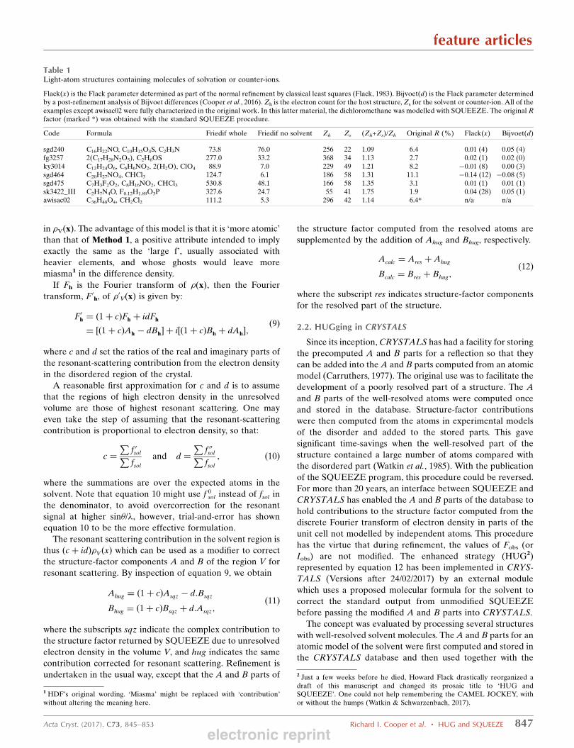
Table 1Light-atom structures containing molecules of solvation or counter-ions.
Flack(x) is the Flack parameter determined as part of the normal refinement by classical least squares (Flack, 1983). Bijvoet(d) is the Flack parameter determinedby a post-refinement analysis of Bijvoet differences (Cooper et al., 2016). Zh is the electron count for the host structure, Zs for the solvent or counter-ion. All of theexamples except awisac02 were fully characterized in the original work. In this latter material, the dichloromethane was modelled with SQUEEZE. The original Rfactor (marked *) was obtained with the standard SQUEEZE procedure.
Code Formula Friedif whole Friedif no solvent Zh Zs (Zh+Zs)/Zh Original R (%) Flack(x) Bijvoet(d)
sgd240 C16H22NO, C10H15O4S, C2H3N 73.8 76.0 256 22 1.09 6.4 0.01 (4) 0.05 (4)fg3257 2(C17H28N2O5), C2H6OS 277.0 33.2 368 34 1.13 2.7 0.02 (1) 0.02 (0)ky3014 C12H24O6, C6H6NO2, 2(H2O), ClO4 88.9 7.0 229 49 1.21 8.2 �0.01 (8) 0.00 (3)sgd464 C20H27NO4, CHCl3 124.7 6.1 186 58 1.31 11.1 �0.14 (12) �0.08 (5)sgd475 C7H3F2O2, C8H16NO2, CHCl3 530.8 48.1 166 58 1.35 3.1 0.01 (1) 0.01 (1)sk3422_III C2H7N4O, F0.12H1.89O3P 327.6 24.7 55 41 1.75 1.9 0.04 (28) 0.05 (1)awisac02 C36H48O4, CH2Cl2 111.2 5.3 296 42 1.14 6.4* n/a n/a
1 HDF’s original wording. ‘Miasma’ might be replaced with ‘contribution’without altering the meaning here.
2 Just a few weeks before he died, Howard Flack drastically reorganized adraft of this manuscript and changed its prosaic title to ‘HUG andSQUEEZE’. One could not help remembering the CAMEL JOCKEY, withor without the humps (Watkin & Schwarzenbach, 2017).
electronic reprint

main structure in a normal refinement. These refinements
were compared with a refinement in which the A and B parts
were from a solvent which was SQUEEZEd and HUGged.
The agreement between the HUGged and atomic structure
amplitudes can be estimated by
RðAvsÞ ¼P jAvhug � AvatomjP jAvatomj
ð13Þ
RðDsÞ ¼P jDhug �DatomjP jDatomj
; ð14Þ
where Avatom and Datom are the average and difference of
structure-factor magnitudes of a Bijvoet pair computed from a
fully atomic model, and Avhug and Dhug are equivalent values
computed from a HUGged model. The agreement can be
visualized in plots of Dhug versus Datom.
3. Results
Table 1 lists solvated structures selected from the recent
literature where the molecules of interest contained only light
atoms and the solvents were reasonably well defined: sgd240
(Chernega et al., 2009), fg3257 (Bojarska et al., 2012), ky3014
(Shi et al., 2012), sgd464 (Davies et al., 2013a), sgd475 (Davies
et al., 2013b), sk3422 (Fabry et al., 2012) and awisac02 (Qian et
al., 2016). The absolute structures were confirmed by re-
refining the atomic model in CRYSTALS. The solvents were
then excluded from the structure-factor calculation and
modelled using the standard SQUEEZE procedure. The
outputs from SQUEEZE were HUGged as explained above,
and the structures rerefined. The applicability of the proce-
dure was assessed by comparing the Flack(x) (Flack, 1983)
and Bijvoet(d) (Cooper et al., 2016) parameters determined
from the atomic model and the HUGged model, and by
plotting Ds, the computed Bijvoet difference, for one model
against the other.
The effect of modelling regions of the crystal structure
containing strong resonant scatterers with scattering from an
electron-density map with no resonant scattering effects may
be demonstrated by a comparison of the absolute structure
statistics for sgd464: the Flack(x) and Bijvoet(d) parameters
for a complete atomic model are �0.14 (12) and �0.08 (5)
feature articles
848 Richard I. Cooper et al. � HUG and SQUEEZE Acta Cryst. (2017). C73, 845–853
Table 2Structures from the literature were rerefined in CRYSTALS (against F 2 with SHELX-type weights) and the absolute-structure parameters determined.
The solvent/counter-ion was excluded and the structures rerefined using the modified Ahug and Bhug parts of the structure factor computed by SQUEEZE. Flack(x)is the Flack parameter determined as part of the normal refinement by classical least squares (Flack, 1983). Bijvoet(d) is the Flack parameter determined by a post-refinement analysis of Bijvoet differences (Cooper et al., 2016). R(Avs) and R(Ds) are defined in equations 13 and 14. The atomic models for structures marked �were modelled with disordered solvent or counter-ion. The structure marked � was squeezed in the original work. The HUGged R factor and Bijvoet(d) marked †were obtained when the d parameter was multiplied by 1.5.
Code HUGged R (%) HUGged Flack(x) HUGged Bijvoet(d) Space group Electrons found (expected) R(Avs) R(Ds)
sgd240 7.1 0.01 (10) 0.27 (3) P212121 28 (22) 0.06 0.75fg3257 4.1 �0.30 (4) �0.30 (1) P212121 47 (58) 0.05 0.33ky3014� 8.0 0.05 (8) 0.27 (5) Pna21 45 (49) 0.10 0.91sgd464 9.3 0.00 (9) �0.02 (5) P21 54 (58) 0.07 0.44sgd475� 4.0 0.00 (2) 0.00 (1) C2 61 (58) 0.12 0.40sk3422_III 13.8 �0.2 (3) 0.06 (4) Cc 41 (41) 0.51 0.91awisac02� 5.9† 0.1 (2) 0.33 (3) P21 48 (42) n/a n/a
Figure 1Averages (Avs) and differences (Ds) of Bijvoet pairs computed from the HUGged and atomic models of structure sgd240. The gradients of both plots(1.00 and 1.01) show that the HUGged model is a fair approximation to the atomic model; R(Avs) = 0.06 and R(Ds) = 0.80. The abscissa is valuescomputed from the fully resolved atomic model and the ordinate values are computed from the HUGged model.
electronic reprint

respectively, and the R1 value is 11.1%. After the chloroform
molecule is removed and SQUEEZE applied, the remaining
structure can be refined to an R1 value of 9.39%, but the
Flack(x) and Bijvoet(d) parameters are now 4.7 (10) and
0.7 (2) (Table 2).
3.1. Structure sgd240
This is an organic material of known absolute configuration
(from the starting materials) containing a sulfoxide group and
a well-behaved acetonitrile of solvation. The data were
measured with Mo K� radiation. The solvent contains 22
electrons, SQUEEZE returns 114 electrons/cell in the voids,
and �3 electrons/cell outside the voids. The principal normal
and resonant-scattering atom is sulfur in the main molecule.
The resonant scattering from the solvent is marginal so that
the unmodified SQUEEZE refinement is essentially the same
as the modified (Fig. 1).
3.2. Structure fg3257
This is an organic material containing C, H, N and O atoms,
with two independent molecules and one dimethyl sulfoxide
solvent in the asymmetric unit. The absolute structure was
determined from the X-ray diffraction data. Modifying the
SQUEEZE output using constants determined by equation 10
gives a Bijvoet(d) parameter of �0.30 (1). Multiplying c and d
by factors of 1.5 and 2.0 gave Bijvoet(d) values of �0.02 (1)
and 0.12 (0), respectively. The Flack(x) parameter increased
from �0.30 (4) with unscaled d values to �0.00 (2) and
feature articles
Acta Cryst. (2017). C73, 845–853 Richard I. Cooper et al. � HUG and SQUEEZE 849
Figure 3Wilson Plot for structure sgd464. The abscissa is lnðhðF2
obsÞ�i=f 2� Þ and the
ordinate � = (sin�/�)2. Blue circles are calculated from measureddiffraction intensities and red circles are calculated from the final atomicmodel (for comparison). The up-turn at about � = 0.35 is characteristic ofdata being recorded beyond the real diffraction limit and thereforeconsisting mainly of noise.
Figure 4The A and B parts from SQUEEZE for structure sgd464 were modified toinclude resonant scattering from the solvent and were used withoutcontributions from the host molecule to compute calculated structurefactors and phases, which were then used to compute a Fourier synthesis.The electron density (arbitrary contour levels where red > blue > green)shows a good representation of the solvent. The chloroform molecule hasbeen overlaid as a guide to the eye. Chloroform contains 58 electrons andthe unmodified SQUEEZE map contains 54 electrons per void.
Figure 2Averages and differences of Bijvoet pairs for structure fg3257. The gradient of the averages (0.98) suggests that the A and B parts from SQUEEZE arereasonably well scaled and the gradient of the differences (0.36) suggests that the d factor used to generate the resonant differences should be multipliedby about 0.3.
electronic reprint
�0.13 (2) with the increasing scaling factors. The near-unity
value of the ratio (Zh + Zs)/Zh (1.13) suggests that the scaling
of the A and B parts from SQUEEZE is almost correct,
confirmed by the gradient (0.98) of the plot of the averaged
Bijvoet pairs of the HUGged model versus the atomic model
(Fig. 2); the need for scaling of d is demonstrated in the right-
hand plot.
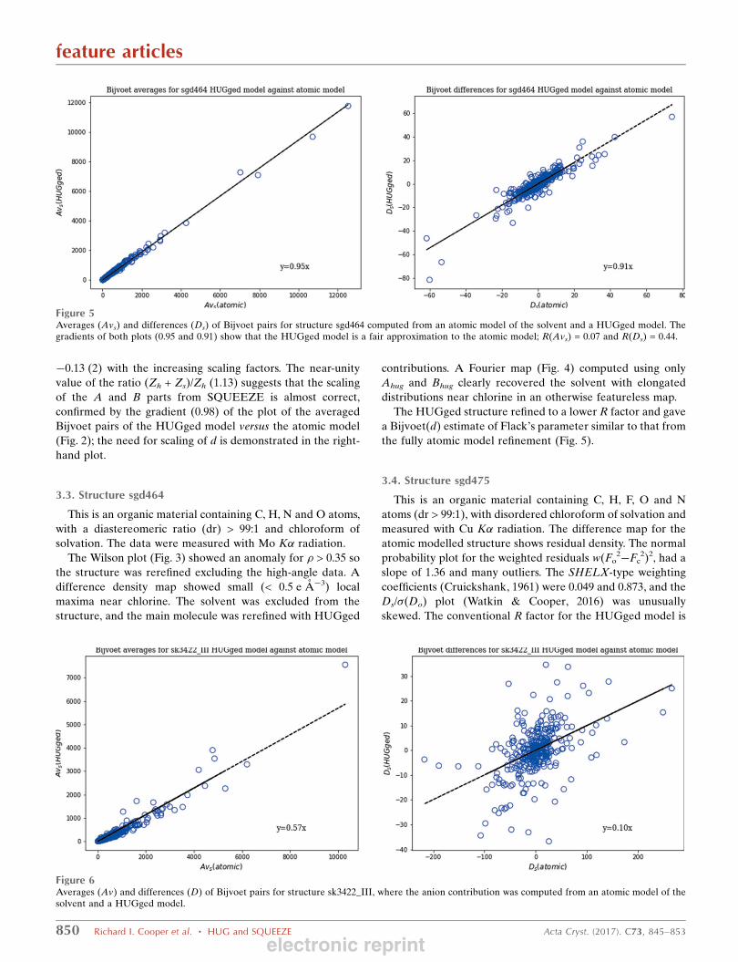
3.3. Structure sgd464
This is an organic material containing C, H, N and O atoms,
with a diastereomeric ratio (dr) > 99:1 and chloroform of
solvation. The data were measured with Mo K� radiation.
The Wilson plot (Fig. 3) showed an anomaly for � > 0.35 so
the structure was rerefined excluding the high-angle data. A
difference density map showed small (< 0.5 e A�3) local
maxima near chlorine. The solvent was excluded from the
structure, and the main molecule was rerefined with HUGged
contributions. A Fourier map (Fig. 4) computed using only
Ahug and Bhug clearly recovered the solvent with elongated
distributions near chlorine in an otherwise featureless map.
The HUGged structure refined to a lower R factor and gave
a Bijvoet(d) estimate of Flack’s parameter similar to that from
the fully atomic model refinement (Fig. 5).
3.4. Structure sgd475
This is an organic material containing C, H, F, O and N
atoms (dr > 99:1), with disordered chloroform of solvation and
measured with Cu K� radiation. The difference map for the
atomic modelled structure shows residual density. The normal
probability plot for the weighted residuals w(Fo2�Fc
2)2, had a
slope of 1.36 and many outliers. The SHELX-type weighting
coefficients (Cruickshank, 1961) were 0.049 and 0.873, and the
Ds/�(Do) plot (Watkin & Cooper, 2016) was unusually
skewed. The conventional R factor for the HUGged model is
feature articles
850 Richard I. Cooper et al. � HUG and SQUEEZE Acta Cryst. (2017). C73, 845–853
Figure 5Averages (Avs) and differences (Ds) of Bijvoet pairs for structure sgd464 computed from an atomic model of the solvent and a HUGged model. Thegradients of both plots (0.95 and 0.91) show that the HUGged model is a fair approximation to the atomic model; R(Avs) = 0.07 and R(Ds) = 0.44.
Figure 6Averages (Av) and differences (D) of Bijvoet pairs for structure sk3422_III, where the anion contribution was computed from an atomic model of thesolvent and a HUGged model.
electronic reprint
higher than that for the atomic model, but the Bijvoet(d)
estimate of absolute structure is still reliably determined. The
unmodified SQUEEZE map contains 61 electrons per void;
R(Avs) = 0.12 and R(Ds) = 0.40.
3.5. Structure sk3422_III
This is an organic salt consisting of C, H, N and O atoms,
containing a cation and a disordered mixture of hydrogen
phosphite (H2O3P�) and hydrogen fluorophosphonate
(HFO3P�) anions in an 88:12 ratio. Although the Wilson plot
looked normal, the N(z) plot contained bumpy deviations
from the theoretical acentric curve. Refinement of the atomic
model gave a conventional R factor of 1.87% (SHELX-type
weighing parameters of 0.032, 0.000). The refinement of the
HUGged model was more problematic (conventional R =
13.8%). A SHELX-type weighting scheme could not be
determined automatically and the parameters (0.40, 0.00)
were set manually to get a roughly flat distribution of resi-
duals. Not unsurprisingly, the absolute structure analysis of the
HUGged data was also unsatisfactory. One possibility for
these difficulties may have been failures in the interface
between PLATON and CRYSTALS, but this is unlikely
because the R1 value computed from the SQUEEZEd data in
CRYSTALS was 17%, comparable with a value of 17%
computed by PLATON and 15% computed by SHELXL
(Version 2014/7; Sheldrick, 2015).
The average of the Bijvoet pairs determined by HUGged
SQUEEZE was approximately one-half of that determined
feature articles
Acta Cryst. (2017). C73, 845–853 Richard I. Cooper et al. � HUG and SQUEEZE 851
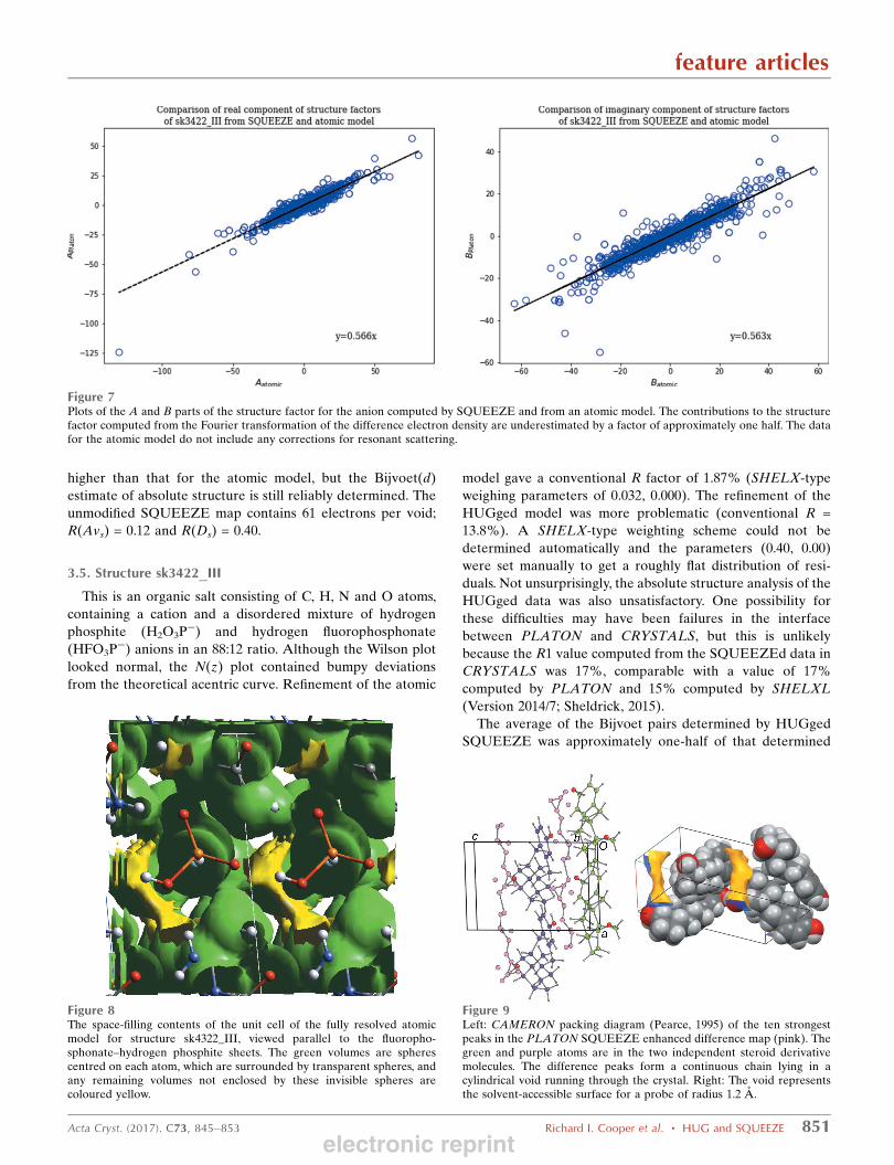
Figure 7Plots of the A and B parts of the structure factor for the anion computed by SQUEEZE and from an atomic model. The contributions to the structurefactor computed from the Fourier transformation of the difference electron density are underestimated by a factor of approximately one half. The datafor the atomic model do not include any corrections for resonant scattering.
Figure 8The space-filling contents of the unit cell of the fully resolved atomicmodel for structure sk4322_III, viewed parallel to the fluoropho-sphonate–hydrogen phosphite sheets. The green volumes are spherescentred on each atom, which are surrounded by transparent spheres, andany remaining volumes not enclosed by these invisible spheres arecoloured yellow.
Figure 9Left: CAMERON packing diagram (Pearce, 1995) of the ten strongestpeaks in the PLATON SQUEEZE enhanced difference map (pink). Thegreen and purple atoms are in the two independent steroid derivativemolecules. The difference peaks form a continuous chain lying in acylindrical void running through the crystal. Right: The void representsthe solvent-accessible surface for a probe of radius 1.2 A.
electronic reprint

from an atomic model for the anion (Fig. 6). This led us to
suspect that the root problem was the quality of the difference
density maps. Plots of the A and B parts for the anion alone
computed by SQUEEZE or an atomic model showed the
same discrepancy (Fig. 7).
In sgd240, the ratio of the electron count of the atoms in the
main moiety to that in the solvent was 11.6:1. In sk3422_III,
the ratio is only 1.3:1. The column headed (Zh + Zs)/Zh in
Table 1 shows that almost half of the total scattering is due to
the anion, which could have an influence in the scaling, but
clearly there is some other unidentified factor influencing the
poor performance of sk4322_III. The crystal structure consists
of layers of hydrogen-bonded chains of fluorophosphonate–
hydrogen phosphite sandwiched between layers of the organic
cations. Fig. 8 shows the space-filling contents of the unit cell
(MCE Version 2005 2.3.01) (Rohlıcek & Husak, 2007). The
yellow regions represent inaccessible volumes in the structure,
and it may be these which contribute to the problems.
3.6. Structure awisac02
This steroid derivative has a known absolute configuration.
There are two molecules in the asymmetric unit, which are
conformationally almost identical except for one hydroxy H
atom, but not related by any approximate symmetry element.
The original authors could not locate an atomic model for the
included solvent and SQUEEZEd the residual electron
density, interpreting the electron count for the solvent-acces-
sible volume as a disordered molecule of methylene
dichloride. Examination of the peaks found in the PLATON
difference synthesis showed that the residual density formed a
continuous chain in a channel through the structure (Fig. 9).
HUGging the SQUEEZE output for a single molecule of
CH2Cl2 reduced the conventional R factor to 6.56%. However,
the electron count in the cell voids from PLATON (95 e�) is
more than that for two single molecules (84 e�) of CH2Cl2.
Multiplying c and d (equation 10) by 1.5 (i.e. three molecules
of the solvent per unit cell) reduced the R factor to 5.92. The
refined Flack(x) of 0.1 (2) and Bijvoet(d) of 0.33 (3) are on the
correct side of 0.5, but are not convincing. The Hooft P(2)
probability does not compute and the P(3) probability is
strongly in favour of a twinned material.
3.7. Simulated diffuse solvent
During the review of this manuscript, one referee was
interested to know how HUGging would perform if the strong
resonant scatterers in the solvent were highly disordered. This
feature articles
852 Richard I. Cooper et al. � HUG and SQUEEZE Acta Cryst. (2017). C73, 845–853
Figure 10The modified structure of sgd464, showing the chloroform solvent at thebottom left of the image, with three individual Cl atoms replaced by aconvolution of the scattering factors of three Cl atoms and a ring. Theposition, orientation and size of the ring were determined by fittingthrough the original Cl-atom positions.
Figure 11For sgd464, the Fourier synthesis calculated from simulated data showsthat the electron density is distributed as intended. Mesh contours are setat arbitrary positive values.
Figure 12For sgd464, the Fourier synthesis computed from structure factorsincluding the SQUEEZEd A and B parts is a close approximation to theoriginal simulated data. Mesh contours are set at arbitrary positive values.
electronic reprint

situation could be studied by altering the model of compound
sgd464, which contains a well-ordered chloroform of solva-
tion. In order to simulate a very disordered solvent, the three
Cl atoms were replaced by an annular distribution equivalent
to the three Cl atoms (Schroder et al., 2004) (Fig. 10).
The Flack(x) parameter was set at 0.02 and the Uiso value
for the ring set at 0.06 A2. The structure factors computed
from this model were treated as (error free) observations, but
retaining the estimated standard uncertainties of the original
data; the R factor was 0.03%. The Fourier synthesis calculated
from this simulated data had the distributed electron density
shown in Fig. 11.
The whole chloroform residue, including the C and H
atoms, was deleted and the data SQUEEZEd. The Fourier
synthesis computed from structure factors including the
squeezed A and B parts was a close approximation to the
original synthesized data (Fig. 12). Refinement of the structure
including the SQUEEZEd A and B parts gave an R factor of
1.6% and a Flack(x) parameter of 2.9. HUGging and
reweighting the SQUEEZEd data gave an R factor of 1.7%
and a Flack(x) parameter of 0.02 (3), admirably close to the
value of 0.02 used in simulating the data. The Bijvoet(d)
parameter was 0.02 (1).
4. Conclusions
These preliminary observations show that an approximation
to the resonant scattering can be computed for a disordered
solvent molecule or counter-ion which may be adequate for
the determination of absolute structure. It seems that the
greatest chance of success occurs when the solvent/counter-ion
has significant resonant scattering, but its real scattering must
not overwhelm that of the host molecules.
Since the HUG algorithm is only a post-processing of the
output from SQUEEZE, the success of the method is critically
dependent on the applicability of SQUEEZE. The computa-
tion of c and d (equation 10) has no knowledge of the distri-
bution in the voids of the strong resonant scatterers so that the
HUG procedure can only be expected to be indicative of the
absolute structure. Except for awisac02 and the simulated data
above (x3.7), in the cases examined here, the solvent had been
modelled by discrete atoms so that the target results were
known. This will not be the case in real-life applications, but it
seems that if the absolute configuration of an enantiopure all
light-atom material is required, it makes sense to attempt to
recrystallize it from a solvent containing strong resonant
scatterers, even if there is a likelihood that these may be
incorporated as disordered solvent.
Funding information
Funding for this research was provided by: EPSRC (grant No.
EP/K013009/1 to RIC).
References
Bojarska, J., Maniukiewicz, W., Sieron, L., Fruzinski, A., Kopczacki,P., Walczynski, K. & Remko, M. (2012). Acta Cryst. C68, o341–o343.
Carruthers, J. R. (1977). In Proceedings of the 4th EuropeanCrystallographic Meeting (ECM-4), Oxford, UK, 30 August–3September 1977. Abstract Ob. 2.
Chernega, A. N., Davies, S. G., Goodwin, C. J., Hepworth, D.,Kurosawa, W., Roberts, P. M. & Thomson, J. E. (2009). Org. Lett.11, 3254–3257.
Cooper, R. I., Watkin, D. J. & Flack, H. D. (2016). Acta Cryst. C72,261–267.
Cruickshank, D. W. J. (1961). In Computing Methods and the PhaseProblem, edited by R. Pepinsky, J. M. Robertson & J. C. Speakman,Paper No. 6. Oxford: Pergamon Press.
Davies, S. G., Figuccia, A. L. A., Fletcher, A. M., Roberts, P. M. &Thomson, J. E. (2013a). Org. Lett. 15, 2042–2045.
Davies, S. G., Fletcher, A. M., Roberts, P. M., Thomson, J. E. &Zammit, C. M. (2013b). Chem. Commun. 49, 7037–7039.
Fabry, J., Fridrichova, M., Dusek, M., Fejfarova, K. & Krupkova, R.(2012). Acta Cryst. C68, o76–o83.
Flack, H. D. (1983). Acta Cryst. A39, 876–881.Flack, H. D. & Shmueli, U. (2007). Acta Cryst. A63, 257–265.Parsons, S., Flack, H. D. & Wagner, T. (2013). Acta Cryst. B69, 249–
259.Pearce, L. J. (1995). PhD thesis, University of Oxford, England.Qian, M., Engler-Chiurazzi, E. B., Lewis, S. E., Rath, N. P., Simpkins,
J. W. & Covey, D. F. (2016). Org. Biomol. Chem. 14, 9790–9805.Rohlıcek, J. & Husak, M. (2007). J. Appl. Cryst. 40, 600–601.Schroder, L., Watkin, D. J., Cousson, A., Cooper, R. I. & Paulus, W.
(2004). J. Appl. Cryst. 37, 545–550.Sheldrick, G. M. (2015). Acta Cryst. C71, 3–8.Shi, P., Zhang, L. & Ye, Q. (2012). Acta Cryst. C68, o266–o269.Sluis, P. van der & Spek, A. L. (1990). Acta Cryst. A46, 194–201.Spek, A. L. (2015). Acta Cryst. C71, 9–18.Watkin, D. & Schwarzenbach, D. (2017). J. Appl. Cryst. 50, 666–
667.Watkin, D. J., Carruthers, J. R. & Betteridge, P. W. (1985).CRYSTALS User Guide. Chemical Crystallography Laboratory,University of Oxford, England.
Watkin, D. J. & Cooper, R. I. (2016). Acta Cryst. B72, 661–683.
feature articles
Acta Cryst. (2017). C73, 845–853 Richard I. Cooper et al. � HUG and SQUEEZE 853electronic reprint
Related Documents