Front Biosci (Landmark Ed). 2013 Jan 1;18:626-37. 1 Hsp60 and human aging: Les liaisons dangereuses Francesco CAPPELLO, 1,2 Everly CONWAY DE MACARIO, 3 Antonella MARINO GAMMAZZA, 1,2 Anna M. CZARNECKA, 4 Felicia FARINA, 1 Giovanni ZUMMO, 1 and Alberto J. L. MACARIO 2,3 1 Department of Experimental Biomedicine and Clinical Neurosciences, Human Anatomy Section Emerico Luna, University of Palermo, Palermo, Italy. 2 IEMEST, Istituto Euro-Mediterraneo di Scienza e Tecnologia, Palermo, Italy. 3 Department of Microbiology and Immunology, School of Medicine, University of Maryland at Baltimore, and IMET, Baltimore (MD), USA 4 Department of Laboratory of Molecular Oncology, Department of Oncology, Military Institute of Medicine, ul. Szaserów 128, 01-141 Warsaw, Poland. TABLE OF CONTENTS 1. Abstract 2. Introduction 3. Essential background on chaperonology 4. Hsp60 molecular anatomy 5. Extracellular Hsp60 and immune system activation 6. Hsp60 in cell aging 7. Hsp60 in aging-related diseases 7.1. Atherosclerosis and heart failure 7.2. Neurodegenerative disorders 7.3. Degenerative joint diseases 7.4. Other pathologies 8. Summary and perspective 9. Acknowledgements 10. References 1. ABSTRACT Stressors can cause abnormal intracellular accumulation of Hsp60 and its localization in extramitochondrial sites, secretion, and circulation, with immune system activation. Dysfunction of chaperones associated with their quantitative and qualitative decline with aging (chaperonopathies of aging) characterizes senescence and is a potential causal factor in the physiological deterioration that occurs with it. The role of Hsp60 in aging is not easy to elucidate, because aging is accompanied by pathologies (e.g., cardiovascular and neurodegenerative disorders, osteoporosis, diabetes, cancer, etc.) in which Hsp60 has been implicated but, although those disorders are more frequent in the elderly, they are not unique to them. Therefore, it is difficult to determine what is due to aging and what to an associated disease that can occur regardless of age. Does Hsp60 contribute to the pathogenesis? How and when does Hsp60 interact with the immune system and, thus, contributes to the initiation-progression of the generalized chronic inflammation characteristic of aging? These and related issues are discussed here in the light of reports showing the participation of Hsp60 in aging-associated disorders. 2. INTRODUCTION Genes for molecular chaperones, and their protein products were identified in the early 1960’s and 1970’s, respectively (1, 2). After that, the study of chaperones, many of which are heat-shock proteins (Hsps), was very active in prokaryotic and eukaryotic systems, including the bacterial prokaryotes Escherichia coli and Bacillus subtilis, and the eukaryotes Drosophila, and a variety of mammals, plants and aquatic organisms (3-8). In the early 1990’s, a molecular chaperone gene was identified for the first time by cloning and sequencing in a prokaryote of the phylogenetic Domain Archaea (9). In later times, a number of studies have examined the role of molecular chaperones in protein folding inside the cell, and

Welcome message from author
This document is posted to help you gain knowledge. Please leave a comment to let me know what you think about it! Share it to your friends and learn new things together.
Transcript

Front Biosci (Landmark Ed). 2013 Jan 1;18:626-37.
1
Hsp60 and human aging: Les liaisons dangereuses
Francesco CAPPELLO,1,2 Everly CONWAY DE MACARIO,
3 Antonella MARINO
GAMMAZZA,1,2 Anna M. CZARNECKA,
4 Felicia FARINA,
1 Giovanni ZUMMO,
1 and Alberto J.
L. MACARIO2,3
1Department of Experimental Biomedicine and Clinical Neurosciences, Human Anatomy Section Emerico
Luna, University of Palermo, Palermo, Italy. 2IEMEST, Istituto Euro-Mediterraneo di Scienza e Tecnologia, Palermo, Italy.
3Department of Microbiology and Immunology, School of Medicine, University of Maryland at Baltimore,
and IMET, Baltimore (MD), USA 4Department of Laboratory of Molecular Oncology, Department of Oncology, Military Institute of
Medicine, ul. Szaserów 128, 01-141 Warsaw, Poland.
TABLE OF CONTENTS
1. Abstract
2. Introduction
3. Essential background on chaperonology
4. Hsp60 molecular anatomy
5. Extracellular Hsp60 and immune system activation
6. Hsp60 in cell aging
7. Hsp60 in aging-related diseases
7.1. Atherosclerosis and heart failure
7.2. Neurodegenerative disorders
7.3. Degenerative joint diseases
7.4. Other pathologies
8. Summary and perspective
9. Acknowledgements
10. References
1. ABSTRACT
Stressors can cause abnormal intracellular accumulation of Hsp60 and its localization in
extramitochondrial sites, secretion, and circulation, with immune system activation. Dysfunction of
chaperones associated with their quantitative and qualitative decline with aging (chaperonopathies of
aging) characterizes senescence and is a potential causal factor in the physiological deterioration that
occurs with it. The role of Hsp60 in aging is not easy to elucidate, because aging is accompanied by
pathologies (e.g., cardiovascular and neurodegenerative disorders, osteoporosis, diabetes, cancer, etc.) in
which Hsp60 has been implicated but, although those disorders are more frequent in the elderly, they are
not unique to them. Therefore, it is difficult to determine what is due to aging and what to an associated
disease that can occur regardless of age. Does Hsp60 contribute to the pathogenesis? How and when does
Hsp60 interact with the immune system and, thus, contributes to the initiation-progression of the
generalized chronic inflammation characteristic of aging? These and related issues are discussed here in
the light of reports showing the participation of Hsp60 in aging-associated disorders.
2. INTRODUCTION
Genes for molecular chaperones, and their protein products were identified in the early 1960’s
and 1970’s, respectively (1, 2). After that, the study of chaperones, many of which are heat-shock proteins
(Hsps), was very active in prokaryotic and eukaryotic systems, including the bacterial prokaryotes
Escherichia coli and Bacillus subtilis, and the eukaryotes Drosophila, and a variety of mammals, plants
and aquatic organisms (3-8). In the early 1990’s, a molecular chaperone gene was identified for the first
time by cloning and sequencing in a prokaryote of the phylogenetic Domain Archaea (9). In later times, a
number of studies have examined the role of molecular chaperones in protein folding inside the cell, and

Front Biosci (Landmark Ed). 2013 Jan 1;18:626-37.
2
chaperones were considered intracellular proteins (10-12). However, in the last several years evidence for
extracellular chaperones has progressively accumulated and, nowadays, there is little doubt that various
types of molecular chaperones can reside inside and outside cells with defined functions in both locations
(12-15). In most recent times, significant advances have occurred in the understanding of detailed
structure-function relationships in chaperones from the prokaryotes Archaea (16-18).
This article has been written for a Special Issue on Frontiers in Molecular Medicine and,
consequently, deals with a theme currently at the outer edge of science: the active participation of
defective molecular chaperones in pathogenesis. Diseases in which the primary cause, or one of the most
important secondary causes, is a defective chaperone have been called chaperonopathies (19, 20). This
unifying concept, encompassing a wide range of pathological conditions with diverse signs and symptoms
but sharing important features, is the foundation for the outlining of a new area of Medicine. The
scientific, medical, and practical advantages of such unifying approach are multiple and have been
discussed elsewhere (21).
The main objective of this article is to present some of the chaperonopathies, focusing on the
chaperone Hsp60, that affect aging individuals to illustrate how chaperone defects play a role in
senescence and aging-related diseases by failing to interact correctly with physiological partners or by
interacting with the wrong partner, namely by getting into “dangereuse liaisons.”
The literature on chaperones is abundant, so as to satisfy the bibliographic appetite of interested
readers who are not specialists in this article’s topic we have cited, whenever possible, review articles that
summarize key issues and provide a rich list of references to original work. In addition, we have
discussed some original data from various laboratories, including ours that we considered to be
illustrative of important aspects of chaperone failure, and its consequences, during aging. We have also
highlighted some of the critical points that merit further investigation with priority.
3. ESSENTIAL BACKGROUND ON CHAPERONOLY
As it may be realized from the preceding paragraphs, chaperonology is an emerging area of
science encompassing the study of molecular chaperones in all their aspects, normal and abnormal, as a
unit of related topics pertaining to chaperones in physiology and pathology examined from various angles
(22). Many chaperones are heat shock proteins (Hsps) and, in this article, we will use the terms chaperone
and Hsp interchangeably. This field is important because defective chaperones can contribute to the
pathogenesis of a number of diseases, now referred to as chaperonopathies (19, 20, 23). Chaperonopathies
can be genetic or acquired, the latter being quite common (20, 24).
The role of chaperones in human cell physiology changes from embryo/foetal life through
adulthood to old age, and they are involved in cell senescence (25-27). A number of studies have shown
that the stress-induced levels of chaperones tend to decrease with age (28). Pathological post-translational
modifications causing malfunction of chaperones seem to be implicated in the aging process by affecting
other molecules and supramolecular structures, cells, and tissues (20). There is some evidence indicating
that the age-associated appearance of defective chaperones (chaperonopathies of aging) contributes to the
accumulation of defective non-chaperone proteins (proteinopathies of aging) (27, 29-36). This
accumulation, in turn, causes a quantitative deficiency of chaperones, which thus become insufficient to
deal with the increased demand from proteins in need of assistance for folding, refolding, translocation,
etc. (30-32). Alternatively, chaperonopathies and proteinopathies of aging may start independently of one
another – perhaps simultaneously – and progress in parallel, in which case the chaperonopathies would
have a negative impact on protein homeostasis and, thus, contribute to the aggravation of the
proteinopathies (27, 35).
In this article, we will concentrate on Hsp60, the eukaryotic mitochondrial chaperonin that is
also found outside mitochondria, and discuss its role in cell senescence and organismal aging.
4. HSP60 MOLECULAR ANATOMY

Front Biosci (Landmark Ed). 2013 Jan 1;18:626-37.
3
Chaperonins are a subset of chaperones highly conserved during evolution and with essential
roles in cell physiology. They are classified in two groups: Group I, present in bacteria (e.g., GroEL) and
eukaryotic organelles (e.g., mitochondrial Hsp60 also called Cpn60), and Group II, found in archaea (e.g.,
thermosome subunits) and eukaryotic cytoplasm (e.g., CCT or TriC subunits) (37-40).
Hsp60 works in mitochondria together with its co-chaperonin Hsp10 (or Cpn10). In mammalian
cells, Hsp60 and Hsp10 are important mitochondrial molecules, playing key roles in both unstressed and
stressed cells (41, 42). The human genes for Hsp60 and Hsp10 are localised head-to-head on chromosome
2 and share a bidirectional promoter (43). This probably means that the DNA sequences encoding these
chaperonins moved together to the nucleus from a bacterium, according to the endosymbiotic theory (44).
Most of what we know about the eukaryotic Hsp60 and Hsp10 structures and functions derives from
studies on their prokaryotic homologues, the bacterial GroEL and GroES, respectively, to which they are
evolutionarily related.
The typical chaperonin machine in bacteria is formed by 14 GroEL molecules arranged in two
stacked heptameric rings delimiting a barrel-like container with an inner cavity large enough to
accommodate client polypeptides of up to nearly 60 kDa (40, 45, 46) (Figure 1A). GroES also forms a
heptameric ring, which associates with the GroEL barrel at one of its ends, serving as a sort of lid to the
GroEL-complex cavity (47). In addition, some information suggests that eukaryotic mitochondrial Hsp60
can form not only the typical two-ringed barrel, but it can also function as a single heptameric ring (48-
50) (Figure 1B). Moreover, the majority of the client proteins are affected by the inactivation of Hsp60,
but only a small subset of them are affected by the lack of Hsp10, suggesting that in eukaryotic cells
Hsp60 and Hsp10 do not always act together as a functional unit (Figure 1C) (51). Unfortunately, the full
range of Hsp60-dependent protein substrates in humans has not yet been fully delineated and this lack of
information curtails research on the Hsp60 chaperonopathies.
Apart from mitochondria, Hsp60 can be found in other subcellular compartments (e.g.,
zymogenic granules) as well as in the cytosol and on the cell membrane (52-54). In this context, one of
the most interesting issues for investigation is the structure and function of this chaperonin in sites
different from the canonical intra-mitochondrial location. For example, some data indicate that cytosolic
Hsp60, that is involved in apoptosis activation (55), is sometimes in a monomeric form (56), while other
results suggest equilibrium between monomeric and heptameric forms (57). It is likely that both situations
are not mutually exclusive. Furthermore, it is also possible that various oligomeric forms of different
multiplicities, depending mainly on the Hsp60 concentration in the cytosol, can occur and function.
In what concerns membrane-associated Hsp60, it is now clear that this association is a pre-
requisite for its secretion into the extracellular environment by secretory mechanisms involving the lipid
rafts/exosomes pathway, in both stressed and tumor cells (54, 58). In the extracellular environment,
Hsp60 encounters leukocytes and participates in immune system regulation, as discussed in the following
section.
5. EXTRACELLULAR Hsp60 AND IMMUNE SYSTEM ACTIVATION
Hsp60 has been for long time considered an intracellular protein but the reality is that it can also
be found in the extracellular milieu as well as in the bloodstream and its plasmatic levels seem to be
genetically controlled (59). A work on 60 subjects aged between 20 and 96 years of age showed that the
serum levels of Hsp60, but not those of anti-Hsp60 auto-antibodies, declined with aging (60). The reason
why the antibodies do not decline, and may even increase with age, could be that the immune system
reacts not only to the autologous Hsp60 but also to its homolog from microbial pathogens usually present
as persistent infections in the elderly (61). Furthermore, it is possible that post-translation modifications
occurring in Hsp60 as the age progresses make the autologous chaperonin immunogenic, thus stimulating
autoantibody production. Hence, although the number of Hsp60 molecules decreases with age, their
immunogenicity-antigenicity would increase.
An open question is how and when (i.e., during fetal development and after birth) Hsp60 is
released outside the cell. It has recently been shown that Hsp60 is secreted from normal adult rat

Front Biosci (Landmark Ed). 2013 Jan 1;18:626-37.
4
cardiomyocytes via the exosomal pathway (58) but, to our knowledge, no other similar observations have
been reported regarding normal human cells. We recently demonstrated with cultured cell lines that viable
tumor, but not normal cells secrete Hsp60 into the extracellular milieu by the lipid rafts-exosomal
pathway (54). The Hsp60 secretion pathway(s) of human senescent cells are currently poorly known.
Independently from the via of release, it is well established that presence of Hsp60 in the
extracellular space facilitates the contact with the immune system cells, promoting their activation; for
example, we recently showed that Hsp60 is present in macrophages of colon mucosa from patients with
ulcerative colitis and its levels decreased after an effective therapy, thus suggesting its role in maintaining
mucosal inflammation in these patients (62). Analogously, in another work on bronchial mucosa from
chronic obstructive pulmonary disease patients we found increased levels of Hsp60, compared to smoking
and non-smoking controls, both in epithelium and lamina propria, and, in the latter, this chaperonin
localized into neutrophils (manuscript submitted). In both cases, Hsp60 binding with inflammatory cells
may trigger or perpetuate immune system activation and, thus, disease progression. Although it is
possible that inflammatory cells in the airways are able to produce their own Hsp60, we cannot exclude
that, at least in part, Hsp60 reaches inflammatory cells after release from other cytotypes (e.g., epithelial
cells) and interact with receptors localised on the inflammatory-cell surface.
However, there is some controversy concerning whether or not immune-cell activation by
receptor-binding Hsp60 (e.g., using toll-like receptors) requires bacterial-derived products such as LPS,
flagellin, or lipoprotein (15, 63), a concern that has been ruled out for other Hsps (as Hsp70) by the use of
recombinant proteins isolated from insects and of other appropriate controls (64).
From the interstitium, Hsp60 can reach the bloodstream. When in circulation, Hsp60 appears to
be a key endogenous inflammatory mediator by causing the release of pro-inflammatory cytokines and
nitric oxide by immune competent cells (65). In contrast, other studies have demonstrated that induction
of immunity to Hsp60 can attenuate inflammatory diseases (65, 66). It is very difficult, if not impossible,
at the present time to always distinguish between situations in which Hsp60 plays a passive role as an
autoantigen and situations in which the chaperonin has an active role as a chaperokine (endocrine-like or
signalling function), inducing inflammation and/or an immune response to other antigens. What happens
and when may depend on a variety of factors, not completely understood. In addition, the role of Hsp60
during inflammation seems to depend on the subtypes of activated lymphocytes infiltrating tissues. For
example, in atherosclerosis (ATS) Hsp60 would have pro-inflammatory effects (Figure 2A) (67), whereas
in rheumatoid arthritis, it could have an anti-inflammatory action (Figure 2B) (68).
In any case, given the data available at present, one can assume that the changes in the immune
system response observed during aging are correlated, at least partially, to the decline in Hsp60 levels
and/or to structural changes in this chaperonin due, for example, to post-translational modifications.
Abnormal levels and/or structural damage in Hsp60 will most likely lead to scrambling of its interactions
with immune system components and, consequently, to pathogenesis (27). Hence, a future challenge will
be to shed more light into the nature and types of interactions between Hsp60 and the immune system
during aging.
6. HSP60 IN CELL AGING
Cell aging includes morphological, structural, and biochemical changes as part of a complex
biologic program whereby old individuals accumulate senescent cells in their bodies. Aging cells in
culture become flat and enlarged, developing extensive vacuolization and, in vitro as well as in vivo, they
show modified secretory pathways and a reduced ability to respond to stressors and to divide, with growth
arrest in late stages (69).
Some key components of the senescence process may be regulated by chaperones (70).
Analogously to what happens during the normal-dysplasia-carcinoma transition in various anatomical
sites (71-75), levels of Hsp60 increase in human skin fibroblasts during replicative senescence (69, 76)
(Figure 3), and the rapid increase in the levels of this chaperonin was positively correlated with cell cycle
progression (69). A correlation between increased levels of Hsp60 and senescence in human skin
Front Biosci (Landmark Ed). 2013 Jan 1;18:626-37.
5
fibroblasts was shown to involve interaction between Hsp60 and mtHsp70 (77). These in vitro results are
in agreement with in vivo studies that reported an increased Hsp60 expression in the forearm skin of
elderly subjects in comparison with young individuals, while other Hsps did not show differences (78).
The authors postulated that the chaperonin-level changes occurring with age are the consequence of the
mitochondrial oxidative stress characteristic of cell senescence.
Certain chaperones can inhibit caspase-dependent apoptosis, conferring immortality to the cell
(79). Hsp60 is one of these caspase-dependent apoptosis inhibitors (80). Moreover, exogenous Hsp60
produced by a persistent infection with Chlamydia trachomatis can also block the anti-apoptotic and the
pro-senescence effects of the host’s (endogenous) Hsp60, and in turn favour the active proliferation of
damaged cells (81). Finally, chronic infection with C. trachomatis with host invasion by the bacterial
Hsp60 generates anti-chaperonin antibodies that crossreact with the host’s counterpart and, thereby,
causes a variety of lesions in those locations in which the host’s Hsp60 happens to reside (61).
In summary, the pioneering studies discussed in the preceding paragraphs suggest that Hsp60
plays a role in the cell senescence process but the precise molecular mechanisms are not yet fully
elucidated and deserve further analysis, because their elucidation will provide useful clues for developing
preventive and treatment means applicable to Hsp60 chaperonopathies.
7. HSP60 IN AGING RELATED DESEASES
The number of papers showing the participation of Hsp60 in aging-related disease development
is constantly growing. The involvement of Hsp60 in the pathogenesis of aging-related diseases has been
studied with a number of approaches, both in vivo and in vitro, and at tissue, cellular, and subcellular
levels, as summarized in Table 1. Some of these papers are mainly based on observational studies and a
cause-effect relationship has not been yet properly investigated. Hsp60 in aging individuals seems to have
variable roles, which most likely depend on conditions in the cell, tissue, and organism affected. Hsp60
may metamorphose from a cell-protecting molecule to a dangerous one, thus being the Proteus of human
disease pathogenesis. This is possibly due to largely unknown mechanisms that regulate its gene
expression and to variables in the translation process as well as to post-translational modifications. In
addition, the roles of Hsp60 may be influenced by its interactions with other intracellular proteins and
other processes, including secretion into the extracellular space with the chaperonin becoming an
extracellular chaperone, signal molecule, autoantigen, or cytokine-like or endocrine-like molecule.
The involvement of Hsp60 in age-related tumor pathogenesis has been extensively discussed
elsewhere (82, 83). In the following subsections we will briefly discuss other widespread aging-related
pathological conditions that reduce significantly the quality and extent of life in Western countries.
7.1. Atherosclerosis and heart failure Although atherosclerosis (ATS) has been proposed to be an “autoimmune disease due to an
immune reaction against Hsp60” (67), the exact involvement of Hsp60 in the pathogenesis of ATS is still
confused by contradictory results. Therefore, further studies are necessary to definitively clarify the roles
of Hsp60 as etiologic factor and of the antibodies against it in sustaining the inflammation underlying the
pathogenesis of ATS (84-87). Also, it is pertinent to consider the contribution of molecular mimicry
between human and microbial Hsp60 in generating an autoimmune response that in turn causes
endothelial damage and artery inflammation (88-94). A number of in vivo data strongly support the
involvement of Hsp60 in the pathogenesis of coronary artery disease (59, 87, 95-98), but this view was
not supported by data from others (99).
With regard to heart failure, Hsp60 is gaining ranks as a clinical marker for monitoring this
condition (58, 80, 100). Hsp60 is overexpressed in the cytosol, localises in the cell membrane, and can
also be secreted by stressed myocardiocytes (58, 80). Higher than normal anti-Hsp60 antibody levels
were correlated with higher levels of brain natriuretic peptide, left ventricular end-diastolic dimension and
with the extent of cardiac dysfunction (101). If confirmed, these data may become of clinical utility for
heart failure management.
Front Biosci (Landmark Ed). 2013 Jan 1;18:626-37.
6
7.2. Neurodegenerative disorders Several kinds of stresses, among which nitrosidative stress, may cause accumulation of aberrant
proteins and neuronal cell damage or death (102). Thus, stress-induced proteins like some of the
chaperones have been proposed as protective molecules for the nervous system cells. For example,
overexpression of the hsp60 gene was observed in experimental subarachnoidal haemorrhage in rats,
possibly induced as a protective mechanism (103).
Hsp60 has been proposed as a good histological marker of the normality of unaffected cells in
not damaged areas in pathological brains (104), although it is extremely difficult to value its levels in vivo
in patients with Alzheimer’s, Parkinson’s, Huntington’s, and prion diseases in clinical practice.
Hsp60 levels, as well as those of other stress proteins, were found elevated in lymphocytes from
Alzheimer’s disease patients when compared to controls (105). It could be of some utility to test levels of
Hsp60 in patients with mild cognitive impairment (a clinical condition that precede Alzheimer’s disease
arise), for assessing the potential value of this protein as an early marker of the disease.
Lastly, since Hsp60 plays a role in the pathogenesis of ATS as discussed above, it can be
inferred that by similar mechanisms this chaperonin is implicated in cerebrovascular disorders of the
central nervous system, such as stroke, as also proposed by others (106).
7.3. Degenerative joint diseases
Hsp60 has been implicated in the pathogenesis of degenerative joint disease, both in young and
elderly people (66, 107, 108). It has been postulated that a humoral response against bacterial Hsp60
(exogenous chaperonin) could elicit a cross-reaction against the infected-host’s Hsp60 (endogenous
chaperonin) and other antigens in the synovial tissue in rheumatoid arthritis, thus perpetuating the local
inflammatory and destructive processes (108, 109).
7.4. Other pathologies Hsp60 levels and the presence of anti-chaperonin auto-antibodies have been studied in relation to
onset and progression of other aging-related diseases different from those already discussed above, such
as atrial fibrillation (110-112), glaucoma (113, 114), periodontitis (115, 116), osteoporosis (117, 118),
type II diabetes (119), and chronic obstructive pulmonary disease (120, 121). In these instances it is of the
essence to determine to what extent Hsp60 contributes to pathogenesis when defective or to protection
when normal, for elucidating the molecular mechanisms by which this chaperonin might trigger the
initiation, and/or modify the course, of these age-related disorders.
8. SUMMARY AND PERSPECTIVES
A positive correlation between longevity and capacity for mounting a strong heat-shock response
has been reported (122, 123), implying that chaperones are key components of adaptive mechanisms for
survival. Unfortunately, chaperone levels and/or functionality generally decrease with age. For example,
Hsp60 could be overloaded by the increasing demand due to the accumulation of damaged proteins that
occurs during senescence, which could result in a widespread chaperoning deficiency and lead to the
onset of aging-related disorders (30, 124). A shift in the balance between misfolded proteins and available
chaperonins, and other Hsps, in aging organisms, can bring about defects in signal transduction, protein
transport, cellular organization, and immune functions (30, 32).
All the information available at the present time indicates that Hsp60 is a key factor for protein
homeostasis and cell survival, and that this chaperonin is involved in various ways in the onset and
progression of aging-related diseases. We have surveyed in this review some of the pathological
conditions in which Hsp60 is implicated but there are many others which could also be listed here. In
addition, one has to assume that other syndromes and diseases will in the near future be found to have a
pathogenetic component involving Hsp60. In all these conditions, already described or to be found, it is
necessary to investigate in depth the mechanism of Hsp60 participation and, thus, uncover clues useful for
the development of diagnostic and therapeutic means, which ought to help considerably the management
of aging-related disorders.

Front Biosci (Landmark Ed). 2013 Jan 1;18:626-37.
7
9. ACKNOWLEDGEMENTS
This study was supported by funds of the Istituto Euro-Mediterraneo di Scienza e Tecnologia,
Palermo, Italy.
10. REFERENCES
1. F. Ritossa: A new puffing pattern induced by heat shock and DNP in Drosophila. Experentia 18, 571
(1962)
2. A. Tissières, HK Mitchell, UM Tracy: Protein synthesis in salivary glands of Drosophila
melanogaster: relation to chromosome puffs. J Mol Biol 84(3), 389-98 (1974)
3. F.C. Neidhardt, R.A. VanBogelen, V. Vaughn: The genetics and regulation of heat-shock proteins.
Annu Rev Genet 8, 295-329 (1984)
4. J.R. Subjeck, T. Shyy: Stress protein system of mammalian cells. Am J Physiol 250, C1 (1986)
5. K. Watson: Microbial stress proteins. Adv Microbial Physiol 31,183 (1990)
6. R.J. Ellis , S.M. Van der Vies : Molecular chaperones. Annu Rev Biochem 60, 321-47 (1991)
7. C. Georgopoulos, W.J. Welch: Role of the major heat shock proteins as molecular chaperones. Annu
Rev Cell Biol 9, 601-34 (1993)
8. B.M. Sanders: Stress protein in aquatic organisms: an environmental perspective. Crit Rev Toxicol 23,
49 (1993)
9. A.J.L. Macario, C.B. Dugan, E. Conway de Macario: A dnaK homolog in the archaebacterium
Mathanosarcina mazei S6. Gene 108, 133 (1991)
10. J. Frydman: Folding of newly translated proteins in vivo: the role of molecular chaperones. Annu Rev
Biochem 70, 603-47 (2001)
11. F.U. Hartl, M. Hayer-Hartl: Molecular chaperones in the cytosol: from nascent chain to folded
protein. Science 295, 1852-8 (2002)
12. J. Haak, K.C. Kregel: A cell stress odyssey. Novartis Found. Symp 291, 3-15 (2008)
13. F. Cappello, E. Conway de Macario, L. Marasa, G. Zummo, A.J.L. Macario: Hsp60 expression, new
locations, functions, and perspectives for cancer diagnosis and therapy. Cancer Biol, Ther 7, 801-809
(2008)
14. B. Henderson: Integrating the cell stress response: a new view of molecular chaperones as
immunological and physiological homeostatic regulators. Cell Biochem Funct 28, 1-14 (2010)
15. B. Henderson, S.K. Calderwood, A.R. Coates, I. Cohen, W. Van Eden, T. Lehner, A.G. Pockley:
Caught with their PAMPs down? The extracellular signaling actions of molecular chaperones are not due
to microbial contaminants. Cell Stress Chaperones 15, 123-41 (2010)
16. H. Luo, F.T. Robb: A modulator domain controlling thermal stability in the Group II chaperonins of
Archaea. Arch Biochem Biophys (2011) (In press.)
17. N.R. Douglas, S. Reissmann, J. Zhang, B. Chen, J. Jakana, R. Kumar, W. Chiu, J. Frydman: Dual
action of ATP hydrolysis couples lid closure to substrate release into the group II chaperonin chamber.
Cell 144(2), 240-52 (2011)
18. J. Zhang, B. Ma, F. Dimaio, N.R. Douglas, L.A. Joachimiak, D. Baker, J. Frydman, M. Levitt, W.
Chiu: Cryo-EM structure of a Group II chaperonin in the prehydrolysis ATP-bound state leading to lid
closure. Structure 19(5), 633-9 (2011)

Front Biosci (Landmark Ed). 2013 Jan 1;18:626-37.
8
19. A.J.L. Macario, E. Conway de Macario: The pathology of anti-stress mechanisms: A new frontier.
Stress 7, 243-249 (2004)
20. A.J.L. Macario, E. Conway de Macario: Sick chaperones, cellular stress, and disease. N Engl J Med
353, 1489-1501 (2005)
21. A.J.L. Macario: The chaperonopathies: a new field of medicine for study by physicians and all others
involved in medical sciences and public health. Medicine & Health 2012 published by Going
International, Vienna, Austria.
22. A.J.L. Macario, E. Conway de Macario: Chaperonopathies and chaperonotherapy. FEBS Letters 581,
3681-3688 (2007a)
23. A.J.L. Macario: Heat-shock proteins and molecular chaperones: Implications for pathogenesis,
diagnostics, and therapeutics. Intl J Clin Lab Res 25, 59-70 (1995)
24. A.J.L. Macario, T.M. Grippo, E. Conway de Macario: Genetic disorders involving molecular-
chaperone genes: A perspective. Genet Med 7, 3-12 (2005)
25. A.J.L. Macario, E. Conway de Macario: Molecular chaperones and age-related degenerative
disorders. Adv Cell Aging Gerontol 7, 131-162 (2001a)
26. A.J.L. Macario, E. Conway de Macario: Sick chaperones and ageing: A perspective. Ageing Res Rev
1, 295-311 (2002)
27. A.J.L Macario, F. Cappello, G. Zummo, E. Conway de Macario: Chaperonopathies of senescence and
the scrambling of the interactions between the chaperoning and the immune systems. Ann N Y Acad Sci
1197, 85-93 (2010)
28. R. Njemini, C. Demanet, T. Mets: Aging-related differences in basal heat shock protein 70 levels in
lymphocytes are linked to altered frequencies of lymphocyte subsets. Aging Cell 7, 498-505 (2008)
29. C. Soti, A.S. Sredhart, P. Csermely: Apoptosis, necrosis and cellular senescence: chaperone
occupancy as a potential switch. Aging Cell 2, 39-45 (2003)
30. C. Soti, P. Csermely: Aging and molecular chaperones. Exp Gerontol 38, 1037-1040 (2003)
31. C. Soti, P. Csermely: Aging cellular networks: chaperones as major participants. Exp Gerontol 42,
113-119 (2007a)
32. C. Soti, P. Csermely: Protein stress and stress proteins: Implication in aging and disease. J Biosci 32,
511-515 (2007b)
33. J. Bhattacharyya, E.V. Shipova, P. Santhoshkumar, K.K. Sharma, B.J. Ortwerth: Effect of a single
AGE modification on the structure and chaperone activity of human alphaB-crystallin. Biochemistry 46,
14682-14692 (2007)
34. P.A. Kumar, M.S. Kumar, G.B. Reddy: Effect of glycation on alpha-crystallin structure and
chaperone-like function. Biochem J 408, 251-258 (2007)
35. A.J.L. Macario, E. Conway de Macario: Chaperonopathies by defect, excess, or mistake. Ann N Y
Acad Sci 1113, 178-191 (2007b)
36. E.T. Soo, Y.K. Ng, B.H. Bay, G.W. Yip: Heat shock proteins and neurodegenerative disorders.
Scientific World J 8, 270-274 (2008)
37. A.J.L. Macario, E. Conway de Macario: The molecular chaperone system and other anti-stress
mechanisms in archaea. Front Biosci 6, d262-283 (2001b)
38. A.J.L. Macario, M. Lange, B.K. Haring, E. Conway de Macario: Stress genes and proteins in the
Archaea. Microbiol Mol Biol Reviews 63, 923-967 (1999)
39. A.J.L. Macario, M. Malz, E. Conway de Macario: Evolution of assisted protein folding: the
distribution of the main chaperoning systems within the phylogenetic domain archaea. Front Biosci 9,
1318-1332 (2004)

Front Biosci (Landmark Ed). 2013 Jan 1;18:626-37.
9
40. A.L. Horwich, W.A. Fenton, E. Chapman, G.W. Farr: Two families of chaperonins: physiology and
mechanism. Annu Rev Cell Dev Biol 23, 115-45 (2007)
41. S. Jindal, A.K. Dudani, B. Singh, C.B. Harley, R.S. Gupta: Primary structure of a human
mitochondrial protein homologous to the bacterial and plant chaperonins and to the 65-kilodalton
mycobacterial antigen. Mol Cell Biol 9, 2279-2283 (1989)
42. S.K. Sadacharan, A.C. Cavanagh, R.S. Gupta: Immunoelectron microscopy provides evidence for the
presence of mitochondrial heat shock 10-kDa protein (chaperonin 10) in red blood cells and a variety of
secretory granules. Histochem Cell Biol 116, 507-517 (2001)
42. J.J. Hansen, P. Bross, M. Westergaard, M.N. Nielsen, H. Eiberg, A.D. Borglum, J. Mogensen, K.
Kristiansen, L. Bolund, N. Gregersen: Genomic structure of the human mitochondrial chaperonin genes:
HSP60 and HSP10 are localised head to head on chromosome 2 separated by a bidirectional promoter.
Hum Genet 112, 71-77 (2003)
44. J.L. Blanchard, M. Lynch: Organellar genes: why do they end up in the nucleus? Trends Genet 16,
315-320 (2000)
45. K. Braig, Z. Otwinowski, R. Hegde, D.C. Boisvert, A. Joachimiak, A.L. Horwich, P.B. Sigler: The
crystal structure of the bacterial chaperonin GroEL at 2.8 A. Nature 371, 578-586 (1994)
46. N.A. Ranson, H.E. White, H.R. Saibil: Chaperonins. Biochem J 333, 233-242 (1998)
47. J.F. Hunt, A.J. Weaver, S.J. Landry, L. Gierasch, J. Deisenhofer: The crystal structure of the GroES
co-chaperonin at 2.8 A resolution. Nature 379, 37-45 (1996)
48. G. Levy-Rimler, R.E. Bell, N. Ben-Tal, A. Azem: Type I chaperonins: not all are created equal. FEBS
Lett. 529, 1-5 (2002)
49. K.L. Nielsen, N.J. Cowan: A single ring is sufficient for productive chaperonin-mediated folding in
vivo. Mol Cell 2, 93-99 (1998)
50. K.L. Nielsen, N. McLennan, M. Masters, N.J. Cowan: A single-ring mitochondrial chaperonin
(Hsp60-Hsp10) can substitute for GroEL-GroES in vivo. J Bacteriol 181, 5871-5875 (1999)
51. Y. Dubaquie, R. Looser, U. Funfschilling, P. Jeno, S. Rospert: Identification of in vivo substrates of
the yeast mitochondrial chaperonins reveals overlapping but non-identical requirement for hsp60 and
hsp10. EMBO J 17, 5868-5876 (1998)
52. J.D. Cechetto, B.J. Soltys, G.S. Gupta: Localization of mitochondrial 60-kD heat shock chaperonin
protein (Hsp60) in pituitary growth hormone secretory granules and pancreatic zymogen granules. J
Histochem Cytochem 48, 45-56 (2000)
53. B.J. Soltys, R.S. Gupta: Immunoelectron microscopic localization of the 60-kDa heat shock
chaperonin protein (Hsp60) in mammalian cells. Exp Cell Res 222(1), 16-27 (1996)
54. A.M. Merendino, F. Bucchieri, C. Campanella, V. Marcianò, A. Ribbene, S. David, G. Zummo, G.
Burgio, D.F. Corona, E. Conway de Macario, A.J.L. Macario, F. Cappello: Hsp60 is actively secreted by
human tumor cells. PLoS ONE 5, e9247 (2010)
55. D. Chandra, G. Choy, D.G. Tang: Cytosolic accumulation of HSP60 during apoptosis with or without
apparent mitochondrial release: evidence that its pro-apoptotic or pro-survival functions involve
differential interactions with caspase-3. J Biol Chem 282, 31289-301 (2007)
56. H. Taguchi, Y. Makino, M. Yoshida: Monomeric chaperonin-60 and its 50-kDa fragment possess the
ability to interact with non-native proteins, to suppress aggregation, and to promote protein folding. J Biol
Chem 269, 8529-8534 (1994)
57. G. Levy-Rimler, P. Viitanen, C. Weiss, R. Sharkia, A. Greenberg, A. Niv, A. Lustig, Y. Delarea, A.
Azem: The effect of nucleotides and mitochondrial chaperonin 10 on the structure and chaperone activity
of mitochondrial chaperonin 60. Eur J Biochem. 268, 3465-3472 (2001)
58. S. Gupta, A.A. Knowlton: HSP60 trafficking in adult cardiac myocytes: role of the exosomal
pathway. Am J Physiol Heart Circ Physiol 292, H3052-3056 (2007)

Front Biosci (Landmark Ed). 2013 Jan 1;18:626-37.
10
59. A. Shamaei-Tousi, A. Steptoe, K. O'Donnell, J. Palmen, J.W. Stephens, S.J. Hurel, M. Marmot, K.
Homer, F. D'Aiuto, A.R. Coates, S.E. Humphries, B. Henderson: Plasma heat shock protein 60 and
cardiovascular disease risk: the role of psychosocial, genetic, and biological factors. Cell Stress
Chaperones 12, 384-392 (2007)
60. I.M. Rea, S. McNerlan, A.G. Pockley: Serum heat shock protein and anti-heat shock protein antibody
levels in aging. Exp Gerontol 36, 341-35 (2001)
61. F. Cappello, E. Conway de Macario, V. Di Felice, G. Zummo, A.J.L. Macario: Chlamydia
trachomatis infection and anti-Hsp60 immunity: The two sides of the coin. PLoS Pathog 5(8), e1000552
(2009)
62. G. Tomasello, V. Rodolico, M. Zerilli, A. Martorana, F. Bucchieri, A. Pitruzzella, A.M. Gammazza,
S. David, F. Rappa, G. Zummo, P. Damiani, S. Accomando, M. Rizzo, E. Conway de Macario, A.J.L.
Macario, F. Cappello: Changes in immunohistochemical levels and subcellular localization after therapy
and correlation and colocalization with CD68 suggest a pathogenetic rRole of Hsp60 in ulcerative colitis.
Appl Immunohistochem Mol Morphol, (2011)
63. M.F. Tsan, B. Gao: Heat shock proteins and immune system. J Leuk Biol 85, 905-910 (2009)
64. A. De Maio: Extracellular heat-shock proteins, cellular export vesicles and the stress observational
system. A form of communication during injury, infection and cell damage. Cell Stress Chaperones 16,
235-249 (2011)
65. W. Van Eden, G. Wick, S. Albani, I. Cohen: Stress, heat shock proteins, and autoimmunity: how
immune responses to heat shock proteins are to be used for the control of chronic inflammatory diseases.
Ann N Y Acad Sci 1113, 217-237 (2007)
66. J.A. van Roon, W. van Eden, J.L. van Roy, F.J. Lafeber, J.W. Bijlsma: Stimulation of suppressive T
cell responses by human but not bacterial 60-kD heat-shock protein in synovial fluid of patients with
rheumatoid arthritis. J Clin Invest 100, 459-463 (1997)
67. G. Wick: Atherosclerosis--an autoimmune disease due to an immune reaction against heat-shock
protein 60. Herz 25, 87-90 (2000)
68. F.J. Quintana, A. Momran, P. Carmi, F. Mor, I.R. Cohen: Hsp60 as a target of anti-ergotypic
regulatory T cells. PLoS ONE 3(12), e4026 (2008)
69. V. Di Felice, N. Ardizzone, V. Marcianò, T. Bartolotta, F. Cappello, F. Farina, G. Zummo:
Senescence-associated HSP60 expression in normal human skin fibroblasts. Anat Rec 284A, 446-453
(2005a)
70. C.C. Deocaris, S.C. Kaul, R. Wadhwa: On the brotherhood of the mitochondrial chaperones mortalin
and heat shock protein 60. Cell Stress Chaperones 11, 116-128 (2006)
71. F. Cappello, M. Bellafiore, A. Palma, V. Marciano, G. Martorana, P. Belfiore, A. Martorana, F.
Farina, G. Zummo, F. Bucchieri: Expression of 60-kD heat shock protein increases during carcinogenesis
in the uterine exocervix. Pathobiology 70, 83-88 (2002)
72. F. Cappello, M. Bellafiore, A. Palma, S. David, V. Marcianò, T. Bartolotta, C. Sciumè, G. Modica, F.
Farina, G. Zummo, F. Bucchieri: 60KDa chaperonin (HSP60) is over-expressed during colorectal
carcinogenesis. Eur J Histochem 47, 105-110 (2003a)
73. F. Cappello, F. Rappa, S. David, R. Anzalone, G. Zummo: Immunohistochemical evaluation of
PCNA, p53, HSP60, HSP10 and MUC-2 presence and expression in prostate carcinogenesis. Anticancer
Res 23, 1325-1331 (2003b)
74. F. Cappello, S. David, F. Rappa, F. Bucchieri, L. Marasà, T.E. Bartolotta, F. Farina, G. Zummo: The
expression of HSP60 and HSP10 in large bowel carcinomas with lymph node metastases BMC Cancer 5,
139(2005a)
75. F. Cappello, A Di Stefano, SE D'Anna, CF Donner, G Zummo: Immunopositivity of heat shock
protein 60 as a biomarker of bronchial carcinogenesis. Lancet Oncol 6, 816 (2005b)

Front Biosci (Landmark Ed). 2013 Jan 1;18:626-37.
11
76. Y.H. Lee, J.C. Lee, H.J. Moon, J.E. Jung, M. Sharma, B.H. Park, H.K. Yi, E.C. Jhee: Differential
effect of oxidative stress on the apoptosis of early and late passage human diploid fibroblasts: implication
of heat shock protein 60. Cell Biochem Funct 26, 502-508 (2008)
77. Z. Kaul, T. Yaguchi, S.C. Kaul, R. Wadhwa: Quantum dot-based protein imaging and functional
significance of two mitochondrial chaperones in cellular senescence and carcinogenesis. Ann N Y Acad
Sci 1067, 469-473 (2006)
78. P. Gromov, G.L. Skovgaard, H. Palsdottir, I. Gromova, M. Østergaard, J.E Celis: Protein profiling of
the human epidermis from the elderly reveals up-regulation of a signature of interferon-gamma-induced
polypeptides that includes manganese-superoxide dismutase and the p85beta subunit of
phosphatidylinositol 3-kinase. Mol Cell Proteomics 2, 70-84 (2003)
79. J. Nylandsted, M. Rohde, K. Brand, L. Bastholm, F. Elling, M. Jäättelä: Selective depletion of heat
shock protein 70 (Hsp70) activates a tumor-specific death program that is independent of caspases and
bypasses Bcl-2. Proc Natl Acad Sci USA 97, 7871-7876 (2000)
80. L. Lin, S.C. Kim, Y. Wang, S. Gupta, B. Davis, S.I. Simon, G. Torre-Amione, A.A. Knowlton:
HSP60 in heart failure: abnormal distribution and role in cardiac myocyte apoptosis. Am J Physiol Heart
Circ Physiol 293, H2238-H2247 (2007)
81. V. Di Felice, S. David, F. Cappello, F. Farina, G. Zummo: Is chlamydial heat shock protein 60 a risk
factor for oncogenesis?. Cell Mol Life Sci 62, 4-9 (2005b)
82. F. Cappello, A.M. Czarnecka, G. La Rocca, A. Di Stefano, G. Zummo, A.J.L. Macario: Hsp60 and
Hsp10 as antitumor molecular agents. Cancer Biol Ther 6, 487-489 (2007)
83. F. Cappello, S. David, G. Peri, F. Farina, E. Conway de Macario, A.J.L. Macario, G. Zummo: Hsp60:
molecular anatomy and role in colorectal cancer diagnosis and treatment. Front Biosci 3, 341-51 (2011)
84. Q. Xu, G. Luef, S. Weimann, R.S. Gupta, H. Wolf, G. Wick: Staining of endothelial cells and
macrophages in atherosclerotic lesions with human heat-shock protein-reactive antisera. Arterioscler
Thromb 13, 1763-1769 (1993)
85. G. Schett, Q. Xu, A. Amberger, R. Van der Zee, H. Recheis, J. Willeit, G. Wick: Autoantibodies
against heat shock protein 60 mediate endothelial cytotoxicity. J Clin Invest 96, 2569-2577 (1995)
86. Q. Xu, G. Schett, H. Perschinka, M. Mayr, G. Egger, F. Oberhollenzer, J. Willeit, S. Kiechl, G. Wick:
Serum soluble heat shock protein 60 is elevated in subjects with atherosclerosis in a general population.
Circulation 102(1), 14-20 (2000)
87. Z. Prohaszka, J. Duba, L. Horvath, A. Csaszar, I. Karadi, A. Szebeni, M. Singh, B. Fekete, L. Romics,
G. Fust: Comparative study on antibodies to human and bacterial 60 kDa heat shock proteins in a large
cohort of patients with coronary heart disease and healthy subjects. Eur J Clin Invest 31, 285-292 (2001)
88. M. Mayr, B. Metzler, S. Kiechl, J. Willeit, G. Schett, Q. Xu, G. Wick: Endothelial cytotoxicity
mediated by serum antibodies to heat shock proteins of Escherichia coli and Chlamydia pneumoniae:
immune reactions to heat shock proteins as a possible link between infection and atherosclerosis.
Circulation 99, 1560-1566 (1999)
89. A. Ciervo, P. Visca, A. Petrucca, L.M. Biasucci, A. Maseri, A. Cassone: Antibodies to 60-kilodalton
heat shock protein and outer membrane protein 2 of Chlamydia pneumoniae in patients with coronary
heart disease. Clin Diag Lab Immunol 9, 66-74 (2002)
90. L.M. Biasucci, G. Liuzzo, A. Ciervo, A. Petrucca, M. Piro, D.J. Angiolillo, F. Crea, A. Cassone, A.
Maseri: Antibody response to chlamydial heat shock protein 60 is strongly associated with acute coronary
syndromes. Circulation 107, 3015-3017 (2003)
91. M. Knoflach, B. Mayrl, C. Mayerl, R. Sedivy, G. Wick: Atherosclerosis as a paradigmatic disease of
the elderly: role of the immune system. Immunol Allergy Clin North Am 23, 117-132 (2003)
92. S. Hoshida, M. Nishino, J. Tanouchi, T. Kishimoto, Y. Yamada: Acute Chlamydia pneumoniae
infection with heat-shock-protein-60-related response in patients with acute coronary syndrome.
Atherosclerosis 183, 109-112 (2005)

Front Biosci (Landmark Ed). 2013 Jan 1;18:626-37.
12
93. L.G. Spagnoli, S. Pucci, E. Bonanno, A. Cassone, F. Sesti, A. Ciervo, A. Mauriello: Persistent
Chlamydia pneumoniae infection of cardiomyocytes is correlated with fatal myocardial infarction. Am J
Pathol 170, 33-42 (2007)
94. V.Y. Hoymans, J.M. Bosmans, P.L. Van Herck, M.M. Ieven, C.J. Vrints: Implications of antibodies
to heat-shock proteins in ischemic heart disease. Int J Cardiol 123, 277-282 (2008)
95. A. Veres, G. Füst, M. Smiela, M. McQueen, A. Horváth, Q. Yi, A. Bíró, J. Pogue, L. Romics, I.
Karádi, M. Singh, J. Gnarpe, Z. Prohászka, S. Yusuf: Heart Outcomes Prevention Evaluation (HOPE)
Study Investigators. Relationship of anti-60 kDa heat shock protein and anti-cholesterol antibodies to
cardiovascular events. Circulation 106, 2775-2780 (2002)
96. J. Wysocki, B. Karawajczyk, J. Gorski, A. Korzeniowski, Z. Mackiewicz, G. Kupryszewski, R.
Glosnicka: Human heat shock protein 60 (409-424) fragment is recognized by serum antibodies of
patients with acute coronary syndromes. Cardiovasc Pathol 11, 238-243 (2002)
97. C. Lenzi, A. Palazzuoli, N. Giordano, G. Alegente, C. Gonnella, M.S. Campagna, A. Santucci, M.
Sozzi, P. Papakostas, F. Rollo, R. Nuti, N. Figura: H. pylori infection and systemic antibodies to CagA
and heat shock protein 60 in patients with coronary heart disease. World J Gastroenterol 12, 7815-7820
(2006)
98. G. Novo, F. Cappello, M. Rizzo, G. Fazio, S. Zambuto, E. Tortorici, A. Marino Gammazza, S.
Corrao, G. Zummo, E. Conway de Macario, A.J.L. Macario, P. Assennato, S. Novo, G. Li Volti: Hsp60
and heme oxygenase-1 (Hsp32) in acute myocardial infarction. Transl Res 157(5), 285-92 2011
99. A. Steptoe, A. Shamaei-Tousi, A. Gylfe, L. Bailey, S. Bergström, A.R. Coates, B. Henderson:
Protective effect of human heat shock protein 60 suggested by its association with decreased
seropositivity to pathogens. Clin Vaccine Immunol 14, 204-207 (2007)
100. N. Latif, P.M. Taylor, M.A. Khan, M.H. Yacoub, M.J. Dunn: The expression of heat shock protein
60 in patients with dilated cardiomyopathy. Basic Res Cardiol 94, 112-119 (1999)
101. D. Giannessi, C. Colotti, M. Maltinti, S. Del Ry, C. Prontera, S. Turchi, A. Labbate, D. Neglia:
Circulating heat shock proteins and inflammatory markers in patients with idiopathic left ventricular
dysfunction: their relationships with myocardial and microvascular impairment. Cell Stress Chaperones
12, 265-274 (2007)
102. T. Nakamura, Z. Gu, S.A. Lipton: Contribution of glutamatergic signaling to nitrosative stress-
induced protein misfolding in normal brain aging and neurodegenerative diseases. Aging Cell 6, 351-359
(2007)
103. M. Satoh, J. Tang, A. Nanda, J.H. Zhang: Heat shock proteins expression in brain stem after
subarachnoid hemorrhage in rats. Acta Neurochir Suppl 86, 477-482 (2003)
104. J.E. Martin, M. Swash, K. Mather, P.N. Leigh: Expression of the human GroEL stress-protein
homologue in the brain and spinal cord. J Neurol Sci 118, 202-206 (1993)
105. V. Calabrese, R. Sultana, G. Scapagnini, E. Guagliano, M. Sapienza, R. Bella, J. Kanski, G. Pennisi,
C. Mancuso, A.M. Stella, D.A. Butterfield: Nitrosative stress, cellular stress response, and thiol
homeostasis in patients with Alzheimer's disease. Antioxid Redox Signal 8, 1975-1986 (2006)
106. Q. Xiao, K. Mandal, G. Schett, G. Mayr, F. Oberhollenzen, J. Willeit, S. Kiechl, Q. Xu: Association
of serum-soluble heat shock protein 60 with carotid atherosclerosis: clinical significance determined in a
follow-up study. Stroke 36, 2571-6 (2005)
107. B. Wilbrink, M. Holewijn, J.W. Bijlsma, J.L. van Roy, W. den Otter, W. van Eden: Suppression of
human cartilage proteoglycan synthesis by rheumatoid synovial fluid mononuclear cells activated with
mycobacterial 60-kd heat-shock protein. Arthritis Rheum 36, 514-518 (1993)
108. V. Krenn, H.P. Vollmers, P. von Landenberg, B. Schmausser, M. Rupp, A. Roggenkamp, H.K.
Müller-Hermelink: Immortalized B-lymphocytes from rheumatoid synovial tissue show specificity for
bacterial HSP60. Virchows Arch 427, 511-518 (1996)

Front Biosci (Landmark Ed). 2013 Jan 1;18:626-37.
13
109. U. Rudolphi, R. Rzepka, S. Batsford, S.H. Kaufmann, K. von der Mark, H.H. Peter, I. Melchers: The
B cell repertoire of patients with rheumatoid arthritis. II. Increased frequencies of IgG+ and IgA+ B cells
specific for mycobacterial heat-shock protein 60 or human type II collagen in synovial fluid and tissue.
Arthritis Rheum 40, 1409-1419 (1997)
110. A.E. Schäfler, K. Kirmanoglou, J. Balbach, P. Pecher, A. Hannekum, B. Schumacher: The
expression of heat shock protein 60 in myocardium of patients with chronic atrial fibrillation. Basic Res
Cardiol 97, 258-61 (2002)
111. M. Yang, H. Tan, L. Cheng, M. He, Q. Wei, R.M. Tanguay, T. Wu: Expression of heat shock
proteins in myocardium of patients with atrial fibrillation. Cell Stress Chaperones 12, 142-50 (2007)
112. H. Cao, L. Xue, X. Xu, Y. Wu, J. Zhu, L. Chen, D. Chen, Y. Chen: Heat shock proteins in
stabilization of spontaneously restored sinus rhythm in permanent atrial fibrillation patients after mitral
valve surgery. Cell Stress Chaperones 2011 Apr 1. [Epub ahead of print]
113. M.B. Wax, G. Tezel, I. Saito, R.S. Gupta, J.B. Harley, Z. Li, C. Romano: Anti-Ro/SS-A positivity
and heat shock protein antibodies in patients with normal-pressure glaucoma. Am J Ophthalmol 125, 145-
157 (1998)
114. M.B. Wax, G. Tezel, J. Yang, G. Peng, R.V. Patil, N. Agarwal, R.M. Sappington, D.J. Calkins:
Induced autoimmunity to heat shock proteins elicits glaucomatous loss of retinal ganglion cell neurons
via activated T-cell-derived fas-ligand. J Neurosci 28, 12085-12096 (2008)
115. S.W. Chung, H.S. Kang, H.R. Park, S.J. Kim, S.J. Kim, J.I. Choi: Immune responses to heat shock
protein in Porphyromonas gingivalis-infected periodontitis and atherosclerosis patients. J Periodontal Res
38, 388-93 (2003)
116. J. Choi, S.Y. Lee, K. Kim, B.K. Choi: Identification of immunoreactive epitopes of the
Porphyromonas gingivalis heat shock protein in periodontitis and atherosclerosis. J Periodontal Res 46,
240-5 (2011)
117. S. Meghji, M. Lillicrap, M. Maguire, P. Tabona, J.S. Gaston, S. Poole, B. Henderson: Human
chaperonin 60 (Hsp60) stimulates bone resorption: structure/function relationships. Bone 33, 419-425
(2003)
118. Y.S. Kim, J.M. Koh, Y.S. Lee, B.J. Kim, S.H. Lee, K.U. Lee, G.S. Kim: Increased circulating heat
shock protein 60 induced by menopause, stimulates apoptosis of osteoblast-lineage cells via up-regulation
of toll-like receptors. Bone 45, 68-76 (2009)
119. A. Mostafazadeh, C. Herder, B. Haastert, P. Hanifi-Moghaddam, N. Schloot, W. Koenig, T. Illig, B.
Thorand, R. Holle, M.B. Eslami, H. Kolb, KORA Group: Association of humoral immunity to human
Hsp60 with the IL-6 gene polymorphism C-174G in patients with type 2 diabetes and controls. Horm
Metab Res 37, 257-263 (2005)
120. F. Cappello, A. Di Stefano, S. David, F. Rappa, R. Anzalone, G. La Rocca, S.E. D'Anna, F. Magno,
C.F. Donner, B. Balbi, G. Zummo: Hsp60 and Hsp10 down-regulation predicts bronchial epithelial
carcinogenesis in smokers with chronic obstructive pulmonary disease. Cancer 107, 2417-2424 (2006)
121. C.A. Carter, M. Misra, S. Pelech: Proteomic analyses of lung lysates from short-term exposure of
Fischer 344 rats to cigarette smoke. J Proteome Res (2011) (In press.)
122. D. Garigan, A.L. Hsu, A.G. Fraser, R.S Kamath, J. Ahringer, C. Kenyon: Genetic analysis of tissue
aging in Caenorhabditis elegans: a role for heat-shock factor and bacterial proliferation. Genetics 161,
1101-1112 (2002)
123. A.L. Hsu, C.T. Murphy, C. Kenyon: Regulation of aging and age-related disease by DAF-16 and
heat-shock factor. Science 300, 1142-1145 (2003)
124. P. Csermely: Chaperone overload is a possible contributor to 'civilization diseases'. Trends Genet 17,
701-704 (2001)

Front Biosci (Landmark Ed). 2013 Jan 1;18:626-37.
14
Abbreviations: Hsps: heath shock proteins; ATS: atherosclerosis; COPD: chronic obstructive pulmonary
disease
Key Words: Hsp60, Chaperoning system, chaperonin, Hsp10, senescence, chaperonopathies,
chaperonotherapy
Send correspondence to: Francesco Cappello, MD, Department of Experimental Biomedicine and
Clinical Neurosciences, Human Anatomy Section “Emerico Luna”. Via del Vespro 129, 90127 Palermo,
Italy. E-mail:[email protected]

Front Biosci (Landmark Ed). 2013 Jan 1;18:626-37.
15
Figure 1. Hsp60 functioning models in human cells. (A) Mammalian mitochondrial Hsp60 forms a
double heptameric-ring barrel-like structure with an inner cavity -- the folding chamber -- (blue cylinder),
analogously to the prokaryotic homolog GroEL (not shown). A client polypeptide, i.e., a polypeptide in
need of assistance for folding (black filament), enters the folding chamber of one of the rings (the light
blue cylinder in this figure) in which it will fold to gain the native conformation after the chamber is
occluded by the binding of Hsp10 (the homolog of the prokaryotic GroES; represented as a red cap). The
process requires energy from ATP hydrolysis. Another chaperoning round begins with the dissociation of
Hsp10, the release of the folded protein (four-pointed star) and ADP, and the binding of ATP to the other
ring. In addition, there is information suggesting that a similar folding process is carried out by a single
Hsp60 heptameric ring with Hsp10 (B), or even by a single ring without participation of Hsp10 (C), but
these two possibilities, particularly the last shown in (C) are still controversial.

Front Biosci (Landmark Ed). 2013 Jan 1;18:626-37.
16
Figure 2. Hsp60 can have both pro- and anti-inflammatory effects. (A) Stress (e.g., hypertension) on
vascular endothelial cells (top red thin rectangles) can determine Hsp60 (small blue squares)
overexpression, cell membrane localization and secretion (I-II). In turn, Hsp60 can have pro-
inflammatory effects on immune cells (lymphocytes, round blue circles with a nucleus; and macrophages,
stars with a nucleus; both shown here inside a blood vessel) infiltrating the vessel wall (II-III), which
induces, as a feedback effect, a perpetuation of inflammation (III-IV); finally, atherosclerotic lesions
develop (IV-V). An incipient atherosclerosis plaque is shown in V (see Ref. 67). (B) In an injured joint
(e.g., in rheumatoid arthritis) with inflammation of synovial tissue (I-II) (Hsp60 and lymphocytes are
represented as in A, above), Hsp60 peptide administration (III) can exert an anti-inflammatory effect (IV)
and contribute to joint lesion improvement (see Ref. 68).
Front Biosci (Landmark Ed). 2013 Jan 1;18:626-37.
17
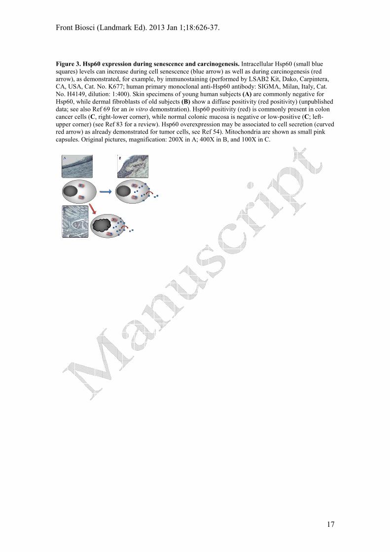
Figure 3. Hsp60 expression during senescence and carcinogenesis. Intracellular Hsp60 (small blue
squares) levels can increase during cell senescence (blue arrow) as well as during carcinogenesis (red
arrow), as demonstrated, for example, by immunostaining (performed by LSAB2 Kit, Dako, Carpintera,
CA, USA, Cat. No. K677; human primary monoclonal anti-Hsp60 antibody: SIGMA, Milan, Italy, Cat.
No. H4149, dilution: 1:400). Skin specimens of young human subjects (A) are commonly negative for
Hsp60, while dermal fibroblasts of old subjects (B) show a diffuse positivity (red positivity) (unpublished
data; see also Ref 69 for an in vitro demonstration). Hsp60 positivity (red) is commonly present in colon
cancer cells (C, right-lower corner), while normal colonic mucosa is negative or low-positive (C; left-
upper corner) (see Ref 83 for a review). Hsp60 overexpression may be associated to cell secretion (curved
red arrow) as already demonstrated for tumor cells, see Ref 54). Mitochondria are shown as small pink
capsules. Original pictures, magnification: 200X in A; 400X in B, and 100X in C.

Front Biosci (Landmark Ed). 2013 Jan 1;18:626-37.
18
Table 1. Aging-related pathologies in which Hsp60 is believed to play a pathogenetic role
Damaged
tissue/organ
Cells involved in
physiopathogenesis
Disease References
Vessels Endothelial cells,
macrophages
Vasculitis,
atherosclerosis
84-86, 88, 91
Heart Myocardiocytes Myocarditis, infarct,
heart failure
18, 58, 59, 80, 87, 89,
90, 92-94, 95-98, 100,
101
Conducting system cells Atrial fibrillation 110-112
Brain Neurons, glia Neurodegenerative
disorders
103-105
Joints Synoviocytes Degenerative joint
disease, rheumatoid
arthritis
66, 107-109
Bone Osteoblasts, osteoclasts Osteoporosis 117, 118
Pancreas Beta-cells Type II Diabetes 119
Bronchi Epithelial cells Chronic obstructive
pulmonary disease
120, 121
Eye Trabecular endothelial-
like cells of the
iridocorneal angle
Glaucoma 113
Oral cavity Gingival cells Periodontitis 115, 116
Related Documents