How We Know It Hurts: Item Analysis of Written Narratives Reveals Distinct Neural Responses to Others’ Physical Pain and Emotional Suffering Emile Bruneau*, Nicholas Dufour, Rebecca Saxe Department of Brain and Cognitive Sciences, Massachusetts Institute of Technology, Cambridge, Massachusetts, United States of America Abstract People are often called upon to witness, and to empathize with, the pain and suffering of others. In the current study, we directly compared neural responses to others’ physical pain and emotional suffering by presenting participants (n = 41) with 96 verbal stories, each describing a protagonist’s physical and/or emotional experience, ranging from neutral to extremely negative. A separate group of participants rated ‘‘how much physical pain’’, and ‘‘how much emotional suffering’’ the protagonist experienced in each story, as well as how ‘‘vivid and movie-like’’ the story was. Although ratings of Pain, Suffering and Vividness were positively correlated with each other across stories, item-analyses revealed that each scale was correlated with activity in distinct brain regions. Even within regions of the ‘‘Shared Pain network’’ identified using a separate data set, responses to others’ physical pain and emotional suffering were distinct. More broadly, item analyses with continuous predictors provided a high-powered method for identifying brain regions associated with specific aspects of complex stimuli – like verbal descriptions of physical and emotional events. Citation: Bruneau E, Dufour N, Saxe R (2013) How We Know It Hurts: Item Analysis of Written Narratives Reveals Distinct Neural Responses to Others’ Physical Pain and Emotional Suffering. PLoS ONE 8(4): e63085. doi:10.1371/journal.pone.0063085 Editor: Katsumi Watanabe, University of Tokyo, Japan Received October 10, 2012; Accepted March 30, 2013; Published April 26, 2013 Copyright: ß 2013 Bruneau et al. This is an open-access article distributed under the terms of the Creative Commons Attribution License, which permits unrestricted use, distribution, and reproduction in any medium, provided the original author and source are credited. Funding: Funding for this work was provided by the Air Force Office of Scientific Research, managed through the Office of Naval Research, grant number N000140910845. The funders had no role in study design, data collection and analysis, decision to publish, or preparation of the manuscript. Competing Interests: The authors have declared that no competing interests exist. * E-mail: [email protected] Introduction At the 1994 Olympics, the young running phenomenon Mary Decker turned the corner in the 3000-meter race she was favoured to win, and then suddenly collided with her running nemesis, Zola Budd, falling hard to the ground on her hip. A picture captures the moment as Mary Decker, tears streaming down her face and mouth open in anguish, watches as the runners continue without her. People who saw that event first hand, who have seen the Pulitzer prize-winning photograph, or even who just read this brief verbal description of the event, respond to two distinct (though related) aspects of Decker’s experience: the physical pain in her injured body, and the emotional suffering as she watched her Olympic dreams recede in front of her. In real life, misfortunes often combine physical pain and emotional suffering. Events that are emotionally painful without a direct physical cause (grieving over the loss of a loved one, or agonizing over unrequited love) are described in language borrowed from physical pain (‘‘feeling like you were hit in the gut’’, ‘‘love hurts’’). Conversely, simple physical injuries neverthe- less elicit strong emotions: fear, anger, anxiety, shame. When watching or reading about these events, do we recognize another person’s physical pain and understand their emotional suffering using a single unified neural system? Or are there distinct neural systems for these two processes? Recent neuroimaging studies have found evidence both hypotheses. On the one hand, a group of brain regions collectively called the ‘Shared Pain network,’ including parts of bilateral anterior insula (AI) and anterior middle cingulate cortex (AMCC), are recruited both when participants experience physical pain, and when they observe others experiencing similar pain [1–7] (but see [8,9]). Activity in AI and AMCC is correlated with trial-by-trial measurements of the intensity of physical pain experienced [10] or observed [11]. The response in these regions is influenced by the affective aspects of painful experiences, and not just the sensory aspects (for more details see [12]). For example, activity in insula and AMCC is modulated by participants’ anxiety and fear associated with anticipating pain, even prior to any actual painful sensation [13,14]. Finally, there is evidence that these same regions are recruited when experiencing, or witnessing another person experience, purely ‘‘social’’ suffering, e.g. during exclusion from a social interaction [15,16]. These results have been interpreted as evidence that AI and AMCC are the primary brain regions involved in responses to others’ physical and emotional suffering. On the other hand, other recent studies find that thinking about another person’s feelings of guilt, embarrassment and/or grief does not elicit activity in AI or AMCC [17,18]. Rather, thinking about another person’s feelings seems to predominantly lead to activity in a region of medial prefrontal cortex (DMPFC). The DMPFC is active while participants read verbal stories describing individuals experiencing emotional loss [19], and while participants read stories or look at cartoons, and then make inferences about the characters’ emotions [20,21]. Individuals with more activity in DMPFC while observing others’ suffering later offer more help to alleviate that suffering [16,22], and individuals who reported more frequently helping friends in their daily lives (in a diary study) show PLOS ONE | www.plosone.org 1 April 2013 | Volume 8 | Issue 4 | e63085

Welcome message from author
This document is posted to help you gain knowledge. Please leave a comment to let me know what you think about it! Share it to your friends and learn new things together.
Transcript
How We Know It Hurts: Item Analysis of WrittenNarratives Reveals Distinct Neural Responses to Others’Physical Pain and Emotional SufferingEmile Bruneau*, Nicholas Dufour, Rebecca Saxe
Department of Brain and Cognitive Sciences, Massachusetts Institute of Technology, Cambridge, Massachusetts, United States of America
Abstract
People are often called upon to witness, and to empathize with, the pain and suffering of others. In the current study, wedirectly compared neural responses to others’ physical pain and emotional suffering by presenting participants (n = 41) with96 verbal stories, each describing a protagonist’s physical and/or emotional experience, ranging from neutral to extremelynegative. A separate group of participants rated ‘‘how much physical pain’’, and ‘‘how much emotional suffering’’ theprotagonist experienced in each story, as well as how ‘‘vivid and movie-like’’ the story was. Although ratings of Pain,Suffering and Vividness were positively correlated with each other across stories, item-analyses revealed that each scale wascorrelated with activity in distinct brain regions. Even within regions of the ‘‘Shared Pain network’’ identified using a separatedata set, responses to others’ physical pain and emotional suffering were distinct. More broadly, item analyses withcontinuous predictors provided a high-powered method for identifying brain regions associated with specific aspects ofcomplex stimuli – like verbal descriptions of physical and emotional events.
Citation: Bruneau E, Dufour N, Saxe R (2013) How We Know It Hurts: Item Analysis of Written Narratives Reveals Distinct Neural Responses to Others’ PhysicalPain and Emotional Suffering. PLoS ONE 8(4): e63085. doi:10.1371/journal.pone.0063085
Editor: Katsumi Watanabe, University of Tokyo, Japan
Received October 10, 2012; Accepted March 30, 2013; Published April 26, 2013
Copyright: � 2013 Bruneau et al. This is an open-access article distributed under the terms of the Creative Commons Attribution License, which permitsunrestricted use, distribution, and reproduction in any medium, provided the original author and source are credited.
Funding: Funding for this work was provided by the Air Force Office of Scientific Research, managed through the Office of Naval Research, grant numberN000140910845. The funders had no role in study design, data collection and analysis, decision to publish, or preparation of the manuscript.
Competing Interests: The authors have declared that no competing interests exist.
* E-mail: [email protected]
Introduction
At the 1994 Olympics, the young running phenomenon Mary
Decker turned the corner in the 3000-meter race she was favoured
to win, and then suddenly collided with her running nemesis, Zola
Budd, falling hard to the ground on her hip. A picture captures the
moment as Mary Decker, tears streaming down her face and
mouth open in anguish, watches as the runners continue without
her. People who saw that event first hand, who have seen the
Pulitzer prize-winning photograph, or even who just read this brief
verbal description of the event, respond to two distinct (though
related) aspects of Decker’s experience: the physical pain in her
injured body, and the emotional suffering as she watched her
Olympic dreams recede in front of her.
In real life, misfortunes often combine physical pain and
emotional suffering. Events that are emotionally painful without
a direct physical cause (grieving over the loss of a loved one, or
agonizing over unrequited love) are described in language
borrowed from physical pain (‘‘feeling like you were hit in the
gut’’, ‘‘love hurts’’). Conversely, simple physical injuries neverthe-
less elicit strong emotions: fear, anger, anxiety, shame.
When watching or reading about these events, do we recognize
another person’s physical pain and understand their emotional
suffering using a single unified neural system? Or are there distinct
neural systems for these two processes? Recent neuroimaging
studies have found evidence both hypotheses. On the one hand,
a group of brain regions collectively called the ‘Shared Pain
network,’ including parts of bilateral anterior insula (AI) and
anterior middle cingulate cortex (AMCC), are recruited both when
participants experience physical pain, and when they observe
others experiencing similar pain [1–7] (but see [8,9]). Activity in
AI and AMCC is correlated with trial-by-trial measurements of
the intensity of physical pain experienced [10] or observed [11].
The response in these regions is influenced by the affective aspects
of painful experiences, and not just the sensory aspects (for more
details see [12]). For example, activity in insula and AMCC is
modulated by participants’ anxiety and fear associated with
anticipating pain, even prior to any actual painful sensation
[13,14]. Finally, there is evidence that these same regions are
recruited when experiencing, or witnessing another person
experience, purely ‘‘social’’ suffering, e.g. during exclusion from
a social interaction [15,16]. These results have been interpreted as
evidence that AI and AMCC are the primary brain regions
involved in responses to others’ physical and emotional suffering.
On the other hand, other recent studies find that thinking about
another person’s feelings of guilt, embarrassment and/or grief does
not elicit activity in AI or AMCC [17,18]. Rather, thinking about
another person’s feelings seems to predominantly lead to activity in
a region of medial prefrontal cortex (DMPFC). The DMPFC is
active while participants read verbal stories describing individuals
experiencing emotional loss [19], and while participants read
stories or look at cartoons, and then make inferences about the
characters’ emotions [20,21]. Individuals with more activity in
DMPFC while observing others’ suffering later offer more help to
alleviate that suffering [16,22], and individuals who reported more
frequently helping friends in their daily lives (in a diary study) show
PLOS ONE | www.plosone.org 1 April 2013 | Volume 8 | Issue 4 | e63085
greater DMPFC response to depictions of emotional suffering
[23]. These results suggest that AI and AMCC are predominantly
recruited when witnessing others in physical pain, and DMPFC is
more implicated in empathic responses to emotional suffering.
One interpretive challenge for prior studies, however, may be
that – as in our initial example – witnessing another person’s
misfortune often carries elements of both physical pain and
emotional suffering. Watching someone receive an electric shock,
or be poked by a painful needle, we may spontaneously anticipate
their fear and sadness, as well as their physical discomfort. Hearing
about someone heading into a terrifying final exam, we may
spontaneously imagine the clenched gut and sweaty palms of their
physical experience. How can these correlated features of
experience be separated in an experiment?
The present study sought to disentangle the naturally con-
founded experiences of witnessing physical pain and emotional
suffering using a parametric item-analysis [24]. In the study,
participants read verbal stories that varied in how much physical
pain and how much emotional suffering the protagonist endured.
We obtained behavioral ratings of each story from a separate
group of participants, on how much physical pain and emotional
suffering the main character experienced, and how vivid the verbal
description was; we then compared these ratings with brain
responses to each story across participants.
These data represent a subset of the data that were analyzed
previously [25] (see methods for details). As with most neuroima-
ging studies, stories in this previous analysis were assigned to
categories based on the subjective judgment of the experimenter:
for example, a story about a boy who falls while walking on a picket
fence and is left hanging by his broken leg is categorized as
‘‘Physical Pain’’, and a story about a father and son who find out
that the child has terminal cancer is categorized as ‘‘Emotional
Pain’’. Within a condition, the intensity of pain and suffering
inevitably varied, and this was treated as a source of noise: the
story about the boy with the broken leg and a story about a girl
who burns her finger on a hot light bulb are treated equally as
‘‘Physical Pain’’ items. However, this standard fMRI analysis
technique ignores two important points: first, physical pain and
emotional suffering are conflated in each category (the boy left
hanging on the fence certainly felt physical pain, but also certainly
suffered from the terror, embarrassment and confusion); and
second, variability in the stories could help to illuminate brain
regions that are sensitive to that variation, rather than merely
being treated as noise. Therefore, in the present item analysis
[26,27] we use independent ratings to simultaneously characterize
each item across multiple dimensions (e.g. both how much
physical pain and how much emotional suffering each story
depicts), and treat variability of items (e.g. burning a finger versus
breaking a leg) within ‘conditions’ as an opportunity for additional
statistical leverage on the effects of interest. Since so many
phenomena of interest to social neuroscience are continuous
rather than categorical (e.g. fear, disgust, pain), and inter-related
rather than orthogonal (e.g. pain and suffering, shame and
humiliation), this analysis strategy could be leveraged effectively in
other studies involving complex social stimuli, and therefore could
be of great utility to social cognitive neuroscience.
Methods
ParticipantsfMRI data were collected from 41 naı̈ve right-handed
participants (18–37 years old (mean 23.064.8 s.d.), 25 females).
A separate group of fourteen participants (19–33 years old (mean
23.564.1 s.d.), 8 female) engaged in the localizer experiment. The
study protocol was approved by MIT’s Committee on the Use of
Humans as Experimental Subjects, and was conducted at the
Athinoula A. Martinos Imaging Center in the McGovern Institute
for Brain Research at the Massachusetts Institute of Technology.
All participants were proficient English speakers, had normal or
corrected to normal vision, gave written informed consent, and
were compensated for their time.
The item ratings were obtained from approximately 30
‘‘workers’’ per story on Amazon’s Mechanical Turk (mTurk), an
online interface; stories were presented one at a time, and
individual workers rated stories ad libitum. These workers gave
informed consent to participate, and were compensated per story,
in accordance with the requirements of MIT’s Committee on the
Use of Humans as Experimental Subjects.
Design and MaterialsForty-eight verbal stories involving mild to extreme physical or
emotional pain were constructed (‘‘high pain/suffering’’); a milder
form of each story was also created, in which the characters and
scene remained unchanged but the physical or emotional pain
were eliminated or lessened (‘‘low pain/suffering’’). This resulted
in 96 total stories. Stories varied in the depicted environment, the
number of people involved, and age and gender of the protagonist,
but were approximately matched for length (mean: 46.9 words
63.5 s.d.) and complexity (Flesch reading level: 86.366.2 s.d.).
(For sample stories, see Table 1; for full list of stimuli, see
Methods S1).
An additional 48 stories involving false beliefs were also
presented but were excluded from the analysis. In these stories,
the protagonist’s false beliefs always led to emotions (there were no
stories in which a character experienced physical pain based on
a false belief), and could therefore bias our analysis to find
differences between emotional and physical stories, which truly
reflected only the presence of false beliefs.
Half the participants in the study were told to ‘‘rate the pain/
suffering’’ experienced by the protagonist of each story, and half
the participants were told to ‘‘actively empathize’’ with the
protagonist of each story [25]. We have previously reported that
participants’ explicit task had little effect on neural responses to
these stimuli; we therefore average across both groups of
participants in the current analysis.
Behavioral ratings. The 96 short stories were presented one
at a time, using the online interface of mTurk. mTurk ‘‘workers’’
rated each story on three separate 9-point scales (anchored at
‘‘none at all’’ and ‘‘extreme’’):
Pain: ‘‘How much physical pain was the main character in?’’
Suffering: ‘‘How much emotional suffering did the main
character experience?’’
Vividness: ‘‘How vivid and ‘movie-like’ was this scenario to
you?’’
The 96 stories were each rated by 40 people for ‘‘pain’’ and
‘‘suffering’’, and by 20 people for ‘‘vividness’’. A worker’s
responses to an item were dropped if: 1. they gave any response
except ‘9’ (‘‘completely agree’’) for a ‘catch’ question (‘‘I read the
story and answered all the questions honestly’’), or 2. the response
times were unreasonably fast (unrealistic reading rates of .600
words per minute). These restrictions resulted in the exclusion of
approximately 15% of the responses. After excluding these data,
each story was rated across the dimensions of ‘‘pain’’ and
‘‘suffering’’ by 26–40 people (mean 34.664.0 s.d.), and across
the dimension of ‘‘vividness’’ by 12–20 people (1762.1).
fMRI imaging. Participants in the fMRI study each read 48
of the 96 stories. The sample of items was approximately balanced
across participants (to ensure every participant saw approximately
Neural Responses to Others’ Pain and Suffering
PLOS ONE | www.plosone.org 2 April 2013 | Volume 8 | Issue 4 | e63085
12 stories with the highest levels of emotional suffering and
physical pain) but otherwise randomly chosen. The number of
participants who saw each specific story was 19.963.8 s.d. (range:
11–29).
Each run consisted of 4 blocks, separated by 12 s fixation
periods; within each block, 3 stories were presented for 16 s each,
separated by 2 s. In total, each run contained 12 stories, 2 from
each of the original six conditions, in counterbalanced order, and
lasted 4.6 minutes; the experiment consisted of 6 total runs. While
in the scanner, participants were asked to respond to each story
using a MRI-safe 4-button box.
Image Acquisition and Analysis. Participants were scanned
using a Siemens Magnetom Tim Trio 3T System (Siemens
Solutions, Erlangen, Germany) in the Athinoula A. Martinos
Imagining Center at the McGovern Institute for Brain Research at
MIT using 30 4-mm-thick near axial slices with whole brain
coverage (TR = 2 s, TE = 30 ms, flip angle = 90). The
experiment used a block design, and was modeled using a boxcar
regressor.
MRI data were analyzed using SPM8 (http://www.fil.ion.ucl.
ac.uk/spm/software/spm8/), and custom software. Each partic-
ipant’s data were motion corrected and normalized onto
a common brain space (Montreal Neurological Institute, MNI,
Template). Data were smoothed using a Gaussian filter (full width
half maximum = 5 mm).
For whole brain analyses, we first built a modified linear model
of the experimental design to analyze the BOLD response in each
voxel. The model included both covariates of interest (the stimulus
items) and nuisance covariates – run effects, an intercept term, and
global signal. We modeled each item as a box-car (matching the
onset and duration of each story) convolved with a standard
hemodynamic response function (HRF). Time-series data were
subjected to a high-pass filter (128 Hz).
Average beta responses were calculated across participants for
each story. Random Effects analysis was performed across the
item-wise beta-images [26], using the ratings on each question
(Pain, Suffering, Vividness) as user-defined regressors. These
analyses used corrected p thresholds of p,0.05 in a voxel-wise
analysis using Statistical non-Parametric Mapping (SnPM).
Because these predictors were correlated, it was interesting to
ask whether they nevertheless predict independent components of
the neural response; therefore analyses were also conducted
including all three regressors simultaneously in a single model,
with p thresholds at k.10, p,0.001, uncorrected.
For regions of interest analyses, a set of ROIs was generated
from a separate study in which participants directly viewed
another person experiencing a painful and unpleasant electrical
shock in real time. In this ‘‘Pain Localizer’’ study, participants in
the scanner directly observed a confederate’s hand as the
confederate received a painful and unpleasant electric shock
(versus a barely perceptible, non-painful shock) to the visible hand.
Based on activity in this task, we identified 8 functional regions of
interest (ROIs) involved in experiencing and/or perceiving pain or
suffering: bilateral secondary sensory cortex (l and r SII), bilateral
insula, anterior middle cingulate cortex (AMCC), bilateral
thalamus, and dorsal medial prefrontal cortex (DMPFC) (for
details, see Methods S1 and [25]). Then, the average response (i.e.
beta value) within each ROI for the current participants was
determined for each of the 96 stories.
Principal Component Analysis was applied to the 8696 matrix
of the average betas for the 96 stories in 8 ROIs. These
components were then correlated with the behavioral ratings of
Pain, Suffering and Vividness. Factor correlations were de-
termined for each behavioral rating individually, and also for the
residual of each rating after the other two ratings had been
accounted for.
To ensure that the ratings of Pain and Suffering explained
variance in the neural data beyond the variance that could be
explained by the original binary categorization of the stories, we
performed a two-step regression: first, we accounted for the
variance in the item-wise neural data within the regions of interest
using the categorical regressors of Pain (a ‘1’ for stories designed to
focus on Physical Pain, and a ‘0’ for all other stories) and Suffering
(a ‘1’ for stories designed to focus on Emotional Suffering, and a ‘0’
for all other stories). Second, we used the behavioral ratings of
Pain and Suffering for each story as continuous regressors and
determined if these regressors explained the residual variance.
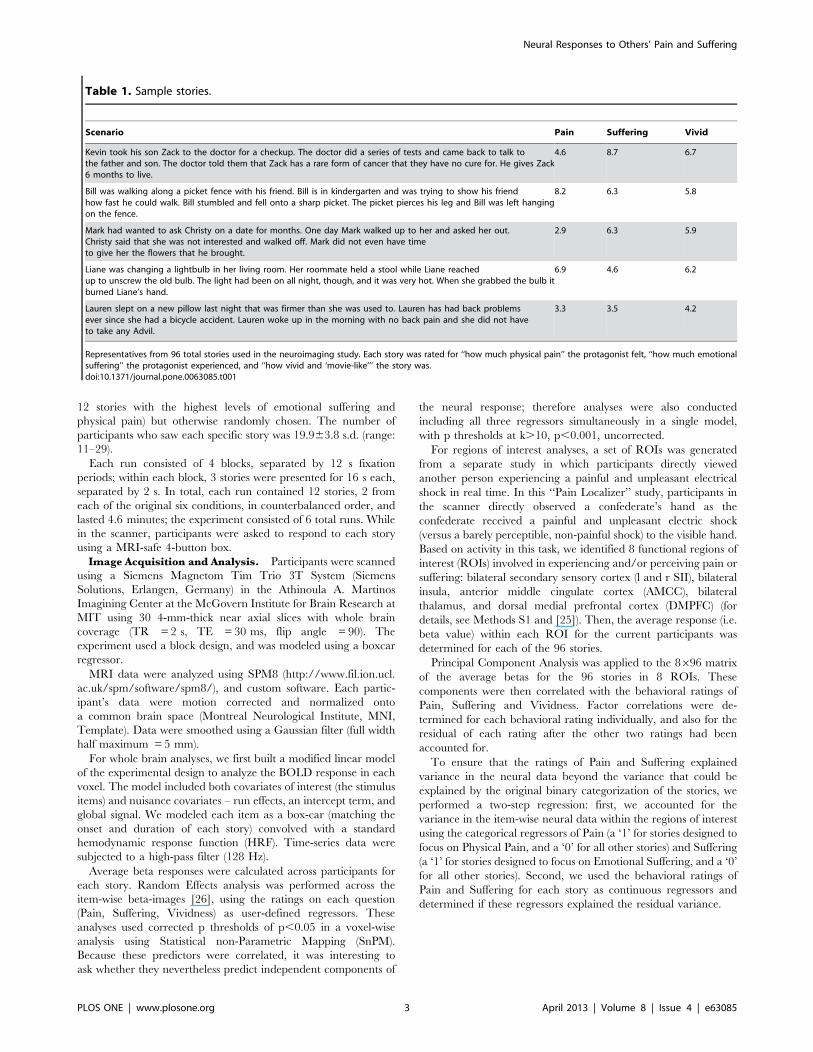
Table 1. Sample stories.
Scenario Pain Suffering Vivid
Kevin took his son Zack to the doctor for a checkup. The doctor did a series of tests and came back to talk tothe father and son. The doctor told them that Zack has a rare form of cancer that they have no cure for. He gives Zack6 months to live.
4.6 8.7 6.7
Bill was walking along a picket fence with his friend. Bill is in kindergarten and was trying to show his friendhow fast he could walk. Bill stumbled and fell onto a sharp picket. The picket pierces his leg and Bill was left hangingon the fence.
8.2 6.3 5.8
Mark had wanted to ask Christy on a date for months. One day Mark walked up to her and asked her out.Christy said that she was not interested and walked off. Mark did not even have timeto give her the flowers that he brought.
2.9 6.3 5.9
Liane was changing a lightbulb in her living room. Her roommate held a stool while Liane reachedup to unscrew the old bulb. The light had been on all night, though, and it was very hot. When she grabbed the bulb itburned Liane’s hand.
6.9 4.6 6.2
Lauren slept on a new pillow last night that was firmer than she was used to. Lauren has had back problemsever since she had a bicycle accident. Lauren woke up in the morning with no back pain and she did not haveto take any Advil.
3.3 3.5 4.2
Representatives from 96 total stories used in the neuroimaging study. Each story was rated for ‘‘how much physical pain’’ the protagonist felt, ‘‘how much emotionalsuffering’’ the protagonist experienced, and ‘‘how vivid and ‘movie-like’’’ the story was.doi:10.1371/journal.pone.0063085.t001
Neural Responses to Others’ Pain and Suffering
PLOS ONE | www.plosone.org 3 April 2013 | Volume 8 | Issue 4 | e63085
Results
Behavioral resultsAcross stories, ratings of Pain, Suffering and Vividness were all
significantly positively correlated with each other (Pain-Suffering:
pearson’s r = 0.43; Pain-Vivid: r = 0.52; Suffering-Vivid: r = 0.76,
all p-values ,0.001). We next tested whether these behaviorally
inter-correlated features of the stories were represented in similar
or distinct brain regions.
Whole brain random effects analysesWe performed three whole-brain item-wise Random Effects
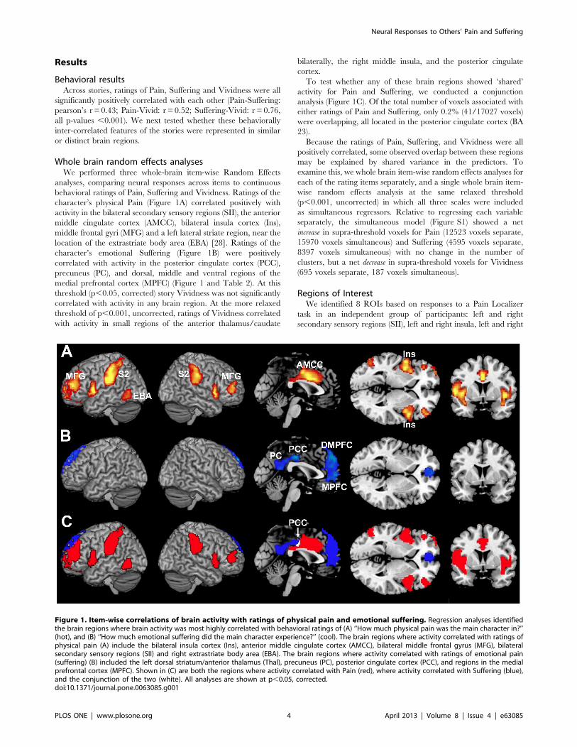
analyses, comparing neural responses across items to continuous
behavioral ratings of Pain, Suffering and Vividness. Ratings of the
character’s physical Pain (Figure 1A) correlated positively with
activity in the bilateral secondary sensory regions (SII), the anterior
middle cingulate cortex (AMCC), bilateral insula cortex (Ins),
middle frontal gyri (MFG) and a left lateral striate region, near the
location of the extrastriate body area (EBA) [28]. Ratings of the
character’s emotional Suffering (Figure 1B) were positively
correlated with activity in the posterior cingulate cortex (PCC),
precuneus (PC), and dorsal, middle and ventral regions of the
medial prefrontal cortex (MPFC) (Figure 1 and Table 2). At this
threshold (p,0.05, corrected) story Vividness was not significantly
correlated with activity in any brain region. At the more relaxed
threshold of p,0.001, uncorrected, ratings of Vividness correlated
with activity in small regions of the anterior thalamus/caudate
bilaterally, the right middle insula, and the posterior cingulate
cortex.
To test whether any of these brain regions showed ‘shared’
activity for Pain and Suffering, we conducted a conjunction
analysis (Figure 1C). Of the total number of voxels associated with
either ratings of Pain and Suffering, only 0.2% (41/17027 voxels)
were overlapping, all located in the posterior cingulate cortex (BA
23).
Because the ratings of Pain, Suffering, and Vividness were all
positively correlated, some observed overlap between these regions
may be explained by shared variance in the predictors. To
examine this, we whole brain item-wise random effects analyses for
each of the rating items separately, and a single whole brain item-
wise random effects analysis at the same relaxed threshold
(p,0.001, uncorrected) in which all three scales were included
as simultaneous regressors. Relative to regressing each variable
separately, the simultaneous model (Figure S1) showed a net
increase in supra-threshold voxels for Pain (12523 voxels separate,
15970 voxels simultaneous) and Suffering (4595 voxels separate,
8397 voxels simultaneous) with no change in the number of
clusters, but a net decrease in supra-threshold voxels for Vividness
(695 voxels separate, 187 voxels simultaneous).
Regions of InterestWe identified 8 ROIs based on responses to a Pain Localizer
task in an independent group of participants: left and right
secondary sensory regions (SII), left and right insula, left and right
Figure 1. Item-wise correlations of brain activity with ratings of physical pain and emotional suffering. Regression analyses identifiedthe brain regions where brain activity was most highly correlated with behavioral ratings of (A) ‘‘How much physical pain was the main character in?’’(hot), and (B) ‘‘How much emotional suffering did the main character experience?’’ (cool). The brain regions where activity correlated with ratings ofphysical pain (A) include the bilateral insula cortex (Ins), anterior middle cingulate cortex (AMCC), bilateral middle frontal gyrus (MFG), bilateralsecondary sensory regions (SII) and right extrastriate body area (EBA). The brain regions where activity correlated with ratings of emotional pain(suffering) (B) included the left dorsal striatum/anterior thalamus (Thal), precuneus (PC), posterior cingulate cortex (PCC), and regions in the medialprefrontal cortex (MPFC). Shown in (C) are both the regions where activity correlated with Pain (red), where activity correlated with Suffering (blue),and the conjunction of the two (white). All analyses are shown at p,0.05, corrected.doi:10.1371/journal.pone.0063085.g001
Neural Responses to Others’ Pain and Suffering
PLOS ONE | www.plosone.org 4 April 2013 | Volume 8 | Issue 4 | e63085
anterior thalamus, anterior middle cingulate cortex (AMCC) and
dorsomedial prefrontal cortex (DMPFC). In each ROI, average
beta values were extracted for each story.
In a principal component analysis (PCA) of the responses of
these eight regions across items, the first principal component
explained 60% of the variance. The DMPFC, left thalamus and
right thalamus ROIs loaded positively on this component, while
the remaining 5 ROIs (left and right SII, left and right insula,
AMCC) loaded negatively (Table 3). Also, this first principle
component was strongly positively correlated with ratings of
Suffering (r = 0.35, p,0.0005), negatively correlated with ratings
of Pain (r =20.27, p,0.01), and uncorrelated with ratings of
Vividness (r = 0.17, p = 0.11), even though ratings of Pain,
Suffering, and Vividness were positively correlated across items.
We also conducted this analysis using only the residual variance
for each of these ratings that was unshared with the other two
ratings. Again, the residual variance in Suffering and Pain were
strongly correlated with the first principal component of ROI
responses (Suffering: r = 0.38, p,0.0005; Pain: r =20.44,
p,0.0001), while Vividness was not (r =20.01, p = 0.95).
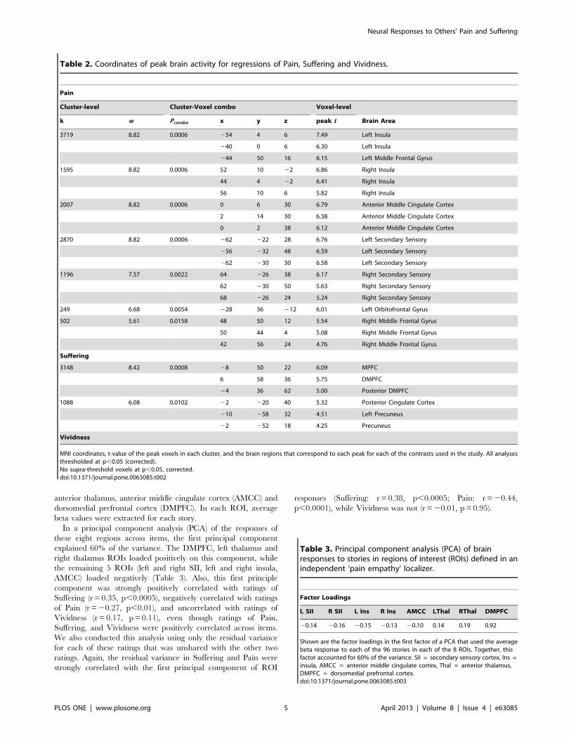
Table 2. Coordinates of peak brain activity for regressions of Pain, Suffering and Vividness.
Pain
Cluster-level Cluster-Voxel combo Voxel-level
k w Pcombo x y z peak t Brain Area
3719 8.82 0.0006 254 4 6 7.49 Left Insula
240 0 6 6.30 Left Insula
244 50 16 6.15 Left Middle Frontal Gyrus
1595 8.82 0.0006 52 10 22 6.86 Right Insula
44 4 22 6.41 Right Insula
56 10 6 5.82 Right Insula
2007 8.82 0.0006 0 6 30 6.79 Anterior Middle Cingulate Cortex
2 14 30 6.38 Anterior Middle Cingulate Cortex
0 2 38 6.12 Anterior Middle Cingulate Cortex
2870 8.82 0.0006 262 222 28 6.76 Left Secondary Sensory
256 232 48 6.59 Left Secondary Sensory
262 230 30 6.58 Left Secondary Sensory
1196 7.57 0.0022 64 226 38 6.17 Right Secondary Sensory
62 230 50 5.63 Right Secondary Sensory
68 226 24 5.24 Right Secondary Sensory
249 6.68 0.0054 228 36 212 6.01 Left Orbitofrontal Gyrus
502 5.61 0.0158 48 50 12 5.54 Right Middle Frontal Gyrus
50 44 4 5.08 Right Middle Frontal Gyrus
42 56 24 4.76 Right Middle Frontal Gyrus
Suffering
3148 8.42 0.0008 28 50 22 6.09 MPFC
6 58 36 5.75 DMPFC
24 36 62 5.00 Posterior DMPFC
1088 6.08 0.0102 22 220 40 5.32 Posterior Cingulate Cortex
210 258 32 4.51 Left Precuneus
22 252 18 4.25 Precuneus
Vividness
MNI coordinates, t-value of the peak voxels in each cluster, and the brain regions that correspond to each peak for each of the contrasts used in the study. All analysesthresholded at p,0.05 (corrected).No supra-threshold voxels at p,0.05, corrected.doi:10.1371/journal.pone.0063085.t002
Table 3. Principal component analysis (PCA) of brainresponses to stories in regions of interest (ROIs) defined in anindependent ‘pain empathy’ localizer.
Factor Loadings
L SII R SII L Ins R Ins AMCC LThal RThal DMPFC
20.14 20.16 20.15 20.13 20.10 0.14 0.19 0.92
Shown are the factor loadings in the first factor of a PCA that used the averagebeta response to each of the 96 stories in each of the 8 ROIs. Together, thisfactor accounted for 60% of the variance. SII = secondary sensory cortex, Ins =insula, AMCC = anterior middle cingulate cortex, Thal = anterior thalamus,DMPFC = dorsomedial prefrontal cortex.doi:10.1371/journal.pone.0063085.t003
Neural Responses to Others’ Pain and Suffering
PLOS ONE | www.plosone.org 5 April 2013 | Volume 8 | Issue 4 | e63085
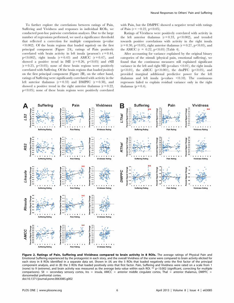
To further explore the correlations between ratings of Pain,
Suffering and Vividness and responses in individual ROIs, we
conducted post-hoc pairwise correlation analyses. Due to the large
number of regressions performed, we used a significance threshold
that reflected a correction for multiple comparisons (p-value
,0.002). Of the brain regions that loaded negatively on the first
principal component (Figure 2A), ratings of Pain positively
correlated with brain activity in left insula (pearson’s r = 0.44,
p,0.002), right insula (r = 0.43) and AMCC (r = 0.47), and
showed a positive trend in lSII (r = 0.26, p,0.05) and rSII
(r = 0.25, p,0.05); none of these brain regions were positively
correlated with Suffering. Of the brain regions that loaded positively
on the first principal component (Figure 2B), on the other hand,
ratings of Suffering were significantly correlated with activity in the
left anterior thalamus (r = 0.33) and DMPFC (r = 0.38), and
showed a positive trend in the right anterior thalamus (r = 0.22,
p,0.05); none of these brain regions were positively correlated
with Pain, but the DMPFC showed a negative trend with ratings
of Pain (r =20.22, p,0.05).
Ratings of Vividness were positively correlated with activity in
the left anterior thalamus (r = 0.33, p,0.002), and trended
towards positive correlations with activity in the right insula
(r = 0.30, p,0.05), right anterior thalamus (r = 0.27, p,0.05), and
the AMCC (r = 0.22, p,0.05) (Table 4).
After accounting for variance explained by the original binary
categories of the stimuli (physical pain, emotional suffering), we
found that the continuous measures still explained significant
variance in the left and right SII (p-values ,0.01), the right insula
(p,0.01), the aMCC (p,0.001), the dmPFC (p,0.05), and
provided marginal additional predictive power for the left
thalamus and left insula (p-values ,0.10). The continuous
regressors failed to explain residual variance only in the right
thalamus (p = 0.4).
Figure 2. Ratings of Pain, Suffering and Vividness compared to brain activity in 8 ROIs. The average ratings of Physical Pain andEmotional Suffering experienced by the protagonist in each story, and the overall Vividness of the scene were compared to brain activity elicited foreach story in 8 ROIs identified in a separate data set. Shown in (A) are the 5 ROIs that loaded negatively onto the first factor of the principalcomponent analysis, and in (B) the 3 ROIs that loaded positively onto that first factor. Pain, Suffering and Vividness were rated on a scale from 1(none) to 9 (extreme), and brain activity was measured as the average beta value within each ROI. ** p,0.002 (significant, correcting for multiplecomparisons). SII = secondary sensory cortex, Ins = insula, AMCC = anterior middle cingulate cortex, Thal = anterior thalamus, DMPFC =dorsomedial prefrontal cortex.doi:10.1371/journal.pone.0063085.g002
Neural Responses to Others’ Pain and Suffering
PLOS ONE | www.plosone.org 6 April 2013 | Volume 8 | Issue 4 | e63085
Discussion
In our stimuli, as in many real life experiences, the physical
pain, emotional suffering, and vividness of the experience were all
positively correlated. Positive correlations between distinct features
of complex social stories raise a challenge for neuroimaging
experiments. In the current experiment, we addressed that
challenge using parametric item analysis. This analysis allowed
us to go beyond the limits of both standard subtraction techniques,
which require that stimuli be assigned to a small number of
discrete conditions, and of regions of interest analyses, which focus
on constrained brain regions, to look at the association and
dissociation between continuous cognitive processes.
Using item analysis, we found that different brain regions are
correlated with the amount of physical pain versus emotional
suffering depicted in verbal stories. Across 96 stories, the amount
of physical pain experienced by the protagonist predicted neural
activity in bilateral secondary sensory regions, bilateral insulae,
anterior middle cingulate cortex, bilateral middle frontal gyri and
a left lateral extrastriate region, possibly the left EBA [29]. By
contrast, the amount of emotional suffering depicted in the same
stories was correlated with activity in the precuneus, posterior
cingulate, and medial prefrontal cortex. Converging results
appeared in whole brain and regions of interest analyses, and in
spite of the fact that ratings of Pain, Suffering and Vividness were
positively correlated in the stimuli. These results therefore strongly
suggest that people have distinct neural responses to other people
experiencing physical pain versus emotional suffering.
The parametric item analysis has multiple methodological
advantages over more standard subject-wise analyses used in our
prior papers [19,25]: (1) the behavioral ratings used to predict
neural response to each item were assigned by naive participants
(rather than by the experimenter), (2) multiple correlated
dimensions of the stimuli could be studied simultaneously, (3) we
explicitly tested whether the observed relationships can be
generalized beyond the current sample of stimuli, by treating
items as a random effect [24,26,27] (4) the statistical power of the
analyses is related to the number of items (n = 96), which is greater
than the number of participants (n = 41), and (5) variance within
a ‘condition’ could be used as statistical leverage, rather than
ignored as noise. Item analysis can thus provide strong evidence
for the association between a brain region’s activity and intrinsic
dimensions of high-level complex stimuli, like vignettes about
other people (rather than participants’ specific experiences of the
stimuli).
Consistent with our previous analyses of these data [25], the
current results show that simply reading about another person’s
physical pain can produce activity in the ‘Shared Pain network’. In
prior research, these regions (especially bilateral insula and middle
cingulate cortex) were recruited when participants viewed images
of body parts threatened by needles and knives [4,7,30,31],
watched videos of people’s faces while they undergo painful
physical therapy [5,11], were cued that a loved one was receiving
an electric shock [6], or were reminded of a documentary about
another’s pain [3,32]. In the current experiment, these same
regions’ responses were robustly correlated with continuous ratings
of physical pain experienced by a (fictional) stranger in a verbal
story. It is particularly interesting that we observed significant
responses in right and left secondary sensory cortex to verbal
stories (although note that the responses in the sensory regions
were low overall); just imagining sensory experiences appears to be
sufficient to modulate sensory cortices [33]. These results may
have practical implications, since written stories can be transmitted
so much further, and faster, than direct dyadic social interactions.
One striking feature of these results is that they suggest
a functional divide between two subsets of the ‘Shared Pain
network’. We identified 8 regions of interest based on a standard
Pain Localizer task, in which participants experienced, and
directly witnessed another person experiencing, painful and
unpleasant electric shocks to the hand. This localizer is most
likely to identify brain regions associated with the perception of
acute, temporary, concrete, discrete, physical pain. For this reason
it is particularly interesting that all of these regions are modulated
by verbal stories describing painful experiences that are longer
lasting, more distant in space and time and presented more
abstractly – and for some regions, experiences that are more
emotionally than physically painful.
Interestingly, rather than being explained by 2 separate
components of equal weight, the data in the ROIs were best
described by a single component of the regions’ responses across
items, which accounted for 60% of the variance. This component
had a positive loading of left thalamus, right thalamus and DMPF,
and a negative loading of bilateral insula, bilateral sensory regions,
and the AMCC. This component in turn correlated positively with
ratings of emotional Suffering and negatively with ratings of
physical Pain, but was not correlated with ratings of story
Vividness. While the overall PCA suggests that activity in the
ROIs is best explained by a single, anti-correlated component,
follow-up pairwise correlations within each ROI indicated that
most regions were correlated with either the physical Pain or the
emotional Suffering ratings. Analysis within the ROIs showed that
only the middle cingulate region showed any hint of a positive
correlation with both dimensions, and only the DMPFC showed
a significant positive correlation with one (Suffering) and a negative
correlation with the other (Pain).
The distinction between the two sets of brain regions responding
to others physical pain versus emotional suffering may reflect
distinct evolutionary histories. It is possible that responses to
others’ physical pain evolved earlier, followed by a second system
that evolved in evolutionarily more recent regions (i.e. prefrontal
cortex) as human social cognition developed. If, as suggested by
the PCA, these two neural responses are anti-correlated, rather
than simply uncorrelated, this could also be adaptive. It may be
prudent to prioritize attention to another’s physical pain over their
emotional suffering until an immediate physical threat is
Table 4. Item-wise correlations between brain activity andbehavioral ratings within specific regions of interest.
Suffering Pain Vivid
lSII 20.19 0.26* 0.02
rSII 20.09 0.25* 0.00
lIns 0.01 0.44** 0.17
rIns 0.19 0.43** 0.30*
aMCC 0.21* 0.47** 0.22*
lThal 0.33** 0.17 0.33**
rThal 0.21* 0.07 0.27*
dmPFC 0.38** 20.22* 0.19
Behavioral ratings for each story for Pain, Suffering and Vividness werecorrelated with average brain activity in 8 regions of interest defined in anindependent data set. SII = secondary sensory cortex, Ins = insula, AMCC =anterior middle cingulate cortex, Thal = anterior thalamus, DMPFC =dorsomedial prefrontal cortex.*p,0.05.**p,0.002.doi:10.1371/journal.pone.0063085.t004
Neural Responses to Others’ Pain and Suffering
PLOS ONE | www.plosone.org 7 April 2013 | Volume 8 | Issue 4 | e63085
addressed: coming upon the scene of a child hanging by their leg
on a picket fence, or finding a relative gored by a wild animal, first
attending to liberating them from the physically dangerous
situation, and then addressing their emotional trauma may be
more likely to save their lives. Further studies, potentially
employing temporally sensitive methods like EEG or MEG, may
help to resolve this issue of uncorrelated versus anti-correlated
activity in the brain regions responsive to others’ physical Pain and
emotional Suffering.
How do the current results fit with the previous literature? In
apparent contrast to the dissociation observed here, multiple prior
studies have reported simultaneous activity in regions that we
found to be uncorrelated with each other in the current analyses,
such as AI, AMCC and DMPFC [1,3,11,34]. We suggest that in
those stimuli, as in real life, physical pain almost always includes
some aspect of emotional suffering, so both processes may have
been initiated in these previous studies. In our story stimuli, ratings
of pain and suffering were strongly positively correlated. In the
story empathy experiment we were able to distinguish these
regions, by using a continuous parametric item analysis to separate
correlations with emotional suffering versus physical pain in the
same stimuli.
Our results converge with considerable prior evidence that
regions in the MPFC are critical for affective perspective-taking
[35–37]. DMPFC activity is observed when stimuli elicit a strong
sense of a target’s emotions – including people’s faces [1,5,11] or
the context of their suffering [5]. By contrast, just seeing images of
a body part threatened by a needle, for example, does not lead to
activity in MPFC [4,7,30,31]. Similarly, a recent meta-analysis
found that regions in the MPFC were recruited when a physically
present live person was subjected to a painful experience (as it was
in our Pain Localizer experiment), but not when similar injuries
were depicted in photographs (note that the MPFC region in the
current analysis is closer to the region labeled ‘‘VMPFC’’ than
‘‘DMPFC’’, in the meta-analysis [32]).
Identifying the MPFC as a region that recognizes emotional
suffering, but not physical pain, provides an interesting in-
terpretation of a prior result on inter-group empathic failures.
Black and white Americans, looking at images of other black and
white Americans after a natural disaster (Hurricane Katrina),
showed activity in both ‘pain’ (e.g. insula) and ‘suffering’ (e.g.
MPFC) brain regions, but MPFC activity was reduced for
outgroup targets [22]. In another study, neural responses in
Arabs, Israelis and South Americans to stories involving ingroup,
conflict outgroup and ‘distant’ outgroup protagonists experiencing
either pain or suffering was examined. Activity in the DMPFC and
VMPFC was reduced when reading about a distant outgroup
member’s emotional suffering [19]. These studies suggest that
neural responses to observed emotional suffering may be more
affected by inter-group differences than responses to physical pain.
The one area where brain activity correlated with both Pain and
Suffering measures was a small region in the posterior cingulate
cortex (PCC; Brodmann’s Area 23). Cytoarchitectural data and
functional connectivity suggest that the cingulate is a highly
compartmentalized structure [38,39]: while the AMCC (BA 24)
has numerous thalamic inputs and is strongly associated with first
and second-hand pain, the PCC (BA 23) immediately caudal to
the AMCC shares reciprocal connections with high-level visual
cortex [38], and this particular region of the PCC has been shown
to activate when making moral judgments, particularly if those
judgments are difficult [40,41]. In the present study, PCC activity
while making judgments about others’ pain/suffering was sensitive
to the extremity of another’s suffering. Together, this suggests that
the PCC is sensitive to rating others’ emotional experiences per se,
or to enhanced imagination of these emotional experiences as the
severity increases. A more detailed analysis would be necessary to
determine if the slight overlap of Pain and Suffering in this region
represents a meaningful recruitment across conditions or a re-
cruitment of different neural populations within the same region.
Conclusion
In sum, using both whole brain and region of interest item-
analyses, we found that the brain regions associated with the
‘Shared Pain network’ (secondary sensory, anterior middle
cingulate and bilateral insulae) were recruited more for stories
involving physical pain, while a distinct set of brain regions
(especially in medial prefrontal cortex) were recruited more for
stories involving emotional suffering. These brain regions thus
seem to be involved, respectively, in representing another person’s
physical pain and emotional suffering. These neural systems may
therefore provide a foundation for two distinct aspects of human
empathy. Note, though, that the neural response measured here
represents just the first steps in a full-blown empathic response
[42]. While a neural representation of another’s pain and suffering
may precipitate empathic concern and helping behavior, this pro-
social response is by no means inevitable. Understanding if
someone is suffering is presumably just as important to an
interrogator as it is to a social worker: representing another’s pain
and suffering could also be the first step to exploitation or even
feeling delight. Determining which part of the activity observed
here, or which additional downstream responses, represent true
empathic concern will be a focus of future research.
Supporting Information
Figure S1 Whole brain item-analysis using ratings ofPain, Suffering and Vividness as simultaneous regres-sors. (A) Ratings of Pain (hot), (B) ratings of Suffering (cool), and
(C) ratings of Vividness (green).
(JPG)
Methods S1
(DOCX)
Acknowledgments
The authors thank David Feder and Alek Chakroff for their technical
assistance.
Author Contributions
Conceived and designed the experiments: EB RS. Performed the
experiments: EB ND. Analyzed the data: EB ND. Wrote the paper: EB
RS.
References
1. Botvinick M, Jha A, Bylsma L, Fabian S, Solomon P, et al. (2005) Viewing facial
expressions of pain engages cortical areas involved in the direct experience of
pain. Neuroimage 25: 312–319.
2. Gu X, Han S (2007) Attention and reality constraints on the neural processes of
empathy for pain. Neuroimage 36: 256–267.
3. Immordino-Yang M, McColl A, Damasio H, Damasio A (2009) Neural
correlates of admiration and compassion. Proceedings of the National Academy
of Sciences 106: 8021.
4. Jackson P, Meltzoff A, Decety J (2005) How do we perceive the pain of others? A
window into the neural processes involved in empathy. Neuroimage 24: 771–
779.
Neural Responses to Others’ Pain and Suffering
PLOS ONE | www.plosone.org 8 April 2013 | Volume 8 | Issue 4 | e63085
5. Lamm C, Batson C, Decety J (2007) The neural substrate of human empathy:
effects of perspective-taking and cognitive appraisal. Journal of cognitiveneuroscience 19: 42–58.
6. Singer T, Seymour B, O’Doherty J, Kaube H, Dolan R, et al. (2004) Empathy
for pain involves the affective but not sensory components of pain. Science 303:1157–1162.
7. Xu X, Zuo X, Wang X, Han S (2009) Do you feel my pain? Racial groupmembership modulates empathic neural responses. Journal of Neuroscience 29:
8525–8529.
8. Corradi-Dell’Acqua C, Hofstetter C, Vuilleumier P (2011) Felt and seen painevoke the same local patterns of cortical activity in insular and cingulate cortex.
The Journal of Neuroscience 31: 17996–18006.9. Morrison I, Downing P (2007) Organization of felt and seen pain responses in
anterior cingulate cortex. Neuroimage 37: 642–651.10. Peyron R, Laurent B, Garcia-Larrea L (2000) Functional imaging of brain
responses to pain. A review and meta-analysis (2000). Neurophysiologie
Clinique/Clinical Neurophysiology 30: 263–288.11. Saarela M, Hlushchuk Y, Williams A, Schurmann M, Kalso E, et al. (2007) The
compassionate brain: humans detect intensity of pain from another’s face.Cerebral Cortex 17: 230–237.
12. Fitzgibbon BM, Giummarra MJ, Georgiou-Karistianis N, Enticott PG,
Bradshaw JL (2010) Shared pain: from empathy to synaesthesia. Neuroscience& Biobehavioral Reviews 34: 500–512.
13. Atlas LY, Bolger N, Lindquist MA, Wager TD (2010) Brain mediators ofpredictive cue effects on perceived pain. The Journal of Neuroscience 30: 12964.
14. Wiech K, Lin C, Brodersen KH, Bingel U, Ploner M, et al. (2010) Anteriorinsula integrates information about salience into perceptual decisions about pain.
The Journal of Neuroscience 30: 16324.
15. Eisenberger N, Lieberman M, Williams K (2003) Does rejection hurt? An fMRIstudy of social exclusion. Science 302: 290.
16. Masten CL, Morelli SA, Eisenberger NI (2010) An fMRI investigation ofempathy for social pain and subsequent prosocial behavior. Neuroimage.
17. Takahashi H, Matsuura M, Yahata N, Koeda M, Suhara T, et al. (2006) Men
and women show distinct brain activations during imagery of sexual andemotional infidelity. Neuroimage 32: 1299–1307.
18. Takahashi H, Yahata N, Koeda M, Matsuda T, Asai K, et al. (2004) Brainactivation associated with evaluative processes of guilt and embarrassment: An
fMRI study. Neuroimage 23: 967–974.19. Bruneau EG, Dufour N, Saxe R (2012) Social cognition in members of conflict
groups: behavioural and neural responses in Arabs, Israelis and South
Americans to each other’s misfortunes. Phil Trans R Soc B 367: 717–730.20. Hooker C, Verosky S, Germine L, Knight R, D’Esposito M (2010) Neural
activity during social signal perception correlates with self-reported empathy.Brain Research 1308: 100–113.
21. Hooker CI, Verosky SC, Germine LT, Knight RT, Dı́Esposito M (2008)
Mentalizing about emotion and its relationship to empathy. Social cognitive andaffective neuroscience 3: 204.
22. Mathur V, Harada T, Lipke T, Chiao J (2010) Neural basis of extraordinaryempathy and altruistic motivation. Neuroimage 51: 1468–1475.
23. Rameson LT, Morelli SA, Lieberman MD (2011) The Neural Correlates ofEmpathy: Experience, Automaticity, and Prosocial Behavior. Journal of
cognitive neuroscience: 1–11.
24. Clark HH (1973) The language-as-fixed-effect fallacy: A critique of language
statistics in psychological research. Journal of verbal learning and verbal
behavior 12: 335–359.
25. Bruneau EG, Pluta A, Saxe R (2012) Distinct roles of the ‘Shared Pain’and
‘Theory of Mind’networks in processing others’ emotional suffering. Neurop-
sychologia 50: 219–231.
26. Bedny M, Aguirre GK, Thompson-Schill SL (2007) Item analysis in functional
magnetic resonance imaging. Neuroimage 35: 1093–1102.
27. Dodell-Feder D, Koster-Hale J, Bedny M, Saxe R (2011) fMRI item analysis in
a theory of mind task. Neuroimage 55: 705–712.
28. Downing PE, Jiang Y, Shuman M, Kanwisher N (2001) A cortical area selective
for visual processing of the human body. Science 293: 2470–2473.
29. Downing PE, Jiang Y, Shuman M, Kanwisher N (2001) A cortical area selective
for visual processing of the human body. Science 293: 2470.
30. Han S, Fan Y, Xu X, Qin J, Wu B, et al. (2009) Empathic neural responses to
others’ pain are modulated by emotional contexts. Human brain mapping 30:
3227–3237.
31. Morrison I, Lloyd D, Di Pellegrino G, Roberts N (2004) Vicarious responses to
pain in anterior cingulate cortex: Is empathy a multisensory issue? Cognitive,
Affective, & Behavioral Neuroscience 4: 270.
32. Lamm C, Decety J, Singer T (2010) Meta-analytic evidence for common and
distinct neural networks associated with directly experienced pain and empathy
for pain. Neuroimage 54: 2492–2502.
33. Keysers C, Wicker B, Gazzola V, Anton J, Fogassi L, et al. (2004) A Touching
Sight: SII/PV Activation during the Observation and Experience of Touch.
Neuron 42: 335–346.
34. Zaki J, Ochsner K, Hanelin J, Wager T, Mackey S (2007) Different circuits for
different pain: Patterns of functional connectivity reveal distinct networks for
processing pain in self and others. Social neuroscience 2: 276–291.
35. Shamay-Tsoory S, Aharon-Peretz J, Perry D (2008) Two systems for empathy:
a double dissociation between emotional and cognitive empathy in inferior
frontal gyrus versus ventromedial prefrontal lesions. Brain.
36. Shamay-Tsoory S, Tibi-Elhanany Y, Aharon-Peretz J (2006) The ventromedial
prefrontal cortex is involved in understanding affective but not cognitive theory
of mind stories. Social neuroscience 1: 149–166.
37. Schnell K, Bluschke S, Konradt B, Walter H (2010) Functional relations of
empathy and mentalizing: An fMRI study on the neural basis of cognitive
empathy. Neuroimage.
38. Vogt BA (2005) Pain and emotion interactions in subregions of the cingulate
gyrus. Nature Reviews Neuroscience 6: 533–544.
39. Yu C, Zhou Y, Liu Y, Jiang T, Dong H, et al. (2010) Functional segregation of
the human cingulate cortex is confirmed by functional connectivity based
neuroanatomical parcellation. Neuroimage.
40. Greene JD, Nystrom LE, Engell AD, Darley JM, Cohen JD (2004) The neural
bases of cognitive conflict and control in moral judgment. Neuron 44: 389–400.
41. Raine A, Yang Y (2006) Neural foundations to moral reasoning and antisocial
behavior. Social cognitive and affective neuroscience 1: 203.
42. Batson C (2009) These things called empathy: eight related by distinct
phenomena. In: Decety J, Ickes W, editors. The social neuroscience of empathy.
Cambridge, MA: MIT Press. 3–15.
Neural Responses to Others’ Pain and Suffering
PLOS ONE | www.plosone.org 9 April 2013 | Volume 8 | Issue 4 | e63085
Related Documents