4 How the Dynamic Properties and Functional Mechanisms of GPCRs Are Modulated by Their Coupling to the Membrane Environment Sayan Mondal, George Khelashvili, Niklaus Johner, and Harel Weinstein Abstract Experimental observations of the dependence of function and organization of G protein-coupled receptors (GPCRs) on their lipid environment have stimulated new quantitative studies of the coupling between the proteins and the membrane. It is important to develop such a quantitative understanding at the molecular level because the effects of the coupling are seen to be physiologically and clinically significant. Here we review findings that offer insight into how membrane-GPCR coupling is connected to the structural characteristics of the GPCR, from sequence to 3D structural detail, and how this coupling is involved in the actions of ligands on the receptor. The application of a recently developed computational approach designed for quantitative evaluation of membrane remodeling and the energetics of membrane-protein interactions brings to light the importance of the radial asymmetry of the membrane-facing surface of GPCRs in their interaction with the surrounding membrane. As the radial asymmetry creates adjacencies of hydrophobic and polar residues at specific sites of the GPCR, the ability of membrane remodeling to achieve complete hydrophobic matching is limited, and the residual mismatch carries a significant energy cost. The adjacencies are shown to be affected by ligand-induced conformational changes. Thus, functionally important organization of GPCRs in the cell membrane can depend both on ligand-determined properties S. Mondal • G. Khelashvili • N. Johner Department of Physiology and Biophysics, Weill Cornell Medical College, Cornell University, Room E-509, 1300 York Avenue, 10065 New York City,NY, USA H. Weinstein () Department of Physiology and Biophysics, Weill Cornell Medical College, Cornell University, Room E-509, 1300 York Avenue, 10065 New York City,NY, USA The HRH Prince Alwaleed Bin Talal Bin Abdulaziz Alsaud Institute for Computational Biomedicine, Weill Cornell Medical College, Cornell University, New York, NY 10065, USA e-mail: [email protected] M. Filizola (ed.), G Protein-Coupled Receptors - Modeling and Simulation, Advances in Experimental Medicine and Biology 796, DOI 10.1007/978-94-007-7423-0__4, © Springer ScienceCBusiness Media Dordrecht 2014 55

Welcome message from author
This document is posted to help you gain knowledge. Please leave a comment to let me know what you think about it! Share it to your friends and learn new things together.
Transcript
4How the Dynamic Propertiesand Functional Mechanisms of GPCRsAre Modulated by Their Couplingto the Membrane Environment
Sayan Mondal, George Khelashvili, Niklaus Johner,and Harel Weinstein
Abstract
Experimental observations of the dependence of function and organizationof G protein-coupled receptors (GPCRs) on their lipid environment havestimulated new quantitative studies of the coupling between the proteinsand the membrane. It is important to develop such a quantitativeunderstanding at the molecular level because the effects of the couplingare seen to be physiologically and clinically significant. Here wereview findings that offer insight into how membrane-GPCR couplingis connected to the structural characteristics of the GPCR, fromsequence to 3D structural detail, and how this coupling is involved inthe actions of ligands on the receptor. The application of a recentlydeveloped computational approach designed for quantitative evaluationof membrane remodeling and the energetics of membrane-proteininteractions brings to light the importance of the radial asymmetry ofthe membrane-facing surface of GPCRs in their interaction with thesurrounding membrane. As the radial asymmetry creates adjacencies ofhydrophobic and polar residues at specific sites of the GPCR, the abilityof membrane remodeling to achieve complete hydrophobic matching islimited, and the residual mismatch carries a significant energy cost. Theadjacencies are shown to be affected by ligand-induced conformationalchanges. Thus, functionally important organization of GPCRs in thecell membrane can depend both on ligand-determined properties
S. Mondal • G. Khelashvili • N. JohnerDepartment of Physiology and Biophysics, Weill CornellMedical College, Cornell University, Room E-509, 1300York Avenue, 10065 New York City, NY, USA
H. Weinstein (�)Department of Physiology and Biophysics, Weill CornellMedical College, Cornell University, Room E-509, 1300York Avenue, 10065 New York City, NY, USA
The HRH Prince Alwaleed Bin Talal Bin AbdulazizAlsaud Institute for Computational Biomedicine, WeillCornell Medical College, Cornell University, New York,NY 10065, USAe-mail: [email protected]
M. Filizola (ed.), G Protein-Coupled Receptors - Modeling and Simulation, Advancesin Experimental Medicine and Biology 796, DOI 10.1007/978-94-007-7423-0__4,© Springer ScienceCBusiness Media Dordrecht 2014
55
56 S. Mondal et al.
and on the lipid composition of various membrane regions with differentremodeling capacities. That this functionally important reorganization canbe driven by oligomerization patterns that reduce the energy cost of theresidual mismatch, suggests a new perspective on GPCR dimerization andligand-GPCR interactions. The relation between the modulatory effectson GPCRs from the binding of specific cell-membrane components, e.g.,cholesterol, and those produced by the non-local energetics of hydropho-bic mismatch are discussed in this context.
Keywords
GPCR • Transmembrane proteins • Structure and remodeling of lipidmembranes • Hydrophobic mismatch • Lipid/protein interactions •CTMD • Continuum elastic theory • Molecular dynamics simulations •Residual exposure • Residual mismatch • Membrane deformation en-ergy • Oligomerization • Serotonin receptor • Dopamine receptor •Rhodopsin • Cholesterol/GPCR binding • DHA • Structure-functionrelations in GPCRs • Functional microdomains in GPCRs
Abbreviations
5HT 5-hydroxytryptamineCTMD Continuum-Molecular Dynamics
hybrid approachDHA ¨-3 Docosahexaenoic AcidGPCR G protein-coupled receptorMD Molecular DynamicsSASA Solvent Accessible Surface AreaSDPC 1-stearoyl-2-docosahexaenoyl-sn-
Glycero-3-phosphocholineSDPE 1-stearoyl-2-docosahexaenoyl-sn-
Glycero-3-phosphoethanolamineSM/FM Sequence Motif/ Functional
MicrodomainTM Transmembrane segment
4.1 Introduction
A rich body of knowledge and informationconcerning the molecular pharmacology ofthe receptors, and their coupling to signaltransduction cascades in the cell, is offering afunctional context for the recently determinedstructures of GPCRs (Palczewski et al. 2000;Ruprecht et al. 2004; Cherezov et al. 2007;Rasmussen et al. 2007, 2011; Jaakola et al.2008; Park et al. 2008; Scheerer et al. 2008;
Warne et al. 2008; Chien et al. 2010; Wuet al. 2010; Choe et al. 2011; Lebon et al.2011; Rosenbaum et al. 2011; Xu et al. 2011;Granier et al. 2012; Haga et al. 2012; Hansonet al. 2012; Liu et al. 2012; Wu et al. 2012;Manglik et al. 2012). The literature aboundsin such information, obtained from decades ofdetailed examination of the molecular structuraldeterminants for the pharmacologically andphysiologically measured ligand-recognitionproperties and mechanisms of receptor activation.Such structure-based details have emerged fromexperiments in which the receptors were probedwith structurally defined ligands and mutationsto reveal mechanistic details in activation anddimerization (Ballesteros et al. 2001; Han et al.2009; Strader et al. 1987; Suryanarayana et al.1992; Luttrell et al. 1999; Liapakis et al. 2000;Barak et al. 1995; Chelikani et al. 2007; Fritzeet al. 2003; Green and Liggett 1994; Guo et al.2005; Javitch et al. 1995a, b, 1998; Kahsai et al.2011; Kofuku et al. 2012; O’Dowd et al. 1988;Prioleau et al. 2002; Shi and Javitch 2004; Shiet al. 2002). Together, computational modeling,mutagenesis, and crystallography have shownthat each of the five GPCR families share highlyconserved sequence motifs (SMs) that constitutefunctional microdomains (FMs) mediatingreceptor activation (Ballesteros et al. 1998;
4 How the Dynamic Properties and Functional Mechanisms of GPCRs Are Modulated. . . 57
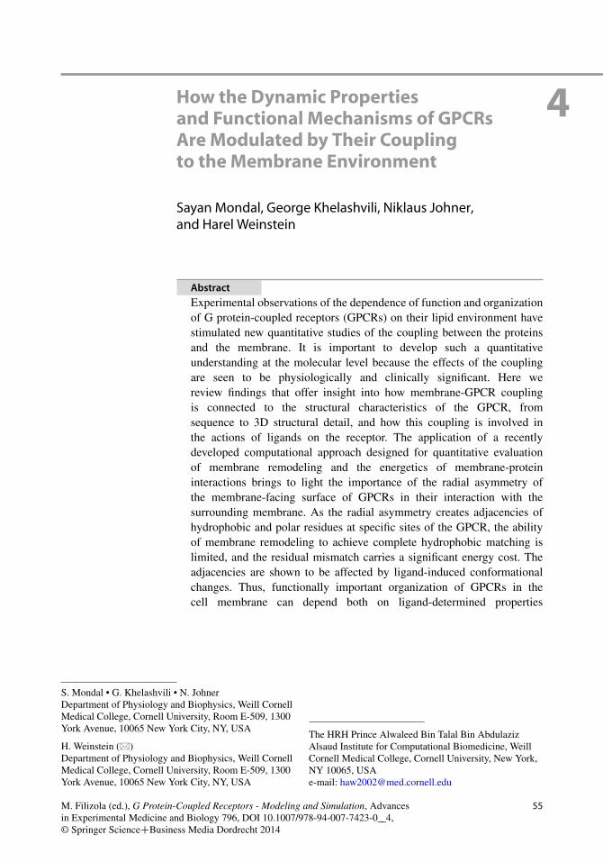
Fig. 4.1 The position of Structural Motifs recognizedas Functional Microdomains (SM/FMs) in GPCRs, il-lustrated in the molecular model of 5HT2AR (Reprintedwith permission from Shan et al. 2012)
Visiers et al. 2002; Prioleau et al. 2002; Baraket al. 1995; Fritze et al. 2003; Lagerstrom andSchioth 2008; Rosenbaum et al. 2009; Palczewskiet al. 2000; Warne et al. 2008; Cherezov et al.2007; Weinstein 2005). Prominent examples ofthese SM/FMs from Class-A GPCRs include the“arginine cage” around the E/DRY motif in TM3(Ballesteros et al. 1998), and the NPxxY motif inTM7 (Ballesteros et al. 1998; Barak et al. 1995;Fritze et al. 2003; Prioleau et al. 2002). Figure 4.1illustrates some of these SM/FMs in a structuralmodel of the serotonin receptor 5HT2AR.
With the focus on the conformational changesof structural elements of the GPCR proteins, themechanistic models have largely ignored the in-volvement of the membrane environment in thediscriminant properties of the GPCRs, and thedifferential ligand-determined effects. This situa-tion is now changing rapidly, because the mem-brane environment has emerged as an essentialpart of functional mechanisms including the ac-tivation, coupling, and cell-surface organizationof the GPCRs. In particular, the conformationsof the GPCRs in the various states of activation– both ligand-determined and constitutive – cou-
ple the response of specific membrane regionsto the transition among such conformations. Thiscoupling is becoming recognized as a determi-nant factor in (i)-the specificity of ligand binding,and (ii)-the responses elicited from the variousactivated states. Importantly, the effects of themembrane environment are strongly dependenton the lipid composition and hence can varybetween different cell types, and also within dif-ferent regions of the plasma membrane of thesame cell. For example, cholesterol, a compo-nent of biological membranes with a relativeabundance that varies among cell types and evenwithin different regions of the same plasma mem-brane, has been shown to affect GPCR signaling.Functionally important receptor properties suchas thermal stability, ligand binding, and local-ization in the cell membrane have been shownto be cholesterol-dependent for beta2-adrenergic,5HT1A, oxytocin, and metabotropic glutamatereceptors (Xiang et al. 2002; Pucadyil and Chat-topadhyay 2004; Gimpl and Fahrenholz 2002;Eroglu et al. 2003). Rhodopsin has served as aprototype for elucidating the role of the lipidenvironment. In systematic studies, the activa-tion and the photochemical function of rhodopsinwere found to be affected by the cholesterolconcentration, as well as by molecular-level char-acteristics of the lipids such as their acyl chainlength, unsaturation of the acyl chain, headgroupcharge, and headgroup size (Brown 1994; Brownet al. 2002; Botelho et al. 2002, 2006; Gibsonand Brown 1991, 1993). To take the exampleof acyl chain unsaturation, the equilibrium be-tween the inactive Metarhodopsin I and the activeMetarhodopsin II was shown to shift in favorof Metarhodopsin II with increasing molar frac-tion of lipids containing the polyunsaturated ¨-3 docosahexaenoic (DHA) tail, as compared tomonounsaturated lipid tails (Brown 1994). Suchmodulation is physiologically and clinically sig-nificant, as dietary deficiency in ¨-3 fatty acidsis well-known to be implicated in a wide vari-ety of diseases (Innis 2008; Lavie et al. 2009;Neuringer et al. 1984; Stahl et al. 2008), atleast some of which involve the modulation ofGPCR function by the lipid environment. In ratmodels, for example, deficiency of dietary ¨-3fatty acids impaired photochemical function, with
58 S. Mondal et al.
the Metarhodopsin II formation being both lowerand slower in response to dim light stimulus (Niuet al. 2002).
As the structures of GPCRs become knownfrom crystallography at increasingly highresolution, and various biophysical methodsof investigation offer increasingly detailedquantitative information about mechanisticelements of GPCR function, it becomes possibleto focus on the structural context of the mech-anisms. In parallel, this development requires acorresponding evaluation of the demonstratedrole of the lipid environment using appropriatequantitative methods to assess the membrane-protein coupling components. Achieving thisgoal requires the development and applicationof quantitative approaches, capable of relatingthe lipid-protein interactions to the structuralcharacteristics of the receptor in its variousstates, and especially in the functional context ofligand-induced conformational rearrangements.Significant progress in this direction has beenachieved with biophysical methods anchored infundamental experimental data, and formulatedin the framework of physics-based computationalmodeling approaches ranging from moleculardynamics simulations to continuum-based andmean field methods. We review, in Sect. 4.2,the new types of insights attained from theapplication of such approaches. In Sect. 4.3, wepresent the essential methodological advancesthat enabled quantitative investigation of therelation between lipid-protein interactions andthe GPCR structure. To enable clear referencesto structural elements in different GPCRs,residues and motifs are identified throughout thechapter with the Ballesteros-Weinstein notation(Ballesteros and Weinstein 1995).
4.2 Relation Between ReceptorStructure and Its Interactionwith the Membranein a Functional Context
This section reviews findings that demonstratehow the membrane interacts with the embeddedGPCR, i.e., the nature of the membrane-GPCR
coupling. The focus is on the manner inwhich such coupling is (a)-connected tothe structural/sequence characteristics of thereceptor, and (b)-involved in the modes andconsequences of receptor-ligand interactions.
4.2.1 How GPCRs Coupleto the SurroundingMembrane
Results from experimental investigations and avariety of theoretical and computational studiessuggest that two major aspects of the couplingbetween the membrane and the embedded GPCRhave the greatest impact on the functional prop-erties of the receptors: (1) the interaction ofindividual lipid molecules with specific sites ofthe receptor molecule, and (2) the mismatch be-tween the hydrophobic/hydrophilic character ofthe membrane and the membrane-facing surfaceof the receptor. As detailed below, a significantpart of this mismatch persists even after themembrane is remodeled around the embeddedprotein, giving rise to an energy cost that affectsthe functional properties and spatial organiza-tion of the GPCRs. This is due to the inherentasymmetry of the membrane-facing surface ofthe embedded protein, caused by the differencesamong the TM segments. Consequently, it is animportant common characteristic of the energet-ics of membrane coupling for multi-TM segmentproteins.
4.2.1.1 Specific Interactions of LipidMoleculeswith Membrane-EmbeddedGPCRs
With the details obtained from atomisticMolecular Dynamics (MD) simulations it hasbecome possible to discern the dynamics ofmembrane components surrounding the receptormolecules, such as cholesterol, DHA, and variousother lipids. The simulations revealed both thenature of the interactions of the various lipidmolecules with specific sites of the GPCR,and the effect on the membrane and theproteins as a whole. For example, in a 1.6 �s
4 How the Dynamic Properties and Functional Mechanisms of GPCRs Are Modulated. . . 59
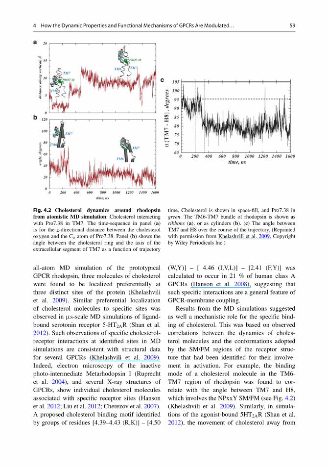
Fig. 4.2 Cholesterol dynamics around rhodopsinfrom atomistic MD simulation. Cholesterol interactingwith Pro7.38 in TM7. The time-sequence in panel (a)is for the z-directional distance between the cholesteroloxygen and the C’ atom of Pro7.38. Panel (b) shows theangle between the cholesterol ring and the axis of theextracellular segment of TM7 as a function of trajectory
time. Cholesterol is shown in space-fill, and Pro7.38 ingreen. The TM6-TM7 bundle of rhodopsin is shown asribbons (a), or as cylinders (b). (c) The angle betweenTM7 and H8 over the course of the trajectory. (Reprintedwith permission from Khelashvili et al. 2009. Copyrightby Wiley Periodicals Inc.)
all-atom MD simulation of the prototypicalGPCR rhodopsin, three molecules of cholesterolwere found to be localized preferentially atthree distinct sites of the protein (Khelashviliet al. 2009). Similar preferential localizationof cholesterol molecules to specific sites wasobserved in �s-scale MD simulations of ligand-bound serotonin receptor 5-HT2AR (Shan et al.2012). Such observations of specific cholesterol-receptor interactions at identified sites in MDsimulations are consistent with structural datafor several GPCRs (Khelashvili et al. 2009).Indeed, electron microscopy of the inactivephoto-intermediate Metarhodopsin I (Ruprechtet al. 2004), and several X-ray structures ofGPCRs, show individual cholesterol moleculesassociated with specific receptor sites (Hansonet al. 2012; Liu et al. 2012; Cherezov et al. 2007).A proposed cholesterol binding motif identifiedby groups of residues [4.39–4.43 (R,K)] – [4.50
(W,Y)] – [ 4.46 (I,V,L)] – [2.41 (F,Y)] wascalculated to occur in 21 % of human class AGPCRs (Hanson et al. 2008), suggesting thatsuch specific interactions are a general feature ofGPCR-membrane coupling.
Results from the MD simulations suggestedas well a mechanistic role for the specific bind-ing of cholesterol. This was based on observedcorrelations between the dynamics of choles-terol molecules and the conformations adoptedby the SM/FM regions of the receptor struc-ture that had been identified for their involve-ment in activation. For example, the bindingmode of a cholesterol molecule in the TM6-TM7 region of rhodopsin was found to cor-relate with the angle between TM7 and H8,which involves the NPxxY SM/FM (see Fig. 4.2)(Khelashvili et al. 2009). Similarly, in simula-tions of the agonist-bound 5HT2AR (Shan et al.2012), the movement of cholesterol away from
60 S. Mondal et al.
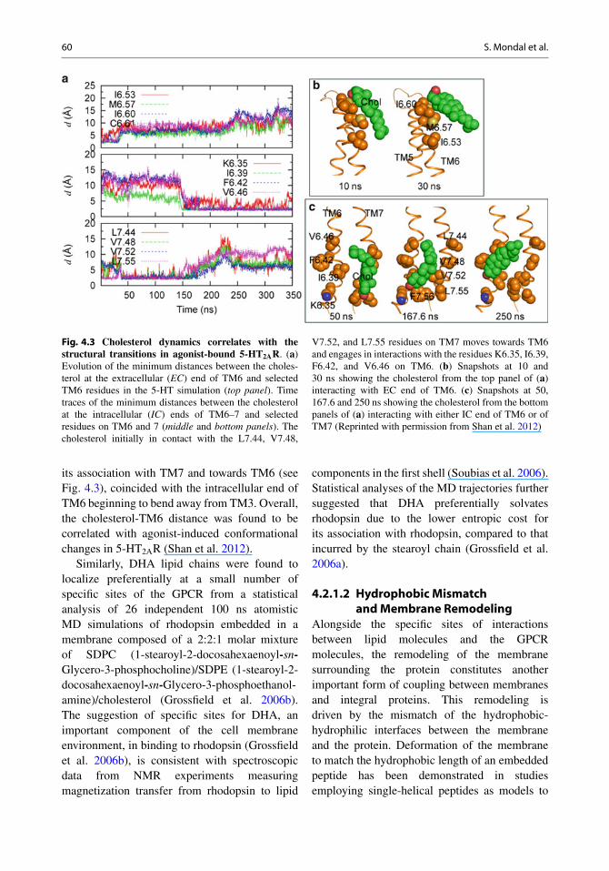
Fig. 4.3 Cholesterol dynamics correlates with thestructural transitions in agonist-bound 5-HT2AR. (a)Evolution of the minimum distances between the choles-terol at the extracellular (EC) end of TM6 and selectedTM6 residues in the 5-HT simulation (top panel). Timetraces of the minimum distances between the cholesterolat the intracellular (IC) ends of TM6–7 and selectedresidues on TM6 and 7 (middle and bottom panels). Thecholesterol initially in contact with the L7.44, V7.48,
V7.52, and L7.55 residues on TM7 moves towards TM6and engages in interactions with the residues K6.35, I6.39,F6.42, and V6.46 on TM6. (b) Snapshots at 10 and30 ns showing the cholesterol from the top panel of (a)interacting with EC end of TM6. (c) Snapshots at 50,167.6 and 250 ns showing the cholesterol from the bottompanels of (a) interacting with either IC end of TM6 or ofTM7 (Reprinted with permission from Shan et al. 2012)
its association with TM7 and towards TM6 (seeFig. 4.3), coincided with the intracellular end ofTM6 beginning to bend away from TM3. Overall,the cholesterol-TM6 distance was found to becorrelated with agonist-induced conformationalchanges in 5-HT2AR (Shan et al. 2012).
Similarly, DHA lipid chains were found tolocalize preferentially at a small number ofspecific sites of the GPCR from a statisticalanalysis of 26 independent 100 ns atomisticMD simulations of rhodopsin embedded in amembrane composed of a 2:2:1 molar mixtureof SDPC (1-stearoyl-2-docosahexaenoyl-sn-Glycero-3-phosphocholine)/SDPE (1-stearoyl-2-docosahexaenoyl-sn-Glycero-3-phosphoethanol-amine)/cholesterol (Grossfield et al. 2006b).The suggestion of specific sites for DHA, animportant component of the cell membraneenvironment, in binding to rhodopsin (Grossfieldet al. 2006b), is consistent with spectroscopicdata from NMR experiments measuringmagnetization transfer from rhodopsin to lipid
components in the first shell (Soubias et al. 2006).Statistical analyses of the MD trajectories furthersuggested that DHA preferentially solvatesrhodopsin due to the lower entropic cost forits association with rhodopsin, compared to thatincurred by the stearoyl chain (Grossfield et al.2006a).
4.2.1.2 Hydrophobic Mismatchand Membrane Remodeling
Alongside the specific sites of interactionsbetween lipid molecules and the GPCRmolecules, the remodeling of the membranesurrounding the protein constitutes anotherimportant form of coupling between membranesand integral proteins. This remodeling isdriven by the mismatch of the hydrophobic-hydrophilic interfaces between the membraneand the protein. Deformation of the membraneto match the hydrophobic length of an embeddedpeptide has been demonstrated in studiesemploying single-helical peptides as models to
4 How the Dynamic Properties and Functional Mechanisms of GPCRs Are Modulated. . . 61
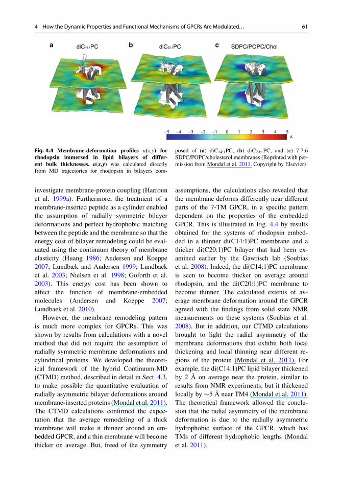
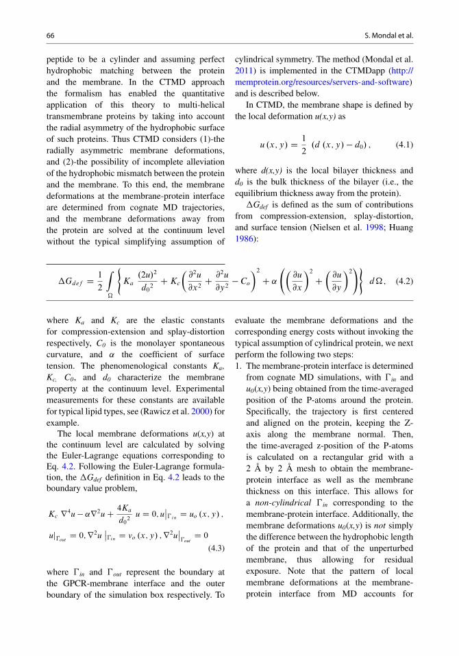
Fig. 4.4 Membrane-deformation profiles u(x,y) forrhodopsin immersed in lipid bilayers of differ-ent bulk thicknesses. u(x,y) was calculated directlyfrom MD trajectories for rhodopsin in bilayers com-
posed of (a) diC14:1PC, (b) diC20:1PC, and (c) 7:7:6SDPC/POPC/cholesterol membranes (Reprinted with per-mission from Mondal et al. 2011. Copyright by Elsevier)
investigate membrane-protein coupling (Harrounet al. 1999a). Furthermore, the treatment of amembrane-inserted peptide as a cylinder enabledthe assumption of radially symmetric bilayerdeformations and perfect hydrophobic matchingbetween the peptide and the membrane so that theenergy cost of bilayer remodeling could be eval-uated using the continuum theory of membraneelasticity (Huang 1986; Andersen and Koeppe2007; Lundbæk and Andersen 1999; Lundbaeket al. 2003; Nielsen et al. 1998; Goforth et al.2003). This energy cost has been shown toaffect the function of membrane-embeddedmolecules (Andersen and Koeppe 2007;Lundbaek et al. 2010).
However, the membrane remodeling patternis much more complex for GPCRs. This wasshown by results from calculations with a novelmethod that did not require the assumption ofradially symmetric membrane deformations andcylindrical proteins. We developed the theoret-ical framework of the hybrid Continuum-MD(CTMD) method, described in detail in Sect. 4.3,to make possible the quantitative evaluation ofradially asymmetric bilayer deformations aroundmembrane-inserted proteins (Mondal et al. 2011).The CTMD calculations confirmed the expec-tation that the average remodeling of a thickmembrane will make it thinner around an em-bedded GPCR, and a thin membrane will becomethicker on average. But, freed of the symmetry
assumptions, the calculations also revealed thatthe membrane deforms differently near differentparts of the 7-TM GPCR, in a specific patterndependent on the properties of the embeddedGPCR. This is illustrated in Fig. 4.4 by resultsobtained for the systems of rhodopsin embed-ded in a thinner di(C14:1)PC membrane and athicker di(C20:1)PC bilayer that had been ex-amined earlier by the Gawrisch lab (Soubiaset al. 2008). Indeed, the di(C14:1)PC membraneis seen to become thicker on average aroundrhodopsin, and the di(C20:1)PC membrane tobecome thinner. The calculated extents of av-erage membrane deformation around the GPCRagreed with the findings from solid state NMRmeasurements on these systems (Soubias et al.2008). But in addition, our CTMD calculationsbrought to light the radial asymmetry of themembrane deformations that exhibit both localthickening and local thinning near different re-gions of the protein (Mondal et al. 2011). Forexample, the di(C14:1)PC lipid bilayer thickenedby 2 Å on average near the protein, similar toresults from NMR experiments, but it thickenedlocally by �5 Å near TM4 (Mondal et al. 2011).The theoretical framework allowed the conclu-sion that the radial asymmetry of the membranedeformation is due to the radially asymmetrichydrophobic surface of the GPCR, which hasTMs of different hydrophobic lengths (Mondalet al. 2011).
62 S. Mondal et al.
E5.36(polar)
Q5.60(polar)
Gul5.36Gul5.36C2 atoms
C2 atomsPhe5.63 Phe5.63F5.63 (hydrophobic)
ca b
d
N1.33
V2.66, V2.67
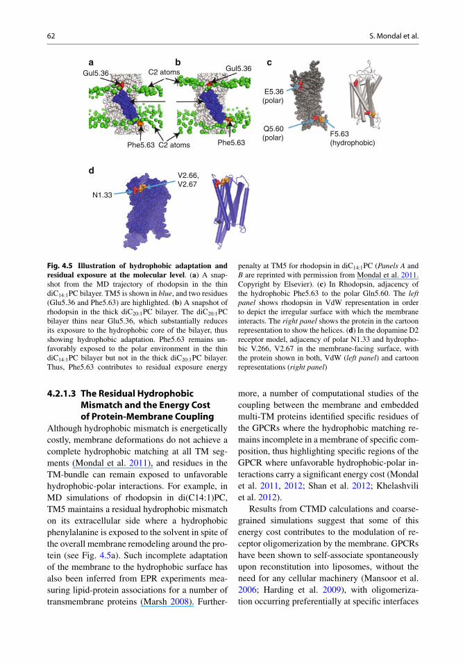
Fig. 4.5 Illustration of hydrophobic adaptation andresidual exposure at the molecular level. (a) A snap-shot from the MD trajectory of rhodopsin in the thindiC14:1PC bilayer. TM5 is shown in blue, and two residues(Glu5.36 and Phe5.63) are highlighted. (b) A snapshot ofrhodopsin in the thick diC20:1PC bilayer. The diC20:1PCbilayer thins near Glu5.36, which substantially reducesits exposure to the hydrophobic core of the bilayer, thusshowing hydrophobic adaptation. Phe5.63 remains un-favorably exposed to the polar environment in the thindiC14:1PC bilayer but not in the thick diC20:1PC bilayer.Thus, Phe5.63 contributes to residual exposure energy
penalty at TM5 for rhodopsin in diC14:1PC (Panels A andB are reprinted with permission from Mondal et al. 2011.Copyright by Elsevier). (c) In Rhodopsin, adjacency ofthe hydrophobic Phe5.63 to the polar Gln5.60. The leftpanel shows rhodopsin in VdW representation in orderto depict the irregular surface with which the membraneinteracts. The right panel shows the protein in the cartoonrepresentation to show the helices. (d) In the dopamine D2receptor model, adjacency of polar N1.33 and hydropho-bic V.266, V2.67 in the membrane-facing surface, withthe protein shown in both, VdW (left panel) and cartoonrepresentations (right panel)
4.2.1.3 The Residual HydrophobicMismatch and the Energy Costof Protein-Membrane Coupling
Although hydrophobic mismatch is energeticallycostly, membrane deformations do not achieve acomplete hydrophobic matching at all TM seg-ments (Mondal et al. 2011), and residues in theTM-bundle can remain exposed to unfavorablehydrophobic-polar interactions. For example, inMD simulations of rhodopsin in di(C14:1)PC,TM5 maintains a residual hydrophobic mismatchon its extracellular side where a hydrophobicphenylalanine is exposed to the solvent in spite ofthe overall membrane remodeling around the pro-tein (see Fig. 4.5a). Such incomplete adaptationof the membrane to the hydrophobic surface hasalso been inferred from EPR experiments mea-suring lipid-protein associations for a number oftransmembrane proteins (Marsh 2008). Further-
more, a number of computational studies of thecoupling between the membrane and embeddedmulti-TM proteins identified specific residues ofthe GPCRs where the hydrophobic matching re-mains incomplete in a membrane of specific com-position, thus highlighting specific regions of theGPCR where unfavorable hydrophobic-polar in-teractions carry a significant energy cost (Mondalet al. 2011, 2012; Shan et al. 2012; Khelashviliet al. 2012).
Results from CTMD calculations and coarse-grained simulations suggest that some of thisenergy cost contributes to the modulation of re-ceptor oligomerization by the membrane. GPCRshave been shown to self-associate spontaneouslyupon reconstitution into liposomes, without theneed for any cellular machinery (Mansoor et al.2006; Harding et al. 2009), with oligomeriza-tion occurring preferentially at specific interfaces
4 How the Dynamic Properties and Functional Mechanisms of GPCRs Are Modulated. . . 63
(Fung et al. 2009). The oligomerization is areversible process (Dorsch et al. 2009; Hern et al.2010), and is driven by hydrophobic mismatch(Botelho et al. 2006). Coarse-grained MD sim-ulations of the interaction of GPCRs in the mem-brane have provided results consistent with theexperimental observations, and offered importantinsights into the energetics of this oligomeriza-tion (Periole et al. 2007, 2012). Such simula-tions, taken together with the quantitative re-sults from CTMD calculations, suggest that theresidual exposure contributes to the organiza-tional role of the hydrophobic mismatch (Mondalet al. 2011, 2012). In particular, these calcu-lations suggest that there is a drive for GPCRoligomerization at specific interfaces that removethe structural context for the residual hydropho-bic mismatch, thereby reducing the hydrophobicmismatch and its energy cost (Mondal et al. 2011,2012).
The detailed quantitative analysis of theincomplete hydrophobic matching associatedwith the protein-membrane coupling of multi-TM-segment proteins, revealed a persistent localexposure of residues to unmatched hydrophobicenvironments, which could not be eliminatedby the overall membrane remodeling. Thiswas termed “residual exposure” (Mondal et al.2011). Calculations in several different GPCR-membrane systems (Mondal et al. 2011, 2012)showed that this residual exposure is due to theradial asymmetry of the GPCR’s hydrophobicsurface. Specifically, if polar and hydrophobicresidues, e.g., in two different TMs, are adjacentin space, membrane deformation would need toproduce a match to both polar and hydrophobicresidues in a small neighborhood, and this may beenergetically unfavorable. Therefore, adjacenciesof hydrophobically disparate residues markpossible sites of residual exposure and thusenergetically costly membrane-protein interac-tions. The CTMD approach that determines theenergetic cost of the residual exposure causedby such adjacencies (Mondal et al. 2011), ispresented in Sect. 4.3 below.
That adjacencies responsible for the resid-ual exposure of multi-TM proteins are a com-mon feature of GPCRs, is evident from the re-cently determined crystal structures and validatedstructural models of a variety of rhodopsin-likeGPCRs, including B2AR, KOR, DOR, 5-HT2AR,and D2R in the literature (Mondal et al. 2011,2012; Shan et al. 2012). Indeed, computationalmodeling with the CTMD method showed thatresidual exposure occurs at these adjacencies indifferent GPCRs and in a number of differentmembranes (see Tables 4.1 and 4.2) (Mondalet al. 2011, 2012; Shan et al. 2012). This isillustrated in Fig. 4.5a–c showing the residualexposure in TM5 of rhodopsin at a site wherePhe and Gln are juxtaposed. Figure 4.5d showsanother example of the adjacency, with the polarN1.33 next to the hydrophobic V2.66 and V2.67in a structural model of the dopamine D2 recep-tor. Here too, the calculations show a residualexposure, with an energy cost of �2 kT at N1.33in a raft-like membrane.
These results from computational modelinghighlight the two key elements of the couplingenergy between the membrane and the GPCR,viz. (1)-the radially asymmetric membrane de-formation, and (2)-the residual exposure remain-ing after the membrane deforms to alleviate thehydrophobic mismatch. The nature of these twoidentifiable elements demonstrates that the cou-pling between the membrane and GPCR proteinsis not a simple result of the difference betweenthe average hydrophobic length of the protein andthat of the pure membrane as previously postu-lated for model peptides (Harroun et al. 1999a,b; Lundbæk and Andersen 1999; Marsh 2008).Rather, the important consequences of the cou-pling emerge from specific features of the proteinsurface, both with respect to residue identityand to local structure. Therefore, very differentconsequences can be expected from even smalldifferences between highly homologous proteins(Mondal et al. 2012), from point mutations, orfrom structural changes due to ligand binding(see next Section).
64 S. Mondal et al.
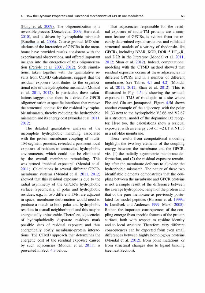
Table 4.1 Residual SAres values (in Å2) and the corresponding �Gres values (in kBT) calculated for TMs of rhodopsinin diC14:1PC, diC16:1PC, diC18:1PC, diC20:1PC, and 7:7:6 SDPC/POPC/cholesterol membranes
diC14:1PC diC16:1PC diC18:1PC diC20:1PC SDPC/POPC/cholesterol
TM SAres �Gres SAres �Gres SAres �Gres SAres �Gres SAres �Gres
1 210 9.9 80 3.8 75 3.5 70 3.3 63 3.02 74 3.5 29 1.4 0 0 32 1.5 27 1.33 N.D.a N.D. N.D. N.D. N.D. N.D. N.D. N.D. N.D. N.D.4 0 0 77 3.6 90 4.3 79 3.7 134 6.35 115 5.4 1 0 9 0.4 7 0.3 17 0.86 36 1.7 31 1.5 56 2.7 38 1.8 51 2.47 119 5.6 44 2.1 22 1.1 0 0 0 0
Reprinted with permission from Mondal et al. (2011). Copyright by ElsevieraN.D. Not Determined
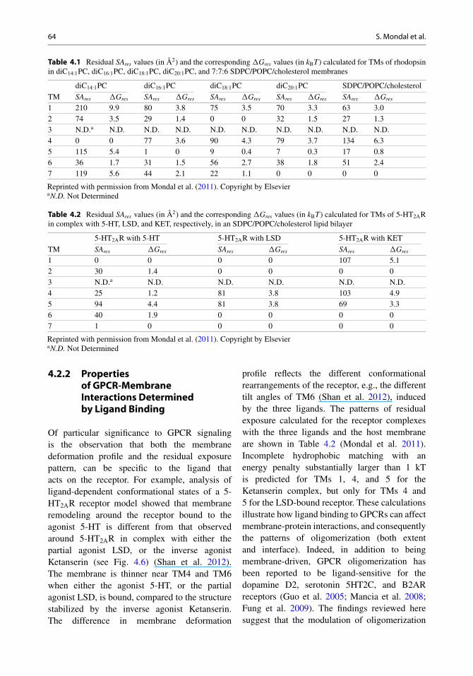
Table 4.2 Residual SAres values (in Å2) and the corresponding �Gres values (in kBT) calculated for TMs of 5-HT2ARin complex with 5-HT, LSD, and KET, respectively, in an SDPC/POPC/cholesterol lipid bilayer
5-HT2AR with 5-HT 5-HT2AR with LSD 5-HT2AR with KET
TM SAres �Gres SAres �Gres SAres �Gres
1 0 0 0 0 107 5.12 30 1.4 0 0 0 03 N.D.a N.D. N.D. N.D. N.D. N.D.4 25 1.2 81 3.8 103 4.95 94 4.4 81 3.8 69 3.36 40 1.9 0 0 0 07 1 0 0 0 0 0
Reprinted with permission from Mondal et al. (2011). Copyright by ElsevieraN.D. Not Determined
4.2.2 Propertiesof GPCR-MembraneInteractions Determinedby Ligand Binding
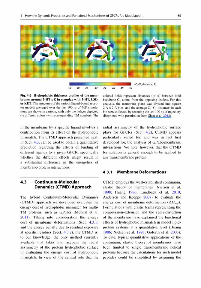
Of particular significance to GPCR signalingis the observation that both the membranedeformation profile and the residual exposurepattern, can be specific to the ligand thatacts on the receptor. For example, analysis ofligand-dependent conformational states of a 5-HT2AR receptor model showed that membraneremodeling around the receptor bound to theagonist 5-HT is different from that observedaround 5-HT2AR in complex with either thepartial agonist LSD, or the inverse agonistKetanserin (see Fig. 4.6) (Shan et al. 2012).The membrane is thinner near TM4 and TM6when either the agonist 5-HT, or the partialagonist LSD, is bound, compared to the structurestabilized by the inverse agonist Ketanserin.The difference in membrane deformation
profile reflects the different conformationalrearrangements of the receptor, e.g., the differenttilt angles of TM6 (Shan et al. 2012), inducedby the three ligands. The patterns of residualexposure calculated for the receptor complexeswith the three ligands and the host membraneare shown in Table 4.2 (Mondal et al. 2011).Incomplete hydrophobic matching with anenergy penalty substantially larger than 1 kTis predicted for TMs 1, 4, and 5 for theKetanserin complex, but only for TMs 4 and5 for the LSD-bound receptor. These calculationsillustrate how ligand binding to GPCRs can affectmembrane-protein interactions, and consequentlythe patterns of oligomerization (both extentand interface). Indeed, in addition to beingmembrane-driven, GPCR oligomerization hasbeen reported to be ligand-sensitive for thedopamine D2, serotonin 5HT2C, and B2ARreceptors (Guo et al. 2005; Mancia et al. 2008;Fung et al. 2009). The findings reviewed heresuggest that the modulation of oligomerization
4 How the Dynamic Properties and Functional Mechanisms of GPCRs Are Modulated. . . 65
Fig. 4.6 Hydrophobic thickness profiles of the mem-branes around 5-HT2AR in complex with 5-HT, LSD,or KET. The structures of the various ligand-bound recep-tor models averaged over the last 100 ns of MD simula-tions are shown in cartoon, with only the helices depicted(in different colors) with corresponding TM numbers. The
colored fields represent distances (in Å) between lipidbackbone C2 atoms from the opposing leaflets. For thisanalysis, the membrane plane was divided into square2 Å � 2 Å bins, and the average C2–C2 distances in eachbin were collected by scanning the last 100 ns of trajectory(Reprinted with permission from Shan et al. 2012)
in the membrane by a specific ligand involves acontribution from its effect on the hydrophobicmismatch. The CTMD approach presented next,in Sect. 4.3, can be used to obtain a quantitativeprediction regarding the effects of binding ofdifferent ligands to a given GPCR, specificallywhether the different effects might result ina substantial difference in the energetics ofmembrane-protein interactions.
4.3 Continuum-MolecularDynamics (CTMD) Approach
The hybrid Continuum-Molecular Dynamics(CTMD) approach we developed evaluates theenergy cost of hydrophobic mismatch for multi-TM proteins, such as GPCRs (Mondal et al.2011). Taking into consideration the energycost of membrane deformations (Sect. 4.3.1)and the energy penalty due to residual exposureat specific residues (Sect. 4.3.2), the CTMD is,to our knowledge, the only method currentlyavailable that takes into account the radialasymmetry of the protein hydrophobic surfacein evaluating the energy cost of hydrophobicmismatch. In view of the central role that the
radial asymmetry of the hydrophobic surfaceplays for GPCRs (Sect. 4.2), CTMD appearsparticularly suited for, and was in fact firstdeveloped for, the analysis of GPCR-membraneinteractions. We note, however, that the CTMDformulation is general enough to be applied toany transmembrane protein.
4.3.1 Membrane Deformations
CTMD employs the well-established continuum,elastic theory of membranes (Nielsen et al.1998; Huang 1986; Lundbaek et al. 2010;Andersen and Koeppe 2007) to evaluate theenergy cost of membrane deformation (�Gdef ).Formulations with elastic terms representing thecompression-extension and the splay-distortionof the membrane have explained the functionaleffects of hydrophobic mismatch in model lipid-protein systems at a quantitative level (Huang1986; Nielsen et al. 1998; Goforth et al. 2003).To date, typical quantitative applications of thecontinuum, elastic theory of membranes havebeen limited to single transmembrane helicalproteins because the calculations for such modelpeptides could be simplified by assuming the
66 S. Mondal et al.
peptide to be a cylinder and assuming perfecthydrophobic matching between the proteinand the membrane. In the CTMD approachthe formalism has enabled the quantitativeapplication of this theory to multi-helicaltransmembrane proteins by taking into accountthe radial asymmetry of the hydrophobic surfaceof such proteins. Thus CTMD considers (1)-theradially asymmetric membrane deformations,and (2)-the possibility of incomplete alleviationof the hydrophobic mismatch between the proteinand the membrane. To this end, the membranedeformations at the membrane-protein interfaceare determined from cognate MD trajectories,and the membrane deformations away fromthe protein are solved at the continuum levelwithout the typical simplifying assumption of
cylindrical symmetry. The method (Mondal et al.2011) is implemented in the CTMDapp (http://memprotein.org/resources/servers-and-software)and is described below.
In CTMD, the membrane shape is defined bythe local deformation u(x,y) as
u .x; y/ D 1
2.d .x; y/ � d0/ ; (4.1)
where d(x,y) is the local bilayer thickness andd0 is the bulk thickness of the bilayer (i.e., theequilibrium thickness away from the protein).
�Gdef is defined as the sum of contributionsfrom compression-extension, splay-distortion,and surface tension (Nielsen et al. 1998; Huang1986):
�Gdef D 1
2
Z
�
(Ka
.2u/2
d02
C Kc
�@2u
@x2C @2u
@y2� Co
�2
C ˛
�@u
@x
�2
C�
@u
@y
�2!)
d�; (4.2)
where Ka and Kc are the elastic constantsfor compression-extension and splay-distortionrespectively, C0 is the monolayer spontaneouscurvature, and ˛ the coefficient of surfacetension. The phenomenological constants Ka,Kc, C0, and d0 characterize the membraneproperty at the continuum level. Experimentalmeasurements for these constants are availablefor typical lipid types, see (Rawicz et al. 2000) forexample.
The local membrane deformations u(x,y) atthe continuum level are calculated by solvingthe Euler-Lagrange equations corresponding toEq. 4.2. Following the Euler-Lagrange formula-tion, the �Gdef definition in Eq. 4.2 leads to theboundary value problem,
Kc r4u � ˛r2u C 4Ka
d02
u D 0; uˇ̌� in
D uo .x; y/ ;
uj�outD 0; r2u
ˇ̌�in D vo .x; y/ ; r2u
ˇ̌�out
D 0
(4.3)
where � in and �out represent the boundary atthe GPCR-membrane interface and the outerboundary of the simulation box respectively. To
evaluate the membrane deformations and thecorresponding energy costs without invoking thetypical assumption of cylindrical protein, we nextperform the following two steps:1. The membrane-protein interface is determined
from cognate MD simulations, with � in andu0(x,y) being obtained from the time-averagedposition of the P-atoms around the protein.Specifically, the trajectory is first centeredand aligned on the protein, keeping the Z-axis along the membrane normal. Then,the time-averaged z-position of the P-atomsis calculated on a rectangular grid with a2 Å by 2 Å mesh to obtain the membrane-protein interface as well as the membranethickness on this interface. This allows fora non-cylindrical � in corresponding to themembrane-protein interface. Additionally, themembrane deformations u0(x,y) is not simplythe difference between the hydrophobic lengthof the protein and that of the unperturbedmembrane, thus allowing for residualexposure. Note that the pattern of localmembrane deformations at the membrane-protein interface from MD accounts for
4 How the Dynamic Properties and Functional Mechanisms of GPCRs Are Modulated. . . 67
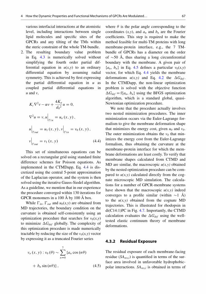
various interfacial interactions at the atomisticlevel, including interactions between singlelipid molecules and specific sites of theGPCRs and any tilting of the TMs withinthe steric constraint of the whole TM-bundle.
2. The resulting boundary value problemin Eq. 4.3 is numerically solved withoutsimplifying the fourth order partial dif-ferential equation in u(x,y) to an ordinarydifferential equation by assuming radialsymmetry. This is achieved by first expressingthe partial differential equation in u ascoupled partial differential equations inu and v,
Kcr2v � ˛v C 4Ka
d02
u D 0;
r2u D v; uˇ̌ˇ�in
D uo .x; y/ ;
uˇ̌ˇ�out
D u1 .x; y/ ; vˇ̌ˇ�in
D v0 .x; y/ ;
vˇ̌ˇ�out
D v1 .x; y/ (4.4)
This set of simultaneous equations can besolved on a rectangular grid using standard finitedifference schemes for Poisson equations. Asimplemented in the CTMDapp, Eq. 4.4 is dis-cretized using the central 5-point approximationof the Laplacian operator, and the system is thensolved using the iterative Gauss-Siedel algorithm.As a guideline, we mention that in our experiencethe procedure converged within 130 iterations forGPCR monomers in a 100 Å by 100 Å box.
While � in, �out and u0(x,y) are obtained fromMD trajectories, the boundary condition on thecurvature is obtained self-consistently using anoptimization procedure that searches for v0(x,y)to minimize �Gdef globally. The complexity ofthis optimization procedure is made numericallytractable by reducing the size of the v0(x,y) vectorby expressing it as a truncated Fourier series
vo .x; y/ W v0 .�/ �7X
nD0
fan cos .n�/
C bn sin .n�/g ; (4.5)
where � is the polar angle corresponding to thecoordinates (x,y), and an and bn are the Fouriercoefficients. This step is required to make themethod feasible for multi-TM proteins with longmembrane-protein interface, e.g., the 7 TM-bundle of GPCRs has a diameter on the orderof �50 Å, thus sharing a long circumferentialboundary with the membrane. A given pair offan, bng in Eq. 4.5 defines a particular v0(x,y)vector, for which Eq. 4.4 yields the membranedeformations u(x,y) and Eq. 4.2 the �Gdef .In the CTMDapp, the non-linear optimizationproblem is solved with the objective function�Gdef D ffan, bng using the BFGS optimizationalgorithm, which is a standard global, quasi-Newtonian optimization procedure.
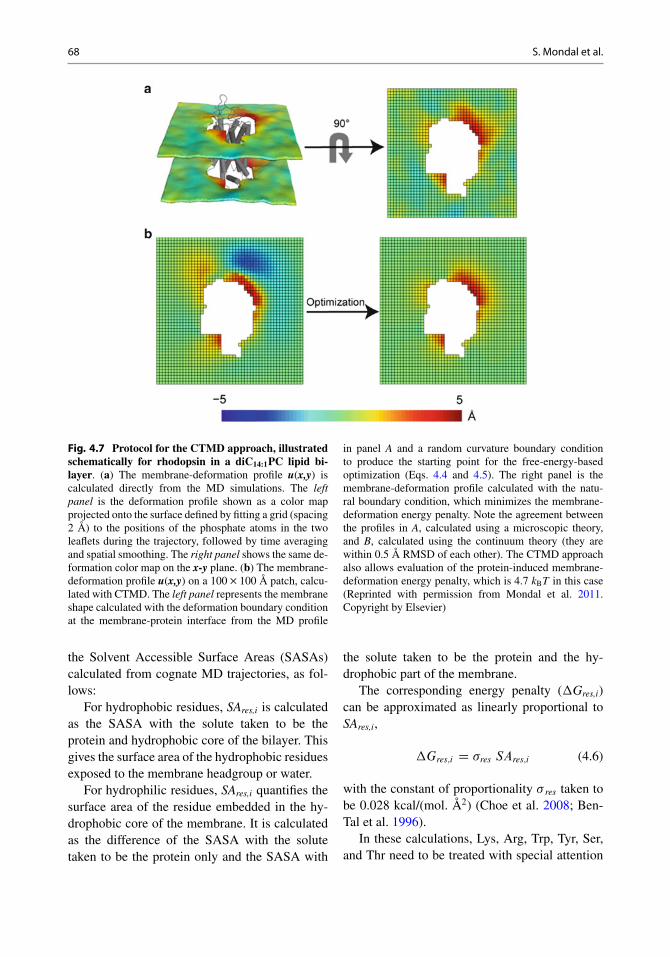
We note that the procedure actually involvestwo nested minimization procedures. The innerminimization occurs via the Euler-Lagrange for-malism to give the membrane deformation shapethat minimizes the energy cost, given u0 and v0.The outer minimization obtains the v0 that min-imizes the energy cost from the Euler-Lagrangeformalism, thus obtaining the curvature at themembrane-protein interface for which the mem-brane deformations are least costly. To verify thatmembrane shapes calculated from CTMD andMD are similar, the macroscopic u(x,y) obtainedby the nested optimization procedure can be com-pared to u(x,y) calculated directly from the cog-nate microscopic MD simulation. The calcula-tions for a number of GPCR-membrane systemshave shown that the macroscopic u(x,y) indeedconverges to a profile similar (within �1 Å)to the u(x,y) obtained from the cognate MDtrajectories. This is illustrated for rhodopsin indi(C14:1)PC in Fig. 4.7. Importantly, the CTMDcalculation evaluates the �Gdef using the well-tested elastic continuum theory of membranedeformations.
4.3.2 Residual Exposure
The residual exposure of each membrane-facingresidue (SAres,i) is quantified in terms of the sur-face area involved in unfavorable hydrophobic-polar interactions. SAres,i is obtained in terms of
68 S. Mondal et al.
Fig. 4.7 Protocol for the CTMD approach, illustratedschematically for rhodopsin in a diC14:1PC lipid bi-layer. (a) The membrane-deformation profile u(x,y) iscalculated directly from the MD simulations. The leftpanel is the deformation profile shown as a color mapprojected onto the surface defined by fitting a grid (spacing2 Å) to the positions of the phosphate atoms in the twoleaflets during the trajectory, followed by time averagingand spatial smoothing. The right panel shows the same de-formation color map on the x-y plane. (b) The membrane-deformation profile u(x,y) on a 100 � 100 Å patch, calcu-lated with CTMD. The left panel represents the membraneshape calculated with the deformation boundary conditionat the membrane-protein interface from the MD profile
in panel A and a random curvature boundary conditionto produce the starting point for the free-energy-basedoptimization (Eqs. 4.4 and 4.5). The right panel is themembrane-deformation profile calculated with the natu-ral boundary condition, which minimizes the membrane-deformation energy penalty. Note the agreement betweenthe profiles in A, calculated using a microscopic theory,and B, calculated using the continuum theory (they arewithin 0.5 Å RMSD of each other). The CTMD approachalso allows evaluation of the protein-induced membrane-deformation energy penalty, which is 4.7 kBT in this case(Reprinted with permission from Mondal et al. 2011.Copyright by Elsevier)
the Solvent Accessible Surface Areas (SASAs)calculated from cognate MD trajectories, as fol-lows:
For hydrophobic residues, SAres,i is calculatedas the SASA with the solute taken to be theprotein and hydrophobic core of the bilayer. Thisgives the surface area of the hydrophobic residuesexposed to the membrane headgroup or water.
For hydrophilic residues, SAres,i quantifies thesurface area of the residue embedded in the hy-drophobic core of the membrane. It is calculatedas the difference of the SASA with the solutetaken to be the protein only and the SASA with
the solute taken to be the protein and the hy-drophobic part of the membrane.
The corresponding energy penalty (�Gres,i)can be approximated as linearly proportional toSAres,i,
�Gres;i D �res SAres;i (4.6)
with the constant of proportionality � res taken tobe 0.028 kcal/(mol. Å2) (Choe et al. 2008; Ben-Tal et al. 1996).
In these calculations, Lys, Arg, Trp, Tyr, Ser,and Thr need to be treated with special attention
4 How the Dynamic Properties and Functional Mechanisms of GPCRs Are Modulated. . . 69
to the structural context. If the polar Lys or Argresidues reside within the hydrophobic part ofthe membrane but near the water-membrane in-terface, their long side chains can extend towardsthe phosphate headgroups so that the polar partof their side-chains interacts with the polar phos-phate headgroups (Strandberg and Killian 2003;Sankararamakrishnan and Weinstein 2002). Thisconformational preference is known as “snorke-ling”, and it has been estimated that as a result,only a small energy penalty is associated withhydrophobic matching by means of snorkeling(Strandberg and Killian 2003). Therefore, Lysand Arg residues near the water-membrane in-terface are not considered in the calculations ofresidual exposure if they are found in the MDsimulations to snorkel to the lipid headgroup re-gion. Interfacial Trp and Tyr are also not consid-ered as candidates for residual exposure, as theircommonly observed interfacial location is con-sidered favorable (Yau et al. 1998). Finally, Serand Thr residues are polar, but when they residewithin the hydrophobic part of the membrane,their polar parts can form H-bonds with the helixbackbone (Gray and Matthews 1984). In suchsituations, the Ser and Thr are interacting directlywith the protein and not the membrane so theywould not be candidates for residual exposure.
4.4 Perspective on “Specific” vs.“Non-specific”GPCR-MembraneInteractions
Functionally relevant interaction of lipids withGPCRs has historically been viewed as being oftwo different types: (1)-specific lipid-proteininteractions, e.g., the binding of individualcholesterol molecules to specific residues ofthe GPCR; and (2)-non-specific interactions,such as hydrophobic mismatch (Botelho et al.2002, 2006; Brown 1994; Gibson and Brown1991, 1993). For example, in affecting rhodopsinactivation, lipids with PE headgroups cancompensate for the absence of lipids with PSheadgroups and lipids with DHA tails, which
has been taken to suggest that the lipid-mediatedeffect on rhodopsin function is not due to lipidsof a specific chemical nature (Botelho et al.2006). Such “non-specific interactions” havebeen interpreted to involve energetically costlyhydrophobic mismatch.
The computational results described hereinidentify the nature of both “specific” and “non-specific” interactions and suggest that the“specific interactions” are local and the “non-specific” hydrophobic mismatch interactionsare non-local in nature. Notably, however,individual lipid molecules that bind to specificsites of the GPCR can also participate inhydrophobic matching. For example, in the1.6 �s simulation of rhodopsin (see Sect. 4.2.1),the cholesterol that binds specifically at theextracellular end of TM2-TM3 partly reducessolvent accessibility at the hydrophobic Leu3.27(Khelashvili et al. 2009). Therefore, what isfundamental to this “specific” interaction is itslocal nature, involving electrostatic/H-bonding/VdW interactions between particular proteinresidues and the particular cholesterol molecule.On the other hand, the “non-specific” nature ofthe hydrophobic mismatch is mitigated by theevidence that the membrane deforms differentlynear TMs of different hydrophobic lengths,and the residual exposure occurs at specificresidues of the receptor depending on the spatialorganization of polar and hydrophobic residues.However, the deformation profile involves themembrane around the GPCR as a whole thussubstantiating the non-local character of theeffect. Consequently, membrane deformationprofiles could indeed be obtained in the CTMDapproach by treating the entire membrane asan elastic continuum and minimizing the totalenergetic cost of the membrane deformations(provided the thickness boundary conditionswere known from atomistic MD simulations).We showed for a number of membrane-GPCRsystems that the membrane deformation profilethus obtained was consistent with results frommicroscopic MD calculations that treat themembrane at the level of lipid molecules (Mondalet al. 2011).
70 S. Mondal et al.
4.5 Conclusion
The multifaceted role of the membrane in GPCRfunction has, of late, come to the forefront ofGPCR research. This has been driven by theexperimental data suggesting that the membraneenvironment affects oligomerization, stability,and activity of GPCRs and the observationof lipid molecules co-crystallized with someGPCR crystals. A central aspect of GPCR-membrane interactions is how the receptorand the membrane couple to each other at themolecular level. Recent computational modelinghas provided significant insight into connectingGPCR-membrane interactions with the receptorstructure. In particular, this review brings to lightthe importance of the radial asymmetry of themembrane-facing surface of GPCRs in theirinteraction with the surrounding membrane. Theradial asymmetry creates a context of adjacenthydrophobic and polar residues at specific sitesof the GPCR where hydrophobic matching mayremain incomplete. Moreover, it is due to thisradial asymmetry that there exist a few specificsites where specific lipid molecules such ascholesterol bind. These interactions have beenshown to be involved in various aspects of thefunction and organization of GPCRs.
We have shown how to take into accountthe radial asymmetry of the hydrophobic surfaceof GPCRs in providing quantitative informationabout their interaction with the membrane. Withthe new understanding gained from the findingsand methods presented here, it becomes possibleto investigate how GPCR mechanisms, includ-ing ligand-determined states and their functionalproperties, are affected in particular lipid envi-ronments. In particular, it becomes possible toquantify the contribution of the lipid-protein in-teractions with respect to (1)-the individual lipidmolecules interacting locally with specific sitesof the GPCR, (2)-the deformation/remodelingof the membrane, and (3)-the residual exposureprofile and its energy cost.
Acknowledgement This work was supported by the Na-tional Institutes of Health grants DA012923-09 and U54-GM087519 to H.W. We also gratefully acknowledge theallocations of computational resources at (1) the Institute
for Computational Biomedicine at Weill Medical Collegeof Cornell University, (2) the New York Blue Gene Com-putational Science facility housed at Brookhaven NationalLab, and (3) NSF Teragrid allocation MCB090022.
References
Andersen OS, Koeppe RE (2007) Bilayer thickness andmembrane protein function: an energetic perspective.Annu Rev Biophys Biomol Struct 36:107–130
Ballesteros JA, Weinstein H (1995) Integrated methodsfor the construction of three-dimensional models andcomputational probing of structure-function relationsin G protein-coupled receptors. In: Sealfon SC (ed)Methods in neurosciences, vol 25. Academic, SanDiego, pp 366–428
Ballesteros J, Kitanovic S, Guarnieri F, Davies P, FrommeBJ, Konvicka K, Chi L, Millar RP, Davidson JS, Wein-stein H (1998) Functional microdomains in G-protein-coupled receptors. J Biol Chem 273(17):10445–10453
Ballesteros JA, Jensen AD, Liapakis G, Rasmussen SGF,Shi L, Gether U, Javitch JA (2001) Activation of theB2-adrenergic receptor involves disruption of an ioniclock between the cytoplasmic ends of transmembranesegments 3 and 6. J Biol Chem 276(31):29171–29177
Barak LS, Menard L, Ferguson SSG, ColapietroAM, Caron MG (1995) The conserved seven-transmembrane sequence NP (X) 2, 3Y of the G-protein-coupled receptor superfamily regulates multi-ple properties of the beta 2-adrenergic receptor. Bio-chemistry 34(47):15407–15414
Ben-Tal N, Ben-Shaul A, Nicholls A, Honig B (1996)Free-energy determinants of alpha-helix insertion intolipid bilayers. Biophys J 70(4):1803–1812
Botelho AV, Gibson NJ, Thurmond RL, Wang Y, BrownMF (2002) Conformational energetics of rhodopsinmodulated by nonlamellar-forming lipids. Biochem-istry 41(20):6354–6368
Botelho AV, Huber T, Sakmar TP, Brown MF (2006)Curvature and hydrophobic forces drive oligomeriza-tion and modulate activity of rhodopsin in membranes.Biophys J 91(12):4464–4477
Brown MF (1994) Modulation of rhodopsin function byproperties of the membrane bilayer. Chem Phys Lipids73(1–2):159–180
Brown MF, Thurmond RL, Dodd SW, Otten D, BeyerK (2002) Elastic deformation of membrane bilayersprobed by deuterium NMR relaxation. J Am Chem Soc124(28):8471–8484
Chelikani P, Hornak V, Eilers M, Reeves PJ, SmithSO, RajBhandary UL, Khorana HG (2007) Role ofgroup-conserved residues in the helical core of beta2-adrenergic receptor. Proc Natl Acad Sci U S A104(17):7027–7032
Cherezov V, Rosenbaum DM, Hanson MA, RasmussenSGF, Thian FS, Kobilka TS, Choi HJ, Kuhn P, WeisWI, Kobilka BK (2007) High-resolution crystal struc-ture of an engineered human beta2-adrenergic G pro-tein coupled receptor. Science 318(5854):1258–1265
4 How the Dynamic Properties and Functional Mechanisms of GPCRs Are Modulated. . . 71
Chien EYT, Liu W, Zhao Q, Katritch V, Won Han G, Han-son MA, Shi L, Newman AH, Javitch JA, Cherezov V,Stevens RC (2010) Structure of the human dopamineD3 receptor in complex with a D2/D3 selective antag-onist. Science 330(6007):1091–1095
Choe S, Hecht KA, Grabe M (2008) A continuum methodfor determining membrane protein insertion energiesand the problem of charged residues. J Gen Physiol131(6):563–573
Choe HW, Kim YJ, Park JH, Morizumi T, Pai EF, KraußN, Hofmann KP, Scheerer P, Ernst OP (2011) Crystalstructure of metarhodopsin II. Nature 471(7340):651–655
Dorsch S, Klotz KN, Engelhardt S, Lohse MJ, BunemannM (2009) Analysis of receptor oligomerization byFRAP microscopy. Nat Methods 6(3):225–230
Eroglu Ç, Brügger B, Wieland F, Sinning I(2003) Glutamate-binding affinity of Drosophilametabotropic glutamate receptor is modulated byassociation with lipid rafts. Proc Natl Acad Sci U S A100(18):10219
Fritze O, Filipek S, Kuksa V, Palczewski K, Hofmann KP,Ernst OP (2003) Role of the conserved NPxxY (x)5, 6F motif in the rhodopsin ground state and duringactivation. Proc Natl Acad Sci U S A 100(5):2290–2295
Fung JJ, Deupi X, Pardo L, Yao XJ, Velez-Ruiz GA,DeVree BT, Sunahara RK, Kobilka BK (2009) Ligand-regulated oligomerization of 2-adrenoceptors in amodel lipid bilayer. EMBO J 28(21):3315–3328
Gibson NJ, Brown MF (1991) Role of phosphatidylserinein the MI-MIII equilibrium of rhodopsin*. BiochemBiophys Res Commun 176(2):915–921
Gibson NJ, Brown MF (1993) Lipid headgroup and acylchain composition modulate the MI-MII equilibriumof rhodopsin in recombinant membranes. Biochem-istry 32(9):2438–2454
Gimpl G, Fahrenholz F (2002) Cholesterol as stabilizerof the oxytocin receptor. Biochimi Biophys ActaBiomembr 1564(2):384–392
Goforth RL, Chi AK, Greathouse DV, Providence LL,Koeppe RE, Andersen OS (2003) Hydrophobic cou-pling of lipid bilayer energetics to channel function. JGen Physiol 121(5):477–493
Granier S, Manglik A, Kruse AC, Kobilka TS, ThianFS, Weis WI, Kobilka BK (2012) Structure of thedelta-opioid receptor bound to naltrindole. Nature485(7398):400–404
Gray TM, Matthews BW (1984) Intrahelical hydrogenbonding of serine, threonine and cysteine residueswithin [alpha]-helices and its relevance to membrane-bound proteins. J Mol Biol 175(1):75–81
Green SA, Liggett SB (1994) A proline-rich region ofthe third intracellular loop imparts phenotypic beta 1-versus beta 2-adrenergic receptor coupling and seques-tration. J Biol Chem 269(42):26215–26219
Grossfield A, Feller SE, Pitman MC (2006a) Contributionof omega-3 fatty acids to the thermodynamics of mem-brane protein solvation. J Phys Chem B 110(18):8907–8909. doi:10.1021/jp060405r
Grossfield A, Feller SE, Pitman MC (2006b) A role fordirect interactions in the modulation of rhodopsin byomega-3 polyunsaturated lipids. Proc Natl Acad Sci US A 103(13):4888–4893
Guo W, Shi L, Filizola M, Weinstein H, Javitch JA (2005)Crosstalk in G protein-coupled receptors: changes atthe transmembrane homodimer interface determineactivation. Proc Natl Acad Sci U S A 102(48):17495–17500
Haga K, Kruse AC, Asada H, Yurugi-Kobayashi T, Shi-roishi M, Zhang C, Weis WI, Okada T, Kobilka BK,Haga T, Kobayashi T (2012) Structure of the humanM2 muscarinic acetylcholine receptor bound to anantagonist. Nature 482:547–551
Han Y, Moreira IS, Urizar E, Weinstein H, Javitch JA(2009) Allosteric communication between protomersof dopamine class A GPCR dimers modulates activa-tion. Nat Chem Biol 5(9):688–695
Hanson MA, Cherezov V, Griffith MT, Roth CB, JaakolaVP, Chien EYT, Velasquez J, Kuhn P, Stevens RC(2008) A specific cholesterol binding site is estab-lished by the 2.8 Å structure of the human beta2-adrenergic receptor. Structure 16(6):897–905
Hanson MA, Roth CB, Jo E, Griffith MT, ScottFL, Reinhart G, Desale H, Clemons B, CahalanSM, Schuerer SC, Sanna MG, Han GW, Kuhn P,Rosen H, Stevens RC (2012) Crystal structure of alipid G protein-coupled receptor. Science 335(6070):851–855
Harding PJ, Attrill H, Boehringer J, Ross S, WadhamsGH, Smith E, Armitage JP, Watts A (2009) Consti-tutive dimerization of the G-protein coupled receptor,neurotensin receptor 1, reconstituted into phospholipidbilayers. Biophys J 96(3):964–973
Harroun TA, Heller WT, Weiss TM, Yang L, HuangHW (1999a) Experimental evidence for hydropho-bic matching and membrane-mediated interactions inlipid bilayers containing gramicidin. Biophys J 76(2):937–945
Harroun TA, Heller WT, Weiss TM, Yang L, HuangHW (1999b) Theoretical analysis of hydrophobicmatching and membrane-mediated interactions in lipidbilayers containing gramicidin. Biophys J 76(6):3176–3185
Hern JA, Baig AH, Mashanov GI, Birdsall B, CorrieJET, Lazareno S, Molloy JE, Birdsall NJM (2010)Formation and dissociation of M1 muscarinic receptordimers seen by total internal reflection fluorescenceimaging of single molecules. Proc Natl Acad Sci U S A107(6):2693–2698
Huang HW (1986) Deformation free energy of bilayermembrane and its effect on gramicidin channel life-time. Biophys J 50(6):1061–1070
Innis SM (2008) Dietary omega 3 fatty acids and thedeveloping brain. Brain Res 1237:35–43
Jaakola VP, Griffith MT, Hanson MA, Cherezov V, ChienEYT, Lane JR, Ijzerman AP, Stevens RC (2008)The 2.6 angstrom crystal structure of a human A2Aadenosine receptor bound to an antagonist. Science322(5905):1211–1217
72 S. Mondal et al.
Javitch JA, Fu D, Chen J (1995a) Residues in the fifthmembrane-spanning segment of the dopamine D2 re-ceptor exposed in the binding-site crevice. Biochem-istry 34(50):16433–16439
Javitch JA, Fu D, Chen J, Karlin A (1995b) Mapping thebinding-site crevice of the dopamine D2 receptor bythe substituted-cysteine accessibility method. Neuron14(4):825–831
Javitch JA, Ballesteros JA, Weinstein H, Chen J (1998)A cluster of aromatic residues in the sixth membrane-spanning segment of the dopamine D2 receptor isaccessible in the binding-site crevice. Biochemistry37(4):998–1006
Kahsai AW, Xiao K, Rajagopal S, Ahn S, Shukla AK,Sun J, Oas TG, Lefkowitz RJ (2011) Multiple ligand-specific conformations of the B2-adrenergic receptor.Nat Chem Biol 7(10):692–700
Khelashvili G, Grossfield A, Feller SE, Pitman MC,Weinstein H (2009) Structural and dynamic effectsof cholesterol at preferred sites of interaction withrhodopsin identified from microsecond length molecu-lar dynamics simulations. Prot Struct Funct Bioinform76(2):403–417
Khelashvili G, Albornoz PBC, Johner N, Mondal S,Caffrey M, Weinstein H (2012) Why GPCRs behavedifferently in cubic and lamellar lipidic mesophases.J Am Chem Soc. doi:10.1021/ja3056485
Kofuku Y, Ueda T, Okude J, Shiraishi Y, Kondo K, MaedaM, Tsujishita H, Shimada I (2012) Efficacy of the B2-adrenergic receptor is determined by conformationalequilibrium in the transmembrane region. Nat Com-mun 3:1045
Lagerstrom MC, Schioth HB (2008) Structural diversity ofG protein-coupled receptors and significance for drugdiscovery. Nat Rev Drug Discov 7(4):339–357
Lavie CJ, Milani RV, Mehra MR, Ventura HO (2009)Omega-3 polyunsaturated fatty acids and cardiovascu-lar diseases. J Am Coll Cardiol 54(7):585–594
Lebon G, Warne T, Edwards PC, Bennett K, Lang-mead CJ, Leslie AGW, Tate CG (2011) Agonist-bound adenosine A2A receptor structures reveal com-mon features of GPCR activation. Nature 474(7352):521–525
Liapakis G, Ballesteros JA, Papachristou S, Chan WC,Chen X, Javitch JA (2000) The forgotten serine: acritical role for Ser-2035.42 in ligand binding to andactivation of the B2-adrenergic receptor. J Biol Chem275(48):37779–37788
Liu W, Chun E, Thompson AA, Chubukov P, Xu F,Katritch V, Han GW, Roth CB, Heitman LH, IjzermanAP, Cherezov V, Stevens RC (2012) Structural basisfor allosteric regulation of GPCRs by sodium ions.Science 337(6091):232–236
Lundbæk JA, Andersen OS (1999) Spring constantsfor channel-induced lipid bilayer deformations esti-mates using gramicidin channels. Biophys J 76(2):889–895
Lundbaek JA, Andersen OS, Werge T, Nielsen C (2003)Cholesterol-induced protein sorting: an analysis ofenergetic feasibility. Biophys J 84(3):2080–2089
Lundbaek JA, Collingwood SA, Ingólfsson HI, KapoorR, Andersen OS (2010) Lipid bilayer regulation ofmembrane protein function: gramicidin channels asmolecular force probes. J R Soc Interface 7(44):373–395
Luttrell LM, Ferguson SSG, Daaka Y, Miller WE, Maud-sley S, Della Rocca GJ, Lin FT, Kawakatsu H, OwadaK, Luttrell DK, Caron MG, Lefkowitz RJ (1999)Beta2-arrestin-dependent formation of beta2 adrener-gic receptor-Src protein kinase complexes. Science283(5402):655–661
Mancia F, Assur Z, Herman AG, Siegel R, Hendrick-son WA (2008) Ligand sensitivity in dimeric associ-ations of the serotonin 5HT2c receptor. EMBO Rep9(4):363–369
Manglik A, Kruse AC, Kobilka TS, Thian FS, MathiesenJM, Sunahara RK, Pardo L, Weis WI, Kobilka BK,Granier S (2012) Crystal structure of the mu-opioidreceptor bound to a morphinan antagonist. Nature485(7398):321–327
Mansoor SE, Palczewski K, Farrens DL (2006) Rhodopsinself-associates in asolectin liposomes. Proc Natl AcadSci U S A 103(9):3060
Marsh D (2008) Energetics of hydrophobic match-ing in lipid-protein interactions. Biophys J 94(10):3996–4013
Mondal S, Khelashvili G, Shan J, Andersen OS, Wein-stein H (2011) Quantitative modeling of membranedeformations by multihelical membrane proteins: ap-plication to G-protein coupled receptors. Biophys J101(9):2092–2101
Mondal S, Khelashvili G, Wang H, Provasi D, AndersenOS, Filizola M, Weinstein H (2012) Interaction withthe membrane uncovers essential differences betweenhighly homologous GPCRs. Biophys J 102(3):514a
Neuringer M, Connor WE, Van Petten C, Barstad L (1984)Dietary omega-3 fatty acid deficiency and visual lossin infant rhesus monkeys. J Clin Invest 73(1):272
Nielsen C, Goulian M, Andersen OS (1998) Energeticsof inclusion-induced bilayer deformations. Biophys J74(4):1966–1983
Niu S-L, Mitchell DC, Litman BJ (2002) Manipulation ofcholesterol levels in rod disk membranes by methyl-beta-cyclodextrin. J Biol Chem 277(23):20139–20145.doi:10.1074/jbc.M200594200
O’Dowd BF, Hnatowich M, Regan JW, Leader WM,Caron MG, Lefkowitz RJ (1988) Site-directed mu-tagenesis of the cytoplasmic domains of the humanbeta 2-adrenergic receptor. Localization of regionsinvolved in G protein-receptor coupling. J Biol Chem263(31):15985–15992
Palczewski K, Kumasaka T, Hori T, Behnke CA,Motoshima H, Fox BA, Trong IL, Teller DC,Okada T, Stenkamp RE (2000) Crystal structureof rhodopsin: a G protein-coupled receptor. Science289(5480):739–745
Park JH, Scheerer P, Hofmann KP, Choe HW, ErnstOP (2008) Crystal structure of the ligand-free G-protein-coupled receptor opsin. Nature 454(7201):183–187
4 How the Dynamic Properties and Functional Mechanisms of GPCRs Are Modulated. . . 73
Periole X, Huber T, Marrink SJ, Sakmar TP (2007)G protein-coupled receptors self-assemble in dynam-ics simulations of model bilayers. J Am Chem Soc129(33):10126–10132
Periole X, Knepp AM, Sakmar TP, Marrink SJ, Huber T(2012) Structural determinants of the supramolecularorganization of G protein-coupled receptors in bilay-ers. J Am Chem Soc 134(26):10959–10965
Prioleau C, Visiers I, Ebersole BJ, Weinstein H, SealfonSC (2002) Conserved helix 7 tyrosine acts as a mul-tistate conformational switch in the 5HT2C receptor.J Biol Chem 277(39):36577–36584
Pucadyil TJ, Chattopadhyay A (2004) Cholesterol mod-ulates ligand binding and G-protein coupling toserotonin 1A receptors from bovine hippocam-pus. Biochimi Biophys Acta Biomembr 1663(1–2):188–200
Rasmussen SGF, Choi HJ, Rosenbaum DM, Kobilka TS,Thian FS, Edwards PC, Burghammer M, Ratnala VRP,Sanishvili R, Fischetti RF, Schertler GFX, Weis WI,Kobilka BK (2007) Crystal structure of the humanbeta2 adrenergic G-protein-coupled receptor. Nature450(7168):383–387
Rasmussen SGF, Choi HJ, Fung JJ, Pardon E, CasarosaP, Chae PS, DeVree BT, Rosenbaum DM, ThianFS, Kobilka TS (2011) Structure of a nanobody-stabilized active state of the beta2 adrenoceptor. Na-ture 469(7329):175–180
Rawicz W, Olbrich KC, McIntosh T, Needham D, EvansE (2000) Effect of chain length and unsaturationon elasticity of lipid bilayers. Biophys J 79(1):328–339
Rosenbaum DM, Rasmussen SG, Kobilka BK (2009) Thestructure and function of G-protein-coupled receptors.Nature 459(7245):356–363
Rosenbaum DM, Zhang C, Lyons JA, Holl R, AragaoD, Arlow DH, Rasmussen SGF, Choi HJ, DeVreeBT, Sunahara RK, Chae PS, Gellman SH, Dror RO,Shaw DE, Weis WI, Caffrey M, Gmeiner P, Ko-bilka BK (2011) Structure and function of an irre-versible agonist-beta 2 adrenoceptor complex. Nature469(7329):236–240
Ruprecht JJ, Mielke T, Vogel R, Villa C, Schertler GFX(2004) Electron crystallography reveals the structureof metarhodopsin I. EMBO J 23(18):3609–3620
Sankararamakrishnan R, Weinstein H (2002) Positioningand stabilization of dynorphin peptides in membranebilayers: the mechanistic role of aromatic and basicresidues revealed from comparative MD simulations.J Phys Chem B 106(1):209–218
Scheerer P, Park JH, Hildebrand PW, Kim YJ, KraußN, Choe HW, Hofmann KP, Ernst OP (2008) Crystalstructure of opsin in its G-protein-interacting confor-mation. Nature 455(7212):497–502
Shan J, Khelashvili G, Mondal S, Mehler EL, WeinsteinH (2012) Ligand-dependent conformations and dy-namics of the serotonin 5-HT2A receptor determineits activation and membrane-driven oligomerizationproperties. PLoS Comput Biol 8(4):e1002473
Shi L, Javitch JA (2004) The second extracellular loopof the dopamine D2 receptor lines the binding-site crevice. Proc Natl Acad Sci U S A 101(2):440–445
Shi L, Liapakis G, Xu R, Guarnieri F, Ballesteros JA,Javitch JA (2002) B2 Adrenergic receptor activation:modulation of the proline kink in transmembrane 6by a rotamer toggle switch. J Biol Chem 277(43):40989–40996
Soubias O, Teague WE, Gawrisch K (2006) Evidence forspecificity in lipid-rhodopsin interactions. J Biol Chem281(44):33233–33241
Soubias O, Niu SL, Mitchell DC, Gawrisch K(2008) Lipid- rhodopsin hydrophobic mismatchalters rhodopsin helical content. J Am Chem Soc130(37):12465–12471
Stahl LA, Begg DP, Weisinger RS, Sinclair AJ (2008) Therole of omega-3 fatty acids in mood disorders. CurrOpin Investig Drugs 9(1):57–64
Strader CD, Sigal IS, Register RB, Candelore MR, RandsE, Dixon RA (1987) Identification of residues requiredfor ligand binding to the beta-adrenergic receptor. ProcNatl Acad Sci U S A 84(13):4384–4388
Strandberg E, Killian JA (2003) Snorkeling of lysine sidechains in transmembrane helices: how easy can it get?FEBS Lett 544(1–3):69–73
Suryanarayana S, Von Zastrow M, Kobilka BK (1992)Identification of intramolecular interactions inadrenergic receptors. J Biol Chem 267(31):21991–21994
Visiers I, Ebersole BJ, Dracheva S, Ballesteros J, SealfonSC, Weinstein H (2002) Structural motifs as functionalmicrodomains in G-protein coupled receptors: ener-getic considerations in the mechanism of activationof the serotonin 5HT2A receptor by disruption of theionic lock of the arginine cage. Int J Quantum Chem88(1):65–75
Warne T, Serrano-Vega MJ, Baker JG, MoukhametzianovR, Edwards PC, Henderson R, Leslie AGW, Tate CG,Schertler GFX (2008) Structure of a beta1-adrenergicG-protein-coupled receptor. Nature 454(7203):486–491
Weinstein H (2005) Hallucinogen actions on 5-HT re-ceptors reveal distinct mechanisms of activation andsignaling by G protein-coupled receptors. AAPS J7(4):871–884
Wu B, Chien EYT, Mol CD, Fenalti G, Liu W,Katritch V, Abagyan R, Brooun A, Wells P, BiFC, Hamel DJ, Kuhn P, Handel TM, Chere-zov V, Stevens RC (2010) Structures of theCXCR4 chemokine GPCR with small-moleculeand cyclic peptide antagonists. Science 330(6007):1066–1071
Wu H, Wacker D, Mileni M, Katritch V, Han GW, VardyE, Liu W, Thompson AA, Huang XP, Carroll FI,Mascarella SW, Westkaemper B, Mosier PD, RothBL, Cherezov V, Stevens RC (2012) Structure of thehuman kappa-opioid receptor in complex with JDTic.Nature 485(7398):327–332
74 S. Mondal et al.
Xiang Y, Rybin VO, Steinberg SF, Kobilka B (2002)Caveolar localization dictates physiologic signalingof beta2-adrenoceptors in neonatal cardiac myocytes.J Biol Chem 277(37):34280–34286
Xu F, Wu H, Katritch V, Han GW, Jacobson KA, GaoZG, Cherezov V, Stevens RC (2011) Structure of an
agonist-bound human A2A adenosine receptor. Sci-ence 332(6027):322–327
Yau WM, Wimley WC, Gawrisch K, WhiteSH (1998) The preference of tryptophan formembrane interfaces. Biochemistry 37(42):14713–14718
Related Documents