Journal of Molecular Graphics and Modelling 29 (2011) 591–596 Contents lists available at ScienceDirect Journal of Molecular Graphics and Modelling journal homepage: www.elsevier.com/locate/JMGM How do carbon nanotubes serve as carriers for gemcitabine transport in a drug delivery system? Uthumporn Arsawang a , Oraphan Saengsawang b,c , Thanyada Rungrotmongkol b,c , Purinchaya Sornmee a , Kitiyaporn Wittayanarakul b,c,e , Tawun Remsungnen d , Supot Hannongbua b,c,∗ a Department of Mathematics, Faculty of Science, Chulalongkorn University, Bangkok 10330, Thailand b Computational Chemistry Unit Cell, Department of Chemistry, Faculty of Science, Chulalongkorn University, Bangkok 10330, Thailand c Center of Innovative Nanotechnology, Chulalongkorn University, Bangkok 10330, Thailand d Department of Mathematics, Faculty of Science, Khon Kaen University, Khonkaen 40002, Thailand e Program of Natural Resource and Environmental Management, School of Science and Technology, Khon Kaen University, Nongkhai Campus, Nongkhai 43000, Thailand article info Article history: Received 24 June 2010 Received in revised form 1 November 2010 Accepted 1 November 2010 Available online 11 November 2010 Keywords: Carbon nanotube Gemcitabine Drug delivery Molecular dynamics simulations Steered molecular dynamics simulations abstract Aiming at understanding the molecular properties of the encapsulation of the anticancer drug gem- citabine in the single-walled carbon nanotube (SWCNT), molecular dynamics (MD) simulations were applied to the two scenarios; that of gemcitabine filling inside the SWCNT, and that of the drug in the free state. Inside the SWCNT, the cytosine ring of gemcitabine was found to form a – stacking confor- mation with the SWCNT surface, and this movement is not along the centerline of the tube from one end to the other of the tube where the distance from the center of gravity of the molecule to the surface is 4.7 ˚ A. A tilted angle of 19 ◦ was detected between the cytosine ring of gemcitabine and the inner surface of SWCNT. In comparison to its conformation in the free form, no significant difference was observed on the torsion angle between the five- (ribose) and the six- (cytosine) membered rings. However, gemcitabine inside the SWCNT was found to have a lower number of solvating water molecules but with a stronger net solvation than the drug in the free state. This is due to the collaborative interactions between gem- citabine and the surface of the SWCNT. In addition, the steered molecular dynamics simulation (SMD) approach was employed to investigate the binding free energy for gemcitabine moving from one end to another end throughout the SWCNT. In excellent agreement with that yielded from the classical MD, the SMD energy profile confirms that the drug molecule prefers to locate inside the SWCNT. © 2010 Elsevier Inc. All rights reserved. 1. Introduction Since the discovery of carbon nanotubes (CNTs) in 1991 [1], they have been considered as the ideal material for a variety of applica- tions owing to their unique properties. These properties include their potential biocompatibility in pharmaceutical drug delivery systems [2–4] and their excellent role as drug carriers with a highly site-selective delivery and sensitivity [5–10]. To accelerate the opti- mal development of CNT as a new effective drug transporter, it is required to better understand the structural properties of the drug–CNT complex. As reported by the Centers for Disease Control and Preven- tion (CDC), cancer is the second leading cause in the number of deaths worldwide [11], and ovarian cancer, found in the female ∗ Corresponding author at: Computational Chemistry Unit Cell, Department of Chemistry, Faculty of Science, Chulalongkorn University, Bangkok 10330, Thailand. Tel.: +66 22 187602; fax: +66 22 187603. E-mail address: [email protected] (S. Hannongbua). reproductive malignant cells [12], is the fifth most common can- cer. Gemcitabine, in combination with carboplatin, is the main anticancer drug used to treat ovarian cancer [13]. Gemcitabine is a pro-drug, and as the active di- and tri-phosphate nucleosides, exhibits cell phase specificity, primarily killing cells undergoing DNA synthesis (S-phase) and also blocking the progression of cells through the G1/S-phase boundary. The cytotoxic effects of gemcitabine are exerted through incorporation of gemcitabine triphosphate (dFdCTP) into DNA, resulting in the inhibition of DNA synthesis and induction of apoptosis. However, this is not cancer cell specific and so the main problem, common to most cancer treat- ments and therapy, is the serious side effects to normal cells. Bone marrow toxicity is one such effect in patients who show adverse reactions to gemcitabine. To avoid such effects, the development of a drug delivery system to transport the drug molecules efficiently and specifically to the targeted tumor cells, without harming the surrounding tissue is one promising approach. This can lead to a more sustained and localized delivery of the drug, reducing the systemic loads and side effects to non-target cells. To this end CNTs have been found to show good carrier properties by serving as a 1093-3263/$ – see front matter © 2010 Elsevier Inc. All rights reserved. doi:10.1016/j.jmgm.2010.11.002

Welcome message from author
This document is posted to help you gain knowledge. Please leave a comment to let me know what you think about it! Share it to your friends and learn new things together.
Transcript
Hd
UPa
b
c
d
e
a
ARRAA
KCGDMS
1
httssmid
td
CT
1d
Journal of Molecular Graphics and Modelling 29 (2011) 591–596
Contents lists available at ScienceDirect
Journal of Molecular Graphics and Modelling
journa l homepage: www.e lsev ier .com/ locate /JMGM
ow do carbon nanotubes serve as carriers for gemcitabine transport in a drugelivery system?
thumporn Arsawanga, Oraphan Saengsawangb,c, Thanyada Rungrotmongkolb,c,urinchaya Sornmeea, Kitiyaporn Wittayanarakulb,c,e, Tawun Remsungnend, Supot Hannongbuab,c,∗
Department of Mathematics, Faculty of Science, Chulalongkorn University, Bangkok 10330, ThailandComputational Chemistry Unit Cell, Department of Chemistry, Faculty of Science, Chulalongkorn University, Bangkok 10330, ThailandCenter of Innovative Nanotechnology, Chulalongkorn University, Bangkok 10330, ThailandDepartment of Mathematics, Faculty of Science, Khon Kaen University, Khonkaen 40002, ThailandProgram of Natural Resource and Environmental Management, School of Science and Technology, Khon Kaen University, Nongkhai Campus, Nongkhai 43000, Thailand
r t i c l e i n f o
rticle history:eceived 24 June 2010eceived in revised form 1 November 2010ccepted 1 November 2010vailable online 11 November 2010
eywords:arbon nanotubeemcitabine
a b s t r a c t
Aiming at understanding the molecular properties of the encapsulation of the anticancer drug gem-citabine in the single-walled carbon nanotube (SWCNT), molecular dynamics (MD) simulations wereapplied to the two scenarios; that of gemcitabine filling inside the SWCNT, and that of the drug in thefree state. Inside the SWCNT, the cytosine ring of gemcitabine was found to form a �–� stacking confor-mation with the SWCNT surface, and this movement is not along the centerline of the tube from one endto the other of the tube where the distance from the center of gravity of the molecule to the surface is4.7 A. A tilted angle of 19◦ was detected between the cytosine ring of gemcitabine and the inner surface ofSWCNT. In comparison to its conformation in the free form, no significant difference was observed on thetorsion angle between the five- (ribose) and the six- (cytosine) membered rings. However, gemcitabine
rug deliveryolecular dynamics simulationsteered molecular dynamics simulations
inside the SWCNT was found to have a lower number of solvating water molecules but with a strongernet solvation than the drug in the free state. This is due to the collaborative interactions between gem-citabine and the surface of the SWCNT. In addition, the steered molecular dynamics simulation (SMD)approach was employed to investigate the binding free energy for gemcitabine moving from one end toanother end throughout the SWCNT. In excellent agreement with that yielded from the classical MD, the
rms t
SMD energy profile confi. Introduction
Since the discovery of carbon nanotubes (CNTs) in 1991 [1], theyave been considered as the ideal material for a variety of applica-ions owing to their unique properties. These properties includeheir potential biocompatibility in pharmaceutical drug deliveryystems [2–4] and their excellent role as drug carriers with a highlyite-selective delivery and sensitivity [5–10]. To accelerate the opti-al development of CNT as a new effective drug transporter, it
s required to better understand the structural properties of the
rug–CNT complex.As reported by the Centers for Disease Control and Preven-ion (CDC), cancer is the second leading cause in the number ofeaths worldwide [11], and ovarian cancer, found in the female
∗ Corresponding author at: Computational Chemistry Unit Cell, Department ofhemistry, Faculty of Science, Chulalongkorn University, Bangkok 10330, Thailand.el.: +66 22 187602; fax: +66 22 187603.
E-mail address: [email protected] (S. Hannongbua).
093-3263/$ – see front matter © 2010 Elsevier Inc. All rights reserved.oi:10.1016/j.jmgm.2010.11.002
hat the drug molecule prefers to locate inside the SWCNT.© 2010 Elsevier Inc. All rights reserved.
reproductive malignant cells [12], is the fifth most common can-cer. Gemcitabine, in combination with carboplatin, is the mainanticancer drug used to treat ovarian cancer [13]. Gemcitabine isa pro-drug, and as the active di- and tri-phosphate nucleosides,exhibits cell phase specificity, primarily killing cells undergoingDNA synthesis (S-phase) and also blocking the progression ofcells through the G1/S-phase boundary. The cytotoxic effects ofgemcitabine are exerted through incorporation of gemcitabinetriphosphate (dFdCTP) into DNA, resulting in the inhibition of DNAsynthesis and induction of apoptosis. However, this is not cancercell specific and so the main problem, common to most cancer treat-ments and therapy, is the serious side effects to normal cells. Bonemarrow toxicity is one such effect in patients who show adversereactions to gemcitabine. To avoid such effects, the development ofa drug delivery system to transport the drug molecules efficiently
and specifically to the targeted tumor cells, without harming thesurrounding tissue is one promising approach. This can lead to amore sustained and localized delivery of the drug, reducing thesystemic loads and side effects to non-target cells. To this end CNTshave been found to show good carrier properties by serving as a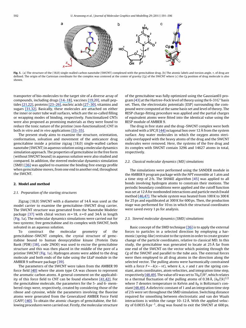
592 U. Arsawang et al. / Journal of Molecular Graphics and Modelling 29 (2011) 591–596
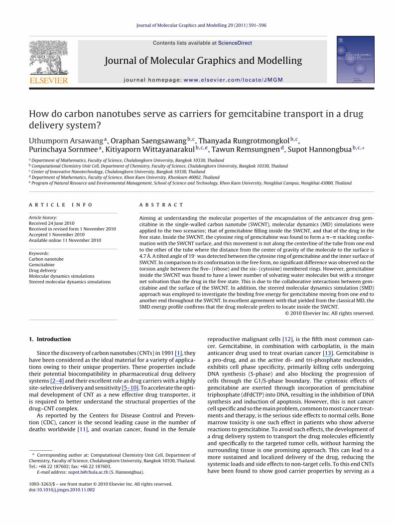
Fig. 1. (a) The structure of the (18,0) single-walled carbon nanotube (SWCNT) complexed with the gemcitabine drug. (b) The atomic labels and torsion angle, �, of drug ared e cents
tctstowrb
cgns(c(wt
2
2
mTp(ts
giBspmA
ftitbra(l
efined. The origin of the Cartesian coordinate for the complex was centered at thhown.
ransporter of bio-molecules to the target site of a diverse array ofompounds, including drugs [14–18], vaccines [19,20], small pep-ides [21,22], proteins [23–26], nucleic acids [27–30], vitamins andugars [31,32]. Basically, these molecules are attached on eitherhe inner or outer tube wall surfaces, which are the so-called fillingr wrapping modes of binding, respectively. Functionalized CNTsere also proposed as promising materials as they were found to
educe the toxic nature of the pristine (non-functionalized) CNT inoth in vitro and in vivo applications [33–35].
The present study aims to examine the structure, orientation,onformation, solvation and movement of the anticancer drugemcitabine inside a pristine zigzag (18,0) single-walled carbonanotube (SWCNT) in aqueous solution using a molecular dynamicsimulation approach. The properties of gemcitabine in the free formwithout SWCNT bound) in aqueous solution were also studied andompared. In addition, the steered molecular dynamics simulationSMD) [36] was applied to examine the binding free energy profilehen gemcitabine moves, from one end to another end, throughout
he SWCNT.
. Model and method
.1. Preparation of the starting structures
Zigzag (18,0) SWCNT with a diameter of 14 A was used as theodel carrier to examine the gemcitabine–SWCNT drug carrier.
he SWCNT structure was generated from the Nanotube Modelerackage [37] with chiral vectors m = 18, n = 0 and 34 A in lengthFig. 1a). The molecular dynamics simulations were carried out forwo systems; free gemcitabine and its complex with SWCNT, botholvated in an aqueous solution.
To construct the molecular geometry of theemcitabine–SWCNT complex, the crystal structure of gemc-tabine bound to human deoxycytidine kinase (Protein Dataank (PDB) [38], code 2NO0) was used to excise the gemcitabinetructure and this was then placed in the middle of the pore of aristine SWCNT (Fig. 1a). Hydrogen atoms were added to the drugolecule and both ends of the tube using the LEaP module in theMBER 9 software package [39].
The parameters of the SWCNT were taken from the AMBER 99orce field [40] where the atom type CA was chosen to representhe aromatic carbon atoms. A general comment on the applicabil-ty of this force field to CNT can be found elsewhere [41,42]. Forhe gemcitabine molecule, the parameters for the 5- and 6- mem-
ered rings were, respectively, created by considering those of theibose and cytosine, while the parameters involving the fluorinetoms were generated from the Generalized AMBER Force FieldGAFF) [40]. To obtain the atomic charges of gemcitabine, the fol-owing procedures were carried out. Firstly, the molecular structureer of gravity (Cg) of the SWCNT where (c) the Cg position of drug molecule is also
of the gemcitabine was fully optimized using the Gaussian03 pro-gram [43] at the Hartree-Fock level of theory using the 6-31G* basisset. Then, the electrostatic potentials (ESP) surrounding the com-pound were computed at the same basis set and level of theory. TheRESP charge-fitting procedure was applied and the partial chargesof equivalent atoms were fitted into the identical value using theRESP module of AMBER 9.
The drug in free state and the drug–SWCNT complex were bothsolvated with a SPC/E [44] octagonal box over 12 A from the systemsurface. Any water molecules in which the oxygen atoms steri-cally overlapped with the heavy atoms of the drug and the SWCNTmolecules were removed. Here, the systems of the free drug andits complex with SWCNT contain 3296 and 14627 atoms in total,respectively.
2.2. Classical molecular dynamics (MD) simulations
The simulations were performed using the SANDER module inthe AMBER 9 program package with the NPT ensemble at 1 atm anda time step of 2 fs. The SHAKE algorithm [45] was applied to allbonds involving hydrogen atoms to constrain their motions. Theperiodic boundary conditions were applied and the cutoff functionwas set at 12 A for nonbonded interactions and particle mesh Ewaldmethod [46,47]. The whole system was heated from 100 K to 300 Kfor 25 ps and equilibrated at 300 K for 600 ps. Then, the productionstage was performed for 10 ns in which the structural coordinateswere saved every 1 ps for analysis.
2.3. Steered molecular dynamics (SMD) simulations
Basic concept of the SMD technique [36] is to apply the externalforces to particles in a selected direction by employing a har-monic (spring-like) restraint to the system in order to create greaterchange of the particle coordinates, relative to classical MD. In thisstudy, the gemcitabine was generated to locate at 25 A far fromone end of the SWCNT on the vector which pointing through theCg of the SWCNT and parallel to the tube axis. The external forceswere then employed to all drug atoms in the direction along theselected vector. The pulling atoms were harmonically constrainedwith a force F = −k(x − vt), where k, x, v and t are the spring con-stant, atom coordinates, atom velocities, and integration time step,respectively [48,49]. The value of k was set to 7kBT/Å2, which relatesto a thermal fluctuation of the pulling atoms of 0.38 A, (kBT/k)1/2
where T denotes temperature in Kelvin and kB is Boltzman’s con-
stant [48,49]. A dielectric constant of 1 and an integration time stepof 2 fs were set throughout the SMD simulation. Switching distancerequired for smoothing between electrostatic and van der Waalsinteractions is within the range 10–12 A. With the applied veloc-ity of 0.0035 A ps−1, drug was found to exit the SWCNT at 600 ps.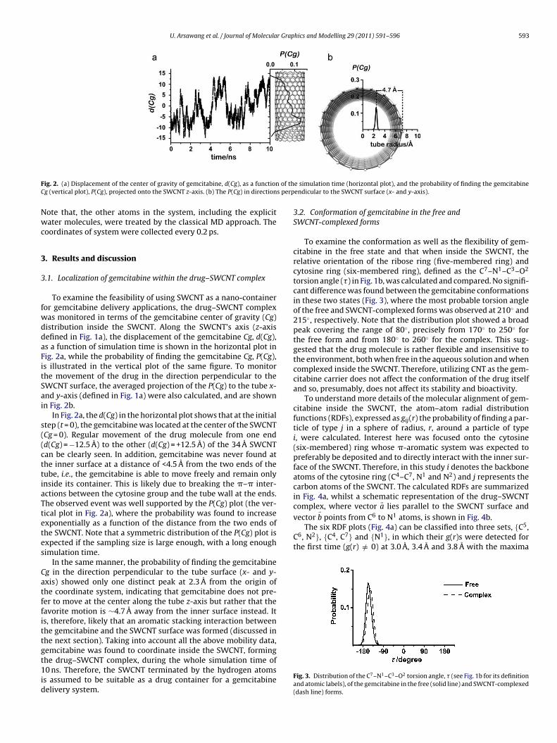
U. Arsawang et al. / Journal of Molecular Graphics and Modelling 29 (2011) 591–596 593
F of thC s perp
Nwc
3
3
fwddaFitSai
s((cttiaTtetes
Catffittgt1id
complex, where vector a lies parallel to the SWCNT surface andvector �b points from C6 to N1 atoms, is shown in Fig. 4b.
The six RDF plots (Fig. 4a) can be classified into three sets, {C5,C6, N2}, {C4, C7} and {N1}, in which their g(r)s were detected forthe first time (g(r) /= 0) at 3.0 A, 3.4 A and 3.8 A with the maxima
ig. 2. (a) Displacement of the center of gravity of gemcitabine, d(Cg), as a functiong (vertical plot), P(Cg), projected onto the SWCNT z-axis. (b) The P(Cg) in direction
ote that, the other atoms in the system, including the explicitater molecules, were treated by the classical MD approach. The
oordinates of system were collected every 0.2 ps.
. Results and discussion
.1. Localization of gemcitabine within the drug–SWCNT complex
To examine the feasibility of using SWCNT as a nano-containeror gemcitabine delivery applications, the drug–SWCNT complexas monitored in terms of the gemcitabine center of gravity (Cg)istribution inside the SWCNT. Along the SWCNT’s axis (z-axisefined in Fig. 1a), the displacement of the gemcitabine Cg, d(Cg),s a function of simulation time is shown in the horizontal plot inig. 2a, while the probability of finding the gemcitabine Cg, P(Cg),s illustrated in the vertical plot of the same figure. To monitorhe movement of the drug in the direction perpendicular to theWCNT surface, the averaged projection of the P(Cg) to the tube x-nd y-axis (defined in Fig. 1a) were also calculated, and are shownn Fig. 2b.
In Fig. 2a, the d(Cg) in the horizontal plot shows that at the initialtep (t = 0), the gemcitabine was located at the center of the SWCNTCg = 0). Regular movement of the drug molecule from one endd(Cg) = −12.5 A) to the other (d(Cg) = +12.5 A) of the 34 A SWCNTan be clearly seen. In addition, gemcitabine was never found athe inner surface at a distance of <4.5 A from the two ends of theube, i.e., the gemcitabine is able to move freely and remain onlynside its container. This is likely due to breaking the �–� inter-ctions between the cytosine group and the tube wall at the ends.he observed event was well supported by the P(Cg) plot (the ver-ical plot in Fig. 2a), where the probability was found to increasexponentially as a function of the distance from the two ends ofhe SWCNT. Note that a symmetric distribution of the P(Cg) plot isxpected if the sampling size is large enough, with a long enoughimulation time.
In the same manner, the probability of finding the gemcitabineg in the direction perpendicular to the tube surface (x- and y-xis) showed only one distinct peak at 2.3 A from the origin ofhe coordinate system, indicating that gemcitabine does not pre-er to move at the center along the tube z-axis but rather that theavorite motion is ∼4.7 A away from the inner surface instead. Its, therefore, likely that an aromatic stacking interaction betweenhe gemcitabine and the SWCNT surface was formed (discussed inhe next section). Taking into account all the above mobility data,
emcitabine was found to coordinate inside the SWCNT, forminghe drug–SWCNT complex, during the whole simulation time of0 ns. Therefore, the SWCNT terminated by the hydrogen atomss assumed to be suitable as a drug container for a gemcitabineelivery system.
e simulation time (horizontal plot), and the probability of finding the gemcitabineendicular to the SWCNT surface (x- and y-axis).
3.2. Conformation of gemcitabine in the free andSWCNT-complexed forms
To examine the conformation as well as the flexibility of gem-citabine in the free state and that when inside the SWCNT, therelative orientation of the ribose ring (five-membered ring) andcytosine ring (six-membered ring), defined as the C7–N1–C3–O2
torsion angle (�) in Fig. 1b, was calculated and compared. No signifi-cant difference was found between the gemcitabine conformationsin these two states (Fig. 3), where the most probable torsion angleof the free and SWCNT-complexed forms was observed at 210◦ and215◦, respectively. Note that the distribution plot showed a broadpeak covering the range of 80◦, precisely from 170◦ to 250◦ forthe free form and from 180◦ to 260◦ for the complex. This sug-gested that the drug molecule is rather flexible and insensitive tothe environment, both when free in the aqueous solution and whencomplexed inside the SWCNT. Therefore, utilizing CNT as the gem-citabine carrier does not affect the conformation of the drug itselfand so, presumably, does not affect its stability and bioactivity.
To understand more details of the molecular alignment of gem-citabine inside the SWCNT, the atom–atom radial distributionfunctions (RDFs), expressed as gij(r) the probability of finding a par-ticle of type j in a sphere of radius, r, around a particle of typei, were calculated. Interest here was focused onto the cytosine(six-membered) ring whose �-aromatic system was expected topreferably be deposited and to directly interact with the inner sur-face of the SWCNT. Therefore, in this study i denotes the backboneatoms of the cytosine ring (C4–C7, N1 and N2) and j represents thecarbon atoms of the SWCNT. The calculated RDFs are summarizedin Fig. 4a, whilst a schematic representation of the drug–SWCNT
�
Fig. 3. Distribution of the C7–N1–C3–O2 torsion angle, � (see Fig. 1b for its definitionand atomic labels), of the gemcitabine in the free (solid line) and SWCNT-complexed(dash line) forms.
594 U. Arsawang et al. / Journal of Molecular Graphics and Modelling 29 (2011) 591–596
F gemcir the SWb
ad(abtiimaf
3S
dapSncFgT
camt
TFSo
and e) were noticeably higher than those in the SWCNT-complexedforms, which imply that water molecules in the first hydration shellbind stronger to the drug atoms in the complex form than those inthe free form. The likely reason for this finding is because of the col-
ig. 4. (a) RDFs centered at the atoms in the cytosine (six-membered) ring of theepresentation of the gemcitabine–SWCNT complex where vector �a lies parallel toetween vectors �a and �b (see text for details of the related distances).
t ∼4.0 A, ∼4.5 A and 4.9 A, respectively. Using the most probableistances from the C6 (∼4.0 A maximum of the C6–C RDF) and N1
∼4.9 A maximum of the N1–C RDF) atoms to the SWCNT surface,nd the N1–C6 distance (2.8 A), the angle between the vectors �a and
� can be estimated (Fig. 4c). The obtained value of 19◦ indicateshe tilted angle representing the configuration of the �–� stackingnteraction between the cytosine ring of the gemcitabine and thenner surface of the SWCNT. This interaction is supposed to be the
ain reason why the preferential mobility of the drug moleculelong the molecular z-axis of the SWCNT takes place at ∼4.7 A farrom the surface of the SWCNT (Fig. 2b).
.3. Solvation of gemcitabine in free solution and complexed withWCNT
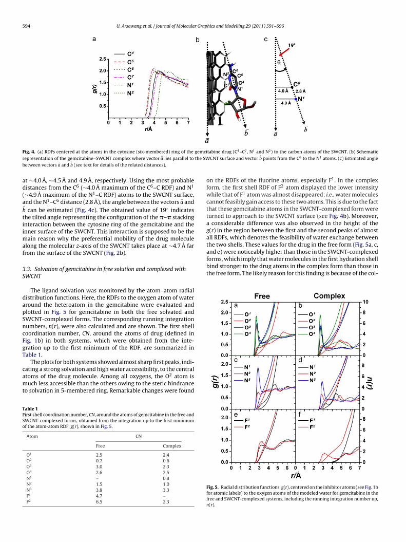
The ligand solvation was monitored by the atom–atom radialistribution functions. Here, the RDFs to the oxygen atom of waterround the heteroatom in the gemcitabine were evaluated andlotted in Fig. 5 for gemcitabine in both the free solvated andWCNT-complexed forms. The corresponding running integrationumbers, n(r), were also calculated and are shown. The first shelloordination number, CN, around the atoms of drug (defined inig. 1b) in both systems, which were obtained from the inte-ration up to the first minimum of the RDF, are summarized inable 1.
The plots for both systems showed almost sharp first peaks, indi-
ating a strong solvation and high water accessibility, to the centraltoms of the drug molecule. Among all oxygens, the O2 atom isuch less accessible than the others owing to the steric hindranceo solvation in 5-membered ring. Remarkable changes were found
able 1irst shell coordination number, CN, around the atoms of gemcitabine in the free andWCNT-complexed forms, obtained from the integration up to the first minimumf the atom-atom RDF, g(r), shown in Fig. 5.
Atom CN
Free Complex
O1 2.5 2.4O2 0.7 0.6O3 3.0 2.3O4 2.6 2.5N1 – 0.8N2 1.5 1.0N3 3.8 3.3F1 4.7 –F2 6.5 2.3
tabine drug (C4–C7, N1 and N2) to the carbon atoms of the SWCNT. (b) SchematicCNT surface and vector �b points from the C6 to the N1 atoms. (c) Estimated angle
on the RDFs of the fluorine atoms, especially F1. In the complexform, the first shell RDF of F2 atom displayed the lower intensitywhile that of F1 atom was almost disappeared; i.e., water moleculescannot feasibly gain access to these two atoms. This is due to the factthat these gemcitabine atoms in the SWCNT-complexed form wereturned to approach to the SWCNT surface (see Fig. 4b). Moreover,a considerable difference was also observed in the height of theg(r) in the region between the first and the second peaks of almostall RDFs, which denotes the feasibility of water exchange betweenthe two shells. These values for the drug in the free form (Fig. 5a, c,
Fig. 5. Radial distribution functions, g(r), centered on the inhibitor atoms (see Fig. 1bfor atomic labels) to the oxygen atoms of the modeled water for gemcitabine in thefree and SWCNT-complexed systems, including the running integration number up,n(r).
U. Arsawang et al. / Journal of Molecular Graphics and Modelling 29 (2011) 591–596 595
F ained( ibose)
li
i(dtosi{m{rsafs
3S
opM[yamdF(
aa(oafmdSfavp
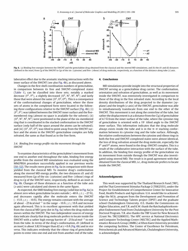
ig. 6. (a) Binding free energies between the SWCNT and the gemcitabine drug obtdefined in the inset) from Cg of the SWCNT to Cgs of the six- (cytosine) and five- (r
aborative effect due to the aromatic stacking interactions with thenner surface of the SWCNT (see also Fig. 4b), as discussed above.
Changes in the first shell coordination numbers of gemcitabine,n comparison between its free and SWCNT-complexed statesTable 1), can be classified into three sets; notably a markedecrease {F1, F2}, a slightly decreased {O3, N1, N2, N3} and lastlyhose that were almost the same {O1, O2, O4}. This is a consequencef the conformational changes of gemcitabine, where the threeets of atoms in the complexed form were located in the follow-ng three configurations relative to the SWCNT surface (Fig. 4b); (i)F1, F2} was tabbed between the SWCNT inner surface and the five-embered ring (almost no space is available for the solvent); (ii)
O3, N1, N2, N3} were positioned in the plane of the six-membereding that is coordinated in the stacked conformation to the SWCNTurface (only half of the space around the atoms can be solvated);nd (iii) {O1, O2, O4} was tilted to point away from the SWCNT sur-ace and the atoms in the SWCNT–gemcitabine complex are fullyolvated, the same as that found as in the free form.
.4. Binding free energy profile via the movement through theWCNT
To examine characteristics of the gemcitabine’s movement fromne end to another end throughout the tube, binding free energyrofile from the steered MD simulations was evaluated using theM/PBSA procedure successfully applied in our previous works
50–52]. The results were given in Fig. 6a in comparison with thoseielded from the classical MD. To monitor drug’s conformationlong the steered MD energy profile, the two distances d1 and d2easured from Cgs of the six- (cytosine) and five- (ribose) rings of
rug to Cg of the SWCNT were, respectively, defined in an inset inig. 6b. Changes of those distances as a function of the tube axisz-axis) were calculated and shown in the same figure.
As expected, the SMD binding free energy (solid line in Fig. 6a) islmost zero when gemcitabine locates outside (−40 A ≤ z ≤ −27 A),nd decreases rapidly after the molecule enters the SWCNT−15 A ≤ z ≤ −10 A). The energy remains constant with the averagef about −25 kcal mol−1 in the range −10 A ≤ z ≤ 10 A and increasegain afterward. This is in excellent agreement with that yieldedrom the classical MD (grey dot in Fig. 6a) when the drug molecule
oves within the SWCNT. The two independent sources of energyata indicate clearly that drug molecule prefers to locate inside the
WCNT with a rather high energy barrier of −25 kcal mol−1 to exitrom the tube. In terms of the drug’s conformation, d1 (defined inn inset) is about 3.82 A longer than d2 at z < 0 A (Fig. 6b) and in viceersa. This indicates evidently that the ribose ring of gemcitabineoints to enter into one end and exit from another end of the tube.from the classical and the steered MD simulations, and (b) the d1 and d2 distancesof drug molecule, respectively, as a function of the distance along tube (z) axis.
4. Conclusions
MD simulations provide insight into the structural properties ofSWCNT serving as a gemcitabine drug carrier. The conformation,orientation and solvation of gemcitabine, as well as its movementinside the SWCNT, was extensively investigated in comparison tothose of the drug in the free solvated state. According to the localdensity distributions of the drug projected to the diameter (xy-plane) and the length (z-axis) of the SWCNT, gemcitabine was ableto simultaneously translocate from one end to the other of theSWCNT. This movement is not along the centerline of the tube, butrather the displacement is at a distance from the C(g) of gemcitabineof 4.7 A from the inner surface of the tube, where the cytosine ringof gemcitabine is oriented with a 19◦ tilted angle to the SWCNTinner surface. This information indicates that the drug moleculealways exists inside the tube and is in the �–� stacking confor-mation between its cytosine ring and the tube surface. Although,the relative conformations between the cytosine and ribose rings inboth the free and SWCNT-complexed states were almost identical,the loss of drug solvation around the drug molecule, especially theF1 and F2 atoms, were found in the drug–SWCNT complex. This is aresult of the collaborative interaction with the surface of the tube.In addition, the binding free energy profile of the gemcitabine viaits movement from outside through the SWCNT was also investi-gated using steered MD. The result is in good agreement with thatobtained from the classical MD, i.e., drug molecule prefers to locateinside the SWCNT.
Acknowledgments
This work was supported by The Thailand Research Fund (TRF),and the Thai Government Stimulus Package 2 (TKK2555), under theProject for Establishment of Comprehensive Center for InnovativeFood, Health Products and Agriculture. U.A. would like to acknowl-edge the M.Sc. program from the Development and Promotion ofScience and Technology Talents project (DPST) and the graduateschool Chulalongkorn University. O.S. thanks the Commission onHigher Education, and T.R. and K.W. thank the Ratchadaphiseksom-phot Endowment Fund from Chulalongkorn University for the PostDoctoral Program. T.R. also thanks the TRF Grant for New Research(Grant No. TRG5280035). The HPC service at National Electronics
and Computer Technology Center (NECTEC) and the Applied Math-ematics Research Group (AMRG) at Khon Kaen University providedthe computing facilities. The Center of Excellence for Petroleum,Petrochemicals and Advanced Materials, Chulalongkorn University,is acknowledged.
5 r Grap
R
[
[[[
[
[
[
[
[[
[
[
[
[
[
[
[
[
[
[
[
[
[
[
[
[
[
[
[[
[
[
[
[
[
[
[
[
[
[
[
96 U. Arsawang et al. / Journal of Molecula
eferences
[1] S. Iijima, Helical microtubules of graphitic carbon, Nature 354 (1991) 56–58.[2] A. Bianco, K. Kostarelos, C.D. Partidos, M. Prato, Biomedical applications of
functionalised carbon nanotubes, Chem. Commun. (2005) 571–577.[3] S.K. Smart, A.I. Cassady, G.Q. Lu, D.J. Martin, The biocompatibility of carbon
nanotubes, Carbon 44 (2006) 1034–1047.[4] A.M. Popov, Y.E. Lozovik, S. Fiorito, L.H. Yahia, Biocompatibility and applications
of carbon nanotubes in medical nanorobots, Int. J. Nanomed. 2 (2007) 361–372.[5] S. Banerjee, T. Hemraj-Benny, S.S. Wong, Covalent surface chemistry of single-
walled carbon nanotubes, Adv. Mater. 17 (2005) 17–29.[6] G. Pastorin, W. Wu, S.B. Wieckowski, J.-P. Briand, K. Kostarelos, M. Prato, et al.,
Double functionalisation of carbon nanotubes for multimodal drug delivery,Chem. Commun. (2006) 1182–1184.
[7] C. Klumpp, K. Kostarelos, M. Prato, A. Bianco, Functionalized carbon nanotubesas emerging nanovectors for the delivery of therapeutics, Biochim. Biophys.Acta: Biomembr. 1758 (2006) 404–412.
[8] K. Kostarelos, L. Lacerda, G. Pastorin, W. Wu, J. WieckowskiSebastien, Luangsivi-lay, et al., Cellular uptake of functionalized carbon nanotubes is independentof functional group and cell type, Nat. Nanotechnol. 2 (2007) 108–113.
[9] V. Raffa, G. Ciofani, S. Nitodas, T. Karachalios, D. D’Alessandro, M. Masini, et al.,Can the properties of carbon nanotubes influence their internalization by livingcells? Carbon 46 (2008) 1600–1610.
10] X. Zhang, L. Meng, Q. Lu, Z. Fei, P.J. Dyson, Targeted delivery and controlledrelease of doxorubicin to cancer cells using modified single wall carbon nan-otubes, Biomaterials 30 (2009) 6041–6047.
11] http://www.cdc.gov/nchs/FASTATS/deaths.htm.12] http://www.reuters.com/article/idUKN1930660220080220.13] M.F. Kose, M.M. Meydanli, G. Tulunay, Gemcitabine plus carboplatin in
platinum-sensitive recurrent ovarian carcinoma, Expert Rev. Anticancer Ther.6 (2006) 437–443.
14] A. Bianco, K. Kostarelos, M. Prato, Applications of carbon nanotubes in drugdelivery, Curr. Opin. Chem. Biol. 9 (2005) 674–679.
15] R.P. Feazell, N. Nakayama-Ratchford, H. Dai, S.J. Lippard, Soluble single-walledcarbon nanotubes as longboat delivery systems for platinum (IV) anticancerdrug design, J. Am. Chem. Soc. 129 (2007) 8438–8439.
16] Z. Liu, X.M. Sun, N. Nakayama-Ratchford, H.J. Dai, Supramolecular chemistryon water-soluble carbon nanotubes for drug loading and delivery, ACS Nano 1(2007) 50–56.
17] Z. Liu, K. Chen, C. Davis, S. Sherlock, Q.Z. Cao, X.Y. Chen, et al., Drug deliverywith carbon nanotubes for in vivo cancer treatment, Cancer Res. 68 (2008)6652–6660.
18] C. Srinivasan, Carbon nanotubes in cancer therapy, Curr. Sci. 94 (2008) 300–301.19] A. Bianco, M. Prato, Can carbon nanotubes be considered useful tools for bio-
logical applications? Adv. Mater. 15 (2003) 1765–1768.20] C. Salvador-Morales, E. Flahaut, E. Sim, J. Sloan, M.L.H. Green, R.B. Sim, Comple-
ment activation and protein adsorption by carbon nanotubes, Mol. Immun. 43(2006) 193–201.
21] D. Pantarotto, C.D. Partidos, R. Graff, J. Hoebeke, J.-P. Briand, M. Prato, et al.,Synthesis, structural characterization, and immunological properties of car-bon nanotubes functionalized with peptides, J. Am. Chem. Soc. 125 (2003)6160–6164.
22] D. Pantarotto, C.D. Partidos, J. Hoebeke, F. Brown, E. Kramer, J.-P. Briand,et al., Immunization with peptide-functionalized carbon nanotubes enhancesvirus-specific neutralizing antibody responses, Chem. Biol. 10 (2003) 961–966.
23] D. Pantarotto, J.-P. Briand, M. Prato, A. Brianco, Translocation of bioactive pep-tides across cell membranes by carbon nanotubes, Chem. Commun. (2004)16–17.
24] N.W.S. Kam, H. Dai, Carbon nanotubes as intracellular protein trans-porters: generality and biological functionality, J. Am. Chem. Soc. 127 (2005)6021–6026.
25] K. Yanagi, K. Iakoubovskii, S. Kazaoui, N. Minami, Y. Maniwa, Y. Miyata, et al.,Light-harvesting function of beta-carotene inside carbon nanotubes, Phys. Rev.B 74 (2006) 155420.
26] K. Yanagi, Y. Miyata, H. Kataura, Highly stabilized �-carotene in carbon nan-
otubes, Adv. Mater. 18 (2006) 437–441.27] H. Gao, Y. Kong, D. Cui, C.S. Ozkan, Spontaneous insertion of DNA oligonu-cleotides into carbon nanotubes, Nano Lett. 3 (2003) 471–473.
28] N.W.S. Kam, Z. Liu, H. Dai, Functionalization of carbon nanotubes via cleavabledisulfide bonds for efficient intracellular delivery of siRNA and potent genesilencing, J. Am. Chem. Soc. 127 (2005) 12492–12493.
[
[
hics and Modelling 29 (2011) 591–596
29] Y. Liu, D.-C. Wu, W.-D. Zhang, X. Jiang, C.-B. He, T.S. Chung, et al.,Polyethylenimine-grafted multiwalled carbon nanotubes for secure noncova-lent immobilization and efficient delivery of DNA, Angew. Chem. Int. Ed. 44(2005) 4782–4785.
30] Z. Liu, M. Winters, M. Holodniy, H.J. Dai, siRNA delivery into human T cellsand primary cells with carbon-nanotube transporters, Angew. Chem. Int. Ed.46 (2007) 2023–2027.
31] Y.H. Xie, A.K. Soh, Investigation of non-covalent association of single-walledcarbon nanotube with amylose by molecular dynamics simulation, Mater. Lett.59 (2005) 971–975.
32] J. Xie, Q. Xue, Q. Zheng, H. Chen, Investigation of the interactions betweenmolecules of [beta]-carotene, vitamin A and CNTs by MD simulations, Mater.Lett. 63 (2009) 319–321.
33] V.L. Colvin, The potential environmental impact of engineered nanomaterials,Nat. Biotechnol. 21 (2003) 1166–1170.
34] C.M. Sayes, F. Liang, J.L. Hudson, J. Mendez, W. Guo, J.M. Beach, et al., Function-alization density dependence of single-walled carbon nanotubes cytotoxicityin vitro, Toxicol. Lett. 161 (2006) 135–142.
35] M. Prato, K. Kostarelos, A. Bianco, Functionalized carbon nanotubes in drugdesign and discovery, Acc. Chem. Res. 41 (2007) 60–68.
36] M. Gao, M. Wilmanns, K. Schulten, Steered molecular dynamics studies of titinII domain unfolding, Biophys. J. 83 (2002) 3435–3445.
37] http://www.jcrystal.com/products/wincnt/, Nanotube Modeler, JCrystalSoftEd., 2004–2005.
38] http://www.drugbank.ca/.39] D.A. Case, T.A. Darden, I.T.E. Cheatham, C.L. Simmerling, J. Wang, R.E. Duke,
et al., AMBER 10, U.O. California Ed., San Francisco, 2008.40] J. Wang, R.M. Wolf, J.W. Caldwell, P.A. Kollman, D.A. Case, Development and
testing of a general amber force field, J. Comput. Chem. 25 (2004) 1157–1174.41] Y. Cheng, D. Li, B. Ji, X. Shi, H. Gao, Structure-based design of carbon nanotubes
as HIV-1 protease inhibitors: atomistic and coarse-grained simulations, J. Mol.Graphics Modell. 29 (2010) 171–177.
42] C. Wei, Adhesion and reinforcement in carbon nanotube polymer composite,Appl. Phys. Lett. 88 (2006) 093108-1–093108-3.
43] M.J.T. Frisch, G.W. Trucks, H.B. Schlegel, G.E. Scuseria, M.A. Robb, J.R. Cheese-man Jr., J.A. Montgomery, T. Vreven, K.N. Kudin, J.C. Burant, J.M. Millam, S.S.Iyengar, J. Tomasi, V. Barone, B. Mennucci, M. Cossi, G. Scalmani, N. Rega, G.A.Petersson, H. Nakatsuji, M. Hada, M. Ehara, K. Toyota, R. Fukuda, J. Hasegawa,M. Ishida, T. Nakajima, Y. Honda, O. Kitao, H. Nakai, M. Klene, X. Li, J.E. Knox, H.P.Hratchian, J.B. Cross, V. Bakken, C. Adamo, J. Jaramillo, R. Gomperts, R.E. Strat-mann, O. Yazyev, A.J. Austin, R. Cammi, C. Pomelli, J.W. Ochterski, P.Y. Ayala, K.Morokuma, G.A. Voth, P. Salvador, J.J. Dannenberg, V.G. Zakrzewski, S. Dapprich,A.D. Daniels, M.C. Strain, O. Farkas, D.K. Malick, A.D. Rabuck, K. Raghavachari, P.Foresman, I. Komaromi, R.L. Martin, D.J. Fox, T. Keith, M.A. Al-Laham, C.Y. Peng,A. Nanayakkara, M. Challacombe, P.M.W. Gill, B. Johnson, W. Chen, M.W. Wong,C. Gonzalez, J.A. Pople, Gaussian 03 Revision C. 02, Gaussian Inc., Wallingford,CT, 2004.
44] H.J.C. Berendsen, J.R. Grigera, T.P. Straatsma, The missing term in effective pairpotentials, J. Phys. Chem. 91 (1987) 6269–6271.
45] S. Miyamoto, P.A. Kollman, Settle: an analytical version of the SHAKE and RAT-TLE algorithm for rigid water models, J. Comput. Chem. 13 (1992) 952–962.
46] T. Darden, D. York, L. Pedersen, Particle mesh Ewald: an N [center-dot]log(N) method for Ewald sums in large systems, J. Chem. Phys. 98 (1993)10089–10092.
47] U. Essmann, L. Perera, M.L. Berkowitz, T. Darden, H. Lee, L.G. Pedersen, A smoothparticle mesh Ewald method, J. Chem. Phys. 103 (1995) 8577–8593.
48] J.C. Phillips, R. Braun, W. Wang, J. Gumbart, E. Tajkhorshid, E. Villa, et al., Scalablemolecular dynamics with NAMD, J. Comput. Chem. 26 (2005) 1781–1802.
49] S. Kumar, C. Huang, G. Zheng, E. Bohm, A. Bhatele, J.C. Phillips, et al., Scalablemolecular dynamics with NAMD on the IBM Blue Gene/L system, IBM J. Res.Dev. 52 (2008) 177–188.
50] T. Rungrotmongkol, N. Nunthaboot, M. Malaisree, N. Kaiyawet, P. Yotmanee, A.Meeprasert, et al., Molecular insight into the specific binding of ADP-ribose tothe nsP3 macro domains of chikungunya and venezuelan equine encephalitisviruses: molecular dynamics simulations and free energy calculations, J. Mol.Graphics Modell. (2010), doi:10.1016/j.jmgm.2010.09.2010.
51] O Aruksakunwong, M. Malaisree, P. Decha, P. Sompornpisut, V. Parasuk, S.Pianwanit, et al., On the lower susceptibility of oseltamivir to influenza neu-raminidase subtype N1 than those in N2 and N9, Biophys. J. 92 (2007) 798–807.
52] M. Malaisree, T. Rungrotmongkol, P. Decha, P. Intharathep, O. Aruksakunwong,S. Hannongbua, Understanding of known drug-target interactions in the cat-alytic pocket of neuraminidase subtype N1, Proteins 71 (2008) 1908–1918.
Related Documents


![A [60]fullerene nanoconjugate with gemcitabine: synthesis ... · Title: A [60]fullerene nanoconjugate with gemcitabine : synthesis, biophysical properties and biological evaluation](https://static.cupdf.com/doc/110x72/608dfcfefb2f9961d327bba6/a-60fullerene-nanoconjugate-with-gemcitabine-synthesis-title-a-60fullerene.jpg)
