Journal of Clinical Immunology, Vol. 27, No. 4, July 2007 ( C 2007) DOI: 10.1007/s10875-007-9084-0 Host Innate Immune Response to Mycobacterium tuberculosis KAMLESH BHATT 1 and PADMINI SALGAME 1,2 Received February 9, 2007; accepted February 14, 2007 Published online: 16 March 2007 This review focuses on recent progress in our understanding of Mycobacterium tuberculosis survival in macrophages, the interaction of M. tuberculosis with Toll-like receptors (TLRs) and the establishment of the link between innate and adaptive immunity, and TLRs and interferon-γ -mediated antimicrobial pathways in macrophages. We also propose a paradigm that TLR2 signaling regulates the magnitude of the host Th1 response leading to either M. tuberculosis persistence and latent infection or replication and disease. KEY WORDS: Mycobaterium tuberculosis; innate immunity; TLR2; dendritic cells; macrophages. INTRODUCTION The naissance of innate immunity was the description of macrophage phagocytosis by Metchnikoff (1). Today, the science of innate immunity is more than just phagocytosis. The innate immune response comprises several different cell types, has its own receptor system to recognize the presence of pathogens, and is a key to the initiation of an adaptive immune response in the host. No wonder, successful pathogens have evolved ways to evade innate immune killing in order to find a niche in the host. In this review, we will discuss host innate immunity generated in response to the pathogen, Mycobacterium tuberculosis (Mtb). Mtb infects approximately one-third of the world’s pop- ulation (2). Close to eight million new cases of tubercu- losis occur each year, accounting for approximately 7% of all deaths and 26% of all avoidable adult deaths in developing countries (3). Despite the implementation of TB control programs, case rates continue to soar where 1 Department of Medicine, Centre for Emerging Pathogens, UMDNJ- New Jersey Medical School, MSB A902, 185 South Orange Avenue, Newark, New Jersey, 07101. 2 To whom correspondence should be addressed to; e-mail: [email protected]. the prevalence of HIV infection is high (4). The situation is further complicated by a worldwide increase in drug resistant and MDR-TB, and the recent reports of XDR TB (5). Thus, the resurgence of TB truly constitutes a global health crisis (6). Tuberculosis begins with the inhalation of Mtb- containing aerosols into the pulmonary alveoli. Here, the bacteria bind to phagocytic receptors and enter resident alveolar macrophages, dendritic cells, and monocytes re- cruited from the bloodstream. Besides expressing phago- cytic receptors, macrophages and dendritic cells also ex- press Toll-like receptors (TLRs) that recognize conserved molecular patterns expressed on pathogens (7–9). Liga- tion of TLRs by these pathogen-specific ligands initiates a signal transduction pathway in the host cell that cul- minates in the activation of NFκ b and the induction of cytokines and chemokines (10) that are crucial to elicit- ing the adaptive immune response against the pathogen. Consequently, activation of TLR is an important link be- tween innate cellular response and the subsequent ac- tivation of adaptive immune defense against microbial pathogens. As depicted in Fig. 1, a small percentage of individuals, despite exposure to Mtb, remain uninfected, most likely due to the expression of high innate immunity. However, in the majority of individuals who are exposed to Mtb, the innate response cannot protect from infection, and effector Th1 cytokines of the adaptive immune response are necessary to restrict bacterial growth and mediate pro- tection. The adaptive immunity generated in these people, although protective, nonetheless does not induce steriliz- ing immunity. These individuals therefore remain latently infected, and are vulnerable to disease reactivation when their immune surveillance weakens or when their immune response is compromised. Reactivation tuberculosis con- tributes significantly to the morbidity and mortality asso- ciated with the disease (11, 12), and is believed to account for a substantial portion of TB cases in HIV-infected in- dividuals (13). In another small proportion of individuals, infection leads directly to primary tuberculosis due to a 347 0271-9142/07/0700-0347/0 C 2007 Springer Science+Business Media, LLC

Welcome message from author
This document is posted to help you gain knowledge. Please leave a comment to let me know what you think about it! Share it to your friends and learn new things together.
Transcript
Journal of Clinical Immunology, Vol. 27, No. 4, July 2007 ( C© 2007)DOI: 10.1007/s10875-007-9084-0
Host Innate Immune Response to Mycobacterium tuberculosis
KAMLESH BHATT1 and PADMINI SALGAME1,2
Received February 9, 2007; accepted February 14, 2007Published online: 16 March 2007
This review focuses on recent progress in our understandingof Mycobacterium tuberculosis survival in macrophages, theinteraction of M. tuberculosis with Toll-like receptors (TLRs)and the establishment of the link between innate and adaptiveimmunity, and TLRs and interferon-γ -mediated antimicrobialpathways in macrophages. We also propose a paradigm thatTLR2 signaling regulates the magnitude of the host Th1 responseleading to either M. tuberculosis persistence and latent infectionor replication and disease.
KEY WORDS: Mycobaterium tuberculosis; innate immunity; TLR2;dendritic cells; macrophages.
INTRODUCTION
The naissance of innate immunity was the description ofmacrophage phagocytosis by Metchnikoff (1). Today, thescience of innate immunity is more than just phagocytosis.The innate immune response comprises several differentcell types, has its own receptor system to recognize thepresence of pathogens, and is a key to the initiation ofan adaptive immune response in the host. No wonder,successful pathogens have evolved ways to evade innateimmune killing in order to find a niche in the host. In thisreview, we will discuss host innate immunity generatedin response to the pathogen, Mycobacterium tuberculosis(Mtb).
Mtb infects approximately one-third of the world’s pop-ulation (2). Close to eight million new cases of tubercu-losis occur each year, accounting for approximately 7%of all deaths and 26% of all avoidable adult deaths indeveloping countries (3). Despite the implementation ofTB control programs, case rates continue to soar where
1Department of Medicine, Centre for Emerging Pathogens, UMDNJ-New Jersey Medical School, MSB A902, 185 South Orange Avenue,Newark, New Jersey, 07101.
2To whom correspondence should be addressed to; e-mail:[email protected].
the prevalence of HIV infection is high (4). The situationis further complicated by a worldwide increase in drugresistant and MDR-TB, and the recent reports of XDRTB (5). Thus, the resurgence of TB truly constitutes aglobal health crisis (6).
Tuberculosis begins with the inhalation of Mtb-containing aerosols into the pulmonary alveoli. Here, thebacteria bind to phagocytic receptors and enter residentalveolar macrophages, dendritic cells, and monocytes re-cruited from the bloodstream. Besides expressing phago-cytic receptors, macrophages and dendritic cells also ex-press Toll-like receptors (TLRs) that recognize conservedmolecular patterns expressed on pathogens (7–9). Liga-tion of TLRs by these pathogen-specific ligands initiatesa signal transduction pathway in the host cell that cul-minates in the activation of NFκb and the induction ofcytokines and chemokines (10) that are crucial to elicit-ing the adaptive immune response against the pathogen.Consequently, activation of TLR is an important link be-tween innate cellular response and the subsequent ac-tivation of adaptive immune defense against microbialpathogens.
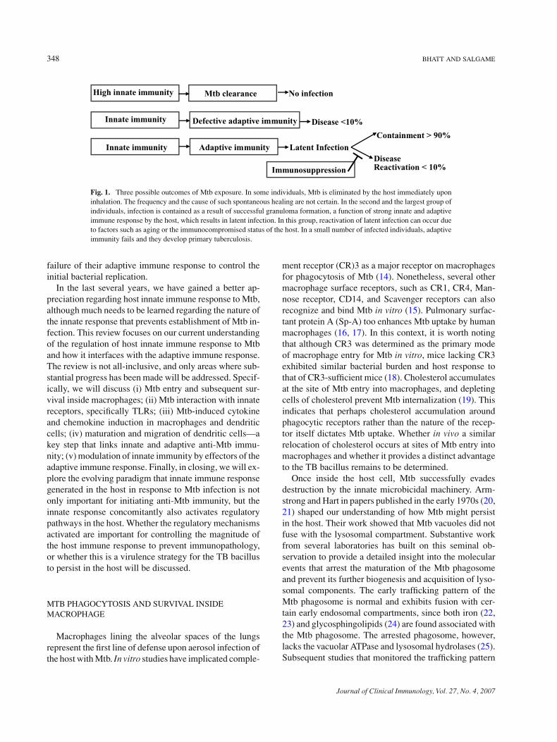
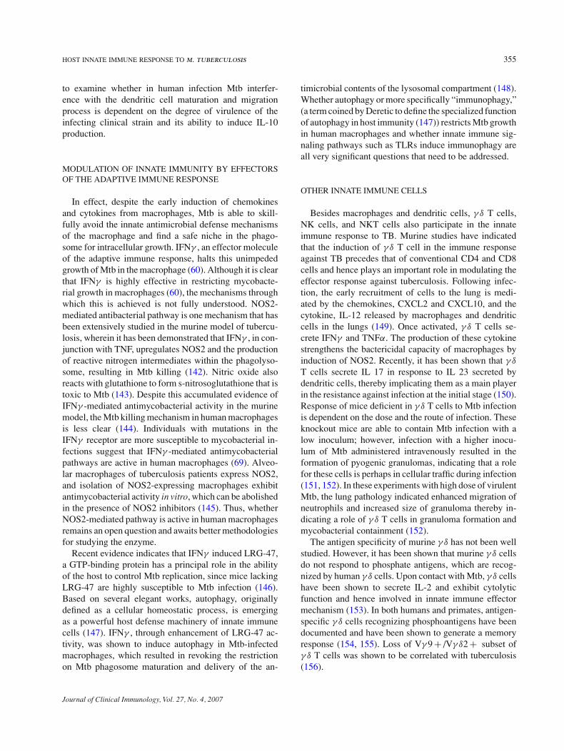
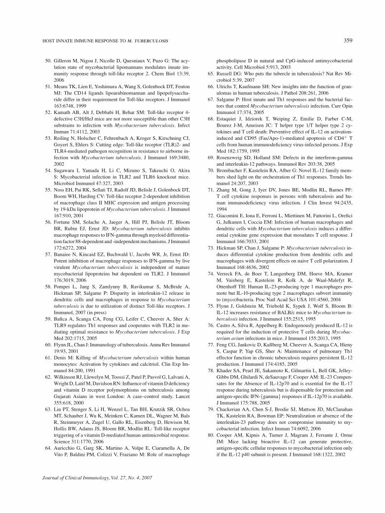
As depicted in Fig. 1, a small percentage of individuals,despite exposure to Mtb, remain uninfected, most likelydue to the expression of high innate immunity. However,in the majority of individuals who are exposed to Mtb,the innate response cannot protect from infection, andeffector Th1 cytokines of the adaptive immune responseare necessary to restrict bacterial growth and mediate pro-tection. The adaptive immunity generated in these people,although protective, nonetheless does not induce steriliz-ing immunity. These individuals therefore remain latentlyinfected, and are vulnerable to disease reactivation whentheir immune surveillance weakens or when their immuneresponse is compromised. Reactivation tuberculosis con-tributes significantly to the morbidity and mortality asso-ciated with the disease (11, 12), and is believed to accountfor a substantial portion of TB cases in HIV-infected in-dividuals (13). In another small proportion of individuals,infection leads directly to primary tuberculosis due to a
3470271-9142/07/0700-0347/0 C© 2007 Springer Science+Business Media, LLC
348 BHATT AND SALGAME
High innate immunity Mtb clearance No infection
Defective adaptive immunityInnate immunity Disease <10%
Immunosuppression
Innate immunity Adaptive immunity Latent InfectionDiseaseReactivation < 10%
Containment > 90%
Fig. 1. Three possible outcomes of Mtb exposure. In some individuals, Mtb is eliminated by the host immediately uponinhalation. The frequency and the cause of such spontaneous healing are not certain. In the second and the largest group ofindividuals, infection is contained as a result of successful granuloma formation, a function of strong innate and adaptiveimmune response by the host, which results in latent infection. In this group, reactivation of latent infection can occur dueto factors such as aging or the immunocompromised status of the host. In a small number of infected individuals, adaptiveimmunity fails and they develop primary tuberculosis.
failure of their adaptive immune response to control theinitial bacterial replication.
In the last several years, we have gained a better ap-preciation regarding host innate immune response to Mtb,although much needs to be learned regarding the nature ofthe innate response that prevents establishment of Mtb in-fection. This review focuses on our current understandingof the regulation of host innate immune response to Mtband how it interfaces with the adaptive immune response.The review is not all-inclusive, and only areas where sub-stantial progress has been made will be addressed. Specif-ically, we will discuss (i) Mtb entry and subsequent sur-vival inside macrophages; (ii) Mtb interaction with innatereceptors, specifically TLRs; (iii) Mtb-induced cytokineand chemokine induction in macrophages and dendriticcells; (iv) maturation and migration of dendritic cells—akey step that links innate and adaptive anti-Mtb immu-nity; (v) modulation of innate immunity by effectors of theadaptive immune response. Finally, in closing, we will ex-plore the evolving paradigm that innate immune responsegenerated in the host in response to Mtb infection is notonly important for initiating anti-Mtb immunity, but theinnate response concomitantly also activates regulatorypathways in the host. Whether the regulatory mechanismsactivated are important for controlling the magnitude ofthe host immune response to prevent immunopathology,or whether this is a virulence strategy for the TB bacillusto persist in the host will be discussed.
MTB PHAGOCYTOSIS AND SURVIVAL INSIDEMACROPHAGE
Macrophages lining the alveolar spaces of the lungsrepresent the first line of defense upon aerosol infection ofthe host with Mtb. In vitro studies have implicated comple-
ment receptor (CR)3 as a major receptor on macrophagesfor phagocytosis of Mtb (14). Nonetheless, several othermacrophage surface receptors, such as CR1, CR4, Man-nose receptor, CD14, and Scavenger receptors can alsorecognize and bind Mtb in vitro (15). Pulmonary surfac-tant protein A (Sp-A) too enhances Mtb uptake by humanmacrophages (16, 17). In this context, it is worth notingthat although CR3 was determined as the primary modeof macrophage entry for Mtb in vitro, mice lacking CR3exhibited similar bacterial burden and host response tothat of CR3-sufficient mice (18). Cholesterol accumulatesat the site of Mtb entry into macrophages, and depletingcells of cholesterol prevent Mtb internalization (19). Thisindicates that perhaps cholesterol accumulation aroundphagocytic receptors rather than the nature of the recep-tor itself dictates Mtb uptake. Whether in vivo a similarrelocation of cholesterol occurs at sites of Mtb entry intomacrophages and whether it provides a distinct advantageto the TB bacillus remains to be determined.
Once inside the host cell, Mtb successfully evadesdestruction by the innate microbicidal machinery. Arm-strong and Hart in papers published in the early 1970s (20,21) shaped our understanding of how Mtb might persistin the host. Their work showed that Mtb vacuoles did notfuse with the lysosomal compartment. Substantive workfrom several laboratories has built on this seminal ob-servation to provide a detailed insight into the molecularevents that arrest the maturation of the Mtb phagosomeand prevent its further biogenesis and acquisition of lyso-somal components. The early trafficking pattern of theMtb phagosome is normal and exhibits fusion with cer-tain early endosomal compartments, since both iron (22,23) and glycosphingolipids (24) are found associated withthe Mtb phagosome. The arrested phagosome, however,lacks the vacuolar ATPase and lysosomal hydrolases (25).Subsequent studies that monitored the trafficking pattern
Journal of Clinical Immunology, Vol. 27, No. 4, 2007
HOST INNATE IMMUNE RESPONSE TO m. tuberculosis 349
of the Mtb phagosome showed that the arrest occurs be-tween the acquisition of the endocytic vesicles Rab5 andRab7. The Mtb phagosome is associated with Rab5 (26),but not Rab7 (27), and furthermore exhibits reduced re-cruitment of the early endosomal autoantigen 1 (EEA1)(28), an effector molecule of Rab5 required for organelletethering and delivery of lysosomal hydrolases, cathep-sins, and vacuolar ATPases from the trans golgi networkto the phagosome. The Mtb phagosome also lacks a spe-cific type III phosphatidylinositol 3-kinase, hVPs34 (29)whose activation product phospatidylinositol 3-phosphateaids the retention of EEA1 to the endosomal membrane(29).
How and what components of Mtb block the Mtbphagosome from undergoing the typical phagosome bio-genesis? The arrest of the Mtb phagosome at the Rab 5stage and its inability to proceed through the maturationpathway, at least partly, results from Mtb-induced inhibi-tion of sphingosine kinase activity and subsequent Ca2+
signaling pathway in the cell, a step necessary for recruit-ment of hVPs35 to membranes of organelles (30). Anotherstudy reported that maturation of the Mtb phagosome isimpeded because Mtb suppresses phagosomal actin as-sembly (31). Yet another study determined that the Mtbphagosome arrest was dependent on its initial fusion withearly endosomes and acquisition of iron (32). Manosy-lated lipoarabinamannan (ManLAM), the Mtb analog ofhost phophatidylinositol-3 phosphate is responsible foractively inhibiting Mtb phagosome from fully maturingand acquiring lysosomal hydrolases (33, 34). Indeed, ithas been shown that phagocytosis of Mtb via binding ofits cell surface LAM to the mannose receptor on humanmacrophages led to the non-fusogenic phenotype of theMtb phagosome (35). Interestingly, other Mtb lipids suchas PIMs enhance Mtb phagosome fusion with early endo-somes, possibly providing the phagosome access to hostnutrients (36). Besides lipids, a protein from Mtb, proteinkinase G (PknG) has also been implicated in interferingwith the transfer of Mtb phagosome to the lysosomal com-partment (37).
Our understanding of Mtb phagocytosis, the biogene-sis of Mtb phagosome, and intracellular growth of Mtb isalmost entirely derived from studies examining interac-tion of Mtb with macrophages. However, it is necessaryto also understand the handling of Mtb inside dendriticcells, since in response to an aerosol challenge with Mtb,dendritic cells take up Mtb, and as will be discussed later,are crucial to linking the innate and adaptive immuneresponses. Besides the expression of CR3 and mannosereceptor, dendritic cells are endowed with an additionalphagocytic receptor for binding Mtb. Dendritic cells ex-press the C-type lectin, DC-SIGN (DC-specific intercel-
lular adhesion molecule-grabbing nonintegrin), and Mtbcan bind DC-SIGN through manLAM expressed on theirsurface. A comparative analysis of Mtb survival within hu-man macrophages and dendritic cells revealed that unlikemacrophages, dendritic cells did not support intracellulargrowth of Mtb (38, 39). Despite being a key player in theinnate response to Mtb, we know very little regarding thetrafficking of Mtb vacuole inside dendritic cells; exceptfor a study that reported that endosomal trafficking is sig-nificantly reduced compared to that in macrophages (39).Clearly, more detailed studies examining the intracellularfate of Mtb inside dendritic cells are needed.
It must be emphasized that Mtb replication in themacrophage is also controlled at the level of host factors.Expression of a candidate gene IntracellularPathogenRe-sistance (lpr1) within the sst1 locus limits Mtb multipli-cation in the host (40). Variants in the human equiva-lent of the lpr1 gene SP110 were shown to be associatedwith genetic susceptibility to TB in a study of familiesin West Africa (41). Another association study in humanTB, also in West Africa, however, found no association ofhuman pulmonary TB with SP110 variants (42). Undoubt-edly, more studies in genetically different populations areneeded to equivocally determine the role of SP100 andother potential candidate genes in susceptibility to TB.
MTB INTERACTION WITH TLRS
Engagement of TLR by Mtb ligands is an early eventin the interaction of Mtb with its host cell. Accumulat-ing data indicate that Mtb expresses a large repertoireof TLR2 ligands. The 19-kDa lipoprotein (LpqH), a se-creted antigen of Mtb, was the first Mtb ligand shownto interact specifically with TLR2 to induce TNFα andnitric oxide production from both murine and humanmacrophages (43). In addition, the 19-kDa lipoproteinis a major inducer of interleukin (IL)-12 production inhuman monocytes (43). LprA (Rv1270) (44) and LprG(Rv1411c) (45) are two other mycobacterial lipoproteinsthat are TLR2 agonists. In addition to lipoproteins, lipo-mannan (46) and phosphatidyl-myo-inositol mannoside(PIM) (47, 48) also interact with TLR2 to initiate cel-lular activation (48). However, with regards to PIMs,Abel et al. (49) demonstrated that PIM structures canalso elicit cellular activation via TLR4. They showedthat PIM was able to induce NFκB activation in a dose-dependent manner in stable TLR4 and MD-2 Ba-F3 trans-formants. A systematic biochemical characterization offour acyl forms of lipomannan (LM) from M. bovis BCGthat differed in their degree of acylation, indicated thatonly the triacylated LM was a potent TLR2 agonist (50).
Journal of Clinical Immunology, Vol. 27, No. 4, 2007
350 BHATT AND SALGAME
Interestingly, ManLAM derived from virulent Mtb failsto activate either TLR2 or TLR4-transfected cells (51). Incontrast, AraLAM purified from fast-growing mycobac-teria is capable of TLR2-mediated cellular activation(51).
Studies aimed at determining the requirement of TLR4in controlling Mtb infection following an aerosol chal-lenge showed that lack of TLR4 did not compromise hostresistance to TB (52, 53). However, a high dose of Mtbinfection did lead to enhanced susceptibility in the ab-sence of TLR4 signaling (49). It is interesting that despitea large collection of TLR2 agonists on the TB bacillus,murine studies indicate that TLR2 is not essential for hostresistance against tuberculosis. In a model of low-doseaerosol infection, TLR2 (53, 54) deficiency did not affecthost defense against Mtb infection. However, in one ofthe two studies (53) with high-dose aerosol infection, arole for TLR2 in host resistance was revealed. The TLR2-deficient mice were not compromised in their ability toinduce Th1 immunity, but on the contrary, exhibited ex-aggerated immunopathology. In vitro studies have shownthat engagement of TLR2 with Mtb ligands induces in-hibition of macrophage MHC class II antigen presenta-tion (55) and also blocks macrophage responsiveness toIFNγ (56, 57). Together with the in vivo studies, these invitro findings that TLR2 signaling negatively modulatesmacrophage functions point to the need for future studiesdesigned to examine whether the negative signaling fromTLR2 curtails Th1 activation, and whether this is impor-tant for balancing protection and immunopathology in thehost.
Our studies examining the in vitro interaction of TLRwith live Mtb reported that in response to Mtb, dendriticcells secreted copious amount of IL-12, while the se-cretion was limited in infected macrophages. The studyalso reported that Mtb induced rapid and significantlyhigher remodeling at the IL-12p40 promoter in dendriticcells in comparison to macrophages. The mechanismbehind the differential remodeling at the IL-12p40 pro-moter and subsequent IL-12 release was shown to be dueto differences in TLR usage by macrophages and den-dritic cells. Mtb induced IL-12 secretion from dendriticcells in a TLR9-dependent manner while in macrophagesit was TLR2-dependent (58). Consistent with this, thegreatest effect on the progression of tuberculosis diseasewas seen in mice doubly deficient in TLR2 and TLR9(59).
Although IFNγ is undoubtedly necessary for resistanceagainst Mtb infection (60), it is of interest that there existantimycobacterial pathways that are independent of IFNγ
and are induced by TLR in human macrophages. For ex-ample, it has been known for some time that activation
of the Vitamin D3 pathway controls Mtb replication inhuman macrophages (61). Also, it had been documentedthat Vitamin D deficiency is a risk factor for tuberculosis(62). Only recently, however, Modlin and colleagues deci-phered the mechanism for Vitamin-D3-mediated antimi-crobial pathway. They demonstrated that TLR2-mediatedactivation of macrophages upregulated the expression ofVitamin D receptor and Vitamin-D-1-hydroxylase genes,leading to the induction of the antimicrobial peptide,cathelicidin (63). The study from Modlin’s group alsoshowed that African American individuals who are moresusceptible to Mtb infection and disease were not effi-cient in inducing the antimicrobial peptide, cathelicidin.The TLR2-mediated innate mechanism of mycobacterialkilling provides a scientific basis for tuberculosis treat-ment of a century ago: exposure to sunlight. Other TLR-induced killing mechanisms may also participate in theinnate response. For instance, CpG, an activator of theTLR9-pathway, also induces rapid antimycobacterial re-sponses in macrophages, in a phospholipase D-dependentmanner (64). These innate mechanisms for killing Mtbprovoke future investigations of whether individuals whonever become infected with Mtb have the capacity to acti-vate these pathways and overpower the Mtb-induced blockin phagosome maturation.
MTB-INDUCED UPSURGE OF CYTOKINEAND CHEMOKINE SECRETION
A major consequence of Mtb interaction with the TLRson macrophages and dendritic cells is the burst in cytokineand chemokine secretion. The induction of these effectormolecules regulates the formation of the granuloma and isresponsible for initiating and shaping the adaptive immuneresponse to Mtb. The contribution of adaptive immunityto the evolving tubercle granuloma in the lung will not beelaborated in this review. The reader is referred to otherreviews on the topic (65–67).
Cytokines Important for Induction of Th1 Immunity
Clearly, induction of cellular Th1 immunity is criti-cal for protection against tuberculosis as evidenced byenhanced disease in the HIV-infected (68) and from ex-periments of nature where individuals carrying defectivegenes for IFNγ R and IL-12R (69) are exquisitely sus-ceptible to intracellular pathogens, including mycobacte-ria. Currently, there are three well-defined cytokines thatsteer naı̈ve T cells toward Th1 commitment (70). IL-12was the first cytokine to be described with potent Th1promoting attributes, followed by the discovery of IL-23
Journal of Clinical Immunology, Vol. 27, No. 4, 2007
HOST INNATE IMMUNE RESPONSE TO m. tuberculosis 351
(shares the p40 component with IL-12) and the recentaddition of IL-27 to this list. Work from several labora-tories has revealed that the three cytokines together or-chestrate Th1 responses, with IL-12 being the prototypicand dominant cytokine that affects both the induction andmaintenance of Th1 immunity. IL-23, on the other hand,has activities on memory T cells and IL-27, secreted priorto IL-12 by antigen-presenting cells, is involved in Th1initiation.
In patients with tuberculosis pleuritis, a clinical formof disease that is mostly self-healing, high IL-12 levelswere found in the pleural fluid (71). Two studies compar-ing murine macrophages and dendritic cells demonstratedthat dendritic cells release significantly higher IL-12 thandid macrophages in response to live Mtb (72, 73). Invitro, Mtb-infected dendritic cells also primed naı̈ve Tcells toward Th1 development, while macrophages didnot; though, IL-23-secreting macrophages were capableof inducing Mtb-specific Th1 response (74).
Early studies in the murine model of tuberculosisclearly demonstrated that the cytokine IL-12 that is nec-essary to drive Th1 responses and IFNγ —the effectormolecule of the Th1 response—were both necessary forprotection against Mtb infection. Mice deficient in the p40component of IL-12 or in IFNγ (GKO) were both highlysusceptible to Mtb infection. Exogenous supplementationof IL-12 at the onset of disease led to reduction in bacterialburden and delayed the lung pathology in the relativelysusceptible Balb/C strain of mice (75). However, IL-12supplementation did not lead to enhanced protection inGKO mice indicating that IFNγ is downstream of IL-12and is the effector molecule mediating protection in thehost. In another study (76), the role of endogenous IL-12was studied by neutralization with anti-IL-12 antibodies.It was found that in Balb/C mice neutralization of IL-12at the onset of infection led to disruption in the abilityof the host to contain infection; however, neutralizationof the cytokine after the onset of infection (third week)did not affect bacterial replication. This suggests that thepresence of IL-12 is more critical during early infectionwhen anti-Mtb adaptive immunity is being shaped towardTh1-type. However, in later studies, reconstitution of IL-12-p40 gene-deficient mice with recombinant IL-12 onlyduring the early phase of infection was determined notto be sufficient to provide long-term immunity, despiteearly control of bacterial growth. Transfer of immuneCD4 T cells from Mtb-infected wild-type mice to Rag−/− provided immunity against infection. However, sim-ilar reconstitution of immune CD4 T cells into Rag−/−mice that were also deficient in IL-12-p40 failed to in-duce protection. This provides experimental evidence thatsustained production of IL-12 throughout the course of
infection is necessary to maintain antibacterial immunityin the host (77). A reason why this study differed fromthe previous where IL-12 was shown not to be neces-sary for long-term immunity may probably be due to thepresence of residual IL-12 activity in the neutralizationexperiments.
To further characterize whether susceptibility to Mtbinfection in the absence of p40 is due to the lack of biolog-ically active IL-12 or is a consequence of defective IL-23production, mice deficient in the specific components ofIL-12 and IL-23, p35 and p19, respectively, were studied.Mice lacking p19 were able to control Mtb infection aswell as the wild-type mice (78, 79). Mice lacking p35were able to control bacterial replication slightly betterthan the p40-deficient mice (79, 80), but in comparison tothe p19 knockout mice exhibited significantly higher bac-terial burden. Mice doubly deficient in p35 and p19 geneswere as susceptible as the p40-gene-deficient mice (79).Exogenous delivery into the lung of IL-23 via adenoviralvectors enhanced anti-Mtb immunity, upregulated IL-17expression, and reduced bacterial burden in the lungs ofMtb-infected mice (81). Together, these data indicate thatIL-23 is less critical for protection against Mtb, and onlyprovides a moderate level of protection to the host in theabsence of biologically active IL-12. On the other hand,IL-12 has a far more vital role in the generation of protec-tive anti-Mtb immunity.
IL-27 is also an IL-12-related cytokine and WSX-1is a component of the IL-27R complex (82). IL-27/IL-27R signaling, interestingly, exhibits both pro- and anti-inflammatory properties. Infection of WSX-1−/− micewith Mtb revealed that, in the absence of IL-27R signal-ing, there was a reduced bacterial burden accompaniedby enhanced CD4 infiltration into the lungs (83). Anothergroup examining Mtb infection in the same WSX-1−/−mice observed increased IL-12-p40 and TNFα expressionand enhanced IFNγ production from CD4 T cells. Thisgroup also reported reduced Mtb burden in the lungs ofinfected WSX-1 knockout mice in comparison with wild-type mice. Despite restricted bacterial growth, the WSX-1knockout mice succumbed to infection due to exaggeratedimmunopathology, a scenario similar to what was first re-ported with Toxoplasma gondii infection in this strain ofknockout mice. Under conditions where pathogens do notinduce a Th1 response in the host, for example in Leish-mania major infection of Balb/c mice, absence of WSX-1 resulted in the generation of protective Th1 responsewith concomitant Th2 downregulation in the host (84).A tenet for future perusal is that the anti-inflammatoryactivity of IL-27 is perhaps more critical than its Th1 pro-moting activity in response to pathogens that have highTh1-inducing potential (85, 86).
Journal of Clinical Immunology, Vol. 27, No. 4, 2007
352 BHATT AND SALGAME
TNF
TNFα plays an important role in regulating the pathol-ogy of tuberculosis (87). TNFα exists in both solubleand membrane bound forms and signals through TNFαR.Mtb infection leads to TNFα secretion by macrophages,dendritic cells, and T cells (60). Secretion of TNFα byMtb-infected macrophages is a potent mechanism to in-duce killing of Mtb via generation of reactive nitrogenintermediates in conjunction with IFNγ (88). An attributeof membrane TNFα is to induce apoptosis of the Mtb-infected alveolar macrophages (89), and thereby indirectlycontribute to the reduction of bacterial burden. TNFα’sability to induce alveolar macrophage apoptosis may alsobe important in the cross-presentation of Mtb antigensfor CD8 cytotoxic T cell priming (90). It has also beensuggested that inhibition of TNFα-mediated macrophageapoptosis is a virulence strategy of Mtb. Avirulent H37Rainduced TNFα-dependent macrophage apoptosis, whilevirulent H37Rv released soluble TNFR2 that reducedTNFα activity and subsequent apoptosis of macrophages(91). Although it needs to be examined in more detail,TNFα has been shown to support the growth of Mtb inhuman monocytes and macrophages (92).
The requirement for TNFα in host defense against Mtbinfection was demonstrated in studies which showed thatmice treated with antibody to TNFα became more sus-ceptible to BCG infection and exhibited malformed gran-ulomas (93). Mtb infection of mice lacking TNF recep-tor or neutralization of TNFα activity in mice also ledto the failure to control bacterial replication resulting inenhanced susceptibility (94). This study indicated thatTNFα contributed to maintaining host resistance by in-ducing the production of reactive nitrogen intermediatesby macrophages. Later studies have indicated that TNFα
also participates in setting the chemokine circuitry in thedeveloping granuloma. Mice lacking TNFα had reducedchemokine expression in lung granulomas (95–97) andthis resulted in reduced T cell infiltration into the lungsand a failure to form a productive granuloma.
For the most part, the immunological forces that controlreactivation remain ill defined, except for the knowledgethat TNFα is a major player (98). Studies in a murine latentmodel of tuberculosis from the Chan and Flynn labora-tories clearly demonstrated that neutralization of TNFα
during the latent/persistent phase induced reactivation inC57BL/6 mice, as indicated by the enhanced bacterial bur-den in the lungs (99). Further, histological examinationof lung tissue from TNFα-neutralized animals revealeda disorganized granuloma with indications of a lack ofcellular turnover and increased fluid accumulation. NOS2expression was attenuated, while IL-10 expression was
upregulated in the lungs. Immunohistochemical analysisindicated an increased presence of apoptotic T cells andmacrophages in the lung, a feature not seen previouslyin other reactivation models. The importance of TNF inmaintaining Mtb in a chronic/persistent phase in mice hasbeen corroborated in humans. It has been observed that theuse of anti-TNFα antibody in patients undergoing treat-ment for rheumatoid arthritis has resulted in reactivationof tuberculosis in some latently infected individuals (100,101).
Mtb-Induced Chemokines
In vitro and in vivo studies provide evidence forthe participation of chemokines in the control of TB.It is present in the innate and adaptive immune re-sponse to Mtb (96, 102). Mtb infection of both hu-man and murine macrophages results in the secretionof a large number of chemokines, including CCL2,CCL3, CCL7, CCL12, CXCL2, and CXCL10 (96). Acomparative chemokine expression analysis showed thatlung interstitial macrophages from a susceptible mousestrain expressed significantly high levels of CXCL13and CXCL14, while higher expression of CXCL9 andCXCL0 was found in macrophages from the resistantstrain of mice (103). Regulation of chemokine pro-duction in macrophages is predominantly regulated byTNFα. Mtb infection of macrophages leads to the pro-duction of TNFα which, in turn, regulates the secre-tion of a plethora of chemokines from macrophages,including CCL2, CCL3, CCL4, CCL5, CXCL10, andCXCL13 (104). Mtb-infected dendritic cells also secretechemokines, including CXCL9, CXCL10, CCL3, andCCL4. CXCL10 secretion was IFNα-dependent and inconjunction with CXCL9 and CXCL3 acted to recruitinflammatory cells to the site of Mtb infection (105).
Studies to examine the role of chemokine andchemokine receptors in host resistance against Mtb in-fection has led to conflicting results, in great part due toredundancy in the function of chemokines and their recep-tors. In a mouse model of tuberculosis, it has been shownthat the first step in recruitment of cells into the lung,specifically recruitment of immature dendritic cells andmonocytes to the site of infection, is mediated by CCR2and, as a consequence, CCR2−/− mice (106) are moresusceptible to Mtb infection. These mice also show de-fective recruitment of dendritic cell to the draining lymphnodes resulting in delayed and reduced priming of naiveT cells. However, a later study (107) indicated that sus-ceptibility of CCR2 knockout mice to Mtb infection wasdose dependent. As seen with high dose infection, a low-dose aerosol challenge of the CCR2 knockout mice with
Journal of Clinical Immunology, Vol. 27, No. 4, 2007
HOST INNATE IMMUNE RESPONSE TO m. tuberculosis 353
Mtb also resulted in reduced cellular migration to thelungs and delayed priming. However, there was no changein bacterial burden or susceptibility to infection. Usingchimeric mice, where either the myeloid or the lymphoidcompartment was lacking CCR2, it was determined thatexpression of CCR2 on macrophages and dendritic cellswas important for the recruitment of T cells to lungs (108).However, mice deficient in CCL2/MCP-1, which is a lig-and for CCR2, do not show reduced susceptibility to Mtbinfection, thereby indicating that in vivo other chemokinessuch as CCL7, CCL8, and CCL12 can compensate for thelack of CCL2/MCP-1 (109).
In addition to its expression on granulocytes andmacrophages, CCR5 is also present on immature den-dritic cells and Mtb modulates its expression. Indeed, my-cobacterial Hsp70 can interact with CCR5 on immaturedendritic cells and induce their maturation and the inter-action also induces IL-12 secretion from dendritic cells(110). Despite CCR5-mediated IL-12 production and theenhanced production of CCR5 ligands, MIP-1α, MIP-1β,and RANTES in the lungs of Mtb-infected mice, absenceof CCR5 did not affect the ability of the host to con-trol bacterial replication in the lung (111, 112). The latterstudy, in addition, observed that CCR5−/− mice exhibitedincreased bacterial burden in the draining lymph nodes(112). The intriguing possibility that CCR5 signals im-pede Mtb-bearing dendritic cell migration resulting in en-hanced accumulation of Mtb in the draining lymph nodesneeds further scrutiny.
CCR7 expression on cells guides their migration to thedraining lymph nodes where its cognate ligands CCL19and CCL21 are present. Indeed, Mtb infection upregulatesCCR7 expression on dendritic cells, but absence of CCR7did not enhance bacterial replication (113). Although itmust be noted that the granulomas of CCR7−/− mice hadaltered granuloma architecture with enhanced inflamma-tion and a lack of follicular B cell architecture. How thesechanges in the granuloma affect host resistance is still notclear. As discussed in the next section, Mtb-infected den-dritic cells migrate to the draining lymph nodes to initiatean immune response. Therefore, it would be important todetermine what chemokine/receptor gradient controls themigration of Mtb-infected dendritic cells from lungs todraining lymph nodes.
CXCR3 is a chemokine receptor preferentially ex-pressed on activated Th1 cells and regulates their mi-gration in response to ligands CXCL10, CXCL9, andCXCL11. Given the importance of Th1 in host resistanceagainst TB, C57BL/6 mice deficient in CXCR3 were stud-ied following Mtb infection. Despite the ability of CXCR3to regulate the migration of Th1 cells, absence of thereceptor did not affect Mtb replication in the host. The
CXCR3 knockout mice, however, did exhibit a neutrophildeficit in the granuloma. The consequence to host resis-tance of reduced neutrophils in the granuloma remainsunclear (114). Contrary findings were reported in a recentstudy that examined mice lacking CXCR3 on the BALB/cbackground. In this study, the CXCR3-deficient miceexhibited heightened resistance to Mtb infection in thechronic phase when compared with wild-type mice (115),and the mice also had enhanced T cell activation (115).The authors of the paper suggest that enhanced resistancein BALB/cCXCR3 knockout mice could have resultedfrom the absence of the immunosuppressive CXCR3–CXCL10 chemokine gradient. Certain chemokine gra-dients, including CXCR3–CXCL10, have recently beenrecognized as immunosuppressive and to interfere withthe formation of the immunological synapse (116). To-gether, these studies highlight the emerging recognitionthat the chemokine circuitry activated during Mtb infec-tion may not only regulate cellular recruitment, but also di-rectly impact on the function of immune cells. The studiesalso highlight the role of genetic differences in regulatingchemokine functions.
IL-10
Dendritic cells and macrophages in response to Mtbproduce the immunosuppressive and anti-inflammatorycytokine IL-10. Interestingly, dendritic cells secrete sub-stantial IL-12 in response to Mtb infection and can primenaive T cells toward Th1-type, despite concomitant se-cretion of IL-10 (73, 117). IL-10-secreting CD8 suppres-sor/regulatory T cells are associated with susceptibility toMtb infection (118) and T cell expressing both IFNγ andIL-10 have been isolated from the bronchoalveolar lavagefluid of TB patients (119). Additionally, depressed T-cellIFNγ responses in pulmonary tuberculosis was shown tobe associated with the induction of IL-10 from monocytes(120). Interestingly, in patients with pleural TB, consid-ered as the resistant and self-healing form of the disease,IL-10 is found along with IFNγ at sites of infection inthe pleural fluid (121). In vitro, IL-10 downregulates theproduction of IL-12 in human monocytes infected withMtb (122). Also, IL-10 down modulates the activity ofCD4 and CD8 T cells via downregulation of costimula-tory molecules on macrophages (123). In addition, IL-10inhibits the proliferation of IFNγ producing T cells and γ δ
T cells (124). Although absence of IL-10 did not enhanceresistance to Mtb infection in IL-10 knockout mice (125),transgenic over-expression of IL-10 resulted in reactiva-tion of chronic disease (126). Similarly, expression of hu-man IL 10 in mice under the control of MHC II promoterenhanced the susceptibility to disease, independent of
Journal of Clinical Immunology, Vol. 27, No. 4, 2007
354 BHATT AND SALGAME
T-cell-derived IL-10. The Mtb-infected macrophagesfrom these transgenic mice exhibited reduced antimy-cobacterial capacity (127). That IL-10 may have a rolein TB is suggested by the fact that polymorphism inmurine SLC11 A1, a tuberculosis susceptibility locus,has been associated with variation in IL-10 production(128). Together, these data suggest that IL-10 is inducedby Mtb and suppresses the generation anti-Mtb immunity.However, Th1 cytokines are often found along with IL-10. Perhaps the relative quantities of the two cytokinesdetermine whether Th1 immunity is suppressed or not.
DENDRITIC CELL MATURATION, MIGRATION,AND ANTIGEN PRESENTATION
Recent work from several laboratories has focused ondissecting the role of dendritic cells in Mtb infection.Upon its interaction with Mtb, dendritic cells undergo arepertoire of phenotypical changes, a process termed asmaturation. This process, which is TLR-dependent, bringsforth three major phenotypic changes in dendritic cells:upregulation of costimulatory molecules-CD40, B7.1 andB7.2, heightened expression of adhesion molecules, andupregulation of chemokine receptor—CCR7. Whereasimmature dendritic cells are efficient at Mtb phagocytosisand exhibit enhanced microbicidal property, maturationendows them with the role of an efficient antigen presenterand initiator of adaptive immune responses (129).
Following Mtb phagocytosis and concomitant TLR ac-tivation, the next step in the development of host immunityis the transport of pathogen from the lung to the drain-ing lymph nodes, where the matured dendritic cells canpresent antigen to naive T cells and initiate the processof adaptive immune response. Although Mtb uptake andengagement of TLR signaling for cellular activation oc-curs in macrophages and dendritic cells, only the lattercell type was shown to acquire the capacity to upreg-ulate CCR7 expression and migrate to draining lymphnodes (130). Consistent with this study that tracked intra-tracheally instilled cells, endogenous lung dendritic cellsalso exhibited similar migratory property. Following in-tratracheal infection of mice with GFP-expressing BCG,dissemination of mycobacteria from the lung was initi-ated by the migration of infected dendritic cells to thedraining lymph nodes (131), despite predominant infec-tion of alveolar macrophages. Another study in BCG-infected mice also demonstrated that dendritic cells, andnot macrophages, were the antigen-presenting cells re-sponsible for priming naı̈ve T cells (132). Direct evidencefor the role of dendritic cells as the priming APC for ini-tiating pulmonary immunity came from the study where
mice depleted of CD11c + dendritic cells exhibited de-layed CD4 responses to Mtb and worsening disease (133).It has been argued that in addition to ferrying antigen forT cell priming, migration of dendritic cells to the lymphnodes may also aid in Mtb dissemination (131).
Are other cell types involved in transporting Mtb anti-gens to the draining lymph nodes? Indeed, a recent re-port implicates neutrophils as the carrier of live BCGfollowing intradermal vaccination from peripheral tissueto the DLN capsule (134). Whether neutrophils partic-ipate in antigen transport during a pulmonary infectionwith Mtb would be worth investigating, particularly sinceneutrophils appear to marginally influence early immuneresponses and the architecture of the ensuing granuloma(135, 136). Following Mtb infection, there is an influxof macrophages and dendritic cells from the peripheryinto the lung. The relative contribution of interstitialversus the newly recruited dendritic cells in Mtb trans-port from lung to the draining lymph nodes is also notclear. However, absence of CCR2 was shown to impairmacrophage and dendritic cells trafficking into the in-fected lungs resulting in susceptibility to Mtb infection(108), suggesting that dendritic cells recruited to the lungmay also function to carry Mtb to the draining lymphnodes.
Collective data indicate that dendritic cells are theantigen-presenting cells that migrate to the draining lymphnodes, and process and present Mtb antigens on MHCClasses I and II to naı̈ve CD4+ and CD8+ cells, respec-tively. This review will not address the mechanisms ofantigen processing and presentation and other molecularevents controlling T cell activation. However, we discusshere one study from Kaufman’s group that demonstrateda detour pathway for how antigens of Mtb, presumablyconfined within the phagosome, are delivered to Class Imolecules. This study demonstrated that dendritic cellstake up apoptotic vesicles containing Mtb, the vesiclesare then degraded in an endosomal-dependent mannerand Mtb antigens are cross-presented on MHC Class Imolecules to CD8 T cells (137).
Since maturation and migration of dendritic cells issuch a key step in linking innate and adaptive immu-nity, it is not surprising that Mtb negatively interfereswith this step. IL-1β release from mycobacteria-infectedantigen-presenting cells inhibits dendritic cell matura-tion (138) and virulent Mtb has been reported to im-pair the maturation of monocyte derived dendritic cells(139). The migration of dendritic cells appears to be reg-ulated to some degree by IL-12p40 homodimers (140).Whether IL-10, which can impede dendritic cell migra-tion (141), functions by downregulating the p40 homod-imers is worth considering. In addition, one also needs
Journal of Clinical Immunology, Vol. 27, No. 4, 2007
HOST INNATE IMMUNE RESPONSE TO m. tuberculosis 355
to examine whether in human infection Mtb interfer-ence with the dendritic cell maturation and migrationprocess is dependent on the degree of virulence of theinfecting clinical strain and its ability to induce IL-10production.
MODULATION OF INNATE IMMUNITY BY EFFECTORSOF THE ADAPTIVE IMMUNE RESPONSE
In effect, despite the early induction of chemokinesand cytokines from macrophages, Mtb is able to skill-fully avoid the innate antimicrobial defense mechanismsof the macrophage and find a safe niche in the phago-some for intracellular growth. IFNγ , an effector moleculeof the adaptive immune response, halts this unimpededgrowth of Mtb in the macrophage (60). Although it is clearthat IFNγ is highly effective in restricting mycobacte-rial growth in macrophages (60), the mechanisms throughwhich this is achieved is not fully understood. NOS2-mediated antibacterial pathway is one mechanism that hasbeen extensively studied in the murine model of tubercu-losis, wherein it has been demonstrated that IFNγ , in con-junction with TNF, upregulates NOS2 and the productionof reactive nitrogen intermediates within the phagolyso-some, resulting in Mtb killing (142). Nitric oxide alsoreacts with glutathione to form s-nitrosoglutathione that istoxic to Mtb (143). Despite this accumulated evidence ofIFNγ -mediated antimycobacterial activity in the murinemodel, the Mtb killing mechanism in human macrophagesis less clear (144). Individuals with mutations in theIFNγ receptor are more susceptible to mycobacterial in-fections suggest that IFNγ -mediated antimycobacterialpathways are active in human macrophages (69). Alveo-lar macrophages of tuberculosis patients express NOS2,and isolation of NOS2-expressing macrophages exhibitantimycobacterial activity in vitro, which can be abolishedin the presence of NOS2 inhibitors (145). Thus, whetherNOS2-mediated pathway is active in human macrophagesremains an open question and awaits better methodologiesfor studying the enzyme.
Recent evidence indicates that IFNγ induced LRG-47,a GTP-binding protein has a principal role in the abilityof the host to control Mtb replication, since mice lackingLRG-47 are highly susceptible to Mtb infection (146).Based on several elegant works, autophagy, originallydefined as a cellular homeostatic process, is emergingas a powerful host defense machinery of innate immunecells (147). IFNγ , through enhancement of LRG-47 ac-tivity, was shown to induce autophagy in Mtb-infectedmacrophages, which resulted in revoking the restrictionon Mtb phagosome maturation and delivery of the an-
timicrobial contents of the lysosomal compartment (148).Whether autophagy or more specifically “immunophagy,”(a term coined by Deretic to define the specialized functionof autophagy in host immunity (147)) restricts Mtb growthin human macrophages and whether innate immune sig-naling pathways such as TLRs induce immunophagy areall very significant questions that need to be addressed.
OTHER INNATE IMMUNE CELLS
Besides macrophages and dendritic cells, γ δ T cells,NK cells, and NKT cells also participate in the innateimmune response to TB. Murine studies have indicatedthat the induction of γ δ T cell in the immune responseagainst TB precedes that of conventional CD4 and CD8cells and hence plays an important role in modulating theeffector response against tuberculosis. Following infec-tion, the early recruitment of cells to the lung is medi-ated by the chemokines, CXCL2 and CXCL10, and thecytokine, IL-12 released by macrophages and dendriticcells in the lungs (149). Once activated, γ δ T cells se-crete IFNγ and TNFα. The production of these cytokinestrengthens the bactericidal capacity of macrophages byinduction of NOS2. Recently, it has been shown that γ δ
T cells secrete IL 17 in response to IL 23 secreted bydendritic cells, thereby implicating them as a main playerin the resistance against infection at the initial stage (150).Response of mice deficient in γ δ T cells to Mtb infectionis dependent on the dose and the route of infection. Theseknockout mice are able to contain Mtb infection with alow inoculum; however, infection with a higher inocu-lum of Mtb administered intravenously resulted in theformation of pyogenic granulomas, indicating that a rolefor these cells is perhaps in cellular traffic during infection(151, 152). In these experiments with high dose of virulentMtb, the lung pathology indicated enhanced migration ofneutrophils and increased size of granuloma thereby in-dicating a role of γ δ T cells in granuloma formation andmycobacterial containment (152).
The antigen specificity of murine γ δ has not been wellstudied. However, it has been shown that murine γ δ cellsdo not respond to phosphate antigens, which are recog-nized by human γ δ cells. Upon contact with Mtb, γ δ cellshave been shown to secrete IL-2 and exhibit cytolyticfunction and hence involved in innate immune effectormechanism (153). In both humans and primates, antigen-specific γ δ cells recognizing phosphoantigens have beendocumented and have been shown to generate a memoryresponse (154, 155). Loss of Vγ 9 + /Vγ δ2 + subset ofγ δ T cells was shown to be correlated with tuberculosis(156).
Journal of Clinical Immunology, Vol. 27, No. 4, 2007
356 BHATT AND SALGAME
NK cells are recruited to the lungs early during Mtbinfection. There they are known to expand and become aprimary source of IFNγ . Activated NK cells are known tocause lysis of infected macrophage by utilizing NK cell re-ceptor in TLR-dependent manner (157). NK cell depletionstudies have shown no change in bacterial burden (158).However, recent studies (159) indicate that NK cells pro-vide resistance during early Mtb infection via productionof IFNγ . The IFNγ activated macrophage in a NOS2-dependent manner and also by regulating neutrophil mi-gration to the lung for controlling lung inflammation. Fu-ture experiments defining the exact contribution of NKcells and the IFNγ secreted from them to innate immu-nity would help understand whether NK cell activationhas a role in preventing infection following exposure toMtb.
NKT cells are TCR-expressing T cells which also ex-press the NK cell marker NK1.1. In mice, NKT cellsare mainly represented by Vα14 NKT cells, while in hu-mans, there is a homologous population of Vα24 NKTcells. NKT cells are known to recognize nonpeptide anti-gens in the context of CD1d. Role of NKT cells in tu-berculosis has been studied in both humans and mice.Human Vα24-restrcited NKT cells are activated by α-galactosylceramide. CD1d-restricted NKT in the presenceof α-galactosylceramide cells restrict the growth of Mtbin a granulolysin-dependent manner (160). It has beenshown that NKT cells induce a granulomatous responseto a glycolipid fraction of Mtb cell wall (161). This findingis further supported by the fact that α-galactosylceramide-activated NKT cells contribute to enhanced resistanceagainst Mtb infection (162).
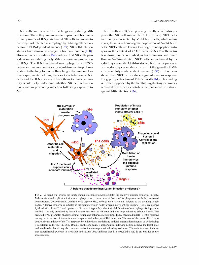
Fig. 2. A paradigm for how the innate immune response to Mtb regulates the adaptive immune response. Initially,Mtb survives and replicates inside macrophages since it can prevent fusion of its phagosome with the lysosomalcompartment. Concomitantly, dendritic cells capture Mtb, undergo maturation, and migrate to the draining lymphnodes. Adaptive response is initiated in the draining lymph nodes wherein naı̈ve antigen-specific T cells are primedby dendritic cells to Th1 and cytotoxic effector cell types. Mycobactericidal function of macrophages is dependenton IFNγ , initially produced by innate immune cells such as NK cells and later on provided by effector T cells. Thesecreted IFNγ promotes phagolysosomal fusion and enhances Mtb killing. TLR2-mediated innate IL-10 is releasedduring the induction of innate immune response and subsequent Th1 induction. The role of the innate IL-10 is tocontrol the magnitude of the Th1 response by either down modulating antigen-presentation function or by inducingT regulatory cells. The TLR2/IL-10 axis, on the one hand, is important for allowing Mtb to achieve the latent stateand, on the other hand, may also cause excessive immunosuppression leading to disease. The unbroken lines indicatethat experimental evidence is available and dashed lines indicate that it is speculative and is an area for futureinvestigation.
Journal of Clinical Immunology, Vol. 27, No. 4, 2007
HOST INNATE IMMUNE RESPONSE TO m. tuberculosis 357
There is still much to learn regarding the contributionof γ δ T cells, NK cells, and NKT cells to the overall innateimmune protection against TB in humans. A next step isto study these cells in the context of protection againstclinical strains of Mtb in humans.
CLOSING THOUGHTS
An emerging principle in intracellular parasitism is thatsuccessful pathogens such as Mtb have acquired the abilityto persist in the host without always inducing disease andmortality (163). The strategy on the part of Mtb is toinduce sufficient Th1 immunity in the host to control itsreplication but not result in its complete eradication. Theadvantage to the host is minimal collateral damage tolung tissue. Thus, Mtb remains dormant in the host fordecades, in a sort of symbiotic relationship. Under certainaltered conditions in the host, Mtb will reactivate andcause immunopathology such as lung cavitation, whichincreases its infectivity and thereby maintains the cycle oftransmission to new hosts.
Although Mtb interacts with several different TLRs onhost cells, we posit that to establish a latent infection Mtbspecifically usurps the innate TLR2 signaling in the hostto blunt Th1 immune responses. Supporting evidence forthe paradigm include (i) Mtb possess a large gamut ofligands for TLR2; (ii) Mtb/TLR2 interaction suppressesmacrophage functions; (iii) innate IL-10 secretion by den-dritic cells and macrophages in response to live Mtb isTLR-2 dependent; and (iv) absence of TLR2 results inexaggerated immunopathology in the host. The mecha-nisms for limiting Th1 response may include inhibition ofantigen-presentation functions and induction of T regula-tory cells (Fig. 2).
A corollary to the paradigm is that virulent strains cantip the balance toward immunosuppression using the sameTLR2 signaling pathway. Although there is no evidencethat TLR2 interaction is necessary for protection againstMtb disease, it must be pointed out that these conclusionsare drawn from studies performed with laboratory strainsof Mtb. It is highly probable that the interaction of clin-ical strains of Mtb with the TLR2 complex does resultin potent immunosuppression in the host. In this regard,it would be interesting to determine if the immunosup-pressive cytokines induced by the phenolic glycolipid ofthe virulent Beijing strains (164) is TLR2-mediated, andwhether the induction of T regulatory cells present in TBpatients (165, 166) is TLR2-dependent.
Clearly, future studies should investigate if the differinginteraction of Mtb clinical strains with the TLR2/IL-10axis is the control switch that determines whether the
outcome of Mtb exposure is latent infection or tuberculo-sis disease.
ACKNOWLEDGMENTS
This work was supported in part by the NIH grantsAI-49778 and AI-55377 to PS.
REFERENCES
1. Metchnikoff E: Immunity to Infective Diseases. London,Cambridge University Press, 1905
2. World Health Organization: Anti-Tuberculosis Drug Resistance inthe World: The WHO/IUTLD Global Project on Anti-TuberculosisDrug Resistance Surveillance. Geneva, Switzerland, World HealthOrganization, 2001
3. Murray JF: Tuberculosis and HIV infection: A global perspective.Respiration 65:335, 1998
4. De Cock KM, Chaisson RE: Will DOTS do it? A reappraisal oftuberculosis control in countries with high rates of HIV infection.Int J Tuberc Lung Dis 3:457, 1999
5. Pablos-Mendez A, Raviglione MC, Laszlo A, Binkin N, RiederHL, Bustreo F, Cohn DL, Lambregts-van Weezenbeek CS, Kim SJ,Chaulet P, Nunn P: Global surveillance for antituberculosis-drugresistance, 1994–1997. World Health Organization–InternationalUnion against Tuberculosis and Lung Disease Working Group onAnti-Tuberculosis Drug Resistance Surveillance. N Engl J Med338:1641, 1998
6. Raviglione MC, Snider DE, Jr, Kochi A: Global epidemiology oftuberculosis. Morbidity and mortality of a worldwide epidemic.JAMA 273:220, 1995
7. Medzhitov R, Janeway CJ: The Toll receptor family and microbialrecognition. Trends Microbiol 10:452, 2000
8. Kaisho T, Akira S: Critical roles of Toll-like receptors in hostdefense. Crit Rev Immunol 20:393, 2000
9. Takeda K, Kaisho T, Akira S: Toll-like receptors. Annu Rev Im-munol 21:335, 2003
10. Aderem A, Ulevitch RJ: Toll-like receptors in the induction of theinnate immune response. Nature 406:782, 2000
11. Stead WW: Pathogenesis of a first episode of chronic pulmonarytuberculosis in man: Recrudescence of residuals of the primaryinfection or exogenous reinfection? Am Rev Respir Dis 95:729,1967
12. Stead WW: The pathogenesis of pulmonary tuberculosis amongolder persons. Am Rev Respir Dis 91:811, 1965
13. Selwyn PA, Hartel D, Lewis VA, Schhoenbaum EE, Vermuns SH,Klein RS, Walker AT, Freidland GH: A prospective study of therisk of tuberculosis among intravenous drug users with humanimmunodeficiency virus infection. N Engl J Med 320:545, 1989
14. Schlesinger LS, Bellinger-Kawahara CG, Payne NR, Horwitz MA:Phagocytosis of Mycobacterium tuberculosis is mediated by hu-man monocyte complement receptors and complement C3. J Im-munol 144:2771, 1990
15. Ernst JD: Macrophage receptors for Mycobacterium tuberculosis.Infect Immun 66:1277, 1998
16. Beharka AA, Gaynor CD, Kang BK, Voelker DR, McCormackFX, Schlesinger LS: Pulmonary surfactant protein A up-regulatesactivity of the mannose receptor, a pattern recognition receptorexpressed on human macrophages. J Immunol 169:3565, 2002
Journal of Clinical Immunology, Vol. 27, No. 4, 2007
358 BHATT AND SALGAME
17. Gaynor CD, McCormack FX, Voelker DR, McGowan SE,Schlesinger LS: Pulmonary surfactant protein A mediates en-hanced phagocytosis of Mycobacterium tuberculosis by a di-rect interaction with human macrophages. J Immunol 155:5343,1995
18. Hu C, Mayadas TN, Tanaka K, Chan J, Salgame P: Mycobacteriumtuberculosis infection in complement receptor 3-deficient mice. JImmunol 165:2596, 2000
19. Gatfield J, Pieters J: Essential role for cholesterol in entry of my-cobacteria into macrophages. Science 288:1647, 2000
20. Armstrong JA, Hart PD: Response of cultured macrophages toM. tuberculosis with observations of fusion of lysosomes withphagosomes. J Exp Med 134:713, 1971
21. Armstrong JA, Hart PD: Phagosome–lysosome interactions in cul-tured macrophages infected with virulent tubercle bacilli. Reversalof the usual nonfusion pattern and observations on bacterial sur-vival. J Exp Med 142:1, 1975
22. Sturgill-Koszycki S, Schaible UE, Russell DG: Mycobacterium-containing phagosomes are accessible to early endosomes and re-flect a transitional state in normal phagosome biogenesis. EMBOJ 15:6960, 1996
23. Clemens DL, Horwitz MA: The Mycobacterium tuberculosisphagosome interacts with early endosomes and is accessible toexogenously administered transferrin. J Exp Med 184:1349, 1996
24. Russell DG, Dant J, Sturgill-Koszycki S: Mycobacterium avium-and Mycobacterium tuberculosis-containing vacuoles are dynamic,fusion-competent vesicles that are accessible to glycosphingolipidsfrom the host cell plasmalemma. J Immunol 156:4764, 1996
25. Sturgill-Koszycki S, Schlesinger PH, Chakraborty P, Haddix PL,Collins HL, Fok AK, Allen RD, Gluck SL, Heuser J, Russell DG:Lack of acidification in Mycobacterium phagosomes produced byexclusion of the vesicular proton-ATPase. Science 263:678, 1994
26. Clemens DL, Lee BY, Horwitz MA: Deviant expression of Rab5 onphagosomes containing the intracellular pathogens Mycobacteriumtuberculosis and Legionella pneumophila is associated with alteredphagosomal fate. Infect Immun 68:2671, 2000
27. Via LE, Deretic D, Ulmer RJ, Hibler NS, Huber LA, Deretic V:Arrest of mycobacterial phagosome maturation is caused by a blockin vesicle fusion between stages controlled by Rab5 and Rab7. JBiol Chem 272:13326, 1997
28. Fratti RA, Backer JM, Gruenberg J, Corvera S, Deretic V: Role ofphosphatidylinositol 3-kinase and Rab5 effectors in phagosomalbiogenesis and mycobacterial phagosome maturation arrest. J CellBiol 154:631, 2001
29. Deretic V, Vergne I, Chua J, Master S, Singh SB, Fazio JA, KyeiG: Endosomal membrane traffic: Convergence point targeted byMycobacterium tuberculosis and HIV. Cell Microbiol 6:999, 2004
30. Kusner DJ: Mechanisms of mycobacterial persistence in tubercu-losis. Clin Immunol 114:239, 2005
31. Anes E, Kuhnel MP, Bos E, Moniz-Pereira J, Habermann A,Griffiths G: Selected lipids activate phagosome actin assemblyand maturation resulting in killing of pathogenic mycobacteria.Nat Cell Biol 5:793, 2003
32. Kelley VA, Schorey JS: Mycobacterium’s arrest of phagosomematuration in macrophages requires Rab5 activity and accessibilityto iron. Mol Biol Cell 14:3366, 2003
33. Fratti RA, Chua J, Vergne I, Deretic V: Mycobacterium tuberculo-sis glycosylated phosphatidylinositol causes phagosome matura-tion arrest. Proc Natl Acad Sci USA 100:5437, 2003
34. Chua J, Vergne I, Master S, Deretic V: A tale of two lipids: My-cobacterium tuberculosis phagosome maturation arrest. Curr OpinMicrobiol 7:71, 2004
35. Kang PB, Azad AK, Torrelles JB, Kaufman TM, BeharkaA, Tibesar E, DesJardin LE, Schlesinger LS: The humanmacrophage mannose receptor directs Mycobacterium tuberculosislipoarabinomannan-mediated phagosome biogenesis. J Exp Med202:987, 2005
36. Vergne I, Fratti RA, Hill PJ, Chua J, Belisle J, Deretic V: Mycobac-terium tuberculosis phagosome maturation arrest: Mycobacterialphosphatidylinositol analog phosphatidylinositol mannoside stim-ulates early endosomal fusion. Mol Biol Cell 15:751, 2004
37. Walburger A, Koul A, Ferrari G, Nguyen L, Prescianotto-BaschongC, Huygen K, Klebl B, Thompson C, Bacher G, Pieters J: Proteinkinase G from pathogenic mycobacteria promotes survival withinmacrophages. Science 304:1800, 2004
38. Bodnar KA, Serbina NV, Flynn JL: Fate of Mycobacterium tuber-culosis within murine dendritic cells. Infect Immun 69:800, 2001
39. Tailleux L, Neyrolles O, Honore-Bouakline S, Perret E, SanchezF, Abastado JP, Lagrange PH, Gluckman JC, Rosenzwajg M,Herrmann JL: Constrained intracellular survival of Mycobac-terium tuberculosis in human dendritic cells. J Immunol 170:1939,2003
40. Pan H, Yan BS, Rojas M, Shebzukhov YV, Zhou H, Kobzik L,Higgins DE, Daly MJ, Bloom BR, Kramnik I: Ipr1 gene mediatesinnate immunity to tuberculosis. Nature 434:767, 2005
41. Tosh K, Campbell SJ, Fielding K, Sillah J, Bah B, Gustafson P,Manneh K, Lisse I, Sirugo G, Bennett S, Aaby P, McAdam KP,Bah-Sow O, Lienhardt C, Kramnik I, Hill AV: Variants in the SP110gene are associated with genetic susceptibility to tuberculosis inWest Africa. Proc Natl Acad Sci USA 103:10364, 2006
42. Thye T, Browne EN, Chinbuah MA, Gyapong J, Osei I, Owusu-Dabo E, Niemann S, Rusch-Gerdes S, Horstmann RD, Meyer CG:No associations of human pulmonary tuberculosis with Sp110 vari-ants. J Med Genet 43:e32, 2006
43. Brightbill HD, Libraty DH, Krutzik SR, Yang RB, Belisle JT,Bleharski JR, Maitland M, Norgard MV, Plevy SE, Smale ST,Brennan PJ, Bloom BR, Godowski PJ, Modlin RL: Host defensemechanisms triggered by microbial lipoproteins through toll-likereceptors. Science 285:732, 1999
44. Pecora ND, Gehring AJ, Canaday DH, Boom WH, Harding CV:Mycobacterium tuberculosis LprA is a lipoprotein agonist of TLR2that regulates innate immunity and APC function. J Immunol177:422, 2006
45. Gehring AJ, Dobos KM, Belisle JT, Harding CV, Boom WH: My-cobacterium tuberculosis LprG (Rv1411c): A novel TLR-2 ligandthat inhibits human macrophage class II MHC antigen processing.J Immunol 173:2660, 2004
46. Quesniaux VJ, Nicolle DM, Torres D, Kremer L, Guerardel Y,Nigou J, Puzo G, Erard F, Ryffel B: Toll-like receptor 2 (TLR2)-dependent-positive and TLR2-independent-negative regulation ofproinflammatory cytokines by mycobacterial lipomannans. J Im-munol 172:4425, 2004
47. Gilleron M, Himoudi N, Adam O, Constant P, Venisse A, Riv-iere M, Puzo G: Mycobacterium smegmatis phosphoinositols-glyceroarabinomannans. Structure and localization of alkali-labile and alkali-stable phosphoinositides. J Biol Chem 272:117,1997
48. Jones BW, Means TK, Heldwein KA, Keen MA, Hill PJ, BelisleJT, Fenton MJ: Different Toll-like receptor agonists induce distinctmacrophage responses. J Leukoc Biol 69:1036, 2001
49. Abel B, Thieblemont N, Quesniaux VJ, Brown N, Mpagi J, MiyakeK, Bihl F, Ryffel B: Toll-like receptor 4 expression is required tocontrol chronic Mycobacterium tuberculosis infection in mice. JImmunol 169:3155, 2002
Journal of Clinical Immunology, Vol. 27, No. 4, 2007
HOST INNATE IMMUNE RESPONSE TO m. tuberculosis 359
50. Gilleron M, Nigou J, Nicolle D, Quesniaux V, Puzo G: The acy-lation state of mycobacterial lipomannans modulates innate im-munity response through toll-like receptor 2. Chem Biol 13:39,2006
51. Means TK, Lien E, Yoshimura A, Wang S, Golenbock DT, FentonMJ: The CD14 ligands lipoarabinomannan and lipopolysaccha-ride differ in their requirement for Toll-like receptors. J Immunol163:6748, 1999
52. Kamath AB, Alt J, Debbabi H, Behar SM: Toll-like receptor 4-defective C3H/HeJ mice are not more susceptible than other C3Hsubstrains to infection with Mycobacterium tuberculosis. InfectImmun 71:4112, 2003
53. Reiling N, Holscher C, Fehrenbach A, Kroger S, Kirschning CJ,Goyert S, Ehlers S: Cutting edge: Toll-like receptor (TLR)2- andTLR4-mediated pathogen recognition in resistance to airborne in-fection with Mycobacterium tuberculosis. J Immunol 169:3480,2002
54. Sugawara I, Yamada H, Li C, Mizuno S, Takeuchi O, AkiraS: Mycobacterial infection in TLR2 and TLR6 knockout mice.Microbiol Immunol 47:327, 2003
55. Noss EH, Pai RK, Sellati TJ, Radolf JD, Belisle J, Golenbock DT,Boom WH, Harding CV: Toll-like receptor 2-dependent inhibitionof macrophage class II MHC expression and antigen processingby 19-kDa lipoprotein of Mycobacterium tuberculosis. J Immunol167:910, 2001
56. Fortune SM, Solache A, Jaeger A, Hill PJ, Belisle JT, BloomBR, Rubin EJ, Ernst JD: Mycobacterium tuberculosis inhibitsmacrophage responses to IFN-gamma through myeloid differentia-tion factor 88-dependent and -independent mechanisms. J Immunol172:6272, 2004
57. Banaiee N, Kincaid EZ, Buchwald U, Jacobs WR, Jr, Ernst JD:Potent inhibition of macrophage responses to IFN-gamma by livevirulent Mycobacterium tuberculosis is independent of maturemycobacterial lipoproteins but dependent on TLR2. J Immunol176:3019, 2006
58. Pompei L, Jang S, Zamlynny B, Ravikumar S, McBride A,Hickman SP, Salgame P: Disparity in interleukin-12 release indendritic cells and macrophages in response to Mycobacteriumtuberculosis is due to utilization of distinct Toll-like receptors. JImmunol, 2007 (in press)
59. Bafica A, Scanga CA, Feng CG, Leifer C, Cheever A, Sher A:TLR9 regulates Th1 responses and cooperates with TLR2 in me-diating optimal resistance to Mycobacterium tuberculosis. J ExpMed 202:1715, 2005
60. Flynn JL, Chan J: Immunology of tuberculosis. Annu Rev Immunol19:93, 2001
61. Denis M: Killing of Mycobacterium tuberculosis within humanmonocytes: Activation by cytokines and calcitriol. Clin Exp Im-munol 84:200, 1991
62. Wilkinson RJ, Llewelyn M, Toossi Z, Patel P, Pasvol G, Lalvani A,Wright D, Latif M, Davidson RN: Influence of vitamin D deficiencyand vitamin D receptor polymorphisms on tuberculosis amongGujarati Asians in west London: A case–control study. Lancet355:618, 2000
63. Liu PT, Stenger S, Li H, Wenzel L, Tan BH, Krutzik SR, OchoaMT, Schauber J, Wu K, Meinken C, Kamen DL, Wagner M, BalsR, Steinmeyer A, Zugel U, Gallo RL, Eisenberg D, Hewison M,Hollis BW, Adams JS, Bloom BR, Modlin RL: Toll-like receptortriggering of a vitamin D-mediated human antimicrobial response.Science 311:1770, 2006
64. Auricchio G, Garg SK, Martino A, Volpe E, Ciaramella A, DeVito P, Baldini PM, Colizzi V, Fraziano M: Role of macrophage
phospholipase D in natural and CpG-induced antimycobacterialactivity. Cell Microbiol 5:913, 2003
65. Russell DG: Who puts the tubercle in tuberculosis? Nat Rev Mi-crobiol 5:39, 2007
66. Ulrichs T, Kaufmann SH: New insights into the function of gran-ulomas in human tuberculosis. J Pathol 208:261, 2006
67. Salgame P: Host innate and Th1 responses and the bacterial fac-tors that control Mycobacterium tuberculosis infection. Curr OpinImmunol 17:374, 2005
68. Estaquier J, Idziorek T, Weiping Z, Emilie D, Farber C-M,Bourez J-M, Ameisen JC: T helper type 1/T helper type 2 cy-tokines and T cell death: Preventive effect of IL-12 on activation-induced and CD95 (Fas/Apo-1)-mediated apoptosis of CD4+ Tcells from human immunodeficiency virus-infected persons. J ExpMed 182:1759, 1995
69. Rosenzweig SD, Holland SM: Defects in the interferon-gammaand interleukin-12 pathways. Immunol Rev 203:38, 2005
70. Brombacher F, Kastelein RA, Alber G: Novel IL-12 family mem-bers shed light on the orchestration of Th1 responses. Trends Im-munol 24:207, 2003
71. Zhang M, Gong J, Iyer DV, Jones BE, Modlin RL, Barnes PF:T cell cytokine responses in persons with tuberculosis and hu-man immunodeficiency virus infection. J Clin Invest 94:2435,1994
72. Giacomini E, Iona E, Ferroni L, Miettinen M, Fattorini L, OreficiG, Julkunen I, Coccia EM: Infection of human macrophages anddendritic cells with Mycobacterium tuberculosis induces a differ-ential cytokine gene expression that mosulates T cell response. JImmunol 166:7033, 2001
73. Hickman SP, Chan J, Salgame P: Mycobacterium tuberculosis in-duces differential cytokine production from dendritic cells andmacrophages with divergent effects on naive T cell polarization. JImmunol 168:4636, 2002
74. Verreck FA, de Boer T, Langenberg DM, Hoeve MA, KramerM, Vaisberg E, Kastelein R, Kolk A, de Waal-Malefyt R,Ottenhoff TH: Human IL-23-producing type 1 macrophages pro-mote but IL-10-producing type 2 macrophages subvert immunityto (myco)bacteria. Proc Natl Acad Sci USA 101:4560, 2004
75. Flynn J, Goldstein M, Triebold K, Sypek J, Wolf S, Bloom B:IL-12 increases resistance of BALB/c mice to Mycobacterium tu-berculosis infection. J Immunol 155:2515, 1995
76. Castro A, Silva R, Appelberg R: Endogenously produced IL-12 isrequired for the induction of protective T cells during Mycobac-terium avium infections in mice. J Immunol 155:2013, 1995
77. Feng CG, Jankovic D, Kullberg M, Cheever A, Scanga CA, HienyS, Caspar P, Yap GS, Sher A: Maintenance of pulmonary Th1effector function in chronic tuberculosis requires persistent IL-12production. J Immunol 174:4185, 2005
78. Khader SA, Pearl JE, Sakamoto K, Gilmartin L, Bell GK, Jelley-Gibbs DM, Ghilardi N, deSauvage F, Cooper AM: IL-23 Compen-sates for the Absence of IL-12p70 and is essential for the IL-17response during tuberculosis but is dispensable for protection andantigen-specific IFN-{gamma} responses if IL-12p70 is available.J Immunol 175:788, 2005
79. Chackerian AA, Chen S-J, Brodie SJ, Mattson JD, McClanahanTK, Kastelein RA, Bowman EP: Neutralization or absence of theinterleukin-23 pathway does not compromise immunity to my-cobacterial infection. Infect Immun 74:6092, 2006
80. Cooper AM, Kipnis A, Turner J, Magram J, Ferrante J, OrmeIM: Mice lacking bioactive IL-12 can generate protective,antigen-specific cellular responses to mycobacterial infection onlyif the IL-12 p40 subunit is present. J Immunol 168:1322, 2002
Journal of Clinical Immunology, Vol. 27, No. 4, 2007
360 BHATT AND SALGAME
81. Happel KI, Lockhart EA, Mason CM, Porretta E, Keoshkerian E,Odden AR, Nelson S, Ramsay AJ: Pulmonary interleukin-23 genedelivery increases local T-cell immunity and controls growth ofMycobacterium tuberculosis in the lungs. Infect Immun 73:5782,2005
82. Hunter CA: New IL-12-family members: IL-23 and IL-27,cytokines with divergent functions. Nat Rev Immunol 5:521,2005
83. Pearl JE, Khader SA, Solache A, Gilmartin L, Ghilardi N, de-Sauvage F, Cooper AM: IL-27 signaling compromises controlof bacterial growth in mycobacteria-infected mice. J Immunol173:7490, 2004
84. Artis D, Johnson LM, Joyce K, Saris C, Villarino A, Hunter CA,Scott P: Cutting edge: Early IL-4 production governs the require-ment for IL-27-WSX-1 signaling in the development of protectiveTh1 cytokine responses following Leishmania major infection. JImmunol 172:4672, 2004
85. Villarino A, Hibbert L, Lieberman L, Wilson E, Mak T, YoshidaH, Kastelein RA, Saris C, Hunter CA: The IL-27R (WSX-1) is re-quired to suppress T cell hyperactivity during infection. Immunity19:645, 2003
86. Villarino AV, Huang E, Hunter CA: Understanding the pro-and anti-inflammatory properties of IL-27. J Immunol 173:715,2004
87. Stenger S: Immunological control of tuberculosis: Role of tumournecrosis factor and more. Ann Rheum Dis 64(Suppl 4):iv24, 2005
88. Chan J, Xing Y, Magliozzo R, Bloom B: Killing of virulent My-cobacterium tuberculosis by reactive nitrogen intermediates pro-duced by activated murine macrophages. J Exp Med 175:1111,1992
89. Keane J, Balcewicz-Sablinska MR, Remold HG, Chupp GL, MeekBB, Fenton MJ, Kornfeld H: Infection by Mycobacterium tuber-culosis promotes human alveolar macrophage apoptosis. InfectImmun 65:298, 1997
90. Winau F, Weber S, Sad S, de Diego J, Hoops SL, Breiden B,Sandhoff K, Brinkmann V, Kaufmann SHE, Schaible UE: Apop-totic vesicles crossprime CD8 T cells and protect against tubercu-losis. Immunity 24:105, 2006
91. Balcewicz-Sablinska MK, Keane J, Kornfeld H, Remold HG:Pathogenic Mycobacterium tuberculosis evades apoptosis of hostmacrophages by release of TNF-R2, resulting in inactivation ofTNF-alpha. J Immunol 161:2636, 1998
92. Engele M, Castiglione K, Schwerdtner N, Wagner M, BolcskeiP, Rollinghoff M, Stenger S: Induction of TNF in human alve-olar macrophages as a potential evasion mechanism of virulentMycobacterium tuberculosis. J Immunol 168:1328, 2002
93. Kindler V, Sappino A-P, Grau GE, Piguet P-F, Vassalli P: Theinducing role of tumor necrosis factor in the development of bac-tericidal granulomas during BCG infection. Cell 56:731, 1989
94. Flynn JL, Goldstein MM, Chan J, Triebold KJ, Pfeffer K,Loenstein CL, Schreiber R, Mak TW, Bloom BR: Tumor necro-sis factor is required in the protective immune response againstMycobacterium tuberculosis in mice. Immunity 2:561, 1995
95. Roach DR, Bean AGD, Demangel C, France MP, Briscoe H,Britton WJ: TNF regulates chemokine induction essential for cellrecruitment, granuloma formation, and clearance of mycobacterialinfection. J Immunol 168:4620, 2002
96. Algood HMS, Chan J, Flynn JL: Chemokines and tuberculosis.Cytokine Growth Factor Rev 14:467, 2003
97. Algood HMS, Lin PL, Flynn JL: Tumor necrosis factor andchemokine interactions in the formation and maintenance of gran-ulomas in tuberculosis. Clin Infect Dis 41:S189, 2005
98. Tufariello JM, Chan J, Flynn JL: Latent tuberculosis: Mechanismsof host and bacillus that contribute to persistent infection. LancetInfect Dis 3:578, 2003
99. Mohan VP, Scanga CA, Yu K, Scott HM, Tanaka KE, Tsang E, TsaiMM, Flynn JL, Chan J: Effects of tumor necrosis factor alpha onhost immune response in chronic persistent tuberculosis: Possiblerole for limiting pathology. Infect Immun 69:1847, 2001
100. Keane J, Gershon S, Wise RP, Mirabile-Levens E, Kasznica J,Schwieterman WD, Siegel JN, Braun MM: Tuberculosis associ-ated with infliximab, a tumor necrosis factor {alpha}-neutralizingagent. N Engl J Med 345:1098, 2001
101. Gardam MA, Keystone EC, Menzies R, Manners S, Skamene E,Long R, Vinh DC: Anti-tumour necrosis factor agents and tubercu-losis risk: Mechanisms of action and clinical management. LancetInfect Dis 3:148, 2003
102. Peters W, Ernst JD: Mechanisms of cell recruitment in the immuneresponse to Mycobacterium tuberculosis. Microbes Infect 5:151,2003
103. Orlova MO, Majorov KB, Lyadova IV, Eruslanov EB, M’lan CE,Greenwood CMT, Schurr E, Apt AS: Constitutive differences ingene expression profiles parallel genetic patterns of susceptibilityto tuberculosis in mice. Infect Immun 74:3668, 2006
104. Tessier P, Naccache P, Clark-Lewis I, Gladue R, Neote K, McCollS: Chemokine networks in vivo: Involvement of C-X-C and C-C chemokines in neutrophil extravasation in vivo in response toTNF-alpha. J Immunol 159:3595, 1997
105. Lande R, Giacomini E, Grassi T, Remoli ME, Iona E, Miettinen M,Julkunen I, Coccia EM: IFN-alpha beta released by Mycobacteriumtuberculosis-infected human dendritic cells induces the expressionof CXCL10: Selective recruitment of NK and activated T cells. JImmunol 170:1174, 2003
106. Peters W, Scott HM, Chambers HF, Flynn JL, Charo IF, ErnstJD: Chemokine receptor 2 serves an early and essential role inresistance to Mycobacterium tuberculosis. Proc Natl Acad Sci USA98:7958, 2001
107. Scott HM, Flynn JL: Mycobacterium tuberculosis in chemokinereceptor 2-deficient mice: Influence of dose on disease progression.Infect Immun 70:5946, 2002
108. Peters W, Cyster JG, Mack M, Schlondorff D, Wolf AJ, Ernst JD,Charo IF: CCR2-dependent trafficking of F4/80dim macrophagesand CD11cdim/intermediate dendritic cells is crucial for T cellrecruitment to lungs infected with Mycobacterium tuberculosis. JImmunol 172:7647, 2004
109. Kipnis A, Basaraba RJ, Orme IM, Cooper AM: Role of chemokineligand 2 in the protective response to early murine pulmonarytuberculosis. Immunology 109:547, 2003
110. Floto RA, MacAry PA, Boname JM, Mien TS, Kampmann B,Hair JR, Huey OS, Houben ENG, Pieters J, Day C, OehlmannW, Singh M, Smith KGC, Lehner PJ: Dendritic cell stimulation bymycobacterial Hsp70 is mediated through CCR5. Science 314:454,2006
111. Badewa AP, Quinton LJ, Shellito JE, Mason CM: Chemokine re-ceptor 5 and its ligands in the immune response to murine tuber-culosis. Tuberculosis 85:185, 2005
112. Algood HM, Flynn JL: CCR5-deficient mice control Mycobac-terium tuberculosis infection despite increased pulmonary lym-phocytic infiltration. J Immunol 173:3287, 2004
113. Kahnert A, Hopken UE, Stein M, Bandermann S, Lipp M,Kaufmann SHE: Mycobacterium tuberculosis triggers formationof lymphoid structure in murine lungs. J Infect Dis 195:46, 2007
114. Seiler P, Aichele P, Bandermann S, Hauser AE, Lu B, GerardNP, Gerard C, Ehlers S, Mollenkopf HJ, Kaufmann SH: Early
Journal of Clinical Immunology, Vol. 27, No. 4, 2007
HOST INNATE IMMUNE RESPONSE TO m. tuberculosis 361
granuloma formation after aerosol Mycobacterium tuberculo-sis infection is regulated by neutrophils via CXCR3-signalingchemokines. Eur J Immunol 33:2676, 2003
115. Chakravarty SD, Xu J, Lu B, Gerard C, Flynn J, Chan J: Thechemokine receptor CXCR3 attenuates the control of chronic My-cobacterium tuberculosis infection in BALB/c mice. J Immunol178:1723, 2007
116. Bromley SK, Peterson DA, Gunn MD, Dustin ML: Cutting edge:Hierarchy of chemokine receptor and TCR signals regulating Tcell migration and proliferation. J Immunol 165:15, 2000
117. Jang S, Uematsu S, Akira S, Salgame P: IL-6 and IL-10 inductionfrom dendritic cells in response to Mycobacterium tuberculosis ispredominantly dependent on TLR2-mediated recognition. J Im-munol 173:3392, 2004
118. Boussiotis VA, Tsai EY, Yunis EJ, Thim S, Delgado JC, DascherCC, Berezovskaya A, Rousset D, Reynes JM, Goldfeld AE: IL-10-producing T cells suppress immune responses in anergic tuber-culosis patients. J Clin Invest 105:1317, 2000
119. Gerosa F, Nisii C, Righetti S, Micciolo R, Marchesini M, CazzadoriA, Trinchieri G: CD4+ T cell clones producing both interferon-gamma and interleukin-10 predominate in bronchoalveolar lavagesof active pulmonary tuberculosis patients. Clin Immunol 92:224,1999
120. Hirsch CS, Toossi Z, Othieno C, Johnson JL, Schwander SK,Robertson S, Wallis RS, Edmonds K, Okwera A, Mugerwa R,Peters P, Ellner JJ: Depressed T-cell interferon-gamma responsesin pulmonary tuberculosis: Analysis of underlying mechanismsand modulation with therapy. J Infect Dis 180:2069, 1999
121. Barnes PF, Lu S, Abrams JS, Wang E, Yamamura M, Modlin RL:Cytokine production at the site of disease in human tuberculosis.Infect Immun 61:3482, 1993
122. Fulton SA, Cross JV, Toossi ZT, Boom WH: Regulation ofinterleukin-12 by interleukin-10, transforming growth factor-beta,tumor necrosis factor-alpha, and interferon-gamma in humanmonocytes infected with Mycobacterium tuberculosis H37Ra. JInfect Dis 178:1105, 1998
123. De La Barrera S, Aleman M, Musella R, Schierloh P, PasquenelliV, Garcia V, Abbate E, Sasiain M, Del C: IL-10 down-regulatescostimulatory molecules on Mycobacterium tuberculosis-pulsedmacrophages and impairs the lytic activity of CD4 and CD8CTL in tuberculosis patients. Clin Exp Immunol 138:128,2004
124. Rojas RE, Balaji KN, Subramanian A, Boom WH: Regulationof human CD4+ alpha beta T-cell-receptor-positive (TCR+) andgamma delta TCR+ T-cell responses to Mycobacterium tubercu-losis by interleukin-10 and transforming growth factor beta. InfectImmun 67:6461, 1999
125. North RJ: Mice incapable of making IL-4 or IL-10 display normalresistance to infection with Mycobacterium tuberculosis. Clin ExpImmunol 113:55, 1998
126. Turner J, Gonzalez-Juarrero M, Ellis DL, Basaraba RJ, Kipnis A,Orme IM, Cooper AM: in vivo IL-10 production reactivates chronicpulmonary tuberculosis in C57BL/6 mice. J Immunol 169:6343,2002
127. Feng CG, Kullberg MC, Jankovic D, Cheever AW, Caspar P,Coffman RL, Sher A: Transgenic mice expressing humaninterleukin-10 in the antigen-presenting cell compartment showincreased susceptibility to infection with Mycobacterium aviumassociated with decreased macrophage effector function and apop-tosis. Infect Immun 70:6672, 2002
128. Awomoyi AA, Marchant A, Howson JM, McAdam KP, Black-well JM, Newport MJ: Interleukin-10, polymorphism in SLC11A1
(formerly NRAMP1), and susceptibility to tuberculosis. J InfectDis 186:1808, 2002
129. Buettner M, Meinken C, Bastian M, Bhat R, Stossel E, FallerG, Cianciolo G, Ficker J, Wagner M, Rollinghoff M, Stenger S:Inverse correlation of maturity and antibacterial activity in humandendritic cells. J Immunol 174:4203, 2005
130. Bhatt K, Hickman SP, Salgame P: Cutting edge: A new approach tomodeling early lung immunity in murine tuberculosis. J Immunol172:2748, 2004
131. Humphreys IR, Stewart GR, Turner DJ, Patel J, Karamanou D,Snelgrove RJ, Young DB: A role for dendritic cells in the dissemi-nation of mycobacterial infection. Microbes Infect 8:1339, 2006
132. Jiao X, Lo-Man R, Guermonprez P, Fiette L, Deriaud E, BurgaudS, Gicquel B, Winter N, Leclerc C: Dendritic cells are host cellsfor mycobacteria in vivo that trigger innate and acquired immunity.J Immunol 168:1294, 2002
133. Tian T, Woodworth J, Skold M, Behar SM: in vivo depletion ofCD11c+ cells delays the CD4+ T cell response to Mycobacteriumtuberculosis and exacerbates the outcome of infection. J Immunol175:3268, 2005
134. Abadie V, Badell E, Douillard P, Ensergueix D, Leenen PJ, TanguyM, Fiette L, Saeland S, Gicquel B, Winter N: Neutrophils rapidlymigrate via lymphatics after Mycobacterium bovis BCG intrader-mal vaccination and shuttle live bacilli to the draining lymph nodes.Blood 106:1843, 2005
135. Pedrosa J, Saunders BM, Appelberg R, Orme IM, Silva MT, CooperAM: Neutrophils play a protective nonphagocytic role in sys-temic Mycobacterium tuberculosis infection of mice. Infect Immun68:577, 2000
136. Tsai MC, Chakravarty S, Zhu G, Xu J, Tanaka K, Koch C,Tufariello J, Flynn J, Chan J: Characterization of the tuberculousgranuloma in murine and human lungs: Cellular composition andrelative tissue oxygen tension. Cell Microbiol 8:218, 2006
137. Schaible UE, Winau F, Sieling PA, Fischer K, Collins HL, HagensK, Modlin RL, Brinkmann V, Kaufmann SH: Apoptosis facilitatesantigen presentation to T lymphocytes through MHC-I and CD1in tuberculosis. Nat Med 9:1039, 2003
138. Makino M, Maeda Y, Mukai T, Kaufmann SH: Impaired maturationand function of dendritic cells by mycobacteria through IL-1beta.Eur J Immunol 36:1443, 2006
139. Hanekom WA, Mendillo M, Manca C, Haslett PA, Siddiqui MR,Barry C, III, Kaplan G: Mycobacterium tuberculosis inhibits matu-ration of human monocyte-derived dendritic cells in vitro. J InfectDis 188:257, 2003
140. Khader SA, Partida-Sanchez S, Bell G, Jelley-Gibbs DM, SwainS, Pearl JE, Ghilardi N, deSauvage FJ, Lund FE, Cooper AM:Interleukin 12p40 is required for dendritic cell migration and Tcell priming after Mycobacterium tuberculosis infection. J ExpMed 203:1805, 2006
141. Demangel C, Bertolino P, Britton WJ: Autocrine IL-10 impairsdendritic cell (DC)-derived immune responses to mycobacte-rial infection by suppressing DC trafficking to draining lymphnodes and local IL-12 production. Eur J Immunol 32:994,2002
142. Flynn JL, Chan J: Immune evasion by Mycobacterium tuberculosis:Living with the enemy. Curr Opin Immunol 15:450, 2003
143. Dayaram YK, Talaue MT, Connell ND, Venketaraman V: Charac-terization of a glutathione metabolic mutant of Mycobacterium tu-berculosis and its resistance to glutathione and nitrosoglutathione.J Bacteriol 188:1364, 2006
144. Thoma-Uszynski S, Stenger S, Takeuchi O, Ochoa MT, En-gele M, Sieling PA, Barnes PF, Rollinghoff M, Bolcskei PL,
Journal of Clinical Immunology, Vol. 27, No. 4, 2007
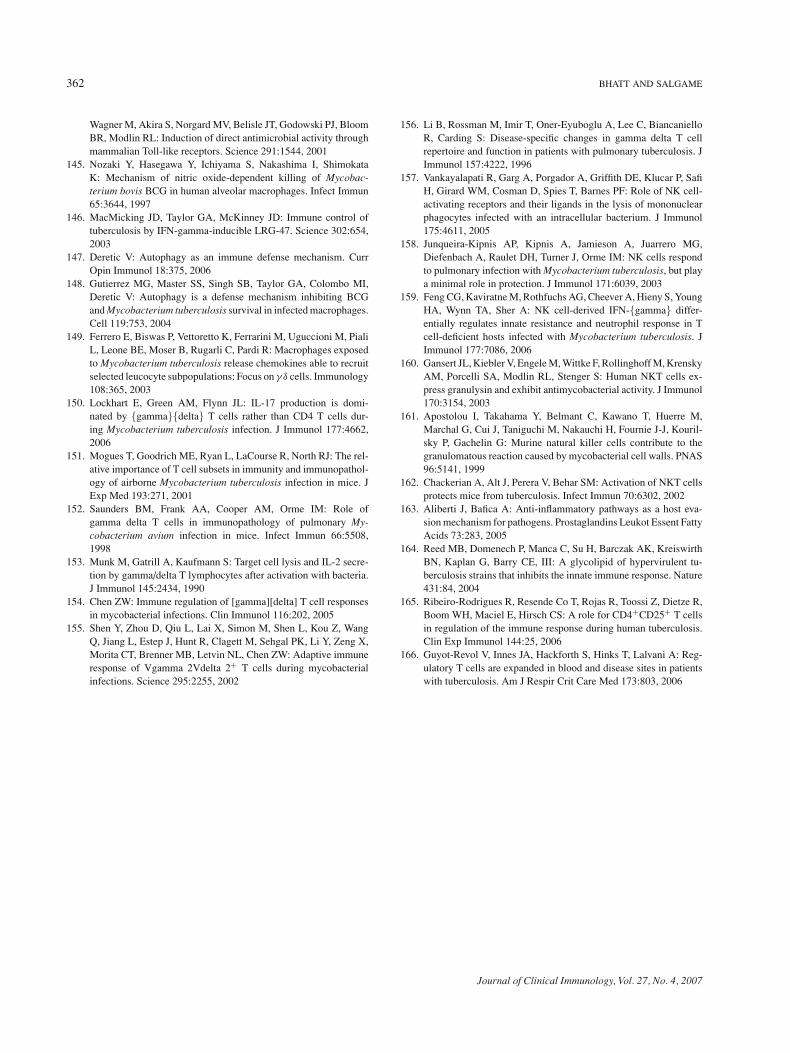
362 BHATT AND SALGAME
Wagner M, Akira S, Norgard MV, Belisle JT, Godowski PJ, BloomBR, Modlin RL: Induction of direct antimicrobial activity throughmammalian Toll-like receptors. Science 291:1544, 2001
145. Nozaki Y, Hasegawa Y, Ichiyama S, Nakashima I, ShimokataK: Mechanism of nitric oxide-dependent killing of Mycobac-terium bovis BCG in human alveolar macrophages. Infect Immun65:3644, 1997
146. MacMicking JD, Taylor GA, McKinney JD: Immune control oftuberculosis by IFN-gamma-inducible LRG-47. Science 302:654,2003
147. Deretic V: Autophagy as an immune defense mechanism. CurrOpin Immunol 18:375, 2006
148. Gutierrez MG, Master SS, Singh SB, Taylor GA, Colombo MI,Deretic V: Autophagy is a defense mechanism inhibiting BCGand Mycobacterium tuberculosis survival in infected macrophages.Cell 119:753, 2004
149. Ferrero E, Biswas P, Vettoretto K, Ferrarini M, Uguccioni M, PialiL, Leone BE, Moser B, Rugarli C, Pardi R: Macrophages exposedto Mycobacterium tuberculosis release chemokines able to recruitselected leucocyte subpopulations: Focus on γ δ cells. Immunology108:365, 2003
150. Lockhart E, Green AM, Flynn JL: IL-17 production is domi-nated by {gamma}{delta} T cells rather than CD4 T cells dur-ing Mycobacterium tuberculosis infection. J Immunol 177:4662,2006
151. Mogues T, Goodrich ME, Ryan L, LaCourse R, North RJ: The rel-ative importance of T cell subsets in immunity and immunopathol-ogy of airborne Mycobacterium tuberculosis infection in mice. JExp Med 193:271, 2001
152. Saunders BM, Frank AA, Cooper AM, Orme IM: Role ofgamma delta T cells in immunopathology of pulmonary My-cobacterium avium infection in mice. Infect Immun 66:5508,1998
153. Munk M, Gatrill A, Kaufmann S: Target cell lysis and IL-2 secre-tion by gamma/delta T lymphocytes after activation with bacteria.J Immunol 145:2434, 1990
154. Chen ZW: Immune regulation of [gamma][delta] T cell responsesin mycobacterial infections. Clin Immunol 116:202, 2005
155. Shen Y, Zhou D, Qiu L, Lai X, Simon M, Shen L, Kou Z, WangQ, Jiang L, Estep J, Hunt R, Clagett M, Sehgal PK, Li Y, Zeng X,Morita CT, Brenner MB, Letvin NL, Chen ZW: Adaptive immuneresponse of Vgamma 2Vdelta 2+ T cells during mycobacterialinfections. Science 295:2255, 2002
156. Li B, Rossman M, Imir T, Oner-Eyuboglu A, Lee C, BiancanielloR, Carding S: Disease-specific changes in gamma delta T cellrepertoire and function in patients with pulmonary tuberculosis. JImmunol 157:4222, 1996
157. Vankayalapati R, Garg A, Porgador A, Griffith DE, Klucar P, SafiH, Girard WM, Cosman D, Spies T, Barnes PF: Role of NK cell-activating receptors and their ligands in the lysis of mononuclearphagocytes infected with an intracellular bacterium. J Immunol175:4611, 2005
158. Junqueira-Kipnis AP, Kipnis A, Jamieson A, Juarrero MG,Diefenbach A, Raulet DH, Turner J, Orme IM: NK cells respondto pulmonary infection with Mycobacterium tuberculosis, but playa minimal role in protection. J Immunol 171:6039, 2003
159. Feng CG, Kaviratne M, Rothfuchs AG, Cheever A, Hieny S, YoungHA, Wynn TA, Sher A: NK cell-derived IFN-{gamma} differ-entially regulates innate resistance and neutrophil response in Tcell-deficient hosts infected with Mycobacterium tuberculosis. JImmunol 177:7086, 2006
160. Gansert JL, Kiebler V, Engele M, Wittke F, Rollinghoff M, KrenskyAM, Porcelli SA, Modlin RL, Stenger S: Human NKT cells ex-press granulysin and exhibit antimycobacterial activity. J Immunol170:3154, 2003
161. Apostolou I, Takahama Y, Belmant C, Kawano T, Huerre M,Marchal G, Cui J, Taniguchi M, Nakauchi H, Fournie J-J, Kouril-sky P, Gachelin G: Murine natural killer cells contribute to thegranulomatous reaction caused by mycobacterial cell walls. PNAS96:5141, 1999
162. Chackerian A, Alt J, Perera V, Behar SM: Activation of NKT cellsprotects mice from tuberculosis. Infect Immun 70:6302, 2002
163. Aliberti J, Bafica A: Anti-inflammatory pathways as a host eva-sion mechanism for pathogens. Prostaglandins Leukot Essent FattyAcids 73:283, 2005
164. Reed MB, Domenech P, Manca C, Su H, Barczak AK, KreiswirthBN, Kaplan G, Barry CE, III: A glycolipid of hypervirulent tu-berculosis strains that inhibits the innate immune response. Nature431:84, 2004
165. Ribeiro-Rodrigues R, Resende Co T, Rojas R, Toossi Z, Dietze R,Boom WH, Maciel E, Hirsch CS: A role for CD4+CD25+ T cellsin regulation of the immune response during human tuberculosis.Clin Exp Immunol 144:25, 2006
166. Guyot-Revol V, Innes JA, Hackforth S, Hinks T, Lalvani A: Reg-ulatory T cells are expanded in blood and disease sites in patientswith tuberculosis. Am J Respir Crit Care Med 173:803, 2006
Journal of Clinical Immunology, Vol. 27, No. 4, 2007
Related Documents


![[Micro] mycobacterium tuberculosis](https://static.cupdf.com/doc/110x72/55d6fc67bb61ebfa2a8b47ea/micro-mycobacterium-tuberculosis.jpg)

