Host immunity during RSV pathogenesis Susan M. Bueno a , Pablo A. González a , Rodrigo Pacheco a , Eduardo D. Leiva a , Kelly M. Cautivo a , Hugo E. Tobar a , Jorge E. Mora a , Carolina E. Prado a , Juan P. Zúñiga a , Jorge Jiménez c , Claudia A. Riedel d , Alexis M. Kalergis a,b, ⁎ a Millennium Nucleus on Immunology and Immunotherapy. Departamento de Genética Molecular y Microbiología, Facultad de Ciencias Biológicas, Pontificia Universidad Católica de Chile, Chile b Departamento de Reumatología, Pontificia Universidad Católica de Chile, Chile c Salud Pública, Pontificia Universidad Católica de Chile, Chile d Universidad Nacional Andrés Bello, Chile Received 7 November 2007; received in revised form 30 January 2008; accepted 17 March 2008 Abstract Infection by respiratory syncytial virus (RSV) is the leading cause of childhood hospitalization as well as a major health and economic burden worldwide. Unfortunately, RSV infection provides only limited immune protection to reinfection, mostly due to inadequate immunological memory, which leads to an exacerbated inflammatory response in the respiratory tract promoting airway damage during virus clearance. This exacerbated and inefficient immune- inflammatory response triggered by RSV, has often been attributed to the induction of a Th2- biased immunity specific for some of the RSV antigens. These features of RSV infection suggest that the virus might possess molecular mechanisms to enhance allergic-type immunity in the host in order to prevent clearance by cytotoxic T cells and ensure survival and dissemination to other hosts. In this review, we discuss recent findings that contribute to explain the components of the innate and adaptive immune response that are involved in RSV-mediated disease exacerbation. Further, the virulence mechanisms used by RSV to avoid activation of protective immune responses are described. © 2008 Elsevier B.V. All rights reserved. KEYWORDS: Respiratory syncytial virus; Immune response; Immunopathology; Th-2 immune response; T cells; Dendritic cells 1. Introduction Respiratory syncicial virus (RSV) is the leading cause of viral bronchiolitis and pneumonia worldwide, infecting more than 70% of children in the first year of life and 100% of children by age 2 [1]. RSV is an enveloped, negative strand RNA virus belonging to the Paramyxoviridae family with a genome that encodes for 11 proteins [2]. Among these proteins, two on the virion surface: F and G, and two non-structural proteins: ⁎ Corresponding author. Departamento de Genética Molecular y Microbiología, Facultad de Ciencias Biológicas, Pontificia Universi- dad Católica de Chile. Alameda #340, Santiago E-8331010, Chile. Tel.: +56 2 686 2842; fax: +56 2 686 2185. E-mail address: [email protected] (A.M. Kalergis). 1567-5769/$ - see front matter © 2008 Elsevier B.V. All rights reserved. doi:10.1016/j.intimp.2008.03.012 www.elsevier.com/locate/intimp International Immunopharmacology (2008) 8, 1320–1329

Welcome message from author
This document is posted to help you gain knowledge. Please leave a comment to let me know what you think about it! Share it to your friends and learn new things together.
Transcript
www.e l sev i e r. com/ loca te / i n t imp
International Immunopharmacology (2008) 8, 1320–1329
Host immunity during RSV pathogenesisSusanM. Bueno a, Pablo A. González a, Rodrigo Pacheco a, Eduardo D. Leiva a,Kelly M. Cautivo a, Hugo E. Tobar a, Jorge E. Mora a, Carolina E. Prado a,Juan P. Zúñiga a, Jorge Jiménez c, Claudia A. Riedel d, Alexis M. Kalergis a,b,⁎
a Millennium Nucleus on Immunology and Immunotherapy. Departamento de Genética Molecular y Microbiología,Facultad de Ciencias Biológicas, Pontificia Universidad Católica de Chile, Chile
b Departamento de Reumatología, Pontificia Universidad Católica de Chile, Chilec Salud Pública, Pontificia Universidad Católica de Chile, Chiled Universidad Nacional Andrés Bello, Chile
Received 7 November 2007; received in revised form 30 January 2008; accepted 17 March 2008
⁎ Corresponding author. DepartamenMicrobiología, Facultad de Ciencias Bdad Católica de Chile. Alameda #340Tel.: +56 2 686 2842; fax: +56 2 686 21
E-mail address: [email protected].
1567-5769/$ - see front matter © 200doi:10.1016/j.intimp.2008.03.012
Abstract
Infection by respiratory syncytial virus (RSV) is the leading cause of childhood hospitalization aswell as a major health and economic burden worldwide. Unfortunately, RSV infection providesonly limited immune protection to reinfection, mostly due to inadequate immunologicalmemory, which leads to an exacerbated inflammatory response in the respiratory tractpromoting airway damage during virus clearance. This exacerbated and inefficient immune-inflammatory response triggered by RSV, has often been attributed to the induction of a Th2-biased immunity specific for some of the RSV antigens. These features of RSV infection suggestthat the virus might possess molecular mechanisms to enhance allergic-type immunity in the hostin order to prevent clearance by cytotoxic Tcells and ensure survival and dissemination to otherhosts. In this review, we discuss recent findings that contribute to explain the components of theinnate and adaptive immune response that are involved in RSV-mediated disease exacerbation.Further, the virulence mechanisms used by RSV to avoid activation of protective immuneresponses are described.© 2008 Elsevier B.V. All rights reserved.
KEYWORDS:Respiratory syncytial virus;Immune response;Immunopathology;Th-2 immune response;T cells;Dendritic cells
to de Genética Molecular yiológicas, Pontificia Universi-, Santiago E-8331010, Chile.85.cl (A.M. Kalergis).
8 Elsevier B.V. All rights reserved.
1. Introduction
Respiratory syncicial virus (RSV) is the leading cause of viralbronchiolitis and pneumonia worldwide, infecting more than70% of children in the first year of life and 100% of children byage 2 [1]. RSV is an enveloped, negative strand RNA virusbelonging to the Paramyxoviridae family with a genome thatencodes for 11 proteins [2]. Among these proteins, two onthe virion surface: F and G, and two non-structural proteins:
1321Host immunity during RSV pathogenesis
NS1 and NS2, constitute key viral components that con-tribute to the infective cycle and to the evasion of the hostimmune response [3–5]. The F protein mediates the fusionbetween the virus and the target cell surface and promotesthe formation of syncytia \ a phenomenon that originatesthe virus name [3].
Despite being highly infective, RSV does not induce aneffective immunological memory and repeated infections aretherefore very frequent [6,7]. Although common RSV symptomsmanifest as rhinitis in adults, severe RSV infection is frequentlyobserved in premature infants, the elderly and immunosu-pressed individuals [8,9]. Furthermore, it has been proposedthat exposure to RSV infection early in life can lead to anincreased susceptibility to suffer from recurrent allergicwheezing and asthma [10]. Considering epidemiological data,RSV is responsible for causing a health problem that is extremelyexpensive for individuals, governments andhealth care systems.Unfortunately, to date there are no commercially availablevaccines against this pathogen. Efforts aimed to develop avaccine against RSV were first carried out with a formalin-inactivated RSV formulation (FI-RSV) in vaccine trials in the mid1960s [11]. However, vaccinated children experienced exacer-bated pulmonary disease and required hospitalization uponsubsequent RSV infection, while non-vaccinated control chil-dren experienced significantly milder symptoms [11,12]. Thefailure of FI-RSVremained unexplained for at least two decades,primarily because of a poor understanding of the immuneresponses triggered by RSV infection. However, recent studieshave suggested that the FI-RSV vaccine failed because of itsability to induce an allergic-like T cell helper-2 (Th2) immuneresponse against the virus [13–15]. This particular Th2 typeresponse is characterized by the activation and proliferation ofCD4+ T cells that secrete a pattern of cytokines promoting theinfiltration of eosinophils and neutrophils into the lung tissues.This inflammatory-allergic cellular environment dampens CD8+
cytotoxic T cell activation and effector functions, such as thesecretion of IFN-γ [16]. As a result, clearance of RSV is delayedand virus spreading promoted. Studies with sera obtained fromchildren immunized with FI-RSV vaccine have shown thatantibodies to the F and G proteins were generated but theyhad low neutralizing capacity [17]. These findings could be dueto a possible disruption of critical epitopes by formalin duringthe process of virus inactivation. Furthermore, excess of theseantibodies may enhance disease by promoting immune complexdeposition and complement activation [17]. Effecting clearanceof RSV would require the induction of a balanced Th1/Th2adaptive immune response that is able to promote theproduction of neutralizing antibodies (preferably mucosal IgA),in addition to the induction of IFN-γ secreting cytotoxic CD8+ Tcells.
Several recent studies have contributed to explain theimpaired adaptive immune response to RSV. Here we discussexperimental evidence providing support for a model forRSV-induced immunopathology and describe virulence fea-tures andmechanisms used by this virus to avoid activation ofan appropriate, protective immune response.
2. RSV infection and innate immune response
Airway epithelial cells are initial targets for RSV infection, aswell as the first site for the activation of an innate immune
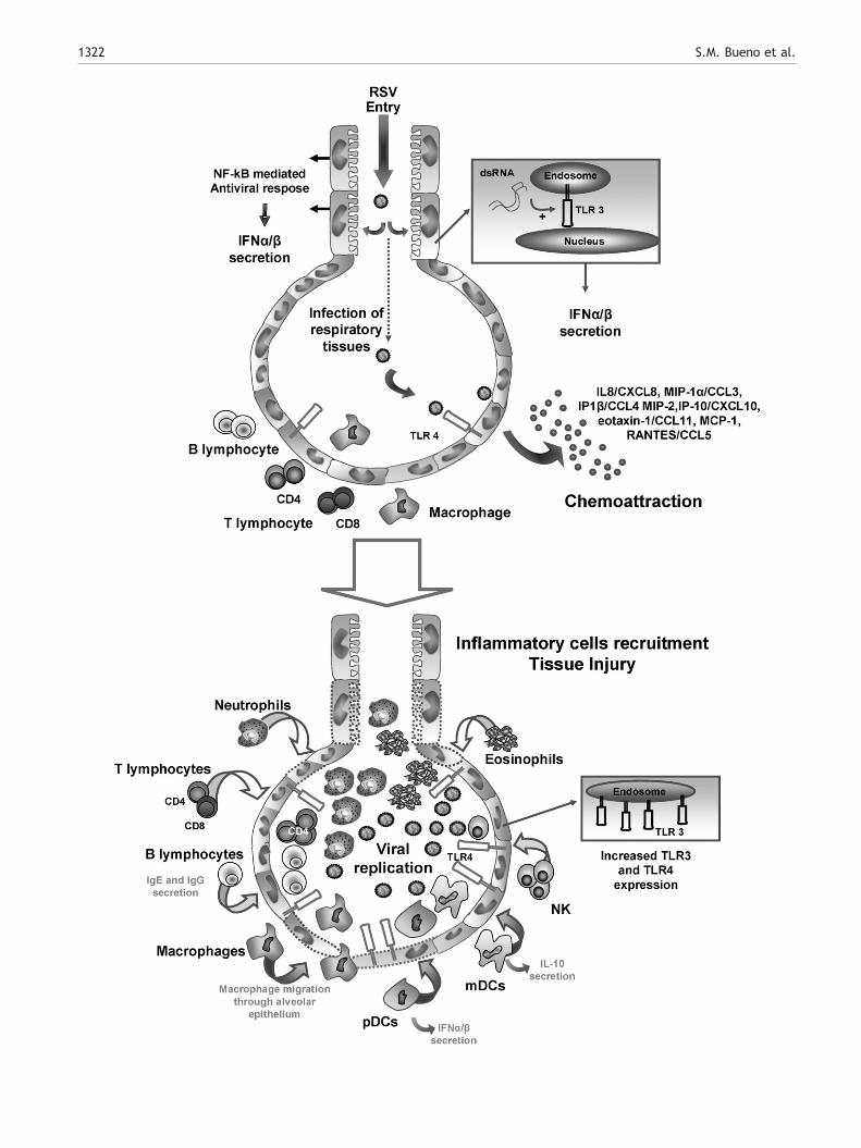
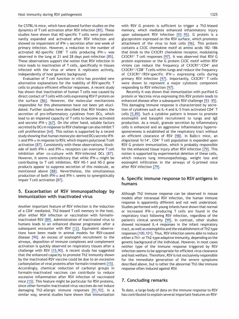
response. After attaching to epithelial cells, RSV induces NF-kB-mediated transcription of genes promoting an anti-viralresponse [18,19]. Accordingly, during the first hours afterinfection, an enhanced expression of genes related with localinflammatory responses, antigen processing and chemoat-traction are observed in the lung epithelium [20]. Theseprocesses promote the production of chemokines and therecruitment of eosinophils, NK cells and CD4+ T cells to theairways (Fig. 1) [21].
Once RSV contacts respiratory epithelial cells, the virus isrecognized by cell surface Toll-like Receptors (TLRs),promoting the secretion of inflammatory cytokines. Amongthe TLRs expressed on the surface of respiratory epithelialcells, the TLR4/CD14 complex is the main extracellularreceptor recognizing RSV, through the binding of the Fusion(F) protein present on the viral envelope [22]. TLR4/CD14engagement by F protein leads to an NF-kB-mediatedcytokine response, including the secretion of IL-8, IL-10,and IL-6 [22,23] and an increase in TLR4 expression onepithelial cells [24]. In addition, several reports havedemonstrated that RSV can also be recognized by TLR3 onrespiratory epithelial cells [18,25,26]. TLR3 is an intracel-lular receptor that recognizes viral replication intermedi-ates, such as dsRNA. Although it has been suggested thatTLR3 is not required for viral clearance, its expression mightbe necessary to regulate the immune environment in the lungepithelia. A recent study has shown that RSV infection inmice lacking TLR3 translates into increased secretion of Th2cytokines and mucus in the lung, as well as enhancedeosinophil recruitment as compared to wild-type mice [25].In addition, increased TLR3 expression is observed onrespiratory epithelial cells after RSV infection, whichprobably can contribute to an increased sensitivity andsecretion of inflammatory cytokines upon contact with RSVor even other microbial components, such as LPS or dsRNA[18]. These observations are consistent with the notion thatRSV might be able to predispose lung tissues to enhancedinflammatory responses to later challenges with virus orbacteria [27,28].
In vitro studies performed on respiratory epithelial cellshave shown that RSV infection promotes the secretion ofchemokines, such as IL-8/CXCL8, MIP-1α/CCL3, MIP-1β/CCL4, MIP-2, IP-10/CXCL10, eotaxin-1/CCL11, macrophagechemoattractant protein 1 (MCP-1) and RANTES/CCL5.Accordingly, expression of these chemokines has beenshown increased in nasal washes from RSV-infected indivi-duals at the time of virus shedding [29–37]. Chemokinessecreted by RSV-infected epithelia promote activation andrecruitment, from blood into infected tissues, of neutrophils(IL-8/CXCL8), monocytes, memory T cells (RANTES/CCL5)and eosinophils (eotaxin-1/CCL11). The recruited immunecells secrete both pro-inflammatory cytokines, such as TNF,IL-6 and IL-8, as well as inhibitory cytokines, such as IL-10[38–40]. Increased secretion of these cytokines probablycontributes to the airway damage caused by RSV infection.Consistent with this notion, in vitro studies have shown thatbronchial epithelial cells secrete higher amounts of IL-8, IL-6and RANTES/CCL5 in response to RSV infection, whencompared to other respiratory virus [16]. Accordingly, invivo chemokine blockade can reduce lung pathology anddamage, as observed in mice treated with anti-RANTESantibodies, which showed a significant decrease in airway
1322 S.M. Bueno et al.
1323Host immunity during RSV pathogenesis
hyperreactivity [37]. Similarly, treatment of mice with Met-RANTES (a competitor for RANTES receptor) reduced therecruitment of inflammatory cells to the lung [32]. Interest-ingly, treatment with RANTES/CCL5 can reduce RSV infectionof HEp-2 cells, probably by blocking the interaction betweenRSV fusion (F) protein and epithelial cell surface proteins[41]. Similarly, infection was also decreased by a biologicallyinactive N-terminally modified Met-CCL5 [41]. In anotherstudy, mice treated with a blocking antibody against CCL11showed reduced lung eosinophilia and disease severity [31].Unexpectedly, treatment with this molecule also causedinhibition of CD4+ but not CD8+ T cell infiltration into thelungs [31]. In agreement with the observations discussedabove, MIP-1α KO mice infected with RSV showed asignificant reduction in lung histopathology as compared towild-type mice. However, no differences on lung viral titerswere observed between MIP-1α KO and WT mice [36]. Takentogether, these data suggest that secretion of chemokines byairway epithelium and infiltrating immune cells can bedetrimental to the host by promoting immunopathology andtissue damage with a minor contribution to viral clearance.Thus, the chemokine milieu is beginning to be considered asan important component during RSV infection and thereforean attractive pharmacological target.
Upon RSV infection, epithelial cells and infiltratingleukocytes produce large amounts of anti-viral molecules,such as type I IFN [42]. These cytokines signal through theIFNAR receptor on the target cell surface and, activateseveral intracellular signaling pathways that involve theactivity of STAT-1 and STAT-2 proteins [42]. Activated STAT-1and -2 bind to Interferon regulatory factor 9 (IRF-9) toassemble an activator complex that translocates to thenucleus and initiates the expression of multiple genes,known as Interferon-stimulated genes (ISGs) [42]. Expressionof these genes triggers several anti-viral functions; such asthe activation of ribonuclease L (RNAseL) that degrades hostand viral RNA. Furthermore, ISGs promote the proliferationand activation of NK cells, as well as their anti-viral capacity[43]. Although plasmacytoid dendritic cell (pDCs) are mainproducers of IFN-α, as described below, epithelial cells alsocan produce significant amounts of type I interferons [44].
However, type I IFNs have been shown to also influenceother immune process during RSV infection. Mice deficienton STAT-1 and -2, two proteins required for IFN α/β and IFN-γinduced signaling, suffer severe inflammation, increasedeosinophil infiltration to the airways and increased Th2cytokines in the lungs in response to RSV infection. [45].Further, it has been observed that IFN-α/β and IFN-γproduced during the innate immune response could alsocontribute to the recruitment of inflammatory cells to thelungs during RSV infection [46]. Mice lacking IFNα/β and IFNγ
Figure. 1 RSV infection modifies the inflammatory environment ininfect airway epithelial cells. As a defense mechanism against viral sactivation of NF-kB and intracellular TLR3 by RSV-derived PAMPs (dsRthe secretion of several chemokines and cytokines. Under normalcirculating in the blood outside alveoli (top panels). Later on infecinflammatory cells into the lungs, which is promoted by the chemokeosinophils, NK cells, mDCs, pDCs, macrophages, B lymphocyes ansurrounding bronchial tubes. Although CD8+ T lymphocyte infiltratio(lower panels). Furthermore, increased expression of TLR3 and TLR4
receptors (IFNαβγR−/−) showed eosinophilia but reducedlymphocyte infiltration to the lungs after a challenge withRSV. However, mice that only lack the IFN-γ receptor, showedonly moderate eosinophilia within the lung [46]. Thus, type IIFNs seems to be critical for the recruitment of inflammatorycells to the lungs, triggered by RSV.
Interestingly, RSV infection promotes a weakened type IIFN response in infected tissues by blocking IFNα/β signaling.Studies using recombinant deletion approaches have demon-strated that RSV proteins NS-1 and NS-2 are necessary toimpair IFN-α/β secretion by epithelial cells [47]. These twoviral proteins act coordinately to decrease STAT-2 mediated-signaling by selectively targeting this receptor for proteaso-mal degradation [48]. Recently, it has been suggested thatNS-1 binds to the proteasome-related proteins elongin C andcullin 2 to form an E3 ubiquitin ligase complex, whichprobably promotes STAT-2 degradation with the assistance ofNS-2 [49]. This mechanism used by RSV to down-regulate typeI IFN signaling and response probably allows successful viralreplicationwithin infected tissues. However, RSValso reducessecretion of type I IFN in RSV-infected tissues, which couldpromote bystander recruitment of inflammatory cells to theairways contributing to lung damage upon viral infection.
3. Dendritic cell function during the immuneresponse to RSV
Dendritic cells (DCs) are ubiquitous professional antigenpresenting cells (APCs) found in lymphoid tissues and non-lymphoid tissues located strategically to capture a diversearray of antigens and present them to T cells as peptidesbound to either MHC class I or class II molecules [50–52].Upon recognition of pathogen associated molecular pat-terns, DCs undergo a phenotypic change, known as matura-tion. As a result of maturation, DCs downmodulate theirphagocytic capacity and up-regulate the expression ofsurface peptide-major histocompatibility complexes(pMHC) and co-stimulatory molecules such as CD80 andCD86 [53]. Additionally, mature DCs increase their migratorycapacity and secrete modulatory cytokines involved in hostdefense, such as IL-12 and type I and type II interferons[54,55]. Concomitantly, DCs undergoing maturation migrateto lymphoid tissues where antigen presentation to specific Tcells takes place and initiate an adaptive immune response[51,56]. Because DCs are key components for the clearanceof pathogens, virulent microorganisms can interfere with DCfunction as a mechanism to impair the proper function ofadaptive immunity [57–60].
However, dendritic cells are a heterogeneous group ofcells, which show phenotypic and functional differences at
the airways. Early after RSV infection, viruses reach alveoli andpreading, infected cells are induced to secrete IFNα/β after theNA). Simultaneously, engagement of surface TLR4 by RSV inducesconditions, lymphocytes and macrophages can only be foundtion, viral replication is accompanied by massive infiltration ofine- and cytokine-rich environment. At this point, neutrophils,d CD4+ T lymphocytes can be observed in alveoli, as well asn can be observed, their effector capacity is widely hamperedby epithelial cells is observed after RSV infection.

1324 S.M. Bueno et al.
priming adaptive immunity [61]. Among the diverse DClineages, two of them predominate and have been extensivelycharacterized, myeloid DCs (mDCs) and plasmacytoid DCs(pDCs). These two DC subtypes work synergistically atinducing efficient anti-viral immune responses [54]. Bystimulating CD4+ and CD8+ T cells, IL-12-producing mDCs arecritical inducers of Th1-polarized adaptive immune responses,which can promote efficient effector responses against virusesand other intracellular pathogens [54,61]. On the other hand,IFN-α secreting pDCs promote anti-viral and immunomodula-tory effects by acting over wider range of cell types [54,62].mDCs and pDCs residing at the airways play pivotal rolesduring innate immune responses against viruses, as well as aregulatory roles in the polarization of Tcell effector mediatedresponses [63]. Considering their key functions as promotersof immunity, it is important to define how mDCs and pDCs cancontribute to development of immune responses to RSV, aswell as the potential virulence mechanisms developed by thisvirus to interfere with DC function.
In vitro experiments have shown that RSV can infect DCsand replicate within these cells [64,65] (Gonzalez andKalergis, unpublished results). DC-RSV interaction can leadto the upregulation of maturation markers on the DC surface,such as CD86 and MHC-II [64–67]. However, RSV-infected DCsseem unable to efficiently prime antigen-specific T cells forIFN-γ secretion [67] (Gonzalez and Kalergis, unpublishedresults), a phenomenon that could be explained by thereduced capacity of RSV-infected DCs to secrete Th1-polarizing cytokines. For example, virulent RSV strainsinhibit IFN-α and IL-12p70 secretion by mDCs [65,68,69],which are key cytokines necessary to promote CD8+ T celleffector functions as well as the induction of memory CD8+ Tcells [70]. Accordingly, another study has demonstrated thatRSV can block IFN-α secretion derived from pDCs in vivo, thusevading the development of a proper anti-viral immuneresponse [44]. However, other studies have shown that RSV-infected pDCs can secrete considerable amounts of IFN-α[66]. These differences might be accounted to the differentstrains of RSV used in these studies.
In vivo studies have shown that, after viral exposure,increased amounts of both myeloid and plasmacytoid DCswith mature phenotypes are observed in the respiratoryairways of mice [71,72]. Moreover, sustained increase forboth mDCs and pDCs is observed in the lungs of mice up to30 days after RSV infection [69,72,73]. It is possible thatproliferation of DC precursors at the site of infection in thelungs in response to GM-CSF secreted by epithelial cells uponRSV infection could contribute to the increased numbers ofDCs [29]. This notion is supported by the expression ofproliferation markers, such as Ki67, by DCs in infected lungs[74]. Interestingly, upon RSV infection, DC precursors in thelung are depleted and thus DC expansion in lungs is notobserved following further viral infections [15].
Recent studies suggest that pulmonary pDCs could play animportant role at modulating the immune response inducedby RSV infection [73]. The selective depletion with pDC-specific antibodies leads to enhanced lung immunopathology[73,74]. As described above, type I IFN might play animportant role at regulating the immune environment duringRSV infection. Since pDCs are the most important IFN-αproducing cells, depletion of these cells might furtherfacilitate the Th2-biased immune response induced by RSV.
In addition, activation of pDCs residing at the airwaysreduces RSV replication and inflammation after infection[15]. Therefore, although in vitro studies have shown thatRSV modulates DC function by reducing its capacity tosecrete key cytokines and prime an efficient anti-viral T cellresponse, in vivo studies would suggest that pulmonary DCsare relevant to control RSV infection and to counteract theimmunopathology induced by this virus. Further studies areneeded to conciliate both in vitro and in vivo observationsregarding the role of DCs during RSV infection.
4. Role of adaptive immunity on RSV infectionand immunopathology
To date, several experimental models and differentapproaches suggest that RSV can modulate the activationof the adaptive immune response in at least two ways. First,RSV seems to block the production/function of cytotoxicmemory T cells against viral antigens. Thus, primaryinfection does not confer protection to subsequent re-infections with antigenically similar RSV strains [75,76].Second, RSV infection and/or vaccination with inactivatedvirus can induce a detrimental, Th2 immune memory thatpromotes lung injury after a second exposure to the virus[77–79].
Early studies reported that monocytes/macrophagessecrete inhibitory molecules that suppress T cells responsesin vitro in response to challenge with RSV [80]. The absenceof efficient murine adaptive immune responses is evidencedby the persistence of RSV in the lungs, despite the presenceof RSV-specific CD4+ and CD8+ Tcells in infected tissues [72].Interestingly, it has become clear that the T cell responsesare specifically impaired in the respiratory tract during RSVinfection [7,81]. It is thought that efficient viral clearancerequires Th1 polarization driven partially by IL-12 secretingmDCs, which promote activation of IFN-γ-producing CD4+ Tcells. IFN-γ in turn promotes cytotoxic T cell function bystimulating CD8+ T cells and NK cells to clear virus-infectedcells, stimulates macrophage phagocytic activity to promoteclearance of dead cells and induces production of neutraliz-ing IgG antibodies by B cells [82]. However, RSV infectionseems able to evade cytotoxic immunity by blocking IFN-γsecretion by RSV-specific Tcells [81]. In Balb/c mice, at leasttwo immunodominant peptides derived from RSV proteinshave been described to bind class I MHC molecules [6]. Theseepitopes have allowed tracking in vivo virus-specific CD8+ Tcells and their activation after RSV infection. Thesepeptides, M282–90 and F85–93, are bound to H-2Kd moleculesand can be used for flow cytometry detection of specific Tcells infiltrating tissues, using MHC-I tetramers [6,81,83].Using this methodology, it has been observed that M2-specific T cells expand, activate and localize in the lungsafter RSV infection. However, these cells showed an impairedeffector activity [81]. A similar behavior was described for F-specific CD8+ T cells [6]. Interestingly, these cells localize inpulmonary tissues for short periods of time and secrete lowamounts of IFN-γ, which contribute to their limited effectoractivity [81]. However, this unresponsiveness of specific CD8+
T cells can be overcome by a treatment with IL-2, both invitro and in vivo [81,84]. Additional immunodominantepitopes derived from RSV proteins have been recognized
1325Host immunity during RSV pathogenesis
for C57BL/6 mice, which have allowed further studies on thedynamics of T cell activation after RSV infection [85]. Thesestudies have shown that M2-specific T cells were predomi-nantly expanded and activated after RSV infection andshowed no impairment of IFN-γ secretion after one week ofprimary infection. However, a reduction in the number ofactivated M2-specific CD8+ T cells producing IFN-γ wasobserved in the lung at 21 and 28 days post-infection [85].These observations support the notion that RSV infection inmice leads to inactivation of T cells, specifically in tissuesinfected with the virus or where it replicates actively,independently of host genetic background.
Evaluation of T cell function in vitro has provided newalternative explanations for the inability of RSV-specific Tcells to produce efficient effector responses. A recent studyhas shown that inactivation of human T cells was caused bydirect contact of Tcells with cells expressing RSV F protein onthe surface [86]. However, the molecular mechanismsresponsible for this phenomenon have not been yet eluci-dated. Further studies have described that RSV hinders thesecretion of pro-inflammatory cytokines from DCs, whichlead to an impaired capacity of T cells to become activatedand secrete IFN-γ [67]. On the other hand, soluble factorssecreted by DCs in response to RSV challenge can also inhibit Tcell proliferation [64]. This notion is supported by a recentstudy showing that humanmonocyte-derived DCs secrete IFN-λ and IFN-α in response to RSV, which impairs polyclonal Tcellactivation [87]. Consistently with these observations, block-ade of both IFN-λ and IFN-α receptors can overcome T cellinhibition after co-culture with RSV-infected DCs [87].However, it seems contradictory that while IFN-α might becontributing to T cell inhibition, RSV NS-1 and NS-2 geneproducts appear to suppress secretion of this molecule, asmentioned above [88]. Nevertheless, the simultaneousproduction of both IFN-α and IFN-λ seems to synergisticallyimpair T cell activation [87].
5. Exacerbation of RSV immunopathology byimmunization with inactivated virus
Another important feature of RSV infection is the inductionof a CD4+ mediated, Th2-biased T cell memory in the host,after either RSV infection or vaccination with formalin-inactivated RSV [89]. Administration of inactivated virus tohumans leads to an enhanced disease progression after asubsequent encounter with RSV [12]. Equivalent observa-tions have been made in animal models for RSV-causeddisease [90]. An excess of eosinophil recruitment to theairways, deposition of immune complexes and complementactivation is quickly observed on respiratory tissues after achallenge with RSV [15,90]. A recent study has suggestedthat the enhanced capacity to promote Th2 immunity shownby the inactivated RSV vaccine could be due to an excessivecarbonylation of viral proteins after formalin treatment [15].Accordingly, chemical reduction of carbonyl groups informalin-inactivated vaccines can contribute to reduceexcessive inflammation after RSV infection of vaccinatedmice [15]. This feature might be particular for RSV proteins,since other formalin-inactivated virus vaccines do not inducedamaging Th2-allergic immune responses [91,92]. In asimilar way, several studies have shown that immunization
with RSV G protein is sufficient to trigger a Th2-biasedmemory, which mediates enhanced inflammatory injuryupon subsequent RSV infection [93–95]. G protein is aglycoprotein expressed on the RSV surface, which promotesattachment of the virus to host cells [96]. This proteincontains a CX3C chemokine motif at amino acids 182–186that binds to the CX3CR1 chemokine receptor, modulatingCX3CR1+ T cell responses [97]. It was observed that RSV Gprotein expression or the G protein CX3C motif within RSVvirions can reduce the frequency of CX3CR1+/CD4+ andCX3CR1+/CD8+ T cells within lungs and reduce the frequencyof CX3CR1+/RSV-specific IFN-γ expressing cells duringprimary RSV infection [97]. Importantly, CX3CR1+ T cellswere shown to represent a major cytotoxic componentresponding to RSV infection [97].
Recently it was shown that immunization with purified Gprotein or Vaccinia virus expressing this RSV protein leads toenhanced disease after a subsequent RSV challenge [93–95].This damaging immune response is characterized by secre-tion of cytokines such as IL-4 and IL-5 from activated CD4+ Tcells [5,89]. Such a cytokine pattern is known to promoteeosinophil and basophil recruitment to lungs and IgEproduction. As a result, granule secretion by inflammatorycells is enhanced and an aggressive inflammatory hyperre-sponsiveness is established at the respiratory tract withoutan efficient clearance of RSV [98]. In Balb/c mice, anoligoclonal Vβ14+, CD4+ T cell population is expanded uponRSV G protein immunization, which is probably responsiblefor the enhanced tissue injury after RSV infection [79]. Thisnotion is supported by experimental deletion of these cells,which reduces lung immunopathology, weight loss andeosinophil infiltration in the airways of G-primed miceafter RSV infection [79].
6. Specific immune response to RSV antigens inhumans
Although Th2 immune response can be observed in mousemodels after intranasal RSV infection, the human immuneresponse is apparently different and not well understood.Studies performed with young infants have shown that normalor increased IFN-γ producing T cells are found in therespiratory tract following RSV infection, regardless of thepatient's clinical severity [99]. In contrast, other studiesshowed increased IL-4 responses in the infant respiratorytract, aswell as eosinophilia and the establishment of Th2 typeresponses [100,101]. Thus, RSV infection seems able to induceeither a Th1- or Th2-type adaptive immunity, depending on thegenetic background of the individual. However, in most casesneither type of the immune response triggered by RSVinfection seems to be appropriate for efficient virus clearanceand host welfare. Therefore, RSV is not exclusively responsiblefor the immediate generation of the severe symptomsdescribed above, but is rather the abnormal Th2-like immuneresponse often induced against RSV.
7. Concluding remarks
To date, a large body of data on the immune response to RSVhas contributed to explain several important features on RSV-
1326 S.M. Bueno et al.
induced pathology. Its seems clear that the overreactingimmune response triggered in some hosts by RSV proteins,which leads to an excessive inflammation and infiltration ofimmune cells into the airways, can be considered as the maincause of tissue damage. Damaging immunity can indeed beobserved upon subsequent RSV infection after vaccinationwith FI-RSV of G protein. In addition, the establishment ofprotective and efficient adaptive immune memory isimpaired probably because RSV infection impedes theactivation of virus-specific Tcell within the lungs. Therefore,efforts aimed to design an effective and safe vaccine againstRSV need to evaluate both, the type of immune responseinduced after vaccination to avoid immunopathology, as wellas the capacity to generate a long-lasting protective immunememory. In addition, further studies are required to under-score the molecular mechanisms used by RSV to interferewith Tcell activation, which could contribute to the design ofnew therapeutic tools to treat or prevent the respiratorydisorders caused by the virus.
Acknowledgements
We would like to thank Dr. J. Reid Schwebach for criticalreview of this manuscript. The authors are supported bygrants FONDECYT no. 1070352, FONDECYT no. 1050979,FONDECYT no. 3060041, FONDECYT no. 3070018, FONDECYTno. 11075060, SavinMuco-Path-INCO-CT-2006-032296; IFS#B/3764-1, FONDEF D04I1075 and Millennium Nucleus onImmunology and Immunotherapy (P04/030-F). PAG and HETare CONICYT fellow.
References
[1] Mejias A, Chavez-Bueno S, Jafri HS, Ramilo O. Respiratorysyncytial virus infections: old challenges and new opportu-nities. Pediatr Infect Dis J 2005;24:S189–96 discussion S96–7.
[2] Hacking D, Hull J. Respiratory syncytial virus—viral biology andthe host response. J Infect 2002;45:18–24.
[3] Harris J, Werling D. Binding and entry of respiratory syncytialvirus into host cells and initiation of the innate immuneresponse. Cell Microbiol 2003;5:671–80.
[4] Srikiatkhachorn A, Braciale TJ. Virus-specific CD8+ T lympho-cytes downregulate T helper cell type 2 cytokine secretion andpulmonary eosinophilia during experimental murine respira-tory syncytial virus infection. J Exp Med 1997;186:421–32.
[5] Srikiatkhachorn A, Braciale TJ. Virus-specific memory andeffector T lymphocytes exhibit different cytokine responses toantigens during experimental murine respiratory syncytialvirus infection. J Virol 1997;71:678–85.
[6] Chang J, Srikiatkhachorn A, Braciale TJ. Visualization andcharacterization of respiratory syncytial virus F-specific CD8(+)T cells during experimental virus infection. J Immunol 2001;167:4254–60.
[7] Braciale TJ. Respiratory syncytial virus and t cells: interplaybetween the virus and the host adaptive immune system. ProcAm Thorac Soc 2005;2:141–6.
[8] Beckham JD, Cadena A, Lin J, Piedra PA, GlezenWP, GreenbergSB, et al. Respiratory viral infections in patients with chronic,obstructive pulmonary disease. J Infect 2005;50: 322–30.
[9] Englund JA, Piedra PA, Jewell A, Patel K, Baxter BB, WhimbeyE. Rapid diagnosis of respiratory syncytial virus infectionsin immunocompromised adults. J Clin Microbiol 1996;34:1649–53.
[10] Harker J, Bukreyev A, Collins PL, Wang B, Openshaw PJM,Tregoning JS. Virally delivered cytokines alter the immuneresponse to future lung infections. J Virol 2007. 01544–07.
[11] Kim HW, Canchola JG, Brandt CD, Pyles G, Chanock RM,Jensen K, et al. Respiratory syncytial virus disease in infantsdespite prior administration of antigenic inactivated vaccine.Am J Epidemiol 1969;89:422–34.
[12] Kapikian AZ, Mitchell RH, Chanock RM, Shvedoff RA, Stewart CE.An epidemiologic study of altered clinical reactivity torespiratory syncytial (RS) virus infection in children previouslyvaccinated with an inactivated RS virus vaccine. Am J Epidemiol1969;89:405–21.
[13] Connors M, Giese NA, Kulkarni AB, Firestone CY, Morse 3rd HC,Murphy BR. Enhanced pulmonary histopathology induced byrespiratory syncytial virus (RSV) challenge of formalin-inacti-vated RSV-immunized BALB/c mice is abrogated by depletion ofinterleukin-4 (IL-4) and IL-10. J Virol 1994;68: 5321–5.
[14] Waris ME, Tsou C, Erdman DD, Zaki SR, Anderson LJ.Respiratory synctial virus infection in BALB/c mice previouslyimmunized with formalin-inactivated virus induces enhancedpulmonary inflammatory response with a predominant Th2-like cytokine pattern. J Virol 1996;70:2852–60.
[15] Moghaddam A, Olszewska W, Wang B, Tregoning JS, Helson R,Sattentau QJ, et al. A potential molecular mechanism forhypersensitivity caused by formalin-inactivated vaccines. NatMed 2006;12:905–7.
[16] Yoon JS, Kim HH, Lee Y, Lee JS. Cytokine induction byrespiratory syncytial virus and adenovirus in bronchialepithelial cells. Pediatr Pulmonol 2007;42:277–82.
[17] Openshaw PJ, Tregoning JS. Immune responses and diseaseenhancement during respiratory syncytial virus infection. ClinMicrobiol Rev 2005;18:541–55.
[18] Groskreutz DJ, Monick MM, Powers LS, Yarovinsky TO, Look DC,Hunninghake GW. Respiratory syncytial virus induces TLR3protein and protein kinase R, leading to increased double-stranded RNA responsiveness in airway epithelial cells. JImmunol 2006;176: 1733–40.
[19] Liu P, Jamaluddin M, Li K, Garofalo R, Casola A, Brasier A.Retinoic acid-inducible gene I mediates early antiviralresponse and toll-like receptor 3 expression in respiratorysyncytial virus-infected airway epithelial cells. J Virol 2007;81:1401–11.
[20] Janssen R, Pennings J, Hodemaekers H, Buisman A, van OostenM, de Rond L, et al. Host transcription profiles upon primaryrespiratory syncytial virus infection. J Virol 2007;81:5958–67.
[21] Hussell T, Openshaw PJ. Intracellular IFN-gamma expression innatural killer cells precedes lung CD8+ T cell recruitmentduring respiratory syncytial virus infection. J Gen Virol1998;79(Pt 11):2593–601.
[22] Kurt-Jones EA, Popova L, Kwinn L, Haynes LM, Jones LP, TrippRA, et al. Pattern recognition receptors TLR4 and CD14mediate response to respiratory syncytial virus. Nat Immunol2000;1:398–401.
[23] Tulic MK, Hurrelbrink RJ, Prele CM, Laing IA, Upham JW, LeSouef P, et al. Polymorphisms mediate impaired responses torespiratory syncytial virus and lipopolysaccharide. J Immunol2007;179:132–40.
[24] Monick MM, Yarovinsky TO, Powers LS, Butler NS, Carter AB,Gudmundsson G, et al. Respiratory syncytial virus up-regulates TLR4 and sensitizes airway epithelial cells toendotoxin. J Biol Chem 2003;278:53035–44.
[25] Rudd BD, Smit JJ, Flavell RA, Alexopoulou L, Schaller MA,Gruber A, et al. Deletion of TLR3 alters the pulmonary immuneenvironment and mucus production during respiratory syncy-tial virus infection. J Immunol 2006;176:1937–42.
[26] Rudd BD, Burstein E, Duckett CS, Li X, Lukacs NW. Differentialrole for TLR3 in respiratory syncytial virus-induced chemokineexpression. J Virol 2005;79:3350–7.

1327Host immunity during RSV pathogenesis
[27] Resch B, Gusenleitner W, Mueller WD. Risk of concurrentbacterial infection in preterm infants hospitalized due torespiratory syncytial virus infection. Acta Paediatrica 2007;96:495–8.
[28] Stark JM, Stark MA, Colasurdo GN, LeVine AM. Decreasedbacterial clearance from the lungs of mice following primaryrespiratory syncytial virus infection. JMed Virol 2006;78: 829–38.
[29] Noah TL, Becker S. Respiratory syncytial virus-induced cy-tokine production by a human bronchial epithelial cell line. AmJ Physiol 1993;265:L472–8.
[30] Sheeran P, Jafri H, Carubelli C, Saavedra J, Johnson C, Krisher K,et al. Elevated cytokine concentrations in the nasopharyngealand tracheal secretions of children with respiratory syncytialvirus disease. Pediatr Infect Dis J 1999;18:115–22.
[31] Matthews SP, Tregoning JS, Coyle AJ, Hussell T, Openshaw PJ.Role of CCL11 in eosinophilic lung disease during respiratorysyncytial virus infection. J Virol 2005;79:2050–7.
[32] Culley FJ, Pennycook AM, Tregoning JS, Dodd JS, Walzl G,Wells TN, et al. Role of CCL5 (RANTES) in viral lung disease. JVirol 2006;80:8151–7.
[33] Culley FJ, Pennycook AM, Tregoning JS, Hussell T, Openshaw PJ.Differential chemokine expression following respiratory virusinfection reflects Th1- or Th2-biased immunopathology. J Virol2006;80:4521–7.
[34] Bermejo-Martin JF, Garcia-Arevalo MC, De Lejarazu RO,Ardura J, Eiros JM, Alonso A, et al. Predominance of Th2cytokines, CXC chemokines and innate immunity mediators atthe mucosal level during severe respiratory syncytial virusinfection in children. Eur Cytokine Netw 2007;18:162–7.
[35] Niimi K, Asano K, Shiraishi Y, Nakajima T, Wakaki M, Kagyo J,et al. TLR3-mediated synthesis and release of eotaxin-1/CCL11from human bronchial smooth muscle cells stimulated withdouble-stranded RNA. J Immunol 2007;178:489–95.
[36] Haeberle HA, Kuziel WA, Dieterich HJ, Casola A, Gatalica Z,Garofalo RP. Inducible expression of inflammatory chemokinesin respiratory syncytial virus-infected mice: role of MIP-1alphain lung pathology. J Virol 2001;75:878–90.
[37] Tekkanat KK, Maassab H, Miller A, Berlin AA, Kunkel SL, LukacsNW. RANTES (CCL5) production during primary respiratorysyncytial virus infection exacerbates airway disease. Eur JImmunol 2002;32:3276–84.
[38] Jafri HS, Chavez-Bueno S, Mejias A, Gomez AM, Rios AM, NassiSS, et al. Respiratory syncytial virus induces pneumonia,cytokine response, airway obstruction, and chronic inflam-matory infiltrates associated with long-term airway hyperre-sponsiveness in mice. J Infect Dis 2004;189:1856–65.
[39] McNamara PS, Flanagan BF, Selby AM, Hart CA, Smyth RL. Pro-and anti-inflammatory responses in respiratory syncytial virusbronchiolitis. Eur Respir J 2004;23:106–12.
[40] Grissell TV, Powell H, Shafren DR, Boyle MJ, Hensley MJ,Jones PD, et al. Interleukin-10 gene expression in acutevirus-induced asthma. Am J Respir Crit Care Med 2005;172:433–9.
[41] Elliott MB, Tebbey PW, Pryharski KS, Scheuer CA, Laughlin TS,Hancock GE. Inhibition of respiratory syncytial virus infectionwith the CC chemokine RANTES (CCL5). J Med Virol 2004;73:300–8.
[42] Garcia-Sastre A, Biron CA. Type 1 interferons and the virus–host relationship: a lesson in detente. Science 2006;312:879–82.
[43] Horvath CM, Stark GR, Kerr IM, Darnell Jr JE. Interactionsbetween STAT and non-STAT proteins in the interferon-stimulated gene factor 3 transcription complex. Mol Cell Biol1996;16:6957–64.
[44] Jewell NA, Vaghefi N, Mertz SE, Akter P, Peebles Jr RS,Bakaletz LO, et al. Differential type I interferon induction byrespiratory syncytial virus and influenza A virus in vivo. J Virol2007;81:9790–800.
[45] Durbin JE, Johnson TR, Durbin RK, Mertz SE, Morotti RA,Peebles RS, et al. The role of IFN in respiratory syncytial viruspathogenesis. J Immunol 2002;168:2944–52.
[46] Johnson TR, Mertz SE, Gitiban N, Hammond S, LeGallo R,Durbin RK, et al. Role for innate IFNs in determining respiratorysyncytial virus immunopathology. J Immunol 2005;174:7234–41.
[47] Schlender J, Bossert B, Buchholz U, Conzelmann KK. Bovinerespiratory syncytial virus nonstructural proteins NS1 and NS2cooperatively antagonize alpha/beta interferon-induced anti-viral response. J Virol 2000;74:8234–42.
[48] Lo MS, Brazas RM, Holtzman MJ. Respiratory syncytial virusnonstructural proteins NS1 and NS2 mediate inhibition of Stat2expression and alpha/beta interferon responsiveness. J Virol2005;79:9315–9.
[49] Elliott J, Lynch OT, Suessmuth Y, Qian P, Boyd CR, Burrows JF,et al. Respiratory syncytial virus NS1 protein degradesSTAT2 by using the elongin–cullin E3 ligase. J Virol 2007;81:3428–36.
[50] Trombetta ES, Mellman I. Cell biology of antigen processing invitro and in vivo. Annu Rev Immunol 2005;23:975-1028.
[51] Banchereau J, Steinman RM. Dendritic cells and the control ofimmunity. Nature 1998;392:245–52.
[52] Vermaelen K, Pauwels R. Pulmonary dendritic cells. Am JRespir Crit Care Med 2005;172:530–51.
[53] Gallucci S, Matzinger P. Danger signals: SOS to the immunesystem. Curr Opin Immunol 2001;13:114–9.
[54] Colonna M, Trinchieri G, Liu YJ. Plasmacytoid dendritic cells inimmunity. Nat Immunol 2004;5:1219–26.
[55] Steinman RM, Hemmi H. Dendritic cells: translating innate toadaptive immunity. Curr Top Microbiol Immunol 2006;311:17–58.
[56] Lanzavecchia A, Sallusto F. Regulation of T cell immunity bydendritic cells. Cell 2001;106:263–6.
[57] Tobar JA, Gonzalez PA, Kalergis AM. Salmonella escape fromantigen presentation can be overcome by targeting bacteria toFc gamma receptors on dendritic cells. J Immunol 2004;173:4058–65.
[58] Bueno SM, Tobar JA, Iruretagoyena MI, Kalergis AM. Molecularinteractions between dendritic cells and Salmonella: escapefrom adaptive immunity and implications on pathogenesis.Crit Rev Immunol 2005;25:389–403.
[59] Weslow-Schmidt JL, Jewell NA, Mertz SE, Simas JP, Durbin JE,Flano E. Type I interferon inhibition and dendritic cellactivation during gammaherpesvirus respiratory infection. JVirol 2007;81:9778–89.
[60] Cush SS, Anderson KM, Ravneberg DH, Weslow-Schmidt JL,Flano E. Memory generation and maintenance of CD8+ T cellfunction during viral persistence. J Immunol 2007;179:141–53.
[61] Liu Y. Dendritic cell subsets and lineages, and theirfunctions in innate and adaptive immunity. Cell 2001;106:259–62.
[62] Barchet W, Cella M, Colonna M. Plasmacytoid dendritic cells—virus experts of innate immunity. Semin Immunol 2005;17:253–61.
[63] Cella M, Facchetti F, Lanzavecchia A, Colonna M. Plasma-cytoid dendritic cells activated by influenza virus and CD40Ldrive a potent TH1 polarization. Nat Immunol 2000;1:305–10.
[64] de Graaff PMA, de Jong EC, van Capel TM, van Dijk MEA, RohollPJM, Boes J, et al. Respiratory syncytial virus infection ofmonocyte-derived dendritic cells decreases their capacity toactivate CD4 T cells. J Immunol 2005;175:5904–11.
[65] Guerrero-Plata A, Casola A, Suarez G, Yu X, Spetch L, PeeplesME, et al. Differential response of dendritic cells to humanmetapneumovirus and respiratory syncytial virus. Am J RespirCell Mol Biol 2006;34:320–9.
1328 S.M. Bueno et al.
[66] Boogaard I, van Oosten M, van Rijt LS, Muskens F, Kimman TG,Lambrecht BN, et al. Respiratory syncytial virus differentiallyactivates murine myeloid and plasmacytoid dendritic cells.Immunology 2007;122:65–72.
[67] Bartz H, Turkel O, Hoffjan S, Rothoeft T, Gonschorek A,Schauer U. Respiratory syncytial virus decreases the capacityof myeloid dendritic cells to induce interferon-g in naïve Tcells. Immunology 2003;109:49–57.
[68] Schlender J, Hornung V, Finke S, Gunthner-Biller M, Marozin S,Brzozka K, et al. Inhibition of toll-like receptor 7- and 9-mediated alpha/beta interferon production in human plas-macytoid dendritic cells by respiratory syncytial virus andmeasles virus. J Virol 2005;79:5507–15.
[69] Wang H, Peters N, Schwarze J. Plasmacytoid dendritic cellslimit viral replication, pulmonary inflammation, and airwayhyperresponsiveness in respiratory syncytial virus infection. JImmunol 2006;177:6263–70.
[70] Mescher MF, Curtsinger JM, Agarwal P, Casey KA, Gerner M,Hammerbeck CD, et al. Signals required for programmingeffector and memory development by CD8+ T cells. ImmunolRev 2006;211:81–92.
[71] Gill MA, Palucka AK, Barton T, Ghaffar F, Jafri H, Banchereau J,et al. Mobilization of plasmacytoid and myeloid dendriticcellstomucosal sites in childrenwith respiratory syncytial virusand other viral respiratory infections. J Infect Dis 2005;1991:1105–15.
[72] Beyer M, Bartz H, Hörner K, Dotths S, Koerner-Rettberg C,Schwarze J. Sustained increases in numbers of pulmonarydendritic cells after respiratory syncytial virus infection. Jallergy clin immunol 2004;113:127–33.
[73] Smit JJ, Rudd BD, Lukacs NW. Plasmacytoid dendritic cellsinhibit pulmonary immunopathology and promote clearanceof respiratory syncytial virus. J Exp Med 2006;203:1153–9.
[74] Wang H, Peters N, Laza-Stanca V, Nawroly N, Johnston SL,Schwarze J. Local CD11c+ MHC class II-precursors generatelung dendritic cells during respiratory viral infection, but aredepleted in the process. J Immunol 2006;177:2536–42.
[75] Singleton R, Etchart N, Hou S, Hyland L. Inability to evoke along-lasting protective immune response to respiratorysyncytial virus infection in mice correlates with ineffectivenasal antibody responses. J Virol 2003;77:11303–11.
[76] Hall CB, Walsh EE, Long CE, Schnabel KC. Immunity to andfrequency of reinfection with respiratory syncytial virus. JInfect Dis 1991;163:693–8.
[77] Hussell T, Spender LC, Georgiou A, O'Garra A, Openshaw PJ.Th1 and Th2 cytokine induction in pulmonary T cells duringinfection with respiratory syncytial virus. J Gen Virol 1996;77(Pt 10):2447–55.
[78] Connors M, Kulkarni AB, Firestone CY, Holmes KL, Morse 3rdHC, Sotnikov AV, et al. Pulmonary histopathology induced byrespiratory syncytial virus (RSV) challenge of formalin-inactivated RSV-immunized BALB/c mice is abrogated bydepletion of CD4+ T cells. J Virol 1992;66:7444–51.
[79] Varga SM, Wang X, Welsh RM, Braciale TJ. Immunopathology inRSV infection is mediated by a discrete oligoclonal subset ofantigen-specific CD4+ T cells. Immunity 2001;15: 637–46.
[80] Roberts Jr NJ, Prill AH, Mann TN. Interleukin 1 and interleukin1 inhibitor production by human macrophages exposed toinfluenza virus or respiratory syncytial virus. Respiratorysyncytial virus is a potent inducer of inhibitor activity. J ExpMed 1986;163:511–9.
[81] Chang J, Braciale TJ. Respiratory syncytial virus infectionsuppresses lung CD8+ T-cell effector activity and peripheralCD8+ T-cell memory in the respiratory tract. Nat Med2002;8:54–60.
[82] Kidd P. Th1/Th2 balance: the hypothesis, its limitations, andimplications for health and disease. Altern Med Rev 2003;8:223–46.
[83] Kalergis AM, Goyarts EC, Palmieri E, Honda S, Zhang W,Nathenson SG. A simplified procedure for the preparation ofMHC/peptide tetramers: chemical biotinylation of anunpaired cysteine engineered at the C-terminus of MHC-I. JImmunol Methods 2000;234:61–70.
[84] Chang J, Choi SY, Jin HT, Sung YC, Braciale TJ. Improvedeffector activity and memory CD8 T cell development by IL-2expression during experimental respiratory syncytial virusinfection. J Immunol 2004;172:503–8.
[85] Lukens MV, Claassen EAW, de Graaff PMA, van Dijk MEA,Hoogerhout P, Toebes M, et al. Characterization of the CD8+ Tcell responses directed against respiratory syncytial virusduring primary and secondary infection in C57BL/6 mice.Virology 2006;352:157–68.
[86] Schlender J, Walliser G, Fricke J, Conzelmann KK. Respiratorysyncytial virus fusion protein mediates inhibition of mitogen-induced T-cell proliferation by contact. J Virol 2002;76:1163–70.
[87] Chi B, Dickensheets HL, Spann KM, Alston MA, Luongo C,Dumoutier L, et al. Alpha and lambda interferon togethermediate suppression of CD4 T cells induced by respiratorysyncytial virus. J Virol 2006;80:5032–40.
[88] Spann KM, Tran KC, Chi B, Rabin RL, Collins PL. Suppression ofthe induction of alpha, beta, and gamma interferons by theNS1 and NS2 proteins of human respiratory syncytial virus inhuman epithelial cells and macrophages. J Virol2004;78:4363–9.
[89] Openshaw PJ, Culley FJ, Olszewska W. Immunopathogenesis ofvaccine-enhanced RSV disease. Vaccine 2001;20(Suppl 1):S27–31.
[90] Polack FP, Teng MN, Collins PL, Prince GA, Exner M, Regele H,et al. A role for immune complexes in enhanced respiratorysyncytial virus disease. J Exp Med 2002;196:859–65.
[91] De Jesus NH. Epidemics to eradication: the modern history ofpoliomyelitis. Virol J 2007;4:70.
[92] Youn HJ, Ko SY, Lee KA, Ko HJ, Lee YS, Fujihashi K, et al. Asingle intranasal immunization with inactivated influenzavirus and alpha-galactosylceramide induces long-term pro-tective immunity without redirecting antigen to the centralnervous system. Vaccine 2007;25:5189–98.
[93] Tebbey PW, Hagen M, Hancock GE. Atypical pulmonaryeosinophilia is mediated by a specific amino acid sequenceof the attachment (G) protein of respiratory syncytial virus. JExp Med 1998;188:1967–72.
[94] Openshaw PJM, Clarke SL, Record FM. Pulmonary eosinophilicresponse to respiratory syncytial virus infection in micesensitized to the major surface glycoprotein G. Int Immunol1992;4:493–500.
[95] Hancock GE, Speelman DJ, Heers K, Bortell E, Smith J, CoscoC. Generation of atypical pulmonary inflammatory responsesin BALB/c mice after immunization with the native attach-ment (G) glycoprotein of respiratory syncytial virus. J Virol1996;70: 7783–91.
[96] Tripp RA, Jones LP, Haynes LM, Zheng HQ, Murphy PM,Anderson LJ. CX3C chemokine mimicry by respiratory syncy-tial virus G glycoprotein. Nat Immunol 2001;2:732–8.
[97] Harcourt J, Alvarez R, Jones LP, Henderson C, Anderson LJ,Tripp RA. Respiratory syncytial virus G protein and G proteinCX3C motif adversely affect CX3CR1+ T cell responses. JImmunol 2006;176:1600–8.
[98] Becker Y. Respiratory syncytial virus (RSV) evades thehuman adaptive immune system by skewing the Th1/Th2cytokine balance toward increased levels of Th2 cytokinesand IgE, markers of allergy—a review. Virus Genes 2006;33:235–52.
[99] van Benten IJ, van Drunen CM, Koevoet JL, Koopman LP, HopWC, Osterhaus AD, et al. Reduced nasal IL-10 and enhancedTNFalpha responses during rhinovirus and RSV-induced upper

1329Host immunity during RSV pathogenesis
respiratory tract infection in atopic and non-atopic infants. JMed Virol 2005;75:348–57.
[100] Lindemans CA, Kimpen JLL, Luijk B, Heidema J, Kanters D, vander Ent CK, et al. Systemic eosinophil response induced byrespiratory syncytial virus. Clin Exp Immunol 2006;144:409–17.
[101] Kristjansson S, Bjarnarson SP, Wennergren G, Palsdottir AH,Arnadottir T, Haraldsson A, et al. Respiratory syncytial virusand other respiratory viruses during the first 3 months of lifepromote a local TH2-like response. J Allergy Clin Immunol2005;116:805–11.
Related Documents