Homologous Recombination Mediates Functional Recovery of Dysferlin Deficiency following AAV5 Gene Transfer William E. Grose 1,4 , K. Reed Clark 1,4 , Danielle Griffin 1,4 , Vinod Malik 1,4 , Kimberly M. Shontz 1,4 , Chrystal L. Montgomery 1,4 , Sarah Lewis 1,4 , Robert H. Brown, Jr. 5 , Paul M. L. Janssen 3 , Jerry R. Mendell 1,2,4 *, Louise R. Rodino-Klapac 1,4 * 1 Department of Pediatrics, The Ohio State University, Columbus, Ohio, United States of America, 2 Department of Neurology, The Ohio State University, Columbus, Ohio, United States of America, 3 Department of Physiology and Cell Biology, The Ohio State University, Columbus, Ohio, United States of America, 4 Center for Gene Therapy, The Research Institute at Nationwide Children’s Hospital, Columbus, Ohio, United States of America, 5 Department of Neurology, The University of Massachusetts Medical School, Worcester, Massachusetts, United States of America Abstract The dysferlinopathies comprise a group of untreatable muscle disorders including limb girdle muscular dystrophy type 2B, Miyoshi myopathy, distal anterior compartment syndrome, and rigid spine syndrome. As with other forms of muscular dystrophy, adeno-associated virus (AAV) gene transfer is a particularly auspicious treatment strategy, however the size of the DYSF cDNA (6.5 kb) negates packaging into traditional AAV serotypes known to express well in muscle (i.e. rAAV1, 2, 6, 8, 9). Potential advantages of a full cDNA versus a mini-gene include: maintaining structural-functional protein domains, evading protein misfolding, and avoiding novel epitopes that could be immunogenic. AAV5 has demonstrated unique plasticity with regards to packaging capacity and recombination of virions containing homologous regions of cDNA inserts has been implicated in the generation of full-length transcripts. Herein we show for the first time in vivo that homologous recombination following AAV5.DYSF gene transfer leads to the production of full length transcript and protein. Moreover, gene transfer of full-length dysferlin protein in dysferlin deficient mice resulted in expression levels sufficient to correct functional deficits in the diaphragm and importantly in skeletal muscle membrane repair. Intravascular regional gene transfer through the femoral artery produced high levels of transduction and enabled targeting of specific muscle groups affected by the dysferlinopathies setting the stage for potential translation to clinical trials. We provide proof of principle that AAV5 mediated delivery of dysferlin is a highly promising strategy for treatment of dysferlinopathies and has far- reaching implications for the therapeutic delivery of other large genes. Citation: Grose WE, Clark KR, Griffin D, Malik V, Shontz KM, et al. (2012) Homologous Recombination Mediates Functional Recovery of Dysferlin Deficiency following AAV5 Gene Transfer. PLoS ONE 7(6): e39233. doi:10.1371/journal.pone.0039233 Editor: Paul McNeil, Medical College of Georgia, United States of America Received February 10, 2012; Accepted May 17, 2012; Published June 15, 2012 Copyright: ß 2012 Grose et al. This is an open-access article distributed under the terms of the Creative Commons Attribution License, which permits unrestricted use, distribution, and reproduction in any medium, provided the original author and source are credited. Funding: This work was supported by the Day Foundation, MDA, and Jesse’s Journey Foundation for Gene and Cell Therapy. The muscle physiology core is supported by National Institutes of Health (NIH) P30 NS045758. The project described was also supported by Award Number UL1RR025755 from the National Center For Research Resources. The content is solely the responsibility of the authors and does not necessarily represent the official views of the National Center For Research Resources or the National Institutes of Health. Dr. Rodino-Klapac was supported by an NIH sponsored NRSA Fellowship (1F32AR055008). The funders had no role in study design, data collection and analysis, decision to publish, or preparation of the manuscript. Competing Interests: The authors have declared that no competing interests exist. * E-mail: [email protected] (JRM); [email protected] (LRRK) Introduction Mutations in the dysferlin gene cause allelic autosomal recessive disorders including limb girdle muscular dystrophy type 2B (LGMD2B), Miyoshi myopathy [1,2] and distal anterior compart- ment myopathy [3,4,5], collectively known as the dysferlinopa- thies. A less common phenotype of dysferlin deficiency presents with rigid spine syndrome [1,2,3,4,6]. Typically patients present in their early twenties with slowly progressive weakness and high serum creatine kinase (CK) [7]. Approximately one-third of patients become wheelchair-dependent within 15 years of onset. Clinically the heart is only mildly affected in one third of cases [8] and cognitive function is spared. The phenotypic variants with a relatively restricted distribution of muscle weakness set the stage for potential regional vascular gene replacement therapy that could impact quality of life for this disorder [9,10]. Single nucleotide changes [11,12], the typical DYSF gene mutation, also favors success in gene transfer serving to protect the transgene product from immunorejection. The dysferlin gene is large, with 55 exons so far identified spanning at least 150 kb of genomic DNA. These exons predict a cDNA of approximately 6.5 kb and a protein of 2,088 amino acids [1,2,11,13]. Dysferlin is a 237 kDa protein composed of a C- terminal hydrophobic transmembrane domain and a longer cytoplasmic oriented hydrophilic region with multiple C2 domains with implications for calcium and phospholipid binding [14]. Recent work has shown that loss of dysferlin compromises Ca 2+ - dependent membrane repair in skeletal muscle [15,16]. Dysferlin- null muscle fibers fail to exclude dye entry even in the presence of Ca 2+ strongly suggesting that Ca 2+ -dependent membrane repair requires dysferlin [17]. There is also evidence from LGMD2B patients that ultrastructural membrane defects are a present and PLoS ONE | www.plosone.org 1 June 2012 | Volume 7 | Issue 6 | e39233

Welcome message from author
This document is posted to help you gain knowledge. Please leave a comment to let me know what you think about it! Share it to your friends and learn new things together.
Transcript
Homologous Recombination Mediates FunctionalRecovery of Dysferlin Deficiency following AAV5 GeneTransferWilliam E. Grose1,4, K. Reed Clark1,4, Danielle Griffin1,4, Vinod Malik1,4, Kimberly M. Shontz1,4,
Chrystal L. Montgomery1,4, Sarah Lewis1,4, Robert H. Brown, Jr.5, Paul M. L. Janssen3,
Jerry R. Mendell1,2,4*, Louise R. Rodino-Klapac1,4*
1 Department of Pediatrics, The Ohio State University, Columbus, Ohio, United States of America, 2 Department of Neurology, The Ohio State University, Columbus, Ohio,
United States of America, 3 Department of Physiology and Cell Biology, The Ohio State University, Columbus, Ohio, United States of America, 4 Center for Gene Therapy,
The Research Institute at Nationwide Children’s Hospital, Columbus, Ohio, United States of America, 5 Department of Neurology, The University of Massachusetts Medical
School, Worcester, Massachusetts, United States of America
Abstract
The dysferlinopathies comprise a group of untreatable muscle disorders including limb girdle muscular dystrophy type 2B,Miyoshi myopathy, distal anterior compartment syndrome, and rigid spine syndrome. As with other forms of musculardystrophy, adeno-associated virus (AAV) gene transfer is a particularly auspicious treatment strategy, however the size ofthe DYSF cDNA (6.5 kb) negates packaging into traditional AAV serotypes known to express well in muscle (i.e. rAAV1, 2, 6,8, 9). Potential advantages of a full cDNA versus a mini-gene include: maintaining structural-functional protein domains,evading protein misfolding, and avoiding novel epitopes that could be immunogenic. AAV5 has demonstrated uniqueplasticity with regards to packaging capacity and recombination of virions containing homologous regions of cDNA insertshas been implicated in the generation of full-length transcripts. Herein we show for the first time in vivo that homologousrecombination following AAV5.DYSF gene transfer leads to the production of full length transcript and protein. Moreover,gene transfer of full-length dysferlin protein in dysferlin deficient mice resulted in expression levels sufficient to correctfunctional deficits in the diaphragm and importantly in skeletal muscle membrane repair. Intravascular regional genetransfer through the femoral artery produced high levels of transduction and enabled targeting of specific muscle groupsaffected by the dysferlinopathies setting the stage for potential translation to clinical trials. We provide proof of principlethat AAV5 mediated delivery of dysferlin is a highly promising strategy for treatment of dysferlinopathies and has far-reaching implications for the therapeutic delivery of other large genes.
Citation: Grose WE, Clark KR, Griffin D, Malik V, Shontz KM, et al. (2012) Homologous Recombination Mediates Functional Recovery of Dysferlin Deficiencyfollowing AAV5 Gene Transfer. PLoS ONE 7(6): e39233. doi:10.1371/journal.pone.0039233
Editor: Paul McNeil, Medical College of Georgia, United States of America
Received February 10, 2012; Accepted May 17, 2012; Published June 15, 2012
Copyright: � 2012 Grose et al. This is an open-access article distributed under the terms of the Creative Commons Attribution License, which permitsunrestricted use, distribution, and reproduction in any medium, provided the original author and source are credited.
Funding: This work was supported by the Day Foundation, MDA, and Jesse’s Journey Foundation for Gene and Cell Therapy. The muscle physiology core issupported by National Institutes of Health (NIH) P30 NS045758. The project described was also supported by Award Number UL1RR025755 from the NationalCenter For Research Resources. The content is solely the responsibility of the authors and does not necessarily represent the official views of the National CenterFor Research Resources or the National Institutes of Health. Dr. Rodino-Klapac was supported by an NIH sponsored NRSA Fellowship (1F32AR055008). The fundershad no role in study design, data collection and analysis, decision to publish, or preparation of the manuscript.
Competing Interests: The authors have declared that no competing interests exist.
* E-mail: [email protected] (JRM); [email protected] (LRRK)
Introduction
Mutations in the dysferlin gene cause allelic autosomal recessive
disorders including limb girdle muscular dystrophy type 2B
(LGMD2B), Miyoshi myopathy [1,2] and distal anterior compart-
ment myopathy [3,4,5], collectively known as the dysferlinopa-
thies. A less common phenotype of dysferlin deficiency presents
with rigid spine syndrome [1,2,3,4,6]. Typically patients present in
their early twenties with slowly progressive weakness and high
serum creatine kinase (CK) [7]. Approximately one-third of
patients become wheelchair-dependent within 15 years of onset.
Clinically the heart is only mildly affected in one third of cases [8]
and cognitive function is spared. The phenotypic variants with a
relatively restricted distribution of muscle weakness set the stage
for potential regional vascular gene replacement therapy that
could impact quality of life for this disorder [9,10]. Single
nucleotide changes [11,12], the typical DYSF gene mutation, also
favors success in gene transfer serving to protect the transgene
product from immunorejection.
The dysferlin gene is large, with 55 exons so far identified
spanning at least 150 kb of genomic DNA. These exons predict a
cDNA of approximately 6.5 kb and a protein of 2,088 amino acids
[1,2,11,13]. Dysferlin is a 237 kDa protein composed of a C-
terminal hydrophobic transmembrane domain and a longer
cytoplasmic oriented hydrophilic region with multiple C2 domains
with implications for calcium and phospholipid binding [14].
Recent work has shown that loss of dysferlin compromises Ca2+-
dependent membrane repair in skeletal muscle [15,16]. Dysferlin-
null muscle fibers fail to exclude dye entry even in the presence of
Ca2+ strongly suggesting that Ca2+-dependent membrane repair
requires dysferlin [17]. There is also evidence from LGMD2B
patients that ultrastructural membrane defects are a present and
PLoS ONE | www.plosone.org 1 June 2012 | Volume 7 | Issue 6 | e39233
contributing factor to disease pathology [18,19]. The importance
of this system is emphasized when considering that skeletal muscle
is mechanically active and predisposed to injury; thus, a robust
membrane resealing mechanism must be present. Absent or
mutant dysferlin leads to impaired membrane repair and a cascade
of events starting with muscle fiber necrosis resulting in muscle
fiber loss and progressive limb weakness [16,17].
Packaging limitations of AAV, estimated at ,5 kb, present
obstacles for gene replacement strategies requiring cDNA cassettes
exceeding this size constraint [20,21,22]. Alternative methods to
bypass packaging limits include miniaturizing genes and trans-
splicing approaches. These tactics have often compromised
function and often result in reduced levels of gene expression at
standard dosing levels [23,24,25,26]. A recent report suggests that
the dysferlin gene can be delivered to muscle using a dual trans-
splicing vector strategy with functional improvement of the defect
in membrane repair [27]. A second study using AAV to deliver a
naturally occurring minidysferlin protein also showed some
improvement in the membrane repair defect [28]. A sentinel
report indicating that AAV5 can package large transcripts up to
8.9 kb in size encouraged translational investigators to pursue gene
replacement requiring inserts as large as the dysferlin gene [29,30].
Subsequent studies showed the mechanism for AAV5 mediated
delivery of genes .5 kb was homologous recombination of 59 and
39 products of partially packaged virions rather than ability to
package intact full size genomes [31,32]. In our own studies, we
attempted to take advantage of the potential for AAV5 to deliver a
dysferlin expression cassette of 7.7 kb in a single vector, including
an optimized cDNA and muscle specific promoter to avoid off
target effects. Our findings demonstrate highly favorable results
with full restoration of dysferlin without compromise in function.
In the diaphragm muscle of a mouse model of dysferlin deficiency,
tetanic force was restored to normal and there was full resistance
to fatigue. Importantly, the newly restored dysferlin fully repaired
membrane defects in dysferlin deficient mice. In addition,
rAAV5.DYSF was successfully delivered through isolated limb
perfusion to the limb muscles preferentially affected in entities such
as Miyoshi myopathy and distal anterior compartment myopathy.
Even more proximal sites can potentially be transduced by
vascular delivery as we have demonstrated in the non-human
primate [10]. Of particular note, consistent with recent observa-
tions, the full-length dysferlin product that restored these favorable
results was mediated by recombination of homologous region of
,1 kb present in 59 and 39 packaged vector genomes, [29,31,32].
Herein we present our findings that AAV5 delivery is a viable
treatment modality for dysferlin gene replacement with far
reaching implications for other monogenic disorders caused by
mutations in large genes.
Results
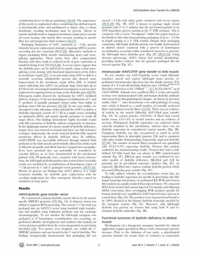
rAAV5.dysferlin gene transfer vectorWe constructed a human dysferlin cassette driven by the muscle
specific MHCK7 promoter [33] (Fig. 1A). A chimeric intron was
added to augment RNA processing. The cassette (7.7 kb total) was
packaged into an AAV2/5 vector using standard triple transfec-
tion and purified using iodixanol gradients and ion exchange
chromatography. To test whether the full-length transgene was
packaged or if homologous recombination was occurring, we
performed alkaline electrophoresis and southern blot analysis on
vector genomes purified from the vector preparation as previously
described [32]. Two probes were designed, one within the 59
MHCK7 promoter and one located in the 39 end of dysferlin. The
findings unequivocally demonstrated that packaging did not
exceed ,5.2 kb with either probe consistent with recent reports
[29,31,32] (Fig. 1B). AAV is known to package single strand
genomes with 39 to 59 polarity into the pre-formed particle in an
ATP dependent process starting at the 39 ITR terminus. This is
consistent with a vector ‘‘breakpoint’’ within this region based on
the Southern blot data demonstrating packaging of up to ,5.2 kb
in length starting at a 39 ITR termini. Despite these packaging
constraints, full-length dysferlin protein was readily demonstrated
in skeletal muscle consistent with a process of homologous
recombination occurring within transduced myocytes to generate
the full-length intact dysferlin gene (Fig. 2C) [29,31,32]. Using
electron microscopy, AAV5 virions had normal morphology,
providing further evidence that the genomes packaged did not
exceed capacity (Fig. 1C).
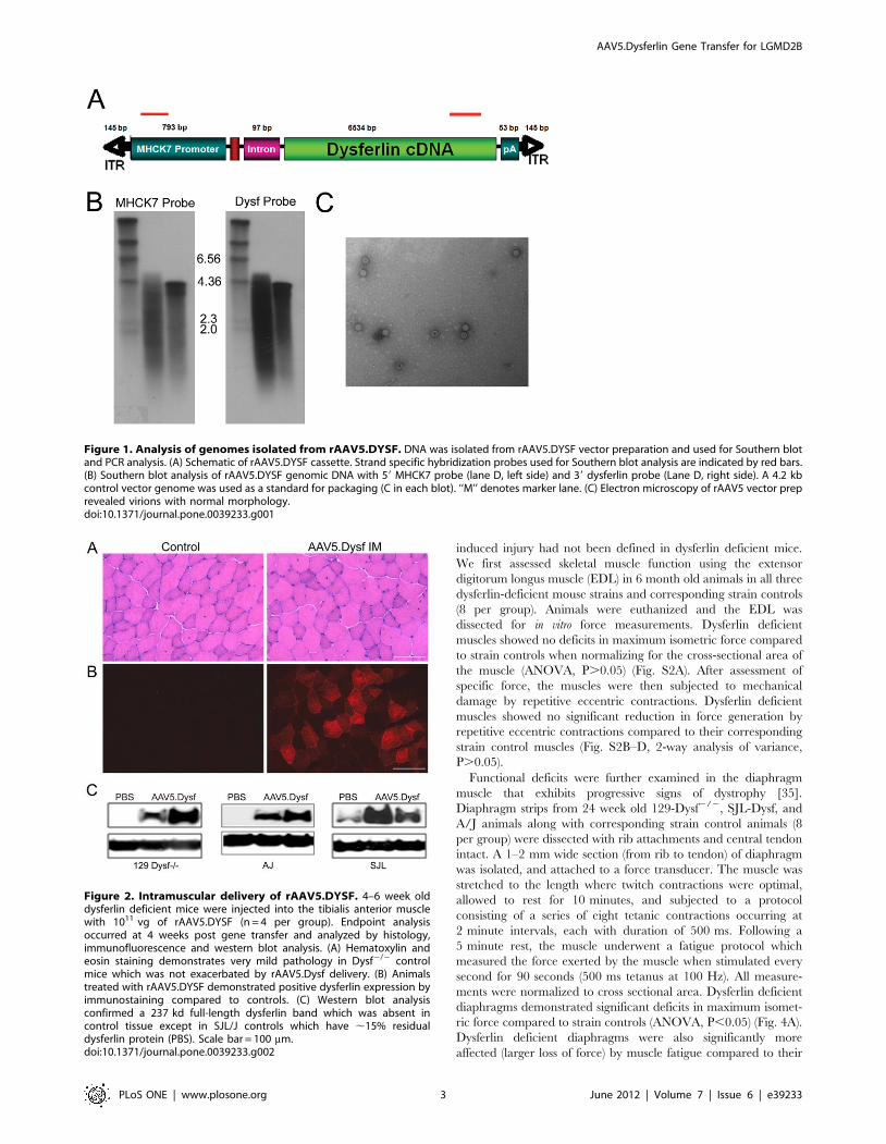
Intramuscular rAAV5.DYSF gene transfer to limb muscleTo test whether our AAV5.dysferlin vector could efficiently
transduce muscle and express full-length intact protein, we
performed intramuscular injections into the tibialis anterior (TA)
muscle of 4–6 week old dysferlin deficient mice 129-Dysftm1Kcam/J
(hereafter referred to as the 129Dysf2/2) [17,34,35] with1011 vg of
rAAV5.DYSF. Animals were sacrificed after 4 weeks and muscle
sections were immunostained with anti-human dysferlin antibody
and assessed for histopathological changes. As shown by previous
studies, Dysf2/2 mice demonstrate very mild pathology in young
mice which is limited to a small number of centrally nucleated
fibers and isolated necrotic fibers [15,35]. There was no evidence
of toxicity in the muscle following AAV5.Dysf gene transfer
(Fig. 2A). In control muscles, 3.860.8% of fibers had central
nuclei versus 2.961.6% in treated muscles and no evidence of
necrosis. Widespread dysferlin expression was achieved which
correctly localized to the sarcolemma, whereas there was no
dysferlin expression in contralateral control muscles (Fig. 2B).
Cytoplasm dysferlin was also encountered as noted in newly
regenerating fibers in dystrophic patients [36] and is consistent
with other studies demonstrating exogenously expressed dysferlin
[27,28]. The number of muscle fibers transduced was quantified
with 67.3614.4% expressing dysferlin. Western blot analysis
confirmed the immunostaining results. Treated mice had a well-
defined 237 kDa band that was absent in PBS control-treated
animals (Fig. 2C). Efficient gene transfer was confirmed in two
other models of dysferlin deficiency, SJL-Dysf and A/J for
potential use in pre-clinical outcomes analyses (Fig. 2C). As
expected, SJL-Dysf mice express residual dysferlin protein which
increased following gene transfer (Fig. 2C).
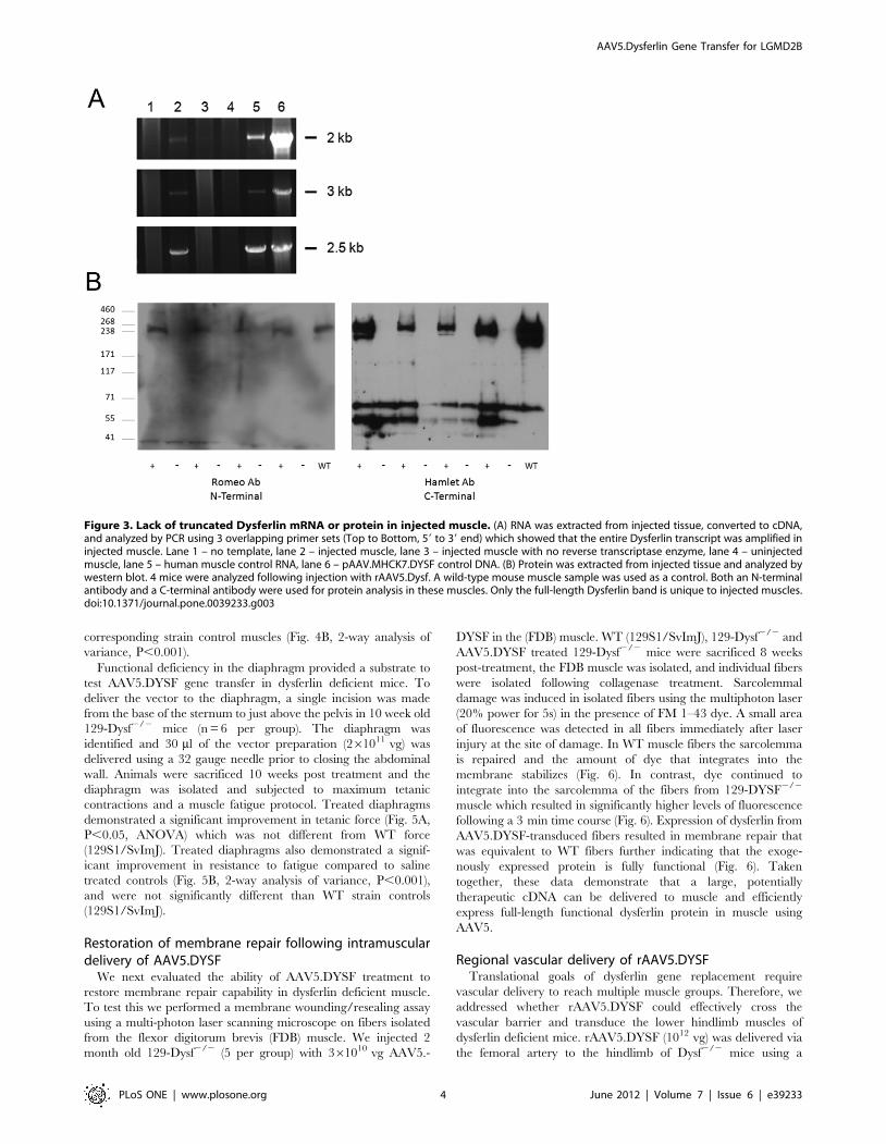
To fully address whether the recombination events that are
leading to dysferlin expression are specific in generating only full-
length transcript and protein, we performed RT-PCR and western
blot analysis on samples isolated from injected tissue. We extracted
RNA from treated and control injected TA muscles and following
cDNA conversion, three overlapping PCR products specific for
human dysferlin were amplified in AAV5 injected tissue and not in
control tissue (Fig. 3A). The products were sequenced and found to
be 100% identical to the human dysferlin transcript encoded by
the transgene cassette (Fig. S1). Moreover, only full-length
dysferlin was present on western blot using both N and C-
terminal dysferlin antibodies (Fig. 3B).
Functional outcomes of dysferlin deficiency in skeletalmuscle
Development of a therapeutic transgene intended for clinical
application requires preclinical efficacy with a functional outcome
measure. Prior to the initiation of our study, a physiological
outcome measure for tetanic force or resistance to contraction
AAV5.Dysferlin Gene Transfer for LGMD2B
PLoS ONE | www.plosone.org 2 June 2012 | Volume 7 | Issue 6 | e39233
induced injury had not been defined in dysferlin deficient mice.
We first assessed skeletal muscle function using the extensor
digitorum longus muscle (EDL) in 6 month old animals in all three
dysferlin-deficient mouse strains and corresponding strain controls
(8 per group). Animals were euthanized and the EDL was
dissected for in vitro force measurements. Dysferlin deficient
muscles showed no deficits in maximum isometric force compared
to strain controls when normalizing for the cross-sectional area of
the muscle (ANOVA, P.0.05) (Fig. S2A). After assessment of
specific force, the muscles were then subjected to mechanical
damage by repetitive eccentric contractions. Dysferlin deficient
muscles showed no significant reduction in force generation by
repetitive eccentric contractions compared to their corresponding
strain control muscles (Fig. S2B–D, 2-way analysis of variance,
P.0.05).
Functional deficits were further examined in the diaphragm
muscle that exhibits progressive signs of dystrophy [35].
Diaphragm strips from 24 week old 129-Dysf2/2, SJL-Dysf, and
A/J animals along with corresponding strain control animals (8
per group) were dissected with rib attachments and central tendon
intact. A 1–2 mm wide section (from rib to tendon) of diaphragm
was isolated, and attached to a force transducer. The muscle was
stretched to the length where twitch contractions were optimal,
allowed to rest for 10 minutes, and subjected to a protocol
consisting of a series of eight tetanic contractions occurring at
2 minute intervals, each with duration of 500 ms. Following a
5 minute rest, the muscle underwent a fatigue protocol which
measured the force exerted by the muscle when stimulated every
second for 90 seconds (500 ms tetanus at 100 Hz). All measure-
ments were normalized to cross sectional area. Dysferlin deficient
diaphragms demonstrated significant deficits in maximum isomet-
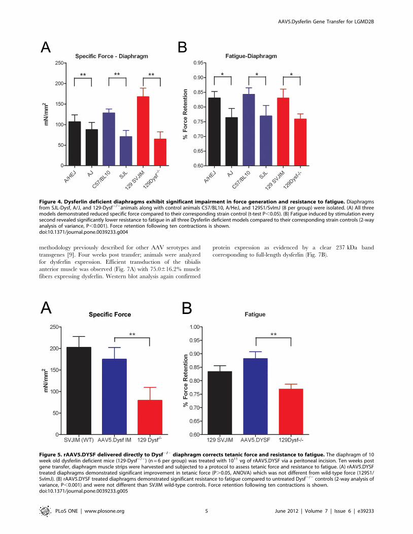
ric force compared to strain controls (ANOVA, P,0.05) (Fig. 4A).
Dysferlin deficient diaphragms were also significantly more
affected (larger loss of force) by muscle fatigue compared to their
Figure 1. Analysis of genomes isolated from rAAV5.DYSF. DNA was isolated from rAAV5.DYSF vector preparation and used for Southern blotand PCR analysis. (A) Schematic of rAAV5.DYSF cassette. Strand specific hybridization probes used for Southern blot analysis are indicated by red bars.(B) Southern blot analysis of rAAV5.DYSF genomic DNA with 59 MHCK7 probe (lane D, left side) and 39 dysferlin probe (Lane D, right side). A 4.2 kbcontrol vector genome was used as a standard for packaging (C in each blot). ‘‘M’’ denotes marker lane. (C) Electron microscopy of rAAV5 vector preprevealed virions with normal morphology.doi:10.1371/journal.pone.0039233.g001
Figure 2. Intramuscular delivery of rAAV5.DYSF. 4–6 week olddysferlin deficient mice were injected into the tibialis anterior musclewith 1011 vg of rAAV5.DYSF (n = 4 per group). Endpoint analysisoccurred at 4 weeks post gene transfer and analyzed by histology,immunofluorescence and western blot analysis. (A) Hematoxylin andeosin staining demonstrates very mild pathology in Dysf2/2 controlmice which was not exacerbated by rAAV5.Dysf delivery. (B) Animalstreated with rAAV5.DYSF demonstrated positive dysferlin expression byimmunostaining compared to controls. (C) Western blot analysisconfirmed a 237 kd full-length dysferlin band which was absent incontrol tissue except in SJL/J controls which have ,15% residualdysferlin protein (PBS). Scale bar = 100 mm.doi:10.1371/journal.pone.0039233.g002
AAV5.Dysferlin Gene Transfer for LGMD2B
PLoS ONE | www.plosone.org 3 June 2012 | Volume 7 | Issue 6 | e39233
corresponding strain control muscles (Fig. 4B, 2-way analysis of
variance, P,0.001).
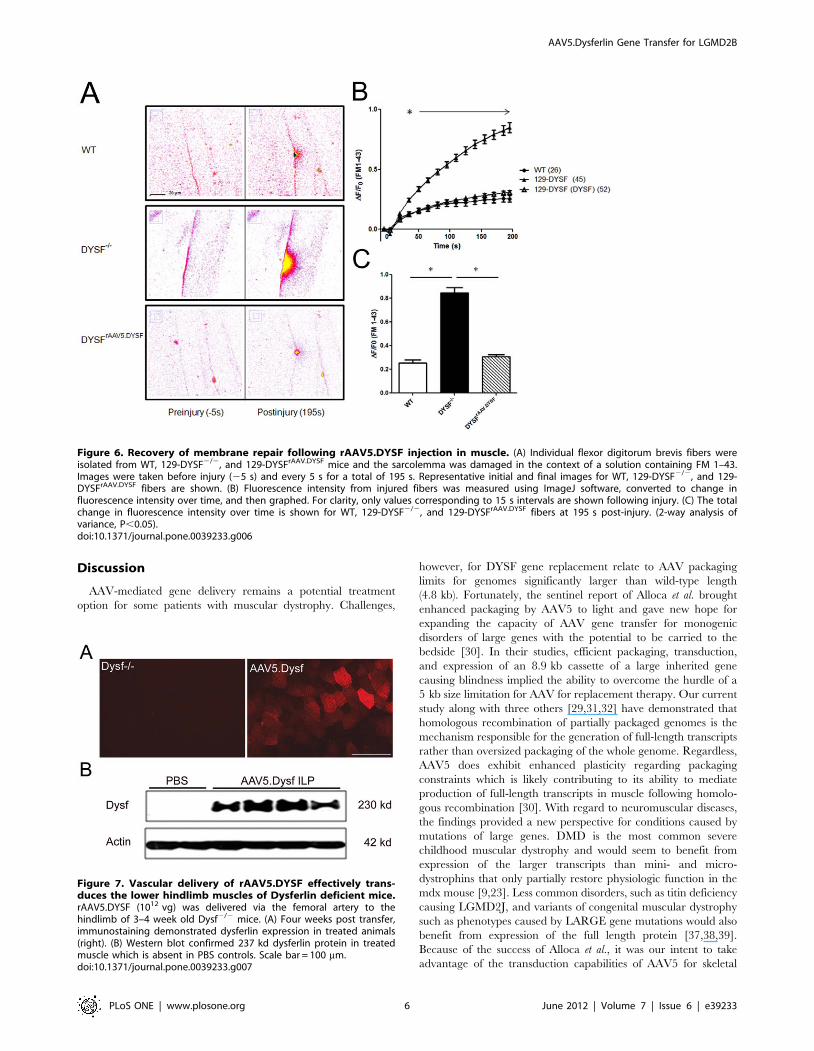
Functional deficiency in the diaphragm provided a substrate to
test AAV5.DYSF gene transfer in dysferlin deficient mice. To
deliver the vector to the diaphragm, a single incision was made
from the base of the sternum to just above the pelvis in 10 week old
129-Dysf2/2 mice (n = 6 per group). The diaphragm was
identified and 30 ml of the vector preparation (261011 vg) was
delivered using a 32 gauge needle prior to closing the abdominal
wall. Animals were sacrificed 10 weeks post treatment and the
diaphragm was isolated and subjected to maximum tetanic
contractions and a muscle fatigue protocol. Treated diaphragms
demonstrated a significant improvement in tetanic force (Fig. 5A,
P,0.05, ANOVA) which was not different from WT force
(129S1/SvImJ). Treated diaphragms also demonstrated a signif-
icant improvement in resistance to fatigue compared to saline
treated controls (Fig. 5B, 2-way analysis of variance, P,0.001),
and were not significantly different than WT strain controls
(129S1/SvImJ).
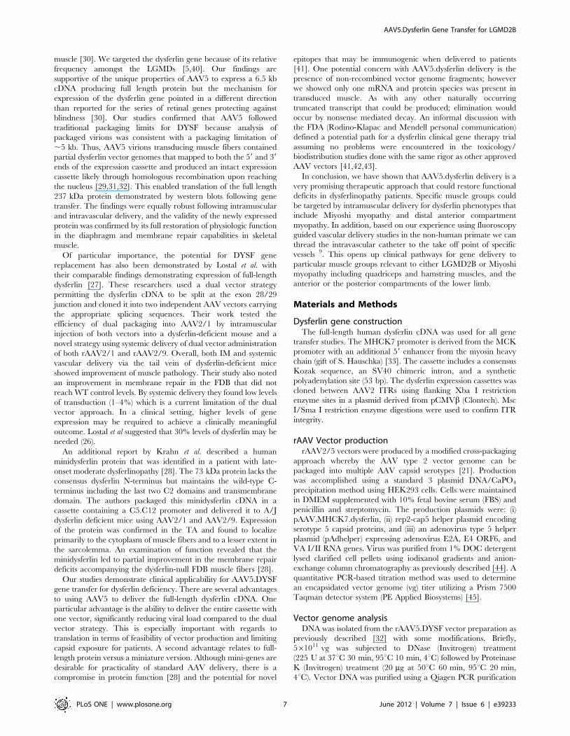
Restoration of membrane repair following intramusculardelivery of AAV5.DYSF
We next evaluated the ability of AAV5.DYSF treatment to
restore membrane repair capability in dysferlin deficient muscle.
To test this we performed a membrane wounding/resealing assay
using a multi-photon laser scanning microscope on fibers isolated
from the flexor digitorum brevis (FDB) muscle. We injected 2
month old 129-Dysf2/2 (5 per group) with 361010 vg AAV5.-
DYSF in the (FDB) muscle. WT (129S1/SvImJ), 129-Dysf2/2 and
AAV5.DYSF treated 129-Dysf2/2 mice were sacrificed 8 weeks
post-treatment, the FDB muscle was isolated, and individual fibers
were isolated following collagenase treatment. Sarcolemmal
damage was induced in isolated fibers using the multiphoton laser
(20% power for 5s) in the presence of FM 1–43 dye. A small area
of fluorescence was detected in all fibers immediately after laser
injury at the site of damage. In WT muscle fibers the sarcolemma
is repaired and the amount of dye that integrates into the
membrane stabilizes (Fig. 6). In contrast, dye continued to
integrate into the sarcolemma of the fibers from 129-DYSF2/2
muscle which resulted in significantly higher levels of fluorescence
following a 3 min time course (Fig. 6). Expression of dysferlin from
AAV5.DYSF-transduced fibers resulted in membrane repair that
was equivalent to WT fibers further indicating that the exoge-
nously expressed protein is fully functional (Fig. 6). Taken
together, these data demonstrate that a large, potentially
therapeutic cDNA can be delivered to muscle and efficiently
express full-length functional dysferlin protein in muscle using
AAV5.
Regional vascular delivery of rAAV5.DYSFTranslational goals of dysferlin gene replacement require
vascular delivery to reach multiple muscle groups. Therefore, we
addressed whether rAAV5.DYSF could effectively cross the
vascular barrier and transduce the lower hindlimb muscles of
dysferlin deficient mice. rAAV5.DYSF (1012 vg) was delivered via
the femoral artery to the hindlimb of Dysf2/2 mice using a
Figure 3. Lack of truncated Dysferlin mRNA or protein in injected muscle. (A) RNA was extracted from injected tissue, converted to cDNA,and analyzed by PCR using 3 overlapping primer sets (Top to Bottom, 59 to 39 end) which showed that the entire Dysferlin transcript was amplified ininjected muscle. Lane 1 – no template, lane 2 – injected muscle, lane 3 – injected muscle with no reverse transcriptase enzyme, lane 4 – uninjectedmuscle, lane 5 – human muscle control RNA, lane 6 – pAAV.MHCK7.DYSF control DNA. (B) Protein was extracted from injected tissue and analyzed bywestern blot. 4 mice were analyzed following injection with rAAV5.Dysf. A wild-type mouse muscle sample was used as a control. Both an N-terminalantibody and a C-terminal antibody were used for protein analysis in these muscles. Only the full-length Dysferlin band is unique to injected muscles.doi:10.1371/journal.pone.0039233.g003
AAV5.Dysferlin Gene Transfer for LGMD2B
PLoS ONE | www.plosone.org 4 June 2012 | Volume 7 | Issue 6 | e39233
methodology previously described for other AAV serotypes and
transgenes [9]. Four weeks post transfer; animals were analyzed
for dysferlin expression. Efficient transduction of the tibialis
anterior muscle was observed (Fig. 7A) with 75.0616.2% muscle
fibers expressing dysferlin. Western blot analysis again confirmed
protein expression as evidenced by a clear 237 kDa band
corresponding to full-length dysferlin (Fig. 7B).
Figure 4. Dysferlin deficient diaphragms exhibit significant impairment in force generation and resistance to fatigue. Diaphragmsfrom SJL-Dysf, A/J, and 129-Dysf2/2animals along with control animals C57/BL10, A/HeJ, and 129S1/SvImJ (8 per group) were isolated. (A) All threemodels demonstrated reduced specific force compared to their corresponding strain control (t-test P,0.05). (B) Fatigue induced by stimulation everysecond revealed significantly lower resistance to fatigue in all three Dysferlin deficient models compared to their corresponding strain controls (2-wayanalysis of variance, P,0.001). Force retention following ten contractions is shown.doi:10.1371/journal.pone.0039233.g004
Figure 5. rAAV5.DYSF delivered directly to Dysf2/2 diaphragm corrects tetanic force and resistance to fatigue. The diaphragm of 10week old dysferlin deficient mice (129-Dysf2/2) (n = 6 per group) was treated with 1011 vg of rAAV5.DYSF via a peritoneal incision. Ten weeks postgene transfer, diaphragm muscle strips were harvested and subjected to a protocol to assess tetanic force and resistance to fatigue. (A) rAAV5.DYSFtreated diaphragms demonstrated significant improvement in tetanic force (P.0.05, ANOVA) which was not different from wild-type force (129S1/SvImJ). (B) rAAV5.DYSF treated diaphragms demonstrated significant resistance to fatigue compared to untreated Dysf2/2 controls (2-way analysis ofvariance, P,0.001) and were not different than SVJIM wild-type controls. Force retention following ten contractions is shown.doi:10.1371/journal.pone.0039233.g005
AAV5.Dysferlin Gene Transfer for LGMD2B
PLoS ONE | www.plosone.org 5 June 2012 | Volume 7 | Issue 6 | e39233
Discussion
AAV-mediated gene delivery remains a potential treatment
option for some patients with muscular dystrophy. Challenges,
however, for DYSF gene replacement relate to AAV packaging
limits for genomes significantly larger than wild-type length
(4.8 kb). Fortunately, the sentinel report of Alloca et al. brought
enhanced packaging by AAV5 to light and gave new hope for
expanding the capacity of AAV gene transfer for monogenic
disorders of large genes with the potential to be carried to the
bedside [30]. In their studies, efficient packaging, transduction,
and expression of an 8.9 kb cassette of a large inherited gene
causing blindness implied the ability to overcome the hurdle of a
5 kb size limitation for AAV for replacement therapy. Our current
study along with three others [29,31,32] have demonstrated that
homologous recombination of partially packaged genomes is the
mechanism responsible for the generation of full-length transcripts
rather than oversized packaging of the whole genome. Regardless,
AAV5 does exhibit enhanced plasticity regarding packaging
constraints which is likely contributing to its ability to mediate
production of full-length transcripts in muscle following homolo-
gous recombination [30]. With regard to neuromuscular diseases,
the findings provided a new perspective for conditions caused by
mutations of large genes. DMD is the most common severe
childhood muscular dystrophy and would seem to benefit from
expression of the larger transcripts than mini- and micro-
dystrophins that only partially restore physiologic function in the
mdx mouse [9,23]. Less common disorders, such as titin deficiency
causing LGMD2J, and variants of congenital muscular dystrophy
such as phenotypes caused by LARGE gene mutations would also
benefit from expression of the full length protein [37,38,39].
Because of the success of Alloca et al., it was our intent to take
advantage of the transduction capabilities of AAV5 for skeletal
Figure 6. Recovery of membrane repair following rAAV5.DYSF injection in muscle. (A) Individual flexor digitorum brevis fibers wereisolated from WT, 129-DYSF2/2, and 129-DYSFrAAV.DYSF mice and the sarcolemma was damaged in the context of a solution containing FM 1–43.Images were taken before injury (25 s) and every 5 s for a total of 195 s. Representative initial and final images for WT, 129-DYSF2/2, and 129-DYSFrAAV.DYSF fibers are shown. (B) Fluorescence intensity from injured fibers was measured using ImageJ software, converted to change influorescence intensity over time, and then graphed. For clarity, only values corresponding to 15 s intervals are shown following injury. (C) The totalchange in fluorescence intensity over time is shown for WT, 129-DYSF2/2, and 129-DYSFrAAV.DYSF fibers at 195 s post-injury. (2-way analysis ofvariance, P,0.05).doi:10.1371/journal.pone.0039233.g006
Figure 7. Vascular delivery of rAAV5.DYSF effectively trans-duces the lower hindlimb muscles of Dysferlin deficient mice.rAAV5.DYSF (1012 vg) was delivered via the femoral artery to thehindlimb of 3–4 week old Dysf2/2 mice. (A) Four weeks post transfer,immunostaining demonstrated dysferlin expression in treated animals(right). (B) Western blot confirmed 237 kd dysferlin protein in treatedmuscle which is absent in PBS controls. Scale bar = 100 mm.doi:10.1371/journal.pone.0039233.g007
AAV5.Dysferlin Gene Transfer for LGMD2B
PLoS ONE | www.plosone.org 6 June 2012 | Volume 7 | Issue 6 | e39233
muscle [30]. We targeted the dysferlin gene because of its relative
frequency amongst the LGMDs [5,40]. Our findings are
supportive of the unique properties of AAV5 to express a 6.5 kb
cDNA producing full length protein but the mechanism for
expression of the dysferlin gene pointed in a different direction
than reported for the series of retinal genes protecting against
blindness [30]. Our studies confirmed that AAV5 followed
traditional packaging limits for DYSF because analysis of
packaged virions was consistent with a packaging limitation of
,5 kb. Thus, AAV5 virions transducing muscle fibers contained
partial dysferlin vector genomes that mapped to both the 59 and 39
ends of the expression cassette and produced an intact expression
cassette likely through homologous recombination upon reaching
the nucleus [29,31,32]. This enabled translation of the full length
237 kDa protein demonstrated by western blots following gene
transfer. The findings were equally robust following intramuscular
and intravascular delivery, and the validity of the newly expressed
protein was confirmed by its full restoration of physiologic function
in the diaphragm and membrane repair capabilities in skeletal
muscle.
Of particular importance, the potential for DYSF gene
replacement has also been demonstrated by Lostal et al. with
their comparable findings demonstrating expression of full-length
dysferlin [27]. These researchers used a dual vector strategy
permitting the dysferlin cDNA to be split at the exon 28/29
junction and cloned it into two independent AAV vectors carrying
the appropriate splicing sequences. Their work tested the
efficiency of dual packaging into AAV2/1 by intramuscular
injection of both vectors into a dysferlin-deficient mouse and a
novel strategy using systemic delivery of dual vector administration
of both rAAV2/1 and rAAV2/9. Overall, both IM and systemic
vascular delivery via the tail vein of dysferlin-deficient mice
showed improvement of muscle pathology. Their study also noted
an improvement in membrane repair in the FDB that did not
reach WT control levels. By systemic delivery they found low levels
of transduction (1–4%) which is a current limitation of the dual
vector approach. In a clinical setting, higher levels of gene
expression may be required to achieve a clinically meaningful
outcome. Lostal et al suggested that 30% levels of dysferlin may be
needed (26).
An additional report by Krahn et al. described a human
minidysferlin protein that was identified in a patient with late-
onset moderate dysferlinopathy [28]. The 73 kDa protein lacks the
consensus dysferlin N-terminus but maintains the wild-type C-
terminus including the last two C2 domains and transmembrane
domain. The authors packaged this minidysferlin cDNA in a
cassette containing a C5.C12 promoter and delivered it to A/J
dysferlin deficient mice using AAV2/1 and AAV2/9. Expression
of the protein was confirmed in the TA and found to localize
primarily to the cytoplasm of muscle fibers and to a lesser extent in
the sarcolemma. An examination of function revealed that the
minidysferlin led to partial improvement in the membrane repair
deficits accompanying the dysferlin-null FDB muscle fibers [28].
Our studies demonstrate clinical applicability for AAV5.DYSF
gene transfer for dysferlin deficiency. There are several advantages
to using AAV5 to deliver the full-length dysferlin cDNA. One
particular advantage is the ability to deliver the entire cassette with
one vector, significantly reducing viral load compared to the dual
vector strategy. This is especially important with regards to
translation in terms of feasibility of vector production and limiting
capsid exposure for patients. A second advantage relates to full-
length protein versus a miniature version. Although mini-genes are
desirable for practicality of standard AAV delivery, there is a
compromise in protein function [28] and the potential for novel
epitopes that may be immunogenic when delivered to patients
[41]. One potential concern with AAV5.dysferlin delivery is the
presence of non-recombined vector genome fragments; however
we showed only one mRNA and protein species was present in
transduced muscle. As with any other naturally occurring
truncated transcript that could be produced; elimination would
occur by nonsense mediated decay. An informal discussion with
the FDA (Rodino-Klapac and Mendell personal communication)
defined a potential path for a dysferlin clinical gene therapy trial
assuming no problems were encountered in the toxicology/
biodistribution studies done with the same rigor as other approved
AAV vectors [41,42,43].
In conclusion, we have shown that AAV5.dysferlin delivery is a
very promising therapeutic approach that could restore functional
deficits in dysferlinopathy patients. Specific muscle groups could
be targeted by intramuscular delivery for dysferlin phenotypes that
include Miyoshi myopathy and distal anterior compartment
myopathy. In addition, based on our experience using fluoroscopy
guided vascular delivery studies in the non-human primate we can
thread the intravascular catheter to the take off point of specific
vessels 9. This opens up clinical pathways for gene delivery to
particular muscle groups relevant to either LGMD2B or Miyoshi
myopathy including quadriceps and hamstring muscles, and the
anterior or the posterior compartments of the lower limb.
Materials and Methods
Dysferlin gene constructionThe full-length human dysferlin cDNA was used for all gene
transfer studies. The MHCK7 promoter is derived from the MCK
promoter with an additional 59 enhancer from the myosin heavy
chain (gift of S. Hauschka) [33]. The cassette includes a consensus
Kozak sequence, an SV40 chimeric intron, and a synthetic
polyadenylation site (53 bp). The dysferlin expression cassettes was
cloned between AAV2 ITRs using flanking Xba I restriction
enzyme sites in a plasmid derived from pCMVb (Clontech). Msc
I/Sma I restriction enzyme digestions were used to confirm ITR
integrity.
rAAV Vector productionrAAV2/5 vectors were produced by a modified cross-packaging
approach whereby the AAV type 2 vector genome can be
packaged into multiple AAV capsid serotypes [21]. Production
was accomplished using a standard 3 plasmid DNA/CaPO4
precipitation method using HEK293 cells. Cells were maintained
in DMEM supplemented with 10% fetal bovine serum (FBS) and
penicillin and streptomycin. The production plasmids were: (i)
pAAV.MHCK7.dysferlin, (ii) rep2-cap5 helper plasmid encoding
serotype 5 capsid proteins, and (iii) an adenovirus type 5 helper
plasmid (pAdhelper) expressing adenovirus E2A, E4 ORF6, and
VA I/II RNA genes. Virus was purified from 1% DOC detergent
lysed clarified cell pellets using iodixanol gradients and anion-
exchange column chromatography as previously described [44]. A
quantitative PCR-based titration method was used to determine
an encapsidated vector genome (vg) titer utilizing a Prism 7500
Taqman detector system (PE Applied Biosystems) [45].
Vector genome analysisDNA was isolated from the rAAV5.DYSF vector preparation as
previously described [32] with some modifications. Briefly,
561011 vg was subjected to DNase (Invitrogen) treatment
(225 U at 37uC 30 min, 95uC 10 min, 4uC) followed by Proteinase
K (Invitrogen) treatment (20 mg at 50uC 60 min, 95uC 20 min,
4uC). Vector DNA was purified using a Qiagen PCR purification
AAV5.Dysferlin Gene Transfer for LGMD2B
PLoS ONE | www.plosone.org 7 June 2012 | Volume 7 | Issue 6 | e39233
kit according to the manufacturer’s instructions (Qiagen Inc, Cat
No. 28104). Southern blot analysis was performed as previously
described [32]. Fifteen ng of probe (MHCK7 and Dysferlin) was
used for each Southern blot hybridization.
RNA analysisRNA was isolated from TA muscles using an RNeasyH Mini Kit
(QIAGEN). cDNA was synthesized from 100 ng RNA using the
High Capacity cDNA Reverse Transcription kit (Applied
BiosystemsTM) and PCR was performed using PFU Ultra II
(Strategene) with the following primers: Dysf 1F (59ATGCT-
GAGGGTCTTCATCCTCTA39) – Dysf 1976R (59ACCA-
CAGGTTTCACGTTACC39), Dysf 1957F (59GGTAACGT-
GAAACCTGTGGT39) – Dysf 5005R
(59TGTTCTCCAGGTCGACGAC39), Dysf 3820F
(59TTGAGCTCATCCAGAGAGAGAAGC39) – Dysf 6243R
(59TCAGCTGAAGGGCTTCACCAG39). Human skeletal mus-
cle total RNA (Biochain) and the pAAV.MHCK7.Dysf plasmid
(50 ng) were used as positive controls. PCR products were gel
purified using the Qiaquick Gel Extraction Kit (QIAGEN) and
sequenced using the following primers: Dysf 1F, Dysf 499R (59
CAGTGAGTCCCTGGTCCTCT39), Dysf 501F (59 AGAT-
GAGGCGGAGCCATTCC39), Dysf 900F (59GTTCCGGATG-
GACGTGGGCA39), Dysf 1976R (59ACCACAGGTTT-
CACGTTACC39), Dysf 1957F, Dysf 2400R
(59TCCCTGCAGCATCCAGATGA39), Dysf 3339R
(59CCTCAAGGGCAAACACAGCTGC39), Dysf 3377F
(59TTCCATGTCCGTCTCCACCTTG39), Dysf 3820F, Dysf
4750F (59 ATTGTCCGAGCATTTGGCCT39), Dysf 5005R
(59TGTTCTCCAGGTCGACGAC39), Dysf 5750F (59
CCTTTGATGATTTTCTGGGC39), and Dysf 6243R. The
sequenced cDNA from injected tissue was then compared to the
reference Dysferlin sequence (Genbank# NM_003494.3) using
the ClustalW online alignment tool.
Dysferlin deficient mouse strainsStocks of A/J, A/HeJ, SJL/J, C57BL/10, 129S1/SvImJ, and
129-Dysf2/2 mice were bred and maintained as homozygous
animals in standardized conditions in the Vivarium at the
Research Institute at Nationwide Children’s Hospital. They were
maintained on Teklad Global Rodent Diet (3.8% Fiber, 18.8%
Protein, 5% fat chow) with a 12:12 h dark:light cycle. Procedures
used in the experiments were approved by the Institutional Animal
Care and Use Committee at Nationwide Childrens Hospital
(AR08-00017).
rAAV5.DYSF intramuscular gene transferThe anterior compartment (containing tibialis anterior muscle
and EDL) of 4–6 week old dysferlin deficient mice was injected
with 1011 vg of rAAV5.DYSF diluted in normal saline buffer
(50 ml volume). Control mice were sham injected with normal
saline. The TA and EDL muscles from both limbs were harvested
at 4 weeks post administration to assess efficiency of gene transfer.
For histological analysis, muscles were embedded in 7% gum
tragacanth and flash frozen in isopentane cooled in liquid
nitrogen. Cryostat sections (12 mm) were collected for immuno-
histochemistry.
Intramuscular injections into the diaphragmTen week old dysferlin deficient mice (n = 6 per group) were
treated with 261011 vg of rAAV5.DYSF via a peritoneal incision.
Anesthetized mice (Ketamine/Xylazine 100 mg/kg and 10 mg/
kg, respectively) were secured to a warm surgical table and the
abdomen prepped and draped in a sterile fashion. A single incision
was made from the base of the sternum to just above the pelvis
(approximately 1 cm incision). The diaphragm was identified and
30 ml of the vector preparation in sterile saline was delivered using
a 32 gauge needle. The abdominal wall was closed with 4.0 Vicryl
Plus continuous sutures and skin wound closed with sterile surgical
staples. Mice were treated with a post-op dose of Buprenorphine
0.01 mg/kg subcutaneously for pain. Ten weeks post gene
transfer; diaphragm muscle strips were harvested and subjected
to a protocol to assess resistance to fatigue.
Membrane Repair AssayThe ability of the sarcolemma to repair following injury was
assessed on at least 5 129-SVLMJ, 129-Dysf2/2, and 129-
DysfrAAV5.DYSF mice at 4.5 months of age. Individual fibers were
isolated from the flexor digitorum brevis muscle following
treatment with a 2% collagenase solution. The fibers were washed
in PBS and placed in glass bottom dishes in the presence of
2.5 mM FMH 1–43 (InvitrogenH) with or without 1.5 mM Ca2+.
Membrane damage was induced with a FluoViewH FV1000 two-
photon confocal laser-scanning microscope (Olympus). A circular
area (diameter, 5 mm) on the edge of the sarcolemma was
irradiated at 20% power for 5 s. Images were captured 5 s prior to
injury and every 5 s after injury until 3 min post irradiation. For
every image, the fluorescence intensity surrounding the site of
damage was analyzed (ImageJ). At least 25 fibers were evaluated
for each condition (2-way analysis of variance, P,0.05).
Vascular deliveryEight adult dysferlin deficient mice (4–6 weeks of age) were
perfused with 1012 vg rAAV5.DYSF in 100 mL of normal saline as
described [9]. Briefly, mice were anesthetized and the femoral
bundle was visualized via a small incision proximal to the mid-
thigh. Blood flow through the femoral artery was controlled by
catheter placement using a customized heat pulled polypropylene
10 (PE-10) catheter placed into the femoral artery. Prior to vector
administration, the arterial catheter was flushed (pre-flush) with
100 ml sterile normal saline. Immediately prior to vector admin-
istration all blood flow to the extremity was impeded (isolated limb
perfusion – ILP) by tightening the ligature at the mid-thigh.
rAAV5.DYSF was perfused through the femoral artery in 100 ml
of sterile Tris buffered saline administered at a rate of
approximately 2 ml per second (over 60–80 seconds). After
10 minutes of maintained vascular occlusion, 100 ml of normal
saline was administered to the arterial catheter (again at about 2 ml
per second) as a post-flush and the tourniquet was then released.
The wound was flushed with sterile normal saline and closed with
a 6-0 proline suture.
ImmunohistochemistryImmunostaining for dysferlin was performed on all transduced
tissue to assess efficacy of gene transfer. Tissue sections were
incubated with dysferlin rabbit anti-human monoclonal (Lifespan
Biosciences, Inc., Cat #LS-C138735) antibody to detect dysferlin
at a dilution of 1:100 in block (20% goat serum, 0.1% triton X-100
in PBS) applied for 1 hour at 25uC, in a wet chamber. Sections
were washed with PBS 3620 minutes, reblocked, and then
incubated for 30 minutes at 25uC with an Alexa 568 goat anti
rabbit antibody at a dilution of 1:300 (Molecular Probes). Sections
were washed in PBS for 3620 minutes and mounted with
Vectashield mounting medium (Vector Laboratories). Images
were captured with a Zeiss Axioskop2 Plus Microscope and
AxioCam MRC5 camera. Four random 206 images (each field
with an average of 150 muscle fibers) were captured per muscle
AAV5.Dysferlin Gene Transfer for LGMD2B
PLoS ONE | www.plosone.org 8 June 2012 | Volume 7 | Issue 6 | e39233
and the number of fibers with dysferlin staining was counted and
expressed as percent of total number of fibers.
MorphometricsCentralized nuclei counts were performed on sections of TA
muscles stained with hematoxylin and eosin (H&E) from
AAV5.DYSF treated Dysf2/2 animals. TAs from the contralateral
extremity served as controls. Four random 206 fields of 12 mm
sections for each muscle were captured and the number of fibers
with central nuclei counted and expressed as a percentage of the
total number of fibers.
Western Blot AnalysisThe TA was isolated from both the treated and contralateral
limbs. From each muscle, fresh frozen serial tissue sections were
taken for both immunohistochemistry and for western blot
analysis. Muscle samples harvested from rAAV5.DYSF and
control groups were compared with wild-type mice for levels of
vector-mediated dysferlin compared to endogenous dysferlin.
Protein (25 mg) extracted from treated and control samples was
separated by SDS-PAGE (3–8% Novex NuPAGE gradient gels,
Invitrogen), blotted on PVDF membrane and probed with
dysferlin rabbit anti-human monoclonal (Lifespan Biosciences,
Inc.) primary antibody at a dilution of 1:1000, NCL-Hamlet
(Novocastra) primary antibody at a dilution of 1:1,000 or actin
antibody (NCL-MSA) at a dilution of 1:6,000 followed by
horseradish-peroxidase labeled goat anti-mouse IgG or hoserad-
ish-peroxidase labeled goat anti-rabbit IgG (1:5,000, Millipore).
Immunoreactive bands were detected with ECL Plus detection
system (GE Healthcare) and signal captured on Hyperfilm ECL
(Amersham).
Force generation and protection from eccentriccontractions in EDL
SJL-Dysf, A/J, and 129-Dysf2/2 animals along with control
animals C57/BL10, A/HeJ, and 129S1/SvImJ (8 per group) were
assessed for physiological deficits in the extensor digitorum longus
(EDL) muscle at 24 weeks (when histopathological features are
present) as previously described [9,46]. Mice were euthanized and
the EDL was removed, the tendons sutured, and bathed in
oxygenated circulating Krebs-Henseleit solution at 30uC in an
organ bath. For the procedure, one end of the muscle was tied to a
force transducer and the other to a high-speed linear servo-
controlled motor. The muscle was mounted in the set-up at slack
length with a resting tension of 1 g for 10 minutes without
electrical stimulation. Stimulation was delivered via two parallel
platinum-iridium electrodes on either side of the muscle. Muscles
were adjusted to optimum length (L0), defined as the length for
maximal twitch and subjected to an isometric tetanus of 150 Hz.
Following a 5 minute rest period, muscles were subjected to an
eccentric contraction protocol consisting of a series of 10 isometric
700 ms tetani, at 2 minute intervals, with a 5% lengthening of the
muscles (0.5 fiber length per second for duration of 200 ms) when
maximal force had developed at 500 ms. After the tetanus ended
(at t = 700 ms), the muscle was brought back to initial length (at
the same speed as the stretch), allowing for full relaxation to the
initial length. For comparative purposes, all force measurements
are expressed per unit cross-sectional area (normalized isometric
force or tension: mN/mm2). Cross-sectional area (CSA) is
calculated using the following equation, CSA = (muscle mass in
g)/[(optimal fiber length in cm)6(muscle density in g/cm3)], where
muscle density is 1.06 g/cm3.
Diaphragm Tetanic Contraction and Muscle FatigueMethods
As a second approach, the diaphragm will be tested as a target
for a therapeutic outcome measure. SJL-Dysf, A/J, and 129-
Dysf2/2 animals along with control animals C57/BL10, A/HeJ,
and 129S1/SvImJ (8 per group) were assessed at 24 weeks. Mice
were euthanized and the diaphragm was dissected with rib
attachments and central tendon intact, and placed in K-H buffer
at 37 C as previously described [47,48]. A 1–2 mm wide section
(with length from rib to tendon) of diaphragm was isolated, and
attached to a force transducer. The diaphragm strip was looped
around a basket assembly attached to the transducer (the rib
cartilage serves as the anchor), and the tendon was pierced by a
pin. The muscle was stretched to optimal length for measurement
of twitch contractions, and then allowed to rest for 10 minutes
before initiation of the tetanic protocol. The protocol consisted of
a series of eight tetanic contractions occurring at 2 minute
intervals, each with duration of 500 ms. The force was recorded
for each stimulus, and normalized to account for muscle width and
length. The muscle was rested for 5 minutes before starting the
muscle fatigue protocol. This protocol measures the force exerted
by the muscle when stimulated every second for 90 seconds
(500 ms tetanus at 100 Hz). Following the muscle fatigue protocol,
the muscle strip was removed from the apparatus, the rib cartilage
removed and weighed.
Supporting Information
Figure S1 RNA analysis from injected tissue. Sequence
analysis of cDNA from rAAV.DYSF-injected muscle. cDNA from
injected tissues was sequenced completely and then aligned to the
reference Dysferlin sequence containing UTRs. The sequenced
cDNA aligns exactly with the reference.
(PDF)
Figure S2 Functional assessment of the EDL muscle inDysf2/2 mice. SJL-Dysf, A/J, and 129-Dysf2/2 animals along
with control animals C57/BL10, A/HeJ, and 129S1/SvImJ (8 per
group) were assessed for physiological deficits in the EDL. (A)
Dysferlin deficient muscles showed no deficits in maximum
isometric force compared to strain controls when normalizing
for the cross-sectional area of the muscle (ANOVA, P.0.05). (B–
D) Muscles were subjected to mechanical damage by 10 repetitive
eccentric contractions. Dysferlin deficient muscles were not
significantly more affected (larger loss of force) by repetitive
eccentric contractions compared to their corresponding strain
control muscles (2-way analysis of variance, P.0.05)
(TIF)
Acknowledgments
We thank Nationwide Children’s Viral Vector Core for vector production
and Stephen D. Hauschka for the MHCK7 promoter. We also thank
Nancy Davis and Jianchao Zhang for technical assistance.
Author Contributions
Conceived and designed the experiments: WEG JRM LRK. Performed the
experiments: WEG KRC DG VM KMS CLM SL PMLJ LRK. Analyzed
the data: WEG JRM LRK. Contributed reagents/materials/analysis tools:
RHB. Wrote the paper: WEG JRM LRK.
AAV5.Dysferlin Gene Transfer for LGMD2B
PLoS ONE | www.plosone.org 9 June 2012 | Volume 7 | Issue 6 | e39233
References
1. Bashir R, Britton S, Strachan T, Keers S, Vafiadaki E, et al. (1998) A gene
related to Caenorhabditis elegans spermatogenesis factor fer-1 is mutated inlimb-girdle muscular dystrophy type 2B. Nat Genet 20: 37–42.
2. Liu J, Aoki M, Illa I, Wu C, Fardeau M, et al. (1998) Dysferlin, a novel skeletalmuscle gene, is mutated in Miyoshi myopathy and limb girdle muscular
dystrophy. Nat Genet 20: 31–36.
3. Illa I, Serrano-Munuera C, Gallardo E, Lasa A, Rojas-Garcia R, et al. (2001)Distal anterior compartment myopathy: a dysferlin mutation causing a new
muscular dystrophy phenotype. Ann Neurol 49: 130–134.4. Seror P, Krahn M, Laforet P, Leturcq F, Maisonobe T (2008) Complete fatty
degeneration of lumbar erector spinae muscles caused by a primary
dysferlinopathy. Muscle Nerve 37: 410–414.5. Rosales XQ, Gastier-Foster JM, Lewis S, Vinod M, Thrush DL, et al. (2010)
Novel diagnostic features of dysferlinopathies. Muscle Nerve 42: 14–21.6. Nagashima T, Chuma T, Mano Y, Goto Y, Hayashi YK, et al. (2004)
Dysferlinopathy associated with rigid spine syndrome. Neuropathology 24: 341–346.
7. Klinge L, Dean AF, Kress W, Dixon P, Charlton R, et al. (2008) Late onset in
dysferlinopathy widens the clinical spectrum. Neuromuscul Disord 18: 288–290.8. Rosales XQ, Moser SJ, Tran T, McCarthy B, Dunn N, et al. (2011)
Cardiovascular magnetic resonance of cardiomyopathy in limb girdle musculardystrophy 2B and 2I. J Cardiovasc Magn Reson 13: 39.
9. Rodino-Klapac LR, Janssen PM, Montgomery CL, Coley BD, Chicoine LG, et
al. (2007) A translational approach for limb vascular delivery of the micro-dystrophin gene without high volume or high pressure for treatment of
Duchenne muscular dystrophy. J Transl Med 5: 45.10. Rodino-Klapac LR, Montgomery CL, Bremer WG, Shontz KM, Malik V, et al.
(2010) Persistent expression of FLAG-tagged micro dystrophin in nonhumanprimates following intramuscular and vascular delivery. Mol Ther 18: 109–117.
11. Aoki M, Liu J, Richard I, Bashir R, Britton S, et al. (2001) Genomic
organization of the dysferlin gene and novel mutations in Miyoshi myopathy.Neurology 57: 271–278.
12. Takahashi T, Aoki M, Tateyama M, Kondo E, Mizuno T, et al. (2003) Dysferlinmutations in Japanese Miyoshi myopathy: relationship to phenotype. Neurology
60: 1799–1804.
13. Liu J, Wu C, Bossie K, Bejaoui K, Hosler BA, et al. (1998) Generation of a 3-MbPAC contig spanning the Miyoshi myopathy/limb-girdle muscular dystrophy
(MM/LGMD2B) locus on chromosome 2p13. Genomics 49: 23–29.14. Therrien C, Dodig D, Karpati G, Sinnreich M (2006) Mutation impact on
dysferlin inferred from database analysis and computer-based structuralpredictions. J Neurol Sci 250: 71–78.
15. Bansal D, Miyake K, Vogel SS, Groh S, Chen CC, et al. (2003) Defective
membrane repair in dysferlin-deficient muscular dystrophy. Nature 423: 168–172.
16. Lennon NJ, Kho A, Bacskai BJ, Perlmutter SL, Hyman BT, et al. (2003)Dysferlin interacts with annexins A1 and A2 and mediates sarcolemmal wound-
healing. J Biol Chem 278: 50466–50473.
17. Han R, Campbell KP (2007) Dysferlin and muscle membrane repair. Curr OpinCell Biol 19: 409–416.
18. Piccolo F, Moore SA, Ford GC, Campbell KP (2000) Intracellular accumulationand reduced sarcolemmal expression of dysferlin in limb–girdle muscular
dystrophies. Ann Neurol 48: 902–912.19. Cenacchi G, Fanin M, De Giorgi LB, Angelini C (2005) Ultrastructural changes
in dysferlinopathy support defective membrane repair mechanism. J Clin Pathol
58: 190–195.20. Dong JY, Fan PD, Frizzell RA (1996) Quantitative analysis of the packaging
capacity of recombinant adeno-associated virus. Hum Gene Ther 7: 2101–2112.21. Gao G, Vandenberghe LH, Wilson JM (2005) New recombinant serotypes of
AAV vectors. Curr Gene Ther 5: 285–297.
22. Hermonat PL, Quirk JG, Bishop BM, Han L (1997) The packaging capacity ofadeno-associated virus (AAV) and the potential for wild-type-plus AAV gene
therapy vectors. FEBS Lett 407: 78–84.23. Harper SQ, Hauser MA, DelloRusso C, Duan D, Crawford RW, et al. (2002)
Modular flexibility of dystrophin: implications for gene therapy of Duchenne
muscular dystrophy. Nat Med 8: 253–261.24. Wells DJ, Wells KE, Asante EA, Turner G, Sunada Y, et al. (1995) Expression of
human full-length and minidystrophin in transgenic mdx mice: implications forgene therapy of Duchenne muscular dystrophy. Hum Mol Genet 4: 1245–1250.
25. Duan D, Yue Y, Yan Z, Engelhardt JF (2000) A new dual-vector approach toenhance recombinant adeno-associated virus-mediated gene expression through
intermolecular cis activation. Nat Med 6: 595–598.
26. Ghosh A, Yue Y, Long C, Bostick B, Duan D (2007) Efficient whole-body
transduction with trans-splicing adeno-associated viral vectors. Mol Ther 15:
750–755.
27. Lostal W, Bartoli M, Bourg N, Roudaut C, Bentaib A, et al. (2010) Efficient
recovery of dysferlin deficiency by dual adeno-associated vector-mediated gene
transfer. Hum Mol Genet 19: 1897–1907.
28. Krahn M, Wein N, Bartoli M, Lostal W, Courrier S, et al. (2010) A naturally
occurring human minidysferlin protein repairs sarcolemmal lesions in a mouse
model of dysferlinopathy. Sci Transl Med 2: 50ra69.
29. Lai Y, Yue Y, Duan D (2010) Evidence for the failure of adeno-associated virus
serotype 5 to package a viral genome . or = 8.2 kb. Mol Ther 18: 75–79.
30. Allocca M, Doria M, Petrillo M, Colella P, Garcia-Hoyos M, et al. (2008)
Serotype-dependent packaging of large genes in adeno-associated viral vectors
results in effective gene delivery in mice. J Clin Invest 118: 1955–1964.
31. Dong B, Nakai H, Xiao W (2010) Characterization of genome integrity for
oversized recombinant AAV vector. Mol Ther 18: 87–92.
32. Wu Z, Yang H, Colosi P (2010) Effect of genome size on AAV vector packaging.
Mol Ther 18: 80–86.
33. Salva MZ, Himeda CL, Tai PW, Nishiuchi E, Gregorevic P, et al. (2007) Design
of tissue-specific regulatory cassettes for high-level rAAV-mediated expression in
skeletal and cardiac muscle. Mol Ther 15: 320–329.
34. Bittner RE, Anderson LV, Burkhardt E, Bashir R, Vafiadaki E, et al. (1999)
Dysferlin deletion in SJL mice (SJL-Dysf) defines a natural model for limb girdle
muscular dystrophy 2B. Nat Genet 23: 141–142.
35. Ho M, Post CM, Donahue LR, Lidov HG, Bronson RT, et al. (2004) Disruption
of muscle membrane and phenotype divergence in two novel mouse models of
dysferlin deficiency. Hum Mol Genet 13: 1999–2010.
36. Roche JA, Ru LW, O’Neill AM, Resneck WG, Lovering RM, et al. (2011)
Unmasking potential intracellular roles for dysferlin through improved
immunolabeling methods. J Histochem Cytochem 59: 964–975.
37. Barresi R, Michele DE, Kanagawa M, Harper HA, Dovico SA, et al. (2004)
LARGE can functionally bypass alpha-dystroglycan glycosylation defects in
distinct congenital muscular dystrophies. Nat Med 10: 696–703.
38. Bushby KM, Beckmann JS (2003) The 105th ENMC sponsored workshop:
pathogenesis in the non-sarcoglycan limb-girdle muscular dystrophies, Naarden,
April 12–14, 2002. Neuromuscul Disord 13: 80–90.
39. Longman C, Brockington M, Torelli S, Jimenez-Mallebrera C, Kennedy C, et
al. (2003) Mutations in the human LARGE gene cause MDC1D, a novel form of
congenital muscular dystrophy with severe mental retardation and abnormal
glycosylation of alpha-dystroglycan. Hum Mol Genet 12: 2853–2861.
40. Moore SA, Shilling CJ, Westra S, Wall C, Wicklund MP, et al. (2006) Limb-
girdle muscular dystrophy in the United States. J Neuropathol Exp Neurol 65:
995–1003.
41. Mendell JR, Campbell K, Rodino-Klapac L, Sahenk Z, Shilling C, et al. (2010)
Dystrophin immunity in Duchenne’s muscular dystrophy. N Engl J Med 363:
1429–1437.
42. Mendell JR, Rodino-Klapac LR, Rosales XQ, Coley BD, Galloway G, et al.
(2010) Sustained alpha-sarcoglycan gene expression after gene transfer in limb-
girdle muscular dystrophy, type 2D. Ann Neurol 68: 629–638.
43. Mendell JR, Rodino-Klapac LR, Rosales-Quintero X, Kota J, Coley BD, et al.
(2009) Limb-girdle muscular dystrophy type 2D gene therapy restores alpha-
sarcoglycan and associated proteins. Ann Neurol 66: 290–297.
44. Clark KR, Liu X, McGrath JP, Johnson PR (1999) Highly purified recombinant
adeno-associated virus vectors are biologically active and free of detectable
helper and wild-type viruses. Hum Gene Ther 10: 1031–1039.
45. Schnepp BC, Jensen RL, Chen CL, Johnson PR, Clark KR (2005)
Characterization of adeno-associated virus genomes isolated from human
tissues. J Virol 79: 14793–14803.
46. Martin PT, Xu R, Rodino-Klapac LR, Oglesbay E, Camboni M, et al. (2009)
Overexpression of Galgt2 in skeletal muscle prevents injury resulting from
eccentric contractions in both mdx and wild-type mice. Am J Physiol Cell
Physiol 296: C476–488.
47. Rafael-Fortney JA, Chimanji NS, Schill KE, Martin CD, Murray JD, et al.
(2011) Early treatment with lisinopril and spironolactone preserves cardiac and
skeletal muscle in Duchenne muscular dystrophy mice. Circulation 124: 582–
588.
48. Beastrom N, Lu H, Macke A, Canan BD, Johnson EK, et al. (2011) mdx(cv)
mice manifest more severe muscle dysfunction and diaphragm force deficits than
do mdx Mice. Am J Pathol 179: 2464–2474.
AAV5.Dysferlin Gene Transfer for LGMD2B
PLoS ONE | www.plosone.org 10 June 2012 | Volume 7 | Issue 6 | e39233
Related Documents