Effect of HIV on Measles Antibody Responses • JID 2009:200 (1 October) • 1031 MAJOR ARTICLE HIV-1 Infection in Zambian Children Impairs the Development and Avidity Maturation of Measles Virus–Specific Immunoglobulin G after Vaccination and Infection Nitya Nair, 1 William J. Moss, 1,2 Susana Scott, 5 Nanthalile Mugala, 6 Zaza M. Ndhlovu, 1 Kareem Lilo, 1 Judith J. Ryon, 1 Mwaka Monze, 7 Thomas C. Quinn, 3,4 Simon Cousens, 5 Felicity Cutts, 5 and Diane E. Griffin 1 1 W. Harry Feinstone Department of Molecular Microbiology and Immunology and 2 Department of Epidemiology, Bloomberg School of Public Health, and 3 Division of Infectious Diseases, Department of Medicine, School of Medicine, Johns Hopkins University, Baltimore, and 4 Division of Intramural Research, National Institute of Allergy and Infectious Diseases, National Institutes of Health, Bethesda, Maryland; 5 Department of Epidemiology and Population Health, London School of Hygiene and Tropical Medicine, London, United Kingdom; 6 Health Services and Systems Program and 7 Virology Laboratory, University Teaching Hospital, Lusaka, Zambia Background. Endemic transmission of measles continues in many countries that have a high human im- munodeficiency virus (HIV) burden. The effects that HIV infection has on immune responses to measles and to measles vaccine can impact measles elimination efforts. Assays to measure antibody include the enzyme immu- noassay (EIA), which measures immunoglobulin G (IgG) to all measles virus (MV) proteins, and the plaque reduction neutralization (PRN) assay, which measures antibody to the hemagglutinin and correlates with protection. Antibody avidity may affect neutralizing capacity. Methods. HIV-infected and HIV-uninfected Zambian children were studied after measles vaccination (n p ) or MV infection ( ). Laboratory or wild-type MV strains were used to infect Vero or Vero/signaling 44 n p 57 lymphocyte-activation molecule (SLAM) cells in PRN assays. IgG to MV was measured by EIA, and avidity was determined by ammonium thiocyanate dissociation. Results. HIV infection impaired EIA IgG responses after vaccination and measles but not PRN responses measured using laboratory-adapted MV. Avidity was lower among HIV-infected children 3 months after vaccination and 1 and 3 months after measles. Neutralization of wild-type MV infection of Vero/SLAM cells correlated with IgG avidity. Conclusion. Lower antibody quality and quantity in HIV-infected children after measles vaccination raise challenges for assuring the long-term protection of these children. Antibody quality in children receiving antiret- roviral therapy requires assessment. Until the recent acceleration of measles control efforts, measles was a leading cause of vaccine-preventable mortality in children !5 years of age in low-income countries [1]. Many deaths due to measles occurred in sub-Saharan Africa, where almost 90% of global pe- diatric HIV infections occur [2, 3]. Although measles Received 6 December 2008; accepted 8 May 2009; electronically published 24 August 2009. Reprints or correspondence: Dr Diane E. Griffin, W. Harry Feinstone Dept of Molecular Microbiology and Immunology, Johns Hopkins Bloomberg School of Public Health, 615 N Wolfe St, Rm E5132, Baltimore, MD 21205 (dgriffin@jhsph .edu). The Journal of Infectious Diseases 2009; 200:1031–8 2009 by the Infectious Diseases Society of America. All rights reserved. 0022-1899/2009/20007-0004$15.00 DOI: 10.1086/605648 deaths in Africa have been greatly reduced, sustaining these reductions requires maintaining high levels of vac- cine coverage and vaccine effectiveness. Infants born to HIV-infected women have lower levels of measles virus (MV)–specific transplacental antibody and often be- come susceptible to infection before administration of the live attenuated measles vaccine at 9 months of age [3, 4]. In addition, HIV infection is associated with a Potential conflicts of interest: none reported. Presented in part: 48th Interscience Conference on Antimicrobial Agents and Chemotherapy/46th Infectious Diseases Society of America Annual Meeting, Washington, DC, 25–28 October 2008 (abstract 810). Financial support: National Institute of Allergy and Infectious Diseases (grant AI23047 to D.E.G. and Division of Intramural Research support to T.C.Q.); Wellcome Trust–Burroughs Fund Infectious Disease Initiative (grant GR059114MA to W.J.M.); Bill and Melinda Gates Foundation (grant 3522 to D.E.G.). at Stanford University Libraries on April 3, 2014 http://jid.oxfordjournals.org/ Downloaded from

Welcome message from author
This document is posted to help you gain knowledge. Please leave a comment to let me know what you think about it! Share it to your friends and learn new things together.
Transcript

Effect of HIV on Measles Antibody Responses • JID 2009:200 (1 October) • 1031
M A J O R A R T I C L E
HIV-1 Infection in Zambian Children Impairsthe Development and Avidity Maturation of MeaslesVirus–Specific Immunoglobulin G after Vaccinationand Infection
Nitya Nair,1 William J. Moss,1,2 Susana Scott,5 Nanthalile Mugala,6 Zaza M. Ndhlovu,1 Kareem Lilo,1 Judith J. Ryon,1
Mwaka Monze,7 Thomas C. Quinn,3,4 Simon Cousens,5 Felicity Cutts,5 and Diane E. Griffin1
1W. Harry Feinstone Department of Molecular Microbiology and Immunology and 2Department of Epidemiology, Bloomberg School of PublicHealth, and 3Division of Infectious Diseases, Department of Medicine, School of Medicine, Johns Hopkins University, Baltimore, and 4Divisionof Intramural Research, National Institute of Allergy and Infectious Diseases, National Institutes of Health, Bethesda, Maryland; 5Departmentof Epidemiology and Population Health, London School of Hygiene and Tropical Medicine, London, United Kingdom; 6Health Services and SystemsProgram and 7Virology Laboratory, University Teaching Hospital, Lusaka, Zambia
Background. Endemic transmission of measles continues in many countries that have a high human im-munodeficiency virus (HIV) burden. The effects that HIV infection has on immune responses to measles and tomeasles vaccine can impact measles elimination efforts. Assays to measure antibody include the enzyme immu-noassay (EIA), which measures immunoglobulin G (IgG) to all measles virus (MV) proteins, and the plaquereduction neutralization (PRN) assay, which measures antibody to the hemagglutinin and correlates with protection.Antibody avidity may affect neutralizing capacity.
Methods. HIV-infected and HIV-uninfected Zambian children were studied after measles vaccination (n p) or MV infection ( ). Laboratory or wild-type MV strains were used to infect Vero or Vero/signaling44 n p 57
lymphocyte-activation molecule (SLAM) cells in PRN assays. IgG to MV was measured by EIA, and avidity wasdetermined by ammonium thiocyanate dissociation.
Results. HIV infection impaired EIA IgG responses after vaccination and measles but not PRN responsesmeasured using laboratory-adapted MV. Avidity was lower among HIV-infected children 3 months after vaccinationand 1 and 3 months after measles. Neutralization of wild-type MV infection of Vero/SLAM cells correlated withIgG avidity.
Conclusion. Lower antibody quality and quantity in HIV-infected children after measles vaccination raisechallenges for assuring the long-term protection of these children. Antibody quality in children receiving antiret-roviral therapy requires assessment.
Until the recent acceleration of measles control efforts,
measles was a leading cause of vaccine-preventable
mortality in children !5 years of age in low-income
countries [1]. Many deaths due to measles occurred in
sub-Saharan Africa, where almost 90% of global pe-
diatric HIV infections occur [2, 3]. Although measles
Received 6 December 2008; accepted 8 May 2009; electronically published 24August 2009.
Reprints or correspondence: Dr Diane E. Griffin, W. Harry Feinstone Dept ofMolecular Microbiology and Immunology, Johns Hopkins Bloomberg School ofPublic Health, 615 N Wolfe St, Rm E5132, Baltimore, MD 21205 ([email protected]).
The Journal of Infectious Diseases 2009; 200:1031–8� 2009 by the Infectious Diseases Society of America. All rights reserved.0022-1899/2009/20007-0004$15.00DOI: 10.1086/605648
deaths in Africa have been greatly reduced, sustaining
these reductions requires maintaining high levels of vac-
cine coverage and vaccine effectiveness. Infants born to
HIV-infected women have lower levels of measles virus
(MV)–specific transplacental antibody and often be-
come susceptible to infection before administration of
the live attenuated measles vaccine at 9 months of age
[3, 4]. In addition, HIV infection is associated with a
Potential conflicts of interest: none reported.Presented in part: 48th Interscience Conference on Antimicrobial Agents and
Chemotherapy/46th Infectious Diseases Society of America Annual Meeting,Washington, DC, 25–28 October 2008 (abstract 810).
Financial support: National Institute of Allergy and Infectious Diseases (grantAI23047 to D.E.G. and Division of Intramural Research support to T.C.Q.); WellcomeTrust–Burroughs Fund Infectious Disease Initiative (grant GR059114MA to W.J.M.);Bill and Melinda Gates Foundation (grant 3522 to D.E.G.).
at Stanford University L
ibraries on April 3, 2014
http://jid.oxfordjournals.org/D
ownloaded from

1032 • JID 2009:200 (1 October) • Nair et al
greater severity of measles [5], higher measles mortality [6],
and prolonged MV RNA shedding [7]. As antiretroviral therapy
becomes more available, the quality of the immune responses
of HIV-infected individuals to measles vaccine and measles will
become increasingly important for measles control efforts [8].
Neutralizing antibody provides the best correlate of protec-
tion from MV infection [9]. In a study in Zambia, the quantity
of neutralizing antibody initially produced in response to mea-
sles vaccination at age 9 months, as measured by the standard
plaque reduction neutralization (PRN) assay, did not appear
to differ between HIV-infected and HIV-uninfected children,
but titers waned rapidly in HIV-infected children, suggesting
that B cells failed to mature into long-lived plasma cells [10].
A study in Malawi using an enzyme immunoassay (EIA) for
measurement of MV-specific immunoglobulin G (IgG) after
vaccination at age 6 and 9 months with the same vaccine used
in Zambia showed no significant difference in response to the
first dose but lower rates of seroconversion after the second
dose in HIV-infected children [11]. A study in the United States
found lower titers and lower avidity [12] in HIV-infected chil-
dren. It is not known whether differences between these results
reflect differences in the vaccines delivered, the populations
studied, assay sensitivity, or the types of antibodies being mea-
sured by each assay.
EIA measures IgG to many MV proteins, including nonpro-
tective antibody to the abundant nucleocapsid (N) protein,
whereas the PRN assay measures protective antibody to the
hemagglutinin (H) protein [9]. H has 2 overlapping binding
sites that interact variably with the 2 known cellular receptors,
the signaling lymphocyte-activation molecule (SLAM; CD150)
and the membrane cofactor protein (CD46) [13–16]. Wild-
type MV strains that cause natural disease preferentially bind
to SLAM, which is expressed on activated T cells, B cells, and
antigen-presenting cells, whereas laboratory-adapted MV
strains used in PRN assays can also bind to CD46, which is
expressed on all nucleated cells [17, 18].
MV H binds to SLAM with higher affinity than CD46, so
antibody with higher avidity may be required to neutralize the
wild-type MV interaction with SLAM than to neutralize the
interaction between laboratory-adapted MV and CD46 [13, 19].
Higher-avidity antibodies will bind at lower concentrations and
are more likely to be protective [20]. Avidity maturation occurs
in the germinal centers of secondary lymphoid tissue and is
correlated with the development of long-lived antibody-se-
creting plasma cells [21], so impaired avidity maturation in
response to vaccination may contribute to failure of protection.
Furthermore, low-avidity antibody may predispose to forma-
tion of immune complexes in the event of wild-type MV in-
fection, as observed for atypical measles after immunization
with a formalin-inactivated vaccine [22].
To better understand the effect of HIV infection on antibody
responses to MV and to determine the influence of assay type
on the results, we studied the development of antibody avidity,
IgG isotypes, specificity for MV proteins, and neutralizing ca-
pacity after vaccination or natural measles.
METHODS
Study populations. Samples were collected during a study of
the immunogenicity of the Edmonston-Zagreb measles vaccine
(Berna Biotec) delivered to Zambian children at 9 months of
age from 2000 through 2002 [10]. A questionnaire was ad-
ministered and blood was collected at the time of vaccination
and 3 months after vaccination. Plasma was available from 44
vaccinated children (23 boys), including 29 HIV-uninfected and
15 HIV-infected children.
Samples were also available from a study of children with
measles admitted to the University Teaching Hospital in Lusaka,
Zambia, from 1998 through 2000 [23]. A questionnaire was
administered and blood was collected at hospital admission, at
discharge, and at 1- and 3-month follow-up visits. Plasma sam-
ples were available from 57 hospitalized children (26 boys), of
which 33 were from HIV-uninfected children and 24 were from
HIV-infected children. Measles was confirmed by detection of
MV-specific immunoglobulin M by EIA (Wampole Laborato-
ries). For analysis, samples were grouped on the basis of the
number of days after the onset of rash: entry (0–7 days; n p
), discharge (8–21 days; ), first follow-up (22–63 days;47 n p 27
), and second follow-up (64–175 days; ).n p 53 n p 21
For both groups, HIV infection was confirmed by detection
of plasma HIV RNA using a reverse-transcriptase polymerase
chain reaction assay (Amplicor HIV-1 Monitor, version 1.5;
Roche Molecular Systems). At the time of the study, children
were not receiving antiretroviral treatment in Zambia. The per-
centage of CD4+ T cells in whole blood was determined using
a FACScan flow cytometer and CellQuest software (version 1.2;
Becton Dickinson). For all children, written informed consent
was obtained from parents or guardians before enrollment.
Studies were approved by the institutional review boards of the
University of Zambia, the London School of Hygiene and Trop-
ical Medicine, and the Johns Hopkins Bloomberg School of
Public Health.
MV strains. The Zambia strain of wild-type MV (D2 ge-
notype) was recovered by inoculating B95a cells with a naso-
pharyngeal swab sample collected in 2001 from an 11-month-
old boy. Stocks of Zambia wild-type virus were grown in Vero
cells expressing SLAM [24]. Chicago-1 MV stocks were grown
by infecting Vero cells at a multiplicity of 0.001 which provided
a high titer stock by 5 days after infection. Edmonston MV
stocks (Center for Biologics Evaluation and Research, Food and
Drug Administration) were prepared and used as described
previously [10, 25].
Assays for MV-specific neutralizing antibody. A modified
at Stanford University L
ibraries on April 3, 2014
http://jid.oxfordjournals.org/D
ownloaded from

Effect of HIV on Measles Antibody Responses • JID 2009:200 (1 October) • 1033
Figure 1. Geometric mean titers (GMTs) of measles virus (MV)–specificneutralizing antibodies after vaccination and after natural infection inhuman immunodeficiency virus (HIV)–infected and HIV-uninfected children.Plaque reduction neutralization (PRN) after vaccination was measuredusing the Edmonston strain of MV to infect Vero cells [10] (A). PRN afternatural MV infection was measured using the Chicago-1 strain of MV toinfect Vero (B ) or Vero/signaling lymphocyte-activation molecule (C ) cells.White bars indicate HIV-uninfected children, checkered bars indicate HIV-infected children, error bars indicate interquartile ranges, and dashedlines indicate the generally accepted protective level of PRN antibody(120 mIU/mL).
Figure 2. Measles virus–specific immunoglobulin G antibody re-sponses, determined by enzyme immunoassay after vaccination (A) andinfection (B ) in human immunodeficiency virus (HIV)–uninfected (whiteboxes) and HIV-infected (checkered boxes) children. Boxes indicate upperand lower quartiles, lines inside boxes indicate medians, and whiskerbars indicate the 10th and 90th percentiles. P values were determinedby the Wilcoxon signed-rank test or the Wilcoxon rank-sum test. OD405,optical density read at 405 nm.
PRN assay was used [10]. Plasma samples were tested in parallel
with the Second International World Health Organization Se-
rum Standard 66/202 (5000 mIU/mL). For samples from vac-
cinated children, the assay was previously performed using the
Edmonston strain of MV for infection of Vero cells [10]. For
samples from patients with measles, the assay was performed
using both the Chicago-1 and Zambia strains of MV for in-
fection of Vero or Vero/SLAM cells. Efficiency of plaque for-
mation by Chicago-1 MV is similar on Vero and Vero/SLAM
cells, and the uncorrected PRN titers for the international stan-
dard serum are similar for Chicago-1 on Vero (2310) and Vero/
SLAM (2244) cells. The standard serum has a PRN titer of 1872
for Zambia MV on Vero/SLAM cells.
Assays for MV-specific EIA antibody and avidity. MV-spe-
cific IgG binding antibody, avidity, and isotypes were measured
by EIA, as described elsewhere [26]. Briefly, 96-well Maxisorp
plates (Nalgene Nunc) were coated overnight at 4�C with 1 mg
of Edmonston MV-infected Vero cell lysate (Advanced Bio-
technologies) per well diluted in NaHCO3 (pH 9.6), incubated
with plasma diluted 1:100 in blocking buffer (2% skim milk
in phosphate-buffered saline [PBS]), and detected with alka-
line phosphatase–conjugated goat anti–human IgG (Accurate
Chemicals). The substrate used was p-nitrophenyl phosphate
at Stanford University L
ibraries on April 3, 2014
http://jid.oxfordjournals.org/D
ownloaded from

1034 • JID 2009:200 (1 October) • Nair et al
Figure 3. Isotypes of measles virus–specific immunoglobulin G (IgG)responses after vaccination (A) and infection (B ), as determined by enzymeimmunoassay. OD655, optical density read at 655 nm.
Figure 4. Measles virus–specific immunoglobulin G (IgG) avidity mat-uration after vaccination (A) and infection (B ) in human immunodeficiencyvirus (HIV)–uninfected (white boxes) and HIV-infected (checkered boxes)children. Boxes indicate upper and lower quartiles, lines inside boxesindicate median values, and whisker bars indicate 10th and 90th per-centiles. P values were determined by the Wilcoxon-signed rank test orthe Wilcoxon rank-sum test.
(Sigma). Absorbance was read at 405 nm using SOFTmax PRO
software (version 3.1.1; Molecular Devices), and data are ex-
pressed as optical density (OD) values. A PBS negative control
and a laboratory standard positive control ( ) wereOD 1 2.5
included in each assay.
Avidity was measured as described elsewhere [26]. Briefly,
plates were incubated with plasma samples as described above,
washed and incubated at room temperature for 15 min with
50 mL of increasing concentrations of ammonium thiocyanate
(NH4SCN) (0–3 mol/L) in 0.5 mol/L increments. The plates
were washed, and IgG was detected as described above. The
avidity index (AI) for each sample was defined as the concen-
tration of NH4SCN required to reduce antibody binding by
50% [27, 28]. Samples with absorbance readings of !0.3 in the
absence of NH4SCN were not analyzed for avidity.
MV protein–specific IgG was measured using the above-de-
scribed protocol except that plates were coated with lysates of L
cells expressing Edmonston MV H (1:20) or F (1:10) proteins
[29] or with N protein expressed in baculovirus (0.5 mg/mL)
[30]. L cell lysates were stored in PBS with 100 mmol/L glycine,
1% Triton X-100 and protease inhibitors. For determination of
IgG isotypes, horseradish peroxidase–conjugated sheep anti–hu-
man IgG1, IgG2, IgG3, and IgG4 (The Binding Site) were used
as secondary antibodies. The substrate used was 3,3′,5,5′-tetra-
methylbenzidine (Sigma), and absorbance was read at 655 nm.
Statistical analysis. The Wilcoxon signed-rank test was used
to compare values at vaccination and 3 months after vaccination
and to compare changes in values in hospitalized children with
time. The Wilcoxon rank-sum test was used to compare unpaired
samples between HIV-uninfected and HIV-infected children. The
Spearman rank correlation coefficient was used to determine
correlations between results of different assays. All comparisons
were performed using Stata software (release 10.0, StataCorp).
RESULTS
Responses to measles vaccine. To assess the neutralizing ca-
pacity of plasma samples, PRN assays were performed using
at Stanford University L
ibraries on April 3, 2014
http://jid.oxfordjournals.org/D
ownloaded from
Effect of HIV on Measles Antibody Responses • JID 2009:200 (1 October) • 1035
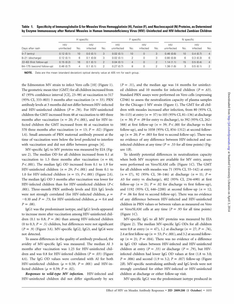
Table 1. Specificity of Immunoglobulin G for Measles Virus Hemagglutinin (H), Fusion (F), and Nucleocapsid (N) Proteins, as Determinedby Enzyme Immunoassay after Natural Measles in Human Immunodeficiency Virus (HIV)–Uninfected and HIV-Infected Zambian Children
Days after rash
H specific F specific N specific
HIVuninfected No.
HIVinfected No.
HIVuninfected No.
HIVinfected No.
HIVuninfected No.
HIVinfected No.
0–7 (entry) 0.12 (0.1) 10 0.0 (0.1) 3 0.02 (0.1) 10 0 3 0.46 (0.6) 10 0.4 (0.7) 4
8–21 (discharge) 0.12 (0.1) 6 0.1 (0.0) 3 0.02 (0.1) 2 0 3 0.80 (0.8) 6 0.3 (0.3) 6
22–63 (first follow-up) 0.15 (0.2) 16 0.1 (0.1) 2 0.04 (0.1) 4 0 2 1.14 (1.1) 15 0.5 (0.4) 4
64–175 (second follow-up) 0.46 (0.7) 4 0.1 (0.1) 2 0.27 (0.7) 8 0 2 1.38 (1.6) 3 0.5 (0.1) 2
NOTE. Data are the mean (standard deviation) optical density value at 405 nm for each group.
the Edmonston MV strain to infect Vero cells [10] (Figure 1).
The geometric mean titer (GMT) for all children increased from
47 (95% confidence interval [CI], 23–98) at vaccination to 517
(95% CI, 333–803) 3 months after vaccination ( ). PRNn p 33
antibody levels at 3 months did not differ between HIV-infected
and HIV-uninfected children ( ). For HIV-uninfectedP p .78
children the GMT increased from 48 at vaccination to 485 three
months after vaccination ( ; ), and for HIV-in-n p 20 P ! .001
fected children the GMT increased from 46 at vaccination to
570 three months after vaccination ( ; ) (Figuren p 13 P p .02
1A). Small amounts of PRN maternal antibody present at the
time of vaccination were below the level predicted to interfere
with vaccination and did not differ between groups [4].
MV-specific IgG to MV proteins was measured by EIA (Fig-
ure 2). The median OD for all children increased from 0.1 at
vaccination to 1.5 three months after vaccination ( ;n p 44
). The median IgG OD increased from 0.1 to 1.9 forP ! .001
HIV-uninfected children ( ; ) and from 0.1 ton p 29 P ! .001
1.0 for HIV-infected children ( ; ) (Figure 2A).n p 15 P ! .001
The median IgG OD 3 months after vaccination was lower for
HIV-infected children than for HIV-uninfected children (P !
). Three-month PRN antibody levels and EIA IgG levels.001
were not strongly correlated (for HIV-infected children, r p
and ; for HIV-uninfected children, and�0.10 P p .73 r p 0.4
).P p .08
IgG1 was the predominant isotype, and IgG1 levels appeared
to increase more after vaccination among HIV-uninfected chil-
dren (0.1 to 0.8; ) than among HIV-infected childrenP p .06
(0 to 0.3; ) children, but differences were not significantP p .5
( ) (Figure 3A). MV-specific IgG2, IgG3, and IgG4 wereP p .9
not detected.
To assess differences in the quality of antibody produced, the
avidity of MV-specific IgG was measured. The median AI 3
months after vaccination was 1.25 for HIV-uninfected chil-
dren and was 0.8 for HIV-infected children ( ) (FigureP p .03
4A). The IgG OD values were correlated with AI for both
HIV-uninfected children ( ; ) and HIV-in-r p 0.58 P p .001
fected children ( ; ).r p 0.59 P p .02
Responses to wild-type MV infection. HIV-infected and
HIV-uninfected children did not differ significantly by sex
( ), and the median age was 14 months for uninfect-P p .11
ed children and 10 months for infected children ( ).P p .63
Standard PRN assays were performed on Vero cells (expressing
CD46) to assess the neutralization capacity of plasma samples
for the Chicago-1 MV strain (Figure 1). The GMT for all chil-
dren with measles increased after infection, from 80 (95% CI,
56–115) at entry ( ) to 105 (95% CI, 81–136) at dischargen p 37
( ; for entry vs discharge), to 392 (95% CI, 262–n p 30 P p .09
588) at first follow-up ( ; for discharge vs firstn p 39 P ! .001
follow-up), and to 1058 (95% CI, 854–1312) at second follow-
up ( ; for first vs second follow-up). There wasn p 20 P p .003
no evidence of any difference between HIV-infected and un-
infected children at any time ( for all time points) (Fig-P p .53
ure 1B).
To identify potential differences in neutralization capacity
when both MV receptors are available for MV entry, assays
were performed on Vero/SLAM cells (Figure 1C). The GMT
for all children with measles was 71 (95% CI, 55–142) at entry
( ), 92 (95% CI, 58–146) at discharge ( ;n p 17 n p 11 P p
for entry vs discharge), 402 (95% CI, 234–690) at first.63
follow-up ( ; for discharge vs first follow-up),n p 21 P p .02
and 1192 (95% CI, 646–2200) at second follow-up ( ;n p 12
for first vs second follow-up). There was no evidenceP p .06
of any difference between HIV-infected and HIV-uninfected
children in PRN values or between values as measured on Vero
or Vero/SLAM cells at any time ( for all time points)P p .93
(Figure 1C).
MV-specific IgG to all MV proteins was measured by EIA
(Figure 2). The median MV-specific IgG ODs for all children
were 0.8 at entry ( ), 1.2 at discharge ( ; ),n p 47 n p 27 P p .74
2.4 at first follow-up ( ; ), and 3.2 at second follow-n p 53 P ! .001
up ( ; ). There was no evidence of a differencen p 21 P p .014
in IgG OD values between HIV-infected and HIV-uninfected
children at entry ( ) or discharge ( ), but HIV-P p .33 P p .79
infected children had lower IgG OD values at first (1.8 vs 3.0;
) and second (1.9 vs 3.2; ) follow-up (FigureP p .006 P p .017
2B). MV-specific neutralizing antibody and IgG levels were not
strongly correlated for either HIV-infected or HIV-uninfected
children at discharge or either follow-up visit.
MV-specific IgG1 was the predominant isotype produced in
at Stanford University L
ibraries on April 3, 2014
http://jid.oxfordjournals.org/D
ownloaded from

1036 • JID 2009:200 (1 October) • Nair et al
Figure 5. Linear regression analysis showing correlation between avid-ity index and plaque reduction neutralization for plasma samples fromchildren with measles tested on Vero (A) and Vero/signaling lymphocyte-activation molecule (SLAM) (B ) cells using Chicago-1 measles virus andon Vero/SLAM cells using a wild-type measles virus isolate from Zam-bia (C ).
response to wild-type MV infection. MV-specific IgG1 levels
increased through the second follow-up visit for HIV-unin-
fected children but plateaued after discharge for HIV-infected
children (Figure 3B). However, there was no statistically sig-
nificant difference in MV-specific IgG1 or IgG3 levels be-
tween HIV-infected and HIV-uninfected children at entry
( and ), discharge ( and ), or firstP p .4 P p .45 P p .6 P p .2
( and ) or second ( and ) fol-P p .08 P p .11 P p .3 P p .2
low-up, perhaps because of limited power to detect a signifi-
cant difference. MV-specific IgG2 and IgG4 antibodies were
not detected.
To determine whether there were differences in the MV pro-
teins recognized by HIV-infected and HIV-uninfected children,
IgG antibodies specific for MV N, H, and F were measured (Table
1). N-specific responses accounted for the largest proportion of
MV-specific IgG among both HIV-infected and HIV-uninfected
children. Both N and H OD values were lower among HIV-
infected children, and F-specific responses were not detected.
To determine the maturation of antibody avidity after natural
MV infection, AIs were determined (Figure 4). The median AI
for all children was 0.3 ( ) at entry, 0.5 ( ) atn p 48 n p 27
discharge, 0.8 at first follow-up ( ), and 1.25 at secondn p 52
follow-up ( ). The median AI for HIV-uninfected chil-n p 22
dren was 0.3 at entry ( ), 0.5 at discharge ( ;n p 29 n p 13
for entry vs discharge), 0.85 at first follow-up (P p .008 n p
; for discharge vs first follow-up), and 1.38 at sec-32 P p .003
ond follow-up ( ; for first vs second follow-n p 14 P p .006
up). The median AI for HIV-infected children was 0.3 at entry
( ), 0.38 at discharge ( ; for entry vs dis-n p 19 n p 14 P p .07
charge), 0.75 at first follow-up ( ; for dischargen p 20 P p .05
vs first follow-up), and 0.8 at second follow-up ( ;n p 8 P p
for first vs second follow-up) (Figure 4B). AI values were.05
lower for HIV-infected children at first follow-up ( )P p .014
and at second follow-up ( ) than for HIV-uninfectedP p 0.04
children. AI did not correlate with the percentage of CD4+ T
cells at entry for HIV-infected or HIV-uninfected children.
To assess the potential relevance of AI to protection from
reinfection, correlation between AI and PRN titers was assessed
(Figure 5). There was no strong evidence of a correlation with
AI when PRN was measured using the Chicago-1 strain of MV
on Vero cells ( ; ) (Figure 5A) or Vero/SLAMr p 0.618 P p .139
cells ( ; ) (Figure 5B). However, AI and neu-r p 0.158 P p .783
tralization were correlated when PRN was measured using the
wild-type Zambia MV strain for infection of Vero/SLAM cells
( ; ) (Figure 5C).r p 0.78 P p .002
DISCUSSION
In the present study, we have shown that HIV infection influ-
ences both the quantity and quality of the antibody produced
in response to measles vaccination and to measles. Identifica-
tion of these defects depends on the tests used to assess MV-
specific antibody. We found that HIV infection does not affect
production of neutralizing antibody, as measured by the stan-
dard PRN assay, during the first 3 months after immunization
at age 9 months or after natural infection. However, vaccine-
induced neutralizing antibody wanes rapidly, suggesting that a
defect in the quality of the response exists [10].
EIA, using a lysate of MV-infected cells as antigen, measures
at Stanford University L
ibraries on April 3, 2014
http://jid.oxfordjournals.org/D
ownloaded from

Effect of HIV on Measles Antibody Responses • JID 2009:200 (1 October) • 1037
IgG binding to many MV proteins but is dominated by the
response to N, and EIA is less sensitive than the PRN assay for
detecting immunity to MV when antibody levels are low [31,
32]. Our analysis by EIA showed that both the amount and
the avidity of MV-specific IgG produced were impaired by HIV
infection. The development of high-avidity antibody correlated
with the ability to neutralize the infection of Vero/SLAM cells
with a wild-type strain of MV. These experiments suggest that
HIV-infected children are less well protected from MV infection
by vaccination than are HIV-uninfected children. This sugges-
tion is supported by the observation that HIV-infected children
hospitalized with measles in Zambia are significantly more
likely than HIV-uninfected children to have a history of vac-
cination [6]. Furthermore, specific aspects of the antibody tests
chosen to measure the response to measles need to be consid-
ered in designing studies to assess immune responses in this
population.
These experiments also confirm previous observations that
EIA responses to vaccination are lower in HIV-infected children
than in HIV-uninfected children [11, 12]. Prior experiments
also showed that antibody levels waned over time and were not
augmented by repeat vaccination [12]. It is presumed that the
differences in the results obtained by the 2 methods are due
to the differences in the specificities being analyzed. EIA an-
tibody was primarily IgG1 and was directed against N, whereas
PRN antibody can be of any subclass and is primarily directed
against specific epitopes on H. Understanding the isotype pro-
file is likely to be important because IgG subclasses differ in
many biologic properties—including half-life, Fc receptor bind-
ing, and complement activation—that alter effectiveness.
The mechanisms by which HIV infection impairs IgG re-
sponses and avidity maturation have yet to be fully elucidated.
Because most children are infected with HIV during the per-
inatal period before encountering MV antigens, avidity mat-
uration occurs in the context of an already-impaired immune
system. Avidity maturation is T cell dependent and requires the
activation-induced, cytidine deaminase–mediated process of so-
matic hypermutation of the variable regions of antibody genes
[33, 34], all of which are impaired in HIV-infected individuals
[35–37]. We did not observe a correlation between avidity and
CD4+ T cell counts, but it is likely that HIV-induced function-
al alteration of CD4+ T cells contributes to impaired antibody
responses.
HIV can also have direct effects on the B cell compartment
[38]. Ongoing HIV replication is associated with B cell dys-
regulation, increased B cell activation and turnover, and an
increase in immature and transitional B cells [39, 40]. There
is loss of memory B cells and decreases in levels of previously
induced antibody [39, 41–43]. HIV Nef protein can penetrate
bystander B cells and inhibit class-switch recombination and
IgG production [44]. In addition, chemokine and chemokine
receptor expression important for B cell migration within lym-
phoid tissues, for germinal center formation, and for B cell
homing to bone marrow is decreased by HIV infection [39].
Germinal center abnormalities are seen throughout the course
of HIV infection as HIV uptake by follicular dendritic cells and
can lead to overload and network disruption [39, 45]. It is
likely that HIV-induced effects on T and B cells, on germinal
center integrity, and on antigen presentation all contribute to
the reduced quantity and quality of MV-specific antibody ob-
served in Zambian children.
The observation that neutralization and avidity were not
correlated when laboratory-adapted strains of MV were used
to infect Vero or Vero/SLAM cells but were correlated when a
wild-type field isolate was used to infect Vero/SLAM cells in-
dicates the importance of the high-affinity interaction between
SLAM and the H protein of field isolates. Furthermore, it sug-
gests that the avidity of the antibody induced by vaccination
is important for protection against wild-type MV infection.
Our findings show that HIV impairs qualitative features of
the antibody response to MV vaccination and infection. In-
creased access to antiretroviral treatment—with particular em-
phasis on reducing mother-to-child transmission—should re-
duce the prevalence of HIV among children. For children who
are infected, treatment is increasingly available, and the re-
sponse of HIV-infected children upon treatment to measles
vaccination and revaccination requires evaluation. The present
study underscores the need for a variety of assays to effectively
measure the immunogenicity of measles vaccine in HIV-in-
fected persons and for the additional information provided by
measurement of avidity.
Acknowledgments
We thank Judy Beeler and Susette Audet of the Food and Drug Ad-ministration for performing PRN assays after vaccination and Brandyn Laufor expert technical assistance.
References
1. Wolfson LJ, Strebel PM, Gacic-Dobo M, Hoekstra EJ, McFarland JW,Hersh BS. Has the 2005 measles mortality reduction goal beenachieved? A natural history modelling study. Lancet 2007; 369:191–200.
2. Cutts FT, Henao-Restrepo A, Olive JM. Measles elimination: progressand challenges. Vaccine 1999; 17(Suppl 3):S47–52.
3. Moss WJ, Cutts F, Griffin DE. Implications of the HIV epidemic forcontrol and eradication of measles. Clin Infect Dis 1999; 29:106–12.
4. Scott S, Moss WJ, Cousens S, et al. The influence of HIV-1 exposureand infection on levels of passively acquired antibodies to measles virusin Zambian infants. Clin Infect Dis 2007; 45:1417–24.
5. Centers for Disease Control. Measles in HIV-infected children, UnitedStates. MMWR Morb Mortal Wkly Rep 1988; 37:183–6.
6. Moss WJ, Fisher C, Scott S, et al. HIV type 1 infection is a risk factorfor mortality in hospitalized Zambian children with measles. Clin InfectDis 2008; 46:523–7.
7. Permar SR, Moss WJ, Ryon JJ, et al. Prolonged measles virus sheddingin human immunodeficiency virus–infected children, detected by re-
at Stanford University L
ibraries on April 3, 2014
http://jid.oxfordjournals.org/D
ownloaded from
1038 • JID 2009:200 (1 October) • Nair et al
verse transcriptase–polymerase chain reaction. J Infect Dis 2001; 183:532–8.
8. Scott S, Mossong J, Moss WJ, Cutts FT, Cousens S. Predicted impactof the HIV-1 epidemic on measles in developing countries: results froma dynamic age-structured model. Int J Epidemiol 2008; 37:356–67.
9. Chen RT, Markowitz LE, Albrecht P, et al. Measles antibody: reeval-uation of protective titers. J Infect Dis 1990; 162:1036–42.
10. Moss WJ, Scott S, Mugala N, et al. Immunogenicity of standard-titermeasles vaccine in HIV-1–infected and uninfected Zambian children:an observational study. J Infect Dis 2007; 196:347–55.
11. Helfand RF, Witte D, Fowlkes A, et al. Evaluation of the immuneresponse to a 2-dose measles vaccination schedule administered at 6and 9 months of age to HIV-infected and HIV-uninfected children inMalawi. J Infect Dis 2008; 198:1457–65.
12. Brunell PA, Vimal V, Sandu M, Courville TM, Daar E, Israele V. Ab-normalities of measles antibody response in human immunodeficien-cy virus type 1 (HIV-1) infection. J Acquir Immune Defic Syndr HumRetrovirol 1995; 10:540–8.
13. Hashiguchi T, Kajikawa M, Maita N, et al. Crystal structure of measlesvirus hemagglutinin provides insight into effective vaccines. Proc NatlAcad Sci U S A 2007; 104:19535–40.
14. Dorig RE, Marcil A, Chopra A, Richardson CD. The human CD46molecule is a receptor for measles virus (Edmonston strain). Cell1993; 75:295–305.
15. Naniche D, Varior-Krishnan G, Cervoni F, et al. Human membranecofactor protein (CD46) acts as a cellular receptor for measles virus.J Virol 1993; 67:6025–32.
16. Tatsuo H, Ono N, Tanaka K, Yanagi Y. SLAM (CDw150) is a cellularreceptor for measles virus. Nature 2000; 406:893–8.
17. Yanagi Y, Takeda M, Ohno S. Measles virus: cellular receptors, tropismand pathogenesis. J Gen Virol 2006; 87:2767–79.
18. Erlenhofer C, Duprex WP, Rima BK, ter Meulen V, Schneider-SchauliesJ. Analysis of receptor (CD46, CD150) usage by measles virus. J GenVirol 2002; 83:1431–6.
19. Santiago C, Bjorling E, Stehle T, Casasnovas JM. Distinct kinetics forbinding of the CD46 and SLAM receptors to overlapping sites in themeasles virus hemagglutinin protein. J Biol Chem 2002; 277:32294–301.
20. Olszewska W, Obeid OE, Steward MW. Protection against measlesvirus-induced encephalitis by anti-mimotope antibodies: the role ofantibody affinity. Virology 2000; 272:98–105.
21. Phan TG, Paus D, Chan TD, et al. High affinity germinal center Bcells are actively selected into the plasma cell compartment. J Exp Med2006; 203:2419–24.
22. Polack FP, Hoffman SJ, Crujeiras G, Griffin DE. A role for nonpro-tective complement-fixing antibodies with low avidity for measles virusin atypical measles. Nat Med 2003; 9:1209–13.
23. Moss WJ, Monze M, Ryon JJ, Quinn TC, Griffin DE, Cutts F. Pro-spective study of measles in hospitalized, human immunodeficiencyvirus (HIV)–infected and HIV-uninfected children in Zambia. ClinInfect Dis 2002; 35:189–96.
24. Ono N, Tatsuo H, Hidaka Y, Aoki T, Minagawa HY. Measles viruseson throat swabs from measles patients use signaling lymphocytic ac-tivation molecule (CDw150) but not CD46 as a cellular receptor. JVirol 2001; 75:4399–401.
25. Cohen BJ, Audet S, Andrews N, Beeler J. Plaque reduction neutrali-zation test for measles antibodies: description of a standardised lab-oratory method for use in immunogenicity studies of aerosol vacci-nation. Vaccine 2007; 26:59–66.
26. Nair N, Gans H, Lew-Yasukawa L, Long-Wagar AC, Arvin A, GriffinDE. Age-dependent differences in IgG isotype and avidity induced by
measles vaccine received during the first year of life. J Infect Dis 2007;196:1339–45.
27. Macdonald RA, Hosking CS, Jones CL. The measurement of relativeantibody affinity by ELISA using thiocyanate elution. J Immunol Meth-ods 1988; 106:191–4.
28. Pullen GR, Fitzgerald MG, Hosking CS. Antibody avidity determi-nation by ELISA using thiocyanate elution. J Immunol Methods 1986;86:83–7.
29. Beauverger P, Buckland R, Wild F. Establishment and characterisationof murine cells constitutively expressing the fusion, nucleoprotein andmatrix proteins of measles virus. J Virol Methods 1993; 44:199–210.
30. Hummel KB, Erdman DD, Heath J, Bellini WJ. Baculovirus expressionof the nucleoprotein gene of measles virus and utility of the recom-binant protein in diagnostic enzyme immunoassays. J Clin Microbiol1992; 30:2874–80.
31. van den HS, Gageldonk-Lafeber AB, Van Binnendijk RS, van Gagel-donk PG, Berbers GA. Comparison of measles virus-specific antibodytitres as measured by enzyme-linked immunosorbent assay and virusneutralisation assay. Vaccine 2003; 21:4210–4.
32. Cohen BJ, Dobias D, Andrews N. Comparison of plaque reductionneutralisation test (PRNT) and measles virus-specific IgG ELISA forassessing immunogenicity of measles vaccination. Vaccine 2008; 26:6392–7.
33. Cozine CL, Wolniak KL, Waldschmidt TJ. The primary germinal cen-ter response in mice. Curr Opin Immunol 2005; 17:298–302.
34. Dudley DD, Chaudhuri J, Bassing CH, Alt FW. Mechanism and con-trol of V(D)J recombination versus class switch recombination: simi-larities and differences. Adv Immunol 2005; 86:43–112.
35. Scamurra RW, Miller DJ, Dahl L, et al. Impact of HIV-1 infection onVH3 gene repertoire of naive human B cells. J Immunol 2000; 164:5482–91.
36. Wisnewski A, Cavacini L, Posner M. Human antibody variable regiongene usage in HIV-1 infection. J Acquir Immune Defic Syndr HumRetrovirol 1996; 11:31–8.
37. Poli G, Pantaleo G, Fauci AS. Immunopathogenesis of human im-munodeficiency virus infection. Clin Infect Dis 1993; 17:S224-9.
38. Moir S, Fauci AS. B cells in HIV infection and disease. Nat Rev Im-munol 2009; 9:235–45.
39. Cagigi A, Nilsson A, De MA, Chiodi F. B cell immunopathology dur-ing HIV-1 infection: lessons to learn for HIV-1 vaccine design. Vac-cine 2008; 26:3016–25.
40. Malaspina A, Moir S, Ho J, et al. Appearance of immature/transition-al B cells in HIV-infected individuals with advanced disease: correla-tion with increased IL-7. Proc Natl Acad Sci USA 2006; 103:2262–7.
41. Moir S, Malaspina A, Pickeral OK, et al. Decreased survival of B cellsof HIV-viremic patients mediated by altered expression of receptorsof the TNF superfamily. J Exp Med 2004; 200:587–99.
42. De MA, Nilsson A, Titanji K, et al. Mechanisms of hypergammaglob-ulinemia and impaired antigen-specific humoral immunity in HIV-1infection. Blood 2004; 103:2180–6.
43. Titanji K, De MA, Cagigi A, et al. Loss of memory B cells impairsmaintenance of long-term serologic memory during HIV-1 infection.Blood 2006; 108:1580–7.
44. Qiao X, He B, Chiu A, Knowles DM, Chadburn A, Cerutti A. Humanimmunodeficiency virus 1 Nef suppresses CD40-dependent immu-noglobulin class switching in bystander B cells. Nat Immunol 2006; 7:302–10.
45. Taruishi M, Terashima K, Dewan Z, et al. Role of follicular dendriticcells in the early HIV-1 infection: in vitro model without specific an-tibody. Microbiol Immunol 2004; 48:693–702.
at Stanford University L
ibraries on April 3, 2014
http://jid.oxfordjournals.org/D
ownloaded from
Related Documents