HIV-Associated Disruption of Tight and Adherens Junctions of Oral Epithelial Cells Facilitates HSV-1 Infection and Spread Irna Sufiawati 1¤ , Sharof M. Tugizov 1,2 * 1 Department of Medicine, University of California San Francisco, San Francisco, California, United States of America, 2 Department of Orofacial Sciences, University of California San Francisco, San Francisco, California, United States of America Abstract Herpes simplex virus (HSV) types 1 and 2 are the most common opportunistic infections in HIV/AIDS. In these immunocompromised individuals, HSV-1 reactivates and replicates in oral epithelium, leading to oral disorders such as ulcers, gingivitis, and necrotic lesions. Although the increased risk of HSV infection may be mediated in part by HIV-induced immune dysfunction, direct or indirect interactions of HIV and HSV at the molecular level may also play a role. In this report we show that prolonged interaction of the HIV proteins tat and gp120 and cell-free HIV virions with polarized oral epithelial cells leads to disruption of tight and adherens junctions of epithelial cells through the mitogen-activated protein kinase signaling pathway. HIV-induced disruption of oral epithelial junctions facilitates HSV-1 paracellular spread between the epithelial cells. Furthermore, HIV-associated disruption of adherens junctions exposes sequestered nectin-1, an adhesion protein and critical receptor for HSV envelope glycoprotein D (gD). Exposure of nectin-1 facilitates binding of HSV-1 gD, which substantially increases HSV-1 infection of epithelial cells with disrupted junctions over that of cells with intact junctions. Exposed nectin-1 from disrupted adherens junctions also increases the cell-to-cell spread of HSV-1 from infected to uninfected oral epithelial cells. Antibodies to nectin-1 and HSV-1 gD substantially reduce HSV-1 infection and cell-to-cell spread, indicating that HIV-promoted HSV infection and spread are mediated by the interaction of HSV gD with HIV- exposed nectin-1. Our data suggest that HIV-associated disruption of oral epithelial junctions may potentiate HSV-1 infection and its paracellular and cell-to-cell spread within the oral mucosal epithelium. This could be one of the possible mechanisms of rapid development of HSV-associated oral lesions in HIV-infected individuals. Citation: Sufiawati I, Tugizov SM (2014) HIV-Associated Disruption of Tight and Adherens Junctions of Oral Epithelial Cells Facilitates HSV-1 Infection and Spread. PLoS ONE 9(2): e88803. doi:10.1371/journal.pone.0088803 Editor: Claude Krummenacher, University of Pennsylvania School of Veterinary Medicine, United States of America Received October 29, 2013; Accepted January 15, 2014; Published February 21, 2014 Copyright: ß 2014 Sufiawati, Tugizov. This is an open-access article distributed under the terms of the Creative Commons Attribution License, which permits unrestricted use, distribution, and reproduction in any medium, provided the original author and source are credited. Funding: This project was supported by National Institutes of Health grants R01 DE023315, R21 DE016009, and R21 DE021011, and the NCI/UCSF Cancer Center grant P30 CA 82103 (to ST). The authors acknowledge the financial support of Directorate General of Higher Education, Ministry of National Education Indonesia, through the Doctoral Sandwich Program Scholarship (to IS). The funders had no role in study design, data collection and analysis, decision to publish, or preparation of the manuscript. Competing Interests: The authors have declared that no competing interests exist. * E-mail: [email protected] ¤ Current address: Department of Oral Medicine, Faculty of Dentistry, University of Padjadjaran, Bandung, Indonesia Introduction Herpes simplex virus type 1 (HSV-1) is a common oral pathogen that causes multiple oral disorders such as ulcers, necrotic lesions, and gingivostomatitis. Oral epithelium is also infected with HSV-2 [1], but to a lesser extent. HIV infection leads to reactivation and spread of herpesviruses, including HSV-1 and - 2, in oral and genital mucosa [2,3,4,5,6,7,8,9,10]. HIV infection causes attenuation of the immune system by substantially depleting CD4 + T cells in peripheral blood, lymphoid organs, and mucosal tissues, leading to CD8 + T cell dysfunction [11,12,13]. HIV- mediated depletion and dysfunction of CD4 + /CD8 + T immune cells can lead to the activation of herpesviruses [2,3,4,5,6], which are usually latent under normal immune surveillance [14]. In addition to attenuation of the immune system, HIV in- fection can impair the barrier function of various mucosal epithelia, including oral, intestinal and anogenital mucosa [15,16,17,18,19,20]. This in turn may facilitate the spread of opportunistic infections, including HSV-1/2, throughout the epithelium. HIV tat and gp120 proteins play an important role in the impairment of the mucosal barrier by disrupting epithelial tight junctions (TJs). HIV tat and gp120 are transactivator and envelope proteins that activate multiple signaling pathways, including mitogen-activated protein kinase (MAPK) signaling, which lead to disruption of TJs through aberrant internalization of TJ proteins and their down-regulation and/or proteasome- mediated degradation [21,22,23,24,25,26,27,28,29,30,31,32]. Nectin-1 is a poliovirus receptor-related protein 1 (PRR1/ HveC/CD111) and a Ca2+-independent cell adhesion protein of the immunoglobulin superfamily [33,34]. Nectin-1 binds to HSV glycoprotein D (gD), facilitating entry of virions into epithelial cells and cell-to-cell spread of progeny virions [35,36,37,38,39,40,41,42]. Nectin-1 is sequestered in the intercel- lular junctions, limiting the access of HSV [43]. In this study we wanted to explore the role of HIV-associated disruption of oral mucosal epithelium in HSV-1 infection and spread by using polarized oral keratinocytes as a model system. Our data show that HIV tat and gp120 proteins disrupt oral PLOS ONE | www.plosone.org 1 February 2014 | Volume 9 | Issue 2 | e88803

Welcome message from author
This document is posted to help you gain knowledge. Please leave a comment to let me know what you think about it! Share it to your friends and learn new things together.
Transcript

HIV-Associated Disruption of Tight and AdherensJunctions of Oral Epithelial Cells Facilitates HSV-1Infection and SpreadIrna Sufiawati1¤, Sharof M. Tugizov1,2*
1 Department of Medicine, University of California San Francisco, San Francisco, California, United States of America, 2 Department of Orofacial Sciences, University of
California San Francisco, San Francisco, California, United States of America
Abstract
Herpes simplex virus (HSV) types 1 and 2 are the most common opportunistic infections in HIV/AIDS. In theseimmunocompromised individuals, HSV-1 reactivates and replicates in oral epithelium, leading to oral disorders such asulcers, gingivitis, and necrotic lesions. Although the increased risk of HSV infection may be mediated in part by HIV-inducedimmune dysfunction, direct or indirect interactions of HIV and HSV at the molecular level may also play a role. In this reportwe show that prolonged interaction of the HIV proteins tat and gp120 and cell-free HIV virions with polarized oral epithelialcells leads to disruption of tight and adherens junctions of epithelial cells through the mitogen-activated protein kinasesignaling pathway. HIV-induced disruption of oral epithelial junctions facilitates HSV-1 paracellular spread between theepithelial cells. Furthermore, HIV-associated disruption of adherens junctions exposes sequestered nectin-1, an adhesionprotein and critical receptor for HSV envelope glycoprotein D (gD). Exposure of nectin-1 facilitates binding of HSV-1 gD,which substantially increases HSV-1 infection of epithelial cells with disrupted junctions over that of cells with intactjunctions. Exposed nectin-1 from disrupted adherens junctions also increases the cell-to-cell spread of HSV-1 from infectedto uninfected oral epithelial cells. Antibodies to nectin-1 and HSV-1 gD substantially reduce HSV-1 infection and cell-to-cellspread, indicating that HIV-promoted HSV infection and spread are mediated by the interaction of HSV gD with HIV-exposed nectin-1. Our data suggest that HIV-associated disruption of oral epithelial junctions may potentiate HSV-1infection and its paracellular and cell-to-cell spread within the oral mucosal epithelium. This could be one of the possiblemechanisms of rapid development of HSV-associated oral lesions in HIV-infected individuals.
Citation: Sufiawati I, Tugizov SM (2014) HIV-Associated Disruption of Tight and Adherens Junctions of Oral Epithelial Cells Facilitates HSV-1 Infection andSpread. PLoS ONE 9(2): e88803. doi:10.1371/journal.pone.0088803
Editor: Claude Krummenacher, University of Pennsylvania School of Veterinary Medicine, United States of America
Received October 29, 2013; Accepted January 15, 2014; Published February 21, 2014
Copyright: � 2014 Sufiawati, Tugizov. This is an open-access article distributed under the terms of the Creative Commons Attribution License, which permitsunrestricted use, distribution, and reproduction in any medium, provided the original author and source are credited.
Funding: This project was supported by National Institutes of Health grants R01 DE023315, R21 DE016009, and R21 DE021011, and the NCI/UCSF Cancer Centergrant P30 CA 82103 (to ST). The authors acknowledge the financial support of Directorate General of Higher Education, Ministry of National Education Indonesia,through the Doctoral Sandwich Program Scholarship (to IS). The funders had no role in study design, data collection and analysis, decision to publish, orpreparation of the manuscript.
Competing Interests: The authors have declared that no competing interests exist.
* E-mail: [email protected]
¤ Current address: Department of Oral Medicine, Faculty of Dentistry, University of Padjadjaran, Bandung, Indonesia
Introduction
Herpes simplex virus type 1 (HSV-1) is a common oral
pathogen that causes multiple oral disorders such as ulcers,
necrotic lesions, and gingivostomatitis. Oral epithelium is also
infected with HSV-2 [1], but to a lesser extent. HIV infection leads
to reactivation and spread of herpesviruses, including HSV-1 and -
2, in oral and genital mucosa [2,3,4,5,6,7,8,9,10]. HIV infection
causes attenuation of the immune system by substantially depleting
CD4+ T cells in peripheral blood, lymphoid organs, and mucosal
tissues, leading to CD8+ T cell dysfunction [11,12,13]. HIV-
mediated depletion and dysfunction of CD4+/CD8+ T immune
cells can lead to the activation of herpesviruses [2,3,4,5,6], which
are usually latent under normal immune surveillance [14].
In addition to attenuation of the immune system, HIV in-
fection can impair the barrier function of various mucosal
epithelia, including oral, intestinal and anogenital mucosa
[15,16,17,18,19,20]. This in turn may facilitate the spread of
opportunistic infections, including HSV-1/2, throughout the
epithelium. HIV tat and gp120 proteins play an important role
in the impairment of the mucosal barrier by disrupting epithelial
tight junctions (TJs). HIV tat and gp120 are transactivator and
envelope proteins that activate multiple signaling pathways,
including mitogen-activated protein kinase (MAPK) signaling,
which lead to disruption of TJs through aberrant internalization of
TJ proteins and their down-regulation and/or proteasome-
mediated degradation [21,22,23,24,25,26,27,28,29,30,31,32].
Nectin-1 is a poliovirus receptor-related protein 1 (PRR1/
HveC/CD111) and a Ca2+-independent cell adhesion protein
of the immunoglobulin superfamily [33,34]. Nectin-1 binds to
HSV glycoprotein D (gD), facilitating entry of virions into
epithelial cells and cell-to-cell spread of progeny virions
[35,36,37,38,39,40,41,42]. Nectin-1 is sequestered in the intercel-
lular junctions, limiting the access of HSV [43].
In this study we wanted to explore the role of HIV-associated
disruption of oral mucosal epithelium in HSV-1 infection and
spread by using polarized oral keratinocytes as a model system.
Our data show that HIV tat and gp120 proteins disrupt oral
PLOS ONE | www.plosone.org 1 February 2014 | Volume 9 | Issue 2 | e88803
epithelial TJs and adherens junctions (AJs), leading to the
paracellular spread of HSV, which may lead to rapid dissemina-
tion of virus within the mucosal environment and to saliva,
increasing the risk of spreading viral infection to others. HIV tat/
gp120-induced disruption of AJs exposes nectin-1 for HSV-1
binding. Furthermore, HIV-associated disruption of AJs and
exposure of nectin-1 promote HSV-1 infection and cell-to-cell
spread of the virus, leading to the rapid progression of HSV-
caused mucosal lesions and ulcers.
Materials and Methods
Ethics StatementThis study was conducted according to the principles expressed
in the Declaration of Helsinki. The study was approved by the
Committee on Human Research of the University of California–
San Francisco (IRB approval #: H8597-30664-03). All subjects
provided written informed consent for the collection of samples
and subsequent analysis.
Establishment of Polarized Oral Epithelial CellsTo establish polarized cells from primary oral epithelial cells, we
propagated primary keratinocytes from adult tonsil tissue samples,
as described in our previous work [44,45]. Tonsil epithelial cell
lines were grown in culture medium SAGM (Lonza Inc.,
Allendale, NJ) and incubated at 37uC in a humidified incubator
containing 5% CO2. Polarized cells were established in 0.4-mm
Transwell two-chamber filter inserts (12-well inserts) as described
previously [45,46,47]. The polarity of epithelial cells was
confirmed by immunodetection of TJ protein zonula occludens-1
(ZO-1), and measurement of transepithelial resistance (TER) and
paracellular permeability [45]. TER was measured with an
epithelial Millicell-ERS volt-ohm-meter (Millipore Corp., Bill-
erica, MA). Paracellular permeability was evaluated by adding
horseradish peroxidase (HRP)-conjugated goat anti-donkey IgG
(Fab’)2 (Jackson ImmunoResearch Laboratories, Inc., West Grove,
PA) to the upper filter compartment and photometrically assaying
the medium from the lower compartment for HRP with o-
phenylenediamine dihydrochloride as the substrate [45,48].
Detection of IgG in the lower chamber indicated leakage of IgG
from the upper chamber.
Viruses and Viral ProteinsHIV-1SF33 dual-tropic virus was propagated in peripheral blood
mononuclear cells as described [46,49]. HIV was inactivated by
UV irradiation at 100 mJ/cm2 [50]. The virus stocks were grown
and quantitated for p24 antigen using a p24 ELISA assay and
stored in aliquots at 80uC. Recombinant HIV-1 (Bal strain) wild-
type tat and inactive mutant tat proteins were purchased from
ImmunoDX, LLC (Woburn, MA). Mutant tat was generated by
substitution of the basic arginine-rich domain at 49–57 aa and the
integrin-binding RGD motif in the C terminus with alanines.
HIV-1 (Bal strain) gp120 was provided by the NIH AIDS
Research Reagent Program. gp120 was inactivated by incubation
at 85uC for 30 min [24]. All proteins were stored at –80uC in the
dark before use.
HSV-1 (strain F) was grown and titered in Vero cells [51]. The
truncated form of HSV-1 gD ectodomain gD(306t), which
contains 1–306 residues of the ectodomain and lacks transmem-
brane and cytoplasmic domains [52], was provided by Gary H.
Cohen and Roselyn J. Eisenberg (Department of Microbiology,
School of Dental Medicine, University of Pennsylvania, Philadel-
phia, PA). HSV-1 gD(306t) was produced in the baculovirus
system [52].
Treatment of Polarized Cells with Cell-free HIV Virions,HIV Proteins tat and gp120, and HSV gD
Infectious or UV-inactivated HIV-1SF33 was added to the apical
surface of polarized epithelial cells at 20 ng/ml of p24 antigen.
Active and inactive recombinant tat and/or gp120 (10 ng/ml of
each) were added to polarized tonsil cells from both apical and
basolateral surfaces. HSV-1 gD (306t) at 10 ng/ml was added to
polarized cells from both surfaces. Culture medium was changed
daily to add fresh virus or proteins. Disruption of TJs and AJs was
examined by immunostaining of their marker proteins ZO-1 and
E-cadherin, respectively. Expression and localization of ZO-1 and
E-cadherin as a ring shape was considered an intact junction.
Diffuse, dot-like patterns in the cytoplasm were considered
disrupted junctions [15]. Disruption of epithelial junctions was
also monitored by measuring TER and paracellular permeability
[46,47].
Immunofluorescence AssayPolarized epithelial monolayers were fixed with 4% parafor-
maldehyde for 20 min, permeabilized with 0.05% Triton X-100,
and washed three times with PBS. To determine disruption of TJs,
we immunostained cells with mouse monoclonal antibodies against
ZO-1 (Invitrogen/Life Technologies, Austin, TX) for 1 h at room
temperature. For detection of AJs, polarized cells were stained
with rabbit antibodies to E-cadherin and mouse monoclonal
antibodies to nectin-1 (both from Invitrogen/Life Technologies).
To determine HSV-1 infection, we immunostained infected cells
with mouse antibodies against HSV-1 ICP4 and gB (both from
Santa Cruz Biotechnology, Inc., Dallas, TX), rabbit anti-gD
(HSV-1) (MyBioSource, San Diego, CA), and goat anti-HSV-1/2
antiserum (AbD Serotec, Raleigh, NC). All antibodies were diluted
1:50 in blocking solution (3% BSA and 0.03% saponin in PBS).
Monolayers were washed and then incubated for 25 min with
FITC- or TRITC-labeled secondary antibodies (Vector Labora-
tories, Inc., Burlingame, CA). Cell nuclei were counterstained with
TO-PRO-3 (blue) or propidium iodide (red) (Molecular Probes/
Life Technologies). Cells were analyzed by using a Leica SP5 laser
confocal microscope.
Assay for HSV-1 Spread through a Paracellular Route ofDisrupted Polarized Oral Epithelial Cells
For the HSV-1 paracellular spread assay, HSV-1 at a
multiplicity of infection (MOI) of 10 PFU per cell was added to
the apical surface of polarized monolayers, and cells were
incubated at 37uC for 1, 2, or 4 h. At the various time points,
culture medium was collected from the basolateral chamber of
Transwell inserts and used to infect Vero cells, which were grown
on cover slips on 24-well plates. After 4 h, cells were fixed for the
immunofluorescence assay. HSV infection was quantified by
detection of HSV-1 ICP4 protein, and the percentage of infected
Vero cells was determined.
HSV Infection and Cell-to-Cell Spread in Polarized TonsilEpithelial Cells
To examine HSV-1 infection in polarized tonsil epithelial cells,
we added HSV-1 to apical and basolateral membranes at an MOI
of 4 and 40 PFU per cell, respectively. This resulted in the equal
infection of virus at approximately 4 virions per cell from both
membranes, because about 90% of basolaterally added virions
could be trapped in the filter pores, and about 10% of virions
reach the basolateral membranes [45,47,53]. Cells were cultured
for 24 h and fixed and immunostained for HSV-1 gB using mouse
monoclonal antibodies (Santa Cruz Biotechnology).
HIV, Epithelial Junctions, Herpes Simplex Virus
PLOS ONE | www.plosone.org 2 February 2014 | Volume 9 | Issue 2 | e88803
To examine the role of nectin-1 in HSV-1 infection, we
preincubated polarized cells with mouse monoclonal antibody to
nectin-1 (clone CK-8; Invitrogen/Life Technologies) at 10 mg/ml
for 1 h, and cells were then infected with HSV-1. After 24 h, cells
were fixed, and HSV infection was evaluated by immunostaining
of cells with polyclonal goat anti-HSV-1/2 antibodies (AbD
Serotec). HSV infection was quantified by counting infected cells,
and the percentage of infected cells was determined.
To determine cell-to-cell spread of HSV-1 in polarized
epithelial cells, a viral plaque assay was used [47]. Polarized cells
were infected with HSV-1 from the basolateral surface at an MOI
of 0.01 PFU per cell, and cells were incubated for 2 h at 37uC.
The cells were then washed once with serum-free medium and
overlaid with serum-free medium containing 0.5% methylcellulose
from apical and basolateral chambers; this prevents the spread of
released virions but allows cell-to-cell spread of progeny virions.
Cells were cultured for 3 days. To detect HSV-1-infected viral foci
or plaques, cells were fixed and immunostained with goat
polyclonal anti-HSV-1 antibodies (AbD Serotec). The number of
viral plaques was counted on a minimum of 3 filter inserts for each
experiment, and average plaque numbers were expressed per
insert. HSV-1 cell-to-cell spread was evaluated by quantitative
analysis of HSV-1-infected plaques. Foci containing 5 or more
infected cells were considered plaques, and cell numbers in each
plaque were counted. A minimum of 30 plaques for each
experimental condition were evaluated and the average number
of cells per plaque was expressed.
For inhibition of HSV-1 cell-to-cell spread after 2 h of infection,
cells were washed, and culture medium with antibodies to nectin-1
(Invitrogen/Life Technologies) and/or HSV-1 gD (MyBioSource,
San Diego, CA) (both at 10 mg/ml) was added to polarized cells
from apical and basolateral chambers. Cells were grown for
3 days, and culture medium was changed daily to add fresh
antibodies. Viral plagues were quantitated as described above.
Western Blot AssayFor Western blot detection of E-cadherin and nectin-1,
polarized tonsil epithelial cells were extracted, and proteins were
separated on a 16% SDS-polyacrylamide gel and blotted with
anti-E-cadherin and nectin-1 antibodies (both from Invitrogen/
Life Technologies). The protein bands were visualized using ECL
detection reagents (GE Healthcare Bio-Sciences, Pittsburgh, PA),
and equal protein loading was confirmed by detection of b-actin
(Ambion/Life Technologies).
Detection of Nectin-1 and HSV-1 gD on the Cell Surfaceby Domain-Selective Labeling Assay
Polarized cells were labeled with 200 mg/ml sulfo-NHS-LC-
biotin (Thermo Fisher Scientific Inc., Rockford, IL) from apical or
basolateral membranes for 30 min as described [44,47]. Cells were
washed with Tris saline (10 mM Tris-HCl pH 7.4, 120 mM
NaCl), and extracted in lysis buffer containing 1% Nonidet P-40,
1% sodium deoxycholate, 0.1% sodium dodecyl sulfate (SDS),
1 mM phenylmethylsulfonyl fluoride (PMSF), and aprotinin
(1 mg/ml). Biotinylated proteins were precipitated with streptavi-
din–agarose beads in lysis buffer (Thermo Fisher Scientific,
Waltham, MA), followed by separation on a 4–20% Tris-glycine
SDS-polyacrylamide gel and transferred to nitrocellulose mem-
branes (GE Healthcare, Germany). Nectin-1 was detected using
mouse monoclonal (Invitrogen/Life Technologies) antibodies. To
determine binding of HSV-1 gD to nectin-1, we used the
truncated form of HSV-1 gD ectodomain gD(306t). HSV-
1 gD(306t) at 20 mg/ml was added to apical or basolateral
membranes for 30 min, and cells were washed and subjected to
domain-selective labeling from appropriate cell membranes. HSV-
1 gD was detected using rabbit polyclonal antibodies (MyBio-
Source). Bands were visualized using the ECL detection system
(GE Healthcare).
MAPK Activation AssayPolarized tonsil epithelial cells were incubated with HIV tat
and/or gp120 recombinant proteins and UV-inactivated or active
cell-free HIV virions. One set of cells was treated with MAPK
inhibitor U0126 (Sigma) at 1 mM. The absence of toxic effect of
1 mM U0126 on polarized cells was confirmed by MTT cell
viability assay (Biotium Inc.) and measurement of TER. After
5 days, cells were extracted and proteins were separated on a 4–
20% Tris-glycine SDS-polyacrylamide gel and transferred to
nitrocellulose membranes (GE Healthcare). MAPK signaling
activity was measured by detection of phosphorylated and total
ERK1/2 protein using mouse monoclonal antibodies (Cell
Signaling Technology, Danvers, MA) that recognize phosphory-
lated (Clone E10, #9106) and nonphosphorylated (#9102)
ERK1/2. Protein bands were visualized using the ECL detection
system (GE Healthcare). Inhibition of MAPK activity by U0126
was quantitated by measuring the intensity of pixels (mean density)
in protein bands using Image J software.
Statistical AnalysisData are presented as mean 6 standard error of the mean
(SEM) of two experiments performed in duplicate. The differences
between control cells (untreated or treated with inactive tat/
gp120) and experimental cells (treated with active tat/gp120 and
HIV virions) were analyzed using Student’s t test. A p value of
,0.05 was considered significant.
Results
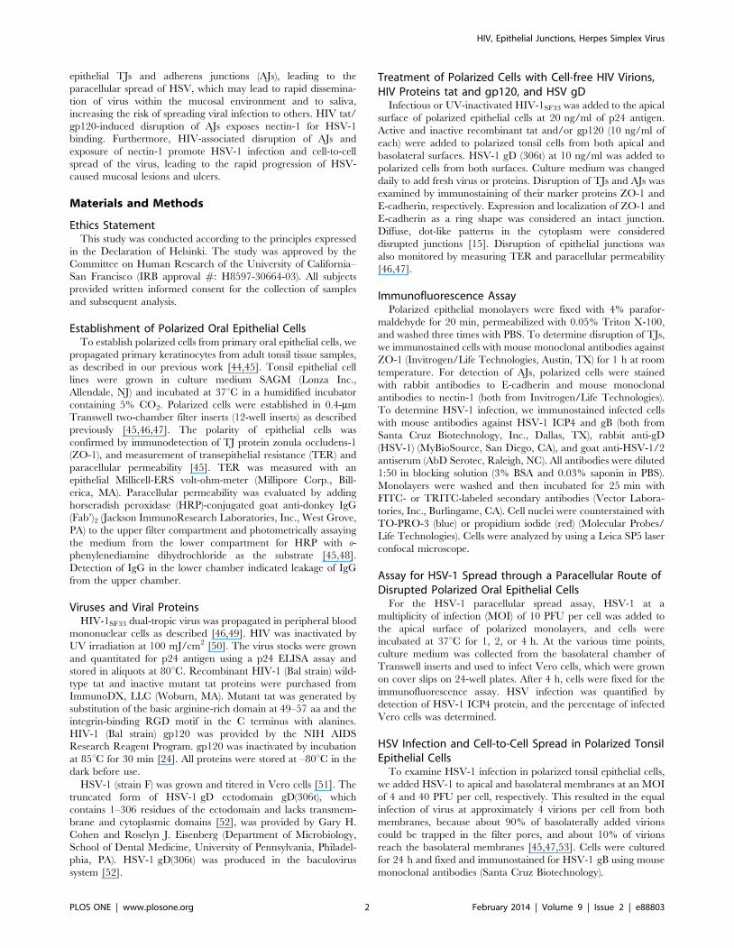
HIV Tat- and gp120-Induced Disruption of TightJunctions of Polarized Oral Epithelial Cells FacilitatesParacellular Spread of HSV-1
The tight junctions of oropharyngeal mucosal epithelium have
critical barrier functions for protection against paracellular spread
of various pathogens, including viruses. We have shown that HIV-
associated disruption of tight junctions of oral mucosal epithelium
facilitates paracellular spread of human papillomavirus [15]. HIV
tat and gp120 had the greatest effect on TJ disruption when cells
were treated with a combination of the two proteins for 5 days,
and there was no toxic effect on the cells [15]. Thus, to determine
if HIV tat- and gp120-induced TJ disruption facilitates HSV-1
paracellular spread, we treated polarized tonsil epithelial cells with
a combination of recombinant tat and gp120 at 10 ng/ml each,
which is similar to physiological levels in HIV-infected individuals
[54,55,56,57] (Fig. 1A). As negative controls we used mutant tat
protein, which lacks the basic and integrin-binding domains, and
heat-inactivated gp120; both are functionally inactive proteins
[15,21,58,59,60]. Culture medium was changed daily to include
fresh proteins, and disruption of TJs was confirmed on day 5 in
two inserts of each plate by three independent assays in the
following order: (i) measurement of TER, (ii) evaluation of
paracellular permeability, and (iii) immunostaining of cells for TJ
protein ZO-1. Measurement of TER showed that the electrical
resistance of cells treated with active forms of tat and gp120 was
substantially reduced, in contrast to that of TER in control cells,
either untreated or treated with inactive tat and gp120 (Fig. 1A,
upper panel). Consistent with the TER data, paracellular leakage
of IgG (Fab’)2 from the apical chamber to the basolateral chamber
was detected only when cells were treated with active tat/gp120
HIV, Epithelial Junctions, Herpes Simplex Virus
PLOS ONE | www.plosone.org 3 February 2014 | Volume 9 | Issue 2 | e88803
(Fig. 1A, lower panel). Finally, localization of ZO-1 in polarized
cells treated with active tat/gp120 was diffuse cytoplasmic,
indicating disruption of TJs (Fig. 1B). Polarized cells treated with
inactive tat and gp120 and untreated control cells showed the
localization of ZO-1 as a ring shape, which is typical for intact TJs
(Fig. 1B).
After confirmation of TJ disruption, we added HSV-1 to the
apical surface of polarized cells at an MOI of 10 PFU per cell for
1, 2, or 4 h. Measurement of TER in control cells exposed to
HSV-1 showed that TER was not reduced at any time point (data
not shown), indicating that HSV does not alter the TJs of tonsil
epithelial cells. At various times, the culture medium in the lower
chamber was collected for the HSV-1 infectivity assay in Vero
cells. Virus-containing basolateral culture medium was added to
Vero cells, and culture was maintained for 4 h. Vero cells were
then fixed and immunostained for HSV-1 ICP-4, which is
immediate-early protein expressed at 4 h after infection (Fig. 1C)
[61]. Confocal immunofluorescence analysis of Vero cells showed
that ICP4 was expressed in the nuclei of cells treated with active
tat/gp120 and exposed to HSV-1. In contrast, ICP4 was not
detected in Vero cells that were untreated or treated with inactive
tat/gp120 and exposed to HSV-1. Quantitative analysis of HSV-
infected Vero cells indicated that HSV paracellular spread
through tat/gp120-disrupted TJs occurred after 1 h of exposure
to HSV-1 from the apical surface (Fig. 1D). During the next 2 and
4 h of HSV-1 exposure, the paracellular spread of HSV increased
in a time-dependent fashion. These data indicate that HIV tat/
gp120-induced disruption of TJs of tonsil epithelial cells facilitates
the paracellular spread of HSV-1.
To determine if interaction of HSV-1 with oral epithelial cells
disrupts epithelial TJs, polarized tonsil epithelial cells were treated
with one of the key glycoproteins of HSV-1, the soluble gD(306t),
which contains 1–306 residues in its extracellular domain. Cells
were treated with 10 ng/ml of gD for 5 days and culture media
with fresh protein was changed every day. Measurement of TER
and immunostaining of ZO-1 showed that HSV-1 gD did not
reduce TER and did not alter localization of ZO-1 of polarized
cells (data not shown), indicating that HSV gD is not involved in
disruption of TJs.
Figure 1. HIV tat- and gp120-induced disruption of TJs of oral epithelial cells facilitates the paracellular spread of HSV-1. A (upperpanel). Polarized tonsil epithelial cells were treated with active or inactive recombinant HIV tat and gp120 in combination for 5 days, and TER wasthen measured. A (lower panel). The same cells were used to evaluate paracellular permeability after 5 days of treatment, as determined by leakageof IgG (Fab’)2 from the apical chamber to the basolateral chamber. OD, optical density. B. The same cells were immunostained for ZO-1 (green). Cellnuclei are stained in blue. C. HSV-1 at an MOI of 10 PFU per cell was added to the upper chamber of polarized cells, and culture medium wascollected from the basolateral chamber after 1, 2, or 4 h. HSV-1 paracellular spread was confirmed by detection of ICP4 protein in Vero cells (green)4 h after infection. Cell nuclei were stained with propidium iodide (red). Yellow indicates colocalization of ICP4 with the nuclear marker. D. HSV-1paracellular spread was quantified by counting of HSV-1-infected Vero cells in 10 random microscopic fields and determining the percentage of cellspositive for ICP4. A, D: Error bars indicate SEM (n = 3).doi:10.1371/journal.pone.0088803.g001
HIV, Epithelial Junctions, Herpes Simplex Virus
PLOS ONE | www.plosone.org 4 February 2014 | Volume 9 | Issue 2 | e88803
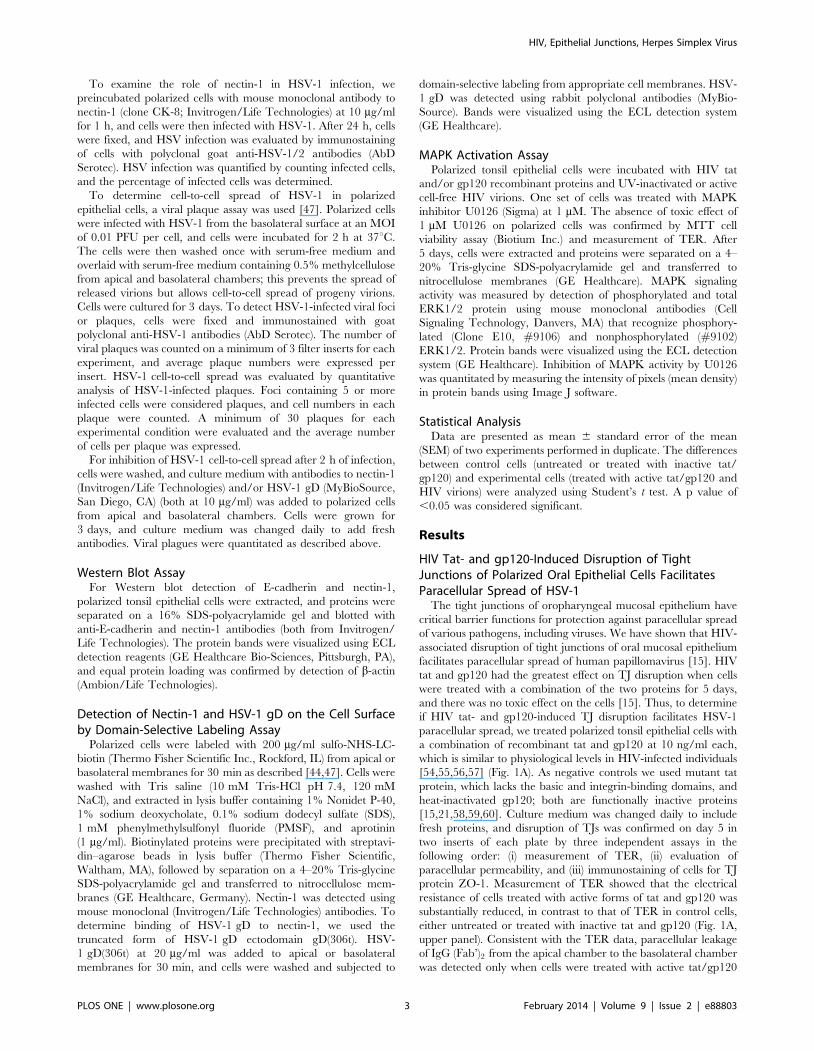
Interaction of HIV Virions with Mucosal EpitheliumDisrupts Epithelial Tight Junctions and Promotes HSVParacellular Spread
To determine whether direct interaction of cell-free HIV virions
with mucosal epithelium disrupts the TJs, we exposed polarized
tonsil epithelial cells to dual X4- and R5-tropic HIV-1SF33 for
5 days. In parallel experiments UV-inactivated virions were used.
Culture medium was changed daily to add fresh virus, and TER
was measured daily. In untreated control cells, TER increased
over the 5 days. In contrast, TER gradually declined in cells
exposed to either inactivated or active HIV virions (Fig. 2A). The
HIV-induced reduction in TER was detected only with prolonged
exposure to virus (4–5 days); with shorter treatment (1–3 days) it
was not. Analysis of paracellular leakage of IgG (Fab’)2 confirmed
the TER data; i.e., both inactive and active HIV disrupted
epithelial TJs (data not shown). To examine the paracellular
spread of HSV through HIV-disrupted tonsil epithelial cells, we
added HSV to the apical membranes of polarized cells for 1, 2, or
4 h and tested the basolateral medium for HSV infection in Vero
cells. Quantitative analysis of HSV-infected Vero cells showed
HSV paracellular spread through cells disrupted by both HIV-
inactivated and HIV-active virus (Fig. 2B). HSV paracellular
spread was not detected through control cells with intact TJs.
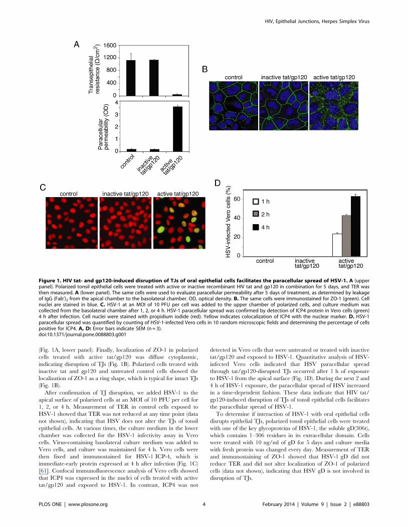
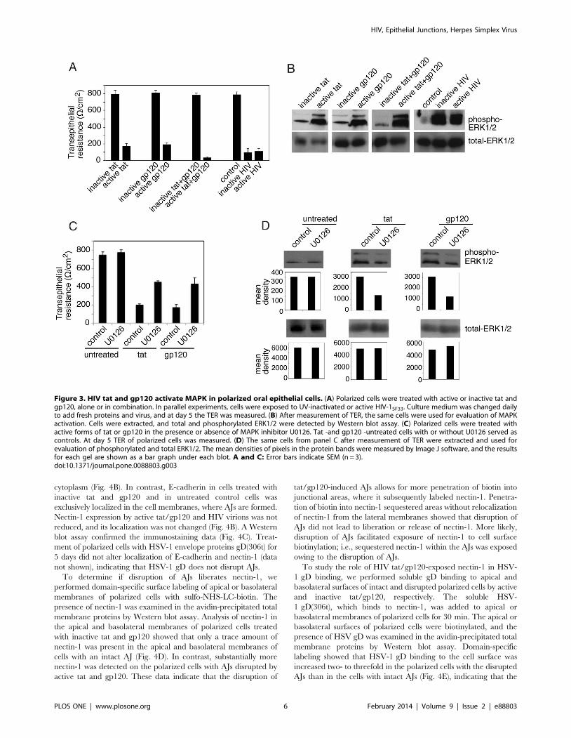
HIV tat and gp120 and Cell-free HIV Virions InduceActivation of MAPK Signaling in Polarized Oral EpithelialCells
MAPK activation is a key mechanism for the disruption of TJs,
and HIV tat binds to b1 and av integrins [59,60,62,63], which
leads to induction of MAPK activation in endothelial cells
[21,22,23]. Binding of HIV gp120 to chemokine receptors
CXCR4 and CCR5 in lymphocytes and macrophages also leads
to induction of MAPK [64,65,66,67]. We have shown that
polarized oral epithelial cells express to b1 and v integrins, and
CCR5 and CXCR4 [44,46,68], indicating availability of HIV tat
and gp120 receptors in oral epithelium. To determine if HIV tat
and gp120 interaction with these receptors induces MAPK in oral
epithelial cells, we examined the phosphorylation of MAPKs
ERK1/2 in polarized tonsil epithelial cells. Cells were treated with
tat or gp120 alone or in combination for 5 days. Also, tonsil cells
were incubated with UV-inactivated and infectious cell-free HIV
virions. TER was drastically reduced (80–90%) in cells treated
with active tat and/or gp120 and with active or inactive HIV
virions, compared to the TER of untreated control cells and cells
treated with inactive tat and/or gp120 (Fig. 3A). After confirma-
tion of TJ disruption, cells were extracted and examined for total
and phosphorylated ERK1/2 by Western blot assay (Fig. 3B).
These results show that both active tat and gp120 induce ERK1/2
phosphorylation and that treatment of cells with a combination of
tat and gp120 substantially increases ERK1/2 phosphorylation
over that in cells treated with tat or gp120 alone. Analysis of
MAPK activation in the cells incubated with UV-inactivated and
infectious HIV virions showed that both inactive and active virions
induce phosphorylation of ERK1/2.
To confirm the role of MAPK signaling in HIV-associated
disruption of TJs, polarized cells were treated with tat or gp120 in
the presence or absence of MAPK inhibitor U0126 at 1 mM
(Fig. 3C). Culture media with fresh proteins and inhibitor were
changed every day, and at day 5 TER was measured. Comparison
of TER in tat or gp120-treated cells with and without U0126
showed that TER was increased about 2 fold in cells with U0126
compared to cells without MAPK inhibitor (Fig. 3C). In the
presence of U0126, TER reached almost 60% of its normal level
in tat or gp120-untreated cells. The presence of U0126 in tat- or
gp120-untreated cells did not alter the TER. Analysis of ERK1/2
phosphorylation in tat- or gp120-treated cells in the presence and
absence of U0126 showed that U0126 induced approximately
50% reduction of ERK1/2 phosphorylation, which was well
correlated with the increase of TER (Fig. 3D). These findings
indicate that inhibition of MAPK signaling prevented tat and
gp120-induced disruption of tight junctions, i.e., HIV-induced
MAPK activation is critical for disruption of TJs in oral epithelial
cells.
HIV tat and gp120 and Cell-free HIV Virions DisruptAdherens Junctions of Oral Epithelial Cells and ExposeNectin-1
Activation of MAPK signaling may also lead to disruption of
AJs [69,70]. Dissociation of AJs by calcium depletion liberates
nectin-1, facilitating HSV-1 infection [43]. We hypothesized that
HIV-induced MAPK activation may also disrupt AJs, liberate
nectin-1, and thereby promote HSV-1 infection. To test this
hypothesis, we investigated the role of HIV tat/gp120 and cell-free
HIV virions in the disruption of AJs, liberation of nectin-1, and
binding of nectin-1 to HSV gD.
Coimmunostaining of nectin-1 and E-cadherin in polarized
tonsil epithelial cells showed that these two adhesion proteins were
colocalized (Fig. 4A). To examine whether the HIV tat and gp120
and virions disrupt the AJs, we treated polarized tonsil cells with
inactive or active tat and gp120 in combination and cell-free HIV
virions for 5 days. Immunostaining of E-cadherin in these cells
showed that active tat/gp120 as well as cell-free HIV virions
substantially reduced or completely inhibited expression of E-
cadherin and altered its localization from cell membrane to
Figure 2. HIV cell-free virion-associated disruption of epithelialtight junctions facilitates HSV paracellular spread. A. Polarizedtonsil epithelial cells were incubated with dual X4- and R5-tropic HIV-1SF33 for 5 days. One set of cells was exposed to UV-inactivated virions.Culture medium was changed daily to add fresh virus, and TER wasmeasured. B. HSV-1 was added to the apical surface of polarized cellsupon complete disruption of TJs at 5 days. HSV paracellular spread at 1,2, and 4 h after incubation was examined in Vero cells grown in thebasolateral chamber of filter inserts by immunostaining of ICP4 protein.HSV-1 paracellular spread was quantified by counting HSV-1-infectedVero cells, and the percentage of cells positive for ICP4 was determined.A, B: Error bars indicate SEM (n = 3).doi:10.1371/journal.pone.0088803.g002
HIV, Epithelial Junctions, Herpes Simplex Virus
PLOS ONE | www.plosone.org 5 February 2014 | Volume 9 | Issue 2 | e88803
cytoplasm (Fig. 4B). In contrast, E-cadherin in cells treated with
inactive tat and gp120 and in untreated control cells was
exclusively localized in the cell membranes, where AJs are formed.
Nectin-1 expression by active tat/gp120 and HIV virions was not
reduced, and its localization was not changed (Fig. 4B). A Western
blot assay confirmed the immunostaining data (Fig. 4C). Treat-
ment of polarized cells with HSV-1 envelope proteins gD(306t) for
5 days did not alter localization of E-cadherin and nectin-1 (data
not shown), indicating that HSV-1 gD does not disrupt AJs.
To determine if disruption of AJs liberates nectin-1, we
performed domain-specific surface labeling of apical or basolateral
membranes of polarized cells with sulfo-NHS-LC-biotin. The
presence of nectin-1 was examined in the avidin-precipitated total
membrane proteins by Western blot assay. Analysis of nectin-1 in
the apical and basolateral membranes of polarized cells treated
with inactive tat and gp120 showed that only a trace amount of
nectin-1 was present in the apical and basolateral membranes of
cells with an intact AJ (Fig. 4D). In contrast, substantially more
nectin-1 was detected on the polarized cells with AJs disrupted by
active tat and gp120. These data indicate that the disruption of
tat/gp120-induced AJs allows for more penetration of biotin into
junctional areas, where it subsequently labeled nectin-1. Penetra-
tion of biotin into nectin-1 sequestered areas without relocalization
of nectin-1 from the lateral membranes showed that disruption of
AJs did not lead to liberation or release of nectin-1. More likely,
disruption of AJs facilitated exposure of nectin-1 to cell surface
biotinylation; i.e., sequestered nectin-1 within the AJs was exposed
owing to the disruption of AJs.
To study the role of HIV tat/gp120-exposed nectin-1 in HSV-
1 gD binding, we performed soluble gD binding to apical and
basolateral surfaces of intact and disrupted polarized cells by active
and inactive tat/gp120, respectively. The soluble HSV-
1 gD(306t), which binds to nectin-1, was added to apical or
basolateral membranes of polarized cells for 30 min. The apical or
basolateral surfaces of polarized cells were biotinylated, and the
presence of HSV gD was examined in the avidin-precipitated total
membrane proteins by Western blot assay. Domain-specific
labeling showed that HSV-1 gD binding to the cell surface was
increased two- to threefold in the polarized cells with the disrupted
AJs than in the cells with intact AJs (Fig. 4E), indicating that the
Figure 3. HIV tat and gp120 activate MAPK in polarized oral epithelial cells. (A) Polarized cells were treated with active or inactive tat andgp120, alone or in combination. In parallel experiments, cells were exposed to UV-inactivated or active HIV-1SF33. Culture medium was changed dailyto add fresh proteins and virus, and at day 5 the TER was measured. (B) After measurement of TER, the same cells were used for evaluation of MAPKactivation. Cells were extracted, and total and phosphorylated ERK1/2 were detected by Western blot assay. (C) Polarized cells were treated withactive forms of tat or gp120 in the presence or absence of MAPK inhibitor U0126. Tat -and gp120 -untreated cells with or without U0126 served ascontrols. At day 5 TER of polarized cells was measured. (D) The same cells from panel C after measurement of TER were extracted and used forevaluation of phosphorylated and total ERK1/2. The mean densities of pixels in the protein bands were measured by Image J software, and the resultsfor each gel are shown as a bar graph under each blot. A and C: Error bars indicate SEM (n = 3).doi:10.1371/journal.pone.0088803.g003
HIV, Epithelial Junctions, Herpes Simplex Virus
PLOS ONE | www.plosone.org 6 February 2014 | Volume 9 | Issue 2 | e88803
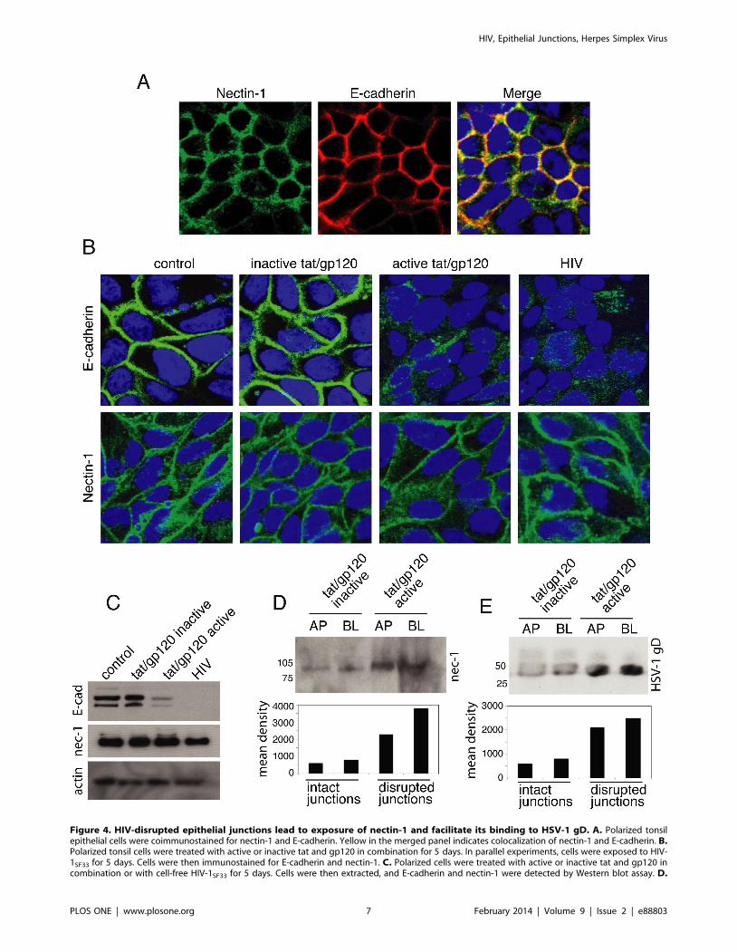
Figure 4. HIV-disrupted epithelial junctions lead to exposure of nectin-1 and facilitate its binding to HSV-1 gD. A. Polarized tonsilepithelial cells were coimmunostained for nectin-1 and E-cadherin. Yellow in the merged panel indicates colocalization of nectin-1 and E-cadherin. B.Polarized tonsil cells were treated with active or inactive tat and gp120 in combination for 5 days. In parallel experiments, cells were exposed to HIV-1SF33 for 5 days. Cells were then immunostained for E-cadherin and nectin-1. C. Polarized cells were treated with active or inactive tat and gp120 incombination or with cell-free HIV-1SF33 for 5 days. Cells were then extracted, and E-cadherin and nectin-1 were detected by Western blot assay. D.
HIV, Epithelial Junctions, Herpes Simplex Virus
PLOS ONE | www.plosone.org 7 February 2014 | Volume 9 | Issue 2 | e88803
exposure of nectin-1 is facilitated by HSV-1 gD binding to
disrupted epithelial cells.
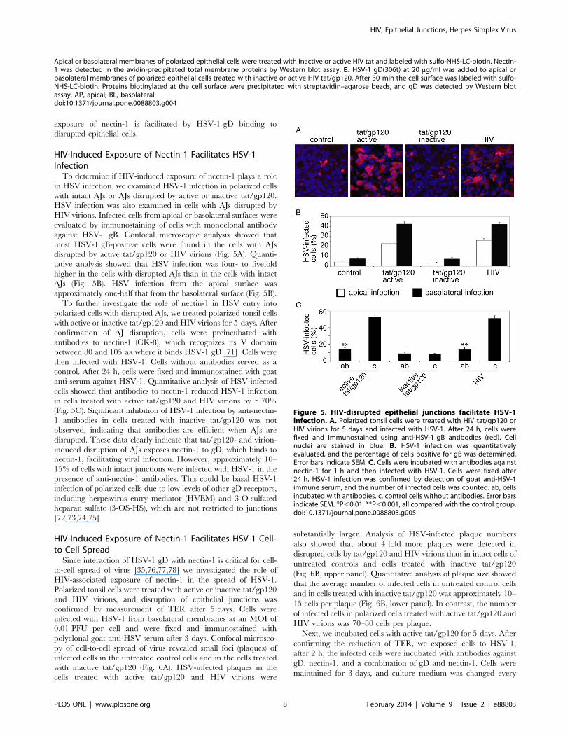
HIV-Induced Exposure of Nectin-1 Facilitates HSV-1Infection
To determine if HIV-induced exposure of nectin-1 plays a role
in HSV infection, we examined HSV-1 infection in polarized cells
with intact AJs or AJs disrupted by active or inactive tat/gp120.
HSV infection was also examined in cells with AJs disrupted by
HIV virions. Infected cells from apical or basolateral surfaces were
evaluated by immunostaining of cells with monoclonal antibody
against HSV-1 gB. Confocal microscopic analysis showed that
most HSV-1 gB-positive cells were found in the cells with AJs
disrupted by active tat/gp120 or HIV virions (Fig. 5A). Quanti-
tative analysis showed that HSV infection was four- to fivefold
higher in the cells with disrupted AJs than in the cells with intact
AJs (Fig. 5B). HSV infection from the apical surface was
approximately one-half that from the basolateral surface (Fig. 5B).
To further investigate the role of nectin-1 in HSV entry into
polarized cells with disrupted AJs, we treated polarized tonsil cells
with active or inactive tat/gp120 and HIV virions for 5 days. After
confirmation of AJ disruption, cells were preincubated with
antibodies to nectin-1 (CK-8), which recognizes its V domain
between 80 and 105 aa where it binds HSV-1 gD [71]. Cells were
then infected with HSV-1. Cells without antibodies served as a
control. After 24 h, cells were fixed and immunostained with goat
anti-serum against HSV-1. Quantitative analysis of HSV-infected
cells showed that antibodies to nectin-1 reduced HSV-1 infection
in cells treated with active tat/gp120 and HIV virions by ,70%
(Fig. 5C). Significant inhibition of HSV-1 infection by anti-nectin-
1 antibodies in cells treated with inactive tat/gp120 was not
observed, indicating that antibodies are efficient when AJs are
disrupted. These data clearly indicate that tat/gp120- and virion-
induced disruption of AJs exposes nectin-1 to gD, which binds to
nectin-1, facilitating viral infection. However, approximately 10–
15% of cells with intact junctions were infected with HSV-1 in the
presence of anti-nectin-1 antibodies. This could be basal HSV-1
infection of polarized cells due to low levels of other gD receptors,
including herpesvirus entry mediator (HVEM) and 3-O-sulfated
heparan sulfate (3-OS-HS), which are not restricted to junctions
[72,73,74,75].
HIV-Induced Exposure of Nectin-1 Facilitates HSV-1 Cell-to-Cell Spread
Since interaction of HSV-1 gD with nectin-1 is critical for cell-
to-cell spread of virus [35,76,77,78] we investigated the role of
HIV-associated exposure of nectin-1 in the spread of HSV-1.
Polarized tonsil cells were treated with active or inactive tat/gp120
and HIV virions, and disruption of epithelial junctions was
confirmed by measurement of TER after 5 days. Cells were
infected with HSV-1 from basolateral membranes at an MOI of
0.01 PFU per cell and were fixed and immunostained with
polyclonal goat anti-HSV serum after 3 days. Confocal microsco-
py of cell-to-cell spread of virus revealed small foci (plaques) of
infected cells in the untreated control cells and in the cells treated
with inactive tat/gp120 (Fig. 6A). HSV-infected plaques in the
cells treated with active tat/gp120 and HIV virions were
substantially larger. Analysis of HSV-infected plaque numbers
also showed that about 4 fold more plaques were detected in
disrupted cells by tat/gp120 and HIV virions than in intact cells of
untreated controls and cells treated with inactive tat/gp120
(Fig. 6B, upper panel). Quantitative analysis of plaque size showed
that the average number of infected cells in untreated control cells
and in cells treated with inactive tat/gp120 was approximately 10–
15 cells per plaque (Fig. 6B, lower panel). In contrast, the number
of infected cells in polarized cells treated with active tat/gp120 and
HIV virions was 70–80 cells per plaque.
Next, we incubated cells with active tat/gp120 for 5 days. After
confirming the reduction of TER, we exposed cells to HSV-1;
after 2 h, the infected cells were incubated with antibodies against
gD, nectin-1, and a combination of gD and nectin-1. Cells were
maintained for 3 days, and culture medium was changed every
Apical or basolateral membranes of polarized epithelial cells were treated with inactive or active HIV tat and labeled with sulfo-NHS-LC-biotin. Nectin-1 was detected in the avidin-precipitated total membrane proteins by Western blot assay. E. HSV-1 gD(306t) at 20 mg/ml was added to apical orbasolateral membranes of polarized epithelial cells treated with inactive or active HIV tat/gp120. After 30 min the cell surface was labeled with sulfo-NHS-LC-biotin. Proteins biotinylated at the cell surface were precipitated with streptavidin–agarose beads, and gD was detected by Western blotassay. AP, apical; BL, basolateral.doi:10.1371/journal.pone.0088803.g004
Figure 5. HIV-disrupted epithelial junctions facilitate HSV-1infection. A. Polarized tonsil cells were treated with HIV tat/gp120 orHIV virions for 5 days and infected with HSV-1. After 24 h, cells werefixed and immunostained using anti-HSV-1 gB antibodies (red). Cellnuclei are stained in blue. B. HSV-1 infection was quantitativelyevaluated, and the percentage of cells positive for gB was determined.Error bars indicate SEM. C. Cells were incubated with antibodies againstnectin-1 for 1 h and then infected with HSV-1. Cells were fixed after24 h, HSV-1 infection was confirmed by detection of goat anti-HSV-1immune serum, and the number of infected cells was counted. ab, cellsincubated with antibodies. c, control cells without antibodies. Error barsindicate SEM. *P,0.01, **P,0.001, all compared with the control group.doi:10.1371/journal.pone.0088803.g005
HIV, Epithelial Junctions, Herpes Simplex Virus
PLOS ONE | www.plosone.org 8 February 2014 | Volume 9 | Issue 2 | e88803
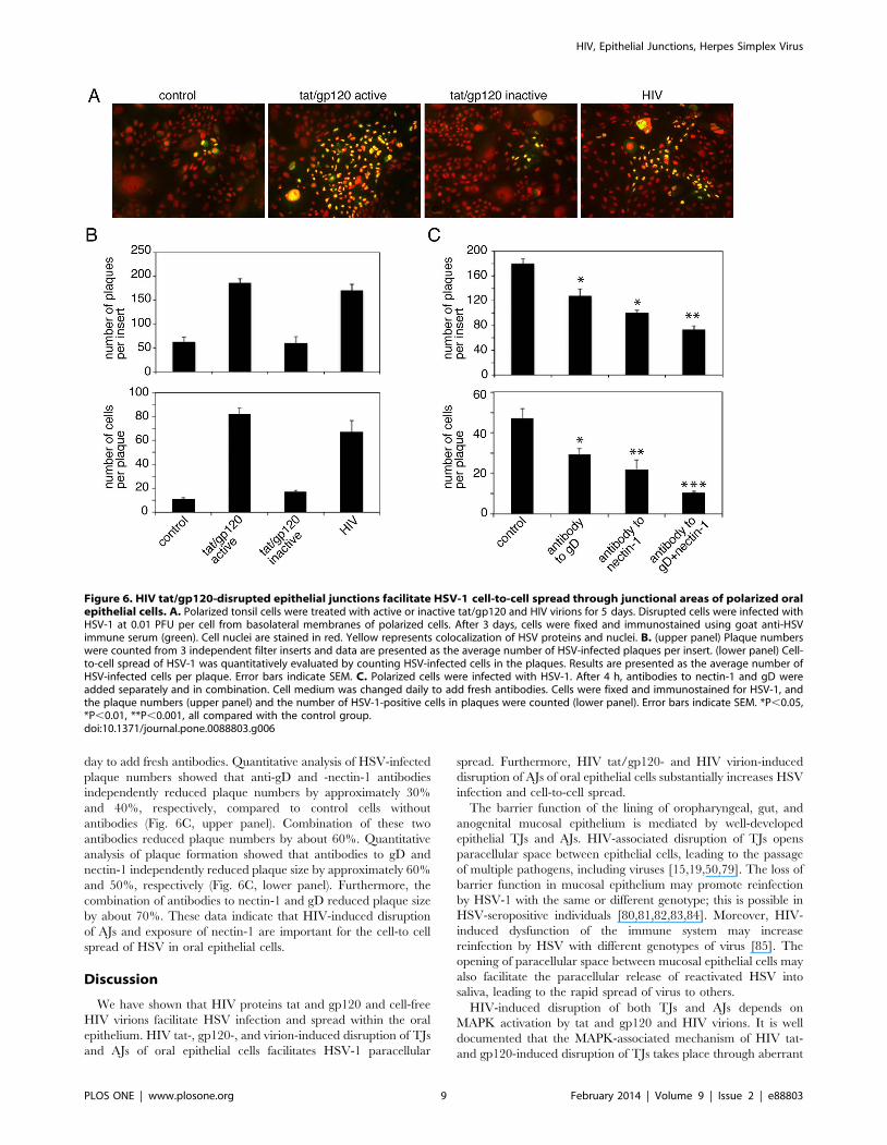
day to add fresh antibodies. Quantitative analysis of HSV-infected
plaque numbers showed that anti-gD and -nectin-1 antibodies
independently reduced plaque numbers by approximately 30%
and 40%, respectively, compared to control cells without
antibodies (Fig. 6C, upper panel). Combination of these two
antibodies reduced plaque numbers by about 60%. Quantitative
analysis of plaque formation showed that antibodies to gD and
nectin-1 independently reduced plaque size by approximately 60%
and 50%, respectively (Fig. 6C, lower panel). Furthermore, the
combination of antibodies to nectin-1 and gD reduced plaque size
by about 70%. These data indicate that HIV-induced disruption
of AJs and exposure of nectin-1 are important for the cell-to cell
spread of HSV in oral epithelial cells.
Discussion
We have shown that HIV proteins tat and gp120 and cell-free
HIV virions facilitate HSV infection and spread within the oral
epithelium. HIV tat-, gp120-, and virion-induced disruption of TJs
and AJs of oral epithelial cells facilitates HSV-1 paracellular
spread. Furthermore, HIV tat/gp120- and HIV virion-induced
disruption of AJs of oral epithelial cells substantially increases HSV
infection and cell-to-cell spread.
The barrier function of the lining of oropharyngeal, gut, and
anogenital mucosal epithelium is mediated by well-developed
epithelial TJs and AJs. HIV-associated disruption of TJs opens
paracellular space between epithelial cells, leading to the passage
of multiple pathogens, including viruses [15,19,50,79]. The loss of
barrier function in mucosal epithelium may promote reinfection
by HSV-1 with the same or different genotype; this is possible in
HSV-seropositive individuals [80,81,82,83,84]. Moreover, HIV-
induced dysfunction of the immune system may increase
reinfection by HSV with different genotypes of virus [85]. The
opening of paracellular space between mucosal epithelial cells may
also facilitate the paracellular release of reactivated HSV into
saliva, leading to the rapid spread of virus to others.
HIV-induced disruption of both TJs and AJs depends on
MAPK activation by tat and gp120 and HIV virions. It is well
documented that the MAPK-associated mechanism of HIV tat-
and gp120-induced disruption of TJs takes place through aberrant
Figure 6. HIV tat/gp120-disrupted epithelial junctions facilitate HSV-1 cell-to-cell spread through junctional areas of polarized oralepithelial cells. A. Polarized tonsil cells were treated with active or inactive tat/gp120 and HIV virions for 5 days. Disrupted cells were infected withHSV-1 at 0.01 PFU per cell from basolateral membranes of polarized cells. After 3 days, cells were fixed and immunostained using goat anti-HSVimmune serum (green). Cell nuclei are stained in red. Yellow represents colocalization of HSV proteins and nuclei. B. (upper panel) Plaque numberswere counted from 3 independent filter inserts and data are presented as the average number of HSV-infected plaques per insert. (lower panel) Cell-to-cell spread of HSV-1 was quantitatively evaluated by counting HSV-infected cells in the plaques. Results are presented as the average number ofHSV-infected cells per plaque. Error bars indicate SEM. C. Polarized cells were infected with HSV-1. After 4 h, antibodies to nectin-1 and gD wereadded separately and in combination. Cell medium was changed daily to add fresh antibodies. Cells were fixed and immunostained for HSV-1, andthe plaque numbers (upper panel) and the number of HSV-1-positive cells in plaques were counted (lower panel). Error bars indicate SEM. *P,0.05,*P,0.01, **P,0.001, all compared with the control group.doi:10.1371/journal.pone.0088803.g006
HIV, Epithelial Junctions, Herpes Simplex Virus
PLOS ONE | www.plosone.org 9 February 2014 | Volume 9 | Issue 2 | e88803
internalization of TJ proteins and their down-regulation and/or
proteasome-mediated degradation [21,22,23,24,25,26,27,28,29,
30,31,32]. Here we have shown for the first time that HIV tat
and gp120 proteins and HIV virions also induce dissociation of
AJs. It has been reported that activation of the MAPK ERK
pathway in epithelial cells induces expression of transforming
growth factor-b1, vgixg causes internalization of E-cadherin
from AJs into cytoplasm, leading to the epithelial-to-mesenchy-
mal transition (EMT) [86]. MAPK activation also induces the
expression of fibroblast growth factor-2, which causes E-cadherin
down-regulation via EMT mechanisms [87]. Our data show that
HIV virions and tat/gp120 induce activation of MAPK ERK1/2
and reduce E-cadherin expression, also altering its localization
from membrane to cytoplasm. These findings suggest that HIV
induces the disruption of AJs through the EMT phenotype, which
is a well-coordinated epigenetic process [88]. HIV has previously
been shown to cause EMT in renal epithelium. HIV infection is
associated with kidney failure due to severe nephropathy,
characterized by the loss of the renal epithelial phenotype and
the acquisition of mesenchymal features, including de-differenti-
ation, depolarization, and proliferation [89,90,91,92,93]. Our data
also consistently show that treatment of polarized oral epithelial
cells with HIV tat and gp120 and HIV virions induces disruption
of TJs and AJs and therefore the depolarization of epithelial cells.
UV irradiation of HIV virions did not affect the role of HIV in the
disruption of cell junctions and depolarization, indicating that
HIV infection is not critical for disruption of cell junctions.
The major receptor for HSV-1 gD nectin-1 is an adhesion
protein associated with AJs. Nectin-1 has hemophilic interactions
with itself and heterophilic interactions with nectins 3 and 4; such
interactions within the lateral membranes of epithelial cells form
AJs [94]. Dissociation of AJs by calcium depletion liberates nectin-
1, indicating that it is sequestered within the AJs [43]. However, in
our study HIV tat/gp120- and HIV-induced disruption of AJs did
not alter localization of nectin-1 from lateral membranes of
polarized cells, suggesting that, in this case, release or liberation of
nectin-1 may not occur. Rather, our results show that HIV-
associated disruption of AJs leads to the exposure or unmasking of
nectin-1 from its sequestered areas. HIV-induced exposure of
nectin-1 is a key factor for HSV infection in the lateral membranes
of oral epithelial cells. Nectin-1 has three Ig-like extracellular
domains; HSV gD binds to V domain, which is the distal Ig-like
domain [95,96]. Antibodies to the V domain of nectin-1 and to
HSV gD reduce HSV-1 infection, indicating that HIV-induced
disruption of AJs exposes the V domain of nectin-1 to the HSV-
1 gD. This notion is well supported by the lack of inhibitory effect
of anti-nectin-1 antibodies to HSV-1 infection in cells with intact
AJs; i.e., nectin-1 is hidden within the intact AJs and thus is not
accessible to the HSV gD. HSV-1 entry may occur by direct fusion
of the viral envelope with the plasma membrane or by endocytosis
of virions into the cytoplasm and subsequent fusion of the virion
envelope with endosomal membranes [97,98]. Mechanisms of
HSV gD and –nectin-1-mediated viral entry via HIV-disrupted
junctional areas need to be clarified.
HIV-induced disruption of AJs also promotes the cell-to-cell
spread of HSV-1, indicating that the availability of the nectin-1 V
domain on the lateral membranes of epithelial cells is critical for
the spread of progeny virions. It has been well documented that
the extracellular V domain of nectin-1 is critical for HSV cell-to-
cell spread, in contrast to the cytoplasmic tail and transmembrane
domains of nectin-1, which do not play a critical role in virus
spread [76]. HSV-1 does not spread from nectin-1-positive to
nectin-1-negative cells [77], suggesting that the disruption of AJs
may efficiently promote virus spread as it exposes nectin-1 from
neighboring epithelial cells. During the spread of HSV, newly
synthesized gD replaces the nectin-1 in infected cells at junctions
with uninfected cells [78]. This requires that unoccupied nectin-1
binding sites be available from hemophilic or heterophilic
interactions of nectin-1 from uninfected cells. Thus, the HIV-
induced exposure of nectin-1 promotes binding of gD to
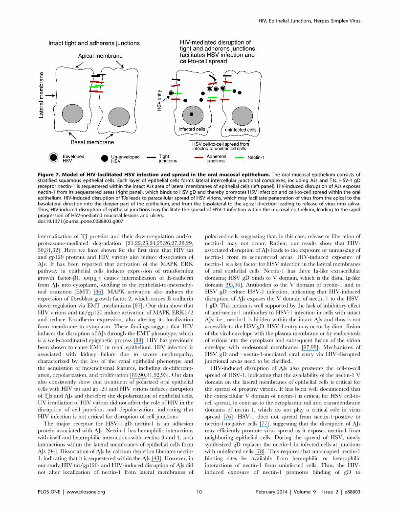
Figure 7. Model of HIV-facilitated HSV infection and spread in the oral mucosal epithelium. The oral mucosal epithelium consists ofstratified squamous epithelial cells. Each layer of epithelial cells forms lateral intercellular junctional complexes, including AJs and TJs. HSV-1 gDreceptor nectin-1 is sequestered within the intact AJs area of lateral membranes of epithelial cells (left panel). HIV-induced disruption of AJs exposesnectin-1 from its sequestered areas (right panel), which binds to HSV gD and thereby promotes HSV infection and cell-to-cell spread within the oralepithelium. HIV-induced disruption of TJs leads to paracellular spread of HSV virions, which may facilitate penetration of virus from the apical to thebasolateral direction into the deeper part of the epithelium, and from the basolateral to the apical direction leading to release of virus into saliva.Thus, HIV-induced disruption of epithelial junctions may facilitate the spread of HSV-1 infection within the mucosal epithelium, leading to the rapidprogression of HSV-mediated mucosal lesions and ulcers.doi:10.1371/journal.pone.0088803.g007
HIV, Epithelial Junctions, Herpes Simplex Virus
PLOS ONE | www.plosone.org 10 February 2014 | Volume 9 | Issue 2 | e88803
unoccupied nectin-1 binding sites from uninfected cells and
therefore may facilitate rapid HSV cell-to-cell spread.
HSV gD also interacts with other receptors. HSV-1/2 gD binds
to HVEM, HSV-1 gD binds to 3-OS-HS, and HSV-2 gD
interacts with nectin-2 [72,73,74,75,99]. Further study may
elucidate the functional role of these receptors in HSV infection
and cell-to-cell spread in HIV-disrupted polarized oral epithelial
cells.
HIV-associated HSV-1 spread in the oral epithelium is a
common manifestation of HIV/AIDS [2,3,4,5,6,7], in which
junctions of oral epithelium are disrupted [15]. HIV-associated
exposure of nectin-1 in oral epithelium could be an important
mechanism for the rapid spread of HSV-1 infection. Furthermore,
our findings may also apply to HIV-associated spread of HSV-2
within the genital mucosa [100,101,102,103,104], because HIV
induces disruption of vaginal and cervical epithelium [50] and
HSV-2 also uses nectin-1 for entry and spread [105].
The presence of HIV tat and gp120 in the mucosal
environment of HIV-infected individuals is critical for the
disruption of epithelial junctions and the exposure of nectin-1.
The presence of HIV virions and HIV gene products in oral and
genital epithelium is well described [15,106,107,108,109,110,111,
112,113,114,115,116,117,118,119], especially in circulating HIV-
infected immune cells [117,118,119]. Secretion of HIV tat and
gp120 into blood has been shown [54,55,56,57,120,121,122]. The
immune cells expressing tat and gp120 have been detected in the
oral and anal mucosa of HIV-infected individuals, including those
on antiretroviral therapy (ART) [15]. HIV in anal tissues,
including tissues from ART-treated donors, replicates and is
infectious [15]. Furthermore, tat and gp120 are detected in the
saliva, and salivary tat penetrates mucosal epithelium [15]. HIV
tat may penetrate cells and tissues through its protein transduction
domain, which is based on the amino acids arginine and lysine.
Internalization of protein into cells and tissues is accomplished by
several mechanisms, including endocytosis and macropinocytosis
[123,124,125,126,127]. Mucosal epithelium may therefore be
exposed to tat and gp120 from multiple sources, including saliva
and circulating immune cells, even among individuals receiving
ART whose HIV viral load is suppressed. Mucosal epithelium may
also serve as an HIV reservoir [118] and as a source of tat and
gp120, which disrupt AJs and expose nectin-1, leading to infection
and spread of HSV-1 and -2.
Incubation of polarized oral epithelial cells with cell-free HIV
virions also caused disruption of TJs and AJs and exposed nectin-
1, leading to HSV-1 infection and spread. Thus, in addition to
secreted tat and gp120, the presence of HIV virions within the
mucosal environment may contribute to the disruption of mucosal
epithelium and thereby the promotion of HSV spread.
In summary, we have shown that HIV proteins tat and gp120
and cell-free virions disrupt oral epithelial junctions and expose
HSV-1 receptor nectin-1, which is sequestered within the AJs.
This is critical for infection by HSV-1 and its rapid paracellular
and cell-to-cell spread (Fig. 7), suggesting that the interaction of
HIV and HSV through the mucosal environment may play an
important role in the development of HIV/AIDS-associated HSV
disease. Combined with other factors, such as HIV/AIDS-caused
reduction of immune response to HSV, this may increase the risk
of subsequent development of HSV-associated diseases. Topical
inhibitors of HIV tat- and gp120-activated MAPK signaling
[21,22,23,24,25,26,128,129,130,131,132,133,134,135], which
plays a key role in disruption of epithelial junctions and exposure
of nectin-1 in HIV-infected individuals, may be useful for reducing
HSV-1 spread within the oral and genital epithelium.
Acknowledgments
We thank Rossana Herrera for technical assistance, Jay Levy for providing
the HIV-SF33 virus strain, Gary Cohen and Roselyn Eisenberg for
providing the HSV-1 gD(306t) protein, and Joel Palefsky for useful
discussions.
Author Contributions
Conceived and designed the experiments: ST. Performed the experiments:
IS ST. Analyzed the data: IS ST. Wrote the paper: ST IS.
References
1. Wald A, Ericsson M, Krantz E, Selke S, Corey L (2004) Oral shedding of
herpes simplex virus type 2. Sexually transmitted infections 80: 272–276.
2. Corey L, Wald A, Celum CL, Quinn TC (2004) The effects of herpes simplex
virus-2 on HIV-1 acquisition and transmission: a review of two overlapping
epidemics. J Acquir Immune Defic Syndr 35: 435–445.
3. Van de Perre P, Segondy M, Foulongne V, Ouedraogo A, Konate I, et al.
(2008) Herpes simplex virus and HIV-1: deciphering viral synergy. Lancet
Infect Dis 8: 490–497.
4. Mbopi-Keou FX, Belec L, Teo CG, Scully C, Porter SR (2002) Synergism
between HIV and other viruses in the mouth. Lancet Infect Dis 2: 416–424.
5. Griffin E, Krantz E, Selke S, Huang ML, Wald A (2008) Oral mucosal
reactivation rates of herpesviruses among HIV-1 seropositive persons. J Med
Virol 80: 1153–1159.
6. Carbone A, Cesarman E, Spina M, Gloghini A, Schulz TF (2009) HIV-
associated lymphomas and gamma-herpesviruses. Blood 113: 1213–1224.
7. Leigh JE, Shetty K, Fidel PL, Jr. (2004) Oral opportunistic infections in HIV-
positive individuals: review and role of mucosal immunity. AIDS Patient Care
STDS 18: 443–456.
8. Posavad CM, Wald A, Kuntz S, Huang ML, Selke S, et al. (2004) Frequent
reactivation of herpes simplex virus among HIV-1-infected patients treated
with highly active antiretroviral therapy. The Journal of infectious diseases 190:
693–696.
9. Dinotta F, De Pasquale R, Nasca MR, Tedeschi A, Micali G (2009)
Disseminated herpes simplex infection in a HIV+ patient. Giornale italiano
di dermatologia e venereologia : organo ufficiale, Societa italiana di
dermatologia e sifilografia 144: 205–209.
10. Kebede Y, Dorigo-Zetsma W, Mengistu Y, Mekonnen Y, Schaap A, et al.
(2004) Transmission of herpes simplex virus Type 2 among factory workers in
Ethiopia. The Journal of infectious diseases 190: 365–372.
11. Levy JA (2009) HIV pathogenesis: 25 years of progress and persistent
challenges. AIDS 23: 147–160.
12. Feller L, Lemmer J (2007) Aspects of immunopathogenic mechanisms of HIV
infection. SADJ 62: 432–434, 436.
13. Feller L, Khammissa RA, Wood NH, Meyerov R, Lemmer J (2008) Insights
into immunopathogenic mechanisms of HIV infection: high levels of immune
activation and HIV fitness. SADJ 63: 552, 554, 556–557.
14. Roizman B, Whitley RJ, Lopez C, editors (1993) The Human Herpesviruses.
New York: Raven Press.
15. Tugizov SM, Herrera R, Chin-Hong P, Veluppillai P, Greenspan D, et al.
(2013) HIV-associated disruption of mucosal epithelium facilitates paracellular
penetration by human papillomavirus. Virology 446: 378–388.
16. Kapembwa MS, Fleming SC, Orr M, Wells C, Bland M, et al. (1996) Impaired
absorption of zidovudine in patients with AIDS-related small intestinal disease.
Aids 10: 1509–1514.
17. Obinna FC, Cook G, Beale T, Dave S, Cunningham D, et al. (1995)
Comparative assessment of small intestinal and colonic permeability in HIV-
infected homosexual men. Aids 9: 1009–1016.
18. Kapembwa MS, Fleming SC, Sewankambo N, Serwadda D, Lucas S, et al.
(1991) Altered small-intestinal permeability associated with diarrhoea in
human-immunodeficiency-virus-infected Caucasian and African subjects. Clin
Sci (Lond) 81: 327–334.
19. Maingat F, Halloran B, Acharjee S, van Marle G, Church D, et al. (2011)
Inflammation and epithelial cell injury in AIDS enteropathy: involvement of
endoplasmic reticulum stress. FASEB journal : official publication of the
Federation of American Societies for Experimental Biology 25: 2211–2220.
20. Sankaran S, George MD, Reay E, Guadalupe M, Flamm J, et al. (2008) Rapid
onset of intestinal epithelial barrier dysfunction in primary human immuno-
deficiency virus infection is driven by an imbalance between immune response
and mucosal repair and regeneration. J Virol 82: 538–545.
21. Toschi E, Bacigalupo I, Strippoli R, Chiozzini C, Cereseto A, et al. (2006)
HIV-1 Tat regulates endothelial cell cycle progression via activation of the
Ras/ERK MAPK signaling pathway. Mol Biol Cell 17: 1985–1994.
HIV, Epithelial Junctions, Herpes Simplex Virus
PLOS ONE | www.plosone.org 11 February 2014 | Volume 9 | Issue 2 | e88803
22. Pu H, Tian J, Andras IE, Hayashi K, Flora G, et al. (2005) HIV-1 Tat protein-
induced alterations of ZO-1 expression are mediated by redox-regulated ERK
1/2 activation. J Cereb Blood Flow Metab 25: 1325–1335.
23. Andras IE, Pu H, Tian J, Deli MA, Nath A, et al. (2005) Signaling mechanisms
of HIV-1 Tat-induced alterations of claudin-5 expression in brain endothelial
cells. J Cereb Blood Flow Metab 25: 1159–1170.
24. Bai L, Zhang Z, Zhang H, Li X, Yu Q, et al. (2008) HIV-1 Tat protein alter
the tight junction integrity and function of retinal pigment epithelium: an
in vitro study. BMC Infect Dis 8: 77.
25. Zhong Y, Smart EJ, Weksler B, Couraud PO, Hennig B, et al. (2008) Caveolin-
1 regulates human immunodeficiency virus-1 Tat-induced alterations of tight
junction protein expression via modulation of the Ras signaling. J Neurosci 28:
7788–7796.
26. Song L, Ge S, Pachter JS (2007) Caveolin-1 regulates expression of junction-
associated proteins in brain microvascular endothelial cells. Blood 109: 1515–
1523.
27. Kanmogne GD, Schall K, Leibhart J, Knipe B, Gendelman HE, et al. (2007)
HIV-1 gp120 compromises blood-brain barrier integrity and enhances
monocyte migration across blood-brain barrier: implication for viral neuro-
pathogenesis. J Cereb Blood Flow Metab 27: 123–134.
28. Kanmogne GD, Primeaux C, Grammas P (2005) HIV-1 gp120 proteins alter
tight junction protein expression and brain endothelial cell permeability:
implications for the pathogenesis of HIV-associated dementia. J Neuropathol
Exp Neurol 64: 498–505.
29. Nakamuta S, Endo H, Higashi Y, Kousaka A, Yamada H, et al. (2008) Human
immunodeficiency virus type 1 gp120-mediated disruption of tight junction
proteins by induction of proteasome-mediated degradation of zonula
occludens-1 and -2 in human brain microvascular endothelial cells.
J Neurovirol 14: 186–195.
30. Shin K, Fogg VC, Margolis B (2006) Tight junctions and cell polarity. Annual
review of cell and developmental biology 22: 207–235.
31. Umeda K, Ikenouchi J, Katahira-Tayama S, Furuse K, Sasaki H, et al. (2006)
ZO-1 and ZO-2 independently determine where claudins are polymerized in
tight-junction strand formation. Cell 126: 741–754.
32. Shen L, Turner JR (2005) Actin depolymerization disrupts tight junctions via
caveolae-mediated endocytosis. Molecular biology of the cell 16: 3919–3936.
33. Takahashi K, Nakanishi H, Miyahara M, Mandai K, Satoh K, et al. (1999)
Nectin/PRR: an immunoglobulin-like cell adhesion molecule recruited to
cadherin-based adherens junctions through interaction with Afadin, a PDZ
domain-containing protein. The Journal of cell biology 145: 539–549.
34. Takai Y, Nakanishi H (2003) Nectin and afadin: novel organizers of
intercellular junctions. Journal of cell science 116: 17–27.
35. Tiwari V, Oh MJ, Kovacs M, Shukla SY, Valyi-Nagy T, et al. (2008) Role for
nectin-1 in herpes simplex virus 1 entry and spread in human retinal pigment
epithelial cells. The FEBS journal 275: 5272–5285.
36. Karaba AH, Kopp SJ, Longnecker R (2011) Herpesvirus entry mediator and
nectin-1 mediate herpes simplex virus 1 infection of the murine cornea. Journal
of virology 85: 10041–10047.
37. Connolly SA, Landsburg DJ, Carfi A, Whitbeck JC, Zuo Y, et al. (2005)
Potential nectin-1 binding site on herpes simplex virus glycoprotein d. Journal
of virology 79: 1282–1295.
38. Krummenacher C, Baribaud F, Ponce de Leon M, Baribaud I, Whitbeck JC, et
al. (2004) Comparative usage of herpesvirus entry mediator A and nectin-1 by
laboratory strains and clinical isolates of herpes simplex virus. Virology 322:
286–299.
39. Gianni T, Campadelli-Fiume G, Menotti L (2004) Entry of herpes simplex
virus mediated by chimeric forms of nectin1 retargeted to endosomes or to lipid
rafts occurs through acidic endosomes. Journal of virology 78: 12268–12276.
40. Cocchi F, Lopez M, Dubreuil P, Campadelli Fiume G, Menotti L (2001)
Chimeric nectin1-poliovirus receptor molecules identify a nectin1 region
functional in herpes simplex virus entry. Journal of virology 75: 7987–7994.
41. Struyf F, Posavad CM, Keyaerts E, Van Ranst M, Corey L, et al. (2002) Search
for polymorphisms in the genes for herpesvirus entry mediator, nectin-1, and
nectin-2 in immune seronegative individuals. The Journal of infectious diseases
185: 36–44.
42. Krummenacher C, Nicola AV, Whitbeck JC, Lou H, Hou W, et al. (1998)
Herpes simplex virus glycoprotein D can bind to poliovirus receptor-related
protein 1 or herpesvirus entry mediator, two structurally unrelated mediators of
virus entry. Journal of virology 72: 7064–7074.
43. Yoon M, Spear PG (2002) Disruption of adherens junctions liberates nectin-1
to serve as receptor for herpes simplex virus and pseudorabies virus entry.
Journal of virology 76: 7203–7208.
44. Xiao J, Palefsky JM, Herrera R, Berline J, Tugizov SM (2009) EBV BMRF-2
facilitates cell-to-cell spread of virus within polarized oral epithelial cells.
Virology 388: 335–343.
45. Tugizov SM, Herrera R, Palefsky JM (2013) Epstein-Barr Virus Transcytosis
through Polarized Oral Epithelial Cells. Journal of virology 87: 8179–8194.
46. Tugizov SM, Herrera R, Veluppillai P, Greenspan D, Soros V, et al. (2011)
HIV is inactivated after transepithelial migration via adult oral epithelial cells
but not fetal epithelial cells. Virology 409: 211–222.
47. Tugizov SM, Berline JW, Palefsky JM (2003) Epstein-Barr virus infection of
polarized tongue and nasopharyngeal epithelial cells. Nat Med 9: 307–314.
48. Gulino D, Delachanal E, Concord E, Genoux Y, Morand B, et al. (1998)
Alteration of endothelial cell monolayer integrity triggers resynthesis of vascularendothelium cadherin. J Biol Chem 273: 29786–29793.
49. Tugizov SM, Herrera R, Veluppillai P, Greenspan D, Soros V, et al. (2012)
Differential transmission of HIV traversing fetal oral/intestinal epithelia and
adult oral epithelia. Journal of virology 86: 2556–2570.
50. Nazli A, Chan O, Dobson-Belaire WN, Ouellet M, Tremblay MJ, et al. (2010)Exposure to HIV-1 directly impairs mucosal epithelial barrier integrity
allowing microbial translocation. PLoS pathogens 6: e1000852.
51. Navarro D, Paz P, Pereira L (1992) Domains of herpes simplex virus 1
glycoprotein B that function in virion penetration, cell-to-cell spread, and cellfusion. Virology 186: 99–112.
52. Sisk WP, Bradley JD, Leipold RJ, Stoltzfus AM, Ponce de Leon M, et al. (1994)
High-level expression and purification of secreted forms of herpes simplex virus
type 1 glycoprotein gD synthesized by baculovirus-infected insect cells. Journalof virology 68: 766–775.
53. Krautkramer E, Zeier M (2008) Hantavirus causing hemorrhagic fever with
renal syndrome enters from the apical surface and requires decay-acceleratingfactor (DAF/CD55). Journal of virology 82: 4257–4264.
54. Westendorp MO, Frank R, Ochsenbauer C, Stricker K, Dhein J, et al. (1995)Sensitization of T cells to CD95-mediated apoptosis by HIV-1 Tat and gp120.
Nature 375: 497–500.
55. Xiao H, Neuveut C, Tiffany HL, Benkirane M, Rich EA, et al. (2000) Selective
CXCR4 antagonism by Tat: implications for in vivo expansion of coreceptoruse by HIV-1. Proc Natl Acad Sci U S A 97: 11466–11471.
56. Rychert J, Strick D, Bazner S, Robinson J, Rosenberg E (2010) Detection of
HIV gp120 in plasma during early HIV infection is associated with increasedproinflammatory and immunoregulatory cytokines. AIDS research and human
retroviruses 26: 1139–1145.
57. Poggi A, Zocchi MR (2006) HIV-1 Tat triggers TGF-beta production and NK
cell apoptosis that is prevented by pertussis toxin B. Clinical & developmentalimmunology 13: 369–372.
58. Nagahara H, Vocero-Akbani AM, Snyder EL, Ho A, Latham DG, et al. (1998)Transduction of full-length TAT fusion proteins into mammalian cells: TAT-
p27Kip1 induces cell migration. Nat Med 4: 1449–1452.
59. Barillari G, Sgadari C, Fiorelli V, Samaniego F, Colombini S, et al. (1999) TheTat protein of human immunodeficiency virus type-1 promotes vascular cell
growth and locomotion by engaging the alpha5beta1 and alphavbeta3 integrins
and by mobilizing sequestered basic fibroblast growth factor. Blood 94: 663–672.
60. Barillari G, Sgadari C, Palladino C, Gendelman R, Caputo A, et al. (1999)
Inflammatory cytokines synergize with the HIV-1 Tat protein to promote
angiogenesis and Kaposi’s sarcoma via induction of basic fibroblast growthfactor and the alpha v beta 3 integrin. J Immunol 163: 1929–1935.
61. Su YH, Zhang X, Wang X, Fraser NW, Block TM (2006) Evidence that the
immediate-early gene product ICP4 is necessary for the genome of the herpessimplex virus type 1 ICP4 deletion mutant strain d120 to circularize in infected
cells. Journal of virology 80: 11589–11597.
62. Watson K, Edwards RJ (1999) HIV-1-trans-activating (Tat) protein: both a
target and a tool in therapeutic approaches. Biochem Pharmacol 58: 1521–1528.
63. Vogel BE, Lee SJ, Hildebrand A, Craig W, Pierschbacher MD, et al. (1993) Anovel integrin specificity exemplified by binding of the alpha v beta 5 integrin
to the basic domain of the HIV Tat protein and vitronectin. J Cell Biol 121:461–468.
64. Trushin SA, Algeciras-Schimnich A, Vlahakis SR, Bren GD, Warren S, et al.
(2007) Glycoprotein 120 binding to CXCR4 causes p38-dependent primary T
cell death that is facilitated by, but does not require cell-associated CD4.Journal of immunology 178: 4846–4853.
65. Kinet S, Bernard F, Mongellaz C, Perreau M, Goldman FD, et al. (2002)
gp120-mediated induction of the MAPK cascade is dependent on the
activation state of CD4(+) lymphocytes. Blood 100: 2546–2553.
66. Kuang YQ, Pang W, Zheng YT, Dupre DJ (2012) NHERF1 regulates gp120-induced internalization and signaling by CCR5, and HIV-1 production.
European journal of immunology 42: 299–310.
67. Lee C, Liu QH, Tomkowicz B, Yi Y, Freedman BD, et al. (2003) Macrophage
activation through CCR5- and CXCR4-mediated gp120-elicited signalingpathways. Journal of leukocyte biology 74: 676–682.
68. Xiao J, Palefsky JM, Herrera R, Tugizov SM (2007) Characterization of the
Epstein-Barr virus glycoprotein BMRF-2. Virology 359: 382–396.
69. Chen Y, Lu Q, Schneeberger EE, Goodenough DA (2000) Restoration of tight
junction structure and barrier function by down-regulation of the mitogen-activated protein kinase pathway in ras-transformed Madin-Darby canine
kidney cells. Molecular biology of the cell 11: 849–862.
70. Kuphal S, Poser I, Jobin C, Hellerbrand C, Bosserhoff AK (2004) Loss of E-
cadherin leads to upregulation of NFkappaB activity in malignant melanoma.Oncogene 23: 8509–8519.
71. Krummenacher C, Baribaud I, Ponce de Leon M, Whitbeck JC, Lou H, et al.
(2000) Localization of a binding site for herpes simplex virus glycoprotein D on
herpesvirus entry mediator C by using antireceptor monoclonal antibodies.Journal of virology 74: 10863–10872.
72. Montgomery RI, Warner MS, Lum BJ, Spear PG (1996) Herpes simplex virus-
1 entry into cells mediated by a novel member of the TNF/NGF receptor
family. Cell 87: 427–436.
HIV, Epithelial Junctions, Herpes Simplex Virus
PLOS ONE | www.plosone.org 12 February 2014 | Volume 9 | Issue 2 | e88803
73. Shieh MT, WuDunn D, Montgomery RI, Esko JD, Spear PG (1992) Cell
surface receptors for herpes simplex virus are heparan sulfate proteoglycans.The Journal of cell biology 116: 1273–1281.
74. Terry-Allison T, Montgomery RI, Whitbeck JC, Xu R, Cohen GH, et al.
(1998) HveA (herpesvirus entry mediator A), a coreceptor for herpes simplexvirus entry, also participates in virus-induced cell fusion. Journal of virology 72:
5802–5810.
75. Shukla D, Liu J, Blaiklock P, Shworak NW, Bai X, et al. (1999) A novel role for
3-O-sulfated heparan sulfate in herpes simplex virus 1 entry. Cell 99: 13–22.
76. Even DL, Henley AM, Geraghty RJ (2006) The requirements for herpessimplex virus type 1 cell-cell spread via nectin-1 parallel those for virus entry.
Virus research 119: 195–207.
77. Cocchi F, Menotti L, Dubreuil P, Lopez M, Campadelli-Fiume G (2000) Cell-to-cell spread of wild-type herpes simplex virus type 1, but not of syncytial
strains, is mediated by the immunoglobulin-like receptors that mediate virion
entry, nectin1 (PRR1/HveC/HIgR) and nectin2 (PRR2/HveB). Journal ofvirology 74: 3909–3917.
78. Krummenacher C, Baribaud I, Eisenberg RJ, Cohen GH (2003) Cellular
localization of nectin-1 and glycoprotein D during herpes simplex virusinfection. J Virol 77: 8985–8999.
79. Epple HJ, Schneider T, Troeger H, Kunkel D, Allers K, et al. (2009)
Impairment of the intestinal barrier is evident in untreated but absent insuppressively treated HIV-infected patients. Gut 58: 220–227.
80. Remeijer L, Maertzdorf J, Buitenwerf J, Osterhaus AD, Verjans GM (2002)Corneal herpes simplex virus type 1 superinfection in patients with
recrudescent herpetic keratitis. Investigative ophthalmology & visual science43: 358–363.
81. Sucato G, Wald A, Wakabayashi E, Vieira J, Corey L (1998) Evidence of
latency and reactivation of both herpes simplex virus (HSV)-1 and HSV-2 inthe genital region. The Journal of infectious diseases 177: 1069–1072.
82. Buchman TG, Roizman B, Nahmias AJ (1979) Demonstration of exogenous
genital reinfection with herpes simplex virus type 2 by restriction endonuclease
fingerprinting of viral DNA. The Journal of infectious diseases 140: 295–304.
83. Maertzdorf J, Remeijer L, Van Der Lelij A, Buitenwerf J, Niesters HG, et al.(1999) Amplification of reiterated sequences of herpes simplex virus type 1
(HSV-1) genome to discriminate between clinical HSV-1 isolates. Journal ofclinical microbiology 37: 3518–3523.
84. Stanberry LR, Cunningham AL, Mindel A, Scott LL, Spruance SL, et al.
(2000) Prospects for control of herpes simplex virus disease through
immunization. Clinical infectious diseases : an official publication of theInfectious Diseases Society of America 30: 549–566.
85. Roest RW, Maertzdorf J, Kant M, van der Meijden WI, Osterhaus AD, et al.
(2006) High incidence of genotypic variance between sequential herpes simplexvirus type 2 isolates from HIV-1-seropositive patients with recurrent genital
herpes. The Journal of infectious diseases 194: 1115–1118.
86. Xie L, Law BK, Chytil AM, Brown KA, Aakre ME, et al. (2004) Activation of
the Erk pathway is required for TGF-beta1-induced EMT in vitro. Neoplasia6: 603–610.
87. Lau MT, So WK, Leung PC (2013) Fibroblast growth factor 2 induces E-
cadherin down-regulation via PI3K/Akt/mTOR and MAPK/ERK signalingin ovarian cancer cells. PloS one 8: e59083.
88. Stadler SC, Allis CD (2012) Linking epithelial-to-mesenchymal-transition and
epigenetic modifications. Seminars in cancer biology 22: 404–410.
89. Barisoni L, Bruggeman LA, Mundel P, D’Agati VD, Klotman PE (2000) HIV-
1 induces renal epithelial dedifferentiation in a transgenic model of HIV-associated nephropathy. Kidney international 58: 173–181.
90. Schwartz EJ, Cara A, Snoeck H, Ross MD, Sunamoto M, et al. (2001) Human
immunodeficiency virus-1 induces loss of contact inhibition in podocytes.Journal of the American Society of Nephrology : JASN 12: 1677–1684.
91. Medapalli RK, He JC, Klotman PE (2011) HIV-associated nephropathy:
pathogenesis. Current opinion in nephrology and hypertension 20: 306–311.
92. Bruggeman LA, Ross MD, Tanji N, Cara A, Dikman S, et al. (2000) Renal
epithelium is a previously unrecognized site of HIV-1 infection. Journal of theAmerican Society of Nephrology : JASN 11: 2079–2087.
93. Lu TC, He JC, Wang ZH, Feng X, Fukumi-Tominaga T, et al. (2008) HIV-1
Nef disrupts the podocyte actin cytoskeleton by interacting with diaphanousinteracting protein. The Journal of biological chemistry 283: 8173–8182.
94. Takai Y, Irie K, Shimizu K, Sakisaka T, Ikeda W (2003) Nectins and nectin-
like molecules: roles in cell adhesion, migration, and polarization. Cancer
science 94: 655–667.
95. Cocchi F, Lopez M, Menotti L, Aoubala M, Dubreuil P, et al. (1998) The Vdomain of herpesvirus Ig-like receptor (HIgR) contains a major functional
region in herpes simplex virus-1 entry into cells and interacts physically with theviral glycoprotein D. Proceedings of the National Academy of Sciences of the
United States of America 95: 15700–15705.
96. Krummenacher C, Rux AH, Whitbeck JC, Ponce-de-Leon M, Lou H, et al.
(1999) The first immunoglobulin-like domain of HveC is sufficient to bindherpes simplex virus gD with full affinity, while the third domain is involved in
oligomerization of HveC. Journal of virology 73: 8127–8137.
97. Nicola AV, Straus SE (2004) Cellular and viral requirements for rapidendocytic entry of herpes simplex virus. Journal of virology 78: 7508–7517.
98. Pertel PE, Fridberg A, Parish ML, Spear PG (2001) Cell fusion induced by
herpes simplex virus glycoproteins gB, gD, and gH-gL requires a gD receptor
but not necessarily heparan sulfate. Virology 279: 313–324.
99. Delboy MG, Patterson JL, Hollander AM, Nicola AV (2006) Nectin-2-
mediated entry of a syncytial strain of herpes simplex virus via pH-independentfusion with the plasma membrane of Chinese hamster ovary cells. Virology
journal 3: 105.
100. Smith JS, Robinson NJ (2002) Age-specific prevalence of infection with herpes
simplex virus types 2 and 1: a global review. The Journal of infectious diseases186 Suppl 1: S3–28.
101. Andreoletti L, Piednoir E, Legoff J, Brodard V, Beguinot I, et al. (2005) High
seroprevalence of herpes simplex virus type 2 infection in French humanimmunodeficiency virus type 1-infected outpatients. Journal of clinical
microbiology 43: 4215–4217.
102. Chakraborty N, Bhattacharyya S, De C, Mukherjee A, Bhattacharya D, et al.
(2010) Incidence of multiple Herpesvirus infection in HIV seropositive patients,a big concern for Eastern Indian scenario. Virology journal 7: 147.
103. Turner KR, McFarland W, Kellogg TA, Wong E, Page-Shafer K, et al. (2003)Incidence and prevalence of herpes simplex virus type 2 infection in persons
seeking repeat HIV counseling and testing. Sexually transmitted diseases 30:331–334.
104. Hagan H, Jenness SM, Wendel T, Murrill CR, Neaigus A, et al. (2010) Herpes
simplex virus type 2 associated with HIV infection among New York
heterosexuals living in high-risk areas. International journal of STD & AIDS21: 580–583.
105. Shukla SY, Singh YK, Shukla D (2009) Role of nectin-1, HVEM, and PILR-
alpha in HSV-2 entry into human retinal pigment epithelial cells. Investigative
ophthalmology & visual science 50: 2878–2887.
106. Rodriguez-Inigo E, Jimenez E, Bartolome J, Ortiz-Movilla N, Bartolome VillarB, et al. (2005) Detection of human immunodeficiency virus type 1 RNA by in
situ hybridization in oral mucosa epithelial cells from anti-HIV-1 positivepatients. J Med Virol 77: 17–22.
107. Chou LL, Epstein J, Cassol SA, West DM, He W, et al. (2000) Oral mucosalLangerhans’ cells as target, effector and vector in HIV infection. J Oral Pathol
Med 29: 394–402.
108. Goto Y, Yeh CK, Notkins AL, Prabhakar BS (1991) Detection of proviral
sequences in saliva of patients infected with human immunodeficiency virustype 1. AIDS Res Hum Retroviruses 7: 343–347.
109. Kakizawa J, Ushijima H, Oka S, Ikeda Y, Schroder HC, et al. (1996) Detection
of human immunodeficiency virus-1 DNA, RNA and antibody, and occultblood in inactivated saliva: availability of the filter paper disk method. Acta
Paediatr Jpn 38: 218–223.
110. Liuzzi G, Chirianni A, Clementi M, Bagnarelli P, Valenza A, et al. (1996)
Analysis of HIV-1 load in blood, semen and saliva: evidence for different viralcompartments in a cross-sectional and longitudinal study. Aids 10: F51–56.
111. Maticic M, Poljak M, Kramar B, Tomazic J, Vidmar L, et al. (2000) ProviralHIV-1 DNA in gingival crevicular fluid of HIV-1-infected patients in various
stages of HIV disease. J Dent Res 79: 1496–1501.
112. Qureshi MN, Barr CE, Hewlitt I, Boorstein R, Kong F, et al. (1997) Detectionof HIV in oral mucosal cells. Oral Dis 3 Suppl 1: S73–78.
113. Qureshi MN, Barr CE, Seshamma T, Reidy J, Pomerantz RJ, et al. (1995)Infection of oral mucosal cells by human immunodeficiency virus type 1 in
seropositive persons. J Infect Dis 171: 190–193.
114. Zuckerman RA, Whittington WL, Celum CL, Collis T, Lucchetti A, et al.
(2003) Factors associated with oropharyngeal human immunodeficiency virusshedding. J Infect Dis 188: 142–145.
115. Nuovo GJ, Forde A, MacConnell P, Fahrenwald R (1993) In situ detection of
PCR-amplified HIV-1 nucleic acids and tumor necrosis factor cDNA incervical tissues. Am J Pathol 143: 40–48.
116. Clemetson DB, Moss GB, Willerford DM, Hensel M, Emonyi W, et al. (1993)Detection of HIV DNA in cervical and vaginal secretions. Prevalence and
correlates among women in Nairobi, Kenya. JAMA : the journal of theAmerican Medical Association 269: 2860–2864.
117. Sonza S, Mutimer HP, Oelrichs R, Jardine D, Harvey K, et al. (2001)Monocytes harbour replication-competent, non-latent HIV-1 in patients on
highly active antiretroviral therapy. Aids 15: 17–22.
118. Henning TR, Kissinger P, Lacour N, Meyaski-Schluter M, Clark R, et al.(2010) Elevated cervical white blood cell infiltrate is associated with genital HIV
detection in a longitudinal cohort of antiretroviral therapy-adherent women.
The Journal of infectious diseases 202: 1543–1552.
119. Crowe SM, Sonza S (2000) HIV-1 can be recovered from a variety of cellsincluding peripheral blood monocytes of patients receiving highly active
antiretroviral therapy: a further obstacle to eradication. Journal of leukocyte
biology 68: 345–350.
120. Santosuosso M, Righi E, Lindstrom V, Leblanc PR, Poznansky MC (2009)HIV-1 envelope protein gp120 is present at high concentrations in secondary
lymphoid organs of individuals with chronic HIV-1 infection. The Journal of
infectious diseases 200: 1050–1053.
121. Oh SK, Cruikshank WW, Raina J, Blanchard GC, Adler WH, et al. (1992)Identification of HIV-1 envelope glycoprotein in the serum of AIDS and ARC
patients. Journal of acquired immune deficiency syndromes 5: 251–256.
122. Montagnier L, Clavel F, Krust B, Chamaret S, Rey F, et al. (1985)
Identification and antigenicity of the major envelope glycoprotein oflymphadenopathy-associated virus. Virology 144: 283–289.
123. Kaplan IM, Wadia JS, Dowdy SF (2005) Cationic TAT peptide transduction
domain enters cells by macropinocytosis. Journal of controlled release : official
journal of the Controlled Release Society 102: 247–253.
HIV, Epithelial Junctions, Herpes Simplex Virus
PLOS ONE | www.plosone.org 13 February 2014 | Volume 9 | Issue 2 | e88803
124. Wadia JS, Stan RV, Dowdy SF (2004) Transducible TAT-HA fusogenic
peptide enhances escape of TAT-fusion proteins after lipid raft macropinocy-tosis. Nature medicine 10: 310–315.
125. Mann DA, Frankel AD (1991) Endocytosis and targeting of exogenous HIV-1
Tat protein. The EMBO journal 10: 1733–1739.126. Ferrari A, Pellegrini V, Arcangeli C, Fittipaldi A, Giacca M, et al. (2003)
Caveolae-mediated internalization of extracellular HIV-1 tat fusion proteinsvisualized in real time. Molecular therapy : the journal of the American Society
of Gene Therapy 8: 284–294.
127. Fittipaldi A, Ferrari A, Zoppe M, Arcangeli C, Pellegrini V, et al. (2003) Cellmembrane lipid rafts mediate caveolar endocytosis of HIV-1 Tat fusion
proteins. The Journal of biological chemistry 278: 34141–34149.128. Preiss S, Namgaladze D, Brune B (2007) Critical role for classical PKC in
activating Akt by phospholipase A2-modified LDL in monocytic cells.Cardiovasc Res 73: 833–840.
129. Kawakami Y, Nishimoto H, Kitaura J, Maeda-Yamamoto M, Kato RM, et al.
(2004) Protein kinase C betaII regulates Akt phosphorylation on Ser-473 in acell type- and stimulus-specific fashion. J Biol Chem 279: 47720–47725.
130. Zhou H, Das S, Murthy KS (2003) Erk1/2- and p38 MAP kinase-dependent
phosphorylation and activation of cPLA2 by m3 and m2 receptors. Am J Physiol
Gastrointest Liver Physiol 284: G472–480.
131. Lopez-Bergami P, Habelhah H, Bhoumik A, Zhang W, Wang LH, et al. (2005)
RACK1 mediates activation of JNK by protein kinase C [corrected]. Mol Cell
19: 309–320.
132. Ali AS, Ali S, El-Rayes BF, Philip PA, Sarkar FH (2009) Exploitation of protein
kinase C: a useful target for cancer therapy. Cancer Treat Rev 35: 1–8.
133. Marshall CJ (1996) Cell signalling. Raf gets it together. Nature 383: 127–128.
134. Cai H, Smola U, Wixler V, Eisenmann-Tappe I, Diaz-Meco MT, et al. (1997)
Role of diacylglycerol-regulated protein kinase C isotypes in growth factor
activation of the Raf-1 protein kinase. Mol Cell Biol 17: 732–741.
135. Wang Y, Zhang J, Yi XJ, Yu FS (2004) Activation of ERK1/2 MAP kinase
pathway induces tight junction disruption in human corneal epithelial cells.
Exp Eye Res 78: 125–136.
HIV, Epithelial Junctions, Herpes Simplex Virus
PLOS ONE | www.plosone.org 14 February 2014 | Volume 9 | Issue 2 | e88803
Related Documents


