Chapter 4 History of Antimicrobial Prophylaxis Protocols for Infective Endocarditis Secondary to Dental Procedures Inmaculada Tomás and Maximiliano Álvarez-Fernández Additional information is available at the end of the chapter http://dx.doi.org/10.5772/56118 1. Introduction For several decades, the haematogenous spread of bacteria from the oral cavity has been considered a decisive factor in the pathogenesis of 10% to 15% of episodes of infective endocarditis (IE), suggesting that certain dental procedures may represent a significant risk factor [1]. Nowadays, however, this statement has its detractors; their main argument is that not all patients with heart valves infected by bacteria that typically colonize ecological niches of the oral cavity have undergone dental procedures. Furthermore, there is little evidence to date on the genetic similarity between bacteria isolated from the heart valves, from the bloodstream, and from the oral cavity of patients with IE [2,3]. Apart from its possible involvement in the development of episodes of IE, bacteraemia of oral origin has become of particular interest in the past 2 decades because it has been associated with the progression of atherosclerosis and may thus be related to ischemic processes, although the mechanism of action has not yet been fully elucidated [4-6]. A number of published clinical studies have demonstrated an association between periodontal disease and cardiovascular disease [7-9], and oral bacteria have been detected on heart valves and in atherosclerotic plaques and aortic aneurysms [10-12]. In 1935, Okell and Elliot [13] were the first authors to detect bacteraemia caused by Strepto‐ coccus species (in 64% of cases) after performing dental extractions on 138 patients. A year later, Burket and Burn [14] inoculated pigmented Serratia marcescens into the gingival sulcus of 90 patients before performing dental extractions and they subsequently isolated this bacterium in 20% of post-manipulation blood cultures. Those results confirmed that microorganisms from the oral cavity could enter the bloodstream after dental extraction. Between the mid 1930s © 2013 Tomás and Álvarez-Fernández; licensee InTech. This is an open access article distributed under the terms of the Creative Commons Attribution License (http://creativecommons.org/licenses/by/3.0), which permits unrestricted use, distribution, and reproduction in any medium, provided the original work is properly cited.

Welcome message from author
This document is posted to help you gain knowledge. Please leave a comment to let me know what you think about it! Share it to your friends and learn new things together.
Transcript
Chapter 4
History of Antimicrobial Prophylaxis Protocols forInfective Endocarditis Secondary to Dental Procedures
Inmaculada Tomás andMaximiliano Álvarez-Fernández
Additional information is available at the end of the chapter
http://dx.doi.org/10.5772/56118
1. Introduction
For several decades, the haematogenous spread of bacteria from the oral cavity has beenconsidered a decisive factor in the pathogenesis of 10% to 15% of episodes of infectiveendocarditis (IE), suggesting that certain dental procedures may represent a significant riskfactor [1]. Nowadays, however, this statement has its detractors; their main argument is thatnot all patients with heart valves infected by bacteria that typically colonize ecological nichesof the oral cavity have undergone dental procedures. Furthermore, there is little evidence todate on the genetic similarity between bacteria isolated from the heart valves, from thebloodstream, and from the oral cavity of patients with IE [2,3].
Apart from its possible involvement in the development of episodes of IE, bacteraemia of oralorigin has become of particular interest in the past 2 decades because it has been associatedwith the progression of atherosclerosis and may thus be related to ischemic processes, althoughthe mechanism of action has not yet been fully elucidated [4-6]. A number of published clinicalstudies have demonstrated an association between periodontal disease and cardiovasculardisease [7-9], and oral bacteria have been detected on heart valves and in atheroscleroticplaques and aortic aneurysms [10-12].
In 1935, Okell and Elliot [13] were the first authors to detect bacteraemia caused by Strepto‐coccus species (in 64% of cases) after performing dental extractions on 138 patients. A year later,Burket and Burn [14] inoculated pigmented Serratia marcescens into the gingival sulcus of 90patients before performing dental extractions and they subsequently isolated this bacteriumin 20% of post-manipulation blood cultures. Those results confirmed that microorganismsfrom the oral cavity could enter the bloodstream after dental extraction. Between the mid 1930s
© 2013 Tomás and Álvarez-Fernández; licensee InTech. This is an open access article distributed under theterms of the Creative Commons Attribution License (http://creativecommons.org/licenses/by/3.0), whichpermits unrestricted use, distribution, and reproduction in any medium, provided the original work isproperly cited.
and the early 1950s, numerous studies were published on the prevalence of post-dentalextraction bacteraemia, with figures that varied between 2% and 83% [15-19]. In the early 1930sthere was a growing awareness of the need for IE prophylaxis in patients with valvular heartdisease undergoing certain dental manipulations, and the first guidelines recommending theuse of certain sulfonamides to prevent IE of oral origin were published at the end of that decade.This chapter first provides a review of development of antimicrobial prophylaxis protocols forIE secondary to dental procedures between 1930 and 1955. Since the American Heart Associ‐ation (AHA) published its first guideline for the prevention of IE secondary to dental proce‐dures in 1955, several international committees formed mainly of cardiologists, infectiousdiseases specialists and pharmacologists have drawn up different prophylactic regimens basedon findings published in the scientific literature. In the second part of this chapter we thereforereview the changes in IE prophylaxis in the guidelines published by the AHA and the BritishSociety of Antimicrobial Chemotherapy (BSAC) between 1960 and 2009, as well as thoserecently drawn up by other societies. Those guidelines provide a description of the susceptiblepatient, the at-risk dental procedures, the influence of the anaesthetic technique applied indental treatment, the antibiotic prophylaxis protocols (antibiotics of choice, dose and route ofadministration) and the use of antiseptic prophylaxis.
2. Development of antimicrobial prophylaxis protocols for infectiveendocarditis secondary to dental procedures: 1930 to 1955
In the early 1930s, Brown and Abrahamson [20,21] were 2 of the pioneers of the application ofIE prophylaxis before performing certain dental manipulations in patients with valvular heartdisease. Those investigators recommended the prophylactic use of autogenous vaccines. In1938, Feldman and Trace [22] suggested cleaning and scraping the teeth before any manipu‐lation in order to reduce contamination of the operative field; they performed only 1 or 2 dentalextractions per session, and followed this by curettage and irrigation of the periodontal pocketswith antiseptics. A year later, Elliott [23] proposed perialveolar cauterization of the gingiva asa prophylactic measure after dental extraction; this technique not only sterilized the sulcus butalso sealed the gingival capillaries, preventing the entry of microorganisms into the blood‐stream. The practice of dental extractions under local anaesthesia with epinephrine by theinfiltration technique was also recommended, as some authors had shown that this type ofanaesthetic applied in this way created a barrier, preventing vascular invasion by the bacterialinoculum [14,22]. Fish and Maclean [24] recommended that teeth be filled with cotton soakedin a paste of zinc oxide and oil of cloves and that this should be renewed every few days; thoseauthors also recommended the administration of a dose of prontosil (azosulfamide) before adental extraction, in addition to cauterization of the gingiva. However, Bender and Pressman[17] soon declared themselves contrary to the use of cauterization to prevent post-dentalextraction bacteraemia, arguing that the teeth extracted in all the published series in which thistechnique was used were single rooted and a maximum of only 2 teeth were extracted in eachsession. According to those authors, cauterization of multirooted teeth damaged the adjacentperiodontal tissues [17].
Recent Advances in Infective Endocarditis54
The first guideline for antibiotic prophylaxis for IE associated with dental manipulations inpatients with valvular heart disease were soon developed and were based on the use of certainsulfonamides [25,26]. In 1939, Long and Bliss [27] published a book titled The Clinical andExperimental Use of Sulfanilamide, Sulfapyridine and Allied Compounds, in which they recom‐mended the prophylactic administration of sulfanilamide to patients with rheumatic heartdisease before performing dental extractions. In 1941, Kolmer and Tuft [28] drew up the mostcomplete prophylactic guidelines published up to that time; those authors did not favour“massive dental extractions” and recommended not extracting more than 2 teeth in a singlesession; they also recommended the use of an autogenous streptococcal vaccine obtained fromculture of the apical area of the first tooth extracted, which was to be administered beforeextraction of the following tooth. On the matter of antibiotic prophylaxis, those investigatorsproposed a regimen based on the use of 15 grains of sulfapyridine every 6 hours, starting 2days before the manipulation and continuing for 2 or 3 days afterwards; they also endorsedthe protocol for the prolonged administration of sulfonamides −previously proposed byThomas et al [25]−for patients with acute rheumatic fever; that protocol consisted of theadministration of 10 grains of sulfanilamide twice a day for a period that ran from Novemberto June [28]. In 1941, Spink [29] indicated that sulfanilamide had to be administered between8 and 12 hours before the dental manipulation in order to achieve a serum concentration of 7mg/100 ml at the time of the manipulation. A year later, Budnitz et al [30] proposed a prophy‐lactic protocol that consisted of an initial dose of 1 g of sulfapyridine followed by 0.5 g every4 hours for 6 to 7 days, performing the dental extraction on the third or fourth day.
In 1943, Northrop and Crowley [31] were the first authors to evaluate the effect of the antibioticsulfathiazole on the prevalence of post-dental extraction bacteraemia; their study group wasformed of 73 patients who received 1 g of sulfathiazole every 4 hours, starting at 4 pm the daybefore the dental treatment and finishing at 12 noon the day of the procedure, 1 to 2 hoursbefore the dental extraction. Blood samples were collected to perform the correspondingcultures at baseline and at 10 seconds and 10 minutes after the manipulation. All the baselineblood cultures and all those collected at 10 minutes after the dental extraction were negative,both in the controls and in individuals receiving antibiotic prophylaxis; however, at 10 secondsafter the dental extraction, 13% of controls presented detectable bacteraemia compared to 4%of those who received antibiotic therapy (with blood levels of sulfathiazole of at least 3mg/100 ml). These authors therefore concluded that a serum concentration of sulfathiazole of4-5 mg/100 ml was effective for the prevention of post-dental extraction bacteraemia [31]. Ayear later, in the Journal of Oral Surgery, the same authors published another study based onthe administration of a single dose of 5 g of sulfathiazole 3 hours before the dental manipula‐tion, observing a reduction in the percentage of post-dental extraction bacteraemia from 16%to 4% [32]. Hopkins [16] and Budnitz et al [30], in their respective studies of patients at risk ofIE, administered sulfanilamide or sulfapyridine before dental extraction; in both series all thepost-dental extraction blood cultures were negative. In 1945, Bender and Pressman [17], in astudy of the prevalence of post-dental extraction bacteraemia, created 3 randomly assignedstudy groups: a control group, a sulfanilamide group (this group was administered 4 doses of1.35 g of the drug the previous day and 2 g 4 hours before the manipulation) and a cauterisationgroup (cauterisation of the free gingival border and of the full depth of the pocket was
History of Antimicrobial Prophylaxis Protocols for Infective Endocarditis Secondary to Dental Procedureshttp://dx.doi.org/10.5772/56118
55
performed after the dental extraction). The mean serum levels of sulfanilamide were 7.5mg/100 ml. In contrast to the results reported previously by other authors [16], the adminis‐tration of sulfanilamide in this study did not reduce the prevalence of immediate post-dentalextraction bacteraemia (83% in the control group versus 77% in the sulfanilamide group),although there was a detectable reduction in the number of positive blood cultures at 10minutes after completion of the manipulation (33% in the control group versus 13% in thesulfanilamide group) and in the number of bacterial species isolated. Those authors indicatedthat the good results reported previously in the literature could be attributable to the absenceof para-aminobenzoic acid (necessary to neutralise the sulfonamides) from the culture mediaused in some studies and based their findings mainly on the bacteriostatic action of this groupof antibiotics [17].
In 1948, Hirsh et al [33] were the first authors to investigate the effect of penicillin on theprevalence of post-dental extraction bacteraemia. The study population was composed of acontrol group of 65 patients and a study group of 65 patients who received 600,000 IU ofpenicillin intramuscularly 3 to 4 hours before the dental extraction. Blood samples werecollected immediately after the completion of surgery and at 10 and 30 minutes. Although theoverall percentage of bacteraemia did not decline significantly (46% in controls versus 37% inthe group that received penicillin), evaluation of only those cultures that were positive forstreptococcal species showed a significant reduction in the prevalence of positive cultures inthe group receiving prophylaxis compared to the control group (15% versus 34%), confirmingthat penicillin was effective in reducing the prevalence of streptococcal bacteraemia, althoughnot bacteraemia caused by other microorganisms. Those authors speculated about 2 possiblemechanisms of action of penicillin in the prevention of bacteraemia secondary to dentalextractions: the first was that the penicillin present in the blood destroyed the microorganismsthat reached the bloodstream, and the second that the antibiotic could inhibit bacterial growthin the oral cavity, thus reducing the size of the inoculum before vascular invasion occurred[33]. In another study on the efficacy of penicillin in the prevention of post-dental extractionbacteraemia published the same year, Glaser et al [34] administered 50,000 IU of penicillinintramuscularly every 2 hours for 24 hours prior to dental extraction, administering the finalinjection approximately 20 minutes before the manipulation. They then determined thesensitivity to penicillin of the microorganisms isolated from the blood cultures of patients whoreceived the antibiotic therapy. In that study, prophylaxis with penicillin significantly reducedthe prevalence of post-dental extraction bacteraemia (by 25%), as well as the number of bacteriaisolated: there was a predominance of α-haemolytic streptococci in the control group (81%versus 29% in the group that received penicillin) and the majority of streptococci isolated inthe penicillin group were non-haemolytic. However, none of the microorganisms isolated inthe subjects who received prophylaxis were resistant to penicillin, confirming that this wasnot the cause of onset of the bacteraemia. Two very interesting findings of that study were thatprophylaxis with penicillin was more effective in patients with periodontal disease and inthose in whom only a single dental extraction was performed. Finally, those authors describeda third mechanism of action of penicillin in the prevention of IE, the inhibition of bacterialgrowth after implantation of the microorganisms on the endocardium and before the resultingdisease became clinically detectable [34]. Rhoads and Schram [35] evaluated the efficacy of
Recent Advances in Infective Endocarditis56

penicillin and a new sulfonamide, 3,4-dimethyl-5-sulfanilamidoisoxazole (Gantrosan), for theprevention of post-dental extraction bacteraemia. Based on their optimal results, those authorswere emphatic in their indication of the need to administer antibiotic therapy prior to per‐forming dental extractions in patients with valvular heart disease [35].
The book on oral surgery published by Thoma in 1948 [36] was the first to include antibioticprophylaxis prior to oral surgical procedures in patients with heart disease, although nospecific regimen was described. In the first edition of Archer’s classic book on oral surgerypublished in 1952 [37], a complex prophylactic regimen was described based on the adminis‐tration of an injection of procaine penicillin G the day before oral surgery and an injection ofcrystalline penicillin G 30 minutes before the procedure, followed by an injection of procainepenicillin G once a day for 3 days and an injection of bicillin together with the final injectionof procaine penicillin G. A very similar antibiotic prophylaxis regimen appeared in anotherbook on oral surgery published by Mead in 1954 [38], but the penicillin was limited to 3 doses:one the day before, one 20 to 30 minutes before the manipulation and the final one the dayafter the intervention.
In 1955, the Committee on Prevention of Rheumatic Fever and IE of the AHA, which at thattime was formed exclusively by 7 physicians, developed the first prophylactic protocol for usein patients with IE undergoing dental procedures [39]. This protocol was recommended inpatients with congenital or rheumatic heart disease who were undergoing dental extractionsor other manipulations that affected the gingival tissues. The AHA experts stated that the aimof prophylaxis was to make high concentrations of the antibiotic available at the time of themanipulation and to maintain the presence of the drug in the bloodstream for several days inorder to eliminate any bacteria that had adhered to the heart valves during the bacteraemicepisode. The method chosen was an intramuscular injection of a dose of 600,000 IU of aqueouspenicillin and 600,000 IU of procaine penicillin dissolved in oil with 2% aluminium mono‐stearate administered 30 minutes before the dental procedure. Alternatively (although lessdesirable), they proposed the oral administration of 250,000-500,000 IU of penicillin 30 minutesbefore each meal and before bedtime, starting 24 hours before the dental treatment andcontinuing for 5 days afterwards, and with an extra dose of 250,000 IU of penicillin immediatelyprior to the manipulation. For patients with a history of allergy to penicillin, the AHArecommended the use of other antibiotics such as oxytetracycline, chlortetracycline orerythromycin for 5 days, with administration starting the day before dental treatment [39].
3. Development of antimicrobial prophylaxis protocols for infectiveendocarditis secondary to dental procedures: 1960 to 2009
Since the AHA published its first protocol for the prevention of IE associated with dentalprocedures, numerous expert committees in different countries have drawn up differentprophylactic regimens, many of which have subsequently been revised and modified basedon subsequent epidemiological and clinical studies (prevalence of bacteraemia secondary todental procedures, studies of the efficacy of antibiotic and antiseptic prophylaxis, pharmaco‐
History of Antimicrobial Prophylaxis Protocols for Infective Endocarditis Secondary to Dental Procedureshttp://dx.doi.org/10.5772/56118
57
kinetics of antibiotic prophylaxis, antimicrobial sensitivity of isolates identified in post-dentalmanipulation blood cultures) and on animal experimentation [40].
The AHA has published 9 IE prophylaxis protocols, the latest revision being in 2007 [39,41-48].The BSAC published its first antibiotic prophylaxis regimen for IE in 1982; this was revisedand modified in 1986, 1990, 1992 and 2006 [49-53]. The European Society of Cardiology (ESC),together with the group of experts of the International Society of Chemotherapy published aEuropean Consensus on IE prophylaxis in 1995 [54]. In 2004, the ESC and the British CardiacSociety (BCS), in association with the Royal College of Physicians (RCP) of London, drew upguidelines for the prevention of IE associated with dental procedures [55,56]. In 2008, theNational Institute for Clinical Excellence (NICE) of the United Kingdom published clinicalguidelines entitled “Prophylaxis against IE: antimicrobial prophylaxis against IE in adults andchildren undergoing interventional procedures” [57]. In that document, the NICE reviewed 4clinical guidelines on the prevention of IE, including those published by the BSAC in 2006 andthe AHA in 2007. The NICE also reviewed the available evidence on the principal issues of IEof oral origin and reported their conclusions. In 2009, the Task Force of the ESC published anew guideline on the prevention, diagnosis and treatment of IE [58].
3.1. Susceptible patients
In its 2 protocols published in the 1960s on the prevention of IE associated with dentalprocedures, the AHA defined subjects considered to be at risk of IE as those with rheumaticheart disease or congenital heart disease [41,42]. In the early seventies, the AHA emphasisedthat IE represented one of the most serious cardiac complications as it was associated with ahigh morbidity and mortality, though it recognised that it was impossible to predict whichpatients with cardiac abnormalities were susceptible to developing IE after interventions(including those performed in the dental setting) [43]. However, they added patients with apast history of IE, including those with no detectable cardiac abnormalities, to the list ofpatients considered to be at risk of IE. For the first time, the AHA indicated that patients whowere candidates for cardiac surgery should undergo an exhaustive dental examination in orderto perform all necessary treatments in the weeks prior to the operation, with the aim ofreducing the risk of postoperative IE. After cardiac surgery, patients would remain indefinitelyin the category labelled at risk of IE (particularly those with prosthetic valves) and wouldtherefore be candidates for antibiotic prophylaxis. In the opinion of the AHA, patients withatrial septal secundum defects repaired surgically by direct suturing, without the need for aprosthetic patch, and patients who had undergone surgical repair of a patent ductus arteriosuswere not at risk of IE; in the AHA’s opinion, those patients would only need to receive antibioticprophylaxis for dental treatment performed during the first 6 months after cardiac surgery [43].
Five years later, in its new guideline, the AHA pointed out that, despite advances in antimicro‐bial chemotherapy and cardiovascular surgery, IE continued to be associated with a signifi‐cant morbidity and mortality [44]. For the first time, this Association listed those cardiacalterations considered to carry a risk of IE and in which the administration of antibioticprophylaxis was indicated; the list included congenital heart disease, acquired valve disease(rheumatic fever), idiopathic hypertrophic subaortic stenosis, mitral valve prolapse with
Recent Advances in Infective Endocarditis58
insufficiency and prosthetic valves, but not the presence of a secundum atrial septal defect. TheAHA stated that mitral valve prolapse was associated with a relatively low incidence of IE andthat the use of prophylaxis in these patients was therefore controversial. Antibiotic prophylax‐is was not recommended for patients after coronary artery surgery, the insertion of pacemak‐ers, those on renal dialysis with arteriovenous fistulae or hydrocephalic patients withventriculoatrial shunts, although the it was added that “It will be the physician or dentist who takesthe final decision about whether the patient requires the administration of antibiotic prophylaxis” [44].
In the first BSAC guideline on the prevention of IE secondary to dental procedures, patientsconsidered to be at risk of IE included those with alterations of the endocardium due tocongenital or acquired disease, those with valvular heart disease and those with prostheticheart valves [49]. In 1984, the AHA stated that certain patients, such as those with prostheticheart valves or surgically constructed systemic-pulmonary shunts, presented a higher risk ofIE than patients with other heart conditions. This was the first guideline to include a discussionof the action to be taken in patients who were anticoagulated with heparin or dicoumarinderivatives, stating that the antibiotic prophylaxis should be administered intravenously ororally, and that intramuscular injections should be avoided because of the risk of causinghaematomas [45].
In 1990, the AHA listed the heart conditions that did and did not require antibiotic prophylaxis[46]. On the subject of heart transplant patients, the AHA briefly commented that some expertsconsidered these patients to be at risk of IE. In the case of patients with severe renal dysfunction,it was suggested that the second dose of antibiotic (gentamycin or vancomycin) proposed insome regimens should be omitted or modified [46]. Concerning the controversy over valveprolapse, in 1990, the BSAC gave its first opinion in favour of prophylaxis in mitral valveprolapse if the prolapse was associated with a systolic murmur [51].
The intense debate about IE prophylaxis that developed during the European Symposium heldin Lyon in 1994 led an international group of experts to draw up a consensus protocol jointlywith the Working Group on Valvular Heart Disease of the ESC [54]. The guideline waspublished in 1995 and it listed the heart conditions that required prophylaxis, establishing forthe first time the conditions or diseases that were considered to carry a high risk of IE, such asprosthetic heart valves, cyanotic congenital heart disease and previous episodes of IE. Thecontroversy concerning the administration of antibiotic prophylaxis in cases of mitral stenosiswithout valve incompetence was also discussed [54].
In 1997, the AHA adopted a more conservative attitude, admitting that the incidence of IEsecondary to medico-surgical interventions in patients with cardiac abnormalities was low[47]. It was suggested that the indication for antibiotic prophylaxis should be conditioned bya number of factors such as the degree of risk of IE associated with the patient’s specific cardiacabnormality, the probability that the procedure performed might cause bacteraemia, possibleadverse reactions to the recommended antibiotics and the cost-benefit relationship of theprophylactic regimens. One of the important novelties introduced by the AHA was thedifferentiation between cardiac diseases with distinct levels of risk of developing IE (as hadpreviously been done by the ESC in the European Consensus of 1995), and consideration ofthe associated morbidity and mortality (Table 1) [47].
History of Antimicrobial Prophylaxis Protocols for Infective Endocarditis Secondary to Dental Procedureshttp://dx.doi.org/10.5772/56118
59
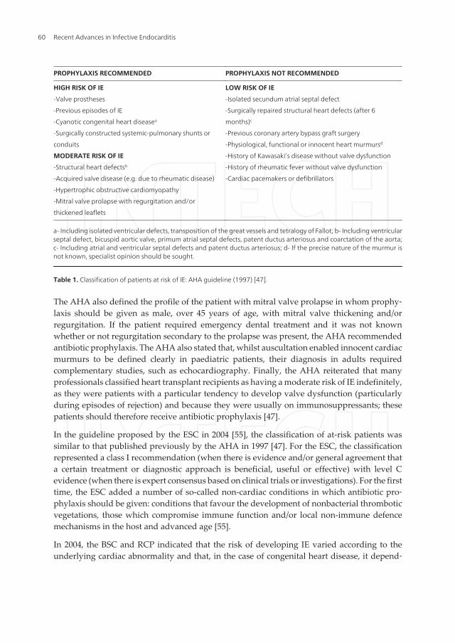
PROPHYLAXIS RECOMMENDED PROPHYLAXIS NOT RECOMMENDED
HIGH RISK OF IE
-Valve prostheses
-Previous episodes of IE
-Cyanotic congenital heart diseasea
-Surgically constructed systemic-pulmonary shunts or
conduits
MODERATE RISK OF IE
-Structural heart defectsb
-Acquired valve disease (e.g. due to rheumatic disease)
-Hypertrophic obstructive cardiomyopathy
-Mitral valve prolapse with regurgitation and/or
thickened leaflets
LOW RISK OF IE
-Isolated secundum atrial septal defect
-Surgically repaired structural heart defects (after 6
months)c
-Previous coronary artery bypass graft surgery
-Physiological, functional or innocent heart murmursd
-History of Kawasaki’s disease without valve dysfunction
-History of rheumatic fever without valve dysfunction
-Cardiac pacemakers or defibrillators
a- Including isolated ventricular defects, transposition of the great vessels and tetralogy of Fallot; b- Including ventricularseptal defect, bicuspid aortic valve, primum atrial septal defects, patent ductus arteriosus and coarctation of the aorta;c- Including atrial and ventricular septal defects and patent ductus arteriosus; d- If the precise nature of the murmur isnot known, specialist opinion should be sought.
Table 1. Classification of patients at risk of IE: AHA guideline (1997) [47].
The AHA also defined the profile of the patient with mitral valve prolapse in whom prophy‐laxis should be given as male, over 45 years of age, with mitral valve thickening and/orregurgitation. If the patient required emergency dental treatment and it was not knownwhether or not regurgitation secondary to the prolapse was present, the AHA recommendedantibiotic prophylaxis. The AHA also stated that, whilst auscultation enabled innocent cardiacmurmurs to be defined clearly in paediatric patients, their diagnosis in adults requiredcomplementary studies, such as echocardiography. Finally, the AHA reiterated that manyprofessionals classified heart transplant recipients as having a moderate risk of IE indefinitely,as they were patients with a particular tendency to develop valve dysfunction (particularlyduring episodes of rejection) and because they were usually on immunosuppressants; thesepatients should therefore receive antibiotic prophylaxis [47].
In the guideline proposed by the ESC in 2004 [55], the classification of at-risk patients wassimilar to that published previously by the AHA in 1997 [47]. For the ESC, the classificationrepresented a class I recommendation (when there is evidence and/or general agreement thata certain treatment or diagnostic approach is beneficial, useful or effective) with level Cevidence (when there is expert consensus based on clinical trials or investigations). For the firsttime, the ESC added a number of so-called non-cardiac conditions in which antibiotic pro‐phylaxis should be given: conditions that favour the development of nonbacterial thromboticvegetations, those which compromise immune function and/or local non-immune defencemechanisms in the host and advanced age [55].
In 2004, the BSC and RCP indicated that the risk of developing IE varied according to theunderlying cardiac abnormality and that, in the case of congenital heart disease, it depend‐
Recent Advances in Infective Endocarditis60
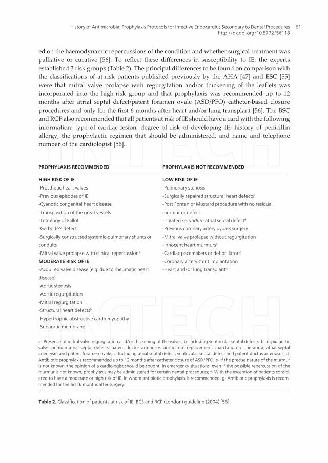
ed on the haemodynamic repercussions of the condition and whether surgical treatment waspalliative or curative [56]. To reflect these differences in susceptibility to IE, the expertsestablished 3 risk groups (Table 2). The principal differences to be found on comparison withthe classifications of at-risk patients published previously by the AHA [47] and ESC [55]were that mitral valve prolapse with regurgitation and/or thickening of the leaflets wasincorporated into the high-risk group and that prophylaxis was recommended up to 12months after atrial septal defect/patent foramen ovale (ASD/PFO) catheter-based closureprocedures and only for the first 6 months after heart and/or lung transplant [56]. The BSCand RCP also recommended that all patients at risk of IE should have a card with the followinginformation: type of cardiac lesion, degree of risk of developing IE, history of penicillinallergy, the prophylactic regimen that should be administered, and name and telephonenumber of the cardiologist [56].
PROPHYLAXIS RECOMMENDED PROPHYLAXIS NOT RECOMMENDED
HIGH RISK OF IE
-Prosthetic heart valves
-Previous episodes of IE
-Cyanotic congenital heart disease
-Transposition of the great vessels
-Tetralogy of Fallot
-Gerbode’s defect
-Surgically constructed systemic-pulmonary shunts or
conduits
-Mitral valve prolapse with clinical repercussiona
MODERATE RISK OF IE
-Acquired valve disease (e.g. due to rheumatic heart
disease)
-Aortic stenosis
-Aortic regurgitation
-Mitral regurgitation
-Structural heart defectsb
-Hypertrophic obstructive cardiomyopathy
-Subaortic membrane
LOW RISK OF IE
-Pulmonary stenosis
-Surgically repaired structural heart defectsc
-Post Fontan or Mustard procedure with no residual
murmur or defect
-Isolated secundum atrial septal defectd
-Previous coronary artery bypass surgery
-Mitral valve prolapse without regurgitation
-Innocent heart murmurse
-Cardiac pacemakers or defibrillatorsf
-Coronary artery stent implantation
-Heart and/or lung transplantg
a- Presence of mitral valve regurgitation and/or thickening of the valves; b- Including ventricular septal defects, bicuspid aorticvalve, primum atrial septal defects, patent ductus arteriosus, aortic root replacement, coarctation of the aorta, atrial septalaneurysm and patent foramen ovale; c- Including atrial septal defect, ventricular septal defect and patent ductus arteriosus; d-Antibiotic prophylaxis recommended up to 12 months after catheter closure of ASD/PFO; e- If the precise nature of the murmuris not known, the opinion of a cardiologist should be sought; in emergency situations, even if the possible repercussion of themurmur is not known, prophylaxis may be administered for certain dental procedures; f- With the exception of patients consid‐ered to have a moderate or high risk of IE, in whom antibiotic prophylaxis is recommended; g- Antibiotic prophylaxis is recom‐mended for the first 6 months after surgery.
Table 2. Classification of patients at risk of IE: BCS and RCP (London) guideline (2004) [56].
History of Antimicrobial Prophylaxis Protocols for Infective Endocarditis Secondary to Dental Procedureshttp://dx.doi.org/10.5772/56118
61
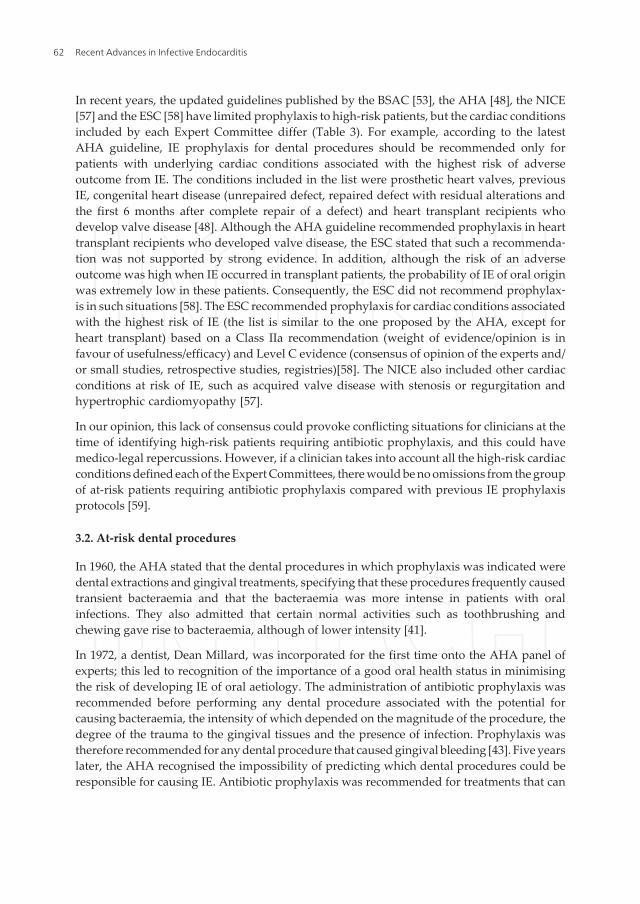
In recent years, the updated guidelines published by the BSAC [53], the AHA [48], the NICE[57] and the ESC [58] have limited prophylaxis to high-risk patients, but the cardiac conditionsincluded by each Expert Committee differ (Table 3). For example, according to the latestAHA guideline, IE prophylaxis for dental procedures should be recommended only forpatients with underlying cardiac conditions associated with the highest risk of adverseoutcome from IE. The conditions included in the list were prosthetic heart valves, previousIE, congenital heart disease (unrepaired defect, repaired defect with residual alterations andthe first 6 months after complete repair of a defect) and heart transplant recipients whodevelop valve disease [48]. Although the AHA guideline recommended prophylaxis in hearttransplant recipients who developed valve disease, the ESC stated that such a recommenda‐tion was not supported by strong evidence. In addition, although the risk of an adverseoutcome was high when IE occurred in transplant patients, the probability of IE of oral originwas extremely low in these patients. Consequently, the ESC did not recommend prophylax‐is in such situations [58]. The ESC recommended prophylaxis for cardiac conditions associatedwith the highest risk of IE (the list is similar to the one proposed by the AHA, except forheart transplant) based on a Class IIa recommendation (weight of evidence/opinion is infavour of usefulness/efficacy) and Level C evidence (consensus of opinion of the experts and/or small studies, retrospective studies, registries)[58]. The NICE also included other cardiacconditions at risk of IE, such as acquired valve disease with stenosis or regurgitation andhypertrophic cardiomyopathy [57].
In our opinion, this lack of consensus could provoke conflicting situations for clinicians at thetime of identifying high-risk patients requiring antibiotic prophylaxis, and this could havemedico-legal repercussions. However, if a clinician takes into account all the high-risk cardiacconditions defined each of the Expert Committees, there would be no omissions from the groupof at-risk patients requiring antibiotic prophylaxis compared with previous IE prophylaxisprotocols [59].
3.2. At-risk dental procedures
In 1960, the AHA stated that the dental procedures in which prophylaxis was indicated weredental extractions and gingival treatments, specifying that these procedures frequently causedtransient bacteraemia and that the bacteraemia was more intense in patients with oralinfections. They also admitted that certain normal activities such as toothbrushing andchewing gave rise to bacteraemia, although of lower intensity [41].
In 1972, a dentist, Dean Millard, was incorporated for the first time onto the AHA panel ofexperts; this led to recognition of the importance of a good oral health status in minimisingthe risk of developing IE of oral aetiology. The administration of antibiotic prophylaxis wasrecommended before performing any dental procedure associated with the potential forcausing bacteraemia, the intensity of which depended on the magnitude of the procedure, thedegree of the trauma to the gingival tissues and the presence of infection. Prophylaxis wastherefore recommended for any dental procedure that caused gingival bleeding [43]. Five yearslater, the AHA recognised the impossibility of predicting which dental procedures could beresponsible for causing IE. Antibiotic prophylaxis was recommended for treatments that can
Recent Advances in Infective Endocarditis62
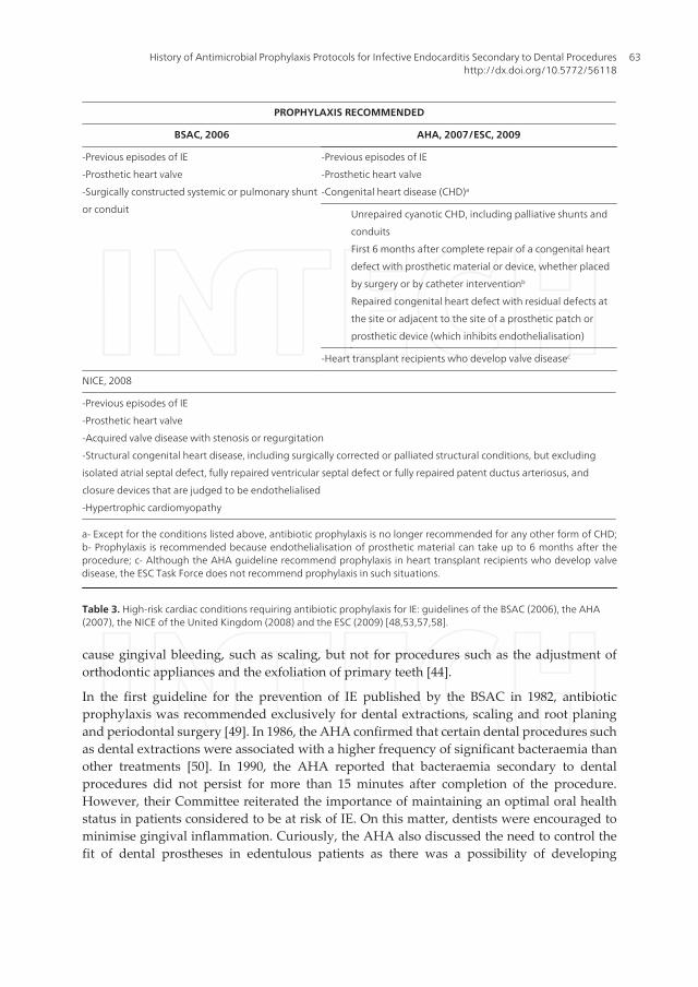
cause gingival bleeding, such as scaling, but not for procedures such as the adjustment oforthodontic appliances and the exfoliation of primary teeth [44].
In the first guideline for the prevention of IE published by the BSAC in 1982, antibioticprophylaxis was recommended exclusively for dental extractions, scaling and root planingand periodontal surgery [49]. In 1986, the AHA confirmed that certain dental procedures suchas dental extractions were associated with a higher frequency of significant bacteraemia thanother treatments [50]. In 1990, the AHA reported that bacteraemia secondary to dentalprocedures did not persist for more than 15 minutes after completion of the procedure.However, their Committee reiterated the importance of maintaining an optimal oral healthstatus in patients considered to be at risk of IE. On this matter, dentists were encouraged tominimise gingival inflammation. Curiously, the AHA also discussed the need to control thefit of dental prostheses in edentulous patients as there was a possibility of developing
PROPHYLAXIS RECOMMENDED
BSAC, 2006 AHA, 2007/ESC, 2009
-Previous episodes of IE
-Prosthetic heart valve
-Surgically constructed systemic or pulmonary shunt
or conduit
-Previous episodes of IE
-Prosthetic heart valve
-Congenital heart disease (CHD)a
Unrepaired cyanotic CHD, including palliative shunts and
conduits
First 6 months after complete repair of a congenital heart
defect with prosthetic material or device, whether placed
by surgery or by catheter interventionb
Repaired congenital heart defect with residual defects at
the site or adjacent to the site of a prosthetic patch or
prosthetic device (which inhibits endothelialisation)
-Heart transplant recipients who develop valve diseasec
NICE, 2008
-Previous episodes of IE
-Prosthetic heart valve
-Acquired valve disease with stenosis or regurgitation
-Structural congenital heart disease, including surgically corrected or palliated structural conditions, but excluding
isolated atrial septal defect, fully repaired ventricular septal defect or fully repaired patent ductus arteriosus, and
closure devices that are judged to be endothelialised
-Hypertrophic cardiomyopathy
a- Except for the conditions listed above, antibiotic prophylaxis is no longer recommended for any other form of CHD;b- Prophylaxis is recommended because endothelialisation of prosthetic material can take up to 6 months after theprocedure; c- Although the AHA guideline recommend prophylaxis in heart transplant recipients who develop valvedisease, the ESC Task Force does not recommend prophylaxis in such situations.
Table 3. High-risk cardiac conditions requiring antibiotic prophylaxis for IE: guidelines of the BSAC (2006), the AHA(2007), the NICE of the United Kingdom (2008) and the ESC (2009) [48,53,57,58].
History of Antimicrobial Prophylaxis Protocols for Infective Endocarditis Secondary to Dental Procedureshttp://dx.doi.org/10.5772/56118
63
bacteraemia because of mucosal ulceration due to poorly fitting prostheses [46]. For its part,the BSAC, in 1992, pronounced for the first time against the use of intraligamental localanaesthesia in patients considered to be at risk of IE [52].
In 1995, the ESC declared that dental treatment constituted the principle risk factor for IE andthat all procedures should therefore be performed under antibiotic prophylaxis, with theexception of superficial fillings and supragingival prosthetic preparations. However, the ESCrecognised that although at-risk dental procedures led to a high prevalence of bacteraemia,this was not predictive of the risk of developing IE. In this context, the duration of theprocedure could represent a possible conditioning factor [54].
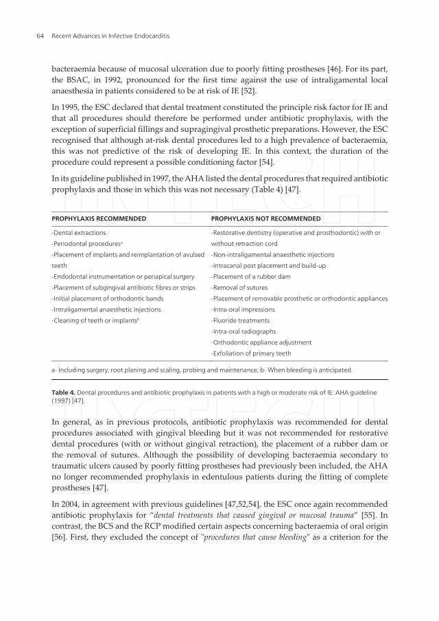
In its guideline published in 1997, the AHA listed the dental procedures that required antibioticprophylaxis and those in which this was not necessary (Table 4) [47].
PROPHYLAXIS RECOMMENDED PROPHYLAXIS NOT RECOMMENDED
-Dental extractions
-Periodontal proceduresa
-Placement of implants and reimplantation of avulsed
teeth
-Endodontal instrumentation or periapical surgery
-Placement of subgingival antibiotic fibres or strips
-Initial placement of orthodontic bands
-Intraligamental anaesthetic injections
-Cleaning of teeth or implantsb
-Restorative dentistry (operative and prosthodontic) with or
without retraction cord
-Non-intraligamental anaesthetic injections
-Intracanal post placement and build-up
-Placement of a rubber dam
-Removal of sutures
-Placement of removable prosthetic or orthodontic appliances
-Intra-oral impressions
-Fluoride treatments
-Intra-oral radiographs
-Orthodontic appliance adjustment
-Exfoliation of primary teeth
a- Including surgery, root planing and scaling, probing and maintenance; b- When bleeding is anticipated.
Table 4. Dental procedures and antibiotic prophylaxis in patients with a high or moderate risk of IE: AHA guideline(1997) [47].
In general, as in previous protocols, antibiotic prophylaxis was recommended for dentalprocedures associated with gingival bleeding but it was not recommended for restorativedental procedures (with or without gingival retraction), the placement of a rubber dam orthe removal of sutures. Although the possibility of developing bacteraemia secondary totraumatic ulcers caused by poorly fitting prostheses had previously been included, the AHAno longer recommended prophylaxis in edentulous patients during the fitting of completeprostheses [47].
In 2004, in agreement with previous guidelines [47,52,54], the ESC once again recommendedantibiotic prophylaxis for “dental treatments that caused gingival or mucosal trauma” [55]. Incontrast, the BCS and the RCP modified certain aspects concerning bacteraemia of oral origin[56]. First, they excluded the concept of "procedures that cause bleeding" as a criterion for the
Recent Advances in Infective Endocarditis64
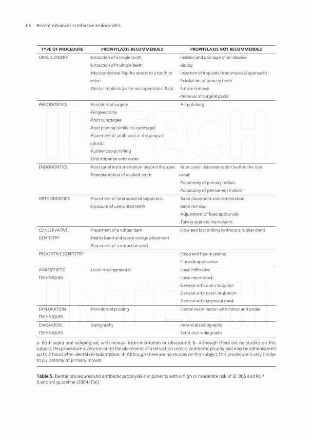
indication for antibiotic prophylaxis in patients at risk of IE; they also re-evaluated thedefinition of "significant bacteraemia" which, according to their new interpretation, was definedas "bacteraemia secondary to a dental procedure that was statistically significant with respect to thebacteraemia present under basal conditions (prior to any manipulation)". Considering these newprovisions, the indication for prophylaxis included not only surgical procedures such as dentalextractions or mucoperiosteal flaps but also other less traumatic procedures such as theplacement of a rubber dam, matrices, wedges or retraction cords (Table 5) [56]. Although thatCommittee recognised the existence of bacteraemia secondary to activities considered to bephysiological (such as toothbrushing), it also recognised the impossibility of administeringprophylaxis for such practices due to the high risk of potentiating the development of bacterialresistance [56].
In 2006, the BSAC summarized the indications for antibiotic prophylaxis for high-risk patientsstating that it should be given for “all dental procedures involving dento-gingival manipulation orendodontics” [53]. According to the latest AHA and ESC guidelines, prophylaxis was recom‐mended for all dental procedures that involved manipulation of gingival tissues or theperiapical region of teeth or perforation of the oral mucosa. This included procedures such asbiopsies, suture removal and placement of orthodontics bands, but it did not include routineanaesthetic injections through non-infected tissue, taking dental radiographs, placement ofremovable prosthodontic or orthodontic appliances, placement of orthodontic brackets, oradjustment of orthodontic appliances [48,58]. The dental procedures with the highest risk ofIE and for which prophylaxis was recommended were associated with a Class IIa recommen‐dation (weight of evidence/opinion is in favour of usefulness/efficacy) and Level C evidence(consensus of opinion of the experts and/or small studies, retrospective studies, registries) [58].There are other events for which prophylaxis was not recommended, such as shedding ofdeciduous teeth and trauma to the lips or oral mucosa [48].
In the latest guidelines published by the BSAC, the AHA, the NICE of the United Kingdom,and the ESC, the emphasis for the cause of IE shifted from procedure-related bacteraemia tocumulative bacteraemia due to everyday oral activities [48,53,57,58]. The NICE considered thatit was biologically implausible that a dental procedure would lead to a greater risk of IE thanregular toothbrushing. On the other hand, even some expert committee guidelines concurredwith the premise “Maintenance of optimal oral hygiene and periodontal health may reduce theincidence of bacteraemia of oral origin and, in the context of a dental procedure, is more important thanprophylactic antibiotics to reduce the risk of IE” [48,58].
The NICE has adopted a drastic stance in this respect, issuing the statement that “antibioticprophylaxis for IE is not recommended in individuals undergoing dental procedures” [58]. Recently,following the introduction in March 2008 of a clinical guideline from NICE recommending thecessation of antibiotic prophylaxis in the United Kingdom, Thornhill et al [60] quantified thechange in the prescription of antibiotic prophylaxis to patients at risk of IE undergoing invasivedental procedures and looked for any concurrent change in the incidence of IE. Despite a 78.6%reduction in the prescription of antibiotic prophylaxis after the introduction of the NICEguideline, that study detected no large increase in the incidence of cases of IE or of IE-relateddeaths over the following 2 years. Those authors concluded that ongoing data monitoring was
History of Antimicrobial Prophylaxis Protocols for Infective Endocarditis Secondary to Dental Procedureshttp://dx.doi.org/10.5772/56118
65
TYPE OF PROCEDURE PROPHYLAXIS RECOMMENDED PROPHYLAXIS NOT RECOMMENDED
ORAL SURGERY -Extraction of a single tooth
-Extraction of multiple teeth
-Mucoperiosteal flap for access to a tooth or
lesion
-Dental implants (as for mucoperiosteal flap)
-Incision and drainage of an abscess
-Biopsy
-Insertion of implants (transmucosal approach)
-Exfoliation of primary teeth
-Suture removal
-Removal of surgical packs
PERIODONTICS -Periodontal surgery
-Gingivectomy
-Root curettagea
-Root planing (similar to curettage)
-Placement of antibiotics in the gingival
sulcusb
-Rubber cup polishing
-Oral irrigation with water
-Air polishing
ENDODONTICS -Root canal instrumentation beyond the apex
-Reimplantation of avulsed teethc
-Root canal instrumentation (within the root
canal)
-Pulpotomy of primary molars
-Pulpotomy of permanent molarsd
ORTHODONTICS -Placement of interproximal separators
-Exposure of unerupted teeth
-Band placement and cementation
-Band removal
-Adjustment of fixed appliances
-Taking alginate impressions
CONSERVATIVE
DENTISTRY
-Placement of a rubber dam
-Matrix band and wood wedge placement
-Placement of a retraction cord
-Slow and fast drilling (without a rubber dam)
PREVENTIVE DENTISTRY -Fossa and fissure sealing
-Fluoride application
ANAESTHETIC
TECHNIQUES
-Local intraligamental -Local infiltrative
-Local nerve block
-General with oral intubation
-General with nasal intubation
-General with laryngeal mask
EXPLORATION
TECHNIQUES
-Periodontal probing -Dental examination with mirror and probe
DIAGNOSTIC
TECHNIQUES
-Sialography -Intra-oral radiographs
-Extra-oral radiographs
a- Both supra and subgingival, with manual instrumentation or ultrasound; b- Although there are no studies on thissubject, this procedure is very similar to the placement of a retraction cord; c- Antibiotic prophylaxis may be administeredup to 2 hours after dental reimplantation; d- Although there are no studies on this subject, this procedure is very similarto pulpotomy of primary molars.
Table 5. Dental procedures and antibiotic prophylaxis in patients with a high or moderate risk of IE: BCS and RCP(London) guideline (2004) [56].
Recent Advances in Infective Endocarditis66
needed to confirm this observation supporting the NICE guideline and that further clinicaltrials should be performed to determine if antibiotic prophylaxis still has a role in protectingsome patients at particularly high risk [60].
3.3. Anaesthetic technique
In 1960, the AHA recommended the administration of antibiotic prophylaxis for any surgicalintervention (including those in the orofacial area) performed under general anaesthesia inpatients considered to be at risk of IE [41]. However, in subsequent protocols published by theAHA, no specific observations were made with regard to the type of anaesthesia used [42-48].
The BSAC, on the other hand, specified for the first time in 1982 that when dental treatmentwas performed under general anaesthesia, special prophylactic protocols should be applied,also considering that "If patients due to undergo a general anaesthesia have prosthetic heart valvesand/or are allergic to penicillin and/or have received prolonged treatment with penicillin and/or havehad previous episodes of IE, their dental problems should be treated in a hospital environment"[49]. The BSAC has maintained that opinion in its protocols on IE prevention published in1986, 1990 and 1992 [50-52]. In 1995, the ESC also included the anaesthetic technique amongthe factors to be taken into account when choosing the prophylactic regimen [54]. In theguideline published by the BCS and RCP in 2004, specific prophylaxis regimens were includedfor dental procedures performed under general anaesthesia [56].
In agreement with the AHA, the latest protocols of the BSAC and ESC on IE preventionrecommend antibiotic prophylaxis irrespective of whether the dental procedure is performedunder general or local anaesthesia [53,58].
3.4. Antibiotics of choice, dose and route of administration
In 1960, the AHA pronounced in favour of administering antibiotic prophylaxis from between24 and 48 hours before the dental procedure, even in the absence of intraoral infections, inorder to reduce the intensity of the post-manipulation bacteraemia [41]. However, in view ofthe problem of bacterial resistance, it was also suggested that prophylaxis could be adminis‐tered immediately before the procedure. According to the AHA, the choice of one or otherregimen depended on the professional, who should evaluate the probability of infection inorder to decide when the prescription of antibiotics was indicated. In contrast to the guidelinepublished in 1955 [39], the exclusively oral protocols were excluded in favour of intramuscularadministration, although penicillin continued to be the antibiotic of choice; the prophylacticregimen consisted of several injections of penicillin from 2 days before up to 2 days after thesession of dental treatment. A combined intramuscular-oral prophylactic regimen was alsoelaborated. For patients with a history of penicillin allergy, the AHA was the first to recom‐mended erythromycin at doses of 250 mg orally 4 times a day (for adults and older children);in small children, the dose of erythromycin was of 20 mg/kg body-weight per day, dividedinto 3 or 4 doses, not exceeding a total dose of 1 g per day [41].
In 1965, the AHA stated that antibiotic prophylaxis should only be administered immediatelybefore the dental procedure and on the subsequent days; this recommendation was based on
History of Antimicrobial Prophylaxis Protocols for Infective Endocarditis Secondary to Dental Procedureshttp://dx.doi.org/10.5772/56118
67
the argument that penicillin did not sterilise the apical foci, and that its excessive use led tothe selection of a resistant oral flora. The AHA also reduced the parenteral regimen to a singleinjection of several penicillins. In those cases in which the complete collaboration of the patientcould be anticipated, an exclusively oral regimen of several doses of penicillin was proposed.Erythromycin was recommended for patients allergic to penicillin [42].
In 1972, the AHA modified its recommendations to include an increase in the initial dosesof penicillin and erythromycin administered orally and the use of erythromycin in patientson prolonged treatments with penicillin, as penicillin-resistant Streptococcus viridans couldpredominate in their oral flora [43]. Five years later, the AHA suggested increasing the initialdose of the antibiotic even further in order to reach higher serum concentrations at themoment at which the microorganism entered the bloodstream [44]. However, they fav‐oured the parenteral regimen, particularly in patients considered to be at high risk of IE. Tworegimens were recommended: regimen A, based on the use of penicillin (erythromycin wasrecommended in patients allergic to penicillin) for parenteral-oral or exclusively oraladministration, and regimen B, which combined penicillin and streptomycin (vancomycinand erythromycin for patients allergic to penicillin) for parenteral-oral administration. Thislatter protocol was reserved for patients with prosthetic heart valves, although patients witha good oral health status could receive the oral prophylaxis regimen for certain non-surgical dental procedures [44].
The BSAC, in its first guideline, suggested a single prophylactic regimen of a single dose ofamoxicillin before the dental procedure for all patients considered to be at risk of IE (includingpatients with prosthetic heart valves) [49]. The BSAC substituted penicillin V, previouslyrecommended by the AHA [44], with amoxicillin due to its more favourable pharmacokineticand pharmacodynamic characteristics. Erythromycin stearate was the antibiotic of choice inpatients allergic to penicillin but because this macrolide has lower activity than amoxicillinagainst some oral streptococci and showed a lower absorption after a single oral dose, theyproposed a second dose 6 hours after completing the dental procedure. One quarter of theadult dose was recommended in children under 5 years of age and a half dose in those of 5 to10 years of age [49]. In contrast to the AHA [44], the BSAC proposed a combined intramuscular-oral regimen in patients undergoing dental treatment under general anaesthesia. Specialprophylactic regimens were proposed for patients being treated in the hospital environment;these regimens were based on the association of amoxicillin and gentamycin or, in patientsunable to receive penicillin, a combination of vancomycin and gentamycin; the following doseswere used in children under 10 years of age: amoxicillin, half the adult dose; gentamycin, 2mg/kg body-weight; and vancomycin, 20 mg/kg body-weight [49].
In its protocol published in 1984, the AHA reduced the dose of the antibiotic after completionof the dental treatment, recommending the administration of penicillin V before the dentalprocedure and a second dose 6 hours after the first. In those patients in whom the oral routewas not available, intramuscular penicillin G was proposed before the procedure and 6 hourslater [45]. The AHA also showed a clear preference for the parenteral route in patients at highrisk of IE and drew up a special regimen for these patients consisting of intramuscular orintravenous ampicillin and gentamycin, together with a second dose of penicillin V orally;
Recent Advances in Infective Endocarditis68
intravenous vancomycin was recommended for patients allergic to penicillin, eliminating thesecond dose of erythromycin [45].
In 1986, the BSAC suggested that vancomycin should be given by slow intravenous infusionover 60 minutes (instead of the previously recommended 30 minutes) to minimise adversereactions such as episodes of hypotension caused by histamine release (red-man syndrome)[50]. As an alternative to the parenteral regimen proposed earlier, the BSAC proposed 2 oralregimens for patients without prosthetic heart valves undergoing dental treatment undergeneral anaesthesia. The first was based on the administration of amoxicillin before anaestheticinduction followed by a second dose in the immediate postoperative period; the secondregimen consisted of the combination of amoxicillin and probenecid administered beforeanaesthesia [50]. For the first time, the BSAC differentiated between patients with prostheticheart valves and other patients considered to be at risk of IE, as the AHA [45] had done in its1984 guideline, proposing specific oral prophylactic regimens for such patients undergoingdental treatment under local anaesthesia [50].
Differing from the BSAC guideline [50], the 1990 AHA guideline continued to favour regi‐mens based on 2 doses. Of particular note amongst the novelties introduced in this protocol wasthe incorporation of amoxicillin as the antibiotic of choice for all groups at risk of IE [46], anapproach that had been adopted by the BSAC in 1982 [49]. According to the AHA, amoxicillin,ampicillin and penicillin showed similar efficacy against α-haemolytic streptococci in vitro butamoxicillin reached higher serum concentrations due to its better gastrointestinal absorption.However, they also defended the use of penicillin V as a suitable alternative for prophylaxis indental procedures. Erythromycin, in its ethylsuccinate or stearate salt preparations, continuedto be the antibiotic of choice in patients allergic to penicillin, being administered 2 hours beforethe procedure to ensure high serum concentrations. For the first time, the AHA recommendedthe administration of clindamycin in patients intolerant to penicillin and erythromycin [46]. Forpatients unable to take oral medication, the AHA drew up a number of regimens for parenter‐al administration as alternatives to the standard protocol, proposing ampicillin (in patients notallergic to penicillin) and clindamycin (in penicillin-allergic patients) as the antibiotics of choice[46]. In contrast to the previous protocols [45], the AHA recommended the administration of thestandard regimen to patients with prosthetic heart valves and other patients considered to be athigh risk of IE (patients with a past history of IE and those with surgically constructed systemic-pulmonary shunts). However, recognising that some professionals preferred parenteralprophylaxis, they also drew up a special parenteral regimen for this type of patient [46].
The prophylactic protocol recommended by the BSAC in 1990 included a new option [51]. Dueto the high prevalence of undesirable gastrointestinal effects caused by erythromycin, andbased on the guideline published in 1984 by the Swiss Expert Committee for the preventionof IE [61], the BSAC proposed the administration of a single oral dose of 600 mg of clindamycin1 hour before the procedure as an alternative in patients with penicillin allergy; the dose ofclindamycin in children under 10 years of age was of 6 mg/kg body-weight [51]. In 1992, theBSAC definitively replaced erythromycin with clindamycin in patients allergic to penicillin,modifying the initial dose in children to 300 mg in those between 5 and 10 years of age and to150 mg in those under 5 years [52]. Due to the high prevalence of adverse effects associated
History of Antimicrobial Prophylaxis Protocols for Infective Endocarditis Secondary to Dental Procedureshttp://dx.doi.org/10.5772/56118
69
with vancomycin and its prolonged duration of administration (around 100 minutes), theBSAC drew up 2 alternative regimens for penicillin-allergic patients with a high risk of IE whowere being treated in the hospital environment. One was based on the intravenous combina‐tion of teicoplanin and gentamycin (in children under 14 years of age the doses were teico‐planin, 6 mg /kg body-weight, and gentamycin, 2 mg/kg body-weight); and the other consistedof an intravenous infusion of clindamycin with a second dose 6 hours after the first. Finally,in patients undergoing dental treatment under general anaesthesia, the BSAC specified thatprophylaxis with amoxicillin should be administered intravenously instead of intramuscular‐ly, particularly in children [52].
In 1995, the ESC performed a critical review of the prophylaxis protocols drawn up by thedifferent national committees, noting clear differences between countries, although allincluded a simple or standard regimen and another more complex regimen for use in specialcircumstances [54]. In general, the standard guidelines consisted of the oral administration ofa single dose of antibiotic which, in the majority of countries, was amoxicillin. Some societiesrecommended the administration of a second dose, particularly in patients considered to beat high risk of IE. In patients allergic to the beta-lactams, the antibiotic of choice was clinda‐mycin at doses between 300 mg and 600 mg, although some countries, for example, Hollandand France, recommended other antibiotics such as erythromycin or pristinamycin [54]. Themore complex regimens were based on the synergistic and prolonged effect provided byseveral doses of different antibiotics with the aim of increasing the safety margin in specialsituations. In an analysis performed by the ESC, it was found that the majority of protocolsrecommended ampicillin or amoxicillin by intravenous infusion followed by a second oraldose 6 hours later; there were only minor differences with respect to the doses used. Althoughsome countries did not use the aminoglycosides, these were recommended in other countriesin patients considered to be at high risk of IE. The most frequently used antibiotic of choice inpatients allergic to penicillin was vancomycin by intravenous infusion; for some scientificsocieties, teicoplanin and clindamycin were possible antimicrobial alternatives [54]. Accordingto the ESC, the choice of the most suitable prophylactic regimen should be based on thefollowing considerations: the heart condition defined as carrying a risk of IE; the type,magnitude and duration of the dental procedure; and the type of anaesthesia used (local orgeneral). The ESC therefore considered the possibility of individualising the antibioticprophylaxis regimen in certain situations [54]. The oral regimen proposed by the ESC consistedof the administration of amoxicillin or clindamycin (in penicillin-allergic patients), whilst thecombination of amoxicillin or ampicillin with gentamycin and a second dose of amoxicillinorally 6 hours later was recommended in the parenteral regimen. In patients allergic topenicillin, the association of vancomycin and gentamycin was recommended, administeringa second dose of vancomycin by intravenous infusion 12 hours after the first dose [54].
The prophylactic protocol recommended by the AHA in 1997 is shown in Table 6 [47]. It is basedon a single dose of amoxicillin administered orally 1 hour before the procedure. In this proto‐col, the dose of amoxicillin was reduced from 3 g to 2 g after confirming that this latter doseprovided adequate serum levels of the drug over several hours and caused fewer adversegastrointestinal effects. Accepting an approach that had been adopted by other societies severalyears earlier [49-52], the AHA recognised that the administration of a second dose of antibioticwas unnecessary, since the serum levels of the drug exceeded the minimum inhibitory concentra‐
Recent Advances in Infective Endocarditis70
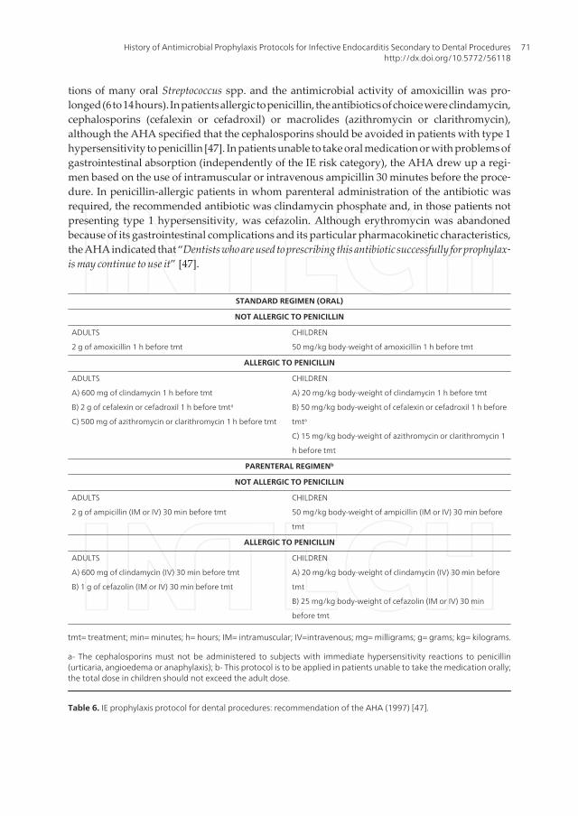
tions of many oral Streptococcus spp. and the antimicrobial activity of amoxicillin was pro‐longed (6 to 14 hours). In patients allergic to penicillin, the antibiotics of choice were clindamycin,cephalosporins (cefalexin or cefadroxil) or macrolides (azithromycin or clarithromycin),although the AHA specified that the cephalosporins should be avoided in patients with type 1hypersensitivity to penicillin [47]. In patients unable to take oral medication or with problems ofgastrointestinal absorption (independently of the IE risk category), the AHA drew up a regi‐men based on the use of intramuscular or intravenous ampicillin 30 minutes before the proce‐dure. In penicillin-allergic patients in whom parenteral administration of the antibiotic wasrequired, the recommended antibiotic was clindamycin phosphate and, in those patients notpresenting type 1 hypersensitivity, was cefazolin. Although erythromycin was abandonedbecause of its gastrointestinal complications and its particular pharmacokinetic characteristics,the AHA indicated that “Dentists who are used to prescribing this antibiotic successfully for prophylax‐is may continue to use it” [47].
STANDARD REGIMEN (ORAL)
NOT ALLERGIC TO PENICILLIN
ADULTS
2 g of amoxicillin 1 h before tmt
CHILDREN
50 mg/kg body-weight of amoxicillin 1 h before tmt
ALLERGIC TO PENICILLIN
ADULTS
A) 600 mg of clindamycin 1 h before tmt
B) 2 g of cefalexin or cefadroxil 1 h before tmta
C) 500 mg of azithromycin or clarithromycin 1 h before tmt
CHILDREN
A) 20 mg/kg body-weight of clindamycin 1 h before tmt
B) 50 mg/kg body-weight of cefalexin or cefadroxil 1 h before
tmta
C) 15 mg/kg body-weight of azithromycin or clarithromycin 1
h before tmt
PARENTERAL REGIMENb
NOT ALLERGIC TO PENICILLIN
ADULTS
2 g of ampicillin (IM or IV) 30 min before tmt
CHILDREN
50 mg/kg body-weight of ampicillin (IM or IV) 30 min before
tmt
ALLERGIC TO PENICILLIN
ADULTS
A) 600 mg of clindamycin (IV) 30 min before tmt
B) 1 g of cefazolin (IM or IV) 30 min before tmt
CHILDREN
A) 20 mg/kg body-weight of clindamycin (IV) 30 min before
tmt
B) 25 mg/kg body-weight of cefazolin (IM or IV) 30 min
before tmt
tmt= treatment; min= minutes; h= hours; IM= intramuscular; IV=intravenous; mg= milligrams; g= grams; kg= kilograms.
a- The cephalosporins must not be administered to subjects with immediate hypersensitivity reactions to penicillin(urticaria, angioedema or anaphylaxis); b- This protocol is to be applied in patients unable to take the medication orally;the total dose in children should not exceed the adult dose.
Table 6. IE prophylaxis protocol for dental procedures: recommendation of the AHA (1997) [47].
History of Antimicrobial Prophylaxis Protocols for Infective Endocarditis Secondary to Dental Procedureshttp://dx.doi.org/10.5772/56118
71

In 2004, the ESC published a guideline on IE prophylaxis which were very similar to the 1997guideline of the AHA [47], except that the use of cephalosporins in patients allergic to penicillinwas excluded [55].
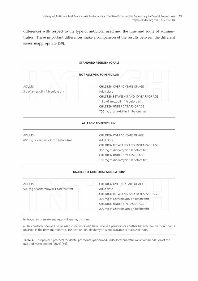
In the prophylaxis protocol for IE secondary to dental procedures drawn up by the BSC andRCP (London) in 2004, prophylaxis was reserved for patients with heart diseases included inthe categories of high and moderate risk of IE, and the prophylactic regimens varied accordingto the type of anaesthesia used [56]. Oral prophylaxis regimens were to be administered inprocedures performed under local anaesthesia and parenteral regimens for those performedunder general anaesthesia (Tables 7 and 8) [56]. In contrast to the 1997 guideline of the AHA[47], the BCS and RCP also provided a special prophylactic regimen for patients with prostheticheart valves and/or previous episodes of IE (Table 9) [56].
The most recent IE prophylaxis protocols published by the BSAC [53], the AHA [48] andthe ESC [58] are very similar and are summarized in Tables 10 and 11. The most recentprophylactic protocol published by the AHA continues to recommend amoxicillin as theantibiotic of choice for oral prophylaxis. For individuals who are allergic to penicillins, theuse of cephalexin or another first-generation oral cephalosporin, clindamycin, azithromy‐cin or clarithromycin is recommended [48]. Because of possible cross-reactions, a cephalo‐sporin must not be administered to patients with a history of anaphylaxis, angioedema orurticaria after treatment with any form of penicillin, including ampicillin or amoxicillin.Patients who are unable to tolerate an oral antibiotic may be treated with intramuscularor intravenous ampicillin, ceftriaxone or cefazolin. For penicillin-allergic patients who areunable to tolerate an oral agent, prophylaxis is recommended with parenteral cefazolin,ceftriaxone or clindamycin [48]. According to the ESC, the main aim of antibiotic prophy‐laxis in patients at risk of IE is to target the oral streptococci. The impact of increasingresistance of these pathogens on the efficacy of antibiotic prophylaxis is unclear. Fluoroqui‐nolones and glycopeptides are not recommended because their efficacy has not beenestablished and because of the potential induction of resistance [58].
It has been estimated that the number of cases of IE that result from dental interventions isvery small. The AHA has therefore concluded that only an extremely small number of casesof IE will be prevented by antibiotic prophylaxis for dental procedures even if such prophy‐lactic regimens are 100% effective [48]. According to the ESC, this observation leads to 2conclusions: (i) IE prophylaxis can at best only protect a small proportion of patients; and (ii)the bacteraemia that causes IE in the majority of patients appears to derive from another source[58]. Finally, the AHA stated the need for prospective placebo-controlled studies of antibioticprophylaxis for IE to evaluate its efficacy in IE prevention [48].
Reviewing the effect of antibiotic prophylaxis on the intensity and duration of bacterae‐mia following dental procedures, the NICE recently concluded that “Antibiotic prophylaxisdoes not eliminate bacteraemia following dental procedures but some studies show that it does reducethe frequency of detection of post-procedure bacteraemia” [57]. This conclusion was reached afteranalysis of a number of studies on the efficacy of antibiotic prophylaxis for the preven‐tion of post-dental manipulation bacteraemia; those studies presented methodological
Recent Advances in Infective Endocarditis72
differences with respect to the type of antibiotic used and the time and route of adminis‐tration. These important differences make a comparison of the results between the differentseries inappropriate [59].
STANDARD REGIMEN (ORAL)
NOT ALLERGIC TO PENICILLIN
ADULTS
3 g of amoxicillin 1 h before tmt
CHILDREN OVER 10 YEARS OF AGE
Adult dose
CHILDREN BETWEEN 5 AND 10 YEARS OF AGE
1.5 g of amoxicillin 1 h before tmt
CHILDREN UNDER 5 YEARS OF AGE
750 mg of amoxicillin 1 h before tmt
ALLERGIC TO PENICILLINa
ADULTS
600 mg of clindamycin 1 h before tmt
CHILDREN OVER 10 YEARS OF AGE
Adult dose
CHILDREN BETWEEN 5 AND 10 YEARS OF AGE
300 mg of clindamycin 1 h before tmt
CHILDREN UNDER 5 YEARS OF AGE
150 mg of clindamycin 1 h before tmt
UNABLE TO TAKE ORAL MEDICATIONb
ADULTS
500 mg of azithromycin 1 h before tmt
CHILDREN OVER 10 YEARS OF AGE
Adult dose
CHILDREN BETWEEN 5 AND 10 YEARS OF AGE
300 mg of azithromycin 1 h before tmt
CHILDREN UNDER 5 YEARS OF AGE
200 mg of azithromycin 1 h before tmt
h= hours; tmt= treatment; mg= milligrams; g= grams.
a- This protocol should also be used in patients who have received penicillin or another beta-lactam on more than 1occasion in the previous month; b- In Great Britain, clindamycin is not available in oral suspension.
Table 7. IE prophylaxis protocol for dental procedures performed under local anaesthesia: recommendation of theBCS and RCP (London) (2004) [56].
History of Antimicrobial Prophylaxis Protocols for Infective Endocarditis Secondary to Dental Procedureshttp://dx.doi.org/10.5772/56118
73
PARENTERAL REGIMEN
NOT ALLERGIC TO PENICILLIN
ADULTS
2 g of amoxicillin or ampicillin (IV) during anaesthetic induction
CHILDREN OVER 10 YEARS OF AGE
Adult dose
CHILDREN BETWEEN 5 AND 10 YEARS OF AGE
500 mg of amoxicillin or ampicillin (IV) during anaesthetic induction
CHILDREN UNDER 5 YEARS OF AGE
250 mg of amoxicillin or ampicillin (IV) during anaesthetic induction
ALLERGIC TO PENICILLINa
ADULTS
300 mg of clindamycin (IV over 10 min) during anaesthetic
induction
150 mg of clindamycin (oral or IV) 6 h after the first dose
CHILDREN OVER 10 YEARS OF AGE
Adult dose
CHILDREN BETWEEN 5 AND 10 YEARS OF AGE
150 mg of clindamycin (IV over 10 min) during anaesthetic induction
CHILDREN UNDER 5 YEARS OF AGE
75 mg of clindamycin (IV over 10 min) during anaesthetic induction
min= minutes; h= hours; IV= intravenous; mg= milligrams; g= grams; kg= kilograms.
a-This protocol should also be used in patients who have received penicillin or another beta-lactam on more than 1occasion in the previous month.
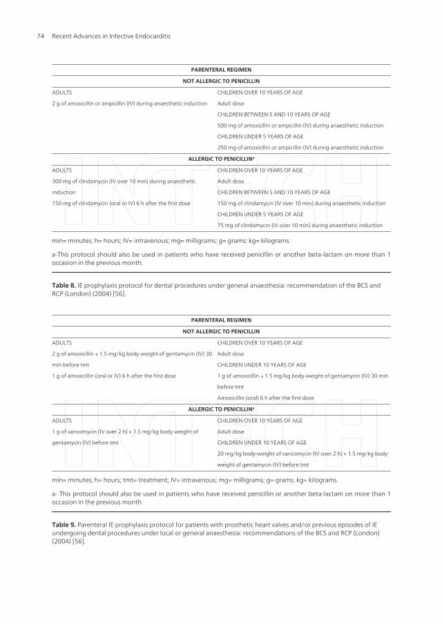
Table 8. IE prophylaxis protocol for dental procedures under general anaesthesia: recommendation of the BCS andRCP (London) (2004) [56].
PARENTERAL REGIMEN
NOT ALLERGIC TO PENICILLIN
ADULTS
2 g of amoxicillin + 1.5 mg/kg body-weight of gentamycin (IV) 30
min before tmt
1 g of amoxicillin (oral or IV) 6 h after the first dose
CHILDREN OVER 10 YEARS OF AGE
Adult dose
CHILDREN UNDER 10 YEARS OF AGE
1 g of amoxicillin + 1.5 mg/kg body-weight of gentamycin (IV) 30 min
before tmt
Amoxicillin (oral) 6 h after the first dose
ALLERGIC TO PENICILLINa
ADULTS
1 g of vancomycin (IV over 2 h) + 1.5 mg/kg body-weight of
gentamycin (IV) before tmt
CHILDREN OVER 10 YEARS OF AGE
Adult dose
CHILDREN UNDER 10 YEARS OF AGE
20 mg/kg body-weight of vancomycin (IV over 2 h) + 1.5 mg/kg body-
weight of gentamycin (IV) before tmt
min= minutes; h= hours; tmt= treatment; IV= intravenous; mg= milligrams; g= grams; kg= kilograms.
a- This protocol should also be used in patients who have received penicillin or another beta-lactam on more than 1occasion in the previous month.
Table 9. Parenteral IE prophylaxis protocol for patients with prosthetic heart valves and/or previous episodes of IEundergoing dental procedures under local or general anaesthesia: recommendations of the BCS and RCP (London)(2004) [56].
Recent Advances in Infective Endocarditis74
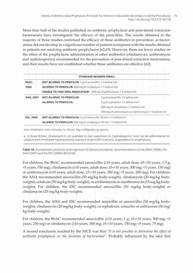
More than half of the studies published on antibiotic prophylaxis and post-dental extractionbacteraemia have investigated the efficacy of the penicillins. The results obtained in themajority of those studies confirmed the efficacy of these antibiotics in prevention, as bacter‐aemia did not develop in a significant number of patients (compared with the results obtainedin patients not receiving antibiotic prophylaxis) [62,63]. However, there are fewer studies onthe effect of the prophylactic administration of other antibiotics (clindamycin, azithromycinand cephalosporins) recommended for the prevention of post-dental extraction bacteraemia,and their results have not established whether these antibiotics are effective [62].
STANDARD REGIMEN (ORAL)
BSAC,
2006
NOT ALLERGIC TO PENICILLIN: 3 g of amoxicillin 1 h before tmt
ALLERGIC TO PENICILLIN: 600 mg of clindamycin 1 h before tmt
UNABLE TO TAKE ORAL MEDICATIONa : 500 mg of azithromycin 1 h before tmt
AHA, 2007 NOT ALLERGIC TO PENICILLIN:
ALLERGIC TO PENICILLIN:
2 g of amoxicillin 1 h before tmt
2 g of cephalexin 1 h before tmtb
600 mg of clindamycin 1 h before tmt
500 mg of azithromycin or clarithromycin 1 h before tmt
ESC, 2009 NOT ALLERGIC TO PENICILLIN: 2 g of amoxicillin 30 min-1 h before tmt
ALLERGIC TO PENICILLIN: 600 mg of clindamycin 30 min-1 h before tmt
tmt= treatment; min= minutes; h= hours; mg= milligrams; g= grams.
a- In Great Britain, clindamycin is not available in oral suspension; b- Cephalosporins must not be administered tosubjects with immediate hypersensitivity reactions to penicillin (urticaria, angioedema or anaphylaxis).
Table 10. IE prophylaxis protocols (oral regimens) for dental procedures: recommendations of the BSAC (2006), theAHA (2007) and the ESC (2009) [48,53,58].
For children, the BSAC recommended amoxicillin (≥10 years, adult dose; ≥5-<10 years, 1.5 g;<5 years, 750 mg), clindamycin (≥10 years, adult dose; ≥5-<10 years, 300 mg; <5 years, 150 mg)or azithromycin (≥10 years, adult dose; ≥5-<10 years, 300 mg; <5 years, 200 mg). For children,the AHA recommended amoxicillin (50 mg/kg body-weight), clindamycin (20 mg/kg body-weight), cefalexin (50 mg/kg body-weight), or azithromycin or clarithromycin (15 mg/kg body-weight). For children, the ESC recommended amoxicillin (50 mg/kg body-weight) orclindamycin (20 mg/kg body-weight).
For children, the AHA and ESC recommended ampicillin or amoxicillin (50 mg/kg body-weight), clindamycin (20 mg/kg body-weight), or cephalexin, cefazolin or ceftriaxone (50 mg/kg body-weight).
For children, the BSAC recommended amoxicillin (≥10 years, 1 g; ≥5-<10 years, 500 mg; <5years, 250 mg) or clindamycin (≥10 years, 300 mg; ≥5-<10 years, 150 mg; <5 years, 75 mg).
A second conclusion reached by the NICE was that “It is not possible to determine the effect ofantibiotic prophylaxis on the duration of bacteraemia”. Probably influenced by the idea that
History of Antimicrobial Prophylaxis Protocols for Infective Endocarditis Secondary to Dental Procedureshttp://dx.doi.org/10.5772/56118
75
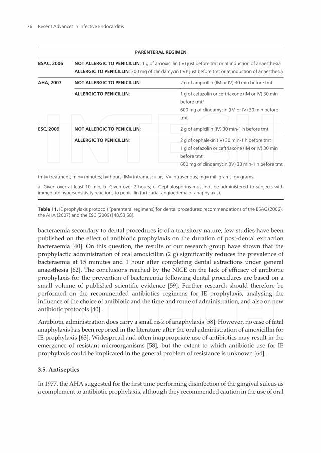
bacteraemia secondary to dental procedures is of a transitory nature, few studies have beenpublished on the effect of antibiotic prophylaxis on the duration of post-dental extractionbacteraemia [40]. On this question, the results of our research group have shown that theprophylactic administration of oral amoxicillin (2 g) significantly reduces the prevalence ofbacteraemia at 15 minutes and 1 hour after completing dental extractions under generalanaesthesia [62]. The conclusions reached by the NICE on the lack of efficacy of antibioticprophylaxis for the prevention of bacteraemia following dental procedures are based on asmall volume of published scientific evidence [59]. Further research should therefore beperformed on the recommended antibiotics regimens for IE prophylaxis, analysing theinfluence of the choice of antibiotic and the time and route of administration, and also on newantibiotic protocols [40].
Antibiotic administration does carry a small risk of anaphylaxis [58]. However, no case of fatalanaphylaxis has been reported in the literature after the oral administration of amoxicillin forIE prophylaxis [63]. Widespread and often inappropriate use of antibiotics may result in theemergence of resistant microorganisms [58], but the extent to which antibiotic use for IEprophylaxis could be implicated in the general problem of resistance is unknown [64].
3.5. Antiseptics
In 1977, the AHA suggested for the first time performing disinfection of the gingival sulcus asa complement to antibiotic prophylaxis, although they recommended caution in the use of oral
PARENTERAL REGIMEN
BSAC, 2006 NOT ALLERGIC TO PENICILLIN: 1 g of amoxicillin (IV) just before tmt or at induction of anaesthesia
ALLERGIC TO PENICILLIN: 300 mg of clindamycin (IV)a just before tmt or at induction of anaesthesia
AHA, 2007 NOT ALLERGIC TO PENICILLIN: 2 g of ampicillin (IM or IV) 30 min before tmt
ALLERGIC TO PENICILLIN: 1 g of cefazolin or ceftriaxone (IM or IV) 30 min
before tmtc
600 mg of clindamycin (IM or IV) 30 min before
tmt
ESC, 2009 NOT ALLERGIC TO PENICILLIN: 2 g of ampicillin (IV) 30 min-1 h before tmt
ALLERGIC TO PENICILLIN: 2 g of cephalexin (IV) 30 min-1 h before tmt
1 g of cefazolin or ceftriaxone (IM or IV) 30 min
before tmtc
600 mg of clindamycin (IV) 30 min-1 h before tmt
tmt= treatment; min= minutes; h= hours; IM= intramuscular; IV= intravenous; mg= milligrams; g= grams.
a- Given over at least 10 min; b- Given over 2 hours; c- Cephalosporins must not be administered to subjects withimmediate hypersensitivity reactions to penicillin (urticaria, angioedema or anaphylaxis).
Table 11. IE prophylaxis protocols (parenteral regimens) for dental procedures: recommendations of the BSAC (2006),the AHA (2007) and the ESC (2009) [48,53,58].
Recent Advances in Infective Endocarditis76
irrigators in patients considered to be at risk of IE, particularly in the presence of deficient oralhygiene habits [44]. This approach was also adopted by the BSAC en 1982 [49], when itrecommended the application of antiseptics at the gingival margins in addition to the pro‐phylactic administration of antibiotics prior to dental manipulations.
In 1990, the AHA recommended the application of chlorhexidine or other antiseptics (povi‐done iodine or a combination of iodine and glycerine) for 3 to 5 minutes around the tooth—aproposal also supported by the BSAC at that time [51]—before performing dental extractionsin patients considered to be at high risk of IE and/or with deficient oral hygiene [46]. Two yearslater, the BSAC specified the form of presentation and the concentration of chlorhexidine tobe used before starting a dental procedure: 1% gel at the gingival margin or 0.2% mouthwashfor 5 minutes [52].
In the European Consensus of 1995, the application of antiseptics was once again recom‐mended as a complementary measure in addition to antibiotic prophylaxis [54]. In its 1997recommendations, the AHA recognised the need to use antiseptic mouthwashes (chlorhex‐idine or povidone iodine) prior to a dental manipulation, although they did not favourtheir application using gingival irrigators and recommended against the continual use ofantiseptics in order to avoid the selection of resistant microorganisms [47]. Paradoxically,in their protocols on the prevention of IE secondary to dental manipulations published in2004, the ESC and the BCS jointly with the RCP made no reference to the use of antisep‐tics before starting a manipulation [55,56].
In 2006, the BSAC recommended that, when possible, and in addition to the antibioticprophylaxis, a pre-operative mouthrinse with 0.2% chlorhexidine gluconate should beperformed, holding the antiseptic in the mouth for 1 minute [53]. In contrast, in its latest IEguideline, the Expert Committee of the AHA did not recommend the use of antisepticprophylaxis before at-risk dental procedures [48].
With regard to the effect of chlorhexidine prophylaxis on the intensity and duration ofbacteraemia following dental procedures, the NICE concluded that “Chlorhexidine used as anoral rinse does not significantly reduce the level of bacteraemia following dental procedures” [57]. Thisconclusion was reached after analysis of certain studies on the efficacy of chlorhexidineprophylaxis for the prevention of post-dental manipulation bacteraemia; those studiespresented methodological differences with respect to the dental procedure performed, theconcentration of chlorhexidine used, and the technique for applying the antiseptic solution(mouthwash and/or irrigation). These important differences make a comparison of the resultsbetween the different series inappropriate [59].
Very few studies have been published on the efficacy of mouth rinsing with 0.2% chlorhexidine(recommended by the BSAC in 2006) for the prevention of post-dental extraction bacteraemia[65]. Our research group demonstrated that initial rinsing with 0.2% chlorhexidine signifi‐cantly reduced the duration of post-dental extraction bacteraemia [66,67]. These results allowus to speculate that the efficacy of antibiotic prophylaxis could be improved by the simulta‐neous application of chlorhexidine prophylaxis, although there is no scientific evidence tosupport this hypothesis.
History of Antimicrobial Prophylaxis Protocols for Infective Endocarditis Secondary to Dental Procedureshttp://dx.doi.org/10.5772/56118
77
The conclusions reached by the NICE on the lack of efficacy of antiseptic prophylaxis for theprevention of bacteraemia following dental procedures are based on a small volume ofpublished scientific evidence [59]. At the present time, the controversies concerning the riskof developing IE of oral origin, the clinical repercussions of bacteraemia of oral origin, theefficacy of antibiotic prophylaxis and the risk-benefit and cost-benefit relationships of antibi‐otic prophylaxis could justify the reappraisal of the need for antibiotic prophylaxis for theprevention of IE currently being undertaken by the scientific community. Further researchshould be encouraged to confirm the efficacy of the recommended chlorhexidine regimensand to investigate new antiseptic protocols [59].
4. Conclusions
Over the past 50 years, prophylactic regimens for the prevention of IE secondary to dentalprocedures have been modified but remain consensus based. The indication for prophylaxisis now limited to patients with the highest risk of IE undergoing the highest risk dentalprocedures. The most recent prophylactic protocols published by the BSAC, the AHA and theESC continue to recommend amoxicillin as the antibiotic of choice for oral prophylaxis. Forindividuals who are allergic to penicillins, the use of clindamycin, cephalexin or another first-generation oral cephalosporin, azithromycin or clarithromycin is recommended. However, theNICE has adopted a drastic stance in this respect, recommending the cessation of antibioticprophylaxis for IE in individuals undergoing dental procedures in the United Kingdom.Further research should be encouraged to determine the impact of this recommendation of theNICE guideline.
All Expert Committees on IE prevention agree on the premise that “Good oral hygiene andregular dental checkups are of particular importance for the prevention of IE of oral origin”.
Acknowledgements
This work was supported by project FIS 2011/PF004 (ref. PI11/01383) from the “Carlos III”Institute of Health, Madrid, Spain.
Author details
Inmaculada Tomás1* and Maximiliano Álvarez-Fernández2
*Address all correspondence to: [email protected]
1 School of Medicine and Dentistry, Santiago de Compostela University, Spain
2 Xeral-Cies Hospital, Vigo, Spain
Recent Advances in Infective Endocarditis78
References
[1] Carmona, I. T, Diz-dios, P, & Scully, C. An update on the controversies in bacterialendocarditis of oral origin. Oral Surgery, Oral Medicine, Oral Pathology, Oral Radi‐ology, and Endodontics (2002). , 93, 660-670.
[2] Pallasch, T. J. Antibiotic prophylaxis: problems in paradise. Dental Clinics of NorthAmerica (2003). , 47, 665-679.
[3] Seymour, R. A, Lowry, R, Whitworth, J. M, & Martin, M. V. Infective endocarditis,dentistry and antibiotic prophylaxis; time for a rethink?. British Dental Journal(2000). , 189, 610-616.
[4] Beck, J, García, R, Heiss, G, Vokonas, P. S, & Offenbacher, S. Periodontal disease andcardiovascular disease. Journal of Periodontology (1996). , 67, 1123-1137.
[5] Destefano, F, Anda, R. F, Kahn, H. S, Williamson, D. F, & Russell, C. M. Dental dis‐ease and risk of coronary heart disease. British Medical Journal (Clinical Researched.) (1993). , 306, 688-691.
[6] Olsen, I. Update on bacteraemia related to dental procedures. Transfusion and Aphe‐resis Science (2008). , 39, 173-178.
[7] Stein, J. M, Kuch, B, Conrads, G, Fickl, S, Chrobot, J, Schulz, S, Ocklenburg, C, &Smeets, R. Clinical periodontal and microbiologic parameters in patients with acutemyocardial infarction. Journal of Periodontology (2009). , 80, 1581-1589.
[8] Monteiro, A. M, Jardini, M. A, Alves, S, Giampaoli, V, Aubin, E. C, Figueiredo-neto,A. M, & Gidlund, M. Cardiovascular disease parameters in periodontitis. Journal ofPeriodontology (2009). , 80, 378-388.
[9] Dietrich, T, Jimenez, M, Krall-kaye, E. A, Vokonas, P. S, & Garcia, R. I. Age-depend‐ent associations between chronic periodontitis/edentulism and risk of coronary heartdisease. Circulation (2008). , 117, 1668-1674.
[10] Gaetti-Jardim E JrMarcelino SL, Feitosa AC, Romito GA, Avila-Campos MJ. Quanti‐tative detection of periodontopathic bacteria in atherosclerotic plaques from coro‐nary arteries. Journal of Medical Microbiology (2009). , 58, 1568-1575.
[11] Nakano, K, Nemoto, H, Nomura, R, Inaba, H, Yoshioka, H, Taniguchi, K, Amano, A,& Ooshima, T. Detection of oral bacteria in cardiovascular specimens. Oral Microbi‐ology and Immunology (2009). , 24, 64-68.
[12] Pucar, A, Milasin, J, Lekovic, V, Vukadinovic, M, Ristic, M, Putnik, M, & Kenney, E.B. Correlation between atherosclerosis and periodontal putative pathogenic bacterialinfections in coronary and internal mammary arteries. Journal of Periodontology(2007). , 78, 677-682.
History of Antimicrobial Prophylaxis Protocols for Infective Endocarditis Secondary to Dental Procedureshttp://dx.doi.org/10.5772/56118
79

[13] Okell, C. C, & Elliott, S. D. Bacteremia and oral sepsis with special reference to theaetiology of subacute endocarditis. Lancet (1935). , 2, 869-872.
[14] Burket, L. W, & Burn, C. G. Bacteremias following dental extraction. Demonstrationof source of bacteria by means of a non-pathogen (Serratia marcescens). Journal ofDental Research (1937). , 16, 521-530.
[15] Palmer, H. R, & Kempf, M. Streptococcus viridans bacteremia following extraction ofteeth; a case of multiple mycotic aneurysms in the pulmonary arteries: report of casesand necropsies. Journal of American Medical Association (1939). , 113, 1788-1792.
[16] Hopkins, J. A. Streptococcus viridans: bacteremia following extraction of the teeth.Journal of American Dental Association (1939). , 26, 2002-2008.
[17] Bender, I. B, & Pressman, R. S. Factors in dental bacteremia. Journal of AmericanDental Association (1945). , 32, 836-853.
[18] Rhoads, P. S, Schram, W. R, & Adair, D. Bacteremia following tooth extraction: pre‐vention with penicillin and UN 445. Journal of American Dental Association (1950). ,41, 55-61.
[19] Robinson, L, Kraus, F. W, Lazansky, J. P, Wheeler, R. E, Gordon, S, & Johnson, V.Bacteremias of dental origin. II. A study of the factors influencing occurrence and de‐tection. Oral Surgery (1950). , 3, 923-926.
[20] Brown, H. H. Tooth extraction and chronic infective endocarditis. Bristish MedicalJournal (1932). , 1, 796-797.
[21] Abrahamson, L. Subacute bacterial endocarditis following removal of septic foci.British Medical Journal (1931). , 2, 8-9.
[22] Feldman, L, & Trace, I. M. Subacute bacterial endocarditis following removal of teethor tonsils. Annals of Internal Medicine (1938). , 11, 2124-2132.
[23] Elliott, S. D. Bacteremia and oral sepsis. Proceedings of the Royal Society of Medicine(1939). , 32, 747-754.
[24] Fish, E. W, & Maclean, I. The distribution of oral streptococci in the tissues. BritishDental Journal (1936). , 61, 336-362.
[25] Thomas, C. B, France, R, & Reichsman, F. Prophylactic use of sulfanilamide. Journalof American Medical Association (1941). , 116, 551-560.
[26] Hupp, J. R. Changing methods of preventing infective endocarditis following dentalprocedures: 1943-1993. Journal of Oral and Maxillofacial Surgery (1993). , 51, 616-623.
[27] Long, P. H, & Bliss, E. A. Clinical use of sulfanilamide, sulfapyridine and allied com‐pounds. New York: MacMillan Co.; (1939).
[28] Kolmer, J. A, & Tuft, L. Clinical immunology, biotherapy and chemotherapy. Phila‐delphia: WB Saunders Co.; (1941).
Recent Advances in Infective Endocarditis80
[29] Spink, W. W. Sulfanilamide and related compounds in general practice. Chicago:Year Book Publishers; (1941).
[30] Budnitz, E, Nizel, A. E, & Berg, L. Prophylactic use of sulfapyridine in patients sus‐ceptible to subacute bacterial endocarditis following dental surgical procedures. Pre‐liminary report. Journal of American Dental Association (1942). , 29, 346-349.
[31] Northrop, P. M, & Crowley, M. C. The prophylactic use of sulfathiazole in transientbacteremia following the extraction of teeth. Journal of Oral Surgery (1943). , 1, 19-29.
[32] Northrop, P. M, & Crowley, M. C. Further studies on the effect of the prophylacticuse of sulfathiazole and sulfamerazine on bacteremia following extraction of teeth.Journal of Oral Surgery (1944). , 2, 134-140.
[33] Hirsh, H. L, Vivino, J. J, Merril, A, & Dowling, H. F. Effect of prophylactically admin‐istered penicillin on incidence of bacteremia following extraction of teeth. Archivesof Internal Medicine (1948). , 81, 868-878.
[34] Glaser, R. J, Dankner, A, Mathes, S. B, & Harford, C. G. Effect of penicillin on the bac‐teremia following dental extraction. American Journal of Medicine (1948). , 4, 55-65.
[35] Rhoads, P. S, & Schram, W. R. Bacteremia following tooth extraction; preventionwith penicillin and dimethyl-5-sulfanilamide-isoxazole (Gantrosan). Proceedings ofTwenty-first Annual Meeting. Journal of Laboratory and Clinical Medicine (1948). , 3,4.
[36] Thoma, K. H. Oral Surgery. St Louis: Mosby Co.; (1948).
[37] Archer, W. H. A manual of oral surgery. Philadelphia: Saunders Co.; (1952).
[38] Mead, S. V. Oral surgery. St Louis: Mosby Co.; (1954).
[39] American Heart AssociationPrevention of rheumatic fever and bacterial endocarditisthrough control of streptococcal infections. Circulation (1955). , 11, 317-320.
[40] Tomás Carmona IDiz Dios P, Scully C. Efficacy of antibiotic prophylactic regimensfor the prevention of bacterial endocarditis of oral origin. Journal of Dental Research(2007). , 86, 1142-1159.
[41] American Heart AssociationPrevention of rheumatic fever and bacterial endocarditisthrough control of streptococcal infections. Circulation (1960). , 21, 151-155.
[42] Wannamaker, L. W, Denny, F. W, Diehl, A, & Jawetz, E. Kirby WMM, Markowitz M,McCarty M, Mortimer EA, Paterson PY, Perry W, Rammelkamp CH Jr, StollermanGH (Committee on Prevention of Rheumatic Fever and Bacterial Endocarditis, Amer‐ican Heart Association). Prevention of bacterial endocarditis. Circulation (1965). , 31,953-954.
[43] American Heart AssociationPrevention of bacterial endocarditis. Journal of Ameri‐can Dental Association (1972). , 85, 1377-1379.
History of Antimicrobial Prophylaxis Protocols for Infective Endocarditis Secondary to Dental Procedureshttp://dx.doi.org/10.5772/56118
81
[44] Kaplan, E. L, Anthony, B. F, Bisno, A, Durack, D, Houser, H, Millard, H. D, Sanford,J, Shulman, S. T, Stollerman, M, & Taranta, A. Wenger N (Committee on RheumaticFever and Bacterial Endocarditis, American Heart Association). Prevention of bacte‐rial endocarditis. Circulation (1977). A-143A.
[45] Shulman, S. T, Amren, D. P, Bisno, A. L, Dajani, A. S, Durack, D. T, Gerber, M. A,Kaplan, E. L, Millard, H. D, Sanders, W. E, & Schwartz, R. H. Watanakunakorn C(Committee on Rheumatic Fever and Infective Endocarditis, American Heart Associ‐ation). Prevention of bacterial endocarditis: a statement for health professionals bythe Committee on cardiovascular disease in the young. Circulation (1984). A-1127A.
[46] Dajani, A. S, Bisno, A. L, Chung, K. J, Durack, D. T, Freed, M, Gerber, M. A, Karchm‐er, A. W, Millard, H. D, Rahimtoola, S, Shulman, S. T, Watanakunakorn, C, & Tau‐bert, K. A. Prevention of bacterial endocarditis: recommendations by the AmericanHeart Association. Journal of American Medical Association (1990). , 264, 2919-2922.
[47] Dajani, A. S, Taubert, K. A, Wilson, W, Bolger, A. F, Bayer, A, Ferrieri, P, Gewitz, M.H, Shulman, S. T, Nouri, S, Newburger, J. W, Hutto, C, Pallasch, T. J, Gage, T. W,Levison, M. E, & Peter, G. Zuccaro G Jr. Prevention of bacterial endocarditis: recom‐mendations by the American Heart Association. Journal of American Medical Asso‐ciation (1997). , 277, 1794-1801.
[48] Wilson, W, Taubert, K. A, Gewitz, M, Lockhart, P. B, Baddour, L. M, Levison, M,Bolger, A, Cabell, C. H, Takahashi, M, Baltimore, R. S, Newburger, J. W, Strom, B. L,Tani, L. Y, Gerber, M, Bonow, R. O, Pallasch, T, Shulman, S. T, Rowley, A. H, Burns,J. C, Ferrieri, P, Gardner, T, Goff, D, & Durack, D. T. Prevention of infective endocar‐ditis: guidelines from the American Heart Association. Circulation (2007). , 116,1736-1754.
[49] [No authors listed]The antibiotic prophylaxis of infective endocarditis: report of aworking party of the British Society for Antimicrobial Chemotherapy. Lancet (1982). ,11, 1323-1326.
[50] Simmons, N. A, Cawson, R. A, Clarke, C. A, Eykyn, S. J, Geddes, A. M, Littler, W. A,Mcgowan, D. A, Oakley, C. M, & Shanson, D. C. Prophylaxis of infective endocardi‐tis. Lancet (1986).
[51] [No authors listed]Antibiotic prophylaxis of infective endocarditis: recommendationsfrom the endocarditis working party of the British Society for Antimicrobial Chemo‐therapy. Lancet (1990). , 13, 88-89.
[52] Simmons, N. A, Ball, A. P, Cawson, R. A, Eykyn, S. J, Littler, W. A, Mcgowan, D. A,Oakley, C. M, & Shanson, D. C. Antibiotic prophylaxis and infective endocarditis.Lancet (1992). , 339, 1292-1293.
[53] Gould, F. K, Elliot, T. S, Foweraker, J, Fulford, M, Perry, J. D, Roberts, G. J, Sandoe, J.A, & Watkin, R. W. Working Party of the British Society for Antimicrobial Chemo‐therapy. Guidelines for the prevention of endocarditis: report of the Working Party
Recent Advances in Infective Endocarditis82
of the British Society for Antimicrobial Chemotherapy. Journal of Antimicrobial Che‐motherapy (2006). , 57, 1035-1042.
[54] Leport, C, Horstkotte, D, & Burckhardt, D. and the group of experts of the Interna‐tional Society for Chemotherapy. European Heart Journal (1995). suppl. B):, 126-131.
[55] Horstkotte, D, Follath, F, Gutschik, E, Lengyel, M, Oto, A, Pavie, A, Soler-soler, J,Thiene, G, Von Graevenitz, A, Priori, S. G, Garcia, M. A, Blanc, J. J, Budaj, A, Cowie,M, Dean, V, & Deckers, J. Fernández Burgos E, Lekakis J, Lindahl B, Mazzotta G,Morais J, Oto A, Smiseth OA, Lekakis J, Vahanian A, Delahaye F, Parkhomenko A,Filipatos G, Aldershvile J, Vardas P; Task Force Members on Infective Endocarditisof the European Society of Cardiology; ESC Committee for Practice Guidelines(CPG); Document Reviewers. Guidelines on prevention, diagnosis and treatment ofinfective endocarditis executive summary: the task force on infective endocarditis ofthe European Society of Cardiology. European Heart Journal (2004). , 25, 267-276.
[56] Dental aspects of endocarditis prophylaxis: new recommendations from a workinggroup of the Bristish Cardiac Society Clinical Practice Committee and Royal Collegeof Physicians Clinical Effectiveness and Evaluation; (2004). http://www.bcs.com/library.accessed June 2012].
[57] National Institute for Health and Clinical ExcellenceProphylaxis against infective en‐docarditis. United Kingdom [WWW document]; 2008. http://www.nice.org.uk/nice‐media/pdf/PIEGuidelines.pdf.accessed June (2012).
[58] Task Force on the PreventionDiagnosis, and Treatment of Infective Endocarditis ofthe European Society of Cardiology; European Society of Clinical Microbiology andInfectious Diseases; International Society of Chemotherapy for Infection and Cancer,Habib G, Hoen B, Tornos P, Thuny F, Prendergast B, Vilacosta I, Moreillon P, of JesusAntunes M, Thilen U, Lekakis J, Lengyel M, Müller L, Naber CK, NihoyannopoulosP, Moritz A, Zamorano JL; ESC Committee for Practice Guidelines, Vahanian A, Au‐ricchio A, Bax J, Ceconi C, Dean V, Filippatos G, Funck-Brentano C, Hobbs R, Kear‐ney P, McDonagh T, McGregor K, Popescu BA, Reiner Z, Sechtem U, Sirnes PA,Tendera M, Vardas P, Widimsky P. Guidelines on the prevention, diagnosis, andtreatment of infective endocarditis (new version 2009): the Task Force on the Preven‐tion, Diagnosis, and Treatment of Infective Endocarditis of the European Society ofCardiology (ESC). European Heart Journal (2009). , 30, 2369-2413.
[59] Tomás, I, Limeres, J, & Diz, P. Confirm the efficacy. British Dental Journal (2008).
[60] Thornhill, M. H, Dayer, M. J, Forde, J. M, Corey, G. R, Hock, G, Chu, V. H, Couper,D. J, & Lockhart, P. B. Impact of the NICE guideline recommending cessation of anti‐biotic prophylaxis for prevention of infective endocarditis: before and after study.British Medical Journal (2011). d2392.
History of Antimicrobial Prophylaxis Protocols for Infective Endocarditis Secondary to Dental Procedureshttp://dx.doi.org/10.5772/56118
83
[61] Schweizerischen Arbeitsgruppe fur Endokarditisprophylaxe: prophylaxe der bakter‐iellen endokarditisSchweizerische Medizinische Wochenschrift (1984). , 114,1146-1152.
[62] Diz Dios PTomás Carmona I, Limeres Posse J, Medina Hernández J, Fernández Fei‐joo J, Álvaréz Fernández M. Comparative efficacies of amoxicillin, clindamycin, andmoxifloxacin in prevention of bacteremia following dental extractions. AntimicrobialAgents and Chemotherapy (2006). , 50, 2996-3002.
[63] Shanson, D. New British and American guidelines for the antibiotic prophylaxis ofinfective endocarditis: do the changes make sense?. A critical review. Current Opin‐ion in Infectious Diseases (2008). , 21, 191-199.
[64] Duval, X, & Leport, C. Prophylaxis of infective endocarditis: current tendencies, con‐tinuing controversies. Lancet Infectious Diseases (2008). , 8, 225-232.
[65] Lockhart, P. B. An analysis of bacteremias during dental extractions. A double-blind,placebo-controlled study of chlorhexidine. Archives of Internal Medicine (1996). ,156, 513-520.
[66] Tomás, I, Álvarez, M, Limeres, J, Tomás, M, Medina, J, Otero, J. L, & Diz, P. Effect ofchlorhexidine mouthwash on the risk of post-extraction bacteremia. Infection Controland Hospital Epidemiology (2007). , 28, 577-582.
[67] Diz, P. Tomás Carmona I, Barbosa M, Amaral B, Cerqueira C, Limeres J, Álvarez M.A chlorhexidine mouthwash reduces the risk of bacteraemia following dental extrac‐tions performed under either general or local anaesthesia. Clinical Research in Cardi‐ology (2007). , 96, 443-444.
Recent Advances in Infective Endocarditis84
Related Documents