American Journal of Life Sciences 2015; 3(6-1): 30-37 Published online November 28, 2015 (http://www.sciencepublishinggroup.com/j/ajls) doi: 10.11648/j.ajls.s.2015030601.15 ISSN: 2328-5702 (Print); ISSN: 2328-5737 (Online) Histopathological Studies on Trichodinosis of Farmed Oreochromis niloticus Mohamed Arafa Adly 1 , Mohamed Abdelaziz Ahmed Abd El-Galil 2 , Fayza M. Soliman 1 , Fatma El Zahraa A. A. Ahmed 1 1 Zoology Department, Faculty of Science, Sohag University, Sohag, Egypt 2 Fish Diseases and Management Department, Faculty of Veterinary Medicine, Sohag University, Sohag, Egypt Email address: [email protected] (M. A. Adly), [email protected] (M. A. A. Abd El-Galil), [email protected] (F. M. Soliman), [email protected] (F. A. A. Ahmed) To cite this article: Mohamed Arafa Adly, Mohamed Abdelaziz Ahmed Abd El-Galil, Fayza M. Soliman, Fatma El Zahraa A. A. Ahmed. Histopathological Studies on Trichodinosis of Farmed Oreochromis niloticus. American Journal of Life Sciences. Special Issue: New Horizons in Basic and Applied Zoological Research. Vol. 3, No. 6-1, 2015, pp. 30-37. doi: 10.11648/j.ajls.s.2015030601.15 Abstract: The present study was planned to study the trichodinosis in the farmed freshwater fish Oreochromis niloticus and investigate the histopathological alterations on the skin and gills. The diseased fish had signs of irritation in the form of erratic swimming, swimming near borders, scratching against hard objects, detached scales, excessive and turbid mucus and ulcerations; and signs of asphyxia in the form of rapid operculum movement, surfacing and piping or gasping. Histopathological examination using light microscopy on the skin of O. niloticus with moderate trichodinosis infection revealed detachment of the epidermis and disarrangement of the collagen bundles in dermis. Heavy infections caused sloughing of the epidermis and the remaining dermis had disarranged collagen bundles and was infiltrated with melanin- carrying cells, forming a thick dark band. Scanning electron micrographs of infected skin surface showed cracked and irregular thickness of squamous epithelium at the whole surface with erosions and marked ulcerations. Histopathological examination using light microscopy on gills of O. niloticus with moderate trichodinosis infection revealed erosions in the epithelial lining cells of the secondary lamellae, causing thinning of their peripheral portions. Heavy infections caused hyperplasia and an intense lamellar epithelial lifting. Scanning electron micrographs of gill arches showed the gill filaments with irregular thickness at their whole lengths. Moreover, filamentary and lamellar surfaces were cracked, spotted and had small notches due to the crawling movement of the Trichodina parasites. Keywords: Trichodinosis, Trichodina sp., O. niloticus, Histopathology, SEM, Skin and Gills 1. Introduction Like humans and other animals, fishes may suffer from diseases which lead to severe economic losses. Fish diseases now are widely spread due to high water pollution which changes the water quality that reduces the immunity of fishes to diseases [1]. The intensification of fish in the farms and/or deteriorating water quality such as unsuitable water temperature, pH, carbon dioxide and free ammonia concentrations create the disease problem [2], also the water pollution accelerate the life cycle of the parasites and promote their spread [3]. Most of fish diseases especially in warm water fishes might be occurred as a result of parasitic infections that are caused by ectoparasitic and/or endoparasitic organisms [1, 4] where a great number of animal species are capable of parasitizing on fish, ranging from microscopic protozoans to grossly visible crustaceans and annelids [5]. Ectoparasites are typically present either on the surface of the fish, within the gills, or both and the ectoparasitic forms are detected in direct microscopic examination of skin and gill scrapings from alive (or freshly killed) fish [6]. Ciliates including Trichodina spp. are the most identified pathogenic protozoan ectoparasites where they can easily spread among most of fish hosts causing serious threats to fish, particularly under culture conditions [7]. Trichodina parasites are circular in shape; side view of the organism reveals a saucer or dome shape that glides rapidly over the gill and skin surfaces [8]. These organisms are 50 microns in diameter, with rows of cilia at both ends and the rest of the body is non-ciliated [9]. Trichodina is a global parasite exists throughout the year [10]. It can parasitize on all fish species and can disperse via translocation of their cultured

Welcome message from author
This document is posted to help you gain knowledge. Please leave a comment to let me know what you think about it! Share it to your friends and learn new things together.
Transcript
American Journal of Life Sciences 2015; 3(6-1): 30-37
Published online November 28, 2015 (http://www.sciencepublishinggroup.com/j/ajls)
doi: 10.11648/j.ajls.s.2015030601.15
ISSN: 2328-5702 (Print); ISSN: 2328-5737 (Online)
Histopathological Studies on Trichodinosis of Farmed Oreochromis niloticus
Mohamed Arafa Adly1, Mohamed Abdelaziz Ahmed Abd El-Galil
2, Fayza M. Soliman
1,
Fatma El Zahraa A. A. Ahmed1
1Zoology Department, Faculty of Science, Sohag University, Sohag, Egypt 2Fish Diseases and Management Department, Faculty of Veterinary Medicine, Sohag University, Sohag, Egypt
Email address: [email protected] (M. A. Adly), [email protected] (M. A. A. Abd El-Galil), [email protected] (F. M. Soliman),
[email protected] (F. A. A. Ahmed)
To cite this article: Mohamed Arafa Adly, Mohamed Abdelaziz Ahmed Abd El-Galil, Fayza M. Soliman, Fatma El Zahraa A. A. Ahmed. Histopathological
Studies on Trichodinosis of Farmed Oreochromis niloticus. American Journal of Life Sciences. Special Issue: New Horizons in Basic and
Applied Zoological Research. Vol. 3, No. 6-1, 2015, pp. 30-37. doi: 10.11648/j.ajls.s.2015030601.15
Abstract: The present study was planned to study the trichodinosis in the farmed freshwater fish Oreochromis niloticus and
investigate the histopathological alterations on the skin and gills. The diseased fish had signs of irritation in the form of erratic
swimming, swimming near borders, scratching against hard objects, detached scales, excessive and turbid mucus and ulcerations;
and signs of asphyxia in the form of rapid operculum movement, surfacing and piping or gasping. Histopathological examination
using light microscopy on the skin of O. niloticus with moderate trichodinosis infection revealed detachment of the epidermis
and disarrangement of the collagen bundles in dermis. Heavy infections caused sloughing of the epidermis and the remaining
dermis had disarranged collagen bundles and was infiltrated with melanin- carrying cells, forming a thick dark band. Scanning
electron micrographs of infected skin surface showed cracked and irregular thickness of squamous epithelium at the whole
surface with erosions and marked ulcerations. Histopathological examination using light microscopy on gills of O. niloticus with
moderate trichodinosis infection revealed erosions in the epithelial lining cells of the secondary lamellae, causing thinning of
their peripheral portions. Heavy infections caused hyperplasia and an intense lamellar epithelial lifting. Scanning electron
micrographs of gill arches showed the gill filaments with irregular thickness at their whole lengths. Moreover, filamentary and
lamellar surfaces were cracked, spotted and had small notches due to the crawling movement of the Trichodina parasites.
Keywords: Trichodinosis, Trichodina sp., O. niloticus, Histopathology, SEM, Skin and Gills
1. Introduction
Like humans and other animals, fishes may suffer from
diseases which lead to severe economic losses. Fish diseases
now are widely spread due to high water pollution which
changes the water quality that reduces the immunity of fishes
to diseases [1]. The intensification of fish in the farms and/or
deteriorating water quality such as unsuitable water
temperature, pH, carbon dioxide and free ammonia
concentrations create the disease problem [2], also the water
pollution accelerate the life cycle of the parasites and promote
their spread [3]. Most of fish diseases especially in warm
water fishes might be occurred as a result of parasitic
infections that are caused by ectoparasitic and/or
endoparasitic organisms [1, 4] where a great number of animal
species are capable of parasitizing on fish, ranging from
microscopic protozoans to grossly visible crustaceans and
annelids [5]. Ectoparasites are typically present either on the
surface of the fish, within the gills, or both and the
ectoparasitic forms are detected in direct microscopic
examination of skin and gill scrapings from alive (or freshly
killed) fish [6]. Ciliates including Trichodina spp. are the most
identified pathogenic protozoan ectoparasites where they can
easily spread among most of fish hosts causing serious threats
to fish, particularly under culture conditions [7].
Trichodina parasites are circular in shape; side view of the
organism reveals a saucer or dome shape that glides rapidly
over the gill and skin surfaces [8]. These organisms are 50
microns in diameter, with rows of cilia at both ends and the
rest of the body is non-ciliated [9]. Trichodina is a global
parasite exists throughout the year [10]. It can parasitize on all
fish species and can disperse via translocation of their cultured
American Journal of Life Sciences 2015; 3(6-1): 30-37 31
fish hosts [6]. It was recorded in Egypt on Oreochromis
niloticus, Clarias gariepinus, Tillapia zilli and
Ctenopharyngodon idella [1, 3, and 11]. Tilapia are
considered as the most popular fish in Egypt, several species
of tilapia are cultured commercially, but Nile tilapia
Oreochromis niloticus is the predominant cultured species in
Egypt and worldwide. The species is favored among
aquaculturists due to its ability to tolerate a wide range of
environmental conditions, fast growth, successful
reproductive strategies, and ability to feed at different trophic
levels [12].
Trichodinosis (slime disease or trichodiniasis) is one of the
protozoan diseases caused by ectoparasitic ciliates called
Trichodina spp. It is frequent in freshwater and marine fishes
that are stressed by harsh winter conditions, overcrowding and
high water pollution. Trichodinosis can be diagnosed based on
the identification of the protozoan parasites on the skin
scrapings and gill arches [13]. In the case of light infection
when a few number of Trichodina parasites are present on fins,
skin and gills, they act as ectocommensals and the infected
tissues remain in a good health condition. The clinical signs
and histopathological changes of trichodinosis were detected
only when high number of Trichodina is present then they
considered a pathogenic ciliates and cause severe injury [2].
This disease is regarded as a main cause of fish mortality in
freshwater farms [14] and it manifests in the form of
restlessness, loss of appetite, loss of condition, signs of
irritation including swimming near borders and scratching
against hard objects. Excessive mucous secretions were
observed in some fishes, and respiratory function can be
impaired in gill infections [6]. Most trichodinids feed on the
disrupted cells of the host gills and skin and may even
penetrate deeply into the tissues causing severe injuries [13].
Histology and Histopathology have a central role in disease
diagnosis by microscopic examination of thin, stained tissue
sections to determine changes in tissues due to pathogens.
In general, fish organs differ from the other exposed
vertebrate organs because of its watery environment. For
example the living epidermal cells of fish skin are in direct
contact with the environment and subjected to at least two
types of stresses, osmotic pressure gradients between the cells
and the water and physical forces originating from the water
itself, the other environmental hazards as rocks and from the
harming organisms such as fungi, bacteria, and water-born
parasites to the skin [15]. The skin of fish mainly consists of
two layers of different composition; the epidermis (the upper
part) and the cutis (the lower part). Some aspects of the
ultrastructure of the skin of several fishes have been studied
before; epidermis [16], dermis [17] and chromatophores [18].
Regarding the gills of bony fishes they are consisting of main
trunk called gill arch which bears two rows of filaments called
primary lamellae that are provided by many regularly
arranged secondary gill lamellae originate perpendicular on
their axis [19]. Concerning the pathogenic effects of
trichodinosis on skin, several investigators showed that the
trichodinosis causes mainly excessive mucus production,
hyperplasia, necrosis and edema [20]. Also epithelial cells
lined both primary and secondary gill lamellae are usually
subjected to severe attack by Trichodina parasites and this
induces excess mucus production and cellular growth leading
to hypertrophy and secondary hyperplasia, sometimes
necrosis or complete destruction of gill epithelium [2].
Because of the presence of Trichodina in all corners of the
globe, and the existence of such protozoan ciliate in a
pathogenic forms due to its high density which lead to serious
disease and high economic loses, and because of the great
economic importance of O. niloticus, this work was carried
out to study the histopathological alterations of trichodinosis
on the skin and gills of such fish species at Sohag
Governorate.
2. Materials and Methods
A total number of 180 farmed O. niloticus were collected
alive from El-Ahaywa fish hatchery at Sohag Governorate and
transported to the fish laboratory of Zoology Dept., Faculty of
Science, Sohag University in plastic containers partially filled
with its local water and aerated by battery aerator [21]. Each
season 45 fish were investigated during the period from
21-12-2011 to 20-12-2012 and the diseased O. niloticus were
processed to histopathological examination.
2.1. Clinical Investigation
Clinical examination was carried out on alive fish. The
external body surface (skin, fins, gills, eyes and other external
features) were examined for the presence of any clinical
abnormalities according to the method described by Noga [22].
Clinical signs and abnormalities appeared on the body surface
of diseased fish were reported and photographed using HP
digital camera 8 mega pixel.
2.2. Parasitological Investigation
Scraps from skin were prepared by curettage of the body
surface and the smears were spread on a dry clean slides with a
drop of water and examined under (40X) lens. Wet smears of
gills were prepared by cutting the gills in petri dish then the
filaments were examined under dissecting microscope. High
density of trichodinids per microscopic field indicates the
infection with trichodinosis [23].
2.3. Histopathological Examinations
2.3.1. Light Microscopy
After clinical and parasitological examinations, skin and
gills were processed for the histological and histopathological
study according to Hibiya [24]. Tissue samples were fixed in
formalin 10% for 48hrs. and were gradually dehydrated in a
series of ascending concentrations of ethyl alcohol, cleared in
methyl benzoate for 8hrs. three times and then followed by
three changes of toluene 2hrs. per each. Next, the samples
were impregnated and embedded in molten paraffin wax and
cooled to harden. Paraffin blocks were then cut into thin
sections (5-7 µm in thickness), mounted onto glass
microscope slides, de-waxed and stained with Haematoxylin
32 Mohamed Arafa Adly et al.: Histopathological Studies on Trichodinosis of Farmed Oreochromis niloticus
and Eosin (H&E). All photographs were taken at the Faculty
of Veterinary medicine under (Labomed) microscope at 100X
magnification using progress capture program (pro 2.5) by
(Ivu 3000) camera; Jenopik, Germany.
2.3.2. Scanning Electron Microscopy (SEM)
Samples of skin and gills were dissected, washed and fixed
in 2.5% glutaraldehyde in 0.1M phosphate buffer (pH 7.4) for
one week in the refrigerator. Then the samples were washed in
the phosphate buffer, dehydrated in ethanol and stored in
anhydrous acetone. They were subjected to critical point
drying using CO2. The dried tissues were mounted, coated
with gold in sputter coater and studied under Phillips-500
Scanning Electron Microscope [25].
3. Results and Discussion
3.1. Clinical Signs
In the case of light infection when few numbers of
Trichodina are present on fins, skin and gills, they act as
ectocommensal protozoans and the infested tissues remain in a
good health condition however most of clinical signs were
noticed in fish with moderate and severe infections [2, 3]. The
recorded clinical signs were noticed only in moderate and
severe infections and the severity of clinical signs were in
correlation with the intensity of trichodinids. The clinical
signs of trichodinosis in O. niloticus were summarized in signs
of skin irritation and respiratory distress. The recorded signs
of asphyxia were rapid operculum movement, surfacing and
piping or gasping. The signs of irritation were noticed in the
form of erratic swimming, swimming near borders, scratching
against hard objects, detached scales, ulcerations on the skin
and formation of excessive and turbid mucus on both skin and
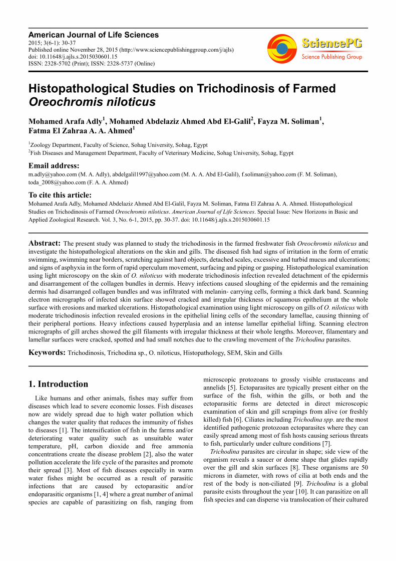
gills in addition to dullness and fin rot (Fig. 1). Signs of
irritation might be attributed to the mechanical action of the
cilia of Trichodina and crawling movement of the parasite on
the surface epithelium of the skin and gills [26]. Signs of
asphyxia may be attributed to the feeding behavior of
Trichodina on the disrupted cells and host’s gills leading to
loss of gill epithelium that, consequently, causes respiratory
function disorders and leads to penetration of the parasite
deeply into the gill tissues [13]. The massive production of
mucus is considered as a defense mechanism to eliminate the
parasite or dilute its irritating effects [2]. These findings
agreed with those found by Soliman et al. [11] who studied the
clinical signs of trichodinosis in the same fish species.
Figure 1. Photograph of diseased O. niloticus fish showing dullness
appearance with detachment of scales (1) and fin rot (2).
3.2. Parasitological Examination
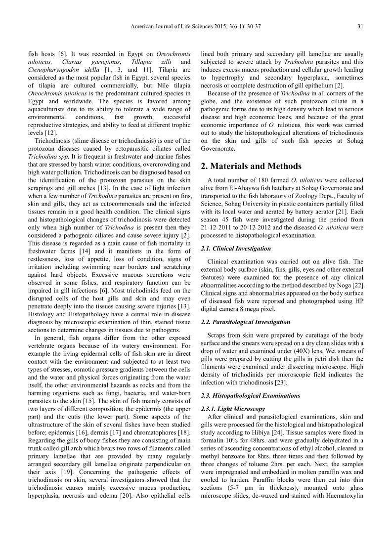
In wet mount preparations, the parasites appeared as
circular or bell-shaped ciliated organisms which were highly
crowding and rapidly motile (Fig. 2 a). Under higher
magnification (40X), the parasite was shown to have several
circular rows of cilia and a circle of more centrally lying
hooklets (Fig. 2 b). The harmful effects of the parasite resulted
from the adhesive disc of Trichodina and the sharp rim of the
border membrane which bite into the surface of the host
epithelial cells, and strongly act as a sucker causing host
irritation. These activities of the parasites, consequently, lead
to severe loss of surface epithelium of skin giving a good
chance for secondary pathogens as bacteria or fungi to invade
the fish [26, 27].
Figure 2. Photograph of wet mount slide shows the Trichodina parasites from
a skin scrap; (a) (100X) and (b) (400X).
3.3. Histological and Histopathological Studies
3.3.1. In Skin
(a). Light Microscopy
Histopathological examination showed that, in contrast to
the normal intact skin of O. niloticus which is composed of
epidermis (E) that consists of stratified squamous epithelium
with mucous cells in between secreting a slimy substance that
covers the whole surface of the skin with a protective layer
against infections [16] and cutis or dermis (D) filled with
American Journal of Life Sciences 2015; 3(6-1): 30-37 33
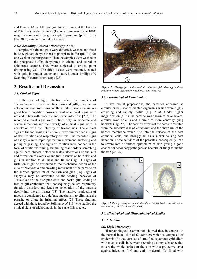
collagen bundles (CB) and subcutaneous skeletal muscles
(SM) (Fig. 3a and b), the skin of O. niloticus with moderate
infection had disorganized and detached epidermis with
vacuolations between cells. The dermis had disarranged
collagen bundles (CB) and was infiltrated with melanin -
carrying cells which aggregated between the skeletal muscles
and the collagen fibers (Fig. 3c and d). These findings agreed
with the results of Hassan [2] who previously reported these
lesions on O. niloticus and O. aureus infected with
trichodinosis and collected from various fish farms of Saudi
Arabia and Huh et al. [28] who reported these lesions on the
infected largemouth bass Micropterus salmoids from North
Carolina, USA.
In severe infection, the epidermis was completely eroded
and sloughed (complete loss). The remaining dermis had
disarranged collagen bundles and was infiltrated with melanin
– carrying cells which aggregated between the muscle layer
and the collagen fibers forming a thick dark band (Fig. 3 e and
f). The latter finding may explain why the heavily infected fish
had the characteristic of dark coloration of body [2]. In
addition to the previous observed lesions, Huh et al. [28]
reported severe epithelial hyperplasia in the epidermis of the
infected largemouth bass Micropterus salmoids from North
Carolina, USA that wasn't observed during this study. Also,
Hassan [2] reported that the dermis was edematous and
infiltrated with leucocytes associated with the
melanin-carrying cells which were observed alone in this
study aggregated between collagen fibers and skeletal
muscles.
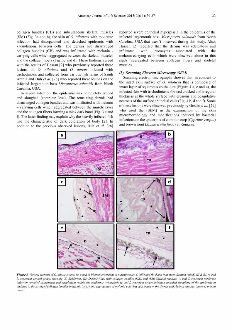
(b). Scanning Electron Microscopy (SEM)
Scanning electron micrographs showed that, in contrast to
the intact skin surface of O. niloticus that is composed of
intact layer of squamous epithelium (Figure 4 a, c and e), the
infected skin with trichodiniasis showed cracked and irregular
thickness at the whole surface with erosions and coagulative
necrosis of the surface epithelial cells (Fig. 4 b, d and f). Some
of these lesions were observed previously by Gostin et al. [29]
who used the (SEM) in the examination of the skin
micromorphology and modifications induced by bacterial
infections on the epidermis of common carp (Cyprinus carpio)
and brown trout (Salmo trutta fario) at Romania.
Figure 3. Vertical sections of O. niloticus skin; (a, c and e) Photomicrographs at magnification (100X) and (b, d and f) at magnification (400X) (H & E). (a and
b) represent control group, showing (E) Epidermis, (D) Dermis filled with collagen bundles (CB), and (SM) Skeletal muscles. (c and d) represent moderate
infection revealed detachment and vaculations within the epidermis (triangles); (e and f) represent severe infection revealed sloughing of the epidermis in
addition to disarranged collagen bundles in dermis (stars) and aggregation of melanin-carrying cells between the dermis and skeletal muscles (arrows) in both
cases.
34 Mohamed Arafa Adly et al.: Histopathological Studies on Trichodinosis of Farmed Oreochromis niloticus
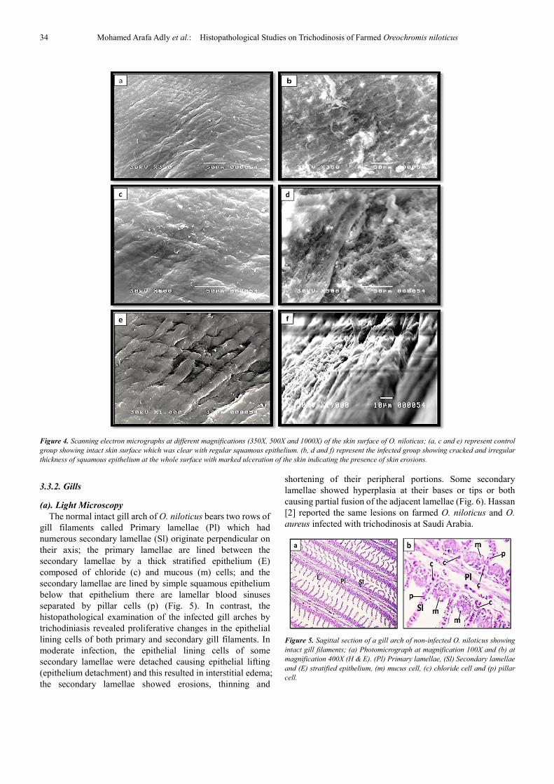
Figure 4. Scanning electron micrographs at different magnifications (350X, 500X and 1000X) of the skin surface of O. niloticus; (a, c and e) represent control
group showing intact skin surface which was clear with regular squamous epithelium. (b, d and f) represent the infected group showing cracked and irregular
thickness of squamous epithelium at the whole surface with marked ulceration of the skin indicating the presence of skin erosions.
3.3.2. Gills
(a). Light Microscopy
The normal intact gill arch of O. niloticus bears two rows of
gill filaments called Primary lamellae (Pl) which had
numerous secondary lamellae (Sl) originate perpendicular on
their axis; the primary lamellae are lined between the
secondary lamellae by a thick stratified epithelium (E)
composed of chloride (c) and mucous (m) cells; and the
secondary lamellae are lined by simple squamous epithelium
below that epithelium there are lamellar blood sinuses
separated by pillar cells (p) (Fig. 5). In contrast, the
histopathological examination of the infected gill arches by
trichodiniasis revealed proliferative changes in the epithelial
lining cells of both primary and secondary gill filaments. In
moderate infection, the epithelial lining cells of some
secondary lamellae were detached causing epithelial lifting
(epithelium detachment) and this resulted in interstitial edema;
the secondary lamellae showed erosions, thinning and
shortening of their peripheral portions. Some secondary
lamellae showed hyperplasia at their bases or tips or both
causing partial fusion of the adjacent lamellae (Fig. 6). Hassan
[2] reported the same lesions on farmed O. niloticus and O.
aureus infected with trichodinosis at Saudi Arabia.
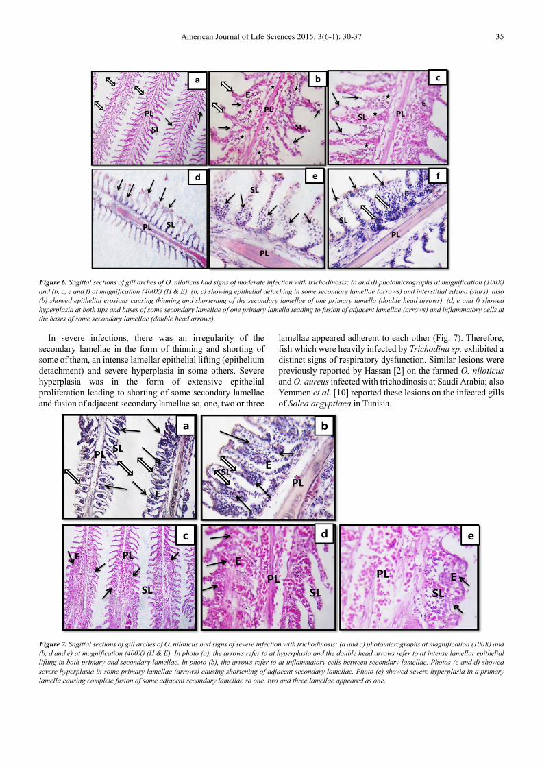
Figure 5. Sagittal section of a gill arch of non-infected O. niloticus showing
intact gill filaments; (a) Photomicrograph at magnification 100X and (b) at
magnification 400X (H & E). (Pl) Primary lamellae, (Sl) Secondary lamellae
and (E) stratified epithelium, (m) mucus cell, (c) chloride cell and (p) pillar
cell.
American Journal of Life Sciences 2015; 3(6-1): 30-37 35
Figure 6. Sagittal sections of gill arches of O. niloticus had signs of moderate infection with trichodinosis; (a and d) photomicrographs at magnification (100X)
and (b, c, e and f) at magnification (400X) (H & E). (b, c) showing epithelial detaching in some secondary lamellae (arrows) and interstitial edema (stars), also
(b) showed epithelial erosions causing thinning and shortening of the secondary lamellae of one primary lamella (double head arrows). (d, e and f) showed
hyperplasia at both tips and bases of some secondary lamellae of one primary lamella leading to fusion of adjacent lamellae (arrows) and inflammatory cells at
the bases of some secondary lamellae (double head arrows).
In severe infections, there was an irregularity of the
secondary lamellae in the form of thinning and shorting of
some of them, an intense lamellar epithelial lifting (epithelium
detachment) and severe hyperplasia in some others. Severe
hyperplasia was in the form of extensive epithelial
proliferation leading to shorting of some secondary lamellae
and fusion of adjacent secondary lamellae so, one, two or three
lamellae appeared adherent to each other (Fig. 7). Therefore,
fish which were heavily infected by Trichodina sp. exhibited a
distinct signs of respiratory dysfunction. Similar lesions were
previously reported by Hassan [2] on the farmed O. niloticus
and O. aureus infected with trichodinosis at Saudi Arabia; also
Yemmen et al. [10] reported these lesions on the infected gills
of Solea aegyptiaca in Tunisia.
Figure 7. Sagittal sections of gill arches of O. niloticus had signs of severe infection with trichodinosis; (a and c) photomicrographs at magnification (100X) and
(b, d and e) at magnification (400X) (H & E). In photo (a), the arrows refer to at hyperplasia and the double head arrows refer to at intense lamellar epithelial
lifting in both primary and secondary lamellae. In photo (b), the arrows refer to at inflammatory cells between secondary lamellae. Photos (c and d) showed
severe hyperplasia in some primary lamellae (arrows) causing shortening of adjacent secondary lamellae. Photo (e) showed severe hyperplasia in a primary
lamella causing complete fusion of some adjacent secondary lamellae so one, two and three lamellae appeared as one.
36 Mohamed Arafa Adly et al.: Histopathological Studies on Trichodinosis of Farmed Oreochromis niloticus
(b). Scanning Electron Microscopy (SEM)
Scanning electron micrographs showed that, in contrast to
the intact surface of the gill arch of O. niloticus which bears
several small filament trunks called primary lamellae that are
provided by many regularly arranged secondary lamellae (Fig.
8 a, c, e), the infected gill filaments appeared cracked with
irregular thickness at their whole lengths due to erosions.
Secondary gill lamellae were greatly thickened curved and
appeared shorter (Fig. 8b, d). The lamellar surface was spotted
with several small notches (n) which may be attributed to the
crawling movement of the Trichodina on their surfaces.
Moreover, some of secondary lamellae were fused to each
other and appeared as one thick lamella because of severe
hyperplasia (Fig. 8d, f). Same lesions were observed by
Hassanain et al. [25] when studding the effect of lead acetate
exposure on fingerlings of Nile Tilapia O. niloticus.
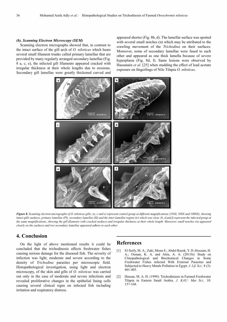
Figure 8. Scanning electron micrographs of O. niloticus gills; (a, c and e) represent control group at different magnifications (350X, 500X and 1000X), showing
intact gills surfaces, primary lamellae (Pl), secondary lamellae (Sl) and the inter-lamellar region (ir) which was clear. (b, d and f) represent the infected group at
the same magnifications, showing the gill filaments with cracked surfaces and irregular thickness at their whole length. Moreover, small notches (n) appeared
clearly on the surfaces and two secondary lamellae appeared adhere to each other.
4. Conclusion
On the light of above mentioned results it could be
concluded that the trichodinosis affects freshwater fishes
causing serious damage for the diseased fish. The severity of
infection was light, moderate and severe according to the
density of Trichodina parasites per microscopic field.
Histopathological investigation, using light and electron
microscopy, of the skin and gills of O. niloticus was carried
out only in the case of moderate and severe infections and
revealed proliferative changes in the epithelial lining cells
causing several clinical signs on infected fish including
irritation and respiratory distress.
References
[1] El-Seify, M. A.; Zaki, Mona S.; Abdel Razek, Y. D.;Hossam, H. A.; Osman, K. A. and Attia, A. A. (2011b): Study on Clinopathological and Biochemical Changes in Some Freshwater Fishes infected With External Parasites and Subjected to Heavy Metals Pollution in Egypt. J. Lif. Sci., 8 (3): 401-405.
[2] Hassan, M. A. H. (1999): Trichodiniasis in Farmed Freshwater Tilapia in Eastern Saudi Arabia. J. KAU: Mar Sci., 10: 157-168.
American Journal of Life Sciences 2015; 3(6-1): 30-37 37
[3] El-Seify, M. A.; Zaki, Mona S.; Abdel Razek, Y. D.; Hossam, H. A.; Osman, K. A. and Attia, A. A. (2011a): Seasonal Variations and Prevalence of Some External Parasites Affecting Freshwater Fishes Reared at Upper Egypt. J. Lif. Sci., 8 (3): 397-400.
[4] Hussain, S.; Hassan, M. Z.; Mukhtar, Y. and Saddiqui, B. N. (2003): Impact of environmental pollution in human behaviour and up-left of awareness level through mass media among the people of Faisalabad city. Int. J. Agric. Biol., 5: 660-661.
[5] Eissa, I. A. M (2002): Parasitic fish diseases in Egypt, 1st. edition, pp: 52-53. Dar El-Nahdda El- Arabia publishing.
[6] Abowei, J. F. N.; Briyai, O. F. and Bassey, S. E. (2011): A Review of Some Basic Parasite Diseases in Culture Fisheries Flagellids, Dinoflagellids and Ichthyophthriasis, Ichthyobodiasis, Coccidiosis, Trichodiniasis, Heminthiasis, Hirudinea Infestation, Crustacean Parsite and Ciliates. Brit. J. of Pharmacol. & Toxicol., 2 (5): 213-226.
[7] Jia-yun, Y.; Xi-lian, L.; Jin-yu, S.; Xiao-yi, P.; Gui-jie, H.; Yang Xu; Wen-lin, Y.; Hongshun, R.; Xiao-lin, L. (2011): Isolation of bioactive components from Chelidonium majus L. with activity against Trichodina sp.. J. Aquacul., 318: 235–238.
[8] Robert, M. D. (2003): protozoan parasites. Southern Regional Aquaculture Center (SRAC) publication no. 4701.
[9] Robert, B. M. Jr. (2013): External Protozoan Diseases of Fish. http://www. cichlidforum. com/articles/diseases_ext_protozoan. php.
[10] Yemmen, C.; Quilichini, Y.; HédiKtari, M.; Marchand, B. and Bahri1, S. (2010/11): Morphological, ecological and histopathological studies of Trichodina gobii Raabe, 1959 (Ciliophora: Peritrichida) infecting the gills of Soleaaegyptiaca. J. Protistol., 6 (4): 258–263.
[11] Soliman Fayza, M.; Abd El-Galil, M. A. A.; Adly, M. A. and Ahmed Fatma El Zahraa, A. A. (2013): Studies on Trichodinosis of Some Cultured Freshwater Fishes at Sohag Governorate. J. life Sci., 10 (4): 1400-1409.
[12] Grammer, G. L.; Slack, W. T.; Peterson, M. S.; and Dugo, M. A. (2012): Nile Tilapia Oreochromis niloticus (Linnaeus, 1758) establishment in temperate Mississippi, USA: multi-year survival confirmed by otolith ages. J. compil. Aquat. Invas., 7 (3): 367–376.
[13] El-Tantawy, S. A. M and El-Sherbiny, H. A. E. (2010): Ectoparasitic trichodinians infecting catfish Clarias gariepinus inhabiting Nile Delta Water of the River Nile, Dakahlia Province, Egypt. J. of Amer. Sci., 6 (9): 656-668.
[14] Abd El-Galil, M. A. A. and Aboelhadid, S. M. (2012): Trials for control of trichodinosis and Gyrodactylosis in hatchery reared Oreochromis niloticus fries by using Garlic. Vet. Parasitol., 185: 57– 63.
[15] Hawkes, J. W. (1974): The Structure of Fish Skin, I. General Organization. Cell Tiss. Res., 149: 147- 158.
[16] Whitear, M. (1971): Cell specialization and sensory function in fish epidermis. J. Zool. (Lond.), 163: 237-264.
[17] Fishelson, L. (1972): Histology and ultrastructure of the skin of Lepadichthys lineatus (Gobiesocidae: Teleostei). Marine Biol., 17: 357-364.
[18] Fujii, R. (1969): Chromatophore and pigments. In: Fish physiology (eds. W. S. Hoar, D. J. Randall), vol. III, p. 307-353. New York: Academic Press.
[19] Figueiredo-Fernandes, A.; Ferreira-Cardoso, J. V.; Garcia-Santos, Sofia; Monteiro, Sandra, M.; Carrola, J.; Matos, P and Fontaínhas-Fernandes, A. (2007): Histopathological changes in liver and gill epithelium of Nile tilapia, Oreochromis niloticus, exposed to waterborne copper1 Pesq. Vet. Bras., 27 (3):103-109.
[20] Abdel-Meguid, M. (2001): Trichodiniasis as a cause of mortality among infected Tilapia zilli with special emphasis on its control using Earthtec. Egypt. J. Aquat. Biol. & Fisk., 5 (2): 95-104.
[21] Abo-Esa, J. F. K. (2008): Study on Some Ectoparasitic Diseases of Catfish, Clarias gariepinus with their Control by Ginger, Zingiber officiale. Medit. Aquacul. J., 1 (1): 1-9.
[22] Noga, E. J. (2000): Fish Diseases. Diagnosis and Treatment. Text book, Iowa State University Press, ISBN 0-8138-2558-X.
[23] Bartholomew, J. L. (2003): Salmonid ceratomyxosis. In: Suggested Procedures for the Detection and Identification of Certain Finfish and Shellfish Pathogens. Blue Book 5th Ed., Volume 2, Fish Health Section, American Fisheries Society.
[24] Hibiya, T. (1982): An Atlas of Fish Histology. No. 1, 1-5, Kodansha Ltd., Tokyo.
[25] Hassanain, M. A.; Abbas, W. T. and Ibrahim, T. B. (2012): Skeletal ossification impairment in Nile Tilapia (Oreochromis niloticus) after exposure to lead acetate. Pakistan J. of Biol. Scis., 15 (15): 729-735.
[26] Abd El-Galil, M. A. A. (1998): Studies on some fish pathogens affecting freshwater fishes in Beni-Suef hatchery. Thesis of M. V. Sc. Fish Diseases and management. Fish Dept., Fac. Vet. Med., Beni-suef, Cairo univ.
[27] Reed, P.; Ruth F. F. and Ruth, E. K. (2003): Monogenean Parasites of Fish. This document is FA-28, one of a series from the Department of Fisheries and Aquatic Sciences, Florida Cooperative Extension Service, Institute of Food and Agricultural Sciences, University of Florida.
[28] Huh, M. D.; Thomas, C. D.; Udomkusnsri, P. and Noga, E. J. (2005): Epidemic trichodinosis associated with sever epidermal hyperplasia in largemouth bass, Micropterus salmoides from North Carolina, USA. J. of Wildlife Dis., 41 (3): 647-653.
[29] Gostin, I. N.; Neagu, A. N. and Vulpe, V. (2011): SEM investigations regarding skin micromorphology and modification induced by bacterial infections in Cyprinus carpio and Salmo trutta fario. Inter. J. of Ener. and Environ., 2 (5): 274-281.
Related Documents