AL-Qadisiya Medical Journal Vol.10 No.18 2014 50 Histopathological changes on Splenomegaly induced in Experimental rats Wistar albino Maha K. Al-Malaak Science collage / Biology Department/ Basrah University (Received 20/1/2013, Accepted 5/3/2013) اﻟﺨﻼﺻﺔ ﺗﮭﺪف اﻟﺪراﺳﺔ اﻟﺤﺎﻟﯿﺔ اﻟﻰ ﺗﻘﯿﯿﻢ اﻟﺘﻐﯿﺮات اﻟﻨﺴﺠﯿﺔ اﻟﻤﺮﺿﯿﺔ ﻓﻲ اﻟﺠﺮذان اﻟﻤﺨﺘﺒﺮﯾﺔ ذات ﺗﻀﺨﻢ اﻟﻄﺤﺎل، اﻟﺜﺎﯾﻮاﺳﺴﺘﺎﻣﯿﺪ وﺗﻠﯿﻒ اﻟﻜﺒﺪ اﻟﻤﺴﺘﺤﺚ ﻋﻦ ﻃﺮﯾﻖ ﺗﺠﺮﯾﻊ ﻣﺮﻛﺐThioacetamide (TAA) . ﻟﻮﺣﻆ اﻟﺘﻠﯿﻒ اﻟﻜﺒﺪي ﻓﻲ ﺟﻤﯿﻊ اﻟﺠﺮذان اﻟﻤﺨﺒﺮﯾﺔ ﺑﻌﺪ اﻟﺤ ﻘﻦ ﺗﺤﺖ اﻟﺒﺮﯾﺘﻮن ﺑﺠﺮﻋﺔ ﻣﻘﺪارھﺎ) 200 ﻣﻠﻐﻢ/ ﻛﻐﻢ( ﻣﻦ وزن اﻟﺠﺴﻢ وﻟﻤﺪة) 8 ( اﺳﺎﺑﯿﻊ ﻣﺘﺘﺎﻟﯿﺔ وﺟﻤﯿﻊ اﻟﻨﺘﺎﺋﺞ ﻗﻮرﻧﺖ ﻣﻊ ﺟﺮذان اﻟﺴﯿﻄﺮة اﻟﺘﻲ ﺣﻘﻨﺖ ﺑﺎﻟﻤﺤﻠﻮل اﻟﻤﻨﻈﻢ. ﺗﻢ ﺗﺴﺠﯿﻞ وزن اﻟﺠﺴﻢ واﻟﻄﺤﺎل واﻟﻜﺒﺪ ﻓﻲ ﺟﺮذان اﻟﺴﯿﻄﺮة واﻟﻤﺠﻤﻮﻋﺔ اﻟﻤﻌﺎﻣﻠﺔ وﻛﺬﻟﻚ ﺣﺴﺒﺖ اﻟﻨﺴﺒﺔ ﻟﻜﻞ ﻣﻦ اﻟﻜﺒﺪ واﻟﻄﺤﺎل اﻟﻰ وزن اﻟﺠﺴﻢ. واﻟﺘﻲ ﺗﺸﻤﻞ اﻟﻠﻮن واﻟﺤﺠﻢ واﻻﺣﺘﻘﺎن وﻋﻼﻣﺎت اﻟﺘﻠﯿﻒ ﺗﻀﻤﻨﺖ اﻟﺪراﺳﺔ اﻟﺘﻐﯿﺮات اﻟﻌﯿﺎﻧﯿﺔ وﻋﺪم اﻻﻧﺘﻈﺎم وﻃﺒﯿﻌﺔ اﻟﺴﻄﺢ ﻟﻜﻞ ﻣﻦ اﻟﻜﺒﺪ واﻟﻄﺤﺎل. اﻇﮭﺮت اﻻﻛﺒﺎد ﻓﻲ اﻟﺠﺮذان اﻟﻤﻌﺎﻣﻠﺔ ﺳﻄﺢ ﻣﺤﺒﺐ وﺧﺸﻦ ﻣﻊ ﻣﻨﺎﻃﻖ ﻣﻦ اﻟﺘﻠﯿﻒ ﺑﺸﻜﻞ ﺑﻘﻊ ﺑﯿﻀﺎء وﻟﻮن ﺷﺎﺣﺐ واﺣﺘﻘﺎن ﻣﻊ زﯾﺎدة ﻓﻲ اﻟ ﺤﺠﻢ ﻛﺬﻟﻚ ﻛﺎن اﻟﻄﺤﺎل ﻣﺘﻀﺨﻢ، اﺣﻤﺮ ﻏﺎﻣﻖ ﻣﻊ ﻣﻨﺎﻃﻖ ﺗﻠﯿﻒ وﺗﺨﺼﺮ وﯾﺒﺪو ھﺶ ﺳﮭﻞ اﻟﺘﺤﻄﻢ واﻟﻨﺰف. اﺷﺎرت اﻟﻨﺘﺎﺋﺞ اﻟﺒﺎﯾﻮﻛﯿﻤﯿﺎﺋﯿﺔ اﻟﻰ ﻧﻘﺼﺎن وﺑﻔﺎرق ﻣﻌﻨﻮي(P<0.05) ﻓﻲ ﻧﺴﺐ اﻻﻟﺒﻮﻣﯿﻦ وﺗﺮﻛﯿﺰ اﻟﺒﺮوﺗﯿﻦ اﻟﻜﻠﻲ ﻓﻲ اﻟﺠﺮذان ذات اﻟﻄﺤﺎل اﻟﻤﺘﻀﺨﻢ ﻣﻘﺎرﻧﺔ ﻣﻊ ﺟﺮذان اﻟﺴﯿﻄﺮة وﻛﺬﻟﻚ ﺗﺜﺒﯿﻂ اﻟﻔﻌ ﺎﻟﯿﺔ اﻻﻧﺰﯾﻤﯿﺔ ﻻﻧﺰﯾﻤﻲ اﻟﻜﺒﺪ) GPT واﻟـGOT ( ﻓﻲ ﻣﺠﻤﻮﻋﺔ اﻟﺘﻠﯿﻒ اﻟﻜﺒﺪي اﻟﻤﺴﺘﺤﺚ واﻟﻄﺤﺎل اﻟﻤﺘﻀﺨﻢ ﻣﻘﺎرﻧﺔ ﻣﻊ اﻟﺴﯿﻄﺮة إذ اﻇﮭﺮت اﻟﻨﺘﺎﺋﺞ زﯾﺎدة ﻣﻌﻨﻮﯾﺔ ﻓﻲ ﺗﺮﻛﯿﺰ ھﺬﯾﻦ اﻻﻧﺰﯾﻤﯿﻦ ﻓﻲ ﻣﺼﻞ اﻟﺪم ﻟﻠﺠﺮذان ذات اﻟﺘﻠﯿﻒ. ﻛﻤﺎ ﺗﻢ ﺗﻘﺪﯾﺮ ﺗﺮﻛﯿﺰ اﻟﺒﻠﯿﺮوﺑﯿﻦ اﻟﻜﻠﻲ ﻓﻲ ﻣﺼﻞ اﻟﺪم ﻟﻠﺠﺮذان ذات اﻟ ﺘﻠﯿﻒ اﻟﻜﺒﺪي اﻟﻤﺴﺘﺤﺚ واﺷﺎرت اﻟﻨﺘﺎﺋﺞ اﻟﻰ ﻓﺮق ﻣﻌﻨﻮي اﺣﺼﺎﺋﻲ ﻋﺎﻟﻲ ﻣﻘﺎرﻧﺔ ﻣﻊ ﺗﺮﻛﯿﺰ اﻟﺒﯿﻠﺮوﺑﯿﻦ ﻓﻲ ﺟﺮذان اﻟﺴﯿﻄﺮة. اﻇﮭﺮت ﻧﺘﺎﺋﺞ اﻟﻔﺤﺺ اﻟﻤﺠﮭﺮي ﻟﻤﻘﺎﻃﻊ ﻓﻲ اﻛﺒﺎد اﻟﺠﺮذان ذات اﻟﺘﻠﯿﻒ اﻟﻜﺒﺪي واﻟﻄﺤﺎل اﻟﻤﺘﻀﺨﻢ اﻟﻰ اﺗﺴﺎع واﺣﺘﻘﺎن اﻻوردة اﻟﺒﻮاﺑﯿﺔ ﻣﻊ زﯾﺎدة ﻓﻲ ﻗﻄﺮ اﻟﻮرﯾﺪ اﻟﺒﻮاﺑﻲ و ﻟﻠﻄﺤﺎل اﻟﻤﺘﻀﺨﻢ ﺑﻔﺎرق ﻣﻌﻨﻮي اﺣﺼﺎﺋﻲ ﻣﻘﺎرﻧﺔ ﻣﻊ اﻟﺴﯿﻄﺮة وﻛﺬﻟﻚ ھﻨﺎك زﯾﺎدة ﻓﻲ اﻗﻄﺎر اﻟﻌﻘﯿﺪات اﻟﻠﻤﻔﯿﺔ ﻋﻨﺪ ﻣﺴﺘﻮىP<0.05 ﻣﻘﺎرﻧﺔ ﻣﻊ ﻣﻌﺪل اﻗﻄﺎرھﺎ ﻓﻲ ﺟﺮذان اﻟﺴﯿﻄﺮة. اﻛﺪت اﻟﺪراﺳﺔ اﻟﺤﺎﻟﯿﺔ ﻋﻠﻰ ﺗﺎﺛﯿﺮ اﻟﻤﺮﻛﺐ(TAA) ﻓﻲ ﺣﺚ اﻟﺘﻐﯿﺮات اﻟﻨﺴﺠﯿﺔ اﻟﻤﺮﺿﯿﺔ ﻓﻲ اﻛﺒﺎد وﻃﺤﺎل اﻟﻤﺠﻤﻮﻋﺔ اﻟﻤﻌﺎﻣﻠﺔ وﻗﻮرﻧﺖ ﻣﻊ ﻣﺠﻤﻮﻋﺔ اﻟﺴﯿﻄﺮة واﺷﺎرت ﻧﺘﺎﺋﺞ اﻟﻔﺤﺺ اﻟﻤﺠﮭﺮي اﻟﻀﻮﺋﻲ ﻟﻠﻤﻘﺎﻃﻊ اﻟﻨﺴﺠﯿﺔ ﻓﻲ اﻟﻜﺒﺪ واﻟﺘﻲ ﺻﺒﻐﺖ ﺑﺎﺳﺘﺨﺪام ﻛﺎﺷﻒ(PAS) اﻟﻰ ﺗﺮﺳﯿﺐ ﺣﺒﯿﺒﺎت اﻟﻜﻼﯾﻜﻮﺟﯿﻦ ﺑﺸﻜﻞ ﺣﺒﯿﺒﺎت ﺧﺸﻨﺔ ﻏﺎﻣﻘﺔ ﻟﻮﻧﮭﺎ اﺣﻤﺮ ﺗﺘﺮﺳﺐ ﻓﻲ ﺳﺎﯾﺘ ﻮﺑﻼزم اﻟﺨﻼﯾﺎ اﻟﻜﺒﺪﯾﺔ، ﻛﺬﻟﻚ ﻟﻮﺣﻆ اﻟﺘﻠﯿﻒ، ﻓﺮط اﻟﺘﻨﺴﺞ، ﺗﻠﯿﻒ اﻟﺤﻮاﺟﺰ اﻟﻜﺒﺪﯾﺔ، ﺗﺮﺳﯿﺐ ﻛﻤﯿﺎت ﻛﺒﯿﺮة ﻣﻦ اﻻﻟﯿﺎف اﻟﻐﺮاوﯾﺔ ﻣﻊ ﺗﺤﻄﻢ اﻟﺘﺮﻛﯿﺐ اﻟﻄﺒﯿﻌﻲ ﻟﻠﻔﺼﯿﺼﺎت اﻟﻜﺒﺪﯾﺔ. اﺷﺎرت اﻟﻨﺘﺎﺋﺞ اﻟﻰ ﺧﻼﯾﺎ ﻛﺒﺪﯾﺔ ﻣﻨﺘﻔﺨﺔ ﺗﺸﺒﮫ اﻟﺒﺎﻟﻮن وﺗﻨﻜﺲ اﻻﻧﻮﯾﺔ ﻣﻊ وﺟﻮد ﻓﺠﻮات ﺗﺘﺠﻤﻊ ﻓﯿﮭﺎ اﻟﺪھﻮن ﻣﻤﺎ ﺗﻌﻄﻲ اﺷﺎرة اﻟﻰ اﻟﺘﻨﻜﺲ اﻟﺪھﻨﻲ ﻓﻲ ﻣﻌﻈﻢ اﻟﻤﻘﺎﻃﻊ اﻟﻨﺴﯿﺠﯿﺔ ﻋﻦ ﻓﺮط اﻟﺘﻨﺴﺞ واﺗﺴﺎع اﻻﻗﻨﯿﺔ اﻟﺼﻔﺮاوﯾﺔ ﻣﻊ ارﺗﺸﺎح ﺧﻼﯾﺎ اﻟﺘﮭﺎﺑﯿﺔ وﻣﻨﺎﻃﻖ ﻣﻦ ﻟﻼﻛﺒﺎد ذات اﻟﺘﻠﯿﻒ، ﻓﻀﻼ اﻟﺘﻠﯿﻒ ﺿﻤﻦ اﻟﻨﺴﯿﺞ اﻟﺤﺸﻮي ﻟﻠﻜﺒﺪ. اﺣﺘﻘﺎن اﻟﻠﺐ اﻻﺣﻤﺮ ﻣﻊ ﺑﻌﺾ اﻟﻌﻘﯿﺪات اﻟﻠﻤﻔﯿﺔ اﻟﻔﻌﺎﻟﺔ وﺑﻌﺾ ﻣﻨﮭﺎ اﻛﺜﺮ ﻣﻦ ﻋ ﻘﯿﺪة ﻟﻤﻔﯿﺔ ﺗﺘﺠﻤﻊ ﻣﻊ ﺑﻌﻀﮭﺎ. زﯾﺎدة ﻓﻲ ﺳﻤﻚ اﻟﺤﻮاﺟﺰ ﻣﻊ و ﺟﻮد ﻋﺪد ﻛﺒﯿﺮ ﻣﻦ اﻟﺨﻼﯾﺎ اﻟﻤﻠﺘﮭﻤﺔ اﻟﺘﻲ ﯾﺘﺮﺳﺐ ﻓﯿﮭﺎ ﺻﺒﻐﺔ اﻟـHemosidrin . ﻟﻮﺣﻆ اﻟﻨﺰف ﻣﻊ اﻋﺪاد ﻣﻦ ﻛﺮﯾﺎت اﻟﺪم اﻟﺤﻤﺮ واﻟﺼﻔﯿﺤﺎت اﻟﺪﻣﻮﯾﺔ اﻟﺘﻲ ﺗﺘﺮﺳﺐ ﻓﻲ اﻻوﻋﯿﺔ اﻟﺪﻣﻮﯾﺔ اﻟﻜﺒﯿﺮة، وھﻨﺎﻟﻚ زﯾﺎدة ﻓﻲ ﺳﻤﻚ اﻟﻤﺤﻔﻈﺔ ﻣﻊ ﻧﺴﯿﺞ ﻣ ﺘﻠﯿﻒ ﻓﻲ ﻣﻨﺎﻃﻖ ﻣﺨﺘﻠﻔﺔ ﻣﻦ اﻟﻨﺴﯿﺞ اﻟﺤﺸﻮي ﻣﻘﺎرﻧﺔ ﻣﻊ اﻟﻤﻘﺎﻃﻊ ﻓﻲ ﻃﺤﺎل ﺟﺮذان اﻟﺴﯿﻄﺮة. Abstract This study was aimed to evaluate histopathological changes in experimental rats with induced splenomegaly and liver fibrosis by administering thioacetamid (TAA). Liver fibrosis was clearly noticed in all experimental rats after intraperitoneally injected with a dose (200mg/kg) body weight for 8 weeks ,while control rats injected with buffered saline.The body , spleen and liver weight were recorded , also ratio of liver and spleen per body weight was estimated.Macroscopical changes including (colour, size, congested, signs of fibrosis, irregularity, nature of surface) regarded to liver and spleen were clarified.

Welcome message from author
This document is posted to help you gain knowledge. Please leave a comment to let me know what you think about it! Share it to your friends and learn new things together.
Transcript

AL-Qadisiya Medical Journal Vol.10 No.18 2014
50
Histopathological changes on Splenomegaly induced in Experimental rats Wistar albino
Maha K. Al-MalaakScience collage / Biology Department/ Basrah University
(Received 20/1/2013, Accepted 5/3/2013)
الخالصةتھدف الدراسة الحالیة الى تقییم التغیرات النسجیة المرضیة في الجرذان المختبریة ذات تضخم الطحال،
لوحظ التلیف .Thioacetamide (TAA)وتلیف الكبد المستحث عن طریق تجریع مركب الثایواسستامید من وزن الجسم ) كغم/ملغم200(قن تحت البریتون بجرعة مقدارھا الكبدي في جمیع الجرذان المخبریة بعد الح
تم تسجیل وزن .اسابیع متتالیة وجمیع النتائج قورنت مع جرذان السیطرة التي حقنت بالمحلول المنظم)8(ولمدة الجسم والطحال والكبد في جرذان السیطرة والمجموعة المعاملة وكذلك حسبت النسبة لكل من الكبد والطحال الى
تضمنت الدراسة التغیرات العیانیة والتي تشمل اللون والحجم واالحتقان وعالمات التلیف .وزن الجسماظھرت االكباد في الجرذان المعاملة سطح محبب وخشن .وعدم االنتظام وطبیعة السطح لكل من الكبد والطحال
حجم كذلك كان الطحال متضخم، مع مناطق من التلیف بشكل بقع بیضاء ولون شاحب واحتقان مع زیادة في ال .احمر غامق مع مناطق تلیف وتخصر ویبدو ھش سھل التحطم والنزف
في نسب االلبومین وتركیز (P<0.05)اشارت النتائج البایوكیمیائیة الى نقصان وبفارق معنوي الیة االنزیمیة البروتین الكلي في الجرذان ذات الطحال المتضخم مقارنة مع جرذان السیطرة وكذلك تثبیط الفع
في مجموعة التلیف الكبدي المستحث والطحال المتضخم مقارنة مع السیطرة ) GOTوالـ GPT(النزیمي الكبد كما تم تقدیر تركیز .إذ اظھرت النتائج زیادة معنویة في تركیز ھذین االنزیمین في مصل الدم للجرذان ذات التلیف
تلیف الكبدي المستحث واشارت النتائج الى فرق معنوي احصائي البلیروبین الكلي في مصل الدم للجرذان ذات الاظھرت نتائج الفحص المجھري لمقاطع في اكباد الجرذان .عالي مقارنة مع تركیز البیلروبین في جرذان السیطرة
ذات التلیف الكبدي والطحال المتضخم الى اتساع واحتقان االوردة البوابیة مع زیادة في قطر الورید البوابي بفارق معنوي احصائي مقارنة مع السیطرة وكذلك ھناك زیادة في اقطار العقیدات اللمفیة للطحال المتضخم و
اكدت الدراسة الحالیة على تاثیر المركب .مقارنة مع معدل اقطارھا في جرذان السیطرة P<0.05عند مستوى (TAA) في حث التغیرات النسجیة المرضیة في اكباد وطحال المجموعة المعاملة وقورنت مع مجموعة
السیطرة واشارت نتائج الفحص المجھري الضوئي للمقاطع النسجیة في الكبد والتي صبغت باستخدام كاشف (PAS) وبالزم الخالیا الى ترسیب حبیبات الكالیكوجین بشكل حبیبات خشنة غامقة لونھا احمر تترسب في سایت
الكبدیة، كذلك لوحظ التلیف، فرط التنسج، تلیف الحواجز الكبدیة، ترسیب كمیات كبیرة من االلیاف الغراویة مع اشارت النتائج الى خالیا كبدیة منتفخة تشبھ البالون وتنكس .تحطم التركیب الطبیعي للفصیصات الكبدیة
تعطي اشارة الى التنكس الدھني في معظم المقاطع النسیجیة االنویة مع وجود فجوات تتجمع فیھا الدھون مما لالكباد ذات التلیف، فضًال عن فرط التنسج واتساع االقنیة الصفراویة مع ارتشاح خالیا التھابیة ومناطق من
احتقان اللب االحمر مع بعض العقیدات اللمفیة الفعالة وبعض منھا اكثر من . التلیف ضمن النسیج الحشوي للكبدزیادة في سمك الحواجز مع و جود عدد كبیر من الخالیا الملتھمة التي یترسب فیھا . قیدة لمفیة تتجمع مع بعضھاع
لوحظ النزف مع اعداد من كریات الدم الحمر والصفیحات الدمویة التي تترسب في . Hemosidrinصبغة الـتلیف في مناطق مختلفة من النسیج الحشوي االوعیة الدمویة الكبیرة، وھنالك زیادة في سمك المحفظة مع نسیج م
.مقارنة مع المقاطع في طحال جرذان السیطرة
AbstractThis study was aimed to evaluate histopathological changes in experimental
rats with induced splenomegaly and liver fibrosis by administering thioacetamid (TAA).Liver fibrosis was clearly noticed in all experimental rats after intraperitoneally injected with a dose (200mg/kg) body weight for 8 weeks ,while control rats injected with buffered saline.The body , spleen and liver weight were recorded , also ratio ofliver and spleen per body weight was estimated.Macroscopical changes including (colour, size, congested, signs of fibrosis, irregularity, nature of surface) regarded to liver and spleen were clarified.
AL-Qadisiya Medical Journal Vol.10 No.18 2014
51
The livers showed coarse granulation surface, fibrous (white) spots, pale color and congested with large size, while spleen appeared enlarged, reddish, with fibrous regions, twister and fragil easy to destruct.Biochemical results revealed to statistical difference (p<0.05) in the rate of albumin and total protein concentration in splenomegaly rats compared to control. Inhibition of both (Gpt and Got) enzymes was estimated in rats with splenomegaly up compared to control rats.There was statistical difference in the mean concentration of total bilirubin in splenomegaly rats in comparison to control rats.The results of microscopic investigation of tissue sections related to rats liver with enlarged spleen showed dilation in portal veins with an increased in (PV) diameter in relation to control rats, also significant increased with splenic lymph nodules diameter in splenomegaly rats compared to control rats.Results on liver sections stained with (PAS) reagent indicated to dense, coarse and red glycogen granules were deposit on hepatocytes also fibrosis, hyperplasia, fibrous septa, large amount of collagen fibers and destruction of normal lobules were observed.The results referred to swollen hepatocytes, pyknotic nuclei, vacuoles with lipid accumulation and moderate steatosis was obvious in all sections of livers with cirrhosis .Hyperplasia, dilated bile ducts and infiltration of inflammatory cells. Spleen sections of splenomegaly rats showed congested red pulp, some lymph nodule s appeared active and more than one aggregated together, thickening in fibrous trabecullae and numerous macrophages with hemosidrin deposite.Hemorrhage with large numbers of erythrocytes and platelets deposit in large blood vessels, thickening in capsule with more fibrous tissue compared with control rats.
Introduction The spleen is an organ has a couple of
important function involving blood cells within the body, it filters blood and removes old and damaged red blood cells, bacteria and other particles as they pass through blood vessels within the spleen, also it produces assists immune system and is considered the draining site for compounds that are administered intravenously (1,2).Weight of spleen is highly variable, in human adults, the spleen generally weights (150-250g) and the elderly spleen is often smaller, even when there is no apparent hypofunction (3). Splenomegaly is often detected in patients with liver cirrhosis and portal hypertension with a prevalence of 60-65%, some cases can be very prominent and cause symptoms like abdominal pain, splenic infarction and cytopenia(4,5).(6)explained why portal hypertension produce congestive splenomegaly and regarded it to the blood reaches the spleen via the splenic artery, a large branch of the celiac artery,
and enters the spleen through four to six branches while venous outflow via four to six venous branches, called the splenic vien which drain into the portal vein .Influence of portal hypertension on the weight of spleen and splenic tissues has been studied and histological changes in rats were similar to the histological findings of splenomegaly accompanying portal hypertension in humans (7).
Splenic enlargement had been found to correlated with increase in the white blood cells but not with that of circulating (CD34) positive cells (8).Study on effect of serious environmental pollutant which toxic effects are associated with oxidative stress showed histological changes on spleen and the spleen white pulp was decreased significantly in treated group compared with control (9). Splenomegaly in liver cirrhosis was not only congestive but there was also significant increase of reticular fibers, red pulp area and angiogenesis, moreover, nitric oxide (No)

AL-Qadisiya Medical Journal Vol.10 No.18 2014
52
reduction resulting from suppression of e-NOS and TNF-α contributed to the increased volume of spleen (10).(11) reffered that chronic hepatitis is a well recognized problem in canine practice and affect dogs of many breeds, however knowledge of its etiology, remains limited .The pathways to splenomegaly was determined in rats with fully developed portal hypertension and the pathogenic mechanisms was
characterized (12,13). (14) suggested that splenectomy may be of a preventive role against CCl4 induced rat liver fibrosis to a certain degree .Since splenomegaly a common cause of liver cirrhosis, the aim of this study was to evaluate histopatholoical changes occur with splenomegaly in relation to acute liver damaged and hyperplasia induced in an experimental model.
Materials and MethodsExperimental animals and mode of
treatment Total number of wister rats (Rattus
norvegicus) (30) rats aged 12 weeks weighing (200-250)g were used in this experiments.Animals were housed in the metal cages under controlled temperature (25±2C°) and constant dark-light cycle (12hr light, 12hr dark). The rats received the same food and water ad libitumthrought the study ,animals divided into two groups, (10)rats as control received only buffered saline (2ml) intraperitoneally. While (20) rats as experimental group treated with (200mg/kg) of body weight intraperitoneally of thioacetamid (TAA) to iniduced liver fibrosis and splenomegaly, the dose gives three times a week for (8 weeks). (TAA) powder dissolved in 0.9% normal saline according to (7,15).After 8 weeks, all the animals were fasted overnight, anaesthesized, body weight was recorded and then the rats were sacrified, the abdomen cut to excised both spleen and liver, both organs weighing to estimated ratio of wet liver and spleen per animal body weight (16,10).Both liver and spleen from each rat fixed with formalin (10%) and some pieces of liver fixed with Bouin`s fixative for glycogen fibrillar matrix deposition.Macroscopical observations of organs included (colour, appearance, shape, nature of liver and spleen surface, regularity, congestion) were recorded. The blood was
immediately collected with sterile tubes, let for 10 minutes to clot, then serum was separated by centrifuging at 2500rpm/min for 15min., stored at (-25C°) for biochemical estimations. All the results expressed as (mean ± SD).Biochemical analysis
Liver parameters like (GPT and GOT) enzymes were estimated in serum, the enzymes activity was determined by kit from Bio-labo-SA company (France) according to(17),total protein was determined by (17) respectively. Albumin concentration was estimated according to(18) and the result compared with standard curve accompanying the kit. Total bilirubin was determined by a method of (19). Tissue processing for light microscopic examination
Samples from spleens and livers related to control and experimental animals were cut to suitable pieces and fixed with formalin (10%) and some pieces of livers fixed with Bouin`s fixative for (12hr) to clarified changes with glycogen .Specimens prepared by paraffin-wax method according to(20)
. After fixation specimens fixed with formalin washed with distilled water while these fixed with Bouin`s fixative washed with 70% alcohol, dehydrated with series of alcohol, cleared with xyelen, infiltrated for (3hr) with mixed of xyelen and paraffin, embedded with paraffine then sectioned to (5μ) thickness

AL-Qadisiya Medical Journal Vol.10 No.18 2014
53
sections ,stained with haematoxylin and eosin .Microscopic examination
All sections were examined and the histopathological changes were evaluated according to fibrosis area, portal vein dilation ,dense of inflammatory cells, glycogen deposition and cellularity of red pulp. Diameter of lymph nodules was determined in all sections of spleen
related to control and treated group .Portal vein diameter was
calculated and regarded abnormal if the mean diameter more than 2.1mm (21).Statistical analysis
All values were expressed as mean ± standard deviation (SD). Means were compared in all results by analysis of variance (ANOVA).Significance levels were established at P<0.05.
Results Macroscopic observations
Results showed that liver has a coarse granulation surface with white fibrous regions, large congested area and large size also irregular margins was noticed, pale color compared with control liver which appeared normal size and red without any abnormal features, (Fig 1,2).Macroscopic observations on spleen treated with (TAA) to induced splenomegaly showed larger size than control with fibrous (white) regions on surface, twister sometimes with irregularity spots, the spleen looked fragil, dark reddish compared with control which it is relatively solid organ (Fig. 3a, b, fig.4).
The body weight of splenomegaly lower at (P<0.05) than the control rats, the body weight mean (215 ± 5g) (235 ± 15g) respectively , weight of liver and spleen recorded after 8 weeks of treatment and both organs has weight higher than control, mean weight of spleen (3.01 ± 0.2g) and liver (14.5 ± 0.16g) compared with control which mean weight of liver and spleen (10.5 ± 0.2g) (1.88 ± 0.15g) respectively. (Table-1).We also determined the ratio of wet liver and spleen to the body weight and the results showed higher ratio on splenomegaly rats than of control (Table-1).
Biochemical analysis Biochemical analysis revealed to
significant difference and decreased with albumin and total protein concentrations in splenomegaly rats compared with control rats,mean concentration was (1.5± 0.3)g/dl (0.85 ± 0.02)g/dl at splenomegaly group compared to albumin concentration and protein content which reached to (2.1 ± 0.3)g/dl and (1.9 ± 0.03)g/dl respectively on control (Table-2).Total bilirubin was recorded and the mean concentration was (1.8± 0.3)mg/dl of splenomegaly group compared to control (0.9 ± 0.02)mg/dl (Table-2).
Microscopic examinationHistomorphometric study
Data showed that portal vein diameter (PVD) was increased on livers sections regarded to an experimental rats,the mean diameter reached to (3.1 ± 0.2)mm in comparison to control which reached to (1.5 ± 0.01)mm (Table-3).Diameter of lymph nodules was determined in both groups, significant increase recorded spleen lymph nodules at splenomegaly rats (10.27 ± 2.8)μm, while mean diameter of lymph nodules on control (6.21 ± 1.4)μm (Table-3).
AL-Qadisiya Medical Journal Vol.10 No.18 2014
54
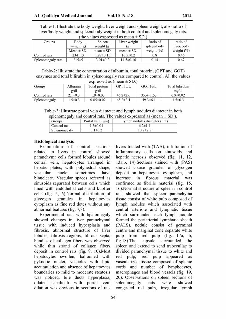
Table-1: Illustrate the body weight, liver weight and spleen weight, also ratio of liver/body weight and spleen/body weight in both control and splenomegaly rats.
(the values expressed as mean ± SD.)Groups Body
weight (g)Spleen
weight (g)Liver weight
(g)Ratio of
spleen/body weight (%)
ratio of liver/body weight (%)Mean ± SD. mean ± SD. mean ± SD.
Control rats 234±13 1.88±0.15 10.5±0.2 0.8 0.46Splenomegaly rats 215±5 3.01±0.2 14.5±0.16 0.14 0.67
Table-2: Illustrate the concentration of albumin, total protein, (GPT and GOT) enzymes and total bilirubin in splenomegaly rats compared to control. All the values
expressed as (mean ± SD.)Groups Albumin
g/dlTotal protein
g/dlGPT Iu/L GOT Iu/L Total bilirubin
mg/dlControl rats 2.1±0.3 1.9±0.03 46.2±2.6 35.4±1.53 0.9±0.02Splenomegaly 1.5±0.3 0.85±0.02 68.2±2.4 49.3±6.1 1.5±0.3
Table-3: Illustrate portal vein diameter and lymph nodules diameter in both splenomegaly and control rats. The values expressed as (mean ± SD.).
Groups Portal vein (μm) Lymph nodules diameter (μm)Control rats 1.5±0.01 6.2±1.4Splenomegaly 3.1±0.2 10.7±2.8
Histological analysis Examination of control sections
related to livers in control showed paranchyma cells formed lobules around central vein, hepatocytes arranged in hepatic plates, with polyhedral shape, vesicular nuclei sometimes have binucleate. Vascular spaces referred as sinusoids separated between cells which lined with endothelial cells and kupffer cells (fig. 5, 6).Normal distribution of glycogen granules in hepatocytes cytoplasm as fine red dotes without any abnormal features (fig. 7,8).
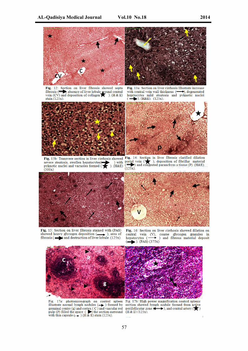
Experimental rats with hpatomegaly showed changes in liver paranchymal tissue with induced hyperplasia and fibrosis, abnormal structure of liver lobules, fibrosis regions, fibrous septa, bundles of collagen fibers was observed while thin strand of collagen fibers deposit in control rats (fig. 9, 10).Most hepatocytes swollen, ballooned with pyknotic nuclei, vacuoles with lipid accumulation and absence of hepatocytes boundaries so mild to moderate steatosis was noticed, bile ducts hyperplasia, dilated canaliculi with portal vein dilation was obvious in sections of rats
livers treated with (TAA), infiltration of inflammatory cells on sinusoids and hepatic necrosis observed (fig. 11, 12, 13a,b, 14).Sections stained with (PAS) showed coarse granules of glycogen deposit on hepatocytes cytoplasm, and increase in fibrous material was confirmed as fibrille material (fig. 15, 16).Normal structure of spleen in control rats showed that spleen parenchyma tissue consist of white pulp composed of lymph nodules which associated with central arteriole and lymphatic tissue which surrounded each lymph nodule formed the periarterial lymphatic sheath (PALS), nodule consist of germinal centre and marginal zone separate white pulp from red pulp (fig. 17a, b, fig.18).The capsule surrounded the spleen and extend to send trabecullae to divided paranchymal tissue to white and red pulp, red pulp appeared as vascularized tissue composed of splenic cords and number of lymphocytes, macrophages and blood vessels (fig. 19, 20). Observations on spleen sections of splenomegaly rats were showed congested red pulp, irregular lymph

AL-Qadisiya Medical Journal Vol.10 No.18 2014
55
nodules masses, dilation on spleen venous sinuses and fibrous trabecullae (fig. 21a,b, 22a,b).Histological examination identified numerous macrophages with hemosiderin deposite, proliferation or hyperplasia with histeocytes and increase with myofibroblasts (fig. 23).Large number of
erythrocytes, platelets deposite in blood vessels, congested and splenic sinuses and more than one lymph nodules aggregated were also noticed (fig. 24). Hemorrhages noticed as yellow or brown pigments in fibrous paranchymal tissue or in the fibrous trabecullae. (fig. 25).

AL-Qadisiya Medical Journal Vol.10 No.18 2014
56
AL-Qadisiya Medical Journal Vol.10 No.18 2014
57

AL-Qadisiya Medical Journal Vol.10 No.18 2014
58

AL-Qadisiya Medical Journal Vol.10 No.18 2014
59
DiscussionThe study was carried out to assess the
histophathogical changes in experimental rats with induced liver fibrosis by TAA and its relationship to splenomegaly caused by .
The TAA model is more reliable and easy to induce liver cirrhosis than the CCl4 model (5). An increase in spleen and liver weight with significance difference in all rats related to splenomegaly group compared to control, this accompanied with decreased in body weight in rats with cirrhotic and splenomegaly compared to control rats, while spleen and liver indices (ratio) per body weight of cirrhotic rats were higher than these of control rats .These results agreed with other studies clarified the relationships between splenomegaly and portal hypertension in patients with liver cirrhosis, splenomegaly not only caused by portal congestion, but is mainly due to tissue hyperplasia and fibrosis, the spleen may enlarged as cause of cirrhosis, portal vein hypertension and other factors (22,23). Also an increase with liver size (hepatomegaly) caused by deposition of fibrillar material in interlobular space, portal vein dilation and hepatic cells hypertrophy which due to an increase on water permeability (24).Total protein and albumin levels on hepatomegaly rats indicated to significance decrease in their concentrations at (P<0.05) compared with control group, this may be related to the effect of TAA as hepatotoxic
compound caused hepatic cells damaged and abnormal synthesis of protein and albumin, results agreed with others studies showed that total protein and albumin levels depressed in hepatotoxic conditions due to disturbances in the carbohydrates, protein, lipid metabolism or abnormal protein biosynthesis in cirrhotic liver or may be regarded to decreased ribosomal RNA in TAA-treated rats (25).
Results referred to decrease with liver enzymes activity (GPT, GOT) in all splenomegaly rats in comparison to control rats, this may be illustrated that TAA induced lipid peroxidations caused damaged of hepatic cells, or necrosis which lead to an excretion of enzymes then elevate the level in serum.Results in agreement with(26) who showed that an increase in liver enzymes an indicator of cellular liver necrosis caused by TAA metabolism which mediated hepatocytes damaged.An increase with bilirubin concentration in serum of splenomegaly group compared to control rats, this may be regarded to the effect of TAA toxicity which lead to bile duct hyperplasia and obstruction, this result discussed by (25) who illustrated that high dose of bilirubin was indication of the increased erythrocytes degeneration rate, the liver excretes the breakdown products of hemoglobin, particularly as bilirubin or may be bile duct obstructed and caused effect on bile transport, the main

AL-Qadisiya Medical Journal Vol.10 No.18 2014
60
component of bile called bilirubin which accumulate in the blood.In this study we recorded the dilation and increase with portal vein diameter in all sections of rats with splenomegaly also there was an increase in lymph nodules diameter of spleen related to these rats compared with control this may be regarded to portal hypertension which induced by toxicity of TAA, also the splenic blood redrains into liver and caused dilation with central vein and portal vein.
These observations also determined by (27) who explained that splenic vein drain into the portal vein, and portal hypertension can produce congestive splenomegaly, the blood flow within the spleen is highly specialized and relates to the different functions of the spleen. Lymph nodules diameter showed an increase with significant difference in all spleen sections from splenomegaly group and this related to the immune response which take place on spleen. Evidence suggests that spleen is relatively insensitive indicator of immunotoxicity (28).
Observations also clarified an increase with glycogen granules deposition in hypertrophic hepatocytes while moderate quantity of glycogen evident in hepatocytes of control rats this caused by cytoplasm changes and disturbance of carbohydrates metabolism due to toxicity of TAA.(24) proved that hepatomegaly caused increase with water permeability inside hepatocytes and an increase with glycogen, fat content which deposite on hepatocytes.Histological investigations elaborate the effect of TAA on both liver and spleen of cirrhotic group compared to control and the results have shown increase with fibrosis appeared as nodular lesions, trabecullae thickening, capsular fibrosis and deposite of fibrous material in both liver and spleen paranchymal tissue, these results agreed with other studies indicated to liver fibrosis which was proven by nodular formation and collagen deposition on liver also appearance of wide spread
fibrosis and lesions on hepatic parenchyma are the main characteristic of liver cirrhosis (29,30).Results of histological observations have shown damaged of hepatocytes, some appeared hypertrophy with pyknotic nuclei and an increase with kupffer cells, inflammatory cells in most liver sections related to hepatomegaly rats also thickening of splenic trabecullae, congested splenic venous and increase with myofibroblast cells, these changes associated with the effect of TAA metabolism which caused necrosis and degenerated of liver and spleen paranchymal tissue. Several experimental studies have shown that the splenomegaly play an inhibitory role in hepatic liver regeneration, there is indicator that humoral factors produced by splenic tissue are carried to the liver through the portal circulation where they inhibit liver regeneration (14,31).
Also an immune cells are involved in mediation of fibrosis including hepatic macrophages (Kupffer cells), Natural Killer (NK) cells, cytotoxic T-cells. Activated macrophages produce pro-inflammatory mediators that further result in the activation of non-specific immunity which lead to the development of myofibroblasts responsible for the production of extra-cellular matrix (ECM) (31).Results identified active lymph nodes in spleen sections regarded to rats with splenomegaly and appeared as more than one lymph nodule aggregated together with large number of lymphocytes in marginal zone, this may by illustrated as immune role of spleen and the cytotoxic effect of TAA.
The marginal zone contain population of lymphocytes and macrophages that are functionally distinct from the cord histiocytes of the red pulp, which at least in animal models seem to be an important in maintaining the anatomic structure of the marginal zone by attracting newly differentiated B-lymphocytes (32,33).Findings from the present study concluded that splenomegaly caused as a result of liver
AL-Qadisiya Medical Journal Vol.10 No.18 2014
61
hyperplasia, cirrhosis and fibrosis induced by toxicity of TAA. The dependent dose very reliable to induced hepatogmegaly and splenomegaly in
experimental rats, on the other hand hepatomegaly model very useful to understand the histopathological changes of splenomegaly.
References 1- U.S. Food and drug administration (USFDA), Department of health and human services, center for drug evaluation and research (CDER), 2001. Guidance for Industry lmmunotoxicology evaluation of investigational new drugs. 2- Braumlwald, E.; Fauci, A.S.; et al. (2007). Harrisons principles of internal medicine. 17th
ed., McGraw Hill. Comp.3- Myers, J. and Segal, R.J. (1974). Weight of the spleen. 1-Range of normal in a non-hospital population. Arch pathol.; 98: 33-35.4- Zhang, J.; Ling, Y.; Luo, B.; Tang, L.; Ryter, S.W.; Stockard, C.R.; Grizzle, W.E. and Fallon, M.B. (2003). Analysis of pulmonary heme oxygenase-1 and nitric synthase alterations in experimental hepatopulmonary syndrome. Gastioenterology. 125(5): 1441-1451.5- Chen, X.C.; Gines, P.; Yang, J.; Summer, S.N.; Falk, S.; Russell, N.S. and Schrier, R.W. (2004). Increased vascular heme oxygenase-1 expression contributes to arterial vasodilation in experimental cirrhosis in rats. Hepatology, 39(4): 1075-1087.6- Scothorne, R.J. (1985). The spleen: structure and function. Histopathology, 9: 663-669.7- Mendez, M.; Sanchez-paton, F.; Casado, I.; Aller, M.; Lopez, L.; Corcura, M.T.; Alonso, M.J.; Nava, M.P. and Arias, J. (2007). Partial portal vein ligation plus thioacetamide: a method to obtain a new model of cirrhosis and chronic portal hypertension in the rat. J. Gastrointest. Surg. 11(2): 187-194.8- Barlak, A.; Kadikoylu, G. and Bolaman, A. (2004). Splenomegaly due to the use of granulocytecology stimulating factor. Turk. J. haematol. 21(2): 93-96.9- Tarasub, N.; Chinnwat, T. and Watcharapora, D. (2009). Histological changes of spleen, stomach and small intestine induced by cadmium in rats and the protective effect of curcumin.10- Radwan,D.; Amin,H. and Ashraf, M. (2010). Histological and immunohistochemical study on rat spleen in experimentally induced liver cirrhosis. Egypt. J. Histol., 33(4): 709-721.11- Raffan, E.; McCallum, A.; Score, T. and Watson, P.J. (2009). Ascites is a negative prognostic indicator in chronic hepatitis in dogs. J. vet. Intern. Med. 23: 63-66.12- Mejias, M.; Garcia-Pras, E.; Gallego, J.; Mendez, R.; Bosch, J. and Fernandez, M. (2010). Relevance of the mTOR signaling pathway in the pathophysiology of splenomegaly in rats with
chronic portal hypertension J. Hepatol. 52(4): 529-539.13- Fernande, Z.M.; Mejias, M.; Garcia-Pars, E.; Mendez, R.; Garcia-pagan, J. and Bosch, J. (2007). Reversal of portal hypertension and hyperdynamic splanchnic circulation by combined vascular endothelial growth factor and platelet-derived growth factor blockade in rats. J. Hepatol. 46(4): 1208-1217.14- Chen, D.; Liu, W.; Leng, E. and Wu, B. (1998). Effect of splenectomy on CCl4 induced liver fibrosis in rats. Chin. Med. J. 111(9): 779-83.15- Aydin, A.F.; Kucku-Kiraz, Z.; Dogru, Abbasogly S.; Gulluoglu, M.; Ugsal, M. and Kocak-Toker, N. (2010). Effect of carnosine against thioactamide induced liver cirrhosis in rats J. Hepatol. 31(1): 67-71.16- Xia, Z.; Wang, G.; Liu, T.; Wang, S.; Wang, B. and Cheng, R. (2010). Expression of NALP3 in the spleen of mice with portal hypertension. J. Huazhong univ. Sci., Techn. Med. Sci. 30(2): 170-172.17- Tietz, N. (1999). Text book of clinical chemistry, 3rd ed., C.A. Burtis, E.R. Ashwood,W.B. Sanders pp. 625-657.18- Daumas, B.; Wafson, W. and Biggs, H. (1971). Albumin standards and the measurements of serum albumin with bromocresol green. 31: 87-96.19- Sinica, A. (1996). Hyperplastic of bile ductules at 3-5 days after treatment with CCl4. Toxicol. Pathol. 24: 90-99.20- Drury, R.A.B.; Wallington, E.A. and Cameron, R. (1967). Carleton`s histological Technique. 4th ed. Oxford unit press New York.21-Lessa.A., Paredes,B., Dias,J., Carvalho,A., Quintanilha,L., Takiya,CH., Tura,B., Rezende,L., Campos,A.,Resende,c.and Regina,G.(2010).Ultrasound imaging in an experimental model of fatty liver disease and cirrhosis in rats.BMC. Veter. Reas., 6:6, dio: 10, 1186/174622- Aiden, M.C. and Karen, M. (2000). Splenomegaly, hypersplenism and coagulation abnormalities in liver disease. Clini. Gastroenter. Vol. 14, pp. 1007-1010.23- Bologensi, M.; Merkel, C.; Sacerdoti, D.; Nava, V. and Gatta, A. (2002). Role of spleen enlargement in cirrhosis with portal hypertension Dig. Liver. Dis, 34(2): 144-50.24- Gressner, O.; Weiskirchen, R. and Gressner, M. (2007). Biomarkers of liver fibrosis:

AL-Qadisiya Medical Journal Vol.10 No.18 2014
62
malfunction tests. Clinica, chimica Acta. Vol. 381(2), pp: 107-113.25- Mir, A.; Anjum, F.; Riaz, N.; Iqbal, H.; Mustatab, H.; Khattak, K.; Khau, A. and Malik, A. (2010). Carbon tetra chloride (CCl4)- induced hepatotoxicity in rats:Curative role of Solanum nigrum. J. Med. plant., Res. 4(23): 2525-2532.26- Fontana, L.; Moreira, E.; Torres, M.I. et al. (1996). Serum amino acid changes in rats with thioacetamide induced liver cirrhosis: Toxicology. 106(1-3): 197-206.27- Elomore, S. (2006). Enhanced histopathology of the spleen. Toxicol., vol. 34(5): 648-655.28- Luster, M.I.; Portier, C.; Pait, D.G.; White, K.L.; Gennings, C.; Munson, A.E. and Rosenthal, G. (1992). Risk assessment in immunotoxicology. 1. Sensitivity and predictability of immune tests. Fundam. Appl. Toxicol. 18: 200-210.29- Sun, F.; Hayami, S.; Ogiri, Y. and et al.(2000). Evaluation of oxidative stress based on
lipid hydroperoxide, vitamin C and vitamin E. during apopotsis and necrosis caused by thioacetamide in rat liver. Biochimica et Biophysica Acta. 1500(2): 181-185.30- Li, M.K. and Crawford, J.M. (2004). The pathology of cholestasis. Seminars in liver disease. 24(1): 21-24.31- Akahoshi, T.; Hashizume, M.; Tanoue, k.; Shimabukuro, R.; Gotoh, N.; Tawikawa, M.; et al. (2002). Role of the spleen in livers fibrosis in rats may be mediated by transforming growth factor beta-1. J. Gastroentero. Hepatol., 17: 59-65.32- Pillai, S.; Cariappa, A. and Moran, S.T. (2005). Marginal zone B lymphocytes. Annu. Rev. Immunol., 23: 161-196.33- Mills, S. (2007). Histology for pathologists. 3rd. ed. Copyright Lippincott. W. and Wilkins. VIII, Hematopoietic sys
Related Documents