Abstract The objective of this study is to investigate in detail the histopathological changes in gill, liver, brain, spinal cord, ovary and kidney of zebra fish exposed to combination pesticide. Twenty one days semi static exposure to sublethal concentration of 8.4 and 4.2 µg/L of Chloropyrifos 50% + Cypermethrin 5% EC was conducted and it was followed with a reversal period of 7 days without pesticide in the exposure water. Three fish were collected from control and treatment group periodically. Whole body fixation of zebra fish as per OECD guidance document 123 was adopted for obtaining the histopathology sections. Structural damages of gill observed were of inflammatory cell infiltration, minimal congestion in primary lamellae, fusion of secondary lamellae, diffused epithelial hyperplasia and multifoci mucus cell hyperplasia. In liver, moderate, diffused to severe cytoplasmic vacuolation, diffused minimal to mild sinusoidal congestion, steatosis, pyknotic, karyorrhectic nuclei with complete dissolution of necrotic hepatocytes was observed. Minimal focal tubular degeneration was observed in kidney. Minimal to mild multifocal follicular atresia was observed in the ovary. In spinal cord neuronal cell degeneration, cytoplasmic vacuolation and enlargement of neuronal body was observed. In brain, mild to moderate demyelination in the neurophil and mild necrotic changes observed in the cerebrum. Some of the lesions were less pronounced after recuperation period. Pesticide in exposure water was analyzed using Agilent QQQ GC-MS/MS equipped with Election Impact Ionization mode. Limit of detection and quantification was 1 µg/L with a Correlation Co-efficient (CC) of 0.999. The recovery of active content of Chlorpyrifos for 4.2 and 8.4 µg/L at 0 hour was 88.1%, 90.3% and at 48 hour was 83.48%, 85.70%, respectively. Similarly, Cypermethrin active content recovered at 0 hour for 4.2 and 8.4 µg/L was 78.74%, 86.24% and at 48 hour was 57.85%, 62.09%, respectively. Further, this is the first study to report pathological findings in Zebrafish exposed to a combination pesticide formulation. The histopathological finding of this investigation reveals the potential toxic hazard from exposure to sub lethal level of the combination pesticide. Few literatures have reported on the mixture toxicity of pesticides and this research work throws light on histopathological lesions of Zebrafish. Further, this is the first study to report spinal lesions in fish due to pesticide exposure. Pesticides usage in pest management or in agriculture practice should be applied safely to prevent pollution and contamination. *Author for correspondence Indian Journal of Science and Technology, Vol 8(18), 68323, August 2015 ISSN (Print) : 0974-6846 ISSN (Online) : 0974-5645 Histopathological Changes in Tissues of Danio rerio Exposed to Sub Lethal Concentration of Combination Pesticide A. Rajini 1* , K. Revathy 2 and G. Selvam 3 1 Sathyabama University, Chennai - 600119, Tamil Nadu, India; [email protected], [email protected] 2 Department of Advanced Zoology, Ethiraj College, University of Madras, Chennai - 600119, Tamil Nadu, India; reva63@rediffmail.com 3 International Institute of Biotechnology and Toxicology, Padappai, Kancheepuram - 601301, Tamil Nadu, India; [email protected] Keywords: Combination Pesticide, Danio rerio, Histopathological Changes

Welcome message from author
This document is posted to help you gain knowledge. Please leave a comment to let me know what you think about it! Share it to your friends and learn new things together.
Transcript

AbstractThe objective of this study is to investigate in detail the histopathological changes in gill, liver, brain, spinal cord, ovary and kidney of zebra fish exposed to combination pesticide. Twenty one days semi static exposure to sublethal concentration of 8.4 and 4.2 µg/L of Chloropyrifos 50% + Cypermethrin 5% EC was conducted and it was followed with a reversal period of 7 days without pesticide in the exposure water. Three fish were collected from control and treatment group periodically. Whole body fixation of zebra fish as per OECD guidance document 123 was adopted for obtaining the histopathology sections. Structural damages of gill observed were of inflammatory cell infiltration, minimal congestion in primary lamellae, fusion of secondary lamellae, diffused epithelial hyperplasia and multifoci mucus cell hyperplasia. In liver, moderate, diffused to severe cytoplasmic vacuolation, diffused minimal to mild sinusoidal congestion, steatosis, pyknotic, karyorrhectic nuclei with complete dissolution of necrotic hepatocytes was observed. Minimal focal tubular degeneration was observed in kidney. Minimal to mild multifocal follicular atresia was observed in the ovary. In spinal cord neuronal cell degeneration, cytoplasmic vacuolation and enlargement of neuronal body was observed. In brain, mild to moderate demyelination in the neurophil and mild necrotic changes observed in the cerebrum. Some of the lesions were less pronounced after recuperation period. Pesticide in exposure water was analyzed using Agilent QQQ GC-MS/MS equipped with Election Impact Ionization mode. Limit of detection and quantification was 1 µg/L with a Correlation Co-efficient (CC) of 0.999. The recovery of active content of Chlorpyrifos for 4.2 and 8.4 µg/L at 0 hour was 88.1%, 90.3% and at 48 hour was 83.48%, 85.70%, respectively. Similarly, Cypermethrin active content recovered at 0 hour for 4.2 and 8.4 µg/L was 78.74%, 86.24% and at 48 hour was 57.85%, 62.09%, respectively. Further, this is the first study to report pathological findings in Zebrafish exposed to a combination pesticide formulation. The histopathological finding of this investigation reveals the potential toxic hazard from exposure to sub lethal level of the combination pesticide. Few literatures have reported on the mixture toxicity of pesticides and this research work throws light on histopathological lesions of Zebrafish. Further, this is the first study to report spinal lesions in fish due to pesticide exposure. Pesticides usage in pest management or in agriculture practice should be applied safely to prevent pollution and contamination.
*Author for correspondence
Indian Journal of Science and Technology, Vol 8(18), 68323, August 2015ISSN (Print) : 0974-6846
ISSN (Online) : 0974-5645
Histopathological Changes in Tissues of Danio rerio Exposed to Sub Lethal Concentration of
Combination PesticideA. Rajini1* , K. Revathy2 and G. Selvam3
1Sathyabama University, Chennai - 600119, Tamil Nadu, India; [email protected], [email protected]
2Department of Advanced Zoology, Ethiraj College, University of Madras, Chennai - 600119, Tamil Nadu, India; [email protected]
3International Institute of Biotechnology and Toxicology, Padappai, Kancheepuram - 601301, Tamil Nadu, India; [email protected]
Keywords: Combination Pesticide, Danio rerio, Histopathological Changes

Histopathological Changes in Tissues of Danio rerio Exposed to Sub Lethal Concentration of Combination Pesticide
Indian Journal of Science and TechnologyVol 8 (18) | August 2015 | www.indjst.org 2
1. Introduction
Synthetic pesticides are important group of aquatic pollutants affecting health of fish1. Physiological and bio-chemical alterations in an animal under any physiological stress can be correlated with the structural and functional changes of cellular proteins2. Wide use of pesticides in agri-culture to control the pests has indirectly created problem of pollution to aquatic environment3. Histopathological evaluation is an important part of the assessment of the adverse effects of xenobiotics on the whole organism4. Exposure to sub-lethal concentrations of environmental chemicals may lead to the histological structure altera-tions which can significantly alter the function of tissues and organs. Histological and ultrastructural changes in cells, tissues or organs can afford good biomarkers of pol-lutant stress5. Histopathological studies are conducted to establish fundamental relationships of contaminant expo-sure and its biological responses6. Due to increased public awareness of the potential of persistent pesticides that cause harm to environment and public health, great stress is being laid for developing least persistent and selective pesticides. Problem of agricultural pest control has been dealt by formulating new and more potential pesticides7. Chlorpyrifos and cypermethrin pesticide combination pesticide are extensively used in pest control and agricul-ture. Chlorpyrifos and cypermethrin residues were found in canal water of all stations at Kedah, Malaysia and these insecticides were transported from their application sites. High concentrations of cypermethrin and chlorpyrifos were detected in December 2010 and March 2011, dur-ing this time the farmers applied insecticides to control pests in rice fields. The highest concentration of 3.97 μg/mL of cypermethrin and 4.42 μg/mL of chlorpyrifos was observed8. Though Pyrethroid when compared to Organophosphate pesticides are reported for less stabil-ity, need is to screen the combination pesticide product in very low concentration. It is hypothesized that sub-lethal concentrations could have detrimental effect to pesticide exposed fish and affect the susceptible organs, thus it can be observed histopathologically. Potential reason for this histopathological evaluation is that there are no published data on this combination pesticide.
2. Materials and MethodsDanio rerio procured from commercial fish farm were
quarantined for 30 days and acclimated in glass aquaria for 7 days in the laboratory condition. Fish were fed with commercial fish pellets. Chlorpyrifos 50% + Cypermethrin 5% EC was purchased from commercial market. Twenty fish in each group was exposed semi-statically for 21 days to the sub lethal concentration of 8.4 and 4.2 µg/L, control group without pesticide was also maintained. The physi-cochemical parameter of exposure water was measured prior to semi static water change, pH (7.5-7.6), water tem-perature (26.8-27.0°C), dissolved oxygen (7.6-7.8 mg/l), conductivity (623-658 µs/cm) and hardness (CaCO3) was 218 - 220 mg/l. During the conduct of the experiment the exposure water was analyzed initially for dose verification of the spiked pesticide at 0th and 48th hour.
The experimental and control fish were sacrificed at the end of 7th, 14th, 21st and 28th day. Fish was euthanized by transferring the fish using a fish net to buffered MS-222 (Tricaine methanesulfonate) solution approximately 2 minutes prior to necropsy. Fish was fixed in whole in mod-ified Davidson’s fixative9 overnight followed by transfer to individual containers of 10 % neutral buffered formalin the next day. For optimal penetration of fixative, a small incision was made near the abdomen by surgical blade. After fixation for 24-30 hours, tissues were dehydrated through a graded series of ethanol, cleared in xylene, and infiltrated with the paraffin. Sections of 5 μm were pre-pared from paraffin blocks by using a rotary microtome. These sections were then stained with Hematoxylin-Eosin. Histopathological lesions were examined and photographed using Leica photomicroscope.
Concentrations of the pesticide 4.2 µg/L and 8.4 µg/L were analyzed at 0th hour and 48th hour. The exposure water sample was analyzed using Agilent QQQ GC-MS/MS equipped with Election Impact Ionization mode. Mass Hunter software supplied by Agilent USA was used for system control and data acquisition. For the separa-tion of residues of chlorpyrifos and cypermethrin HP-5 MS fused silica capillary column (30 m length, 0.25 mm i.d. and 0.25 µm film thickness) was used. Carrier gas was Helium at 1.8 mL/min, temperature of injector was set at 310°C with a split ratio of 1:5 and 310°C as source temperature. The column temperature was maintained at 70°C. The Injection volume was 3.0 µL. Chlorpyrifos and Cypermethrin retention time was 11.8 and 24.1. The exposure water samples was transferred to 250 mL separatory funnel and residues extracted with 2x25 mL of dichloromethane. The dichloromethane layer was col-

A. Rajini , K. Revathy and G. Selvam
Indian Journal of Science and Technology 3Vol 8 (18) | August 2015 | www.indjst.org
lected into a separate flask and evaporated using turbovap, the residues reconstituted with 2 mL hexane.
3. Results The method has the linearity over the concentration range of 1 µg/L to 200 µg/L for chlorpyrifos and cypermethrin. The limit of detection and quantification was 1 µg/L and Correlation Co-efficient (CC) was 0.999 for chlorpyrifos and cypermethrin. Chlorpyrifos active content recovered at 0 hour for 4.2 and 8.4 µg/L was 3.70 (88.1%) and 7.58 (90.3%) and at 48th hour 3.50 (83.48%) and 7.19 (85.70%).
(b)
Figure 1. (a) Standard Chromatogram of Chlorpyrifos – 100 μg/L. (b) Standard Chromatogram of
Cypermethrin – 100 μg/L.
Cypermethrin active content recovered at 0 hour for 4.2 and 8.4 µg/L was 3.31 (78.74%) and 7.24 (86.24%) and at 48th hour it was 2.43 (57.85%) and 5.22(62.09%) (Figure 1 and Figure 2).
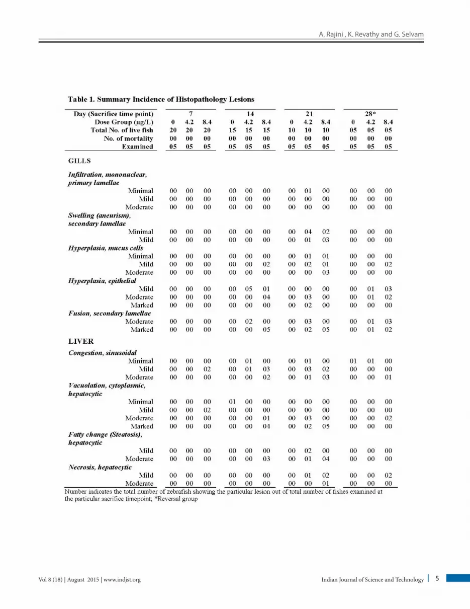
All the histopathological tissues are compared with tissue sections of control group. Histopathological lesions observed in fish exposed to 4.2 and 8.4 µg/L on final sacri-fice on day 7th, 14th, 21st and 31st day when compared with control group are represented in Figure 3, 4 and 5 and histological changes noticed in the pesticide exposed and control fishes are shown in Table 1.
systemadmin
Text Box
(a)

Histopathological Changes in Tissues of Danio rerio Exposed to Sub Lethal Concentration of Combination Pesticide
Indian Journal of Science and TechnologyVol 8 (18) | August 2015 | www.indjst.org 4
(a)
Figure 2. (a) Representative Sample Chromatogram – Concentration 4.2 μg/L. (b)
Representative Sample Chromatogram – Concentration 4.2 μg/L.
Table 1. Summary Incidence of Histopathology Lesions
(b)
A. Rajini , K. Revathy and G. Selvam
Indian Journal of Science and Technology 5Vol 8 (18) | August 2015 | www.indjst.org

Histopathological Changes in Tissues of Danio rerio Exposed to Sub Lethal Concentration of Combination Pesticide
Indian Journal of Science and TechnologyVol 8 (18) | August 2015 | www.indjst.org 6

A. Rajini , K. Revathy and G. Selvam
Indian Journal of Science and Technology 7Vol 8 (18) | August 2015 | www.indjst.org
Figure 3. Histopathological changes observed in liver. Histopathological changes observed in liver of treated zebrafish in comparison with non-treated control fish. A – Normal liver histology of non-treated control fish at the final sacrifice time point; B – Cytoplasmic vacuolation of hepatocytes (encircled) observed in the liver of zebrafish treated with pesticide 8.4 µg/L and sacrificed at 7th day. A minimal to mild sinusoidal congestion (arrows) was observed diffusely; C – Moderate and diffuse cytoplasmic vacuolation observed in the liver of zebrafish treated with pesticide 4.2 µg/L and sacrificed at 14th day; D – Multifocal macrovesicular fatty change (steatosis) observed in the liver of zebrafish treated with pesticide 8.4 µg/L and sacrificed at 14th day; E – Marked to severe cytoplasmic vacuolation intermingled fatty change (steatosis) observed in the liver of zebrafish treated with with pesticide 4.2 µg/L and sacrificed at 21st day. Note the pronounced sinusoidal and venous congestion (arrow). The inner picture shows the high power magnification of cytoplasmic vacuolation; F – Multifocal areas of hepatocyte necrosis observed in the liver of zebrafish treated with pesticide 8.4 µg/L and sacrificed at 21st day. Note the pyknotic and karyorrhectic nuclei and the complete dissolution of majority of the necrosed hepatocytes.

Histopathological Changes in Tissues of Danio rerio Exposed to Sub Lethal Concentration of Combination Pesticide
Indian Journal of Science and TechnologyVol 8 (18) | August 2015 | www.indjst.org 8
Figure 4. Histopathological changes observed in Gill. Histopathological changes observed in gills of treated zebrafish in comparison with non-treated control fish. A – Normal gill histology of non-treated control fish at the final sacrifice time point; B – Inflammatory cell infiltration (arrow) with a minimal congestion (arrow head) in primary lamellae observed in the gill of zebrafish treated with pesticide 8.4 µg/L and sacrificed at 7th day; C – Diffuse epithelial hyperplasia and fusion of secondary lamellae observed in the gill of zebrafish treated with pesticide 4.2 µg/L and sacrificed at 21st day; D – Diffuse epithelial hyperplasia, fusion of secondary lamellae and multifocal mucus cell hyperplasia (arrows) observed in the gill of zebrafish treated with pesticide 8.4 µg/L and sacrificed at 21st day.

A. Rajini , K. Revathy and G. Selvam
Indian Journal of Science and Technology 9Vol 8 (18) | August 2015 | www.indjst.org
Figure 5. Histopathological changes observed in Ovary, Kidney, Cerebrum, Spinal cord . (A) Minimal, multifocal follicular atresia observed in the ovary of zebrafish treated with pesticide 4.2 µg/L and sacrificed at 21st day. Inlet figure shows the higher magnification of an atretic follicle; (B) Mild, multifocal follicular atresia (arrow) observed in the ovary of zebrafish treated with pesticide 8.4 µg/L and sacrificed at 21st day; (C) Minimal, focal tubular degeneration (encircled) observed in the kidney of zebrafish treated with pesticide 4.2 µg/L and sacrificed at 21st day; (D) Mild to moderate tubular degeneration (encircled) along with focal, moderate tubular regeneration (arrow)surrounded by ongoing peritubular inflammation observed in the ovary of zebrafish treated with pesticide 8.4 µg/L and sacrificed at 21st day. (E) Mild to moderate demyelination in the neuropil and mild necrotic changes observed in the cerebrum of zebrafish treated with pesticide 8.4 µg/L and sacrificed at 21st day. (F) Neuronal degeneration characterized by cytoplasmic vacuolation and enlargement of neuronal body (arrow heads) observed in the spinal cord of zebrafish treated with pesticide 8.4 µg/L and sacrificed at 21st day. The arrow indicates normal neuronal body.

Histopathological Changes in Tissues of Danio rerio Exposed to Sub Lethal Concentration of Combination Pesticide
Indian Journal of Science and TechnologyVol 8 (18) | August 2015 | www.indjst.org 10
4. Discussion Pesticides cause physiological and biochemical changes in fish species and influence their activities. Gill is a mul-tifunctional and complex organ with which fish make intimate contact with the surrounding water10. Fusions of lamellae, diffused hyperplasia are due to changes in pavement cells of gill. Inflammatory cell infiltration with congestion is an adaptive response to the pesticide con-taminant. The multifocal mucous cells hyperplasia is a protective mechanism of mucous cells reducing pes-ticide exposure of the gill. Clarius gariepinus exposed to cypermethrin were observed with cellular infiltra-tion and congestion11. Channa punctatus gill showed lamellar fusion of secondary lamellae, congestion and infiltration when exposed to sub-lethal concentrations of Chlorpyrifos12. Multifocal mucous cell hyperplasia was observed in fish exposed to dichlorovos13. In our present study, inflammatory cell infiltration, minimal congestion, diffuses epithelial hyperplasia and fusion of secondary lamellae, multifocal mucus cell hyperplasia was observed and is in accordance with previous reports of 11-13.
Liver regulates metabolism, transforms, excretes xeno-biotics and helps in detoxification. It is a major storage organ of lipids and site of metabolic processes in fish and hepatocytes carry xenobiotics to bile for elimination14. Exposure of malathion for 4 days induced degeneration of hepatocytes characterized by cytoplasmic vacuolization15. After 21 days of chlorpyrifos 20% EC exposure liver of Heteropneustes fossilis was observed with congestion of central vein, degeneration of hepatocytes, cytoplas-mic vacuolization large number of hepatocytes appeared with pyknotic nuclei, thrombosis in hepatoportal blood vessel, haemorrhage around central vein and necrosis in liver has been reported16. Vacuolar degeneration, hyper-trophy in the hepatocytes with nuclear pyknosis and vacuolation of the hepatocytes in liver of fish exposed to sublethal concentration of chlorpyrifos12. In the pres-ent experiment cytoplasmic vacuolation of hepatocytes, multifocal macrovesicular fatty changes (steatosis), pro-nounced sinusoidal and venous congestion and multifocal areas of hepatocyte necrosis, were observed in the liver of fish exposed. The results in the present study could be correlated with previous reports of 12, 15, 16.
Malathion affected detoxifying organ kidney of Channa punctatus and caused shrinkage of glomeruli, degeneration of renal tubules and collecting tubule15. The renal tissues are at risk since they receive large volumes
of blood flow from both renal portal venous system and renal arteries17. Focal necrosis, leucocytic infiltration, shrinkage in renal corpuscles of Tilapia nilotica exposed to chlorpyrifos clearly indicated kidney damage and the pesticide is capable of producing a wider spectrum of sig-nificant histopathologic impairments in fish with even sub lethal concentrations18. Histological damage caused to the fish Cirrhinus mrigala exposed to lethal (5.13 μg/l) and sublethal (1.026 μg/l) concentration of Pyrethroid deriva-tive cypermethrin had induced marked abnormalities in the kidney initiated with disruption of tubular organi-zation19, Vacuolation due to degeneration of cytoplasm, hypertrophy of tubular cells, nuclei of epithelial cells infiltrating into the surrounding tissue and perforation of kidney tubules. Zebrafish exposed to sublethal con-centration of chlorpyrifos caused changes in the kidney, shrunken glomeruli, dilated lumina of the renal tubules20. Non-detoxified pesticide molecules must be eliminated through the kidney of fish and hence, it is susceptible to chemical compounds when exposed to lethal or sublethal dose, Cypermethrin 10% EC eliminated through kidney might have caused degenerative changes in renal tubules and glomeruli, necrosis in the proximal tubule with devel-opment of vacuoles21. The pathological changes observed in the present study such as focal tubular degeneration and regeneration along with peritubular inflammation is in agreement with 15,18-21.
Brain and spinal cord is the important organ it con-trols of movements. In our investigation we did not find any clinical signs of toxicity which could be due to the fact that the sub lethal concentrations did not cause any changes in the synaptic ends of the nerves by Acetylcholine inhibition or alter the voltage-gated sodium channels. Hyperplasia, edema, necrosis and an increase in brain cells were observed in the brain of the fish Cyprinus carpio exposed to sub-lethal concentration of quinol-phos toxicity22. Histological investigation of the brain of Channa punctatus in response to acute and subchronic exposure to the pesticide Chlorpyrifos revealed detach-ment in the superficial zone of the Stratum opticum, Stratum marginale due to degeneration of neuronal cells, spongiosis, congestion, necrosis and appearance of clear areas around the nucleus of mononuclear cells in the lin-ing of the Stratum fibrosum griseum superficiale, Stratum griseum centrale, Stratum album centrale. Granular cells found in the innermost layer of optic tectum, i.e. the Stratum periventriculare severely degenerated and vacu-olized and migrated toward the Torus semicircularis23.

A. Rajini , K. Revathy and G. Selvam
Indian Journal of Science and Technology 11Vol 8 (18) | August 2015 | www.indjst.org
Neuronal degeneration and tissue damages were observed in brain of fish treated with dichlorvos and methyl 24. The 96 hour acute toxicity of cypermethrin to juvenile African catfish was investigated and the brain revealed neuronal degeneration and spongiosis11. In this study brain showed demyelination of neuropil and mild necrotic changes in the cerebrum. Neuronal degeneration, cytoplasmic vacu-olation, and enlargement of neuronal body were observed in the spinal cord. Pathological alteration in the brain is consistent with previous studies of 11, 22-24. This study is the first to report the changes in the spinal cord due to expo-sure to combination pesticide.
In the present study the most notable changes appeared in ovary are atretic follicle, multifocal fol-licular atresia, focal tubular degeneration, peritubular inflammation. Dimethoate organophosphate pesticide in low concentrations revealed disrupted follicular epi-thelial cells, nucleolus condensation of crescent shaped dark granules and degeneration of epithelials cells caus-ing vacuolation25. Channa striatus exposed to sub lethal concentration of cypermethrin revealed degenerated ovary26. The most important significance of follicular atresia during the normal course of reproduction is to limit the number of eggs that could be supported for vitel-logenesis, maturation and ovulation of the female fish27. Gonads are preserved for histopathology to evaluate and assess the reproductive fitness of the fish and it adds to the weight of evidence of other endpoints28. Toxicants produce physiological and biochemical changes in fresh-water organisms29. Chlorpyrifos and Cypermethrin could be Endocrine Disrupting Chemicals and suppress repro-ductive activities and could have direct effects on gonads and gametes quality30.
5. Conclusion The sub lethal concentration of pesticides (Chlorpyrifos 50% + Cypermethrin 5% EC) caused considerable path-ological alterations. Pesticide residues detected above the permissible environmental concentrations impose a significant biological risk, thereby affecting the aquatic fauna. Diligent usage of the pesticide product could pro-tect the environmental contamination.
6. Acknowledgement The author thanks IIBAT management, Staff of Pathology Department and Dr. S. Sathiyanarayanan for guidance and Analytical support.
7. Conflict of Interest Potential conflicts of interest none.
8. References1. Ram NS. Effects of Dimethoate (EC 30%) on gill morphol-
ogy, oxygen consumption and serum electrolyte levels of common carp, Cyprinus Carpio (Linn). International Journal of Scientific Research in Environmental Sciences. 2014; 2(6):192–8.
2. Veeraiah K, Vivek Ch, Srinivas RP, Venkatrao G. Biochemical changes induced by Cypermethrin (10% EC), a Pyrethroid compound in sub-lethal and lethal concentra-tions to the freshwater fish Cirrhinus mrigala (Hamilton). J Atoms and Molecules. 2013; 3(6):625–34.
3. Ganeshwade RM. Histopathological changes in the gills of Puntius Ticto (Ham) under dimethoate toxicity: The BioScan. 2012; 7(3):423–6.
4. Reddy PB, Rawat SS. Assessment of aquatic pollution using histopathology in fish as a protocol. International Research Journal of Environment Sciences. 2013; 2(8): 79–82.
5. Negin S, Mehdi Z. Using of fish pathological alterations to assess aquatic pollution: A review. World Journal of Fish and Marine Sciences. 2012; 4(3):223–31.
6. Pathan TS, Shinde SE, Thete PB, Sonawane DL. Histopathology of liver and kidney of Rasbora danico-nius exposed to paper mill effluent. Research Journal of Biological Sciences. 2010; 5(5):389–94.
7. Anita ST, Sobha K, Tilak KS. Toxicity and histopathologi-cal changes in the three indian major carps, Labeo rohita (Hamilton), Catla catla (Hamilton) and Cirrhinus mrigala (Hamilton) exposed to fenvalerate, International Journal of Plant, Animal and Environmental Sciences. Jan-Mar 2012; 2(1):18–32.
8. Ismail BS, Siti HH, Talib L. Pesticide residue levels in the surface water of the irrigation canals in The Muda Irrigation Scheme Kedah, Malaysia. IJBAS-IJENS. 2012; 12(6):85–90.
9. OECD Series on testing and assessment No. 123, Guidance document on the diagnosis of endocrine-related histopa-

Histopathological Changes in Tissues of Danio rerio Exposed to Sub Lethal Concentration of Combination Pesticide
Indian Journal of Science and TechnologyVol 8 (18) | August 2015 | www.indjst.org 12
thology in fish gonads env/jm/mono. 2010 May 14-31; 1–14.
10. Srivastav AK, Sanjay KS, Sarojni T, Diwakar M, Sunil KS. Morpho-toxicology of chlorpyrifos to prolactin cells of a freshwater catfish, Heteropneustes fossilis. Acta Scientiarum. Biological Sciences Maringa. Oct-Dec 2012; 34(4):443–9.
11. Ayoola SO, Ajani EK. Histopathological effects of Cypermethrin on Juvenile African Catfish (Clarias gari-epinus). World J Biological Research. 2008; 1(2):4(3):1–4.
12. Yogita D, Mishra A. Histopathological alterations in gill and liver anotomy of fresh water, air breathing fish Channa punctatus after Pesticide Hilban® (Chlorpyrifos) Treatment. Adv Biores. 2013; 4(2):57–62.
13. Velmurugan B, Selvanayagam M, Cengiz EI, Unlu E. The effects of monocrotophos to different tissues of freshwater fish Cirrhinus mrigala. Bull Environ Contam Toxicol. 2009; 78:450–4.
14. Patel JM, Bahadur A. Histopathological manifestations of sub lethal toxicity of copper ions in Catla catla. American-Eurasian Journal of Toxicological Sciences. 2011; 3(1):01–5.
15. Afsar S, Magar RS. Effect of Malathion toxicity on detoxi-fying organ of fresh water fish Channa punctatus. IJPCBS. 2013; 3(3):723–8. ISSN: 2249-9504.
16. Hasina BB, Mithra D. Histopathological changes in liver tissue of Heteropneustes fossilis exposed Chlorpyrifos (20% EC). Indian Journal of Applied Research. Jul 2014; 4(7):237–40. ISSN: 2249-555X.
17. Khan S, Sharma N. Histopathological alterations in the kid-ney of Gambusia affinis after exposure to chlorpyrifos. Int J Chem Pharm Sci. 2013; 1(2):122–7.
18. Aliaa IM, Azza GM, Gehad EM. Histological hazards of Chlorpyrifos usage on gills and kidneys of Tilapia nilotica and the role of vitamin E supplement. Egypt Life Science Journal. 2011; 8(4):113–23. ISSN: 1097-8135.
19. Prashanth MS. Histopathological changes observed in the kidney of freshwater fish, Cirrhinus mrigala (Hamilton) exposed to Cypermethrin. Recent Research in Science and Technology. 2011; 3(2):59–65.
20. Scheil V, Zurn A, Kohler H, Triebskorn R. Embryo develop-ment, stress protein (Hsp70) responses, and histo pathology in Zebrafish (Danio rerio) following exposure to nickel chloride, chlorpyrifos, and binary mixtures of them.
Environ Toxicol. 2009; 25(1):83–93. Available from: http:// www.researchgate.net/publication/24176176
21. Manjula SS, Veeraiah K. Effect of Cypermethrin (10% E.C.) on oxygen consumption and histopathology of freshwater fish Cirrhinus mrigala (Hamilton). IOSR-JESTFT. 2014 Oct; 8(10): 12–20. e-ISSN: 2319-2402, p-ISSN: 2319-2399.
22. Ramesh RC, Manjunatha B, Jaffer MG, Srinivasulu M, Juan OT, Shambanagouda MR. Histopathological alterations in the gill, liver and brain of Cyprinus carpio on exposure to quinalphos. American Journal of Life Sciences. 2014; 2(4):211–6.
23. Mishra A, Devi Y. Histopathological alterations in the brain (optic tectum) of the fresh water teleost Channa punctatus in response to acute and subchronic exposure to the pesticide Chlorpyrifos. Acta Histochem. Jan 2014; 116(1):176–81. DOI: 10.1016/j.acthis.2013.07.001.
24. Sukirtha TH, Usharani MV. Effects of organophosphates on acute poisoning and acetyl cholinesterase inhibition in zebra fish. International Journal of Bioassays. 2013; 2(3):575–80.
25. Bhavika D, Shalaka S, Pragna P. Histoarchitectural altera-tions on ovary and testis of Oreochromis mossam bicus (Peters, 1852) exposed to Dimethoate. BioNano frontier. Jul-Dec 2011; 4(2):250–4.
26. Tantarpale VT, Rathod SH. Effect of Cypermethrin on the ovary of freshwater fish Channa striatus. Indian JLSci. 2014; 3(2):87–9.
27. Sharma RK, Bhat RA. Histomorphology of atretic fol licles in rainbow trout (Oncorhynchus mykiss) from Kashmir. Journal of Entomology and Zoology Studies. 2014; 2(4):21–6.
28. OECD Guideline 229. Fish short term reproduction assay adopted. 2012 Oct 2. p. 1–40.
29. Sangeetha S, Sujatha K, Senthilkumaar P, Kalyanaraman V, Eswari S. Acute toxicity of some agriculture fertilizers to fingerlings of Catla catla. Indian Journal of Science and Technology. Jul 2011: 4(7):770–2. ISSN: 0974-6846.
30. Ola-Davies OE, Fagbohun AF, Emikpe BO, Adeyemo OK. Diazinon-induced clastogenity and pathological changes in ovaries and testes of Clarias gariepinus. Agricultural Sciences. 2015; 6:146–51. Available from: http:// dx.doi.org/10.4236/as.2015.61012
Related Documents