MOLECULAR AND CELLULAR BIOLOGY, Feb. 2009, p. 849–860 Vol. 29, No. 3 0270-7306/09/$08.000 doi:10.1128/MCB.01302-08 Copyright © 2009, American Society for Microbiology. All Rights Reserved. Histone Ubiquitination Associates with BRCA1-Dependent DNA Damage Response † Jiaxue Wu, 1 Michael S. Y. Huen, 2 Lin-Yu Lu, 1 Lin Ye, 1 Yali Dou, 3 Mats Ljungman, 4 Junjie Chen, 2 and Xiaochun Yu 1 * Division of Molecular Medicine and Genetics, Department of Internal Medicine, University of Michigan Medical School, 109 Zina Pitcher Place, BSRB 1520, Ann Arbor, Michigan 48109 1 ; Department of Therapeutic Radiology, Yale University School of Medicine, P.O. Box 208040, New Haven, Connecticut 06520 2 ; Department of Pathology, University of Michigan Medical School, Ann Arbor, Michigan 48109 3 ; and Department of Radiation Oncology, University of Michigan Medical School, Ann Arbor, Michigan 48109 4 Received 15 August 2008/Returned for modification 4 September 2008/Accepted 6 November 2008 Histone ubiquitination participates in multiple cellular processes, including the DNA damage response. However, the molecular mechanisms involved are not clear. Here, we have identified that RAP80/UIMC1 (ubiquitin interaction motif containing 1), a functional partner of BRCA1, recognizes ubiquitinated histones H2A and H2B. The interaction between RAP80 and ubiquitinated histones H2A and H2B is increased following DNA damage. Since RAP80 facilitates BRCA1’s translocation to DNA damage sites, our results indicate that ubiquitinated histones H2A and H2B could be upstream partners of the BRCA1/RAP80 complex in the DNA damage response. Moreover, we have found that RNF8 (ring finger protein 8), an E3 ubiquitin ligase, regulates ubiquitination of both histones H2A and H2B. In RNF8-deficient mouse embryo fibroblasts, ubiquitination of both histones H2A and H2B is dramatically reduced, which abolishes the DNA damage-induced BRCA1 and RAP80 accumulation at damage lesions on the chromatin. Taken together, our results suggest that ubiqui- tinated histones H2A and H2B may recruit the BRCA1 complex to DNA damage lesions on the chromatin. Cells encounter enormous DNA damage that is induced by both external and internal hazards. Among various types of DNA damage, DNA double-stand breaks are the most delete- rious type of damage, which may substantially alter genetic information. The proper cellular response to DNA double- stand breaks, including activation of DNA damage checkpoint pathways and DNA repair systems, allows cells to repair dam- age lesions and to avoid genetic instability (16, 45, 46, 69). Following DNA double-stand breaks, a group of DNA damage response factors are accumulated at the DNA damage sites, which is essential to activate DNA damage checkpoints and repair damage lesions (53). One of these important DNA dam- age response proteins is BRCA1. BRCA1 (breast cancer susceptibility gene 1) is an 1,873- amino-acid nuclear polypeptide that contains an N-terminal ring domain and a C-terminal BRCT domain. Accumulated evidence suggests that BRCA1 participates in the DNA dam- age response, including both DNA damage checkpoint activa- tion and DNA damage repair (39, 50, 56). Following DNA double-strand breaks, BRCA1 is phosphorylated by upstream ATM and ATR kinases (8, 12, 13, 55) and controls down- stream Chk1 kinase activity (65), which regulates the damage- induced intra-S-phase checkpoint and the G 2 /M checkpoint (29, 63, 65). BRCA1 also associates with Rad51 (49) and me- diates homologous recombination (37), which is an important mechanism for DNA double-strand break repair in S and G 2 phases. The prerequisite for BRCA1 to participate in these DNA damage responses is that BRCA1 recognizes DNA damage sites. Following DNA double-strand breaks, BRCA1 translo- cates to DNA damage sites and forms nuclear foci, which is also the most direct and obvious evidence of BRCA1 function- ing in the DNA damage response (41, 48). However, the mech- anism underlying this cellular phenomenon is not clear. The C-terminal BRCT domain of BRCA1, a phosphoprotein bind- ing domain (34, 44, 66), is required for BRCA1’s translocation and accumulation at the DNA damage sites (34). Recently, we and others identified two BRCT domain binding partners, CCDC98 (also known as Abraxas) and RAP80 (also known as UIMC1) (1, 25, 26, 32, 51, 59, 64). BRCA1, CCDC98, and RAP80 form a complex. Both CCDC98 and RAP80 are re- quired for DNA damage-induced BRCA1 focus formation (1, 25, 26, 32, 51, 59, 64). Between these two BRCA1-associated proteins, the BRCA1 BRCT domain directly recognizes phos- phorylated Ser406 of CCDC98 (26, 32, 59). While CCDC98 is a mediator between BRCA1 and RAP80, RAP80 indirectly binds to BRCA1 through its interaction with CCDC98 (26, 32, 59). In the absence of RAP80, neither BRCA1 nor CCDC98 could translocate to and accumulate at DNA damage sites, demonstrating that RAP80 is required for targeting this BRCA1 complex to DNA damage sites (26, 32). RAP80 is a 719-amino-acid nuclear protein with an N-ter- minal UIM (ubiquitin-interacting motif) domain and a C-ter- minal zinc finger domain. Structural and functional studies indicate that the N-terminal UIM domain of RAP80 is impor- tant for the DNA damage-induced focus formation of RAP80 (1, 25, 51, 59, 64). This UIM domain contains tandem UIMs that potentially recognize ubiquitin or ubiquitinated proteins * Corresponding author. Mailing address: Division of Molecular Medicine and Genetics, Department of Internal Medicine, University of Michigan Medical School, 109 Zina Pitcher Place, BSRB 1520, Ann Arbor, MI 48109. Phone: (734) 615-4945. Fax: (734) 647-7950. E-mail: [email protected]. † Supplemental material for this article may be found at http://mcb .asm.org/. Published ahead of print on 17 November 2008. 849

Welcome message from author
This document is posted to help you gain knowledge. Please leave a comment to let me know what you think about it! Share it to your friends and learn new things together.
Transcript

MOLECULAR AND CELLULAR BIOLOGY, Feb. 2009, p. 849–860 Vol. 29, No. 30270-7306/09/$08.00�0 doi:10.1128/MCB.01302-08Copyright © 2009, American Society for Microbiology. All Rights Reserved.
Histone Ubiquitination Associates with BRCA1-Dependent DNADamage Response�†
Jiaxue Wu,1 Michael S. Y. Huen,2 Lin-Yu Lu,1 Lin Ye,1 Yali Dou,3 Mats Ljungman,4Junjie Chen,2 and Xiaochun Yu1*
Division of Molecular Medicine and Genetics, Department of Internal Medicine, University of Michigan Medical School, 109 Zina Pitcher Place,BSRB 1520, Ann Arbor, Michigan 481091; Department of Therapeutic Radiology, Yale University School of Medicine, P.O. Box 208040,
New Haven, Connecticut 065202; Department of Pathology, University of Michigan Medical School, Ann Arbor, Michigan 481093;and Department of Radiation Oncology, University of Michigan Medical School, Ann Arbor, Michigan 481094
Received 15 August 2008/Returned for modification 4 September 2008/Accepted 6 November 2008
Histone ubiquitination participates in multiple cellular processes, including the DNA damage response.However, the molecular mechanisms involved are not clear. Here, we have identified that RAP80/UIMC1(ubiquitin interaction motif containing 1), a functional partner of BRCA1, recognizes ubiquitinated histonesH2A and H2B. The interaction between RAP80 and ubiquitinated histones H2A and H2B is increased followingDNA damage. Since RAP80 facilitates BRCA1’s translocation to DNA damage sites, our results indicate thatubiquitinated histones H2A and H2B could be upstream partners of the BRCA1/RAP80 complex in the DNAdamage response. Moreover, we have found that RNF8 (ring finger protein 8), an E3 ubiquitin ligase, regulatesubiquitination of both histones H2A and H2B. In RNF8-deficient mouse embryo fibroblasts, ubiquitination ofboth histones H2A and H2B is dramatically reduced, which abolishes the DNA damage-induced BRCA1 andRAP80 accumulation at damage lesions on the chromatin. Taken together, our results suggest that ubiqui-tinated histones H2A and H2B may recruit the BRCA1 complex to DNA damage lesions on the chromatin.
Cells encounter enormous DNA damage that is induced byboth external and internal hazards. Among various types ofDNA damage, DNA double-stand breaks are the most delete-rious type of damage, which may substantially alter geneticinformation. The proper cellular response to DNA double-stand breaks, including activation of DNA damage checkpointpathways and DNA repair systems, allows cells to repair dam-age lesions and to avoid genetic instability (16, 45, 46, 69).Following DNA double-stand breaks, a group of DNA damageresponse factors are accumulated at the DNA damage sites,which is essential to activate DNA damage checkpoints andrepair damage lesions (53). One of these important DNA dam-age response proteins is BRCA1.
BRCA1 (breast cancer susceptibility gene 1) is an 1,873-amino-acid nuclear polypeptide that contains an N-terminalring domain and a C-terminal BRCT domain. Accumulatedevidence suggests that BRCA1 participates in the DNA dam-age response, including both DNA damage checkpoint activa-tion and DNA damage repair (39, 50, 56). Following DNAdouble-strand breaks, BRCA1 is phosphorylated by upstreamATM and ATR kinases (8, 12, 13, 55) and controls down-stream Chk1 kinase activity (65), which regulates the damage-induced intra-S-phase checkpoint and the G2/M checkpoint(29, 63, 65). BRCA1 also associates with Rad51 (49) and me-diates homologous recombination (37), which is an important
mechanism for DNA double-strand break repair in S and G2
phases.The prerequisite for BRCA1 to participate in these DNA
damage responses is that BRCA1 recognizes DNA damagesites. Following DNA double-strand breaks, BRCA1 translo-cates to DNA damage sites and forms nuclear foci, which isalso the most direct and obvious evidence of BRCA1 function-ing in the DNA damage response (41, 48). However, the mech-anism underlying this cellular phenomenon is not clear. TheC-terminal BRCT domain of BRCA1, a phosphoprotein bind-ing domain (34, 44, 66), is required for BRCA1’s translocationand accumulation at the DNA damage sites (34). Recently, weand others identified two BRCT domain binding partners,CCDC98 (also known as Abraxas) and RAP80 (also known asUIMC1) (1, 25, 26, 32, 51, 59, 64). BRCA1, CCDC98, andRAP80 form a complex. Both CCDC98 and RAP80 are re-quired for DNA damage-induced BRCA1 focus formation (1,25, 26, 32, 51, 59, 64). Between these two BRCA1-associatedproteins, the BRCA1 BRCT domain directly recognizes phos-phorylated Ser406 of CCDC98 (26, 32, 59). While CCDC98 isa mediator between BRCA1 and RAP80, RAP80 indirectlybinds to BRCA1 through its interaction with CCDC98 (26, 32,59). In the absence of RAP80, neither BRCA1 nor CCDC98could translocate to and accumulate at DNA damage sites,demonstrating that RAP80 is required for targeting thisBRCA1 complex to DNA damage sites (26, 32).
RAP80 is a 719-amino-acid nuclear protein with an N-ter-minal UIM (ubiquitin-interacting motif) domain and a C-ter-minal zinc finger domain. Structural and functional studiesindicate that the N-terminal UIM domain of RAP80 is impor-tant for the DNA damage-induced focus formation of RAP80(1, 25, 51, 59, 64). This UIM domain contains tandem UIMsthat potentially recognize ubiquitin or ubiquitinated proteins
* Corresponding author. Mailing address: Division of MolecularMedicine and Genetics, Department of Internal Medicine, Universityof Michigan Medical School, 109 Zina Pitcher Place, BSRB 1520, AnnArbor, MI 48109. Phone: (734) 615-4945. Fax: (734) 647-7950. E-mail:[email protected].
† Supplemental material for this article may be found at http://mcb.asm.org/.
� Published ahead of print on 17 November 2008.
849
(1, 25, 51, 59, 64). Thus, we hypothesize that DNA damage-induced ubiquitination signals recruit the BRCA1 complex toDNA damage sites through the RAP80 UIM domain. Here, wehave identified that the partners of the RAP80 UIM domainare ubiquitinated histones H2A and H2B. Histone H2A andH2B ubiquitination could be the molecular basis to load theBRCA1 complex to DNA damage lesions on the chromatin.
MATERIALS AND METHODS
Plasmids, antibodies, and other materials. S-Flag-biotin (SFB)-tagged full-length RAP80, a UIM domain deletion mutant of RAP80 (�UIM), a zinc fingerdomain deletion mutant of RAP80 (�ZnF), and SFB-tagged full-length HSJ1Awere described previously (25). The RAP80 UIM domain (amino acids [aa] 1 to200), RAP80 �UIM (aa 73 to 126 deleted), RAP80 �UIM1 (aa 78 to 96 deleted),and RAP80 �UIM2 (aa 103 to 120 deleted) were cloned into the pGEX-4T-1vector (Amersham) to generate glutathione S-transferase (GST) fusion proteins.H2A and H2B cDNAs were subcloned into a modified pCDNA3 vector togenerate constructs encoding hemagglutinin (HA)-tagged H2A and H2B. Thepoint mutants of H2A and H2B were generated by using the QuikChangesite-directed mutagenesis kit (Stratagene). Rabbit anti-mouse RNF8, RAP80,and BRCA1 polyclonal antibodies were raised against GST-RNF8 (aa 1 to 324),GST-RAP80 (aa 1 to 354), and GST-BRCA1 (aa 1445 to 1812) fusion proteins,respectively. Rabbit anti-human RAP80, BRCA1, and phospho-H2AX antibod-ies were previously described (25). Antibodies to H2A, the ubiquitinated form ofH2A (ub-H2A), H2B, and H4 antibodies were purchased from Upstate.Anti-HA and anti-�-actin antibodies were purchased from Covance and Sigma.Anti-RING1B and anti-RNF20 antibodies were purchased from MBL andBethyl, respectively.
The small interfering RNA (siRNA) duplexes were purchased from Dharma-con Research (Lafayette, CO). The siRNA sequences targeting Ring1B andRNF20 were 5�-AAC UCA GUU UAU AUG AGU UAC-3� and 5�-AAG AAGGCA GCU GUU GAA GAU-3�, respectively. siRNAs were transfected into thecells using Oligofectamine (Invitrogen) according to the manufacturer’s instruc-tions.
Cell culture and treatment with ionizing radiation. 293T and HeLa cells werecultured in RPMI 1640 medium with 10% fetal bovine serum. RNF8-deficientmouse embryo fibroblasts (MEFs) were cultured in Dulbecco modified Eaglemedium with 10% fetal bovine serum. For ionizing radiation (IR) treatment,cells were irradiated at the indicated doses by using a JL Shepherd 137Cs radi-ation source. Cells were then maintained in the culture conditions for the timepoints specified in the figure legends. H2AX�/� MEFs were a gift from AndreNussenzweig.
Cell lysis, immunoprecipitation, GST pull-down assay, and Western blotting.Cells were lysed with NETN buffer (0.5% NP-40, 50 mM Tris-HCl [pH 8.0], 2mM EDTA, and 100 mM NaCl). For immunoprecipitation and GST pull-downassay, insoluble lysates were collected and sonicated, followed by micrococcalnuclease (Sigma) treatment at room temperature for 10 min. Chromatin-asso-ciated proteins were eluted for further analyses. For acid extraction of histones,insoluble pellets were resuspended in 0.2 M HCl; the acid was neutralized with1 M Tris-HCl (pH 8.5) for further Western blot analysis. Immunoprecipitation,GST pull-down assay, and Western blotting were performed following standardprotocols as described previously (25).
Immunofluorescence staining. Cells grown on coverslips were fixed in 3%paraformaldehyde for 20 min and permeabilized in 0.5% Triton X-100 in phos-phate-buffered saline (PBS) for 5 min at room temperature. Samples wereblocked with 5% goat serum and then incubated with primary antibody for 60min. Samples were washed three times and incubated with secondary antibodyfor 30 min. The coverslips were mounted onto glass slides and visualized with afluorescence microscope. For ubiquitinated H2A staining, cells were treated0.5% Triton X-100 for 5 min before fixation. To visualize IR-induced foci, cellswere cultured on coverslips and treated with 10 Gy IR (1 Gy � 100 rads),followed by recovery for 4 h.
Construction of the lysine-less H2AX mutant. The lysine-less H2AX mutant(NOK) was constructed by overlap PCR to change all lysine resides to arginineresidues using primer pairs KO1F (5� ATG TCG GGC CGC GGC AGG ACTGGC GGC AGG GCC CGC GCC AGG GCC AGG TCG CGC TCG TCGCGC GCC GGC CTC CAG TTC CCA GTG GGC CGT GTA CAC CGG 3�)and KO1B (5� AGC GGT GAG GTA CTC CAG CAC TGC CGC CAG GTACAC TGG CGC GCC GGC GCC AAC GCG CTC GGC GTA GTG GCC CCTCCG CAG CAG CCG GTG TAC ACG G 3�), KO2F (5� AGT ACC TCA CCG
CTG AGA TCC TGG AGC TGG CGG GCA ATG CGG CCC GCG ACAACA GGA GGA CGC GAA TCA TCC CCC GCC ACC TGC AGC TGG 3�)and KO2B (5� ATG TTG GGC AGG ACG CCT CCC TGG GCG ATC GTCACG CCG CCC AGC AGC CTG TTG AGC TCC TCG TCG TTG CGG ATGGCC AGC TGC AGT GG 3�), and KO3F (5� CGT CCT GCC CAA CAT CCAGGC CGT GCT GCT GCC CAG GAG GAC CAG CGC CAC CGT GGGGCC GAG GGC GCC CTC G 3�) and KO3B (5� TTA GTA CTC CTG GGAGGC CTG GGT GGC CCT CCT GCC GCC CGA GGG CGC CCT CGG 3�).Thereafter, wild-type H2AX, S139A mutant, or NOK mutant cDNA was PCRamplified using primers with flanking restriction sites and were subcloned intothe pBabe-puro retroviral vector and packaged in BOSC23 cells for infectingH2AX-deficient cells.
Affinity purification of SFB-tagged RAP80 and mass spectrometry. SFB-tagged RAP80 was stably expressed in K562 cells. Two-liter cultures of cells wereharvested and treated with or without 20 Gy IR. At 4 h after IR treatment, cellswere lysed with 40 ml NETN buffer on ice for 10 min. Cell lysates were centri-fuged at 12,000 � g at 4°C for 20 min. The soluble fraction was collected. Theinsoluble fraction was washed three times with PBS and then treated with 50units of micrococcal nuclease on ice for 1 h and centrifuged at 12,000 � g at 4°Cfor 20 min. The supernatant was the chromatin fraction combined with theNETN soluble faction. The cell lysates were incubated with 500 �l streptavidin-conjugated beads (Amersham) at 4°C for 2 h. The beads were washed three timeswith NETN buffer, and then bead-bound proteins were eluted with 1 ml PBScontaining 2 mM biotin (Sigma). The eluted supernatant was incubated with 50�l S beads (Novagen) at 4°C for 2 hours. The beads were washed three times withNETN buffer and subjected to sodium dodecyl sulfate-polyacrylamide gel elec-trophoresis. The gels were digested, and the peptides were analyzed by liquidchromatography-tandem mass spectrometry. (The common nonspecific associ-ated proteins, including acetyl coenzyme A carboxylase 1, pyruvate carboxylase,methylcrotonoyl coenzyme A carboxylase subunit alpha, and heat shock protein70, are not listed in the mass spectrometry results shown in Table 1, but theseproteins were present in each purification.)
RESULTS
Histones H2A and H2B are ubiquitinated following DNAdamage. To examine the molecular mechanism by whichBRCA1 is recruited to DNA damage sites, we focused ourstudies on RAP80, since the RAP80 UIM domain plays akey role in BRCA1 complex focus formation following DNAdamage. Previously, we have found that RAP80 associatedwith chromatin following DNA damage (25), suggesting thatthere are RAP80 binding partners on the chromatin. Byusing protein affinity purification, we analyzed RAP80-asso-ciated proteins. Besides three known RAP80 partners,CCDC98, BRCC45, and BRCC36, we identified redundantpeptides of histones H2A and H2B from the same purifica-tion (Table 1), suggesting that RAP80 may interact withhistones. In fact, histones H2A and H2B are the most abun-dant ubiquitinated proteins on the chromatin; 10 to 15% ofhistone H2A and 1 to 5% of H2B are ubiquitinated undernormal conditions. Thus, we hypothesize that the RAP80UIM domain recognizes ubiquitinated histones H2A andH2B at the DNA damage sites, which loads the BRCA1complex to DNA damage lesions on the chromatin. To ex-amine our hypothesis, we first determined whether histonesH2A and H2B are ubiquitinated at DNA damage sites.HeLa cells were treated with 10 Gy of IR. By using immu-nofluorescence staining with an antibody against ubiquiti-nated H2A, we found that the ubiquitinated H2A formedDNA damage-induced foci, which colocalized with phospho-H2AX (H2AX) foci, a marker of DNA damage sites (Fig.1A). This suggests that H2A is highly ubiquitinated at theDNA damage sites, which also has been recently reported byother groups (5, 33, 40). H2AX, a variant of H2A thatcontrols DNA damage-induced focus formation by various
850 WU ET AL. MOL. CELL. BIOL.

of other proteins, was also recently shown to be ubiquiti-nated following DNA damage (19, 22, 68). To exclude thepossibility that the observed ubiquitinated H2A foci couldbe solely ubiquitinated H2AX foci, due to a potential riskthat ubiquitinated H2A antibody might also recognize ubiq-uitinated H2AX, we utilized an H2AX mutant (NOK) withall lysine residues mutated to arginine, which could not beubiquitinated before and after DNA damage (see Fig. S1 inthe supplemental material). H2AX�/� cells were reconsti-tuted with either wild-type H2AX or the NOK mutant. DNAdamage-induced ubiquitinated H2A foci could still be ob-served in cells expressing the nonubiquitinatable NOK mu-tant, suggesting that ubiquitinated H2A contributes to atleast part, if not all, of the foci detected by ubiquitinated
H2A antibody (Fig. 1B). Moreover, these ubiquitinatedH2A foci were controlled by H2AX, since reintroducingthe S139A mutant of H2AX into H2AX�/� cells that hadH2AX phosphorylation abolished following DNA damagealso disrupted damage-induced ubiquitinated H2A foci(Fig. 1B).
Due to antibody limitation, we could not directly assessubiquitinated H2B foci. Instead, we examined H2B ubiquiti-nation following DNA damage by Western blotting. Both 293Tand HeLa cells were treated with 0, 10, or 20 Gy of IR. GlobalH2B ubiquitination increased with the increase of IR dose(Fig. 1C), suggesting that DNA damage induces H2B ubiquiti-nation. However, we did not observe any significant increase inH2A ubiquitination following DNA damage in the Western
TABLE 1. Mass spectrometry analysis of RAP80-interacting proteins
IR (Gy) Protein Peptide sequencea Redundancy
0 CCDC98 K.INEM*YASLQEELK.S 3K.INEMYASLQEELK.S 3R.LEHSLYKPQK.G 3R.VPLVVANLGM*SEQLGYK.T 3R.VPLVVANLGMSEQLGYK.T 3
BRCC45 K.LPVDFSNIPTYLLK.D 7R.DQPTLTFQSVYHFTNSGQLYSQAQK.N 4R.ISPM*LSPFISSVVR.N 6R.ISPMLSPFISSVVR.N 6
BRCC36 K.DRVEISPEQLSAASTEAER.L 2K.IHNGSVFTK.N 1R.IHSLTHLDSVTK.I 3R.LAELTGRPMR.V 2R.VEISPEQLSAASTEAER.L 2
H2A
H2B K.AMGIMNSFVNDIFER.I 15
20 CCDC98 K.FFEEDGSLK.E 3K.INEM*YASLQEELK.S 3K.INEMYASLQEELK.S 3R.LEHSLYKPQK.G 3R.VPLVVANLGM*SEQLGYK.T 3R.VPLVVANLGMSEQLGYK.T 3
BRCC45 K.DVNEDPGEDVALLSVSFEDTEATQVYPK.L 7K.LPVDFSNIPTYLLK.D 7K.NNWTGEFSAR.F 7K.VQYVIQGYHK.R 5R.DQPTLTFQSVYHFTNSGQLYSQAQK.N 4R.ISPM*LSPFISSVVR.N 6R.ISPMLSPFISSVVR.N 6
BRCC36 K.DRVEISPEQLSAASTEAER.L 2K.FAYTGTEM*R.T 2K.IHNGSVFTK.N 1R.IEIPIHIVPHVTIGK.V 3R.IHSLTHLDSVTK.I 3R.VEISPEQLSAASTEAER.L 2
H2A R.AGLQFPVGR.I 33
H2B K.AMGIMNSFVNDIFER.I 31K.LLLPGELAK.H 37
a �, Met was oxygenated.
VOL. 29, 2009 HISTONE UBIQUITINATION AND DNA DAMAGE RESPONSE 851

FIG. 1. Ubiquitinated H2A and H2B participate in the DNA damage response. (A) Ubiquitinated H2A forms DNA damage-induced foci and colocalizeswith H2AX. HeLa cells were exposed to 0 or 10 Gy of IR. Four hours after IR, cells were fixed and immunostained with anti-H2AX polyclonal antibody andanti-ubiquitinated H2A (Ub-H2A) monoclonal antibody. DAPI, 4�,6�-diamidino-2-phenylindole. (B) H2AX is required for ubiquitinated H2A focus formationafter DNA damage. H2AX�/� cells were reconstituted with wild-type or S139A or NOK mutant H2AX and irradiated with 10 Gy of IR. Cells were fixed andimmunostained with the indicated antibodies at 4 hours after IR treatment. (C) Histone H2B ubiquitination was induced following IR. 293T or HeLa cells wereexposed to 0, 10, or 20 Gy of IR. Four hours after IR, chromatin fractions were analyzed by Western blotting with anti-H2B (upper panel) and anti-H2A (middlepanel) antibodies. A blot with anti-histone H4 was used as protein loading control (lower panel). (D) Time course of H2B ubiquitination and RAP80 associationwith chromatin following IR. 293T cells were exposed to 20 Gy of IR. Chromatin fractions were analyzed by Western blotting with anti-H2B or anti-RAP80antibodies at the indicated time points. A blot with anti-histone H4 was used as protein loading control.
852

bolt analyses (Fig. 1C), which could probably be masked by ahigh endogenous H2A ubiquitination level without DNA dam-age. Nevertheless, the observed ubiquitinated H2A foci indi-cate that ubiquitinated H2A is highly concentrated at double-strand breaks after IR, making them distinct from backgroundubiquitinated H2A present before DNA damage. Since only1% of H2B was ubiquitinated before IR treatment, we couldobserve the increase in the total ubiquitinated H2B level witha relatively high dose of IR. We also examined the time courseof H2B ubiquitination following DNA damage and found that
at 5 h after IR treatment, H2B ubiquitination increased by amaximum of twofold (Fig. 1D; see Fig. S2 in the supplementalmaterial). Taken together, these results demonstrate that his-tone H2A and H2B are ubiquitinated following DNA damage.DNA double-strand breaks also induce most RAP80 associa-tion with chromatin (25). The increase of chromatin-boundRAP80 correlates with IR-induced H2B ubiquitination, indi-cating that RAP80 may recognize ubiquitinated histones fol-lowing DNA damage (Fig. 1D; see Fig. S2 in the supplementalmaterial).
FIG. 2. RAP80 interacts with ubiquitinated H2A and H2B. (A) Endogenous RAP80 associates with ubiquitinated H2A or H2B in vivo. Thechromatin fraction from 293T cell lysates was immunoprecipitated (IP) with anti-GST (control) or anti-RAP80 antibodies. The precipitatedmaterials were subjected to Western blot analyses by using anti-H2A, anti-ubiquitinated H2A (Ub-H2A), anti-H2B, or anti-RAP80 antibodies.(B) Reverse coimmunoprecipitation was performed by using anti-GST or anti-H2A and -H2B antibody and Western blot analyses with theindicated antibodies. (C) The UIM domain of RAP80 is required for its association with ubiquitinated H2A and H2B. 293T cells were transfectedwith plasmids encoding SFP-tagged RAP80 or its UIM domain deletion mutant. Immunoprecipitation and Western blotting were performed withthe indicated antibodies. (D) The interaction between RAP80 and ubiquitinated H2A and H2B is increased following IR. 293T cells were treatedwith or without 20 Gy of IR. Four hours after IR, chromatin fraction were extracted and used for coimmunoprecipitation and Western blottingwith the indicated antibodies.
VOL. 29, 2009 HISTONE UBIQUITINATION AND DNA DAMAGE RESPONSE 853

FIG. 3. Both UIMs of RAP80 are required for interaction with monoubiquitinated histones H2A and H2B. (A) Primary sequence alignmentof the UIM1 and UIM2 of RAP80. (B) Both UIMs are required for interaction with ubiquitinated H2A and H2B. HA-tagged H2A and H2B wereexpressed in 293T cells. The chromatin fraction of the lysates was incubated with purified GST-RAP80 UIM domain (aa 1 to 200), RAP80 �UIM1&2 (aa 73 to 126 deleted), RAP80 �UIM1 (aa 78 to 96 deleted), or RAP80 �UIM2 (aa 103 to 120 deleted). The UIM-associated proteins wereanalyzed by immunoblotting with the indicated antibodies. Coomassie brilliant blue (CBB) staining was done to ensure the equal loading ofrecombinant GST fusion protein. (C) The zinc finger (ZnF) domain interacts with nonubiquitinated H2B. Free histone was incubated with purifiedGST, GST-RAP80 UIM domain (aa 1 to 200), RAP80 AIR domain (aa 200 to 400), or RAP80 ZnF domain (aa 400 to 600). The associatedproteins were analyzed by immunoblotting with the indicated antibodies. (D) Zinc binding is critical for ZnF domain associates with H2B. Histonewas incubated with purified GST-ZnF or GST-C508A. The associated proteins were analyzed by immunoblotting with anti-H2B antibody. (E) TheZnF domain is important for RAP80 focus formation. HeLa cells transiently expressed Flag-tagged wild-type, �ZnF mutant, or �UIM mutantRAP80. Cells were treated with IR (10 Gy), fixed, and immunostained with anti-Flag (positive transfectants) and anti-H2AX polyclonal antibodyat the indicated time points. One thousand positive transfectants from each transfection were examined by fluorescence microscopy. Thepercentage of focus-positive cells is shown.
854 WU ET AL. MOL. CELL. BIOL.
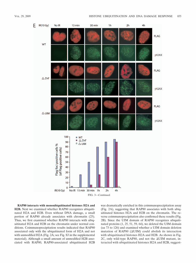
RAP80 interacts with monoubiquitinated histones H2A andH2B. Next we examined whether RAP80 recognizes ubiquiti-nated H2A and H2B. Even without DNA damage, a smallportion of RAP80 already associates with chromatin (25).Thus, we first examined whether RAP80 interacts with ubiq-uitinated H2A and H2B on the chromatin under normal con-ditions. Coimmunoprecipitation results indicated that RAP80associated only with the ubiquitinated form of H2A and notwith unmodified H2A (Fig. 2A; see Fig. S3 in the supplementalmaterial). Although a small amount of unmodified H2B asso-ciated with RAP80, RAP80-associated ubiquitinated H2B
was dramatically enriched in this coimmunoprecipitation assay(Fig. 2A), suggesting that RAP80 associates with both ubiq-uitinated histones H2A and H2B on the chromatin. The re-verse coimmunoprecipitation also confirmed these results (Fig.2B). Since the UIM domain of RAP80 recognizes ubiquiti-nated proteins (1, 25, 51, 59, 64), we deleted the UIM domain(aa 73 to 126) and examined whether a UIM domain deletionmutation of RAP80 (�UIM) could abolish its interactionwith ubiquitinated histones H2A and H2B. As shown in Fig.2C, only wild-type RAP80, and not the �UIM mutant, in-teracted with ubiquitinated histones H2A and H2B, suggest-
FIG. 3—Continued.
VOL. 29, 2009 HISTONE UBIQUITINATION AND DNA DAMAGE RESPONSE 855
856

ing that it is the RAP80 UIM domain that recognizes ubiq-uitinated H2A and H2B. Ubiquitinated histones are themost abundant ubiquitinated proteins in the cell. To exam-ine the specificity of the interaction between the RAP80UIM domain and ubiquitinated histones, we also checkedHJS1A, a UIM domain-containing protein in the nucleus.However, only RAP80, and not HJS1A, could associate withubiquitinated histones, indicating that the UIM domain ofRAP80 specifically recognized ubiquitinated histones (seeFig. S4 in the supplemental material).
Following IR treatment, most RAP80 associates with chro-matin (25). Since the RAP80 UIM domain recognizes ubiq-uitinated histones H2A and H2B, we examined whether theinteraction between RAP80 and ubiquitinated H2A and H2Bwas increased after DNA damage. As expected, DNA damageinduced RAP80’s association with ubiquitinated H2A and H2Bin the chromatin fraction (Fig. 2D). This is consistent with thepurification results for RAP80-interacting proteins (Table 1),suggesting that DNA damage-induced histone ubiquitinationrecruits RAP80 to the chromatin.
In this RAP80 UIM domain, there are two tandem UIMs.These two UIMs have conserved residues to interact with ubiq-uitin, but they differ in the flanking regions (Fig. 3A), indicat-ing that these two UIMs may have different affinities fordifferent ubiquitinated histones (18). We performed a GSTpull-down assay to examine this possibility. The GST-UIMdomain, but not its mutant with both UIMs deleted (GST-�UIM 1&2), could pull down ubiquitinated histones H2A andH2B, further confirming that RAP80 UIMs interact with ubiq-uitinated histones. However, deletion of either UIM abolishedthe interaction between the UIM domain and ubiquitinatedH2A and H2B (Fig. 3B), suggesting that both UIMs have tocooperate together to interact with either ubiquitinated H2Aor ubiquitinated H2B.
In the coimmunoprecipitation assay, we noticed that RAP80associated with a small amount of nonubiquitinated H2B (Fig.2A), suggesting that other regions of RAP80 could also inter-act with H2B regardless of whether it is ubiquitinated or not.Such interaction may determine the binding specificity of theRAP80 UIM domain with ubiquitinated H2A and H2B overother ubiquitinated proteins. Besides the N-terminal UIM do-main, RAP80 also contains two other domains: the Abraxas/CCDC98-interacting region (AIR) in the middle and the C-terminal zinc finger domain (58). We generated GST fusionproteins of all three domains and performed a pull-down assayby using nonubiquitinated free histones. We found that onlythe zinc finger domain, and not the others, specifically inter-acted with H2B, but not H2A or H4 (Fig. 3C). Moreover,
abolishing the Zn binding ability of this zinc finger domain bymutating a conserved cysteine to alanine (C508A) totally dis-rupted this interaction (Fig. 3D). We postulate that the inter-action between the RAP80 zinc finger domain and H2B furtherincreases the affinity between RAP80 and ubiquitinated H2B.To further analyze the function of the zinc finger domain ofRAP80 in the DNA damage response, we examined IR-in-duced focus formation of mutant RAP80 in the absence of thezinc finger domain (�ZnF). Compared with wild-type RAP80,which formed IR-induced foci within 15 minutes, the �ZnFmutant had significantly delayed IR focus formation andstarted to relocate to DNA damage sites after 1 hour of DNAdamage (Fig. 3E). Consistent with our and other previousreports (1, 25, 51, 64), mutant RAP80 without UIM failed torelocate to DNA damage sites (Fig. 3E). These results indicatethat the RAP80 zinc finger domain facilities IR-inducedRAP80 focus formation, possibly by additional interaction withH2B other than with the UIM domain.
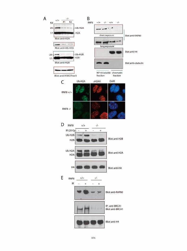
Histone ubiquitination controls RAP80/BRCA1 complex as-sociation with chromatin following DNA damage. To furtherexamine the role of histone ubiquitination in targeting theRAP80/BRCA1 complex to DNA damage sites, we planned toabolish H2A and H2B ubiquitination in vivo. Recent studieshave identified that an E3 ligase, RNF8, controls H2A ubiq-uitination, especially DNA damage-induced H2A ubiquitina-tion (19, 33). We have generated RNF8-deficient MEFs (35).To our surprise, not only 80% of H2A ubiquitination but also90% of H2B ubiquitination was lost in RNF8�/� MEFs (Fig.4A), suggesting that RNF8 not only controls H2A ubiquitina-tion but also regulates H2B ubiquitination. The loss of ubiq-uitinated H2A and H2B correlated with the reduction of chro-matin-associated RAP80 in RNF8�/� cells (Fig. 4B), furtherconfirming the interaction between RAP80 and ubiquitinatedhistones H2A and H2B. Consistently, DNA damage-inducedH2A and H2B ubiquitination was also abrogated in the ab-sence of RNF8 (Fig. 4C and D), and this in turn abolished thedamage-induced association of RAP80 and BRCA1 with chro-matin in RNF8-deficient cells (Fig. 4E). These data togethersuggest that ubiquitinated H2A and H2B could be dockingsites for recruiting the RAP80/BRCA1 complex to the chro-matin following DNA damage.
DISCUSSION
In this study, we have identified that ubiquitinated histonesH2A and H2B are upstream functional partners of theBRCA1/RAP80 complex in the DNA damage response. TheRAP80 UIM domain associated with ubiquitinated H2A and
FIG. 4. Histone H2A and H2B ubiquitination controls RAP80/BRCA1 complex association with chromatin following DNA damage. (A) H2Aand H2B monoubiquitination is dramatically reduced in RNF8-deficient cells. Two different lines of RNF8�/� MEFs were generated. Ubiquiti-nated histones H2A and H2B were analyzed with the indicated antibodies. A blot for trimethylated histone H3K27 was used as protein loadingcontrol. (B) Chromatin-associated RAP80 is reduced in RNF8�/� cells. The NP-40-soluble fraction and chromatin fraction were prepared fromwild-type or RNF8�/� MEFs and subjected to Western blot analysis with the indicated antibodies. (C and D) IR does not induce histone H2A andH2B ubiquitination in RNF8�/� cells. RNF8�/� and RNF8�/� MEFs were treated with or without 20 Gy IR. Four hours later, the cells were fixedand immunostained with anti-ubiquitinated H2A (Ub-H2A) and anti-H2AX antibodies (C). DAPI, 4�,6�-diamidino-2-phenylindole. Alternatively,the chromatin fraction was subjected to Western blotting with the indicated antibodies (D). (E) IR does not induce RAP80/BRCA1 complexassociation with chromatin in the absence of RNF8. RNF8�/� and RNF8�/� MEFs were treated with or without IR (20 Gy). Cells were harvested4 h later. The chromatin fraction was subjected to Western blotting with the indicated antibodies. Anti-H4 was used as a loading control.
VOL. 29, 2009 HISTONE UBIQUITINATION AND DNA DAMAGE RESPONSE 857

H2B, which recruit the BRCA1/RAP80 complex to DNA dam-age lesions on the chromatin. In addition, we have also foundthat RNF8 controls histone H2A and H2B ubiquitination be-fore and after DNA damage, which regulates the BRCA1complex’s translocation to DNA damage sites.
Histone ubiquitination has been known for more than 30years (15). In fact, histone H2A was the first protein known tobe ubiquitinated (15). Monoubiquitinated histones H2A andH2B have been shown to participate in gene transcriptionalregulation (3, 23, 28, 31, 67) and the DNA damage response (4,5, 14, 24, 40). However, the molecular mechanisms underlyingthese histone ubiquitination-dependent functions are not clear.Here, we have shown that histone ubiquitination, like histonephosphorylation and methylation, may function as a platformto recruit the DNA damage response factor BRCA1 to damagelesions on the chromatin. These histone ubiquitination eventsdo not catalyze protesome-dependent protein degradation. In-stead, they may resemble other protein monoubiquitinationsduring endocytosis and protein sorting (38), which function asprotein-protein interaction motifs recognized by other ubiq-uitin binding domains (10). In this study, we may have identi-fied the first ubiquitinated histone binding protein, RAP80.Since ubiquitinated histones are much more abundant thanRAP80 in the nucleus, we postulate that many other ubiquitinbinding proteins may also recognize monoubiquitinated his-tones H2A and H2B and participate in gene transcriptionregulation and the DNA damage response.
We have characterized the interaction between the RAP80UIM domain and monoubiquitinated histones H2A and H2B.Interestingly, the RAP80 UIM domain contains two tandemUIMs. Previous structure analyses suggested that one UIMcould bind to one or two ubiquitins (17, 18, 42, 54). Thus,theoretically, the RAP80 UIM domain could recognize at leasttwo ubiquitins. To our surprise, deleting either UIM abolishedthe interaction between the UIM domain of RAP80 and ubiq-uitinated H2A or H2B, suggesting that these two tandemUIMs are likely to cooperate together and recognize one ubiq-uitinated protein. It is consistent with previous results thatabolishing either UIM will disrupt RAP80 focus formationfollowing DNA damage (1, 25, 51, 59, 64). Further structuralanalyses are needed to examine the molecular details of theinteraction between RAP80 and ubiquitinated histones.
Recent publications showed that H2A and H2AX could bepolyubiquitinated following DNA damage (19, 22, 33, 68). Toavoid antibody cross-reaction, we confirmed that the HA-tagged H2AX could be diubiquitinated (see Fig. S1 in thesupplemental material). Moreover, we have mutated all thelysine residues in H2AX to arginine (NOK mutant). The NOKmutant is phosphorylated at Ser139, but ubiquitination is abol-ished. This mutation still does not affect ubiquitinated H2Afocus formation (Fig. 1B), suggesting that H2AX ubiquitina-tion could only be part of H2A ubiquitination that regulatesthe activation of the DNA damage response. Although wecould not detect major diubiquitination or polyubiquitinationof HA-tagged H2A and H2B (see Fig. S5 in the supplementalmaterial), we could not rule out that a small amount of his-tones H2A and H2B was polyubiquitinated following DNAdamage. Indeed, RAP80 also recognizes K6- or K63-basedpolyubiquitin at the DNA damage sites (36, 43, 51). Thus,
RAP80 might also recognize polyubiquitinated histones orother targets at the DNA damage sites.
Recent studies from our group and others have shown thatRNF8 controls histone H2A and H2AX ubiquitination follow-ing DNA damage (19, 33). We have generated RNF8-deficientMEFs. To our surprise, this single E3 ligase controls not onlyhistone H2A ubiquitination but also histone H2B ubiquitina-tion. To ensure the function of RNF8 in histone ubiquitination,we have generated two different RNF8 knockout mouse celllines and two different deficient MEF lines. Both lines ofRNF8�/� MEFs dramatically lose ubiquitinated histones H2Aand H2B. Previously, the Ring1/Ring2/Bmi1 complex has beenshown to ubiquitinate histone H2A (2, 7, 9, 60), while theRNF20/RNF40 complex ubiquitinates histone H2B (21, 27, 61,70). However, in Ring2 and RNF20 depletion cells, althoughubiquitinated H2A and H2B were dramatically reduced, IRtreatment still could induce H2A and H2B ubiquitination (seeFig. S6 in the supplemental material). This IR-induced histoneubiquitination is controlled by RNF8 and H2AX (Fig. 4C andD; see Fig. S1 and S7 in the supplemental material). Thus, it islikely that RNF8 is important not only for basal level histoneubiquitination but also for IR-induced histone ubiquitination.Moreover, both ubiquitinated H2A and RAP80 formed IR-induced foci in Ring2 and RNF20 depletion cells (see Fig. S6in the supplemental material), suggesting that RNF8-depen-dent IR-induced histone ubiquitination is critical to recruit theRAP80/BRCA1 complex to DNA damage sites. Since theRing2 complex and the RNF20 complex are known E3 ligasesfor histone ubiquitination, regulation of histone ubiquitina-tion could be more complicated than in previously proposedmodels.
Besides RNF8, Ubc13, an E2 conjugase, coordinates to-gether with RNF8 to regulate RAP80/BRCA1 focus formationfollowing DNA damage (20, 58, 68). Ubc13 is known for cat-alyzing K63 polyubiquitin chains on its targets (68). However,two known E2 conjugase partners of Ubc13, MMS2 andUEV1A, are dispensable for ubiquitination foci at the DNAdamage sites (20). Thus, it is likely that only a portion of Ubc13associates with RNF8 and generate ubiquitination signals atthe DNA damage sites.
Following DNA damage, a group of DNA damage check-point proteins and DNA damage repair proteins, includingBRCA1, translocate and accumulate at DNA damage sites.However, the molecular mechanisms by which these DNAdamage response factors are recruited to damaged lesions arenot clear. Recent evidence indicates that DNA damage in-duces substantial chromatin remodeling, including histonephosphorylation, acetylation, and ubiquitination (11, 30, 57).In this study, we found that histone ubiquitination functions asthe platform to recruit DNA damage response factor BRCA1.Besides this finding, it has been shown that MDC1 andMCPH1 recognize phosphorylated Ser139 of histone H2AX(52, 62) and that Crb2 and 53BP1 recognizes histone H4 Lys20methylation (6, 47). Thus, various DNA damage-induced his-tone modifications could function as docking sites to host DNAdamage response factors at damage lesions on the chromatin.
Taken together, our results demonstrate the molecularmechanism by which ubiquitinated histones participate in theDNA damage response.
858 WU ET AL. MOL. CELL. BIOL.
ACKNOWLEDGMENTS
We thank members of the Ben Margolis lab for technical support.We thank Andre Nussenzweig for invaluable reagents.
This work was supported by the Department of Defense (grantBC050367 to X.Y.), the National Institutes of Health (grant CA132755to X.Y.), the University of Michigan Cancer Center, and the GI Pep-tide Research Center of the University of Michigan. X.Y. is a recipientof an AACR-Susan G. Komen for the Cure Career DevelopmentAward for Breast Cancer Research. J.C. is a recipient of an Era ofHope Scholar award from the Department of Defense and is a mem-ber of the Mayo Clinic Breast SPORE program (grant P50 CA116201).J.C. was also supported by the National Cancer Institute (grantsCA89239, CA92312, and CA100109). M.S.Y.H. is supported by anAnna Fuller Fund Fellowship.
REFERENCES
1. Bennett, E. J., and J. W. Harper. 2008. DNA damage: ubiquitin marks thespot. Nat. Struct. Mol. Biol. 15:20–22.
2. Ben-Saadon, R., D. Zaaroor, T. Ziv, and A. Ciechanover. 2006. The poly-comb protein Ring1B generates self atypical mixed ubiquitin chains requiredfor its in vitro histone H2A ligase activity. Mol. Cell 24:701–711.
3. Berger, S. L. 2002. Histone modifications in transcriptional regulation. Curr.Opin. Genet. Dev. 12:142–148.
4. Bergink, S., N. G. Jaspers, and W. Vermeulen. 2007. Regulation of UV-induced DNA damage response by ubiquitylation. DNA Repair (Amster-dam) 6:1231–1242.
5. Bergink, S., F. A. Salomons, D. Hoogstraten, T. A. Groothuis, H. de Waard,J. Wu, L. Yuan, E. Citterio, A. B. Houtsmuller, J. Neefjes, J. H. Hoeijmakers,W. Vermeulen, and N. P. Dantuma. 2006. DNA damage triggers nucleotideexcision repair-dependent monoubiquitylation of histone H2A. Genes Dev.20:1343–1352.
6. Botuyan, M. V., J. Lee, I. M. Ward, J. E. Kim, J. R. Thompson, J. Chen, andG. Mer. 2006. Structural basis for the methylation state-specific recognitionof histone H4-K20 by 53BP1 and Crb2 in DNA repair. Cell 127:1361–1373.
7. Cao, R., Y. Tsukada, and Y. Zhang. 2005. Role of Bmi-1 and Ring1A in H2Aubiquitylation and Hox gene silencing. Mol. Cell 20:845–854.
8. Cortez, D., Y. Wang, J. Qin, and S. J. Elledge. 1999. Requirement of ATM-dependent phosphorylation of brca1 in the DNA damage response to dou-ble-strand breaks. Science 286:1162–1166.
9. de Napoles, M., J. E. Mermoud, R. Wakao, Y. A. Tang, M. Endoh, R.Appanah, T. B. Nesterova, J. Silva, A. P. Otte, M. Vidal, H. Koseki, and N.Brockdorff. 2004. Polycomb group proteins Ring1A/B link ubiquitylation ofhistone H2A to heritable gene silencing and X inactivation. Dev. Cell 7:663–676.
10. Di Fiore, P. P., S. Polo, and K. Hofmann. 2003. When ubiquitin meetsubiquitin receptors: a signalling connection. Nat. Rev. Mol. Cell Biol. 4:491–497.
11. Downs, J. A., M. C. Nussenzweig, and A. Nussenzweig. 2007. Chromatindynamics and the preservation of genetic information. Nature 447:951–958.
12. Gatei, M., S. P. Scott, I. Filippovitch, N. Soronika, M. F. Lavin, B. Weber,and K. K. Khanna. 2000. Role for ATM in DNA damage-induced phosphor-ylation of BRCA1. Cancer Res. 60:3299–3304.
13. Gatei, M., B. B. Zhou, K. Hobson, S. Scott, D. Young, and K. K. Khanna.2001. Ataxia telangiectasia mutated (ATM) kinase and ATM and Rad3related kinase mediate phosphorylation of Brca1 at distinct and overlappingsites. In vivo assessment using phospho-specific antibodies. J. Biol. Chem.276:17276–17280.
14. Giannattasio, M., F. Lazzaro, P. Plevani, and M. Muzi-Falconi. 2005. TheDNA damage checkpoint response requires histone H2B ubiquitination byRad6-Bre1 and H3 methylation by Dot1. J. Biol. Chem. 280:9879–9886.
15. Goldknopf, I. L., C. W. Taylor, R. M. Baum, L. C. Yeoman, M. O. Olson,A. W. Prestayko, and H. Busch. 1975. Isolation and characterization ofprotein A24, a “histone-like” non-histone chromosomal protein. J. Biol.Chem. 250:7182–7187.
16. Harper, J. W., and S. J. Elledge. 2007. The DNA damage response: ten yearsafter. Mol. Cell 28:739–745.
17. Harper, J. W., and B. A. Schulman. 2006. Structural complexity in ubiquitinrecognition. Cell 124:1133–1136.
18. Hirano, S., M. Kawasaki, H. Ura, R. Kato, C. Raiborg, H. Stenmark, and S.Wakatsuki. 2006. Double-sided ubiquitin binding of Hrs-UIM in endosomalprotein sorting. Nat. Struct. Mol. Biol. 13:272–277.
19. Huen, M. S., R. Grant, I. Manke, K. Minn, X. Yu, M. B. Yaffe, and J. Chen.2007. RNF8 transduces the DNA-damage signal via histone ubiquitylationand checkpoint protein assembly. Cell 131:901–914.
20. Huen, M. S., J. Huang, J. Yuan, M. Yamamoto, S. Akira, C. Ashley, W. Xiao,and J. Chen. 2008. Noncanonical E2 variant-independent function of UBC13in promoting checkpoint protein assembly. Mol. Cell. Biol. 28:6104–6112.
21. Hwang, W. W., S. Venkatasubrahmanyam, A. G. Ianculescu, A. Tong, C.Boone, and H. D. Madhani. 2003. A conserved RING finger protein required
for histone H2B monoubiquitination and cell size control. Mol. Cell 11:261–266.
22. Ikura, T., S. Tashiro, A. Kakino, H. Shima, N. Jacob, R. Amunugama, K.Yoder, S. Izumi, I. Kuraoka, K. Tanaka, H. Kimura, M. Ikura, S. Nishikubo,T. Ito, A. Muto, K. Miyagawa, S. Takeda, R. Fishel, K. Igarashi, and K.Kamiya. 2007. DNA damage-dependent acetylation and ubiquitination ofH2AX enhances chromatin dynamics. Mol. Cell. Biol. 27:7028–7040.
23. Jenuwein, T., and C. D. Allis. 2001. Translating the histone code. Science293:1074–1080.
24. Kapetanaki, M. G., J. Guerrero-Santoro, D. C. Bisi, C. L. Hsieh, V. Rapic-Otrin, and A. S. Levine. 2006. The DDB1-CUL4ADDB2 ubiquitin ligase isdeficient in xeroderma pigmentosum group E and targets histone H2A atUV-damaged DNA sites. Proc. Natl. Acad. Sci. USA 103:2588–2593.
25. Kim, H., J. Chen, and X. Yu. 2007. Ubiquitin-binding protein RAP80 me-diates BRCA1-dependent DNA damage response. Science 316:1202–1205.
26. Kim, H., J. Huang, and J. Chen. 2007. CCDC98 is a BRCA1-BRCT domain-binding protein involved in the DNA damage response. Nat. Struct. Mol.Biol. 14:710–715.
27. Kim, J., S. B. Hake, and R. G. Roeder. 2005. The human homolog of yeastBRE1 functions as a transcriptional coactivator through direct activatorinteractions. Mol. Cell 20:759–770.
28. Kouzarides, T. 2007. Chromatin modifications and their function. Cell 128:693–705.
29. Lee, E. Y. 2002. BRCA1 and Chk1 in G2/M checkpoint: a new order ofregulation. Cell Cycle 1:178–180.
30. Lee, K. K., and J. L. Workman. 2007. Histone acetyltransferase complexes:one size doesn’t fit all. Nat. Rev. Mol. Cell Biol. 8:284–295.
31. Li, B., M. Carey, and J. L. Workman. 2007. The role of chromatin duringtranscription. Cell 128:707–719.
32. Liu, Z., J. Wu, and X. Yu. 2007. CCDC98 targets BRCA1 to DNA damagesites. Nat. Struct. Mol. Biol. 14:716–720.
33. Mailand, N., S. Bekker-Jensen, H. Faustrup, F. Melander, J. Bartek, C.Lukas, and J. Lukas. 2007. RNF8 ubiquitylates histones at DNA double-strand breaks and promotes assembly of repair proteins. Cell 131:887–900.
34. Manke, I. A., D. M. Lowery, A. Nguyen, and M. B. Yaffe. 2003. BRCT repeatsas phosphopeptide-binding modules involved in protein targeting. Science302:636–639.
35. Minter-Dykhouse, K., I. Ward, M. S. Huen, J. Chen, and Z. Lou. 2008.Distinct versus overlapping functions of MDC1 and 53BP1 in DNA damageresponse and tumorigenesis. J. Cell Biol. 181:727–735.
36. Morris, J. R., and E. Solomon. 2004. BRCA1:BARD1 induces the formationof conjugated ubiquitin structures, dependent on K6 of ubiquitin, in cellsduring DNA replication and repair. Hum. Mol. Genet. 13:807–817.
37. Moynahan, M. E., J. W. Chiu, B. H. Koller, and M. Jasin. 1999. Brca1controls homology-directed DNA repair. Mol. Cell 4:511–518.
38. Mukhopadhyay, D., and H. Riezman. 2007. Proteasome-independent func-tions of ubiquitin in endocytosis and signaling. Science 315:201–205.
39. Narod, S. A., and W. D. Foulkes. 2004. BRCA1 and BRCA2: 1994 andbeyond. Nat. Rev. Cancer 4:665–676.
40. Nicassio, F., N. Corrado, J. H. Vissers, L. B. Areces, S. Bergink, J. A.Marteijn, B. Geverts, A. B. Houtsmuller, W. Vermeulen, P. P. Di Fiore, andE. Citterio. 2007. Human USP3 is a chromatin modifier required for S phaseprogression and genome stability. Curr. Biol. 17:1972–1977.
41. Paull, T. T., E. P. Rogakou, V. Yamazaki, C. U. Kirchgessner, M. Gellert,and W. M. Bonner. 2000. A critical role for histone H2AX in recruitment ofrepair factors to nuclear foci after DNA damage. Curr. Biol. 10:886–895.
42. Penengo, L., M. Mapelli, A. G. Murachelli, S. Confalonieri, L. Magri, A.Musacchio, P. P. Di Fiore, S. Polo, and T. R. Schneider. 2006. Crystalstructure of the ubiquitin binding domains of rabex-5 reveals two modes ofinteraction with ubiquitin. Cell 124:1183–1195.
43. Polanowska, J., J. S. Martin, T. Garcia-Muse, M. I. Petalcorin, and S. J.Boulton. 2006. A conserved pathway to activate BRCA1-dependent ubiqui-tylation at DNA damage sites. EMBO J. 25:2178–2188.
44. Rodriguez, M., X. Yu, J. Chen, and Z. Songyang. 2003. Phosphopeptidebinding specificities of BRCA1 COOH-terminal (BRCT) domains. J. Biol.Chem. 278:52914–52918.
45. Rouse, J., and S. P. Jackson. 2002. Interfaces between the detection, signal-ing, and repair of DNA damage. Science 297:547–551.
46. Sancar, A., L. A. Lindsey-Boltz, K. Unsal-Kacmaz, and S. Linn. 2004. Mo-lecular mechanisms of mammalian DNA repair and the DNA damage check-points. Annu. Rev. Biochem. 73:39–85.
47. Sanders, S. L., M. Portoso, J. Mata, J. Bahler, R. C. Allshire, and T.Kouzarides. 2004. Methylation of histone H4 lysine 20 controls recruitmentof Crb2 to sites of DNA damage. Cell 119:603–614.
48. Scully, R., J. Chen, R. L. Ochs, K. Keegan, M. Hoekstra, J. Feunteun, andD. M. Livingston. 1997. Dynamic changes of BRCA1 subnuclear locationand phosphorylation state are initiated by DNA damage. Cell 90:425–435.
49. Scully, R., J. Chen, A. Plug, Y. Xiao, D. Weaver, J. Feunteun, T. Ashley, andD. M. Livingston. 1997. Association of BRCA1 with Rad51 in mitotic andmeiotic cells. Cell 88:265–275.
50. Scully, R., and D. M. Livingston. 2000. In search of the tumour-suppressorfunctions of BRCA1 and BRCA2. Nature 408:429–432.
VOL. 29, 2009 HISTONE UBIQUITINATION AND DNA DAMAGE RESPONSE 859

51. Sobhian, B., G. Shao, D. R. Lilli, A. C. Culhane, L. A. Moreau, B. Xia, D. M.Livingston, and R. A. Greenberg. 2007. RAP80 targets BRCA1 to specificubiquitin structures at DNA damage sites. Science 316:1198–1202.
52. Stucki, M., J. A. Clapperton, D. Mohammad, M. B. Yaffe, S. J. Smerdon, andS. P. Jackson. 2005. MDC1 directly binds phosphorylated histone H2AX toregulate cellular responses to DNA double-strand breaks. Cell 123:1213–1226.
53. Su, T. T. 2006. Cellular responses to DNA damage: one signal, multiplechoices. Annu. Rev. Genet. 40:187–208.
54. Swanson, K. A., R. S. Kang, S. D. Stamenova, L. Hicke, and I. Radhakrish-nan. 2003. Solution structure of Vps27 UIM-ubiquitin complex importantfor endosomal sorting and receptor downregulation. EMBO J. 22:4597–4606.
55. Tibbetts, R. S., D. Cortez, K. M. Brumbaugh, R. Scully, D. Livingston, S. J.Elledge, and R. T. Abraham. 2000. Functional interactions between BRCA1and the checkpoint kinase ATR during genotoxic stress. Genes Dev. 14:2989–3002.
56. Venkitaraman, A. R. 2002. Cancer susceptibility and the functions of BRCA1and BRCA2. Cell 108:171–182.
57. Vidanes, G. M., C. Y. Bonilla, and D. P. Toczyski. 2005. Complicated tails:histone modifications and the DNA damage response. Cell 121:973–976.
58. Wang, B., and S. J. Elledge. 2007. Ubc13/Rnf8 ubiquitin ligases control fociformation of the Rap80/Abraxas/Brca1/Brcc36 complex in response to DNAdamage. Proc. Natl. Acad. Sci. USA 104:20759–20763.
59. Wang, B., S. Matsuoka, B. A. Ballif, D. Zhang, A. Smogorzewska, S. P. Gygi,and S. J. Elledge. 2007. Abraxas and RAP80 form a BRCA1 protein complexrequired for the DNA damage response. Science 316:1194–1198.
60. Wang, H., L. Wang, H. Erdjument-Bromage, M. Vidal, P. Tempst, R. S.Jones, and Y. Zhang. 2004. Role of histone H2A ubiquitination in Polycombsilencing. Nature 431:873–878.
61. Wood, A., N. J. Krogan, J. Dover, J. Schneider, J. Heidt, M. A. Boateng, K.
Dean, A. Golshani, Y. Zhang, J. F. Greenblatt, M. Johnston, and A. Shilati-fard. 2003. Bre1, an E3 ubiquitin ligase required for recruitment and sub-strate selection of Rad6 at a promoter. Mol. Cell 11:267–274.
62. Wood, J. L., N. Singh, G. Mer, and J. Chen. 2007. MCPH1 functions in anH2AX dependent but MDC1 independent pathway in response to DNAdamage. J. Biol. Chem. 282:35416–35423.
63. Xu, B., S. Kim, and M. B. Kastan. 2001. Involvement of Brca1 in S-phase andG2-phase checkpoints after ionizing irradiation. Mol. Cell. Biol. 21:3445–3450.
64. Yan, J., Y. S. Kim, X. P. Yang, L. P. Li, G. Liao, F. Xia, and A. M. Jetten.2007. The ubiquitin-interacting motif containing protein RAP80 interactswith BRCA1 and functions in DNA damage repair response. Cancer Res.67:6647–6656.
65. Yarden, R. I., S. Pardo-Reoyo, M. Sgagias, K. H. Cowan, and L. C. Brody.2002. BRCA1 regulates the G2/M checkpoint by activating Chk1 kinaseupon DNA damage. Nat. Genet. 30:285–289.
66. Yu, X., C. C. Chini, M. He, G. Mer, and J. Chen. 2003. The BRCT domainis a phospho-protein binding domain. Science 302:639–642.
67. Zhang, Y. 2003. Transcriptional regulation by histone ubiquitination anddeubiquitination. Genes Dev. 17:2733–2740.
68. Zhao, G. Y., E. Sonoda, L. J. Barber, H. Oka, Y. Murakawa, K. Yamada, T.Ikura, X. Wang, M. Kobayashi, K. Yamamoto, S. J. Boulton, and S. Takeda.2007. A critical role for the ubiquitin-conjugating enzyme Ubc13 in initiatinghomologous recombination. Mol. Cell 25:663–675.
69. Zhou, B. B., and S. J. Elledge. 2000. The DNA damage response: puttingcheckpoints in perspective. Nature 408:433–439.
70. Zhu, B., Y. Zheng, A. D. Pham, S. S. Mandal, H. Erdjument-Bromage, P.Tempst, and D. Reinberg. 2005. Monoubiquitination of human histone H2B:the factors involved and their roles in HOX gene regulation. Mol. Cell20:601–611.
860 WU ET AL. MOL. CELL. BIOL.
Related Documents


