Research Article 1715 Introduction A classical example of unorthodox chromosome segregation is found in sciarid flies (Diptera, Sciaridae) where the selective elimination of paternal chromosomes takes place at different times during development (Metz, 1925; Metz, 1926b; Metz, 1933) (reviewed in Gerbi, 1986; Goday and Esteban, 2001). One of the most complex and bizarre chromosome behaviours occurs in sciarid male meiosis, where the whole paternal chromosome complement is discarded so that only maternally derived chromosomes are included in the sperm nucleus. An additional exceptional feature of male meiosis is the occurrence of non-disjunction of the maternal X chromosome (X m ), which determines the characteristic 3X constitution of the zygote in sciarids (reviewed in Gerbi, 1986; Goday and Esteban, 2001). Since the early findings on the chromosome cell cycle of sciarids, the mechanisms that eliminate paternal chromosomes at meiosis I and non-disjunction of X m chromosome at male meiosis II have been the subject of several studies (reviewed in Gerbi, 1986; Goday and Esteban, 2001). In Fig. 1 we summarise the most relevant chromosomal events occurring during male meiosis that are common to sciarid flies. As shown (Fig. 1A), at the first meiotic division there is no pairing of homologous chromosomes at prophase and the chromosomes do not align in a metaphase-like array (Metz, 1925; Metz, 1926a; Metz, 1926b; Fuge, 1994). Instead, they proceed directly from prometaphase to an ‘anaphase-like’ stage (Fig. 1B). This atypical behaviour is accompanied by the formation of a monopolar spindle generated from a single polar complex that contains ‘giant centrioles’ surrounded by a large amount of pericentriolar material, from which numerous microtubules radiate (Kubai, 1982; Fuge, 1994; Esteban et al., 1997). During the anaphase-I-like stage, the monopolar spindle directs the segregation of maternal chromosomes to the single pole, whereas paternal chromosomes move in the opposite direction into a cytoplasmic bud to be discarded later (Metz, 1925; Smith-Stocking, 1936). Importantly, cytological and ultrastructural data indicate that the two parental sets of chromosomes occupy distinct nuclear compartments in germ nuclei from the initial stages of development until the occurrence of meiosis (Rieffel and Crouse, 1966; Kubai, 1982; Kubai, 1987; Goday and Esteban, 2001; Goday and Ruiz, 2002). Moreover, the precocious intranuclear segregation of the maternal chromosomes with respect to the paternal ones is considered to be essential in determining which chromosomal set will be lost during male meiosis I (Kubai, 1987; Goday and Esteban, 2001). The outstanding feature of meiosis II is the different kinetic behaviour of the maternal X chromosome with respect to maternal autosomes (Metz, 1925; Crouse, 1943) (reviewed in Esteban et al., 1997). By metaphase II (Fig. 1C), whereas maternal autosomes align in a typical metaphase plate, the non-disjoining X m chromosome does not move away from the polar complex formed at meiosis I. Importantly, microtubules radiating from the single pole interact with the X m centromere and maintain the non- disjunction of this chromosome throughout meiosis (Esteban et al., 1997). Finally, at anaphase II (Fig. 1D,E), the autosomal chromatids segregate to opposite poles in the conventional manner by means Summary An extremely unorthodox method of chromosome segregation is found in sciarid flies (Diptera, Sciaridae), where at male meiosis, the whole paternal complement is eliminated and the maternal X chromosome undergoes non-disjunction. At meiosis I, a monopolar spindle directs the segregation of maternal chromosomes to the single pole, whereas paternal chromosomes are discarded. At meiosis II, although maternal autosomes segregate normally, the X chromosome remains undivided. A cis-acting locus within the heterochromatin proximal to the centromere is known to regulate X centromere activity. By immunofluorescence analysis in spermatocytes from Sciara ocellaris and Sciara coprophila, we investigated histone H3 phosphorylation at Ser10, Ser28, Thr3 and Thr11 during male meiosis. We found that chromosome condensation and H3 phosphorylation patterns differ between chromosomes of different parental origin at the time of paternal set elimination. Importantly, at meiosis II, the maternal X chromosome differs from the rest of the chromosomes in that its centromeric region does not become phosphorylated at the four histone H3 sites. We provide here the first evidence linking the under-phosphorylated H3 status of the X chromosome centromeric region with its meiotic non-disjunction in sciarid flies. Our findings strongly support the idea that the deficiency in local H3 phosphorylation inactivates the X centromere at the transition from meiosis I to meiosis II. Key words: H3 phosphorylation, Sciara, Meiosis, Chromosome non-disjunction, Chromosome elimination Accepted 20 January 2011 Journal of Cell Science 124, 1715-1725 © 2011. Published by The Company of Biologists Ltd doi:10.1242/jcs.083022 Histone H3 phosphorylation and non-disjunction of the maternal X chromosome during male meiosis in sciarid flies M. Carmen Escribá 1, *, M. Cecilia Giardini 2, * and Clara Goday 1,‡ 1 Centro de Investigaciones Biológicas, Departamento de Proliferación Celular y Desarrollo, CSIC, Ramiro de Maeztu 9, 28040 Madrid, Spain 2 Instituto de Genética “Ewald A. Favret”, INTA-Castelar, C. C. 25-1712, Argentina *These authors contributed equally to this work ‡ Author for correspondence ([email protected]) Journal of Cell Science

Welcome message from author
This document is posted to help you gain knowledge. Please leave a comment to let me know what you think about it! Share it to your friends and learn new things together.
Transcript

Research Article 1715
IntroductionA classical example of unorthodox chromosome segregation isfound in sciarid flies (Diptera, Sciaridae) where the selectiveelimination of paternal chromosomes takes place at different timesduring development (Metz, 1925; Metz, 1926b; Metz, 1933)(reviewed in Gerbi, 1986; Goday and Esteban, 2001). One of themost complex and bizarre chromosome behaviours occurs in sciaridmale meiosis, where the whole paternal chromosome complementis discarded so that only maternally derived chromosomes areincluded in the sperm nucleus. An additional exceptional feature ofmale meiosis is the occurrence of non-disjunction of the maternalX chromosome (Xm), which determines the characteristic 3Xconstitution of the zygote in sciarids (reviewed in Gerbi, 1986;Goday and Esteban, 2001). Since the early findings on thechromosome cell cycle of sciarids, the mechanisms that eliminatepaternal chromosomes at meiosis I and non-disjunction of Xm
chromosome at male meiosis II have been the subject of severalstudies (reviewed in Gerbi, 1986; Goday and Esteban, 2001). InFig. 1 we summarise the most relevant chromosomal eventsoccurring during male meiosis that are common to sciarid flies. Asshown (Fig. 1A), at the first meiotic division there is no pairing ofhomologous chromosomes at prophase and the chromosomes donot align in a metaphase-like array (Metz, 1925; Metz, 1926a;Metz, 1926b; Fuge, 1994). Instead, they proceed directly fromprometaphase to an ‘anaphase-like’ stage (Fig. 1B). This atypicalbehaviour is accompanied by the formation of a monopolar spindlegenerated from a single polar complex that contains ‘giant
centrioles’ surrounded by a large amount of pericentriolar material,from which numerous microtubules radiate (Kubai, 1982; Fuge,1994; Esteban et al., 1997). During the anaphase-I-like stage, themonopolar spindle directs the segregation of maternal chromosomesto the single pole, whereas paternal chromosomes move in theopposite direction into a cytoplasmic bud to be discarded later(Metz, 1925; Smith-Stocking, 1936). Importantly, cytological andultrastructural data indicate that the two parental sets ofchromosomes occupy distinct nuclear compartments in germ nucleifrom the initial stages of development until the occurrence ofmeiosis (Rieffel and Crouse, 1966; Kubai, 1982; Kubai, 1987;Goday and Esteban, 2001; Goday and Ruiz, 2002). Moreover, theprecocious intranuclear segregation of the maternal chromosomeswith respect to the paternal ones is considered to be essential indetermining which chromosomal set will be lost during malemeiosis I (Kubai, 1987; Goday and Esteban, 2001).
The outstanding feature of meiosis II is the different kineticbehaviour of the maternal X chromosome with respect to maternalautosomes (Metz, 1925; Crouse, 1943) (reviewed in Esteban et al.,1997). By metaphase II (Fig. 1C), whereas maternal autosomesalign in a typical metaphase plate, the non-disjoining Xm
chromosome does not move away from the polar complex formedat meiosis I. Importantly, microtubules radiating from the singlepole interact with the Xm centromere and maintain the non-disjunction of this chromosome throughout meiosis (Esteban et al.,1997). Finally, at anaphase II (Fig. 1D,E), the autosomal chromatidssegregate to opposite poles in the conventional manner by means
SummaryAn extremely unorthodox method of chromosome segregation is found in sciarid flies (Diptera, Sciaridae), where at male meiosis, thewhole paternal complement is eliminated and the maternal X chromosome undergoes non-disjunction. At meiosis I, a monopolarspindle directs the segregation of maternal chromosomes to the single pole, whereas paternal chromosomes are discarded. At meiosisII, although maternal autosomes segregate normally, the X chromosome remains undivided. A cis-acting locus within the heterochromatinproximal to the centromere is known to regulate X centromere activity. By immunofluorescence analysis in spermatocytes from Sciaraocellaris and Sciara coprophila, we investigated histone H3 phosphorylation at Ser10, Ser28, Thr3 and Thr11 during male meiosis.We found that chromosome condensation and H3 phosphorylation patterns differ between chromosomes of different parental origin atthe time of paternal set elimination. Importantly, at meiosis II, the maternal X chromosome differs from the rest of the chromosomesin that its centromeric region does not become phosphorylated at the four histone H3 sites. We provide here the first evidence linkingthe under-phosphorylated H3 status of the X chromosome centromeric region with its meiotic non-disjunction in sciarid flies. Ourfindings strongly support the idea that the deficiency in local H3 phosphorylation inactivates the X centromere at the transition frommeiosis I to meiosis II.
Key words: H3 phosphorylation, Sciara, Meiosis, Chromosome non-disjunction, Chromosome elimination
Accepted 20 January 2011Journal of Cell Science 124, 1715-1725 © 2011. Published by The Company of Biologists Ltddoi:10.1242/jcs.083022
Histone H3 phosphorylation and non-disjunction ofthe maternal X chromosome during male meiosis insciarid fliesM. Carmen Escribá1,*, M. Cecilia Giardini2,* and Clara Goday1,‡
1Centro de Investigaciones Biológicas, Departamento de Proliferación Celular y Desarrollo, CSIC, Ramiro de Maeztu 9, 28040 Madrid, Spain2Instituto de Genética “Ewald A. Favret”, INTA-Castelar, C. C. 25-1712, Argentina*These authors contributed equally to this work‡Author for correspondence ([email protected])
Jour
nal o
f Cel
l Sci
ence

of an asterless bipolar second meiotic spindle (Metz, 1926a; Abbottand Gerbi, 1981; Esteban et al., 1997).
The genetic control of Xm chromosome segregation at malemeiosis was examined in Sciara coprophila, where threeheterochromatic blocks proximal to the centromere contain rDNAsequences (Gerbi and Crouse, 1976; Crouse, 1977; Crouse et al.,1977; Crouse, 1979). A cis-acting locus, the controlling element(CE), regulating X-centromere activity was identified in the middleheterochromatic block (Crouse, 1960; Crouse, 1977; Crouse, 1979;Gerbi, 1986). Its translocation to an autosome provokes non-disjunction of the recipient autosome, whereas the X chromosomelacking the CE segregates normally (Crouse, 1979). In view ofthis, it has been hypothesised that the CE inhibits normalcentromeric function in the Xm chromosome (Gerbi, 1986).However, how this occurs and which specific DNA sequences areinvolved remains undetermined.
In the present work, we have re-examined the process of malemeiosis in Sciara ocellaris and S. coprophila in an attempt tofurther understand the cellular mechanisms leading to both paternalchromosome elimination and Xm chromosome non-disjunction.With this in mind, we decided to analyse conserved chromatinmodifications involved in normal chromosome segregation duringmitosis and meiosis. A good candidate was that of histone H3phosphorylated at four N-terminal residues, Ser10, Ser28, Thr3and Thr11 (reviewed in Nowak and Corces, 2004; Xu et al., 2009).Cell-cycle-dependent phosphorylation of histone H3 (H3-P) occursin most eukaryotes and high levels of H3-P constitute a conservedmark of mitotic cell division. As a general rule, condensedmetaphase chromosomes attain high levels of phosphorylatedhistone H3 and upon exit of mitosis or meiosis, a globaldephosphorylation of histone H3 occurs. Moreover, an increasingamount of data coming from different systems support the idea thatthe four H3-P forms are coordinated both in space and time duringmitosis and meiosis (Xu et al., 2009).
Phosphorylation of H3S10 (H3S10-P) (Gurley et al., 1978; Weiet al., 1998; Hsu et al., 2000; Giet and Glover, 2001) and of H3S28
(H3S28-P) (Goto et al., 1999), are carried out by the mitotic kinaseAurora B that is a component of the chromosomal passengercomplex (CPC) (Giet and Glover, 2001; Ruchaud et al., 2007).Both H3-P modifications are highly conserved among eukaryotesand are crucial for higher-order chromatin compaction duringmitosis and meiosis (reviewed in Hsu et al., 2000; Nowak andCorces, 2004). The temporal correlation of H3S10-P withchromosome condensation during cell cycle progression was firstdemonstrated in Tetrahymena using antibodies against H3S10-P(Wei et al., 1998). Accordingly, mutation of the site H3S10 inTetrahymena disrupts proper chromosome condensation andsegregation both at mitosis and meiosis (Wei et al., 1999). Anumber of studies have shown that in mammals, the cell-cycle-dependent phosphorylation of H3S10 and H3S28 begins at thepericentromeric chromosome regions and spreads throughout thechromosomes during the G2–M phase transition (Goto et al., 1999;Hsu et al., 2000). A similar distribution of H3S10-P linked tochromosome condensation has also been found in flies wheredepletion of Aurora B provoked deficiencies of chromosomecondensation at mitosis (Giet and Glover, 2001). Moreover, inDrosophila spermatocytes undergoing the first meiotic division,metaphase I chromosomes show prominent H3S10-P signals thatdecrease substantially at anaphase and telophase (Krishnamoorthyet al., 2006).
Phosphorylation of H3T3 (H3T3-P) by the kinase Haspin (Daiet al., 2005) and of H3T11 (H3T11-P) by Dlk/ZIP (Preuss et al.,2003) were found to temporally associate with mitosis in turkeyand mammalian cells (Polioudaki et al., 2004; Dai et al., 2005). Thetiming of H3T3 phosphorylation and dephosphorylation is similarto that of H3S10-P, although the strongest presence of H3T3-P atthe inner centromeric regions of the chromosomes suggested amore direct role of H3T3-P in regulation of kinetochore assemblyand functional activity (Dai et al., 2005). In this regard, recentfindings in mammalian cultured cells revealed for the fist time anessential functional role of H3T3 phosphorylation at the centromeresite (Wang et al., 2010). Phosphorylated H3T3 is crucial for the
1716 Journal of Cell Science 124 (10)
Fig. 1. Scheme summarising the most relevant chromosomal events of male meiosis in S. ocellaris. (A)Prophase I. Prophase chromosomes do not pair.Maternal and paternal chromosomes display separate arrangement within the spermatocyte nucleus. A monopolar spindle is formed and non-spindle microtubulesare generated in a cytoplasmic bud region of the spermatocyte. (B)Anaphase-I-like stage. Maternal chromosomes segregate towards the spermatocyte single polarcomplex while the paternal set move into the bud. (C)At metaphase II, whereas the maternal X chromosome remains attached to the polar complex developed inmeiosis I, maternal autosomes align at the equatorial plate and an asterless bipolar second meiotic spindle is developed. (D,E)At anaphase II, whereas the maternalautosomal chromatids segregate towards opposite directions in a conventional manner, the maternal X chromosome remains undivided and attached to the singlepole of the first meiotic spindle that persists throughout meiosis (D). The upper maternal autosomal chromatid group joins the X chromosome to constitute thefuture sperm nucleus, whereas the opposite maternal chromatid group (X-null chromatid set) is captured by bud microtubules and, along with the paternal set, iseliminated from the spermatocyte by a bud excision process (E) (modified from Goday and Esteban, 2001).
Jour
nal o
f Cel
l Sci
ence

localisation of the CPC at centromeres and for the function ofAurora B during mitosis (Wang et al., 2010). Moreover, as shownin Xenopus extracts, defects in the recruitment of the CPC to thechromosomes and in the activation of Aurora B leads to alterationsin spindle assembly around chromosomes (Kelly et al., 2010).
In this work we have analysed the distribution of the four H3-Pforms in spermatocytes of S. ocellaris and S. coprophila during themeiotic divisions. We provide here a comparative location andtiming description of each of the H3-P modification during bothmeiotic divisions. From this analysis, we found that at meiosis I,at the time of paternal chromosome elimination, chromosomecondensation and H3 phosphorylation patterns differ betweenchromosomes of different parental origin. Moreover, andimportantly, we show that non-disjunction of the Xm chromosomeduring meiosis II correlates with the lack of the Xm centromericregion to undergo phosphorylation of histone H3. Our findingsstrongly indicate that the deficiency in H3 phosphorylationinactivates Xm centromere at the transition from meiosis I tomeiosis II. The results also suggest that the CE induces a localdeficiency of H3 phosphorylation in both maternal and paternal Xchromosomes.
ResultsThe chromosomal complement in S. ocellaris premeiotic germnuclei is that of eight chromosomes: three pairs of autosomes (twoacrocentric and one metacentric) and one pair of X chromosomes(acrocentric). In S. coprophila, additional germline-limited ‘L’chromosomes are present in a variable number (1–4). Lchromosomes are metacentric and, in contrast to the regularchromosomal component, are mostly heterochromatic.
Distribution of phosphorylated H3S10 in S. ocellarisspermatocytes during meiotic divisionWe performed the immunodetection of H3S10-P in S. ocellaris inmale meiosis I when the elimination of the whole paternallyinherited complement takes place. As mentioned, in Sciara males,
homologous chromosomes do not pair at prophase nor do theyalign in a metaphase-like array. Instead, they proceed directly fromprophase to an ‘anaphase-like’ stage (Gerbi, 1986; Esteban et al.,1997). Intranuclear H3S10-P labelling was detected in allchromosomes at prophase stage when a significant degree ofchromosome condensation was achieved (Fig. 2A,A�, nucleus onthe right). Fig. 2B–B� shows an example of a DAPI-stained first‘anaphase-like’ figure, together with the corresponding monopolarfirst meiotic spindle evidenced by anti-tubulin staining.Immunolocalisation of H3S10-P (Fig. 2B�) revealed that theantibody associates exclusively to paternal chromosomes (p,segregating towards the cytoplasmic bud), whereas the maternalset (m, already near to the polar complex) is devoid of staining. Inaddition, the chromosomes that displayed H3S10-P signals(paternal set) exhibited a considerably higher degree ofcondensation with respect to the unstained ones (maternal set), asseen by DAPI staining (Fig. 2B). H3S10-P staining differencesbetween the two parental chromosome groups could be observeduntil the end of the ‘anaphase-like’ stage (Fig. 2C,C�), wherepaternal chromosomes, usually tightly grouped and highlyfluorescent with DAPI, are eliminated in buds into the lumen ofthe cyst. From these results, we concluded that in the ‘anaphase-like’ stage of meiosis I, the two chromosomal groups differ inH3S10 phosphorylation and in chromosome condensation levels,with the paternal chromosomes phosphorylated and more highlycondensed than the maternal homologues. Before this stage, asshown in prophase nuclei (Fig. 2A,A�), both maternal and paternalchromosomes had transiently achieved a similar degree ofcondensation and significant levels of H3S10-P. Thus, duringmeiosis I, paternal chromosomes segregating towards thespermatocyte bud remain condensed and H3S10 phosphorylated,whereas the maternal set associated with the polar complex appearsto some extent decondensed and under-phosphorylated at H3S10.
We next examined H3S10-P distribution during the secondmeiotic division. The transition from meiosis I to meiosis II isclearly marked by H3S10 phosphorylation of maternal
1717H3 phosphorylation in Sciara male meiosis
Fig. 2. Distribution of histone H3S10-P in S. ocellaris spermatocytes in the first meiotic division. (A–C�) Chromosome DAPI staining; m and p refer to thematernal and paternal chromosome complement, respectively; asterisk indicates the position of the single spindle pole (polar complex or ‘pc’ visualised by anti-tubulin staining). (A�–C�) Indirect immunolabelling with the H3S10-P antibody and merged images where antibody staining is in red. (B�)Double-immunolabelling with H3S10-P antibody (red) and anti-tubulin antibody (green). (A,A�) Prophase nuclei undergoing chromosome condensation; H3S10-Plabelling is detected in all chromosomes as chromatin condensation increases (nucleus on the right in A�); arrow indicates mitochondria. (B–B�) Anaphase-likestage where the maternal four chromosomes facing the pole (pc in B�) appear less condensed than the paternal set segregating towards the bud; antibody labelling isrestricted to the four paternal chromosomes (B�). (C,C�) End of meiosis I; a partial view of a cyst showing four spermatocytes arranged radially with respect to thelumen of the cyst; maternal chromosomes remain at the pole and lack H3S10-P staining; paternal chromosomes, tightly grouped, exhibit H3S10-P labelling. Scalebars: 10m.
Jour
nal o
f Cel
l Sci
ence

chromosomes together with increasing chromosome condensation(Fig. 3A,A�). At this phase, we frequently observed that one ofthe maternal acrocentric chromosomes was devoid of H3S10-P
signals at one chromosomal end (Fig. 3A,A�, arrow in m). Insome of the spermatocyte squashes, it was also possible to detecta small un-stained chromosomal region in the bulk of the
1718 Journal of Cell Science 124 (10)
Fig. 3. Distribution of histone H3S10-P in S. ocellaris spermatocytes undergoing the second meiotic division. (A–H) Chromatin DAPI staining; m and p referto the maternal and paternal chromosome complement, respectively; asterisk indicates the position of the single spindle pole. (A�–G�) Indirect immunolabellingwith H3S10-P antibody and merged images where antibody staining is in red. (B�,D�,F�,G�) Double-immunolabelling with H3S10-P antibody (red) and anti-tubulin antibody (green). (A,A�) Meiosis I–II transition; maternal and paternal chromosomes exhibit H3S10-P staining except at one end of one of the acrocentricmaternal chromosomes plus at a protruding piece of paternal chromosomes (arrows in m and p). (B–B�) Metaphase II; Xm chromosome is H3S10-P labelled exceptat the chromosome end containing the centromere, as seen by its association to polar microtubules (B�); maternal autosomes at the metaphase plate (two acrocentricand one metacentric, Am in B) are entirely decorated by the antibody (B�); arrows in (B�) indicate the asterless second meiotic spindle and arrowhead denote non-spindle bud microtubules. In the lower part of (B,B�) arrows indicate a small region of paternal chromatin lacking antibody staining. (C,C�) Early anaphase IIwhere maternal autosomes are decorated by the antibody. (D–D�) Anaphase II progression showing that the separated maternal autosomes are entirely labelled(D,D�); in D�, arrows indicate the second meiotic spindle midzone and arrowhead denotes non-spindle bud microtubules. (E,E�) Two nearby spermatocytesshowing the maternal autosomal chromatids moving closer to the X chromosome; antibody staining is progressively reduced starting from the centromeric regions(arrows in E�); Xm chromosome also shows a reduction of H3S10-P signals in both chromatids (arrowheads in E�); long arrows in E,E� denote a non-stained regionprotruding from the paternal chromatin bulk. (F–F�) Two nearby spermatocytes in late anaphase where the maternal chromatids have reached the X chromosome atthe polar complex. (F�)H3S10-P staining is restricted to the tips of the Am chromatids furthest from the direction of movement towards the bud and to paternalchromosomes; insets in F� show enlarged Xm chromosomes where sister chromatids appear are totally separated; arrows in F� indicate the two halves of the secondmeiotic spindle. (G–G�) Spermatocyte at the end of meiosis II; (G) position of the future sperm-nucleus maternal components (Xm+Am) and of the discardedmaternal X-null chromatid set (Am) plus paternal chromosomes (p). (G�)Antibody labelling is restricted to paternal chromosomes. (H)Spermiogenesis; bothspermatid nuclei (arrows) and eliminated chromatin (arrowheads) in the buds are devoid of H3S10-P staining. Scale bar: 10m.
Jour
nal o
f Cel
l Sci
ence

eliminated paternal chromosomes (Fig. 3A,A�, arrow in p). Theanalysis of metaphase II (Fig. 3B–B�). where the non-disjoiningXm chromosome remains attached to the polar complex whereasthe maternal autosomes (Am) align in a metaphase plate, permittedus to identify the Xm chromosome as that lacking H3S10-Psignals at one chromosome end. Moreover, the Xm chromosomeend devoid of the H3S10-P label was the one containing thecentromere, as seen by its typical spatial location and associationwith the polar complex microtubules visualised by anti-tubulinstaining (Fig. 3B�, pc). During anaphase II (Fig. 3C–F�), H3S10-P labelling of maternal autosomes persists until sister chromatidsare completely detached (Fig. 3D–D�). At late anaphase II (Fig.3E–F�), the intensity of H3S10-P staining on the maternalautosomes declined progressively starting from centromericregions of the chromosomes to the more distal ones. Interestingly,the undivided Xm chromosome that remained attached to thepolar complex, also showed a progressive reduction of H3S10-Psignal towards the tip of the sister chromatids (Fig. 3E,E�). At theend of anaphase II, when one maternal group of chromatidsreaches the undisjoined Xm chromosome to constitute the futuresperm nucleus, no H3S10-P signals were detectable in thechromosomes. At this stage, the Xm chromosome sister chromatidscan be discerned because appear separated along their length(insets in Fig. 3F�). Thus, at the conclusion of the second meioticdivision, H3S10-P was only present in the bulk of paternalchromosomes eliminated at the end of meiosis I (Fig. 3G,G�, p);however, during spermatid differentiation, no staining wasdetected with antibody against H3S10-P (Fig. 3H).
Distribution of phosphorylated H3S28 in S. ocellarisspermatocytes during meiotic divisionThe immunolocalisation of H3S28-P in S. ocellaris meioticdivisions (Fig. 4) revealed a very similar pattern to that of histoneH3S10-P. Intranuclear staining of prophase chromosomes wasdetected as chromosomes condense before they enter the ‘anaphase-like’ stage of first meiosis (Fig. 4A,A�). Similarly to H3S10-Pstaining, at meiosis I, only paternal chromosomes were recognisedby the H3S28-P antibody. However, H3S28-P staining appearedless uniform and in a more speckled pattern than that of H3S10-Pin the discarded paternal chromosomes. At the meiosis I–IItransition (Fig. 4C,C�), when maternal chromosomes located at thepolar complex re-condense, they become significantly H3S28phosphorylated, except at one chromosome tip. As found forH3S10-P staining, during metaphase II it was evident that thischromosome corresponds to the Xm chromosome (Fig. 4D,D�).Similarly, during anaphase II progression (Fig. 4E–F�), a reductionof H3S28-P antibody staining was observed in the separatedmaternal chromatids, as well as along the Xm chromosome.
Distribution of phosphorylated H3T3 in S. ocellarisspermatocytes during meiotic divisionIn contrast to staining with antibodies against H3S10-P and H3S28-P, S. ocellaris condensing chromosomes at prophase were devoidof H3T3-P marks (Fig. 5A). H3T3-P signals were first detected atthe ‘anaphase-like’ stage of meiosis I (Fig. 5B,B�) where, similarlyto H3S10-P and H3S28-P staining, H3T3-P labelling was restrictedto the highly condensed paternal set of chromosomes. Likewise,
1719H3 phosphorylation in Sciara male meiosis
Fig. 4. Distribution of histone H3S28-P in S. ocellaris spermatocytes during meiotic divisions. (A–G) Chromosome DAPI staining; m and p refer to thematernal and paternal chromosome complement, respectively; asterisk indicates the position of the single spindle pole. (A�–F�) Indirect inmunolabelling withH3S28-P antibody and merged images where antibody staining is in red. (A,A�) Prophase nucleus showing antibody staining of condensed chromosomes.(B,B�) Anaphase-like stage showing maternal chromosomes devoid of antibody signals in contrast to paternal chromosomes. (C,C�) Two spermatocytes at themeiosis I–II transition; the condensed maternal chromosomes at the pole exhibit H3S28-P staining except at one chromosome end (arrows); paternal chromosomesshow clustered H3S28-P signals, most probably corresponding to the centromeric regions. (D,D�) Metaphase II; the antibody decorates Am chromosomes at theequatorial plate; Xm chromosome exhibits partial H3S28-P staining being the Xm chromosome centromeric end devoid of staining. Chromosomes from two cellsare in this view, and therefore two individual X chromosomes are seen. (E,E�) Early anaphase II. (F,F�) Anaphase II showing the disappearance of H3S28-P signalsat the Xm chromosome and labelling reduction to the Am chromatid ends. (G)Spermiogenesis; spermatid nuclei (arrows) and eliminated chromatin (arrowheads) inbuds lack antibody signals. Scale bar: 10m.
Jour
nal o
f Cel
l Sci
ence

H3T3-P antibody labelling was completely absent in the lesscondensed maternal set located at the polar complex of meiosis I.Moreover, at metaphase II, although the maternal chromosomesware extensively decorated with H3T3-P antibody (Fig. 5C,C�),the non-disjoining Xm chromosome exhibited, again, lack ofantibody staining at the centromeric end. Unlike the H3S10-P andH3S28-P staining, at the onset of anaphase, H3T3-P signals weredramatically reduced in all chromosomes, including the Xm
chromosome (Fig. 5C,C�). From these results, we conclude that inaddition to staining the paternal chromosomes in the late anaphase-like stage of meiosis I, phosphorylated histone H3T3 specificallyassociates to the metaphase stage of meiosis II and that the Xm
chromosome at metaphase is H3T3 underphosphorylated at thecentromeric end.
Distribution of phosphorylated H3T11 in S. ocellarisspermatocytes during meiotic divisionsThe immunolocalisation of H3T11-P revealed that at prophaseall chromosomes were decorated with H3T11-P antibody (arrowin Fig. 6A). At the anaphase-like stage of meiosis I, the antibodyexclusively labelled maternal chromosomes and not the paternalset, as seen in all the spermatocytes of a cyst in Fig. 6A�. Thus,at meiosis I, the H3T11-P staining pattern was the reverse of thatrevealed by H3S10-P, H3S28-P and H3T3-P antibodies. Thisresult was confirmed by double-immunofluorescence with H3T11-
P and H3S10-P antibodies, where, as expected, both parental setswere stained (not shown). At the meiosis I–II transition (Fig.6B,B�), maternal chromosomes remained labelled with H3T11-P, but the Xm chromosome was now unlabelled at the centromericend, as confirmed next in observations of metaphase II (Fig.6C,C�). Thus, these results strongly suggest that the Xm
chromosome at the onset of meiosis II undergoesdephosphorylation of H3T11 at the centromeric end. Thus, theH3T11-P staining pattern at metaphase II (Fig. 6C�) is identicalto that described above for the other histone H3 modifications.At anaphase II (Fig. 6D,D�), however, H3T11-P staining wasdetected along the chromosomes until the end of anaphase unlikethe earlier loss of staining described above for the otherantibodies.
Histone H3 phosphorylation in S. coprophila male meioticdivisionsWe next investigated the distribution of four histone H3-P formsin S. coprophila spermatocytes undergoing meiotic division (Fig.7). In addition to the ordinary chromosomal complement, S.coprophila poses germline L chromosomes that are paternally andmaternally inherited (reviewed in Goday and Esteban, 2001;Greciano and Goday, 2006). In males, all L chromosomes segregatetogether with the maternal set at the first meiotic division. At thesecond meiotic division, L chromosomes undergo normaldisjunction along with the maternal autosomes.
In meiotic prophase nuclei (Fig. 7A–D), immunostaining withH3S10-P, H3S28-P, H3T3-P and H3T11-P antibodies revealedthat the ordinary chromosome complement of S. coprophila exhibitsidentical staining properties to those found in S. ocellaris. That is,all chromosomes appear phosphorylated for H3S10, H3S28 andH3T11, but not for H3T3. L chromosomes, instead, differ withrespect to the ordinary chromosomes in that no labelling wasdetected with H3S10-P and H3S28-P antibodies (Fig. 7A,B).Because L chromosomes are highly heterochromatic, they displaydifferent cell cycle condensation timing. Consequently, the lack ofH3S10-P and H3S28-P signals on the L chromosomes possiblyreflects a lower condensation level of these chromosomes withrespect to the rest, at this particular meiotic stage. However, in allour observations and as shown in Fig. 7D, ordinary chromosomesplus L chromosomes are entirely decorated with H3T11-P antibody.
During S. coprophila meiosis I and meiosis II, the four histoneH3-P forms exhibited identical chromosomal distribution to thatdescribed for S. ocellaris. At meiosis I (Fig. 7E–H), maternalchromosomes plus L chromosomes were labelled only with H3T11-P antibody, whereas the discarding, more condensed, paternal setexhibited H3S10-P, H3S28-P and H3T11-P signals and lackedH3T11-P labelling. At metaphase II (Fig. 7I–L), the maternalautosomes plus the L chromosomes were decorated with the fourantibody staining patterns, whereas the Xm chromosome, whichremained attached to the first single pole was devoid of antibodystaining at the centromeric end. At anaphase II, the four antibodystaining patterns were identical to those found in S. ocellaris (notshown). From these results we conclude that in both Sciara species,the centromeric end of the un-disjoined Xm chromosome isspecifically under-phosphorylated on histone H3.
Another intriguing observation referred to the staining behaviourof paternal chromosome, which, following meiosis I, remainedcongregated in the cytoplasmic bud of the spermatocyte. Asmentioned above, paternal chromosomes in S. ocellaris usuallymove as a tight group into the bud, whereas in S. coprophila,
1720 Journal of Cell Science 124 (10)
Fig. 5. Distribution of histone H3T3-P in S. ocellaris spermatocytes duringmeiotic divisions. (A–C�) Chromosome DAPI staining; m and p refer to thematernal and paternal chromosome complement, respectively; asteriskindicates the position of the single spindle pole. (B�–C�) Indirectimmunolabelling with H3T3-P antibody and merged images where antibodystaining is in red. (A�)Prophase nuclei undergoing chromosome condensationwhere H3T3-P signals are not detected. (B,B�) Late anaphase-like stage ofmeiosis I where maternal chromosomes appear less condensed than thepaternal set; H3T3-P staining is restricted to paternal chromosomes.(C,C�) Three adjacent spermatocytes at meiosis II where one is at metaphase(1) and the other two at anaphase (2,3); at metaphase, H3T3-P antibodydecorates the maternal chromosomes excluding the Xm chromosomecentromeric end; at anaphase, all maternal chromosomes (Xm+Am) are devoidof antibody staining, whereas the eliminated paternal chromosomes maintainH3T3-P signals. Scale bar: 10m.
Jour
nal o
f Cel
l Sci
ence

individual paternal chromosomes can be often identified. In thepresent analysis, on several occasions we first detected in S.ocellaris spermatocytes, a small region protruding from the massof paternal chromatin that was clearly devoid of H3S10-P andH3S28-P staining (arrows in Fig. 3A�,B�,E�). Moreover, in S.coprophila, we were able to discern that one of the acrocentricpaternal chromosomes clearly lacked H3S10-P and H3S28-Plabelling at one chromosome end (Fig. 7J,K). In view of thisfinding, we decided to investigate whether this particular paternalchromosome could correspond to the X chromosome. For thispurpose, we performed in situ hybridisation with a rDNA probeknown to localise in the short arm of the X chromosomes, next tothe centromere (Crouse, 1977; Crouse et al., 1977). Fig. 8A,A�shows a S. coprophila spermatocyte at metaphase II immunostainedfor H3S28-P. The four paternal chromosomes segregated into thebud during meiosis I can be clearly distinguished, including thecentromeric region in three of them that are highly labelled by theantibody. The rDNA FISH analysis showed, as expected, rDNAsequences located at the centromeric end of the maternal Xchromosome devoid of H3S28-P label (Xm in Fig. 8A,A�).Similarly, rDNA signals were detected at the distal part of theH3S28-P-unstained chromosomal region in one of the paternalchromosomes. These results led us to conclude that thischromosome with the unstained tip unequivocally corresponds tothe paternal X chromosome (Xp in Fig. 8A�). Therefore, our datastrongly suggest that at male meiosis, both parental X chromosomesare under-phosphorylated on histone H3 at the chromosomal endcontaining the centromere. Moreover, as expected, in other Sciaradividing tissues, histone H3-P patterns are identical between the X
chromosomes and the rest of the chromosomes (an example ofmitotic neuroblasts immunostained with H3S10-P antibody isshown in Fig. 9).
Distribution and timing of phosphorylated H3 formsduring Sciara male meiosisBased on all our results, in Table 1 we summarise the differentialdistribution of histone H3-P forms in Sciara ordinary chromosomesin relation to the main meiotic stages. At prophase, chromosomecondensation correlated with an increase in the levels of H3phosphorylation at Ser10, Ser28 and Thr11. At the anaphase-likestage, although the condensed paternal chromosomes remainedhighly phosphorylated, maternal chromosomes decondensed tosome extent and became dephosphorylated at histone H3 Ser10and Ser28 but remain phosphorylated at Thr11. As maternalchromosomes initiate recondensation (meiosis I–II transition) toenter metaphase II, significant phosphorylation at Ser10 and Ser28occurred again, and in addition, for the first time at H3T3. Thus,the four H3-P modifications spread over the metaphasechromosomes are also found in the X chromosome, but not at thecentromeric end. Interestingly, phosphorylation at H3T3 is theonly H3 form that is restricted to the metaphase stage.
DiscussionAn intriguing finding of this work is that at the onset of the secondmeiotic division, the maternal X chromosome undergoes normalH3 phosphorylation except at the centromeric end, which is under-phosphorylated for the four H3-P forms. We provide here the firstevidence linking the under-phosphorylated H3 status of the X
1721H3 phosphorylation in Sciara male meiosis
Fig. 6. Distribution of histone H3T11-P in S. ocellaris spermatocytes during meiotic divisions. (A–D�) Chromosome DAPI staining; m and p refer to thematernal and paternal chromosome complement, respectively; asterisk indicates the position of the single spindle pole. (A�,D�) Indirect immunolabelling withH3T11-P antibody and merged images where antibody staining is in red. (A,A�) A cyst with eight spermatocytes at meiosis I (anaphase-like stage) showing thetypical radial arrangement where the discarding paternal chromosomes congregate in buds in the lumen; arrow in A denotes a prophase nucleus from an adjacentcyst, at an earlier stage, where condensed chromosomes are H3T11-P labelled (A�); H3T11-P antibody decorates only maternal chromosomes in all spermatocytes.(B,B�) Two cells at the meiosis I–II transition; maternal chromosomes (Xm+Am) exhibit H3T11-P staining except at one end of the Xm chromosome (arrows).(C,C�) Metaphase II; the antibody decorates the maternal autosomes and the Xm chromosome excluding its centromeric end. (D,D�) Anaphase II showing that thetwo groups of maternal autosomes are entirely labelled except for the tip of the Xm. (E)Spermatocyte at the end of meiosis II; the future sperm-nucleus maternalcomponents (Xm+Am), the discarded maternal X-null chromatid set (Am) and the paternal chromosomes (p), are all devoid of antibody staining. Scale bar: 10m.
Jour
nal o
f Cel
l Sci
ence
chromosome centromeric region with meiotic non-disjunction ofthis chromosome in sciarid flies.
Histone H3 phosphorylation, chromosome condensationand paternal chromosome elimination at meiosis IMany studies indicate that histone H3 phosphorylation is requiredfor chromatin condensation before chromosome segregation atboth mitosis and meiosis (Gurley et al., 1978; Van Hooser et al.,1998; Goto et al., 1999; Wei et al., 1999; Giet and Glover, 2001).Accordingly, we found significant levels of H3S10-P, H3S28-Pand H3T11-P in Sciara spermatocytes undergoing chromosomecondensation at prophase. Moreover, our results also confirmedthat the timing of H3S10 and H3S28 phosphorylation (anddephosphorylation) is very similar, as previously reported in otherdividing cell types (Nowak and Corces, 2004; Xu et al., 2009). Bycontrast, H3T11-P does not always overlap with phosphorylationof H3S10 and H3S28, given that the H3T11-P modification wasalso found in less condensed chromosomes that were under-phosphorylated at H3S10 and H3S28 (see Table 1 and Lchromosomes in Fig. 7). However, H3T3-P specifically associateswith chromosomes displaying the highest degree of compactionduring the meiotic process (metaphase II).
A detailed description of the cellular mechanisms involved inthe highly atypical separation of the two parental sets during malemeiosis I was given in previous studies (Gerbi, 1986; Goday andEsteban, 2001). An important feature is that, before the anaphasemovements of meiosis I, each parental set of chromosomes in theprophase nuclei occupies distinct compartments, with the maternalset always closest to the polar complex of the monopolar spindle(Kubai, 1982; Kubai, 1987). In view of this observation, it wasconcluded that the first meiotic spindle maintains a pre-existingsegregation of the two chromosome sets while the distance betweenthem increases (Kubai, 1982). The proximity of maternal chromatinto a single giant pole generating numerous microtubules ensures
that, following breakdown of the nuclear membrane, the maternalchromosomes are retained at the polar complex during meiosis I(Gerbi, 1986; Goday and Esteban, 2001). When covalent histonemodifications were analysed during germline development in S.ocellaris, differences in the acetylation or methylation of histonesH3 and H4 were found between maternal and parentalchromosomes (Goday and Ruiz, 2002; Greciano and Goday, 2006).Concerning male meiosis I, whereas maternal chromosomes at thepolar complex are acetylated at histones H3 and H4, the eliminatingpaternal chromosomes are, instead, under-acetylated and methylatedat histones H3 and H4 (Goday and Ruiz, 2002; Greciano andGoday, 2006). Here, we show that phosphorylation of histone H3differs between the two separating parental sets of chromosomesat male meiosis I, and that such differences in H3 phosphorylationcoincide with the extent of chromosome condensation. Thus, theunravelling of maternal chromosomes (at the polar complex)correlates temporarily with dephosphorylation at H3S10 andH3S28, whereas the condensed paternal set (segregating to thebud) remains phosphorylated at H3S10 and H3S28. This finding isin good agreement with previous data, where Sciara paternalchromosomes at meiosis I were found to exhibit significantenrichment in H4K20 methylation (Greciano and Goday, 2006), ahistone modification associated with densely packed chromatin(Rice et al., 2002). The present data on histone H3 phosphorylationgive further support to our previous model that relates intranuclearchromosome arrangements, histone covalent modifications andchromosome elimination in Sciara germline nuclei (Greciano andGoday, 2006).
Histone H3 phosphorylation and Xm chromosome non-disjunction at meiosis IIFrom our analysis, the Sciara male meiosis I–II transition is markedby the presence of the four H3-P forms, coincident with a furthercompaction of maternal chromosomes before metaphase II entry
1722 Journal of Cell Science 124 (10)
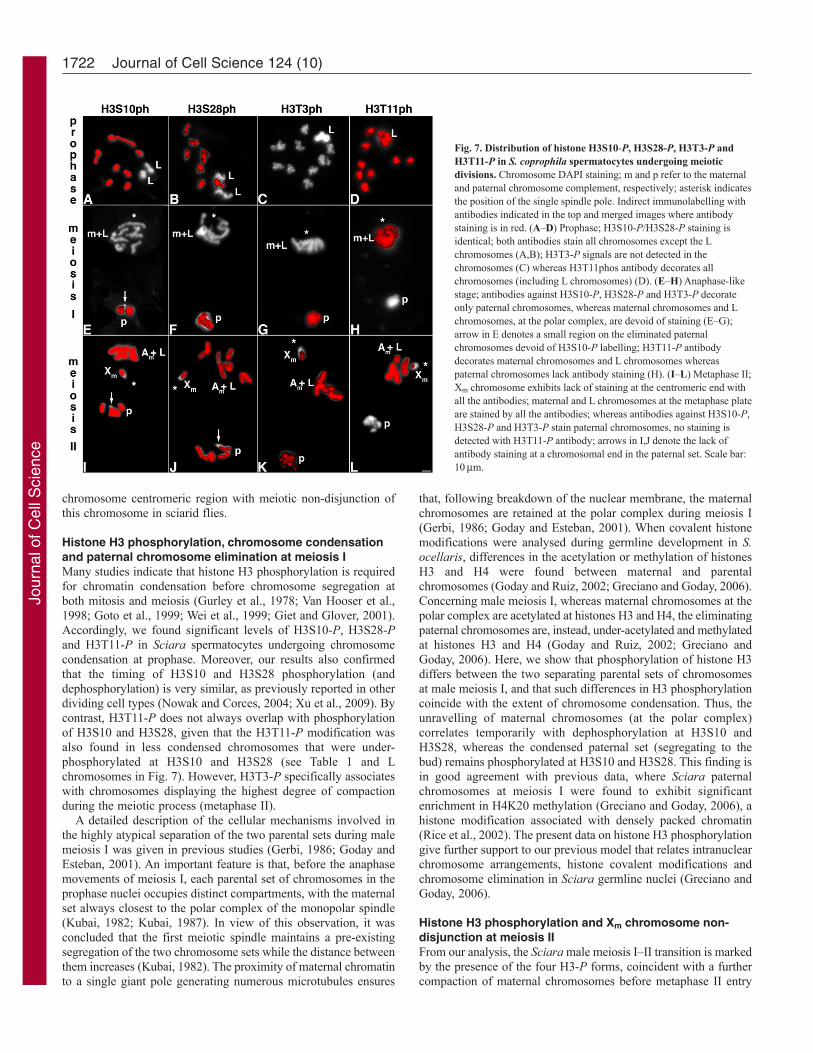
Fig. 7. Distribution of histone H3S10-P, H3S28-P, H3T3-P andH3T11-P in S. coprophila spermatocytes undergoing meioticdivisions. Chromosome DAPI staining; m and p refer to the maternaland paternal chromosome complement, respectively; asterisk indicatesthe position of the single spindle pole. Indirect immunolabelling withantibodies indicated in the top and merged images where antibodystaining is in red. (A–D) Prophase; H3S10-P/H3S28-P staining isidentical; both antibodies stain all chromosomes except the Lchromosomes (A,B); H3T3-P signals are not detected in thechromosomes (C) whereas H3T11phos antibody decorates allchromosomes (including L chromosomes) (D). (E–H) Anaphase-likestage; antibodies against H3S10-P, H3S28-P and H3T3-P decorateonly paternal chromosomes, whereas maternal chromosomes and Lchromosomes, at the polar complex, are devoid of staining (E–G);arrow in E denotes a small region on the eliminated paternalchromosomes devoid of H3S10-P labelling; H3T11-P antibodydecorates maternal chromosomes and L chromosomes whereaspaternal chromosomes lack antibody staining (H). (I–L) Metaphase II;Xm chromosome exhibits lack of staining at the centromeric end withall the antibodies; maternal and L chromosomes at the metaphase plateare stained by all the antibodies; whereas antibodies against H3S10-P,H3S28-P and H3T3-P stain paternal chromosomes, no staining isdetected with H3T11-P antibody; arrows in I,J denote the lack ofantibody staining at a chromosomal end in the paternal set. Scale bar:10m.
Jour
nal o
f Cel
l Sci
ence

(see Table 1). Therefore, H3 phosphorylation of maternalchromosomes congregated at the polar complex site precedes themigration of the autosomes to align in a metaphase II plate. Aparticularly good example is seen in Fig. 4C� where S. ocellarismaternal chromosomes stained with the anti-H3S28-P antibodyline up with the centromeres oriented towards the single pole in theprimary spermatocyte. Taking into consideration all ourobservations, we conclude that it is at this particular ‘pre-metaphasestage’ when local deficiencies in H3 phosphorylation are establishedat the centromeric end of the Xm chromosome. In view of ourresults, it seems very reasonable to question whether such H3phosphorylation modifications are responsible for the persistentassociation of Xm chromosome with the polar complexmicrotubules throughout meiosis II. That is, if the Xm chromosomefailure to align on the metaphase II plate and, consequently, toundergo chromatid disjunction depends on the deficiency in H3phosphorylation at the centromeric region. We believe that this isso, and we discuss this further below in light of recent data thatdemonstrate the crucial role of histone H3 phosphorylation in
centromere function during mammalian cell division (Kelly et al.,2010; Wang et al., 2010).
We have demonstrated that during anaphase II histone H3dephosphorylation at Ser10 and Ser28 begins at the centromericregions of the chromosomes and extends along the chromosomearms. This is consistent with observations of the distribution ofH3S10-P in anaphase chromosomes from Drosophila syncytialmitosis, where dephosphorylation events originate at the centromereregion (Su et al., 1998). In this respect, similar observations wereobtained in early Sciara embryonic somatic divisions (not shown),as well as in neuroblast cells (Fig. 9), suggesting that a gradualH3S10 and H3S28 dephosphorylation pattern might be a generaloccurrence. Consistently, the un-disjoined Xm chromosome lackingH3-P at the centromeric end, also exhibits non-uniformdephosphorylation of H3S10 and H3S28 that, in this chromosome,initiates at the proximal region of chromosome arms. So it seemsthat, at least in the case of the Xm chromosome, phosphorylationor dephosphorylation of histone H3 does not necessarily begin atthe centromere site to extend then along the chromosome arms.However, from our observations in Sciara spermatocytes atanaphase II, a gradual loss of H3T3 and H3T11 phosphorylationalong the chromosome arms could not be discerned. It appears thathistone H3T3 phosphorylation is dramatically reduced at the onsetof anaphase in all chromosomes, whereas phosphorylated H3T11persists until very late anaphase (see Table 1). Taken as awhole, the results support differential phosphorylation and
1723H3 phosphorylation in Sciara male meiosis
Fig. 8. Distribution of rDNA in a S. coprophila spermatocyte atmethaphase II. (A)Chromosome DAPI staining. (A�)rDNA in situhybridisation (green) and H3S28-P immunostaining (red). rDNA sequenceslocate to the centromeric regions of Xm and Xp chromosomes; both Xchromosomes are devoid of H3S28-P signals at the rDNA-chromosomal tip(see arrows in A�); note that the centromeric regions of the rest of thechromosomes are strongly labelled by the antibody; arrows indicate thecentromere position in the paternal chromosomes (chromosome II, III and IV).Scale bar: 10m.
Fig. 9. Distribution of phosphorylated histone H3 at Ser10 in mitoticchromosomes from S. ocellaris neuroblasts. Metaphase (A) and anaphase(B) chromosome DAPI staining. (A�,B�) Indirect immunolabelling withH3S10-P antibody and merged images where antibody staining is in red. Allchromosomes at metaphase are similarly stained by the antibody (A�); middle-anaphase showing a reduction of the antibody staining to the chromosomedistal regions. Scale bar: 10m.
Table 1. Summary of histone H3 phosphorylation distribution during the first and second male meiotic divisions in Sciara
Meiosis IMeiosis II
Prophase Anaphase-like Meiosis I–II Early Latem p m p transition Metaphase anaphase anaphase
Am,Xm Ap,Xp Am,Xm Ap Xp Am Xm Am Xm Am Xm Am Xm
H3S10-P + + – + +/– + +/– + +/– + +/– – –H3S28-P + + – + +/– + +/– + +/– + +/– – –H3T3-P – – – + +/– + +/– + +/– – – – –H3T11-P + + + – – + +/– + +/– + +/– + +/–
m, maternal chromosomes; p, paternal chromosomes; Am, maternal autosomes; Xm, maternal X chromosome; Ap, paternal autosomes; Xp, paternal Xchromosome; +/– denotes antibody staining for a large part of the chromosome except at the centromeric end. Paternal chromosomes are only shown in meiosis Iwhen they are eliminated. L chromosomes (not included in table) behave identically to maternal chromosomes from the initiation of the first meiotic segregationprocess.
Jour
nal o
f Cel
l Sci
ence

dephosphorylation timing between histone H3-P forms along themeiotic process in Sciara. Importantly, all the H3-P forms firstarise at the meiosis I–II transition, and remain until the onset ofanaphase II.
Histone H3 phosphorylation and Xm centromereinactivationThe kinase Aurora B phosphorylates H3S10 and H3S28 and bothH3-P modifications are implicated in chromatin compaction, whichin turn, is required for chromosome congression and properchromosome segregation during mitosis and meiosis (Gurley et al.,1978; Wei et al., 1998; Wei et al., 1999; Goto et al., 1999; Hsu etal., 2000; Giet and Glover, 2001) (reviewed by Nowak and Corces,2004). Aurora B is a component of the CPC (chromosomalpassenger complex) that also contains INCENP, Survivin andBorealin/Dasra (Giet and Glover, 2001; Ruchaud et al., 2007). Atthe beginning of the M phase, the CPC associates with condensingchromatin, accumulates at the inner centromere at metaphase andrelocates onto the central spindle at anaphase (Giet and Glover,2001; Carmena and Earnshaw, 2003). Importantly, the CPC controlschromatin-dependent spindle assembly and processes at thecentromere (Ruchaud et al., 2007). Depletion of Aurora B kinasein Drosophila S2 cells greatly reduces the levels of H3S10phosphorylation and produces important alterations in thecondensation of mitotic chromosome (Giet and Glover, 2001). Oneof the roles of Aurora B, moreover, is to ensure chromosome bi-orientation at metaphase and to correct mono-oriented attachmentsto the spindle (Shannon and Salmon, 2002). Taking this informationinto consideration and given that at metaphase II, the mono-oriented Xm chromosome is clearly under-phosphorylated forH3S10 and H3S28 at the centromere region, it seems reasonableto assume that this region in the Xm chromosome is deficient forAurora B activity and that it is most probably less condensed thana regular centromeric region.
Phosphorylation of H3T3 is dependent on Haspin, a conservedkinase that functions in mitosis and has homologues in all the mainphyla, including fungi, animals and plants (Dai et al., 2005; Higgins,2010). Haspin, localises predominantly to chromosomes, andphosphorylates histone H3T3 during mitosis at the chromosomearms and at the inner centromeres between the regions delineatedby the centromere-specific histone CENP-A (Polioudaki et al.,2004; Dai et al., 2005; Dai et al., 2006; Higgins, 2010).Interestingly, Haspin RNAi in mammalian cells causes partialmetaphase figures with numerous misaligned chromosomes, wheremany of them become trapped near the spindle poles (Dai et al.,2005). Such chromosomes, as demonstrated using anti-centromereantibodies, are constituted by mono-oriented sister chromatid pairs(Dai et al., 2005). It is noteworthy that the Sciara Xm chromosomeat metaphase II displays an identical cytological phenotype to thatdescribed for misaligned chromosomes in Haspin-depleted cells.Considering that the centromeric region of Xm chromosome lacksH3T3-P, it is possible to assume that, similarly to Aurora B, thecentromere of the Xm chromosome is deficient in Haspin kinaseactivity. This could explain the misalignment of Xm chromosomeat metaphase II and its permanent attachment to the spindle poleuntil the end of anaphase II.
Recently, the essential functional role of Haspin during mitoticdivision has been further investigated in mammalian cells (Kellyet al., 2010; Wang et al., 2010). From such studies, it emerged thatHaspin is required for the accumulation of CPC at the centromeres,and that the CPC subunit Survivin binds directly to phosphorylated
H3T3 (Wang et al., 2010). Most important, H3T3-P is recognisedby an evolutionarily conserved binding pocket in the BIR domainof Survivin (Kelly et al., 2010). As concluded, H3T3-P positionsthe CPC to the centromeres to regulate Aurora-B-selected targetsand the interaction between H3T3-P and CPC is mediated bySurvivin (Kelly et al., 2010; Wang et al., 2010). Taking intoconsideration these data and our results, it is highly predictablethat the mere lack of H3T3-P in the centromeric region of the Xm
chromosome (at the meiosis I–II transition) prevents the CPCrecruitment and thus leads to a non-functional centromere.
The CE and histone H3 phosphorylation in the XchromosomeEarly work in sciarid flies identified a cis-acting locus, the CE,which regulates X-centromere activity during S. coprophila malemeiosis and is contained in the heterochromatin proximal to the Xcentromere (Crouse, 1960; Crouse, 1977; Crouse, 1979; Gerbi,1986). Although the molecular nature of the CE is still unknown,it has been considered that it is capable of modifying normalcentromere function of the X chromosome during male meiosis II(Gerbi, 1986). The present results support our view that the CEmodifies the Xm centromeric function, causing Xm chromosomenon-disjunction, by inhibiting global H3 phosphorylation at thecentromeric chromatin. Therefore, it seems that the CE governs theinability of the chromatin at this particular X chromosome regionto become H3 phosphorylated, unlike the rest of the chromosome.Consistently, we found that H3 phosphorylation was also inhibitedat the centromeric region of the paternal X chromosome, whichtogether with the paternal autosomes, is eliminated at meiosis I inboth Sciara species. In this regard, the deficiency in H3phosphorylation at the Xp centromeric region does not affect theXp chromosome segregation modality at meiosis I, in either S.ocellaris or S. coprophila. This interesting observation gives furthersupport to the conclusion that the presence of organisedkinetochores in paternal chromosomes seems not to be necessaryfor their regular elimination at meiosis I (Goday and Esteban,2001).
An understanding of the organisation of the heterochromaticsequences conforming the CE and the mechanisms established toinhibit chromatin H3 phosphorylation in cis are important questionsthat remain to be answered.
Materials and MethodsFly cultureS. ocellaris and S. coprophila were raised at 20°C as described elsewhere (Rieffeland Crouse, 1966).
FixationSciara prepupae were dissected in 15 mM Tris-HCl pH 7.4, 60 mM KCl, 15 mMNaCl, 0.5 mM espermidine, 0.1% Triton X-100 solution to remove the testes. Foreach experiment with a different H3-P antibody at least ten testes were processed.The testes were immediately fixed with 1% paraformaldehyde, 0.1% Triton X-100in PBS for 3 minutes. They were then incubated in 50% acetic acid, 1%paraformaldehyde for 1 minute and squashed. Slides were frozen in liquid N2 toremove coverslips, postfixed in 3.7% formaldehyde for 10 minutes and extensivelywashed in PBS. For mitotic neuroblasts analysis brains were removed from thirdinstar larvae and fixed as above.
Immunostaining and microscopyAll slides were washed in PBS (3�10 minutes), incubated in PBS containing 1%Triton X-100 (10 minutes) and in PBS with 3% BSA and 0.1% Tween for 1 hour atroom temperature. The primary antibodies were rabbit polyclonal anti-H3S10-P,anti-H3S28-P, anti-H3T3-P, anti-H3T11-P (Upstate Biotechnology) diluted 1:40 to1:100; mouse monoclonal anti--tubulin (Amersham) diluted 1:200. Secondaryantibodies were FITC- and Cy3-conjugated anti-rabbit (Southern Biotechnology)and Rhd-conjugated anti-mouse Ig (Dakoppats). Secondary antibodies were diluted1:50 for FITC-conjugated antibody and Rhd-conjugated anti-mouse and 1:800 for
1724 Journal of Cell Science 124 (10)
Jour
nal o
f Cel
l Sci
ence

CY3-conjugated antibody. Primary antibody incubation was at 4°C overnight.Secondary antibody incubation was at room temperature for at least 1 hour. DNAwas visualised with 4�6-diamino-2-phenylindole (DAPI) staining (0.1 mg/ml; 3minutes) and preparations mounted in anti-fading solution. Observations were madeunder epifluorescence optics with a Zeiss axiophot microscope equipped with aLeica CCD camera. Digital images were processed using the AdobePhotoshop PSsoftware.
In situ hybridisationFor fluorescence in situ hybridisation (FISH), the probe pDm238 (Tautz et al., 1988)containing a complete rDNA cistron of Drosophila melanogaster was used. Theprobe was labelled with digoxigenin using the DIG-Nick Translation Mix (RocheDiagnostic), according to the manufacturer’s instructions. Slides previously incubatedwith H3-P antibodies were then processed for in situ hybridisation as describedelsewhere (Goday et al., 2006).
We are grateful to A. Villasante for critical comments andsuggestions. We thank J. Haber for critical reading of the manuscript.This work was supported by grant BFU2008-02947-C02-02/BMC toC.G. and FPI BES-2006-12492 to M.C.E.
ReferencesAbbott, A. G. and Gerbi, S. A. (1981). Spermatogenesis in Sciara coprophila. II.
Precoucious chromosome orientation in meiosis II. Chromosoma 83, 19-27.Carmena, M. and Earnshaw, W. C. (2003). The cellular geography of aurora kinases.
Nat. Rev. Mol. Cell Biol. 4, 842-854.Crouse, H. V. (1943). Translocations in Sciara; their bearing on chromosome behavior and
sex determination. Missouri Univ. Res. Bull. 379, 1-75.Crouse, H. V. (1960). The controlling element in sex chromosome behavior in Sciara.
Genetics 45, 1429-1443.Crouse, H. V. (1977). X heterochromatin subdivision and cytogenetic analysis in Sciara
coprophila (Diptera, Sciaridae). I. Centromere localization. Chromosoma 63, 39-55.Crouse, H. V. (1979). X heterochromatin subdivision and cytogenetic analysis in S.
coprophila (Diptera, Sciaridae). II. The controlling element. Chromosoma 74, 219-239.Crouse, H. V., Gerbi, S. A., Liang, C. M., Magnus, L. and Mercer, I. M. (1977).
Localization of ribosomal DNA within the proximal X heterochromatin of Sciaracoprophila (Diptera, Sciaridae). Chromosoma 64, 305-318.
Dai, J., Sultan, S., Taylor, S. S. and Higgins, J. M. (2005). The kinase haspin is requiredfor mitotic histone H3 Thr 3 phosphorylation and normal metaphase chromosomealignment. Genes Dev. 19, 472-488.
Dai, J., Sullivan, B. A. and Higgins, J. M. (2006). Regulation of mitotic chromosomecohesion by Haspin and Aurora B. Dev. Cell 11, 741-750.
Esteban, M. R., Campos, M. C. C., Perondini, A. L. P. and Goday, C. (1997). Role ofmicrotubules and microtubule organizing centers on meiotic chromosome eliminationin Sciara ocellaris. J. Cell Sci. 110, 721-730.
Fuge, H. (1994). Unorthodox male meiosis in Trichoshia pubescens (Sciaridae)-chromosome elimination involves polar organelle degeneration and monocentric spindlesin first and second division. J. Cell Sci. 107, 299-312.
Gerbi, S. A. (1986). Unusual chromosomes movements in sciarid flies. In Germ-Line-Soma Differentiation. Results and Problems of Cell Diferentiation, Vol. 13 (ed. W.Hennig), pp. 71-104. New York: Springer.
Gerbi, S. A. and Crouse, H. V. (1976). Further studies on the ribosomal cistrons of Sciaracoprophila (Diptera). Genetics 83, 81-90.
Giet, R. and Glover, D. M. (2001). Drosophila aurora B kinase is required for histone H3phosphorylation and condensin recruitment during chromosome condensation and toorganize the central spindle during cytokinesis. J. Cell Biol. 152, 669-682.
Goday, C. and Esteban, M. R. (2001). Chromosome elimination in sciarid flies. BioEssays23, 242-250.
Goday, C. and Ruiz, M. F. (2002). Differential acetylation of histones H3 and H4 inpaternal and maternal germline chromosomes during development of sciarid flies. J.Cell Sci. 115, 4765-4775.
Goday, C., Selivon, D., Perondini, A. L., Greciano, P. G. and Ruiz, M. F. (2006).Cytological characterization of sex chromosomes and ribosomal DNA location inAnastrepha species (Diptera, Tephritidae). Cytogenet. Genome Res. 114, 70-76.
Goto, H., Tomono, Y., Ajiro, K., Kosako, H., Fujita, M., Sakurai, M., Okawa, K.,Iwamatsu, A., Okigaki, T., Takahashi, T. et al. (1999). Identification of a novelphosphorylation site on histone H3 coupled with mitotic chromosome condensation. J.Biol. Chem. 274, 25543-25549.
Greciano, P. G. and Goday, C. (2006). Methylation of histone H3 at Lys4 differs betweenpaternal and maternal chromosomes in Sciara ocellaris germline development. J. CellSci. 119, 4667-4677.
Gurley, L. R., D’Anna, J. A., Barham, S. S., Deaven, L. L. and Tobey, R. A. (1978).Histone phosphorylation and chromatin structure during mitosis in chinese hamstercells. Eur. J. Biochem. 84, 1-15.
Higgins, J. M. (2010). Haspin: a newly discovered regulator of mitotic chromosomebehavior. Chromosoma 119, 137-147.
Hsu, J.-Y., Sun, Z.-W., Li, X., Reuben, M., Tatchell, K., Bishop, D. K., Grushcow, J.M., Brame, C. J., Caldwell, J. A., Hunt, D. F. et al. (2000). Mitotic phosphorylationof histone H3 is governed by Ipl1/aurora kinase and Glc7/PP1 phosphatase in buddingyeast and nematodes. Cell 102, 279-291.
Kelly, A. E., Ghenoiu, C., Xue, J. Z., Zierhut, C., Kimur, H. and Funabiki, H. (2010).Survivin reads phosphorylated histone H3 threonine 3 to activate the mitotic kinaseAurora B. Science 330, 235-239.
Krishnamoorthy, T., Chen, X., Govin, J., Cheung, W. L., Dorsey, J., Schindler, K.,Winter, E., Allis, C. D., Guacci, V., Khochbin, S. et al. (2006). Phosphorylation ofhistone H4 Ser1 regulates sporulation in yeast and is conserved in fly and mousespermatogenesis. Genes Dev. 20, 2580-2592.
Kubai, D. F. (1982). Meiosis in Sciara coprophila: structure of the spindle and chromosomebehavior during the first meiotic division. J. Cell Biol. 93, 655-669.
Kubai, D. F. (1987). Nonrandom chromosome arrangements in germ line nuclei of Sciaracoprophila males: the basis for nonrandom chromosome segregation on the meiosis Ispindle. J. Cell Biol. 105, 2433-2446.
Metz, C. W. (1925). Chromosome behavior in Sciara (Diptera). Anat. Rec. 31, 346-347.Metz, C. W. (1926a). An apparent case of monocentric mitosis in Sciara (Diptera).
Science 63, 190-191.Metz, C. W. (1926b). Genetic evidence of a selective segregation of chromosomes in
Sciara (Diptera). Proc. Natl. Acad. Sci. USA 12, 690-692.Metz, C. W. (1933). Monocentric mitosis with segregation of chromosomes in Sciara and
its bearing on the mechanisms of mitosis. Biol. Bull. 64, 334-347.Nowak, S. J. and Corces, V. G. (2004). Phosphorylation of histone H3: a balancing act
between chromosome condensation and transcriptional activation. Trends Genet. 20,214-220.
Polioudaki, H., Markaki, Y., Kourmouli, N., Dialynas, G., Theodoropoulos, P. A. andSingh, P. B. (2004). Mitotic phosphorylation of histone H3 at threonine 3. FEBS Lett.560, 39-44.
Preuss, U., Landsberg, G. and Scheidtmann, K. H. (2003). Novel mitosis-specificphosphorylation of histone H3 at Thr11 mediated by Dlk/ZIP kinase. Nucleic Acids Res.31, 878-885.
Rice, J. C., Nishioka, K., Sarma, K., Steward, R., Reinberg, D. and Allis, C. D. (2002).Mitotic-specific methylation of histone H4 Lys 20 follows increased PR-Set7 expressionand its localization to mitotic chromosomes. Genes Dev. 16, 2225-2230.
Rieffel, S. M. and Crouse, H. V. (1966). The elimination and differentiation ofchromosomes in the germ line of Sciara. Chromosoma 19, 231-276.
Ruchaud, S., Carmena, M. and Earnshaw, W. C. (2007). Chromosomal passengers:conducting cell division. Nat. Rev. Mol. Cell Biol. 8, 798-812.
Shannon, K. B. and Salmon, E. D. (2002). Chromosome dynamics: new light on AuroraB kinase function. Curr. Biol. 12, 458-460.
Smith-Stocking, H. (1936). Genetic studies on selective segregation of chromosomes inSciara coprophila Lintner. Genetics 21, 421-443.
Su, T. T., Sprenger, F., DiGregorio, P. J., Campbell, S. D. and O’Farrell, P. H. (1998).Exit from mitosis in Drosophila syncytial embryos requires proteolysis and cyclindegradation, and is associated with localized dephosphorylation. Genes Dev. 15, 1493-1503.
Tautz, D., Hancock, J., Webb, D., Tautz, C. and Dover, G. (1988). Complete sequencesof the rRNA genes of Drosophila melanogaster. Mol. Biol. Evol. 5, 366-376.
Van Hooser, A., Goodrich, D. W., Allis, D. C., Brinkley, B. R. and Mancini, M. A.(1998). Histone H3 phosphorylation is required for the initiation, but not maintenance,of mammalian chromosome condensation. J. Cell Sci. 111, 3497-3506.
Wang, F., Dai, J., Daum, J. R., Niedzialkowska, E., Banerjee, B., Stukenberg, P. T.,Gorbsky, G. J. and Higgins, J. M. G. (2010). Histone H3 Thr-3 phosphorylation byHaspin positions Aurora B at centromeres in mitosis. Science 330, 231-235.
Wei, Y., Mizzen, C. A., Cook, R. G., Gorovsky, M. A. and Allis, C. D. (1998).Phosphorylation of histone H3 at serine 10 is correlated with chromosome condensationduring mitosis and meiosis in Tetrahymena. Proc. Natl. Acad. Sci. USA 95, 7480-7484.
Wei, Y., Yu, L., Bowen, J., Gorovsky, M. A. and Allis, C. D. (1999). Phosphorylation ofhistone H3 is required for proper chromosome condensation and segregation. Cell 97,99-109.
Xu, D., Bai, J., Duan, Q., Costa, M. and Dai, W. (2009). Covalent modifications ofhistones during mitosis and meiosis. Cell Cycle 8, 3688-3694.
1725H3 phosphorylation in Sciara male meiosis
Jour
nal o
f Cel
l Sci
ence
Related Documents