Histone H1 limits DNA methylation in Neurospora crassa Michael Seymour ⇤,2 , Lexiang Ji †,2 , Alex M. Santos ‡ , Masayuki Kamei ⇤ , Takahiko Sasaki ⇤ , Evelina Y. Basenko ⇤ , Robert J. Schmitz § , Xiaoyu Zhang ‡ and Zachary A. Lewis ⇤,1 ⇤ Department of Microbiology, University of Georgia, Athens, GA 30602, † Institute of Bioinformatics, University of Georgia, Athens, GA 30602, ‡ Department of Plant Biology, University of Georgia, Athens, GA 30602, § Department of Genetics, University of Georgia, Athens, GA 30602 ABSTRACT Histone H1 variants, known as linker histones, are essential chromatin components in higher eukaryotes, yet compared to the core histones relatively little is known about their in vivo functions. The filamentous fungus Neurospora crassa encodes a single H1 protein that is not essential for viability. To investigate the role of N. crassa H1, we constructed a functional FLAG-tagged H1 fusion protein and performed genomic and molecular analyses. Cell fractionation experiments showed that H1-3XFLAG is a chromatin binding protein. Chromatin-immunoprecipitation combined with sequencing (ChIP-seq) revealed that H1- 3XFLAG is globally enriched throughout the genome with a subtle preference for promoters of expressed genes. In mammals, the stochiometery of H1 impacts nucleosome repeat length. To determine if H1 impacts nucleosome occupancy or nucleosome positioning in N. crassa, we performed micrococcal nuclease digestion in wildtype and the DhH1 strain followed by sequencing (MNase-seq). Deletion of hH1 did not significantly impact nucleosome positioning or nucleosome occupancy. Analysis of DNA methylation by whole-genome bisulfite sequencing (MethylC-seq) revealed a modest but global increase in DNA methylation in the DhH1 mutant. Together, these data suggest that H1 acts as a non-specific chromatin binding protein that can limit accessibility of the DNA methylation machinery in N. crassa. KEYWORDS chromatin histone H1 Nucleosome positioning DNA methylation INTRODUCTION In eukaryotes, packaging of genomic DNA into chromatin is es- sential for genome function. The most basic unit of the chromatin fiber is the nucleosome core particle (NCP), made up of ⇠146 bp of DNA wrapped around a core of histones H3, H4, H2A and H2B (Kornberg and Thomas 1974; Olins and Olins 1974; Luger et al. 1997). In addition to the core histones many organisms encode one or more H1 proteins, also known as linker histones. H1 proteins are evolutionarily unrelated to the core histones and are characterized by a central winged helix domain, or globular domain, flanked by unstructured N- and C-termini (Cerf et al. 1993; Ramakrishnan et al. 1993; Kasinsky et al. 2001). Early studies showed that animal H1 proteins bind outside of the NCP (Baldwin et al. 1975; Shaw et al. 1976) and can protect an additional 20bp of DNA from nuclease Copyright © 2016 Zachary A. Lewis et al. Manuscript compiled: Wednesday 27 th April, 2016% 1 Department of Microbiology, University of Georgia, Athens, GA 30602 [email protected] 2 These authors contributed equally to this work digestion (Whitlock and Simpson 1976; Noll and Kornberg 1977). Subsequent studies revealed that H1 binds near the NCP dyad axis and can interact with DNA as it enters and exits the NCP (recently reviewed in Bednar et al. 2016). Although the interactions between H1 and the NCP have been extensively investigated, H1’s roles in the cell remain poorly understood. In vivo studies of mammalian H1 are complicated by the exis- tence of 11 H1 variants that appear to be partially redundant (Pan and Fan 2015). Deletion of single H1 variants failed to produce significant phenotypes in mice (Fan et al. 2001), but mice lacking three H1 variants are inviable (Fan et al. 2003) and triple-knockout embryonic stem cells (ESCs) are unable to differentiate (Zhang et al. 2012a). These and other data suggest that animal H1 variants cooperate to perform critical functions, influencing gene regulation (Fan et al. 2005; Li et al. 2012b; Zhang et al. 2012b), establishment and/or maintenance of chromatin modification patterns (Li et al. 2012b; Zhang et al. 2012a; Yang et al. 2013; Lu et al. 2013) and forma- tion of higher order chromatin structures (Fan et al. 2005; Geeven et al. 2015). Volume X | April 2016 | 1 INVESTIGATIONS G3: Genes|Genomes|Genetics Early Online, published on May 6, 2016 as doi:10.1534/g3.116.028324 © The Author(s) 2013. Published by the Genetics Society of America.

Welcome message from author
This document is posted to help you gain knowledge. Please leave a comment to let me know what you think about it! Share it to your friends and learn new things together.
Transcript
Histone H1 limits DNA methylation in NeurosporacrassaMichael Seymour⇤,2, Lexiang Ji†,2, Alex M. Santos‡, Masayuki Kamei⇤, Takahiko Sasaki⇤, Evelina Y. Basenko⇤, Robert J.Schmitz§, Xiaoyu Zhang‡ and Zachary A. Lewis⇤,1
⇤Department of Microbiology, University of Georgia, Athens, GA 30602, †Institute of Bioinformatics, University of Georgia, Athens, GA 30602, ‡Department ofPlant Biology, University of Georgia, Athens, GA 30602, §Department of Genetics, University of Georgia, Athens, GA 30602
ABSTRACT Histone H1 variants, known as linker histones, are essential chromatin components in highereukaryotes, yet compared to the core histones relatively little is known about their in vivo functions. Thefilamentous fungus Neurospora crassa encodes a single H1 protein that is not essential for viability. Toinvestigate the role of N. crassa H1, we constructed a functional FLAG-tagged H1 fusion protein and performedgenomic and molecular analyses. Cell fractionation experiments showed that H1-3XFLAG is a chromatinbinding protein. Chromatin-immunoprecipitation combined with sequencing (ChIP-seq) revealed that H1-3XFLAG is globally enriched throughout the genome with a subtle preference for promoters of expressedgenes. In mammals, the stochiometery of H1 impacts nucleosome repeat length. To determine if H1 impactsnucleosome occupancy or nucleosome positioning in N. crassa, we performed micrococcal nuclease digestionin wildtype and the DhH1 strain followed by sequencing (MNase-seq). Deletion of hH1 did not significantlyimpact nucleosome positioning or nucleosome occupancy. Analysis of DNA methylation by whole-genomebisulfite sequencing (MethylC-seq) revealed a modest but global increase in DNA methylation in the DhH1mutant. Together, these data suggest that H1 acts as a non-specific chromatin binding protein that can limitaccessibility of the DNA methylation machinery in N. crassa.
KEYWORDS
chromatinhistone H1NucleosomepositioningDNA methylation
INTRODUCTION
In eukaryotes, packaging of genomic DNA into chromatin is es-sential for genome function. The most basic unit of the chromatinfiber is the nucleosome core particle (NCP), made up of ⇠146 bpof DNA wrapped around a core of histones H3, H4, H2A and H2B(Kornberg and Thomas 1974; Olins and Olins 1974; Luger et al.1997). In addition to the core histones many organisms encode oneor more H1 proteins, also known as linker histones. H1 proteins areevolutionarily unrelated to the core histones and are characterizedby a central winged helix domain, or globular domain, flanked byunstructured N- and C-termini (Cerf et al. 1993; Ramakrishnan et al.1993; Kasinsky et al. 2001). Early studies showed that animal H1proteins bind outside of the NCP (Baldwin et al. 1975; Shaw et al.1976) and can protect an additional 20bp of DNA from nuclease
Copyright © 2016 Zachary A. Lewis et al.Manuscript compiled: Wednesday 27th April, 2016%1Department of Microbiology, University of Georgia, Athens, GA 30602 [email protected] These authors contributed equally to this work
digestion (Whitlock and Simpson 1976; Noll and Kornberg 1977).Subsequent studies revealed that H1 binds near the NCP dyad axisand can interact with DNA as it enters and exits the NCP (recentlyreviewed in Bednar et al. 2016). Although the interactions betweenH1 and the NCP have been extensively investigated, H1’s roles inthe cell remain poorly understood.
In vivo studies of mammalian H1 are complicated by the exis-tence of 11 H1 variants that appear to be partially redundant (Panand Fan 2015). Deletion of single H1 variants failed to producesignificant phenotypes in mice (Fan et al. 2001), but mice lackingthree H1 variants are inviable (Fan et al. 2003) and triple-knockoutembryonic stem cells (ESCs) are unable to differentiate (Zhanget al. 2012a). These and other data suggest that animal H1 variantscooperate to perform critical functions, influencing gene regulation(Fan et al. 2005; Li et al. 2012b; Zhang et al. 2012b), establishmentand/or maintenance of chromatin modification patterns (Li et al.2012b; Zhang et al. 2012a; Yang et al. 2013; Lu et al. 2013) and forma-tion of higher order chromatin structures (Fan et al. 2005; Geevenet al. 2015).
Volume X | April 2016 | 1
INVESTIGATIONS
G3: Genes|Genomes|Genetics Early Online, published on May 6, 2016 as doi:10.1534/g3.116.028324
© The Author(s) 2013. Published by the Genetics Society of America.

Less is known about the functions of H1 in other groups oforganisms, but genetic studies have been carried out in a handfulof microbial model systems. H1 is not essential for viability inthe single-celled Saccharomyces cerevisiae (Patterton et al. 1998) orTetrahymena thermophila (Shen et al. 1995). Similarly, H1-deficientmutants are viable in several filamentous fungi including Neu-rospora crassa (Folco et al. 2003), Aspergillus nidulans (Ramón et al.2000), and Ascobolus immersus (Barra et al. 2000). The yeast H1homolog Hho1p suppresses homologous recombination (Downset al. 2003; Li et al. 2008), impacts ribosomal RNA processing (Levyet al. 2008), and influences chromatin compaction during station-ary phase (Schäfer et al. 2008). In T. thermophila, H1 is requiredfor normal chromatin compaction in macronuclei and influencesexpression of a small number of genes (Shen et al. 1995; Shen andGorovsky 1996). It is important to note that both yeast Hho1pand T. thermophila H1 have atypical protein structures. The yeastprotein contains two globular domains, whereas the T. thermophilaprotein lacks a globular domain altogether. Thus, it is not clear ifthese proteins are functionally analogous to H1 in other organisms.
The filamentous fungi N. crassa, A. nidulans, and A. immersusencode H1 proteins with a canonical tripartite structure, raisingthe possibility that these genetic systems can be used to gain in-sights into H1 function in plants and animals. In N. crassa, an hH1-deficient strain displayed reduced growth and H1 was requiredfor repression of the cfp gene in the presence of ethanol (Folcoet al. 2003). In A. immersus, H1 gene silencing led to increasednuclease accessibility and increased DNA methylation (Barra et al.2000). In contrast, deletion of hhoA in A. nidulans failed to producea phenotype (Ramón et al. 2000). In general, the functions of H1in filamentous fungi remain poorly understood. Moreover, it isnot clear if H1 plays similar roles in fungal and animal cells. Inthe present study, we utilized molecular and genomic approachesto investigate the functions of H1 in the model fungus N. crassa.We confirmed that N. crassa H1 is a chromatin component in vivo,and we found that H1 is not a major determinant of nucleosomepositioning, nucleosome repeat length, or nucleosome occupancy.We report that an H1 fusion protein exhibits enhanced enrichmentat nucleosome free regions and is depleted from coding sequencesof expressed genes in a ChIP-seq assay. We also show that loss ofH1 causes a global increase in DNA methylation.
MATERIALS AND METHODS
Strains, Growth Media, and Molecular AnalysesAll Neurospora strains used in this study are listed in Table 1.Knockout strains of hH1 were generated by the N. crassa geneknockout consortium (Colot et al. 2006) and obtained from theFungal Genetics Stock Center (McCluskey et al. 2010). Neurosporacultures were grown at 32° in Vogel’s minimal medium (VMM) +1.5% sucrose (Davis et al. 1970). Crosses were performed on modi-fied synthetic cross medium at 25° (Davis et al. 1970). For platingassays, N. crassa conidia were plated on VMM with 2.0% sorbose,0.5% fructose, and 0.5% glucose. When relevant, plates included200 µg/mL hygromycin or 400 µg/mL basta (Pall 1993). N. crassatransformation (Margolin et al. 1997), DNA isolation (Pomraninget al. 2009), protein isolation and western blotting (Honda andSelker 2008) were performed as previously described. 3X-FLAGknock-in constructs were made by introduction of linear DNA frag-ments constructed by overlapping PCR using described plasmidvectors (Honda and Selker 2009) and N. crassa fragments generatedusing the following primers: H1 CDS FP 5’- GAG GTC GAC GGTATC GAT AAG CTT AT ATC CAC CGA CAA CAT GTT CGA CTC- 3’; H1 CDS RP 5’ - CCT CCG CCT CCG CCT CCG CCG CCT
CCG CCT GCC TTC TCG GCA GCG GGC TC -3’; H1 UTR FP 5’- TGC TAT ACG AAG TTA TGG ATC CGA GCT CGA CTC GTTCCT TTG GGA TGA T - 3’; H1 UTR RP 5’ - ACC GCG GTG GCGGCC GCT CTA GAA CTA GTT CAT CAA ACC AAA TTC TCGG - 3’. To separate soluble nuclear proteins from the chromatinfraction, cultures were grown overnight and cells were collected,ground in liquid nitrogen, and resuspended in 1 mL of low salt ex-traction buffer (50 mM HEPES-KOH pH 7.5, 150 mM NaCl, 2 mMEDTA, plus protease inhibitor tablets (cat # 11836153001; Roche,Indianapolis, IN). Extracts were centrifuged at 14,000 rpm andthe supernatant containing soluble proteins was saved. The pelletwas resuspended in 1 mL of high salt extraction buffer (50 mMHEPES-KOH pH 7.5, 600 mM NaCl, 2 mM EDTA, plus proteaseinhibitor tablets (cat # 11836153001; Roche, Indianapolis, IN) andsubjected to sonication. Extracts were centrifuged at 14,000 rpmin a microfuge and the supernatant was saved as the chromatinfraction. Both fractions were analyzed by western blotting usinganti-FLAG antibodies (cat # F1804; Sigma-Aldrich) and anti-H3antibodies (cat # 06-755 Millipore).
n Table 1 Strains Used in this study
Lab Strain # Description Source
S1 FGSC #4200 Wild-type mat a
(McCluskey et al.2010)
S2 FGSC #2489 Wild-type mat A
(McCluskey et al.2010)
S240 hH1-3xflag-Hph+mus-52::Basta
this study
FGSC #12224 DhH1::Hph+ mat A (Colot et al. 2006)
S123 Ddim-2::Hph+ matA
(Kouzminova andSelker 2001)
S94 hH1RIP2 (Folco et al. 2003)
DNA Sequencing and Data AnalysisChIP-seq For chromatin immunoprecipitation (ChIP) experiments,5 x106 conidia/ml were inoculated into 50 mL of VMM and in-cubated at 32° for 5 hours. Cells were harvested and ChIP wasperformed as previously described (Sasaki et al. 2014) using anti-FLAG antibodies (cat # F1804; Sigma-Aldrich) or antibodies tothe unphosphorylated C-terminal repeat of RNA polymerase II(8WG16; cat# MMS-126R; Covance). Two biological replicates wereperformed for each experiment. Libraries were prepared using theTruSeq ChIP sample prep kit (Illumina, cat # IP-202-1012) accord-ing to manufacture instructions with the following modification.Library amplification was performed using only 4 cycles of PCR toreduce biased enrichment of GC-rich DNA (Ji et al. 2014). Librarieswere sequenced at the University of Georgia Genomics Facility onan Illumina NextSeq 500 instrument. Reads were aligned to version12 of the N. crassa genome (Refseq Accession # GCF_000182925.2;Galagan et al. 2003) using the Burrows-Wheeler Aligner (BWA ver-sion 0.7.10) (Li and Durbin 2009). To determine if H1-3XFLAG wasenriched over background, coverage was normalized to mitochon-drial DNA as follows. We used BEDtools (version 2.25.0) ’coverage’to calculate read coverage for 1000bp windows across the genome(Quinlan and Hall 2010). We then used BEDtools ’map’ to calcu-late the median coverage for mitochondrial DNA. The coveragefor each 1000 base pair window was then divided by the median
2 | Zachary A. Lewis et al.

coverage for mitochondrial DNA. As a positive control, data froma previously published ChIP experiment for methylated lysine-9of H3 was analyzed (Accession #SRX550120; Sasaki et al. 2014).
The Hypergeometric Optimization of Motif EnRichment(HOMER verson 4.7.2) software package (Heinz et al. 2010) wasused to generate metaplots and heatmaps of enrichment data (an-notatePeaks.pl module; using the -hist and -ghist option, respec-tively). We first created a custom HOMER genome annotationfor Neurospora using a fasta file and a GTF file (SupplementaryFile 1) containing the version 12 genome assemblies and annota-tions, respectively (Galagan et al. 2003). All plots were centeredon transcriptional start sites or transcriptional termination sitesand a window size of 10 bp was specified for all histograms (-hist10). HOMER was also used to construct metaplots of expression-ranked gene groups using the -list option. Genes were assignedinto expression-ranked groups by by expression level determinedby RNA-seq (see below). RPKM values for each quintile groupwere: Q1 (12927.9 - 36.8 ); Q2 (36.8 – 13.8); Q3 (13.8 – 4.9); Q4 (4.9 –0.33); Q5 (0.33 – 0). Thus, most genes in expression group 5 weresilent or rarely expressed.
MNase digestion For micrococcal nuclease (MNase) experiments,5 x106 conidia/mL were inoculated into 50 mL of VMM and in-cubated at 32° for 5 hours. The cell suspension was transferredto a 50 mL conical and centrifuged at 1000 x g for five minutes topellet germinated conidia. Cell pellets were washed with 10 mLPhosphate Buffered Saline (PBS) (Sambrook et al. 1989) and thenresuspended in 10 mL of PBS containing 1% formaldehyde. Thecell suspension was transferred to a 125 mL flask and incubatedwith gentle shaking for 30 minutes at room temperature beforethe crosslinking agent was quenched by addition of 500µL 2.5Mglycine. The cell suspension was transferred to a 50 mL conicaltube and germinating conidia were pelleted by centrifugation for5 minutes at 1000 x g. Cells were washed once in 40 mL of PBSand resuspended in 1 mL of ice-cold PBS. Cells were pelletedby centrifugation for 5 minutes at 5000 x g, and each cell pelletwas resuspended in NPS buffer with calcium chloride (50 mMHEPES-KOH, pH 7.5, 140 mM NaCl, 5 mM MgCl2, 1 mM CaCl2,1% Triton-X 100, 0.1% deoxycholate, 0.5 mM spermidine, 1 mMPMSF plus Roche EDTA-free protease inhibitor tablets [catalog #05892791001] ). Cells were then lysed by gentle sonication using aHeat Systems, Ultrasonics W-380 sonicator with micro tip (Output2.0, Duty Cycle 80% ; 30 1-second pulses). The chromatin fractionwas pelleted by centrifugation for 5 minutes at 14,000 x g. Thesupernatent was discarded and each pellet was resuspended in 1mL of NPS buffer and transferred to a 15 mL conical tube. NPSbuffer with calcium chloride was added to raise the volume to 6mL and the chromatin sample was mixed by pipetting. 700 µLaliquots were transferred to 1.5 mL tubes and 2 units of micrococcalnuclease (cat #2910A; Clontech) were added to each tube. Indi-vidual samples were incubated at 37° for 5, 10, 20, 40, 60 minutes.MNase digestions were stopped by addition of 15µL 0.5M EDTAand 25 µL 4M NaCl and samples were incubated overnight at 65°to reverse cross-links. 6µL RNase A (10mg/ml; Fisher Scientific,cat # BP2529250) was added and samples were incubated for 2hours at 50°. 6µL of 10% SDS and 10µL Proteinase K (10mg/ml;Fisher Scientific, cat # BP1700) was then added and samples wereincubated for 2 hours at 65°. The digested DNA was isolated byphenol-chloroform extraction and precipitated overnight at -20° inethanol and sodium acetate (Sambrook et al. 1989). Digested DNAwas resolved by gel electrophoresis to confirm that digestion wassuccessful.
MNase-seq We constructed sequencing libraries from mono-nucleosomes generated by 20 or 60 minute MNase digestion. Wefirst performed a gel extraction (Qiagen, cat #28706) of mononucle-somal DNA (⇠150bp) and constructed libraries using an IlluminaTruSeq Sample preparation kit (Illumina) according to manufactureinstructions. 50bp paired-end sequencing reads were generatedon a Illumina HiSeq 2500 instrument at the Oregon State Univer-sity genomics core facility. Due to a technical problem duringthe sequencing run, only 44 bp of sequence were obtained forthe read 2 sequence. Sequences were mapped to the N. crassaversion 12 genome assembly (Galagan et al. 2003) using bowtie2(version 2.2.3) (Langmead and Salzberg 2012). To analyze the sizedistributions in wildtype and the hH1 strain, the Picard softwarepackage (http://broadinstitute.github.io/picard) was used to re-move duplicate reads and determine insert size metrics (using’CollectInsertSizeMetrics’). HOMER was used to create metaplotsof MNase data as described above. In all cases, a window size of10bp was used (-hist 10). Metaplots depict only plus strand reads(using the ’-strand +’ option) and thus peaks indicate the left edgeof nucleosomes.
RNA-seq For RNA-seq experiments, 5x10⁶ conidia/mL were in-oculated into 50 mL of VMM containing 2% glucose and grownfor 5 hours at 32°. RNA isolation was performed as described(Bell-Pedersen et al. 1996; Schwerdtfeger and Linden 2001) andstrand-specific RNA-seq libraries were prepared from 5 µg totalRNA. Ribosomal RNAs were depleted using the yeast Ribo-zerokit (cat # MRZY1324 Epicentre) and RNA libraries were generatedwith the Illumina Stranded RNA-seq kit (cat # RS-122-2101). Readswere aligned to version 12 of the N. crassa genome sequence usingTopHat (Trapnell et al. 2009) and expression levels (RPKM) weredetermined using Cufflinks (Trapnell et al. 2012).
MethylC-seq For DNA methylation analysis, conidia were inoc-ulated into 5 mL of VMM and cultures were grown for 48 hoursat 32°. Genomic DNA was isolated using described procedures(Pomraning et al. 2009). MethylC-seq libraries were prepared aspreviously described (Urich et al. 2015). Illumina sequencing wasperformed at the University of Georgia Genomics Facility using anIllumina NextSeq 500 instrument. Sequencing reads were trimmedfor adapters, preprocessed to remove low quality reads and alignedto the N. crassa version 12 genome assembly (Galagan et al. 2003)as described in Schmitz et al. 2013. Mitochondrial DNA sequence(which is fully unmethylated) was used as a control to calculatethe sodium bisulfite reaction non-conversion rate of unmodifiedcytosines. Only cytosine sites with a minimum coverage (set as3) were allowed for subsequent analysis. Binomial test coupledwith Benjamini-Hochberg correction was adopted to determinethe methylation status of each cytosine. Identification of DMRs(Differentially Methylated Regions) was performed as described(Schultz et al. 2015). Methylated regions in wild type were definedpreviously (Basenko et al. 2016). For metaplots, both upstream anddownstream regions were divided into 20 bins each of 50bp inlength for a total 1 Kb in each direction. Methylated regions wereseparated every 5%, for a total of 20 bins. Weighted methylationlevels were computed for each bin as described previously (Schultzet al. 2012).
Data AvailabilityAll strains are listed in Table 1 and available upon request or fromthe Fungal Genetics Stock Center (Manhattan, KS). All sequenc-ing data have been deposited into the NCBI SRA/GEO databases.ChIP-seq, MNase-Seq, and RNA-seq data generated during this
Volume X April 2016 | Genomic analysis of N.crassa H1 | 3

study have been deposited under accession #GSE78157. Controldata from a previously published ChIP-seq experiment for methy-lation of H3 lysine-9 was deposited under accession #SRX550120(Sasaki et al. 2014). MethylC-seq data are deposited under acces-sion #GSE76982 (this study) and #GSE70518 (Basenko et al. 2016).
RESULTS
Construction of an H1-3XFLAG fusion proteinTo investigate the role of H1 in N. crassa cells, we constructedan epitope-tagged version of the protein by introducing codingsequence for a 3X-FLAG tag at the 3’ end of the native hH1 lo-cus (Figure 1A). Primary transformants were crossed to obtain ahomokaryon that was analyzed further. To confirm that the H1-3XFLAG fusion protein is functional, we first compared the growthrate of the hH1::hH1-3xflag-Hph+ strain to wildtype and to an hH1deletion strain obtained from the Neurospora gene knockout con-sortium (Colot et al. 2006). The DhH1 strain displayed a reducedgrowth rate, as reported previously for an H1 loss-of-function al-lele generated by repeat-induced point mutation (Folco et al. 2003).The hH1::hH1-3xflag-Hph+ grew similar to wildtype (Figure 1Band C). We also asked if the H1-3XFLAG protein associates withchromatin. We isolated soluble and chromatin-containing fractions(see Materials and Methods) and performed western blot analysesusing anti-FLAG and anti-H3 antibodies. Western blots probedwith an anti-FLAG antibody revealed a single band. The appar-ent molecular weight was larger than expected based on aminoacid sequence prediction, but the apparent size was consistentwith previous analysis of N. crassa purified by extraction with per-chloric acid (Folco et al. 2003) . We detected H1-3XFLAG in bothsoluble and chromatin fractions with higher levels of H1-3XFLAGobserved in the chromatin fraction (Figure 1D). As expected, H3was exclusively detected in the chromatin fraction. Together, thesedata demonstrate that the H1-3XFLAG construct is functional andthat N. crassa H1 is a component of chromatin.
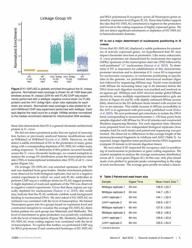
H1 is moderately enriched in promoters and depleted from cod-ing sequences of actively expressed genesTo determine the genome wide distribution of H1-3XFLAG, weperformed chromatin-immunoprecipitation followed by sequenc-ing (ChIP-seq). Inspection of the H1-3XFLAG enrichment datain a genome browser revealed a relatively uniform distributionfor all seven chromosomes. Given that high levels of H1-3XFLAGwere detected in the chromatin fraction by western blotting, wereasoned that the uniform enrichment pattern observed for H1-3XFLAG might reflect global binding of H1 across the genome.To determine if this was the case, we normalized read counts ob-tained for each 1000 base pair window in the nuclear genome tothe median read count obtained for all 1000 base pair windowscovering the mitochondrial genome. This allowed us to calculateenrichment over background because mitochondrial DNA shouldnot be enriched by immunoprecipitation of a nuclear protein. Weplotted normalized enrichment data for all 1000 base pair windowson Linkage Group VII (Figure 2). ChIP-seq experiments using anti-FLAG antibodies led to global enrichment of chromosomal DNAfrom the hH1::hH1-3xflag-Hph+ strain but not from a wildtypenegative control strain. As a positive control, we normalized readcounts obtained in a previously published ChIP-seq experimentperformed with antibodies to H3 methylated at lysine-9 (Sasakiet al. 2014). Results for other chromosomes were comparable. Thisconfirmed that normalization to mitochondrial DNA is an effectivemethod for quantifying enrichment over background. Moreover,
X X
hph+
3xflag
hH1
hH1
hH1-3xflag
Sol Chr
WT
375075
25
20
20
15
α-FLAG
α-H3
MW
Wildtype
A
B
C
ΔhH1
hH1-3Xflag
Sol Chr
25
D
0
10
20
30
20 30 40 50 60
Time (hours)
Dis
tance
(cm
)
WT
hH1−3xflag
ΔhH1
Figure 1 H1-3XFLAG is functional and binds to chromatin.(A) A car-toon illustrating the strategy for introducing a 3x-flag sequence intothe 3’ end of the native hH1 locus by homologous recombination.(B) The linear growth rate of the indicated strains was measuredusing ’race tubes’ for three replicates of each strain. The direction ofgrowth is left to right. (C) Quantification of the linear growth ratedata from panel B as distance (y-axis; cm) versus time (x-axis;hours). The three lines shown for each genotype represent threeindependent race tubes. (D) The soluble (Sol) and the chromatinfractions (Chr) were isolated from wildtype and the hH1-3xflag-Hph+strain and both fractions were analyzed by western blotting usinganti-FLAG and anti-H3 antibodies, as indicated. MW indicates thepositions and sizes in kilodaltons of a prestained protein ladder.
4 | Zachary A. Lewis et al.
0
1
2
3
WT
FLAG
rep1
WT
FLAG
rep2
hH1 3xflag
FLAG
rep1
hH1 3xflag
FLAG
rep2
WT
H3K9me3
Norm
alized E
nri
chm
ent
Linkage Group VII
Figure 2 H1-3XFLAG is globally enriched throughout the N. crassagenome. Normalized read coverage is shown for all 1000 base pairwindows across N. crassa LGVII for anti-FLAG ChIP-seq experi-ments performed with a wildtype negative control strain (no FLAGprotein) and the hH1-3xflag-Hph+ strain (two replicates for eachstrain are shown). Normalized read coverage is also plotted for ananti-H3K9me3 ChIP-seq experiment performed with wildtype. Eachspot depicts the read count for a single 1000bp window normalizedto the median enrichment obtained for mitochondrial DNA windows.
these data demonstrate that H1 is a general chromatin architecturalprotein in N. crassa.
We did not detect prominent peaks that are typical of transcrip-tion factors or previously analyzed histone modifications suchas H3K4me2 or H3K9me3 (Lewis et al. 2009). However, we diddetect a subtle enrichment of H1 in the promoters of many genesalong with a corresponding depletion of H1-3XFLAG within manycoding sequences. To determine if this pattern occurred broadlyacross the N. crassa chromatin landscape, we created metaplots toanalyze the average H1 distribution across the transcriptional startsites (TSS) or transcriptional termination sites (TTS) of all N. crassagenes (Figure 3A).
On average, H1-3XFLAG was enriched upstream of the TSSand was depleted from gene bodies. Similar enrichment patternswere observed for both biological replicates, but not in a negativecontrol experiment in which we used anti-FLAG antibodies toperform ChIP-seq in a wildtype strain (no FLAG-tagged protein).A slight depletion of reads just upstream of the TSS was observedin negative control experiments. Given that these regions are typ-ically depleted for nucleosomes (Sancar et al. 2015), this resultmay indicate that the FLAG antibody exhibits weak backgroundbinding to nucleosomal DNA. We next asked if H1-3XFLAG en-richment was correlated with the level of transcription. We binnedNeurospora genes into five groups based on expression level andconstructed metaplots to visualize the average H1-3XFLAG distri-bution pattern for each group (see Materials and Methods). Thelevel of enrichment in gene promoters was positively correlatedwith the level of transcription (Figure 3B). Similarly, depletion ofH1-3XFLAG from coding regions was correlated with the levelof transcription. To explore this further, we performed ChIP-seqfor RNA polymerase II and constructed heatmaps of H1-3XFLAG
and RNA polymerase II occupancy across all Neurospora genes or-dered by expression level (Figure 3C,D). These data further supportthe idea that H1-3XFLAG enrichment is highest in the promotersand lowest in the coding sequences of highly expressed genes. Wedid not detect significant enrichment or depletion of H1-3XFLAGin heterochromatin domains.
H1 is not a major determinant of nucleosome positioning in N.crassaGiven that H1-3XFLAG displayed a subtle preference for promot-ers of actively expressed genes, we hypothesized that H1 mayimpact chromatin structure at promoters. Like many eukaryotes,N. crassa promoters are characterized by nucleosome free regions(NFRs) upstream of the transcription start site (TSS) followed bywell positioned "+1" nucleosomes (Sancar et al. 2015). To deter-mine if H1 is important for establishment of this characteristicpromoter structure and to determine if N. crassa H1 is importantfor nucleosome occupancy or nuclesome positioning at specificsites in the genome, we performed micrococcal nuclease diges-tion followed by sequencing (MNase-seq). Nuclei were incubatedwith MNase for increasing times (up to 60 minutes) before theDNA from each digestion reaction was purified and resolved onan agarose gel. Wildtype and DhH1 showed similar global MNasesensitivity across multiple experiments (representative gels areshown in Figure 4A and B), with slightly enhanced MNase accessi-bility observed in the H1-deficient strain treated with enzyme forfive or ten minutes. This subtle increase in MNase accessibility inthe DH1 is in agreement with previously published work (Folcoet al. 2003). We next performed gel extractions to isolate the DNAband corresponding to mononucleosomes (⇠150 base pairs) fromsamples digested with MNase for 20 or 60 minutes and constructedIllumina sequencing libraries. For each digestion time, librarieswere prepared from two independent biological replicates (foursamples total for each strain) and paired-end sequencing was per-formed. We observed no differences in the average length of themononucleosomal fragments in wildtype and DhH1 (Table 2). Asexpected, longer digestion times produced shorter DNA fragments(compare 20 minute to 60 minute digestion times).
We next asked if H1 impacted the occupancy and/or position-ing of nucleosomes in promoters or gene coding sequences. Wecreated metaplots to analyze the average nucleosome distributionacross all N. crassa genes (Figure 4C). In this case, only plus strandreads were plotted to generate peaks corresponding to the edgeof each nucleosome. The average gene profile revealed a charac-
n Table 2 Paired-end read insert size
Sample Digest Time Mean Insert Size a
Wildtype replicate 1 20 min 139.9 ±23.7
Wildtype replicate 2 20 min 144.7 ±25.1
DhH1 replicate 1 20 min 144.1 ±22.4
DhH1 replicate 2 20 min 139.6 ±22.7
Wildtype replicate 1 60 min 127.8 ±33.0
Wildtype replicate 2 60 min 126.5 ±28.3
DhH1 replicate 1 60 min 129.8 ±33.9
DhH1 replicate 2 60 min 125.6 ±24.3a ±standard deviation
Volume X April 2016 | Genomic analysis of N.crassa H1 | 5

H1-FLAG
RNA pol II
−1000 −500 0 500 1000
Base coordinates−1000 −500 0 500 1000
Base coordinates
Strain
hH1−3xflag rep1
hH1−3xflag rep2
WT
−1000 −500 0 500 1000
Base coordinates−1000 −500 0 500 1000
Base coordinates
Quintile
Q1
Q2
Q3
Q4
Q5
45
40
35
30
25
20
50
40
30
20
0-250 +250 0-250 +250
A
B
C
TSS TTS
All genes
Expression Quintiles
TSS TTS
0-250 +250 0-250 +250
D
Rela
tive
Enric
hmen
tRe
lativ
e En
richm
ent
Figure 3 H1-3XFLAG is depleted from gene bodies and modestlyenriched in promoters of expressed genes. (A) Metaplots depict theaverage ChIP-seq enrichment pattern across all N. crassa genesfor two replicate FLAG ChIP-seq experiments performed with thehH1-3xflag-Hph+ strain and a wildtype negative control strain (noFLAG-tagged protein). Metaplots are centered on the transcriptionalstart (TSS; left) or the transcriptional termination site (TTS; right).(B) All N. crassa genes were ranked by expression level and splitinto quintile groups. Quintile 1 (Q1) corresponds to genes with thehighest expression level, whereas Q5 corresponds to genes withthe lowest expression. The metaplot depicts the H1-3XFLAG enrich-ment pattern for each expression group across the TSS or TTS. (C)Heatmaps show the distribution of H1-3XFLAG across all N. crassagenes centered on the TSS (left) or TTS (right). Genes are orderedby expression level from highest to lowest. (D) Heatmaps show thedistribution of RNA polymerase II across all N. crassa genes cen-tered on the TSS (left) or TTS (right). Genes are ordered as in C.Data in B-D are from replicate one.
teristic nucleosome free region upstream of the TSS, as reportedpreviously for N. crassa (Sancar et al. 2015). The average nucleo-some position profiles were similar in wildtype and DhH1. BecauseH1 was most enriched in the promoters of highly expressed genes,we next plotted average MNase-seq enrichment profiles for genesgrouped by expression level. Although we detected a subtle in-crease in the size of the NFR from the most expressed genes inthe two 60 minute digestion samples, this difference was not ap-parent in the samples subjected to a 20 minute MNase digestion(Figure S1). The difference was also not apparent in a third in-dependent replicate subjected to single-end Illumina sequencing(Figure S1). Thus, we conclude that H1 is not a major determinantof nucleosome positioning or occupancy in Neurospora. Because itappeared that enrichment of H1-3XFLAG was inversely correlatedwith nucleosome occupancy, we plotted plus strand reads fromreplicate one of the H1-3XFLAG ChIP-seq experiment describedabove to allow direct comparison of nucleosome occupancy andH1-3XFLAG enrichment (Figure 4C - H). This confirmed that thehighest levels of H1 enrichment correspond to sites with the lowestnucleosome occupancy, raising the possibility that H1-3XFLAGbinds to DNA that is free of nucleosomes as well as to nucleosomalDNA in N. crassa. The group of genes including silent or rarelyexpressed genes (Quintile 5) did not exhibit an obvious nucleo-some free region or an ordered array of nucleosomes. This maysuggest that transcription is important for establishment of chro-matin architecture at these genes. However, we can not rule outthe possibility that this result is due to incorrect annotation of theTSS for many genes in this group.
DhH1 exhibits increased DNA methylation in N. crassa hete-rochromatin domains
H1 impacts DNA methylation levels in animals (Fan et al. 2005;Zhang et al. 2012a), plants (Wierzbicki and Jerzmanowski 2005;Zemach et al. 2013; Rea et al. 2012), and the fungus Ascobolus im-mersus (Barra et al. 2000). It was previously reported that H1 didnot impact DNA methylation in N. crassa (Folco et al. 2003); how-ever, Folco and colleagues analyzed only a single methylated re-gion. It remained possible that H1 impacts DNA methylation ina region-specific manner or alternatively, that H1 has subtle im-pact on DNA methylation that may have been overlooked. Weperformed MethylC-seq to analyze DNA methylation across theentire genome at single base-pair resolution. Genomic DNA wasisolated from two replicates each of a wildtype mat A strain, theDhH1 mutant, and a negative control Ddim-2 strain, which lacksDNA methylation altogether (Kouzminova and Selker 2001). Allstrains were grown simultaneously, but the wildtype and Ddim-2data were published previously as part of another study (Basenkoet al. 2016).
To determine if H1 impacts the level or distribution of DNAmethylation in N. crassa, we first plotted DNA methylation levelsacross Linkage Group VII, a 4 Mb chromosome corresponding to⇠10% of the genome. The overall pattern of DNA methylation wassimilar in wildtype and the DhH1 strain. However, we noted thatDNA methylation levels were higher in the DhH1 mutant at mostregions along the chromosome (Figure 5A). We next constructedmetaplots to quantify the average methylation level for all genomicregions that are normally methylated in the wildtype mat A strain(see Materials and Methods). Both DhH1 replicates displayedhigher average DNA methylation levels when compared to thewildtype strain (Figure 5B). To confirm that this wasn’t an artifactdue to differences in strain backgrounds, we compared the level ofmethylation in the DhH1 strain to a wildtype mat a strain and we
6 | Zachary A. Lewis et al.

0.00
0.05
0.10
0.15
0.20
−1000−500 0 500 1000
Base coordinates
Re
lative
En
rich
me
nt
Quintile 1
Sample
WT Rep 1 60 min
WT Rep 2 60 min
ΔhH1 Rep 1 60 min
ΔhH1 Rep 2 60 min
H1-3XFLAG Rep1
0.00
0.05
0.10
0.15
0.20
−1000−500 0 500 1000
Base coordinates
Re
lative
En
rich
me
nt
Quintile 2
0.00
0.05
0.10
0.15
0.20
−1000−500 0 500 1000
Base coordinates
Re
lative
En
rich
me
nt
Quintile 4
0.00
0.05
0.10
0.15
0.20
−1000−500 0 500 1000
Base coordinates
Re
lative
En
rich
me
nt
All Genes
0.00
0.05
0.10
0.15
0.20
−1000−500 0 500 1000
Base coordinates
Re
lative
En
rich
me
nt
Quintile 3
0.00
0.05
0.10
0.15
0.20
−1000−500 0 500 1000
Base coordinates
Re
lative
En
rich
me
nt
Quintile 5
MNase(time)
0 1 5 10 20 40 60
WT
ΔhH1
0 1 5 10 20 40 60
MNase(time)
A
B
D
E G
C F H
L
L
Figure 4 H1 is not a major determinant of nucleosome size or positioning in Neurospora. (A) Wildtype or (B) DhH1 nuclei were isolated andtreated with micrococcal nuclease (MNase) for the indicated times (minutes). DNA was purified, resolved on an agarose gel, and visualizedby staining with Ethidium Bromide. Wildtype and DhH1 display similar digestion kinetics. (C) Metaplots depict the average sequencing depthon the + strand across all N. crassa genes obtained by sequencing mononucleosomal DNA from a 60 minute MNase digest. Plots show datafrom two biological replicates each for wildtype and DhH1. (D - H) All N. crassa genes were ranked by expression level and split into quintilegroups, ranging from highest expression (Quintile 1) to lowest expression (Quintile 5). Metaplots depict the average sequencing coverageon the + strand for each group. Plots show data from two biological replicates each for wildtype and DhH1. The legend in the top right panelcorresponds to plots (C - H). For all plots, the coverage of + strand reads from an H1-3XFLAG ChIP-seq experiment is shown as a dashed line.
analyzed DNA methylation levels in a second hH1 loss of functionstrain in which the hH1 gene was inactivated by repeat-inducedpoint mutation (Folco et al. 2003). In both cases, the level of DNAmethylation was higher in the hH1 mutant strains (Figure 5C,D). Asearch for differentially methylated regions between wildtype andDH1 identified only a single methylated region that was specific tothe DH1 strain (Linkage Group VI; 309590 - 319133). This hyper-methylated sequence corresponds to Sly1-1, a DNA transposonfound in the genomes of some N. crassa isolates (Wang et al. 2015).It is possible that the observed increase in DNA methylation at theSly1-1 locus is due to a site-specific increase in DNA methylation.However, it is also possible that this is actually due to a changein copy number or location of the transposon in the DH1 strainbackground. Additional work is needed to test these possibilities.
Higher methylation levels can result when individual cytosinesare methylated at a higher frequency in a population of nucleior when new cytosines become methylated (or a combination of
both). To determine which of these possibilities was the case inH1-deficient strains, we determined the number of methylatedcytosines in wildtype and DhH1 using a binomial test in combina-tion with multiple testing correction (see Materials and Methods).The number of methylated cytosines increased by ⇠25% in theDH1 strain. We next compared methylation frequency at cytosinesthat were scored as methylated in both strains. This revealed thatshared sites are methylated at higher frequency in nuclei from theDhH1 strain. Thus, loss of H1 leads to a subtle, but global increasein DNA methylation at relics of repeat-induced point mutation.
DISCUSSION
We applied genomic and molecular methods to investigate H1 inthe model fungus N. crassa. We first confirmed that NeurosporaH1 is a chromatin component by constructing a functional epitope-tagged H1 fusion protein and analyzing its localization in cell
Volume X April 2016 | Genomic analysis of N.crassa H1 | 7

0
20
40
50
30
10
Meth
ylatio
n le
vel (
%) wild type
ΔhH1
Δdim-2
0Mb 1Mb 2Mb 3Mb 4Mb
Linkage Group VII
ΔhH1 rep1ΔhH1 rep2wildtype A rep1wildtype A rep2
0
20
30
10
Ave
rage 5
mC
level (%
)
-1kb start stop +1kb -1kb start stop +1kb
(4.5%)808,005
(4.1%)785,051
(5.4%)1,033,481 (5.1%)
970,752
0
4
8
12
wildtype
ΔhH1
Num
ber
of m
C (
x10
5)
ΔhH1 rep1
ΔhH1 rep2
wildtype A rep1
wildtype A rep2
0
5
10
15
Fre
quency
of m
C (
%)
0 10 20 30 40 50 60 70 80 90 100
A
B
E F
Weighted methylated level (%)
0
20
30
10
H1RIP2 rep1H1 RIP2 rep2wildtype A rep1wildtype A rep2
ΔhH1 rep1ΔhH1 rep2wildtype a rep1
0
20
30
10
wildtype a rep2wildtype a rep3
-1kb start stop +1kb
C D
rep1 rep2 rep1 rep2
Figure 5 Increased DNA methylation is observed at most N. crassa heterochromatin domains in H1-deficient strains. (A) The DNA methylationlevel (weighted DNA methylation level (%); see Materials and Methods) is shown for 10 Kb windows across N. crassa Linkage Group VII forwildtype, the DhH1 mutant, and the Ddim-2 strain, which lacks all DNA methylation. (B-D) The metaplots show the average DNA methylationlevel across all previously identified wildtype methylated domains: (B) the wildtype mat A strain and the DhH1 strain; (C) the wildtype mat astrain and the DhH1 strain; and (D) the wildtype mat A strain and the hH1RIP2 strain. Data for at least two independent biological replicatesof each strain are shown. (E) More cytosines are methylated in DhH1. The plot shows the total number of methylated cytosines (see Materi-als and Methods) identified in each wildtype and DhH1 replicate. (F) The level of methylation at individual cytosines is higher in DhH1. Thepercentage of total shared methylated sites (y-axis) versus the level of methylation at individual cytosines (x-axis) is shown.
fractionation and ChIP-seq experiments. This revealed that H1 isglobally enriched throughout the genome, occupying both hete-rochromatic and euchromatic regions. Surprisingly, we observedthe highest enrichment of H1-3XFLAG in promoters of expressedgenes. Although the overall amplitude of enrichment was low,preferential enrichment of H1-3XFLAG was clearly correlated withexpression level. These results may indicate that H1 plays differentroles in fungi and animals. In mouse embryonic stem cells (ESCs),H1c and H1d are depleted from strong promoters and enhancers(Cao et al. 2013). It should be noted that H1 protein levels are signif-
icantly reduced in ESCs compared to differentiated cells (Fan et al.2003), which might explain why H1c and H1d are not detected atESC promoters. This seems unlikely, however, as ChIP-seq analyisof H1b from ESCs and from differentiated cells revealed that thisprotein was similarly enriched at repressed genes and depletedfrom active promoters (Li et al. 2012a). On the other hand, giventhat mammals encode multiple H1 variants it is possible that cer-tain H1 variants will bind preferentially to nucleosome-depletedpromoter regions, similar to the case for Neurospora H1. Indeed,analysis of H1 variants by DamID showed that H1.1 exhibits a dis-
8 | Zachary A. Lewis et al.
tinct localization compared to other histones and is not excludedfrom promoters like the other somatic H1 variants (Izzo et al. 2013).Similarly, the more divergent H1.X variant was recently found tobe enriched at regions with high RNA polymerase II occupancy(Mayor et al. 2015), suggesting that these H1 variants may playroles within active chromatin in higher eukaryotes. Additionalwork to define the specific roles of linker histones in fungal andanimals systems is needed to determine their specific functions atopen chromatin.
One possibility is that N. crassa H1 functions more like animalHMG (High Mobility group) proteins. The chromatin architec-tural protein HMGD1 is enriched in active chromatin, for example(Nalabothula et al. 2014), similar to the results obtained for H1-3XFLAG here. It is important to note that we can not rule out thepossibility that the subtle promoter enrichment observed for H1-3XFLAG is an experimental artifact. This could be the case if thebasic H1 protein interacts non-specifically with DNA sequencesoriginating from open chromatin during the ChIP procedure. Itwas shown that certain highly expressed loci in yeast are proneto artifactual enrichment in ChIP-chip experiments for reasonsthat are not understood (Teytelman et al. 2013). However, we donot think this is the case here because the non-specific enrichmentpatterns observed in yeast are qualitatively different from the en-richment patterns we observe for H1-3XFLAG. Thus, it is likelythat the patterns observed here reflect the in vivo occupancy ofH1. It will be interesting to determine if promoter regions interactwith a sub-population of H1 in which specific residues are post-translationally modified. It will also be interesting to determineif depletion of H1 from coding sequences of expressed genes de-pends on post-translational modification. This seems likely giventhat H1 proteins in plants and animals are extensively modified,much like the core histones (Harshman et al. 2013; Bednar et al.2016; Annalisa and Robert 2015; Kotlinski et al. 2016). Moreover, itwas shown that phosphorylation of H1 was linked to transcriptionby RNA polymerase I and II in humans (Zheng et al. 2010). Animportant goal for future studies will be to determine if N. crassaH1 is post-translationally modified and to determine if differentforms of H1 exhibit distinct localization and/or distinct functions.
We found here that deletion of hH1 from N. crassa did not sub-stantially alter global MNase accessibility or nucleosome posi-tioning. Moreover, H1 did not impact the size of protected DNAfragments produced by MNase treatment or the distance betweenadjacent nucleosomes in genes. These results point to clear dif-ferences in how H1 interacts with chromatin in N. crassa and inanimals. Indeed, H1 depletion caused increased MNase accessibil-ity, altered nucleosome spacing lengths, and reduced chromatincompaction in H1 triple-knockout ESCs (Fan et al. 2005). Our re-sults could indicate that N. crassa H1 does not bind to the linkerDNA and the dyad axis of the NCP as demonstrated for animal H1(Bednar et al. 2016). Another possibility is that N. crassa H1 is moredynamic than H1 in higher eukaryotes. FRAP studies revealed thatmammalian H1 variants exist in high-mobility and low-mobilitypools and that the half-life of fluorescence recovery after H1-GFPbleaching was significantly shorter than for the core histones (Mis-teli et al. 2000; Lever et al. 2000). Interactions between H1 and theNCP may be even more transient in N. crassa such that H1 doesnot interfere with MNase digestion even though it interacts withthe same region of the nucleosome protected by animal H1.
We found increased DNA methylation in H1-deficient cells of N.crassa. H1 affects DNA methylation in both A. thaliana and animalcells, but the relationship between H1 and DNA methylation isdifferent in these systems. In animals, H1 variants promote repres-
sive modifications, including DNA methylation in mammals (Yanget al. 2013) and H3K9me2 in both mammals and Drosophila (Liet al. 2012a; Lu et al. 2013). The observation that H1-deficient cellsexhibit hypermethylation demonstrates that N. crassa H1 is not re-quired to promote DNA methylation or H3K9 methylation, whichdirects DNA methylation in Neurospora (Tamaru and Selker 2001).Similar hypermethylation was reported in A. immersus (Barra et al.2000). Moreover, the DNA methylation phenotypes of N. crassaand A. immersus are reminiscent of A. thaliana H1 depletion lines,where a global increase in DNA methylation was observed in het-erochromatin domains along with loss of DNA methylation ineuchromatic transposon sequences (Wierzbicki and Jerzmanowski2005; Zemach et al. 2013). These results indicate that H1 can limitDNA methylation in plants and fungi. Indeed, depletion of A.thaliana H1 rescued the reduced DNA methylation phenotype ofddm1 plants, leading to the conclusion that DDM1 promotes DNAmethyaltion by removal of H1. The Neurospora LSH/DDM1 ho-molog MUS-30 does not impact DNA methylation levels, thereforewe predict that an DhH1 Dmus-30 double mutant would resemblethe DhH1 single mutant strain (Basenko et al. 2016). Taken together,the data from fungi, plants, and animals may indicate that H1evolved a new function to promote heterochromatic modificationsin the animal lineage.
It is possible that N. crassa H1 limits access of the DNA methyl-transferase DIM-2 or the H3K9 MTase DIM-5KMT1. A recent studyshowed that binding of H1 to the NCP limited the dynamics andmodifiability of the H3 tail in vitro (Stützer et al. 2016), consistentwith this possibility. In addition, increased accessibility of the DIM-2 DNA methyltransferase was linked to hypermethylation in theN. crassa Histone Deacetylase-1-deficient strain (Honda et al. 2012).On the other hand, in A. thaliana H1 was required for imprintingof the MEDEA locus, which involves active removal of methyl cy-tosine bases by DNA glycosylases (Rea et al. 2012). A mechanismfor DNA demethylation has not been described in N. crassa, but itis possible that H1 promotes removal of methylated cytosines inheterochromatin domains. Additional studies are needed to under-stand exactly how H1 impacts DNA methylation levels in N. crassa.Overall, this work adds to the diverse set of phenotypes that havebeen reported following depletion of H1 in plants, animals, andfungi. Future work to investigate H1 in fungal systems is likelyto yield new insights into the evolution and the functions of thisimportant group of proteins.
ACKNOWLEDGMENTS
We would like to thank Cameron Prybol for technical contributionsto the project. This work was funded by a grant from the AmericanCancer Society to ZAL (RSG-14-184-01-DMC), a grant from theNational Institutes of Health to RJS (R00GM100000), and a grantto XZ from the National Science Foundation (#0960425).
LITERATURE CITED
Annalisa, I. and S. Robert, 2015 The role of linker histone H1 mod-ifications in the regulation of gene expression and chromatindynamics. Biochimica et biophysica acta 1859: 486–495.
Baldwin, J. P., P. G. Boseley, E. M. Bradbury, and K. Ibel, 1975 Thesubunit structure of the eukaryotic chromosome. Nature 253:245–249.
Barra, J. L., L. Rhounim, J. L. Rossignol, and G. Faugeron, 2000Histone h1 is dispensable for methylation-associated gene si-lencing in ascobolus immersus and essential for long life span.Molecular and cellular biology 20: 61–69.
Volume X April 2016 | Genomic analysis of N.crassa H1 | 9

Basenko, E. Y., M. Kamei, L. Ji, R. J. Schmitz, and Z. A. Lewis, 2016The lsh/ddm1 homolog mus-30 is required for genome stability,but not for dna methylation in neurospora crassa. PLoS genetics12: e1005790.
Bednar, J., A. Hamiche, and S. Dimitrov, 2016 H1-nucleosomeinteractions and their functional implications. Biochimica et bio-physica acta 1859: 436–43.
Bell-Pedersen, D., J. A. Y. C. Dunlap, and J. J. Loros, 1996 Distinctcis-acting elements mediate clock, light, and developmentalregulation of the neurospora crassa eas (ccg-2) gene. Mol CellBiol 16: 513–521.
Cao, K., N. Lailler, Y. Zhang, A. Kumar, K. Uppal, Z. Liu, E. K. Lee,H. Wu, M. Medrzycki, C. Pan, P.-Y. Ho, G. P. Cooper, X. Dong,C. Bock, E. E. Bouhassira, and Y. Fan, 2013 High-resolutionmapping of h1 linker histone variants in embryonic stem cells.PLoS genetics 9: e1003417.
Cerf, C., G. Lippens, S. Muyldermans, A. Segers, V. Ramakrishnan,S. J. Wodak, K. Hallenga, and L. Wyns, 1993 Homo- and het-eronuclear two-dimensional nmr studies of the globular domainof histone h1: sequential assignment and secondary structure.Biochemistry 32: 11345–11351.
Colot, H. V., G. Park, G. E. Turner, C. Ringelberg, C. M. Crew,L. Litvinkova, R. L. Weiss, K. A. Borkovich, and J. C. Dun-lap, 2006 A high-throughput gene knockout procedure for Neu-rospora reveals functions for multiple transcription factors. Pro-ceedings of the National Academy of Sciences of the UnitedStates of America 103: 10352–7.
Davis, R. H., M. B. Lawless, and L. A. Port, 1970 Arginaselessneurospora: genetics, physiology, and polyamine synthesis. JBacteriol 102: 299–305.
Downs, J. a., E. Kosmidou, A. Morgan, and S. P. Jackson, 2003 Sup-pression of homologous recombination by the saccharomycescerevisiae linker histone. Molecular cell 11: 1685–92.
Fan, Y., T. Nikitina, E. M. Morin-Kensicki, J. Zhao, T. R. Magnuson,C. L. Woodcock, and A. I. Skoultchi, 2003 H1 linker histones areessential for mouse development and affect nucleosome spacingin vivo. Mol Cell Biol 23: 4559–4572.
Fan, Y., T. Nikitina, J. Zhao, T. J. Fleury, R. Bhattacharyya, E. E.Bouhassira, A. Stein, C. L. Woodcock, and A. I. Skoultchi, 2005Histone h1 depletion in mammals alters global chromatin struc-ture but causes specific changes in gene regulation. Cell 123:1199–212.
Fan, Y., A. Sirotkin, R. G. Russell, J. Ayala, and A. I. Skoultchi, 2001Individual somatic h1 subtypes are dispensable for mouse de-velopment even in mice lacking the h1(0) replacement subtype.Molecular and cellular biology 21: 7933–43.
Folco, H. D., M. Freitag, A. Ramon, E. D. Temporini, M. E. Al-varez, I. Garcia, C. Scazzocchio, E. U. Selker, and A. L. Rosa,2003 Histone h1 is required for proper regulation of pyruvatedecarboxylase gene expression in neurospora crassa. Eukaryoticcell 2: 341–350.
Galagan, J. E., S. E. Calvo, K. a. Borkovich, E. U. Selker, N. D.Read, D. Jaffe, W. FitzHugh, L.-J. Ma, S. Smirnov, S. Purcell,B. Rehman, T. Elkins, R. Engels, S. Wang, C. B. Nielsen, J. But-ler, M. Endrizzi, D. Qui, P. Ianakiev, D. Bell-Pedersen, M. A.Nelson, M. Werner-Washburne, C. P. Selitrennikoff, J. a. Kin-sey, E. L. Braun, A. Zelter, U. Schulte, G. O. Kothe, G. Jedd,W. Mewes, C. Staben, E. Marcotte, D. Greenberg, A. Roy, K. Foley,J. Naylor, N. Stange-Thomann, R. Barrett, S. Gnerre, M. Kamal,M. Kamvysselis, E. Mauceli, C. Bielke, S. Rudd, D. Frishman,S. Krystofova, C. Rasmussen, R. L. Metzenberg, D. D. Perkins,S. Kroken, C. Cogoni, G. Macino, D. Catcheside, W. Li, R. J.
Pratt, S. a. Osmani, C. P. C. DeSouza, L. Glass, M. J. Orbach,J. A. Berglund, R. Voelker, O. Yarden, M. Plamann, S. Seiler,J. Dunlap, A. Radford, R. Aramayo, D. O. Natvig, L. a. Alex,G. Mannhaupt, D. J. Ebbole, M. Freitag, I. Paulsen, M. S. Sachs,E. S. Lander, C. Nusbaum, and B. Birren, 2003 The genome se-quence of the filamentous fungus neurospora crassa. Nature 422:859–68.
Geeven, G., Y. Zhu, B. Kim, B. Bartholdy, S.-M. Yang, T. Macfar-lan, W. Gifford, S. Pfaff, M. Verstegen, H. Pinto, M. Vermunt,M. Creyghton, P. Wijchers, J. Stamatoyannopoulos, A. Skoultchi,and W. de Laat, 2015 Local compartment changes and regulatorylandscape alterations in histone h1-depleted cells. Genome Biol16: 289.
Harshman, S. W., N. L. Young, M. R. Parthun, and M. A. Freitas,2013 H1 histones: current perspectives and challenges. Nucleicacids research 41: 9593–609.
Heinz, S., C. Benner, N. Spann, E. Bertolino, Y. C. Lin, P. Laslo,J. X. Cheng, C. Murre, H. Singh, and C. K. Glass, 2010 Sim-ple combinations of lineage-determining transcription factorsprime cis-regulatory elements required for macrophage and bcell identities. Mol Cell 38: 576–589.
Honda, S., Z. a. Lewis, K. Shimada, W. Fischle, R. Sack, and E. U.Selker, 2012 Heterochromatin protein 1 forms distinct complexesto direct histone deacetylation and DNA methylation. NatureStructural & Molecular Biology 19: 471–477.
Honda, S. and E. U. Selker, 2008 Direct interaction between dnamethyltransferase dim-2 and hp1 is required for dna methylationin neurospora crassa. Molecular and cellular biology 28: 6044–55.
Honda, S. and E. U. Selker, 2009 Tools for fungal proteomics: mul-tifunctional neurospora vectors for gene replacement, proteinexpression and protein purification. Genetics 182: 11–23.
Izzo, A., K. Kamieniarz-Gdula, F. Ramírez, N. Noureen, J. Kind,T. Manke, B. vanSteensel, and R. Schneider, 2013 The genomiclandscape of the somatic linker histone subtypes h1.1 to h1.5 inhuman cells. Cell Reports 3: 2142–2154.
Ji, L., T. Sasaki, X. Sun, P. Ma, Z. A. Lewis, and R. J. Schmitz, 2014Methylated dna is over-represented in whole-genome bisulfitesequencing data. Front Genet 5: 341.
Kasinsky, H. E., J. D. Lewis, J. B. Dacks, and J. Ausió, 2001 Originof h1 linker histones. The FASEB journal 15: 34–42.
Kornberg, R. D. and J. O. Thomas, 1974 Chromatin structure;oligomers of the histones. Science 184: 865–868.
Kotlinski, M., K. Rutowicz, L. Knizewski, A. Palusinski, J. Oledzki,A. Fogtman, T. Rubel, M. Koblowska, M. Dadlez, K. Ginalski,and A. Jerzmanowski, 2016 Histone h1 variants in arabidopsisare subject to numerous post-translational modifications, bothconserved and previously unknown in histones, suggesting com-plex functions of h1 in plants. PloS one 11: e0147908.
Kouzminova, E. and E. U. Selker, 2001 Dim-2 encodes a dnamethyltransferase responsible for all known cytosine methy-lation in neurospora. EMBO Journal 20: 4309–4323.
Langmead, B. and S. L. Salzberg, 2012 Fast gapped-read alignmentwith bowtie 2. Nat Methods 9: 357–359.
Lever, M. a., J. P. Th’ng, X. Sun, and M. J. Hendzel, 2000 Rapidexchange of histone h1.1 on chromatin in living human cells.Nature 408: 873–876.
Levy, A., M. Eyal, G. Hershkovits, M. Salmon-Divon, M. Klutstein,and D. J. Katcoff, 2008 Yeast linker histone hho1p is required forefficient rna polymerase i processivity and transcriptional silenc-ing at the ribosomal dna. Proceedings of the National Academyof Sciences of the United States of America 105: 11703–8.
10 | Zachary A. Lewis et al.

Lewis, Z. a., S. Honda, T. K. Khlafallah, J. K. Jeffress, M. Freitag,F. Mohn, D. Schübeler, and E. U. Selker, 2009 Relics of repeat-induced point mutation direct heterochromatin formation inneurospora crassa. Genome research 19: 427–37.
Li, C., J. E. Mueller, M. Elfline, and M. Bryk, 2008 Linker histone h1represses recombination at the ribosomal dna locus in the bud-ding yeast saccharomyces cerevisiae. Molecular microbiology67: 906–19.
Li, H. and R. Durbin, 2009 Fast and accurate short read alignmentwith burrows-wheeler transform. Bioinformatics 25: 1754–1760.
Li, J.-Y., M. Patterson, H. K. A. Mikkola, W. E. Lowry, and S. K.Kurdistani, 2012a Dynamic distribution of linker histone h1.5 incellular differentiation. PLoS genetics 8: e1002879.
Li, X., K. Liu, F. Li, J. Wang, H. Huang, J. Wu, and Y. Shi, 2012bStructure of c-terminal tandem brct repeats of rtt107 proteinreveals critical role in interaction with phosphorylated histoneh2a during dna damage repair. J Biol Chem 287: 9137–9146.
Lu, X., S. N. Wontakal, H. Kavi, B. J. Kim, P. M. Guzzardo, A. V.Emelyanov, N. Xu, G. J. Hannon, J. Zavadil, D. V. Fyodorov, andA. I. Skoultchi, 2013 Drosophila h1 regulates the genetic activityof heterochromatin by recruitment of su(var)3-9. Science (NewYork, N.Y.) 340: 78–81.
Luger, K., a. W. Mäder, R. K. Richmond, D. F. Sargent, and T. J.Richmond, 1997 Crystal structure of the nucleosome core particleat 2.8 a resolution. Nature 389: 251–260.
Margolin, B. S., M. Freitag, and E. U. Selker, 1997 Improved plas-mids for gene targeting at the his-3 locus of neurospora crassaby electroporation. Fungal Genetics Newsletter 44: 34–36.
Mayor, R., A. Izquierdo-Bouldstridge, L. Millán-Ariño, A. Bustillos,C. Sampaio, N. Luque, and A. Jordan, 2015 Genome distribu-tion of replication-independent histone H1 variants shows H1.0associated with nucleolar domains and H1X associated withRNA polymerase II-enriched regions. The Journal of biologicalchemistry 290: 7474–91.
McCluskey, K., A. Wiest, and M. Plamann, 2010 The fungal geneticsstock center: a repository for 50 years of fungal genetics research.J Biosci 35: 119–126.
Misteli, T., a. Gunjan, R. Hock, M. Bustin, and D. T. Brown, 2000 Dy-namic binding of histone h1 to chromatin in living cells. Nature408: 877–881.
Nalabothula, N., G. McVicker, J. Maiorano, R. Martin, J. K.Pritchard, and Y. N. Fondufe-Mittendorf, 2014 The chromatinarchitectural proteins hmgd1 and h1 bind reciprocally and haveopposite effects on chromatin structure and gene regulation.BMC genomics 15: 92.
Noll, M. and R. D. Kornberg, 1977 Action of micrococcal nucleaseon chromatin and the location of histone h1. Journal of molecularbiology 109: 393–404.
Olins, A. L. and D. E. Olins, 1974 Spheroid chromatin units (vbodies). Science (New York, N.Y.) 183: 330–332.
Pall, M. L., 1993 The use of ignite (basta; glufosinate; phos-phinothricin) to select transformants of bar-containing plasmidsin neurospora crassa. Fungal Genetics Newsletter 40: 58.
Pan, C. and Y. Fan, 2015 Role of H1 linker histones in mammaliandevelopment and stem cell differentiation. Biochimica et Bio-physica Acta (BBA) - Gene Regulatory Mechanisms 1859: 496–509.
Patterton, H. G., C. C. Landel, D. Landsman, C. L. Peterson, andR. T. Simpson, 1998 The biochemical and phenotypic characteri-zation of hho1p, the putative linker histone h1 of saccharomycescerevisiae. Journal of Biological Chemistry 273: 7268–7276.
Pomraning, K. R., K. M. Smith, and M. Freitag, 2009 Genome-
wide high throughput analysis of dna methylation in eukaryotes.Methods (San Diego, Calif.) 47: 142–50.
Quinlan, A. R. and I. M. Hall, 2010 Bedtools: a flexible suite ofutilities for comparing genomic features. Bioinformatics 26: 841–842.
Ramakrishnan, V., J. T. Finch, V. Graziano, P. L. Lee, and R. M.Sweet, 1993 Crystal structure of globular domain of histone h5and its implications for nucleosome binding. Nature 362: 219–223.
Ramón, a., M. I. Muro-Pastor, C. Scazzocchio, and R. Gonzalez,2000 Deletion of the unique gene encoding a typical histone h1has no apparent phenotype in aspergillus nidulans. Molecularmicrobiology 35: 223–33.
Rea, M., W. Zheng, M. Chen, C. Braud, D. Bhangu, T. N. Rog-nan, and W. Xiao, 2012 Histone h1 affects gene imprinting anddna methylation in arabidopsis. The Plant journal : for cell andmolecular biology 71: 776–86.
Sambrook, J., E. F. Fritsch, and T. Maniatis, 1989 Molecular Cloning:A Laboratory Manual. Cold Spring Harbor laboratory press.
Sancar, C., N. Ha, R. Yilmaz, R. Tesorero, T. Fisher, M. Brunner, andG. Sancar, 2015 Combinatorial control of light induced chromatinremodeling and gene activation in neurospora. PLoS genetics11: e1005105.
Sasaki, T., K. L. Lynch, C. V. Mueller, S. Friedman, M. Freitag,and Z. A. Lewis, 2014 Heterochromatin controls gammah2alocalization in neurospora crassa. Eukaryot Cell 13: 990–1000.
Schäfer, G., C. R. E. McEvoy, and H.-G. Patterton, 2008 The sac-charomyces cerevisiae linker histone hho1p is essential for chro-matin compaction in stationary phase and is displaced by tran-scription. Proceedings Of The National Academy Of Sciences(Of The United States Of America) 105: 14838–14843.
Schmitz, R. J., Y. He, O. Valdes-Lopez, S. M. Khan, T. Joshi, M. A.Urich, J. R. Nery, B. Diers, D. Xu, G. Stacey, and J. R. Ecker, 2013Epigenome-wide inheritance of cytosine methylation variantsin a recombinant inbred population. Genome Res 23: 1663–1674.
Schultz, M. D., Y. He, J. W. Whitaker, M. Hariharan, E. A. Mukamel,D. Leung, N. Rajagopal, J. R. Nery, M. A. Urich, H. Chen, S. Lin,Y. Lin, I. Jung, A. D. Schmitt, S. Selvaraj, B. Ren, T. J. Sejnowski,W. Wang, and J. R. Ecker, 2015 Human body epigenome mapsreveal noncanonical DNA methylation variation. Nature 523:212–6.
Schultz, M. D., R. J. Schmitz, and J. R. Ecker, 2012 ’leveling’ theplaying field for analyses of single-base resolution dna methy-lomes. Trends Genet 28: 583–585.
Schwerdtfeger, C. and H. Linden, 2001 Blue light adaptation anddesensitization of light signal transduction in neurospora crassa.Molecular Microbiology 39: 1080–1087.
Shaw, B. R., T. M. Herman, R. T. Kovacic, G. S. Beaudreau, and K. E.Van Holde, 1976 Analysis of subunit organization in chickenerythrocyte chromatin. Proceedings of the National Academy ofSciences of the United States of America 73: 505–9.
Shen, X. and M. Gorovsky, 1996 Linker histone h1 regulates specificgene expression but not global transcription in vivo. Cell 86: 475–483.
Shen, X., L. Yu, J. W. Weir, and M. a. Gorovsky, 1995 Linker histonesare not essential and affect chromatin condensation in vivo. Cell82: 47–56.
Stützer, A., S. Liokatis, A. Kiesel, D. Schwarzer, R. Sprangers, J. Söd-ing, P. Selenko, and W. Fischle, 2016 Modulations of dna contactsby linker histones and post-translational modifications deter-mine the mobility and modifiability of nucleosomal h3 tails.Molecular Cell 61: 247–259.
Volume X April 2016 | Genomic analysis of N.crassa H1 | 11
Tamaru, H. and E. U. Selker, 2001 A histone h3 methyltransferasecontrols dna methylation in neurospora crassa. Nature 414: 277–83.
Teytelman, L., D. M. Thurtle, J. Rine, and A. van Oudenaarden,2013 Highly expressed loci are vulnerable to misleading chiplocalization of multiple unrelated proteins. Proceedings of theNational Academy of Sciences of the United States of America110: 18602–7.
Trapnell, C., L. Pachter, and S. L. Salzberg, 2009 Tophat: discover-ing splice junctions with rna-seq. Bioinformatics 25: 1105–1111.
Trapnell, C., A. Roberts, L. Goff, G. Pertea, D. Kim, D. R. Kelley,H. Pimentel, S. L. Salzberg, J. L. Rinn, and L. Pachter, 2012Differential gene and transcript expression analysis of rna-seqexperiments with tophat and cufflinks. Nat Protoc 7: 562–578.
Urich, M. A., J. R. Nery, R. Lister, R. J. Schmitz, and J. R. Ecker,2015 Methylc-seq library preparation for base-resolution whole-genome bisulfite sequencing. Nat Protoc 10: 475–483.
Wang, Y., K. M. Smith, J. W. Taylor, M. Freitag, and J. E. Stajich,2015 Endogenous small rna mediates meiotic silencing of a noveldna transposon. G3 (Bethesda, Md.) 5: 1949–60.
Whitlock, J. P. and R. T. Simpson, 1976 Removal of histone h1exposes a fifty base pair dna segment between nucleosomes.Biochemistry 15: 3307–3314.
Wierzbicki, A. T. and A. Jerzmanowski, 2005 Suppression of his-tone h1 genes in arabidopsis results in heritable developmentaldefects and stochastic changes in dna methylation. Genetics 169:997–1008.
Yang, S.-M., B. J. Kim, L. Norwood Toro, and A. I. Skoultchi, 2013H1 linker histone promotes epigenetic silencing by regulatingboth dna methylation and histone h3 methylation. ProceedingsOf The National Academy Of Sciences (Of The United States OfAmerica) 110: 1708–1713.
Zemach, A., M. Y. Kim, P.-H. Hsieh, D. Coleman-Derr, L. Eshed-Williams, K. Thao, S. L. Harmer, and D. Zilberman, 2013 Thearabidopsis nucleosome remodeler ddm1 allows dna methyl-transferases to access h1-containing heterochromatin. Cell 153:193–205.
Zhang, Y., M. Cooke, S. Panjwani, K. Cao, B. Krauth, P. Y. Ho,M. Medrzycki, D. T. Berhe, C. Pan, T. C. McDevitt, and Y. Fan,2012a Histone h1 depletion impairs embryonic stem cell differ-entiation. PLoS Genet 8: e1002691.
Zhang, Y., Z. Liu, M. Medrzycki, K. Cao, and Y. Fan, 2012b Reduc-tion of hox gene expression by histone h1 depletion. PLoS ONE7.
Zheng, Y., S. John, J. J. Pesavento, J. R. Schultz-Norton, R. L. Schiltz,S. Baek, A. M. Nardulli, G. L. Hager, N. L. Kelleher, and C. A.Mizzen, 2010 Histone h1 phosphorylation is associated withtranscription by rna polymerases i and ii. Journal of Cell Biology189: 407–415.
12 | Zachary A. Lewis et al.
Related Documents