Stem Cell Reports Repor t Histone Demethylases KDM4A and KDM4C Regulate Differentiation of Embryonic Stem Cells to Endothelial Cells Liangtang Wu, 1 Kishore K. Wary, 1 Sergei Revskoy, 1 Xiaopei Gao, 1 Kitman Tsang, 1 Yulia A. Komarova, 1 Jalees Rehman, 1,2, * and Asrar B. Malik 1, * 1 Department of Pharmacology 2 Section of Cardiology, Department of Medicine University of Illinois College of Medicine, Chicago, IL 60612, USA *Correspondence: [email protected] (J.R.), [email protected] (A.B.M.) http://dx.doi.org/10.1016/j.stemcr.2015.05.016 This is an open access article under the CC BY-NC-ND license (http://creativecommons.org/licenses/by-nc-nd/4.0/). SUMMARY Understanding epigenetic mechanisms regulating embryonic stem cell (ESC) differentiation to endothelial cells may lead to increased efficiency of generation of vessel wall endothelial cells needed for vascular engineering. Here we demonstrated that the histone deme- thylases KDM4A and KDM4C played an indispensable but independent role in mediating the expression of fetal liver kinase (Flk)1 and VE-cadherin, respectively, and thereby the transition of mouse ESCs (mESCs) to endothelial cells. KDM4A was shown to bind to his- tones associated with the Flk1 promoter and KDM4C to bind to histones associated with the VE-cadherin promoter. KDM4A and KDM4C were also both required for capillary tube formation and vasculogenesis in mice. We observed in zebrafish that KDM4A depletion induced more severe vasculogenesis defects than KDM4C depletion, reflecting the early involvement of KDM4A in specifying endothelial cell fate. These findings together demonstrate the essential role of KDM4A and KDM4C in orchestrating mESC differentiation to endothelial cells through the activation of Flk1 and VE-cadherin promoters, respectively. INTRODUCTION Endothelial cells derived from differentiation of embryonic stem cells (ESCs) or induced pluripotent cells (iPSCs) hold great promise for regenerating blood vessels in diseases associated with endothelial denudation (Kourembanas, 2014; Yoder, 2012). Studies have described differentiation of endothelial cells from ESCs as mirroring embryonic vascular development (Descamps and Emanueli, 2012; Leeper et al., 2010). The growth factors bone morphoge- netic protein-4 (BMP-4), basic fibroblast growth factor (bFGF), and vascular endothelial growth factor (VEGF) are required for specifying the transition of ESCs to the meso- derm and then to the endothelial cell fate as defined by the appearance of Flk1, CD31, and VE-cadherin-positive cells (Li et al., 2007; Park et al., 2013). Epigenetic regulation through histone modifications is a crucial mechanism mediating lineage-specific gene activa- tion of cells undergoing differentiation (Kooistra and Helin, 2012; Kouzarides, 2007; Ohtani et al., 2011). Histone modifications occurring via mono-methylation, di-methyl- ation, and tri-methylation change histone-DNA binding af- finities and the interactions of specific transcription factors with the promoters (Barski et al., 2007; Kouzarides, 2007; Wang et al., 2007). Demethylases may regulate activation of genes responsible for the transition of pluripotent cells to endothelial cells (Kohler et al., 2013; Marcelo et al., 2013). Here we addressed the role of mouse ESC (mESC) histone demethylation in endothelial cell specification. We demonstrated that histone demethylases KDM4A and KDM4C independently induced demethylation at histone H3K9 to activate Flk1 and VE-cadherin expression and thus enabled the differentiation of mESCs to endothelial cells. KDM4A targeted the Flk1 promoter in the early stage of differentiation, whereas KDM4C targeted the VE-cad- herin promoter later to induce a transition to the endothe- lial cell lineage. Removal of histone methylation marks on Flk1 and VE-cadherin promoters by KDM4A and KDM4C, respectively, is therefore an essential mechanism of endo- thelial cell fate specification and vasculogenesis. RESULTS Time Course of Expression of KDM4A and KDM4C during mESC Differentiation to Endothelial Cells Using an established differentiation protocol employing the growth factors BMP-4, bFGF, and VEGF (Blancas et al., 2008), we generated endothelial cells as defined by co- expression of the surface markers FLK1 and VE-cadherin. Fluorescence-activated cell sorting (FACS) analysis showed 20% FLK1/VE-cadherin double-positive cells on day six (D6) of cell differentiation (Figure 1A). qRT-PCR demon- strated concomitant time-dependent decreases in the expression of the pluripotency regulators Oct4 and Nanog as mESC transitioned into endothelial cells (Figure 1B). To investigate the role of histone demethylases in medi- ating the transition to endothelial cells, we first determined expression levels of 28 histone demethylases in the FLK1/ VE-cadherin-double positive cells derived from mESCs. Stem Cell Reports j Vol. 5 j 1–12 j July 14, 2015 j ª2015 The Authors 1 Please cite this article in press as: Wu et al., Histone Demethylases KDM4A and KDM4C Regulate Differentiation of Embryonic Stem Cells to Endothelial Cells, Stem Cell Reports (2015), http://dx.doi.org/10.1016/j.stemcr.2015.05.016

Welcome message from author
This document is posted to help you gain knowledge. Please leave a comment to let me know what you think about it! Share it to your friends and learn new things together.
Transcript

Please cite this article in press as: Wu et al., Histone Demethylases KDM4A and KDM4C Regulate Differentiation of Embryonic Stem Cells toEndothelial Cells, Stem Cell Reports (2015), http://dx.doi.org/10.1016/j.stemcr.2015.05.016
Stem Cell Reports
ReportHistone Demethylases KDM4A and KDM4C Regulate Differentiation ofEmbryonic Stem Cells to Endothelial Cells
Liangtang Wu,1 Kishore K. Wary,1 Sergei Revskoy,1 Xiaopei Gao,1 Kitman Tsang,1 Yulia A. Komarova,1
Jalees Rehman,1,2,* and Asrar B. Malik1,*1Department of Pharmacology2Section of Cardiology, Department of Medicine
University of Illinois College of Medicine, Chicago, IL 60612, USA
*Correspondence: [email protected] (J.R.), [email protected] (A.B.M.)
http://dx.doi.org/10.1016/j.stemcr.2015.05.016
This is an open access article under the CC BY-NC-ND license (http://creativecommons.org/licenses/by-nc-nd/4.0/).
SUMMARY
Understanding epigenetic mechanisms regulating embryonic stem cell (ESC) differentiation to endothelial cells may lead to increased
efficiency of generation of vessel wall endothelial cells needed for vascular engineering. Here we demonstrated that the histone deme-
thylases KDM4A and KDM4C played an indispensable but independent role in mediating the expression of fetal liver kinase (Flk)1
and VE-cadherin, respectively, and thereby the transition of mouse ESCs (mESCs) to endothelial cells. KDM4Awas shown to bind to his-
tones associated with the Flk1 promoter and KDM4C to bind to histones associated with the VE-cadherin promoter. KDM4A and KDM4C
were also both required for capillary tube formation and vasculogenesis inmice.We observed in zebrafish that KDM4Adepletion induced
more severe vasculogenesis defects thanKDM4Cdepletion, reflecting the early involvement of KDM4A in specifying endothelial cell fate.
These findings together demonstrate the essential role of KDM4A and KDM4C in orchestratingmESC differentiation to endothelial cells
through the activation of Flk1 and VE-cadherin promoters, respectively.
INTRODUCTION
Endothelial cells derived fromdifferentiation of embryonic
stem cells (ESCs) or induced pluripotent cells (iPSCs) hold
great promise for regenerating blood vessels in diseases
associated with endothelial denudation (Kourembanas,
2014; Yoder, 2012). Studies have described differentiation
of endothelial cells from ESCs as mirroring embryonic
vascular development (Descamps and Emanueli, 2012;
Leeper et al., 2010). The growth factors bone morphoge-
netic protein-4 (BMP-4), basic fibroblast growth factor
(bFGF), and vascular endothelial growth factor (VEGF) are
required for specifying the transition of ESCs to the meso-
derm and then to the endothelial cell fate as defined by the
appearance of Flk1, CD31, and VE-cadherin-positive cells
(Li et al., 2007; Park et al., 2013).
Epigenetic regulation through histone modifications is
a crucialmechanismmediating lineage-specific gene activa-
tionof cells undergoingdifferentiation (Kooistra andHelin,
2012; Kouzarides, 2007; Ohtani et al., 2011). Histone
modifications occurring viamono-methylation, di-methyl-
ation, and tri-methylation changehistone-DNAbinding af-
finities and the interactions of specific transcription factors
with the promoters (Barski et al., 2007; Kouzarides, 2007;
Wang et al., 2007). Demethylases may regulate activation
of genes responsible for the transition of pluripotent cells
to endothelial cells (Kohler et al., 2013; Marcelo et al.,
2013). Here we addressed the role of mouse ESC (mESC)
histone demethylation in endothelial cell specification.
We demonstrated that histone demethylases KDM4A and
KDM4C independently induced demethylation at histone
H3K9 to activate Flk1 and VE-cadherin expression and
thus enabled the differentiation of mESCs to endothelial
cells. KDM4A targeted the Flk1 promoter in the early stage
of differentiation, whereas KDM4C targeted the VE-cad-
herin promoter later to induce a transition to the endothe-
lial cell lineage. Removal of histone methylation marks on
Flk1 and VE-cadherin promoters by KDM4A and KDM4C,
respectively, is therefore an essential mechanism of endo-
thelial cell fate specification and vasculogenesis.
RESULTS
Time Course of Expression of KDM4A and KDM4C
during mESC Differentiation to Endothelial Cells
Using an established differentiation protocol employing
the growth factors BMP-4, bFGF, and VEGF (Blancas et al.,
2008), we generated endothelial cells as defined by co-
expression of the surface markers FLK1 and VE-cadherin.
Fluorescence-activated cell sorting (FACS) analysis showed
�20% FLK1/VE-cadherin double-positive cells on day six
(D6) of cell differentiation (Figure 1A). qRT-PCR demon-
strated concomitant time-dependent decreases in the
expression of the pluripotency regulators Oct4 and Nanog
as mESC transitioned into endothelial cells (Figure 1B).
To investigate the role of histone demethylases in medi-
ating the transition to endothelial cells, we first determined
expression levels of 28 histone demethylases in the FLK1/
VE-cadherin-double positive cells derived from mESCs.
Stem Cell Reports j Vol. 5 j 1–12 j July 14, 2015 j ª2015 The Authors 1

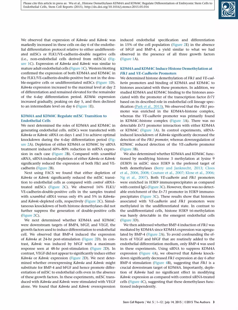
Figure 1. Expression of KDM4A and KDM4C following mESC Differentiation into Endothelial Cells(A) Flow sorting diagram of mESCs differentiated to endothelial cells stained with anti-FLK1 and anti-VE-cadherin antibodies. Cells wereseparated into FLK1/VE-cadherin-double positive endothelial cells or double-negative control cells.(B) Time course of gene expression assessed by qRT-PCR for the pluripotency genes Oct4 and Nanog as well as for endothelial genes Flk1 andVE-cadherin during the 6-day differentiation period (D1 to D6; ES are mESCs); error bars represent mean ± SD (n = 3 independent ex-periments, p < 0.05 by ANOVA).(C) qRT-PCR screen for 28 known histone demethylases was carried out in mESCs differentiated into FLK1/VE-cadherin-double positiveendothelial cells (D6), double-negative control cells (D6), and adult primary mouse endothelial cells. Expression of KDM4a and KDM4c wasmarkedly increased in mESC-derived and mouse endothelial cells as assessed by two-way ANOVA (all values are normalized to 18S inundifferentiated mESCs). Values are mean ± SD (n = 3 independent experiments, *p < 0.001 by two-way ANOVA).(D) Western blotting carried out using anti-KDM4A and anti-KDM4C antibodies in sorted FLK1/VE-cadherin-double-positive cells derivedfrom mESCs at day 6 of differentiation. Representative examples from three independent experiments are shown.(E) Time course of Kdm4a and Kdm4c expression determined by qRT-PCR during the day 1 to day 6 period of endothelial cell differentiationfrom mESCs. Values are mean ± SD (n = 3 independent experiments); gene expression in undifferentiated cells (ES) is set as 1; p < 0.05 byANOVA.
2 Stem Cell Reports j Vol. 5 j 1–12 j July 14, 2015 j ª2015 The Authors
Please cite this article in press as: Wu et al., Histone Demethylases KDM4A and KDM4C Regulate Differentiation of Embryonic Stem Cells toEndothelial Cells, Stem Cell Reports (2015), http://dx.doi.org/10.1016/j.stemcr.2015.05.016
Please cite this article in press as: Wu et al., Histone Demethylases KDM4A and KDM4C Regulate Differentiation of Embryonic Stem Cells toEndothelial Cells, Stem Cell Reports (2015), http://dx.doi.org/10.1016/j.stemcr.2015.05.016
We observed that expression of Kdm4a and Kdm4c was
markedly increased in these cells on day 6 of the endothe-
lial differentiation protocol relative to either undifferenti-
ated mESCs or FLK1/VE-cadherin-double negative cells
(i.e., non-endothelial cells derived from mESCs) (Fig-
ure 1C). Expression of Kdm4a and Kdm4c was similar to
mature adult endothelial cells (Figure 1C).Western blotting
confirmed the expression of both KDM4A and KDM4C in
the FLK1/VE-cadherin-double-positive but not in the dou-
ble-negative cells or undifferentiated mESCs (Figure 1D).
Kdm4a expression increased to the maximal level at day 2
of differentiation and remained elevated for the remainder
of the 6-day differentiation period. KDM4c expression
increased gradually, peaking on day 5, and then declined
to an intermediate level on day 6 (Figure 1E).
KDM4A and KDM4C Regulate mESC Transition to
Endothelial Cells
We next determined the roles of KDM4A and KDM4C in
generating endothelial cells. mESCs were transfected with
Kdm4a or Kdm4c siRNA on days 1 and 3 to achieve optimal
knockdown during the 6-day differentiation period (Fig-
ure 2A). Depletion of either KDM4A or KDM4C by siRNA
treatment induced 60%–80% reduction in mRNA expres-
sion in each case (Figure 2B). Compared with scrambled
siRNA, siRNA-induced depletion of either Kdm4a or Kdm4c
significantly reduced the expression of both Flk1 and VE-
cadherin (Figure 2B).
Next using FACS we found that either depletion of
Kdm4a or Kdm4c significantly reduced the mESC transi-
tion to endothelial cells as compared with control siRNA-
treated mESCs (Figure 2C). We observed 16% FLK1/
VE-cadherin-double-positive cells in the samples treated
with scrambled siRNA versus only 4% and 5% in Kdm4a-
and Kdm4c-depleted cells, respectively (Figure 2C). Simul-
taneous knockdown of both histone demethylases did not
further suppress the generation of double-positive cells
(Figure 2C).
We next determined whether KDM4A and KDM4C
were downstream targets of BMP4, bFGF, and VEGF, the
growth factors used to induce differentiation to endothelial
cell. We observed that BMP-4 induced the expression
of Kdm4a at 24-hr post-stimulation (Figure 2D). In con-
trast, Kdm4c was induced by bFGF with a maximum
response seen at 48-hr post-stimulation (Figure 2D). In
contrast, VEGF did not appear to significantly induce either
Kdm4a or Kdm4c expression (Figure 2D). We next deter-
mined whether overexpressing Kdm4a and Kdm4c might
substitute for BMP-4 and bFGF and hence promote differ-
entiation of mESC to endothelial cells even in the absence
of these growth factors. In these experiments, mESC trans-
duced with Kdm4a and Kdm4c were stimulated with VEGF
alone. We found that Kdm4a and Kdm4c overexpression
induced endothelial specification and differentiation
in 15% of the cell population (Figure 2E) in the absence
of bFGF and BMP-4, a yield similar to what we had
observed in the presence of all three growth factors
(Figure 1A).
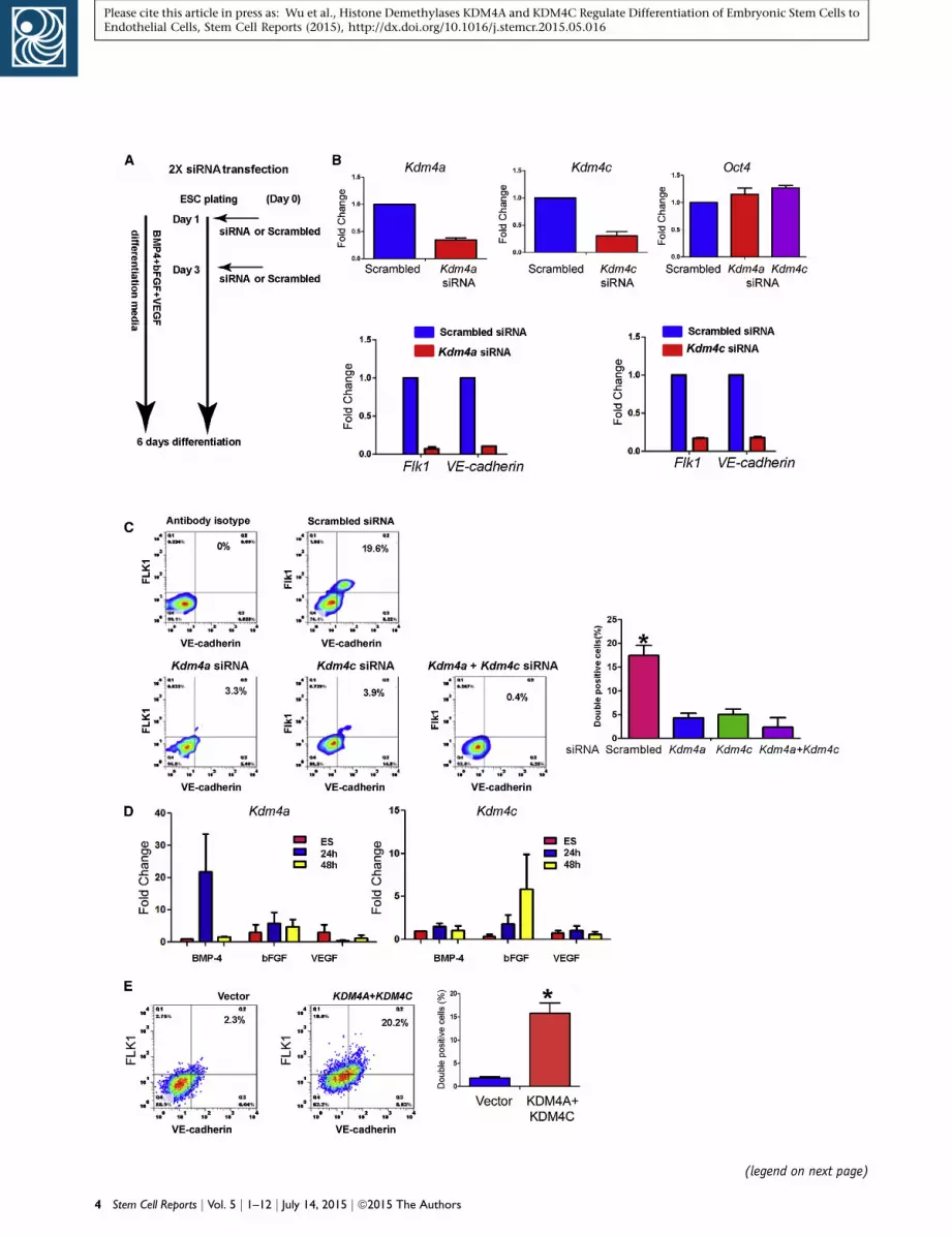
KDM4A andKDM4C InduceHistoneDemethylation at
Flk1 and VE-Cadherin Promoters
We determined histone demethylation of Flk1 and VE-cad-
herin promoters and binding of KDM4A and KDM4C to
histones associated with these promoters. In addition, we
studied KDM4A and KDM4C binding to the histones asso-
ciated with the promoter of the transcription factor Er71
based on its described role in endothelial cell lineage spec-
ification (Park et al., 2013). We observed that the Flk1 pro-
moter was enriched in the KDM4A-histone complex,
whereas the VE-cadherin promoter was primarily found
in KDM4C-histone complex (Figure 3A). There was no
detectable Er71 promoter interaction with either KDM4A
or KDM4C (Figure 3A). In control experiments, siRNA-
induced knockdown of Kdm4a significantly decreased the
detection of the Flk1 promoter, whereas downregulation of
KDM4C reduced detection of the VE-cadherin promoter
(Figure 3B).
We also determined whether KDM4A and KDM4C func-
tioned by modifying histone 3 methylation at lysine 9
(H3K9) in mESC since H3K9 is the preferred target of
both demethylases (Berry and Janknecht, 2013; Cloos
et al., 2006, 2008; Couture et al., 2007; Klose et al., 2006;
Ng et al., 2007). Both VE-cadherin and Flk1 promoters
were enriched in H3K9 immunoprecipitates as compared
with control IgG (Figure 3C). However, there was no detect-
able enrichment of the Er-71 promoter in H3K9 immuno-
precipitates (Figure 3C). These results show that histones
associated with VE-cadherin and Flk1 promoters were
methylated in the undifferentiated state. In contrast to
the undifferentiated cells, histone H3K9 tri-methylation
was barely detectable in the emergent endothelial cells
(Figure 3D).
We then addressed whether BMP-4 induction of Flk1was
mediated by KDM4A since KDM4A expression was upregu-
lated by BMP-4 (Figure 2A). To avoid confounding the ef-
fects of VEGF and bFGF that are routinely added to the
endothelial differentiation medium, only BMP-4 was used
in these experiments. Using siRNA to suppress KDM4A
expression (Figure 4A), we observed that Kdm4a knock-
down significantly decreased Flk1 expression at day 6 after
BMP-4 stimulation (Figure 4B), suggesting that Flk1 is a
crucial downstream target of KDM4A. Importantly, deple-
tion of Kdm4a had no significant effect in modifying
Kdm4c expression as compared with control siRNA-treated
cells (Figure 4C), suggesting that these demethylases func-
tioned independently.
Stem Cell Reports j Vol. 5 j 1–12 j July 14, 2015 j ª2015 The Authors 3
(legend on next page)
4 Stem Cell Reports j Vol. 5 j 1–12 j July 14, 2015 j ª2015 The Authors
Please cite this article in press as: Wu et al., Histone Demethylases KDM4A and KDM4C Regulate Differentiation of Embryonic Stem Cells toEndothelial Cells, Stem Cell Reports (2015), http://dx.doi.org/10.1016/j.stemcr.2015.05.016

Please cite this article in press as: Wu et al., Histone Demethylases KDM4A and KDM4C Regulate Differentiation of Embryonic Stem Cells toEndothelial Cells, Stem Cell Reports (2015), http://dx.doi.org/10.1016/j.stemcr.2015.05.016
KDM4A and KDM4C Are Required for Capillary Tube
Formation and Vasculogenesis In Vivo
mESCs were transfected with either scrambled, Kdm4a, or
Kdm4c siRNA on days 1 and 3 of the differentiation proto-
col. Cells harvested on day 6 were plated onto Matrigel-
coated plates for 16 hr. Control siRNA-treated cells formed
tubes, whereas KDM4A- and KDM4C-depleted cells did
not form tubes (Figure 5A). Experiments were also made
in which unsorted day 6 cells were mixed with Matrigel
and implanted subcutaneously into nude mice. At day 7
post-implantation, Matrigel plugs were retrieved, section,
and stained for PECAM1 andVE-cadherin.We observed PE-
CAM1 and VE-cadherin-positive blood vessels in the con-
trol group pre-treated with scrambled siRNA, whereas there
was a virtual absence of blood vessels when implanted cells
were pre-treatedwith siRNAs targetingKdm4a, orKdm4c, or
both (Figure 5B).
We also used the zebrafish model to address the role of
KDM4A and KDM4C in regulating vascular development
in vivo. Zebrafish embryos were either injected with con-
trol morpholino (MO), or Kdm4a MO, or Kdm4c MO, and
observed for up to 48-hr postfertilization (hpf). In the
control group, endothelial cells grew out from the dorsal
aorta (DA) adjacent to intersomitic boundaries in a stereo-
typical manner at 24 hpf (Figure 5C). Bipolar sprouts grew
toward the dorsal side until they passed the horizontal
myoseptum, where they extended in anterior and poste-
rior directions into two branches that fused with branches
of neighboring sprouts to form the characteristic dorsal
longitudinal anastomotic vessels (DLAVs; Figure 5C).
Downregulation of either KDM4a or KDM4c caused de-
fects in vasculogenesis and angiogenesis. At a low MO
dose (2 ng), most of the embryos of the Kdm4a-MO or
Kdm4c-MO groups demonstrated profoundly defective
intersegmental vessels and complete absence of DLAV at
24 hpf. These defects were more severe in the Kdm4a-
Figure 2. KDM4A and KDM4C Mediate mESC Differentiation to En(A) Diagram showing experimental protocol for siRNA treatment. mESCprotein. The cells were differentiated in medium containing BMP-4, b(B) The effect of Kdm4a and Kdm4c knockdown on expression of endowas 60%–80%. Oct4 expression was not suppressed by either Kdm4aknockdown resulted in 90% and 80%, respectively, reduction in Flk1 an(C) Flow cytometry assessment of cell surface expression of Flk1 andscrambled, Kdm4a, Kdm4c, or Kdm4a and Kdm4c siRNAs. Quantificationdouble positive cells was reduced from 17% to less than 5%; FACS profivalues are mean ± SD; p < 0.05. Combining Kdm4a with Kdm4c siRNA(D) Treatment of mESCs with BMP-4 alone (2 ng/ml) is sufficient to inbFGF alone (10 ng/ml) induced Kdm4c expression at 48-hr post-stimiments). VEGF stimulation did not induce either KDM4A or KDM4C ex(E) Kdm4a and Kdm4c overexpression substitutes for BMP-4 and bFGmedium containing only VEGF. FACS analysis on day 6 demonstratesummarizes data from (n = 3) three independent experiments.
MO group as compared with the Kdm4c-MO group (Fig-
ure 5C). None of the Kdm4a-MO-injected zebrafish sur-
vived by 48 hpf, whereas the Kdm4c-MO-injected group
survived and showed relatively small impairment of
vascular development at this time (Figure 5C). These re-
sults are consistent with the mouse results showing that
KDM4A participated in the early phase of mESC differen-
tiation to endothelial cells and KDM4C mediated sub-
sequent differentiation to mature endothelial cells. The
results also suggest that compensatory mechanisms may
be activated in vivo following Kdm4C knockdown, which
only partially restored vascular development in 48-hpf
embryos.
DISCUSSION
Here we describe the role for histone demethylases KDM4A
and KDM4C in the epigenetic regulation of mESC differen-
tiation to endothelial cells. We showed that KDM4A
and KDM4C targeted the tri-methyl histone 3 lysine 9
(H3K9me3), a known repressive marker of gene regulation
(Black et al., 2012; Berry and Janknecht 2013) at Flk1
and VE-cadherin promoters. Demethylation of H3K9me3
enabled de-repression at the promoters and thereby in-
duced expression of Flk1 andVE-cadherin genes, the lineage
markers of endothelial cells (Kohler et al., 2013; Park et al.,
2013).
Differentiation of ESCs into the mesodermal progenitors
and then into endothelial cells involves the orderly expres-
sion of developmental transcription factors such as Er71
(Kataoka et al., 2011; Lee et al., 2008), upregulation of the
VEGF receptor Flk1 (Kohler et al., 2013; Park et al., 2013),
and expression of endothelial cell-specific proteins such
as VE-cadherin, which allow the cells to form stable
adherens junctions and functional vascular networks
dothelial Cellswere transfected on days 1 and 3 to achieve maximum knockdown ofFGF, and VEGF for 6 days.thelial-specific markers. Efficiency of Kdm4a and Kdm4c knockdownor Kdm4c knockdown (n = 4 per group). Both Kdm4a and Kdm4cd VE-cadherin expression on day 6 (n = 3 independent experiments).VE-cadherin on day 6 of differentiation following treatment within the bar graph shows that the percentage of FLK1 and VE-cadherinle is representative (n = 3 independent experiments). Quantifications did not further suppress differentiation.duced Kdm4a expression at 24-hr post-stimulation. Treatment withulation. Error bars represent mean ± SD (n = 3 independent exper-pression during the 48-hr period.F. mESCs were transfected with Kdm4a and Kdm4c and cultured ind that the fraction of double-positive cells was 20%. Bar graph
Stem Cell Reports j Vol. 5 j 1–12 j July 14, 2015 j ª2015 The Authors 5
Figure 3. KDM4A and KDM4C Bind to His-tones Associated with Flk1 and VE-cad-herin Promoters, Respectively(A) Differentiated cells were subjectedChIP-qPCR analysis using control IgG, anti-KDM4A, and anti-KDM4C antibodies (seeExperimental Procedures). The ChIPs weresubjected to qPCR using oligonucleotides toamplify the promoter regions of VE-cad-herin, Flk1, and the transcription factorEr71. Chromatin binding to IgG-ChIP wasconsidered background. Flk1 promoter wasenriched in KDM4A ChIP, whereas VE-cad-herin promoter was enriched in KDM4C ChIP.Er71-promoter was not detectable (ND) ineither ChIP. Values are mean ± SD (n = 3independent experiments).(B) Kdm4a knockdown prevented enrich-ment of Flk1-promoter in the ChIP assay,whereas Kdm4c knockdown preventedenrichment of the VE-cadherin promoter.Values are mean ± SD (n = 3 independentexperiments).(C) ChIP assay was performed using an anti-histone H3 tri-methylation at lysine (K9)antibody to assess methylation of histonesat the promoters of VE-cadherin, Flk1, andEr71 in undifferentiated mESCs; values aremean ± SD (n = 3 independent experi-ments).(D) Histone 3 tri-methylation at lysine 9(H3K9) was assessed by western blotting inmESCs and FLK1/VE-cadherin-double-posi-tive cells. An antibody against total histoneH3 was used as loading control. The blot isrepresentative of three independent ex-periments. Bar graph (mean ± SD) showssignificant reduction of histone H3 tri-methylation (K9) in endothelial cells ascompared to undifferentiated ESCs (p < 0.05by Student’s t test).
Please cite this article in press as: Wu et al., Histone Demethylases KDM4A and KDM4C Regulate Differentiation of Embryonic Stem Cells toEndothelial Cells, Stem Cell Reports (2015), http://dx.doi.org/10.1016/j.stemcr.2015.05.016
(Park et al., 2013). In the present study, we demonstrated
that KDM4A and KDM4C were essential epigenetic re-
gulators of mESC differentiation to endothelial cells in
mice and also contributed to vascular development in the
zebrafish. KDM4A and KDM4C functioned independently
through histone demethylation at the Flk1 andVE-cadherin
promoters, respectively, to induce the endothelial cell line-
age. Interestingly, we found that Kdm4c knockdown also
suppressed Flk1 expression despite there being no interac-
tion between KDM4C and histones at the Flk1 promoter,
therefore suggesting that KDM4C targets additional genes
regulating Flk1 expression.
6 Stem Cell Reports j Vol. 5 j 1–12 j July 14, 2015 j ª2015 The Authors
Our study examined the role histone demethylases in
the endothelial differentiation of ESCs. Using a screen for
28 histone demethylases in FLK1/VE-cad-double-positive
endothelial cells, we observed that mRNA and protein
expression of Kdm4a and Kdm4c were markedly increased
during endothelial differentiation. Importantly, the levels
of KDM4A andKDM4C increased at day 6 of differentiation
to the same extent seen in adult endothelial cells, suggest-
ing that ESC-derived endothelial cells resembled mature
endothelial cells in their histone demethylation signature.
Although we did not examine human ESC differentiation
to endothelial cells, their emergence may similarly involve
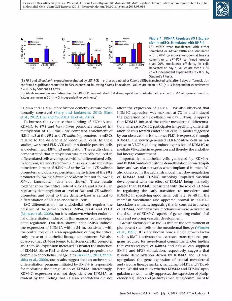
Figure 4. KDM4A Regulates Flk1 Expres-sion in mESCs Stimulated with BMP-4(A) mESCs were transfected with eitherscrambled or Kdm4a siRNA and stimulatedwith BMP-4 to induce mesodermal lineagecommitment. qRT-PCR confirmed greaterthan 90% knockdown efficiency in cellsharvested on day 6; values are mean ± SD(n = 3 independent experiments, p < 0.05 byStudent’s t test).
(B) Flk1 and VE-cadherin expression evaluated by qRT-PCR in either scrambled or Kdm4a siRNA-transfected cells after 6 days differentiationconfirmed significant reduction in Flk1 expression following Kdm4a knockdown. Values are mean ± SD (n = 3 independent experiments;p < 0.05 by Student’s t test).(C) Kdm4c expression was determined by qRT-PCR demonstrated that downregulation of Kdm4a had no effect on Kdm4c gene expression.Values are mean ± SD (n = 3 independent experiments).
Please cite this article in press as: Wu et al., Histone Demethylases KDM4A and KDM4C Regulate Differentiation of Embryonic Stem Cells toEndothelial Cells, Stem Cell Reports (2015), http://dx.doi.org/10.1016/j.stemcr.2015.05.016
KDM4A and KDM4C since histone demethylases are evolu-
tionarily conserved (Berry and Janknecht, 2013; Black
et al., 2012; Hou and Yu, 2010; Ye et al., 2012).
To buttress the evidence that binding of KDM4A and
KDM4C to Flk1 and VE-cadherin promoters reduced tri-
methylation of H3K9me3, we compared enrichment of
H3K9me3 at the Flk1 and VE-cadherin promoters in mESCs
relative to the differentiated endothelial cells. In these
studies, we sorted FLK1/VE-cadherin-double-positive cells
and determined H3K9me3methylation. The results clearly
demonstrated that methylation was markedly reduced in
differentiated cells as compared with undifferentiated cells.
In addition, we knocked down Kdm4a or Kdm4c and deter-
mined enrichment ofH3K9me3 at the Flk1 andVE-cadherin
promoters and observed persistent methylation of the Flk1
promoter following Kdm4a knockdown but not following
Kdm4c knockdown (data not shown). These results
together show the critical role of KDM4A and KDM4C in
regulating demethylation at level of Flk1 and VE-cadherin
promoters and point to these demethylases as central to
differentiation of ESCs to endothelial cells.
ESC differentiation into endothelial cells requires the
presence of the growth factors BMP-4, bFGF, and VEGF
(Blancas et al., 2008), but it is unknown whether endothe-
lial differentiation induced in this manner requires epige-
netic regulation. Our data showed that BMP-4 induced
the expression of KDM4A within 24 hr, consistent with
the central role of KDM4A upregulation during the critical
early phase of endothelial lineage commitment. We also
observed that KDM4A bound to histones on Flk1 promoter
and that Flk1 expression increased 24 hr after the induction
of KDM4A. Since Flk1 enables mesodermal progenitors to
commit to endothelial lineage fate (Park et al., 2013; Yama-
shita et al., 2000), our results suggest that an orchestrated
differentiation program initiated by BMP-4 is responsible
for mediating the upregulation of KDM4A. Interestingly,
KDM4C expression was not dependent on KDM4A, as
evident by the finding that KDM4A knockdown did not
affect the expression of KDM4C. We also observed that
KDM4C expression was maximal at 72 hr and induced
the expression of VE-cadherin on day 5. Thus, it appears
that KDM4A initiated the earlier mesodermal differentia-
tion, whereas KDM4C participates in specifying differenti-
ation of cells toward endothelial cells. A model suggested
by our observations is that once FLK1 is expressed through
KDM4A, the newly generated FLK1-positive cells in res-
ponse to VEGF signaling induce expression of KDM4C to
mediate VE-cadherin expression and thereby the endothe-
lial lineage commitment.
Importantly, endothelial cells generated by KDM4A-
and KDM4C-induced histone demethylation formed capil-
laries and vascular networks when implanted in mice. We
also observed in the zebrafish model that downregulation
of KDM4A and KDM4C orthologs impaired vascular
development with the effect of KDM4A being markedly
greater than KDM4C, consistent with the role of KDM4A
in regulating the early transition to mesoderm and
KDM4C in specifying endothelial cell fate. By 48 hr, the
zebrafish vasculature also appeared normal in KDM4C
knockdown animals, suggesting that in contrast to absence
of KDM4A, compensatory mechanisms were activated in
the absence of KDM4C capable of generating endothelial
cells and restoring vascular development.
Growth factors such as BMP-4 initiate the commitment of
pluripotent stem cells to the mesodermal lineage (Winnier
et al., 1995). It is not known how a single growth factor
such as BMP-4 activates the extensive transcriptional pro-
gram required for mesodermal commitment. Our finding
that overexpression of Kdm4A and Kdm4C can supplant
BMP-4 and bFGF stimulation, respectively, suggests that
histone demethylation driven by KDM4A and KDM4C
upregulates the gene expression of critical mesodermal
and vascular lineagemarkers, including to FLK1 andVE-cad-
herin.We did not studywhether KDM4A and KDM4Cupre-
gulation concomitantly suppresses the expression of plurip-
otency regulators and pathways mediating commitment to
Stem Cell Reports j Vol. 5 j 1–12 j July 14, 2015 j ª2015 The Authors 7
(legend on next page)
8 Stem Cell Reports j Vol. 5 j 1–12 j July 14, 2015 j ª2015 The Authors
Please cite this article in press as: Wu et al., Histone Demethylases KDM4A and KDM4C Regulate Differentiation of Embryonic Stem Cells toEndothelial Cells, Stem Cell Reports (2015), http://dx.doi.org/10.1016/j.stemcr.2015.05.016

Please cite this article in press as: Wu et al., Histone Demethylases KDM4A and KDM4C Regulate Differentiation of Embryonic Stem Cells toEndothelial Cells, Stem Cell Reports (2015), http://dx.doi.org/10.1016/j.stemcr.2015.05.016
non-mesodermal lineages, but this is an important question
that needs to be addressed in future studies. By identifying
the epigenetic switches acting downstream of growth fac-
tors driving vascular differentiation, the present findings
pave the way for novel epigenetic approaches to ensure
highly efficient endothelial differentiation while mini-
mizing the need for exogenous recombinant growth factors.
In contrast to the distinct roles of KDM4A and KDM4C
in mediating the expression of Flk1 and VE-cadherin, we
observed that neither histone demethylase was involved in
mediating the expression of the transcription factor Er71.
The epigenetic regulation of Er71 expression is not known,
but itmay involve other epigeneticmechanisms such as his-
tone de-acetylation or DNA methylation (Park et al., 2013).
One limitation of our study is that we cannot exclude
additional targets of KDM4A and KDM4C besides FLK1
and VE-cadherin or other epigenetic regulatory mecha-
nisms that may also contribute to endothelial lineage
specification. For example, the fact that suppression of
KDM4C also affected the levels of the comparatively early
differentiation marker Flk1 suggests that KDM4C may
have additional targets beyond VE-cadherin that regulate
mesodermal or vascular commitment. Another important
limitation is the focus on Flk1 and VE-cadherin. However,
we chose to focus on these two markers because Flk1 indi-
cates early mesodermal commitment and VE-cadherin is a
highly specific marker of mature endothelial cells, but
future studies may be needed to address whether KDM4A
and KDM4C also regulate the differentiation into subsets
of endothelial cells that do not express VE-cadherin.
In summary, our results elucidate the role of KDM4A and
KDM4C in regulating the differentiation ofmESCs to endo-
thelial cells. KDM4A initiated differentiation by targeting
the Flk1 promoter, whereas KDM4C specified endothelial
cell fate by targeting theVE-cadherin promoter. Vasculogen-
esis in mice and zebrafish was also dependent on KDM4A
and KDM4C. As histone demethylation in Flk1 andVE-cad-
Figure 5. KDM4A and KDM4C Are Required for Capillary Tube Form(A) mESCs were transfected with scrambled, Kdm4a, or Kdm4c siRNA anwere plated on Matrigel, and tube formation was evaluated by phase-cotube formation. Values are mean ± SD (n = 3 independent experimeStudent’s t test).(B) Unsorted differentiated cells treated as in (A) were suspended in MMatrigel plugs were harvested on day 7 and assessed by H&E staining.anti-CD31 and anti-VE-cadherin antibodies. Values are mean ± SD (n =signal intensity (p < 0.05 by Student’s t test).(C) Targeting either Kdm4a or Kdm4c with MOs induced significant defeprominent in the Kdm4a-MO group. In each case, fertilized zebrafish eand the embryos were observed at 24 and 48 hpf. Kdm4amorphants dicomplete lack of DLAV, whereas Kdm4c-MO showed a less severe phenonot survive beyond 36 hpf. In contrast, although the response was dvasculature at 48 hpf similar to control embryos. Bar graphs show bloo(p < 0.05 by ANOVA).
herin promoters by KDM4A and KDM4C acted sequentially
to induce the transition of ESCs to endothelial cells, our
findings raise the intriguingly possibility of activating
these histone demethylases to induce vascular endothelial
differentiation and de novo vasculogenesis.
EXPERIMENTAL PROCEDURES
mESC Culture and ReagentsmESCs (D3) were maintained on gelatin-coated plates in DMEM
supplemented with leukemia inhibitory factor (LIF; Chemicon),
15% defined fetal bovine serum (Hyclone), nonessential amino
acids (GIBCO), 50-mM b-mercaptoethanol (GIBCO), 2 mML-gluta-
mine (Mediatech), 0.1-mg/ml penicillin (Sigma-Aldrich), 0.2-mg/ml
streptomycin (Sigma-Aldrich), and monothioglycerol (MTG), and
ascorbic acid (Sigma-Aldrich).
mESC Differentiation into Endothelial Cells and
siRNA TransfectionmESCs were co-cultured and maintained with fibroblast feeder
cells. For differentiation, cells were cultured feeder free for at least
two passages and seeded on collagen-coated plated 1 day before
initiation of the ESC differentiation. ESC medium was replaced
with endothelial cell differentiation medium containing BMP-4
(2 ng/ml), bFGF (10 ng/ml), and VEGF (50 ng/ml), which was
changed daily (Kohler et al., 2013). KDM4a and KDM4c siRNAs
(Table S1) and scrambled negative control siRNA were transfected
using RNAiMAX Transfection reagent (Invitrogen) followingman-
ufacturer’s protocol. Cells were transfected twice to achieve the
best transfection efficiency.
Flow CytometryFlow cytometric analysis and sorting were performed on a Beck-
man Coulter CyAn II (Beckman Coulter), as previously described
(Kohler et al., 2013). Antibodies purchased from eBiosciences
were specific anti-Flk-1 (anti-mouse CD309 [Flk1]-APC, Clone
Avas12a1, catalog number 17-5821-81), anti-VE-cadherin (anti-
human CD144- PE, Clone 16B1, catalog number 12-1449-82),
ation and Vasculogenesis In Vivod underwent differentiation protocol. Unsorted differentiated cellsntrast microscopy. Knockdown of either Kdm4a or Kdm4c preventednts). Bar graphs of tube lengths in indicated groups (p < 0.05 by
atrigel containing VEGF and injected subcutaneous into nude mice.Bar graphs show vessel area. Immunostaining was performed using3 independent experiments). Bar graphs show CD31 and VE-cadherin
cts in vasculogenesis in zebrafish embryos, with defects being moreggs were injected with 2-ng Control-MO, Kdm4a-MO, or Kdm4c-MO,splayed only residual inter-somite vessels (ISV) derived from DA andtype with reduced number of DLAV at 24 hpf. Kdm4a morphants didelayed in Kdm4c morphants, they developed an apparently normald vessel area and the length of inter-somite vessels at 24 and 48 hpf
Stem Cell Reports j Vol. 5 j 1–12 j July 14, 2015 j ª2015 The Authors 9

Please cite this article in press as: Wu et al., Histone Demethylases KDM4A and KDM4C Regulate Differentiation of Embryonic Stem Cells toEndothelial Cells, Stem Cell Reports (2015), http://dx.doi.org/10.1016/j.stemcr.2015.05.016
and IgG isotype controls. Data were analyzed using the manufac-
turer’s software.
Protein Separation for Western BlottingThe cultured cells were washed twice with PBS and then lysed
with HEPES containing buffer (60 mM KCl, 15 mM NaCl, 15 mM
HEPES [pH7.9], 4mMMgCl2, 0.5-MDTT, 1%TritonX-100, supple-
mented with Proteinase inhibitor). The sample was spun down at
13,000 rpm for 5 min at 4�C, and the supernatant was collected.
Laemmli loading buffer was added into samples, and samples
were heated to 95�C for 5 min. Proteins were separated on 9%
polyacrylamide gels and transferred to polyvinyl difluoride
(PVDF) membranes. Non-specific binding sites were blocked with
5% milk in TBS-T buffer. Polyclonal anti-KDM4A (catalog number
ab105953, Abcam) andKDM4C (catalog number ab93694, Abcam)
antibodies and rabbit anti-H3K9me3 antibody (catalog number
ab8898, Abcam) were used at 1:1,000 dilution. The membrane
was incubated with the primary antibody for 2 hr at room temper-
ature or overnight in a cold room. Goat anti-rabbit or rabbit
anti-mouse secondary antibody from BioRad was used at 1:10,000
dilution and incubated with the membrane for 1 hr at room tem-
perature. Enhanced chemiluminescence (ECL) was used for signal
detection (GE Healthcare).
qRT-PCRTotal RNAwas isolated from cells using the RNeasy plus extraction
kit (QIAGEN) and 1-mg total RNAwas used for reverse transcription
using the High-Capacity cDNA Reverse Transcription Kit (Applied
Biosystems). qRT-PCRwas performed on a 7500 Fast Real Time PCR
System (Applied Biosystems) using SYBR Green PCR mix (Applied
Biosystems). Gene specific (Table S2) and promoter-enhancer
specific oligonucleotides were synthesized by Integrated DNA
Technologies.
Chromatin Immunoprecipitation-qPCR AssayTo analyze the interaction of KDM4A, KDM4C, and histone H3 (tri
methyl K9) with Flk1 and VE-cadherin promoter segments, we used
chromatin immunoprecipitation (ChIP) assay. The assays were
performed according to the manufacturer’s protocol (Abcam).
ChIP entailed the use of specific antibodies and controls such as
IgGs and beads alone. To minimize background binding, samples
were diluted. In brief, cells were cross-linked with 0.75% formalde-
hyde (as the links it forms are reversible) at room temperature for
10 min, stopped by glycine (0.125 M) at room temperature for
5 min, and washed twice with PBS solution. The cells were lysed
with FA buffer (50 mM HEPES-KOH [pH 7.5], 140 mM NaCl,
1 mMEDTA [pH 8.0], 1% Triton X-100, 0.1% sodium deoxycholate,
0.1% SDS, protease Inhibitors) and then subjected to sonication
(20 s for ten times) on ice. Polyclonal anti-KDM4A, anti-KDM4C,
anti-histone H3 (tri methyl K9) antibodies were purchased from
Abcam. Chromatin binding to IgG-ChIP was considered back-
ground. The primers used in ChIP-qPCR were as follows: Er71
promoter, forward, 50-AGCTTCAAACAAAACTCTTC-30 and reverse,
50-TCTTAAGGGGGGAATTTATG-30; Er71_10kb, forward, 50-CTGGTACCTGTCCTGTCTTC-30 and reverse, 50-AATGCAAACTTACAAGTACA-30; Flk1 promoter, forward, 50-CTGTGTTTCCTTAGATCGCG-30 and reverse, 50-CCTGCACCTCGCGCTGGGCA-30; Flk1_10kb,
10 Stem Cell Reports j Vol. 5 j 1–12 j July 14, 2015 j ª2015 The Authors
forward, 50-AGGACTTTTAAATGAATGGA-30 and reverse, 50-TCCAGGCTGTGACTCACAGG-30; VE-cadherin promoter, forward, 50-GGACATCAGTATTCCAGGTA-30 and reverse, 50-GGAACGAGTGGCTCACTCTA-30; and VE-cadherin_10kb, forward, 50-TGACCTGCTTGAGTTCCAGT-30 and reverse, 50-TTTGCATCTACATTCATGAG-30.For ChIP-qPCR, we used 5% of starting chromatin as input, and
the raw Ct value were recorded. Since the starting input was 5%,
dilution factor (DF) of 20 or 4.321 cycles (i.e., log2 of 20) was sub-
tracted from the raw Ct value (i.e., adjusted to 100% input). ChIP-
qPCRwas run in triplicates and Ct values recorded. Data relative to
input were normalized for both background levels and input chro-
matin used for ChIP. Thus, percentage input (y axis) was calculated
from 20*2^ (adjusted input – Ct) and plotted as bar diagram for IgG
and anti-HDM4A and anti-KDM4C antibodies (x axis).
Tube Formation and Matrigel Plug AssaySix-well plates were coated with growth factor-reduced Matrigel
(BD Biosciences) according manufacturer’s instructions. After Ma-
trigel polymerization, 2-million differentiated cells were seeded;
tube-like structures were assessed after 16 hr using phase contrast
microscopy (Olympus).
All mouse experiments were carried out according to protocols
approved by the Animal Care and Use Committee (University of
Illinois at Chicago). Two-million mESCs were transfected with
siRNAs targeting either KDM4a, KDM4c, or both and underwent
the differentiation protocol for 6 days. The differentiated cells
(2 million) for each condition were mixed with Matrigel contain-
ing VEGF and injected into the abdominal subcutaneous skin of
anesthetized nude mice for additional 7 days. All Matrigel plugs
were carefully harvested, fixed with formalin solution, paraffin
embedded, sectioned, and subjected to H&E or immunostaining
staining using anti-CD31 and anti-VE-cadherin antibodies. The
differentiated cells used in the Matrigel studies were not sorted
for endothelial markers.
Zebrafish ExperimentsZebrafish experiments were carried out according to protocols
approved by Animal Care and Use Committee (University of Illi-
nois at Chicago). The breeding colony of zebrafish (Danio rerio,
Sheer strain, Dr. H. Tomasiewicz, University of Wisconsin) was
maintained at standard conditions (28.5�C [pH 7.5]) and staged ac-
cording to morphology (Kimmel et al., 1995). The transgenic line
Tg(fli1:EGFP)y1 (Zebrafish International Resource Center) was
sequentially outcrossed to the Sheer fish to obtain optically trans-
parent fish harboring fli1:EGFP transgene. Homozygous Sheer fish
harboring fli1:EGFP were used to obtain stronger intensity of the
fluorescent signal and a better acquisition of long time-lapse series.
Kdm4A and Kdm4C orthologs are well conserved in vertebrates.
The sequences of zebrafish and human proteins shared 47%
and 50% identity, respectively. For Kdm4a and Kdm4c knockdown,
the following custom translation-blockingMOswere designed and
purchased, along with standard control MOs, from Gene Tools: 50-GGCTGACCGAGTCCTTAACACTGAC-30 (KDMab_ZF MO), 50-CACTTGCCCCAACACCTGCCATCCT-30 (KDMC_ZF MO), as well
as a randomcontrol oligo 25-N and aGFP-positive control.MOs re-
constituted in nuclease-free water were dissolved in nuclease-free
water, and their concentrations were determined with NanoDrop.

Please cite this article in press as: Wu et al., Histone Demethylases KDM4A and KDM4C Regulate Differentiation of Embryonic Stem Cells toEndothelial Cells, Stem Cell Reports (2015), http://dx.doi.org/10.1016/j.stemcr.2015.05.016
Fertilized eggs were collected after timed mating of adult zebrafish
and injected at one- to four-cell stage with the indicated amounts
of MO into the yolk immediately below the blastomeres using a
Pneumatic PicoPump (World Precision Instruments). MOswere in-
jected using 0.1%phenol red in 0.5- to 2-nlDanieau buffer, and the
volume of the injected drop was estimated with a micrometer
scale. The efficacy of a translation-blocking MO was first assessed
by targeting GFP in a strain expressing the GFP gene under the
control of the fli1 promoter. The efficacy of MOs was assessed by
RT-PCR with superscript one-step RT-PCR system (Invitrogen)
and using as template total RNA from either control, Kdm4A or
Kdm4A MO injected embryos. RNA was extracted with Trizol
from 24- and 48-hr pooled embryos and treated with DNase. Em-
bryoswere injectedwith 2-, 5-, or 10-ngMOs in a 1- to 2-nl volume.
Injection of control MOs (Random control oligo and GFP positive
control at 10 ng) verified that injections at this concentration did
not induce MO-mediated toxicity, and the same or lower MO
concentrations were utilized in all experiments. For imaging zebra-
fish vessels, live embryos were dechorionated manually with a mi-
cro tweezers, anesthetized using 0.016% tricaine (Sigma), and
mounted in 1% low melting agarose. Zebrafish embryos were
visualized with stereo zoom microscope (Discovery V8, Zeiss).
For fluorescent microscopy, a GFP filter was used. For visualization
of vasculature formation, zebrafish embryos (24–48 hpf) were
captured by AxioCAm-HR Zeiss Digital Camera. ImageJ (http://
rsb.info.nih.gov/ij/) was used for image processing. Images were
analyzed with Metamorph software (Molecular Devices).
Statistical AnalysisThe statistical data were analyzed using the GraphPad Prism soft-
ware. Results are shown as means ± SD unless specified otherwise.
Comparisons between any two groups were performed using the
two-tailed unpaired Student’s t test. Comparisons of multiple
values among more than two groups were performed using two-
way ANOVA with post hoc Bonferroni’s correction. Differences
were considered statistically significant when p < 0.05.
SUPPLEMENTAL INFORMATION
Supplemental Information includes two tables and can be found
with this article online at http://dx.doi.org/10.1016/j.stemcr.
2015.05.016.
AUTHOR CONTRIBUTIONS
A.B.M., J.R., L.W., and Y.A.K. designed the experiments. L.W. and
S.R. carried out the experiments. L.W., K.K.W., S.R., K.T., Y.A.K.,
J.R., and A.B.M. analyzed the data and wrote the manuscript.
ACKNOWLEDGMENTS
The studies were supported by National Heart, Lung, and
Blood Institute grants T32-HL007829 and HL090152 to A.B.M.,
HL118068 to A.B.M. and J.R., GM094220 to J.R., HL103922 to
Y.A.K., and HL079356 to K.K.W. This paper is dedicated to Liang-
tangWu, the first author of the manuscript, who met an untimely
death during the completion of the revision. He was a passionate
scientist who inspired his colleagues with his creativity and
curiosity.
Received: September 30, 2014
Revised: May 28, 2015
Accepted: May 29, 2015
Published: June 25, 2015
REFERENCES
Barski, A., Cuddapah, S., Cui, K., Roh, T.Y., Schones, D.E.,Wang, Z.,
Wei, G., Chepelev, I., and Zhao, K. (2007). High-resolution
profiling of histone methylations in the human genome. Cell
129, 823–837.
Berry,W.L., and Janknecht, R. (2013). KDM4/JMJD2 histone deme-
thylases: epigenetic regulators in cancer cells. Cancer Res. 73,
2936–2942.
Black, J.C., Van Rechem, C., and Whetstine, J.R. (2012). Histone
lysine methylation dynamics: establishment, regulation, and bio-
logical impact. Mol. Cell 48, 491–507.
Blancas, A.A., Lauer, N.E., andMcCloskey, K.E. (2008). Endothelial
differentiation of embryonic stem cells. Curr. Protoc. Stem Cell
Biol. Chapter 1, 5.
Cloos, P.A., Christensen, J., Agger, K., Maiolica, A., Rappsilber, J.,
Antal, T., Hansen, K.H., and Helin, K. (2006). The putative onco-
gene GASC1 demethylates tri- and dimethylated lysine 9 on his-
tone H3. Nature 442, 307–311.
Cloos, P.A., Christensen, J., Agger, K., and Helin, K. (2008). Erasing
themethylmark: histone demethylases at the center of cellular dif-
ferentiation and disease. Genes Dev. 22, 1115–1140.
Couture, J.F., Collazo, E., Ortiz-Tello, P.A., Brunzelle, J.S., and Trie-
vel, R.C. (2007). Specificity and mechanism of JMJD2A, a trime-
thyllysine-specific histone demethylase. Nat. Struct. Mol. Biol.
14, 689–695.
Descamps, B., and Emanueli, C. (2012). Vascular differentiation
from embryonic stem cells: novel technologies and therapeutic
promises. Vascul. Pharmacol. 56, 267–279.
Hou, H., and Yu, H. (2010). Structural insights into histone lysine
demethylation. Curr. Opin. Struct. Biol. 20, 739–748.
Kataoka, H., Hayashi, M., Nakagawa, R., Tanaka, Y., Izumi, N.,
Nishikawa, S., Jakt, M.L., Tarui, H., and Nishikawa, S. (2011).
Etv2/ER71 induces vascular mesoderm from Flk1+PDGFRa+ prim-
itive mesoderm. Blood 118, 6975–6986.
Kimmel, C.B., Ballard,W.W., Kimmel, S.R., Ullmann, B., and Schil-
ling, T.F. (1995). Stages of embryonic development of the zebrafish.
Dev. Dyn. 203, 253–310.
Klose, R.J., Kallin, E.M., and Zhang, Y. (2006). JmjC-domain-con-
taining proteins and histone demethylation. Nat. Rev. Genet. 7,
715–727.
Kohler, E.E., Wary, K.K., Li, F., Chatterjee, I., Urao, N., Toth, P.T.,
Ushio-Fukai, M., Rehman, J., Park, C., and Malik, A.B. (2013).
Flk1+ and VE-cadherin+ endothelial cells derived from iPSCs reca-
pitulates vascular development during differentiation and display
similar angiogenic potential as ESC-derived cells. PLoS ONE 8,
e85549.
Kooistra, S.M., and Helin, K. (2012). Molecular mechanisms and
potential functions of histone demethylases. Nat. Rev. Mol. Cell
Biol. 13, 297–311.
Stem Cell Reports j Vol. 5 j 1–12 j July 14, 2015 j ª2015 The Authors 11

Please cite this article in press as: Wu et al., Histone Demethylases KDM4A and KDM4C Regulate Differentiation of Embryonic Stem Cells toEndothelial Cells, Stem Cell Reports (2015), http://dx.doi.org/10.1016/j.stemcr.2015.05.016
Kourembanas, S. (2014). Expanding the pool of stem cell therapy
for lung growth and repair. Circulation 129, 2091–2093.
Kouzarides, T. (2007). Chromatin modifications and their func-
tion. Cell 128, 693–705.
Lee, D., Park, C., Lee, H., Lugus, J.J., Kim, S.H., Arentson, E., Chung,
Y.S., Gomez, G., Kyba, M., Lin, S., et al. (2008). ER71 acts down-
stream of BMP, Notch, andWnt signaling in blood and vessel pro-
genitor specification. Cell Stem Cell 2, 497–507.
Leeper, N.J., Hunter, A.L., and Cooke, J.P. (2010). Stem cell therapy
for vascular regeneration: adult, embryonic, and induced pluripo-
tent stem cells. Circulation 122, 517–526.
Li, Z., Wu, J.C., Sheikh, A.Y., Kraft, D., Cao, F., Xie, X., Patel, M.,
Gambhir, S.S., Robbins, R.C., Cooke, J.P., and Wu, J.C. (2007). Dif-
ferentiation, survival, and function of embryonic stem cell derived
endothelial cells for ischemic heart disease. Circulation Suppl. 116,
I46–I54.
Marcelo, K.L., Goldie, L.C., and Hirschi, K.K. (2013). Regulation of
endothelial cell differentiation and specification. Circ. Res. 112,
1272–1287.
Ng, S.S., Kavanagh, K.L., McDonough, M.A., Butler, D., Pilka, E.S.,
Lienard, B.M., Bray, J.E., Savitsky, P., Gileadi, O., von Delft, F., et al.
(2007). Crystal structures of histone demethylase JMJD2A reveal
basis for substrate specificity. Nature 448, 87–91.
12 Stem Cell Reports j Vol. 5 j 1–12 j July 14, 2015 j ª2015 The Authors
Ohtani, K., Vlachojannis, G.J., Koyanagi, M., Boeckel, J.N., Urbich,
C., Farcas, R., Bonig, H., Marquez, V.E., Zeiher, A.M., and Dimm-
eler, S. (2011). Epigenetic regulation of endothelial lineage
committed genes in pro-angiogenic hematopoietic and endothe-
lial progenitor cells. Circ. Res. 109, 1219–1229.
Park, C., Kim, T.M., and Malik, A.B. (2013). Transcriptional regula-
tion of endothelial cell and vascular development. Circ. Res. 112,
1380–1400.
Wang, G.G., Allis, C.D., and Chi, P. (2007). Chromatin remodeling
and cancer, Part I: Covalent histone modifications. Trends Mol.
Med. 13, 363–372.
Winnier, G., Blessing, M., Labosky, P.A., and Hogan, B.L. (1995).
Bone morphogenetic protein-4 is required for mesoderm forma-
tion and patterning in the mouse. Genes Dev. 9, 2105–2116.
Yamashita, J., Itoh, H., Hirashima, M., Ogawa, M., Nishikawa, S.,
Yurugi, T., Naito,M., Nakao, K., andNishikawa, S. (2000). Flk1-pos-
itive cells derived from embryonic stem cells serve as vascular pro-
genitors. Nature 408, 92–96.
Ye, L., Fan, Z., Yu, B., Chang, J., Al Hezaimi, K., Zhou, X., Park, N.H.,
andWang, C.Y. (2012). Histone demethylases KDM4B and KDM6B
promotes osteogenic differentiation of human MSCs. Cell Stem
Cell 11, 50–61.
Yoder, M.C. (2012). Human endothelial progenitor cells. Cold
Spring Harb Perspect Med 2, a006692.
Related Documents