Research Article Histone Deacetylase Inhibitors Increase p27 Kip1 by Affecting Its Ubiquitin-Dependent Degradation through Skp2 Downregulation Adriana Borriello, 1 Silvio Naviglio, 1 Debora Bencivenga, 1 Ilaria Caldarelli, 1 Annunziata Tramontano, 1 Maria Carmela Speranza, 1 Emanuela Stampone, 1 Luigi Sapio, 1 Aide Negri, 2 Adriana Oliva, 1 Antonio Agostino Sinisi, 3 Annamaria Spina, 1 and Fulvio Della Ragione 1 1 Department of Biochemistry, Biophysics and General Pathology, Second University of Naples, 80138 Naples, Italy 2 Dipartimento di Scienze Biomediche Sperimentali e Cliniche, Universit` a degli Studi di Firenze, 50134 Firenze, Italy 3 Dipartimento di Scienze Cardiotoraciche e Respiratorie, Seconda Universit` a di Napoli, 80131 Napoli, Italy Correspondence should be addressed to Adriana Borriello; [email protected] and Silvio Naviglio; [email protected] Received 18 December 2014; Revised 2 August 2015; Accepted 9 August 2015 Academic Editor: Peter Backx Copyright © 2016 Adriana Borriello et al. is is an open access article distributed under the Creative Commons Attribution License, which permits unrestricted use, distribution, and reproduction in any medium, provided the original work is properly cited. Histone deacetylase inhibitors (HDACIs) represent an intriguing class of pharmacologically active compounds. Currently, some HDACIs are FDA approved for cancer therapy and many others are in clinical trials, showing important clinical activities at well tolerated doses. HDACIs also interfere with the aging process and are involved in the control of inflammation and oxidative stress. In vitro, HDACIs induce different cellular responses including growth arrest, differentiation, and apoptosis. Here, we evaluated the effects of HDACIs on p27 Kip1 , a key cyclin-dependent kinase inhibitor (CKI). We observed that HDACI-dependent antiproliferative activity is associated with p27 Kip1 accumulation due to a reduced protein degradation. p27 Kip1 removal requires a preliminary ubiquitination step due to the Skp2-SCF E3 ligase complex. We demonstrated that HDACIs increase p27 Kip1 stability through downregulation of Skp2 protein levels. Skp2 decline is only partially due to a reduced Skp2 gene expression. Conversely, the protein decrease is more profound and enduring compared to the changes of Skp2 transcript. is argues for HDACIs effects on Skp2 protein posttranslational modifications and/or on its removal. In summary, we demonstrate that HDACIs increase p27 Kip1 by hampering its nuclear ubiquitination/degradation. e findings might be of relevance in the phenotypic effects of these compounds, including their anticancer and aging-modulating activities. 1. Introduction e remodelling of nucleosome is a key epigenetic mecha- nism for modulating genome expression. Particularly, it has been postulated that histones postsynthetic modifications represent a “code” that can be recognized by nonhistone proteins, resulting in dynamic complexes capable of regu- lating gene transcription. Several reversible posttranslational changes of histone proteins have been described including lysine acetylation, lysine and arginine methylation, serine phosphorylation, proline isomerization, lysine ubiquitination and sumoylation, ADP-ribosylation, deimination, crotony- lation, propionylation, butyrylation, formylation, hydroxyla- tion, and O-GlcNacylation [1, 2]. Two groups of enzymes, histone acetyltransferases and histone deacetylases (HDACs), determine the pattern of his- tone acetylation. In humans, there are 18 identified HDACs, classified into four major classes, on the basis of sequence homology [3, and references therein]. Class I HDACs (HDAC1, HDAC2, HDAC3, and HDAC8) are strictly related Hindawi Publishing Corporation Oxidative Medicine and Cellular Longevity Volume 2016, Article ID 2481865, 17 pages http://dx.doi.org/10.1155/2016/2481865

Welcome message from author
This document is posted to help you gain knowledge. Please leave a comment to let me know what you think about it! Share it to your friends and learn new things together.
Transcript
Research ArticleHistone Deacetylase Inhibitors Increase p27Kip1 byAffecting Its Ubiquitin-Dependent Degradation throughSkp2 Downregulation
Adriana Borriello,1 Silvio Naviglio,1 Debora Bencivenga,1 Ilaria Caldarelli,1
Annunziata Tramontano,1 Maria Carmela Speranza,1
Emanuela Stampone,1 Luigi Sapio,1 Aide Negri,2 Adriana Oliva,1
Antonio Agostino Sinisi,3 Annamaria Spina,1 and Fulvio Della Ragione1
1Department of Biochemistry, Biophysics and General Pathology, Second University of Naples, 80138 Naples, Italy2Dipartimento di Scienze Biomediche Sperimentali e Cliniche, Universita degli Studi di Firenze, 50134 Firenze, Italy3Dipartimento di Scienze Cardiotoraciche e Respiratorie, Seconda Universita di Napoli, 80131 Napoli, Italy
Correspondence should be addressed to Adriana Borriello; [email protected] Silvio Naviglio; [email protected]
Received 18 December 2014; Revised 2 August 2015; Accepted 9 August 2015
Academic Editor: Peter Backx
Copyright © 2016 Adriana Borriello et al. This is an open access article distributed under the Creative Commons AttributionLicense, which permits unrestricted use, distribution, and reproduction in any medium, provided the original work is properlycited.
Histone deacetylase inhibitors (HDACIs) represent an intriguing class of pharmacologically active compounds. Currently, someHDACIs are FDA approved for cancer therapy and many others are in clinical trials, showing important clinical activities at welltolerated doses. HDACIs also interfere with the aging process and are involved in the control of inflammation and oxidative stress.In vitro, HDACIs induce different cellular responses including growth arrest, differentiation, and apoptosis. Here, we evaluated theeffects of HDACIs on p27Kip1, a key cyclin-dependent kinase inhibitor (CKI).We observed that HDACI-dependent antiproliferativeactivity is associated with p27Kip1 accumulation due to a reduced protein degradation. p27Kip1 removal requires a preliminaryubiquitination step due to the Skp2-SCF E3 ligase complex. We demonstrated that HDACIs increase p27Kip1 stability throughdownregulation of Skp2 protein levels. Skp2 decline is only partially due to a reduced Skp2 gene expression. Conversely, the proteindecrease ismore profound and enduring compared to the changes of Skp2 transcript.This argues forHDACIs effects on Skp2 proteinposttranslational modifications and/or on its removal. In summary, we demonstrate that HDACIs increase p27Kip1 by hamperingits nuclear ubiquitination/degradation.The findings might be of relevance in the phenotypic effects of these compounds, includingtheir anticancer and aging-modulating activities.
1. Introduction
The remodelling of nucleosome is a key epigenetic mecha-nism for modulating genome expression. Particularly, it hasbeen postulated that histones postsynthetic modificationsrepresent a “code” that can be recognized by nonhistoneproteins, resulting in dynamic complexes capable of regu-lating gene transcription. Several reversible posttranslationalchanges of histone proteins have been described includinglysine acetylation, lysine and arginine methylation, serine
phosphorylation, proline isomerization, lysine ubiquitinationand sumoylation, ADP-ribosylation, deimination, crotony-lation, propionylation, butyrylation, formylation, hydroxyla-tion, and O-GlcNacylation [1, 2].
Two groups of enzymes, histone acetyltransferases andhistone deacetylases (HDACs), determine the pattern of his-tone acetylation. In humans, there are 18 identified HDACs,classified into four major classes, on the basis of sequencehomology [3, and references therein]. Class I HDACs(HDAC1, HDAC2, HDAC3, and HDAC8) are strictly related
Hindawi Publishing CorporationOxidative Medicine and Cellular LongevityVolume 2016, Article ID 2481865, 17 pageshttp://dx.doi.org/10.1155/2016/2481865

2 Oxidative Medicine and Cellular Longevity
to the yeast transcriptional regulator RPD3 [2], while class IIHDACs (HDAC4, HDAC5, HDAC6, HDAC7, HDAC9, andHDAC10) share domains with similarity to HDA1, anotherdeacetylase found in yeast.Then, a newmember of theHDACfamily (HDAC11) has been identified. So far, it represents theonly member of class IV HDAC enzymes [3, 4]. Class I, II,and IV HDACs constitute the “classical” HDAC family, witha common catalytic mechanism that requires a zincmetal butdoes not involve a cofactor. Finally, the sirtuin family (sirtuins1–7) belongs to class III HDACs, which share homology withthe yeast SIR2 protein [5]. These enzymes employ a distinctcatalytic mechanism, which is NAD+-dependent.
HDACs of class I are almost ubiquitously expressed,whereas the distribution of class II HDACs is more specific,these enzymes being strongly involved in the developmentand function of skeletal muscle and brain and in the immu-nity system (innate immunity) [4]. The functions and tissuedistribution of class III HDACs (the sirtuin family in higheukaryotes) still require further evaluation. There is howeverample evidence that these enzymes are involved in the ageingprocess, in the response to the caloric restriction, and inmanymetabolic processes [5].
The reprogramming of the physiological histone acetyla-tion process by environmental alterations or drug treatmentsgenerally results in marked phenotypic changes. This isclearly illustrated by the antiproliferative and anticancereffects of HDAC inhibitors (HDACIs), which have beendemonstrated both in cellular and animal models and inclinical trials [6, 7].
HDACIs promote acetylation of histone proteins, allow-ing the transcriptional machinery to access DNA and mod-ulating/enhancing gene transcription, and also of nonhis-tone protein substrates, such as transcription factors, DNA-binding proteins, DNA-repair proteins, signal-transductionfactors, and chaperone proteins. The nonhistone proteinsmodifications are then able to interfere with many funda-mental processes, including gene expression, mRNA half-life, protein stability and activity, and reactive oxygen speciesgeneration [4].
The first HDACI extensively investigated is the shortchain fatty acid butyric acid (BuA). The molecule and itsderivatives (particularly phenylbutyrate, PBuA) induce, inall the cell lines investigated, important morphological andphenotypic changes which vary from cell to cell and includeproliferation arrest, differentiation, and apoptosis [4, 8–10].BuA has also been found to have potentially antineoplas-tic effects on colonic mucosal epithelium [9]. Due to itsantiproliferative activity, BuA has been actively investigatedas a potential anticancer agent [4]. The molecule has alsoshown strong anti-inflammatory and antioxidant effects inseveral disease models such as ulcerative colitis, type 1diabetes, or atherosclerosis [4, 11]. In particular, BuA exhibitsan antioxidant effect by augmenting cellular glutathione,reducing reactive oxygen species levels, and upregulatingseveral glutathione-S-transferase isoforms, implicating theactivation of an antiredox component in the antiproliferativeeffect of BuA. Moreover, the absence of toxicity and thecapability of reactivating gene expression have compellednumerous studies on BuA therapeutic applications.
Therefore, BuA and its derivatives have been evaluated inhuman trials [12, 13] for reexpression of silenced globin genesas a therapy for sickle cell anemia [14] and 𝛽-thalassemia[14, 15]. In addition, the molecule has been in clinical testingfor targeted antitumor therapies, and data on its use in thetreatment of virus-dependentmalignancies (namely, Epstein-Barr virus-derived lymphomas) have been so far encouraging[16]. The utilization of the molecule in therapy is howeverhampered by its extremely short in vivo half-life [4].
This, in turn, stimulated the synthesis of more effi-cacious and stable HDACIs. The novel HDACIs reportedto date might be classified into several structural classes,including hydroxamates, cyclic peptides, aliphatic acids, andaminobenzamides [4, 7]. Trichostatin A was the first nat-ural hydroxamate discovered to inhibit HDACs. Vorinostat(suberanilohydroxamic acid, SAHA) is structurally similarto trichostatin A and a nanomolar inhibitor of partiallypurified HDAC classes I and II [17]; to date, it has beenFDA approved, together with romidepsin, for the treatmentof refractory cutaneous T cell lymphoma (CTCL) and isunder clinical trials for therapy of several othermalignancies.Additional potent HDACIs includeM-carboxycinnamic acidbishydroxamide and its derivative, MS-275, apicidin, andothers [7]. Their use in cancer treatment is under evaluationin clinical trials [4]. In addition, valproic acid (VPA), a drugemployed in the therapy of epilepsy and bipolar syndrome,inhibits HDACs of types I and II [18], suggesting its use as achemotherapeutic agent, too.
Generally, HDACIs cause the arrest of cell proliferationduring the transition fromG1 to S phase, causing the accumu-lation of cells in G1. This event is followed by differentiationor apoptosis, mostly depending on (i) the molecule and itsconcentration, (ii) the treatment duration, and (iii) the cellmodel or, in other words, the genetic lesions present in cancercells.
Cyclin-dependent kinase (CDK) inhibitors (CKIs) act onvarious cyclin-CDK complexes during different phases ofthe cell cycle. Particularly, CKIs, such as p21Cip1, p27Kip1,and p57Kip2, have been shown to mediate the G1 arrest inresponse to an array of stimuli including DNA damage,mitogen deprivation, or drug treatments [19–21].
Here, we report a study on the effect of HDACIson p27Kip1, a tight-binding inhibitor of CDK complexes,belonging to the Cip/Kip family. An extensive literaturedemonstrates that the protein is significantly involved in themodulation of several different cellular processes, includingproliferation, differentiation, apoptosis, cell movement, andmetastasization [20, 21].
We demonstrated that the antiproliferative effect of classI/II HDAC inhibitors is associated with a strong increaseof the CKI and that this event is due to the elongation ofp27Kip1 half-life. A bidimensional analysis of p27Kip1 showsthat the protein accumulates predominantly in a monophos-phorylated isoform. Intriguingly, the impairment of the CKIremoval is associated with the HDACI-dependent decreaseof Skp2, a component of the ubiquitination machinery ofp27Kip1.
Oxidative Medicine and Cellular Longevity 3
2. Materials and Methods
2.1. Materials and Antibodies. BuA, PBuA, VPA, nicoti-namide (NAM), cycloheximide, and the cysteine proteaseinhibitor E64 were supplied by Sigma Chemical Company,St. Louis, MO, USA. The proteasome inhibitors N-acetyl-leucyl-leucyl-norleucinal (ALLN) and clasto-lactacystin 𝛽-lactone (CLBL) were furnished by BioMol Research Lab-oratories Inc., PA, USA. MS-275 was from CalbiochemEMD Biosciences, Inc., La Jolla, CA, USA. Splitomicin wassupplied by Alexis Biochemicals, San Diego, CA, USA.TurboFect TransfectionReagent was purchased fromThermoFisher Scientific Inc. (Waltham, MA, USA). The cell culturemedia and supplements were purchased from Invitrogen s.r.l.(Invitrogen, Carlsbad, CA). pcDNA3-Skp2 and pcDNA3.0-ubiquitin-cMyc were kind gifts of Dr. D. Germain (Depart-ment ofMedicine, Division ofHematology/Oncology,MountSinai School ofMedicine, New York, USA). Validated siRNAsdirected against Skp2 (catalog numbers 1471, 1565, and1655) were furnished by Ambion (Europe) Ltd., Huntingdon,Cambridge, UK.
Monoclonal antibodies (MAb) to p27Kip1 were from BDTransduction Laboratories (Franklin Lakes, NJ, USA). Poly-clonal antibodies (PAb) against Skp2 (H-435), p27Kip1 (C19),p57Kip2 (sc-8298), p21Cip1 (sc-397), CDK2 (M2), phosphoser-ine 10-p27Kip1 (sc-12939-R), phosphothreonine 187-p27Kip1(sc-16324), phospho-GSK3𝛽 (sc-11757), acetyl-histone H3(sc-8655) and MAb to PARP [poly(ADP-ribose)polymerase](sc-8007), GSK3𝛽 (sc-377213), 𝛽-tubulin (sc-5274), and cMyc(sc-40) were from Santa Cruz Biotechnologies (Santa Cruz,CA, USA). PAb against actin and histone H3 were fromSigma. PAb against acetyl-histone H3 were from MilliporeCorporation (Upstate) (Billerica, MA, USA).
2.2. Cell Lines, Treatments, and Cellular Fractionation. Thesource of the cell lines employed and their culture conditionswere previously reported [22–24]. In particular the follow-ing cell lines were employed: Lan-5, SK-N-SH, and SK-N-BE (all from human neuroblastoma), RD (from a humanrhabdomyosarcoma), HeLa (from a human cervical cancer),EPN (from human prostate epithelium), K562 and JURL-MK1 (from human chronic myelogenous leukemias), Caco2(from a colon cancer), and UT-7 (from a megakaryoblasticleukemia). To avoid effects due to the cell-to-cell contactinhibition, all the treatmentswere performed on cultures keptat low density (3-4 × 10⁵ cells/mL in 100mm dishes, for cellsgrowing in suspension).
2.3. Flow Cytometry and Western Blotting Analyses. Cellcycle distribution was studied by propidium iodide stainingand flow cytometric (FACS) analysis using a FACScalibur(Becton Dickinson, CA, USA) as described in [25]. Theobtained results were calculated from 40.000–60.000 eventswith ModFit LTTM software (Becton Dickinson, CA, USA).Total cellular extracts were prepared as in [26]. Nuclearand cytosol extracts were obtained using NE-PER Nuclearand Cytoplasmic Extraction Reagents (Pierce Biotechnology,Rockford, IL, USA) and analyzed by western blotting as
previously described [23, 24]. Immunoblotting signals werequantified by the analysis of scanner band intensities usingImageJ64 software. To determine the effect of BuA on p27Kip1half-life, control and 16-hour BuA-treated cells were grown inpresence of the protein synthesis inhibitor cycloheximide at36 𝜇M final concentration; then samples were withdrawn atdifferent time points and analyzed by immunoblotting.
2.4. Two-Dimensional Electrophoresis Analyses. Two-dimen-sional gel electrophoresis separations were performed as in[26]. For the first dimension Immobiline DryStrip gels, linearpH (3–10 or 4–7), were employed. After isoelectrofocusing,the strips were equilibrated in 50mMTris (pH 8.5), 6M urea,30% glycerol, 2% SDS, 1% DTT, and 0.002% bromophenolblue and loaded onto SDS-polyacrylamide gels for separationaccording to molecular mass. After transfer on nitrocellulosemembranes, the proteins were immunodetected by means ofthe relative specific antiserum.
2.5. Reverse-Transcriptase Polymerase Chain Reaction (RT-PCR) and Quantitative Polymerase Chain Reaction (qPCR).Total RNA was extracted, purified, and retrotranscribed aspreviously described [23]. RT-PCR conditions and primersequences for glyceraldehyde 3-phosphate dehydrogenase(GAPDH), p27Kip1, and Skp2 will be available upon request.In some experiments, the RT-PCR analysis was performed oncells incubated in presence of 36 𝜇M cycloheximide. qPCRanalyses were performed for evaluating the expression ofSkp2. The PCR conditions were described in [23] and thesequences of the employed primers are available on request.
2.6. Plasmid Transfection. pcDNA3-Skp2 and pcDNA3.0-ubiquitin-cMyc were transiently transfected into EPN usingTurboFect Transfection Reagent (Thermo Fisher Scientific)following the manufacturer’s instructions. Briefly, EPN cells,plated in a 6-well tissue culture plate, were transfected withthe selected pcDNA3 (3,0 𝜇g DNA/500,000 cells). At the endof incubation, the cells were washed with fresh medium andtreated as reported in the text. Extracts of the transfectedcells were analyzed by immunoblotting. The treatment withsiRNA against Skp2 was performed as follows. 24 hoursbefore siRNA addition, EPN cells were seeded in 6-well tissueculture plates and cultured in DMEM/F12-HAM (Sigma)supplemented with 5% FBS without antibiotics. Transfec-tion was performed by Lipofectamine 2000 following themanufacturer’s instructions and using 40 nM siRNA finalconcentration. After 48 hours, cells were analyzed for Skp2content by immunoblotting. CDK2 assay was performedessentially as described in [26].
2.7. Statistical Analysis. Experimental data are expressed asmean ± S.D. GraphPad Prism 6 (GraphPad Software, LaJolla, CA,USA)was used for statistical analysis. Comparisonsbetween treated samples and control were performed usingsample 𝑡-test or ANOVA. 𝑃 value < 0.05 was considered to besignificantly different.
4 Oxidative Medicine and Cellular Longevity
3. Results
3.1. The Antiproliferative HDACIs Effect Is Associated withp27Kip1 Accumulation. When K562 cells were treated withdifferent class 1 and 2 HDAC inhibitors (BuA, PBuA, VPA,and MS-275), a clear and statistical significant inhibitionof growth occurred. The effect was evaluated by direct cellcounting andwas expressed as percentage of cells with respectto control (i.e., untreated cells) (Figure 1(a)). Conversely,when these cells were incubated with two powerful inhibitorsof sirtuin proteins (class 3 HDACs), that is, NAM and splito-micin, no effect on the growth ratewas observed (Figure 1(a)).The finding was confirmed by using the additional cell linesreported in Figure 1(b). Identical results were obtained withother cell lines, that is, RD, HeLa, and EPN cells (data notshown). Figure 1(c) confirms the effects of the molecules onhistone acetylation.
Flow cytometry analyses of K562 cells grown in the pres-ence of HDACIs are reported in Figure 1(d). The FACS dataobtained showed that the addition of BuA, PBuA, and VPAinduces, during the first 24 hours, a rapid accumulation ofcells in G1 phase along with a decrease of cells in S phase.TheG2/M cell percentages were almost unchanged. At 48 hours,the patterns changed (equally for BuA, PBuA, and VPA), inthat the number of cells in S phase statistically increasedwhileG2/M cells and G1 cells decreased. These findings argued infavour of inhibitory effects, acting at different time windows,on two restriction points (i.e., G1 → S and S → G2). Theeffect ofMS-275 on cell division cycle appears distinct: the cellpercentage in G1 phase progressively augmented while thosein S andG2/Mphases gradually diminished (Figure 1(d)).Wedid not evidence accumulation of cells in subG1 suggestingthe absence of apoptosis in all the treatments. The absenceof cell programmed death was confirmed by the evaluationof PARP cleavage, a marker of apoptosis. Up to 48 hoursof BuA treatment, no cleavage of the protein was observed(Figure 1(f)). Conversely, a brief treatment of K562 cells withstaurosporine (an apoptotic activator) caused the appearanceof a significant PARP cleavage product (Figure 1(e)).
Since the HDACIs induced, at least during the first 24hours of treatment, the accumulation of cells in G1 phase, weinvestigated the levels of the Cip/Kip family proteinmembersp21Cip1, p27Kip1, and p57Kip2, CKIs involved in the control ofthe G1 → S phase transition.
As reported in Figure 1(e), all the CKIs levels wereincreased by BuA treatment in K562 cells. However, whilethe results on p21Cip1 and p57Kip2 confirmed several previousobservations and, thus, represent well-consolidated findings([27, 28] and references therein), the data regarding p27Kip1appearmost appealing, since scarce knowledge on the activityof HDACIs on this protein is still available [29–31].
We strengthened the view that the BuA effect on p27Kip1was related to HDAC inhibition by employing PBuA, VPA,and MS-275 (Figure 2(a), left). Interestingly, NAM did notinduce p27Kip1 increase (Figure 2(a), right). Moreover, weconfirmed the data obtained in K562 cells by using othercell systems, including EPN, HeLa, HT29, and Caco2 celllines and EBV-transformed lymphoblastoid cells (data not
shown). In particular, EPN, which is a cell line establishedfrom human prostate epithelium, when treated with BuAshowed a very strong effect on the CKI content.Thus, this cellline, together with K562 cells, was employed in the majorityof the subsequent experiments. When time-course analyseswere performed inEPNandK562 cells to evaluate the effect ofBuA treatment on p27Kip1 cellular content, we observed thatthe activity of HDACIs was clearly detected after 16 hours ofincubation (Figure 2(b)).
Thereafter, we investigated if BuA causes the accumula-tion of p27Kip1 in a specific cellular compartment. Indeed,it has been demonstrated that the relocalization of the CKIplays a pivotal role in modulating its metabolism and activity([20, 21] and references therein). However, as shown inFigure 2(c), after 16 hours of treatment, the build-up of theprotein was observed in both nuclei and cytosols of thetreated cells.
3.2.TheHDACI-Dependent p27Kip1 Increase Is due to ReducedProtein Degradation. The analysis of BuA activity on theexpression of p27Kip1 gene (CDKN1B) demonstrated, bothin K562 and in EPN cells, that the compound does notupregulate CDKN1B transcription (Figure 3(a)). Since it hasalso been reported that the cytosolic relocalization of CDK2,a key CDK modulated by p27Kip1, causes the increase of theCKI [32], we also investigated the effect of BuA on the cellulardistribution of the kinase. No changes in the CDK2 localiza-tion were observed after 24 hours of treatment, allowing theexclusion of this mechanism as the cause of p27Kip1 accu-mulation (Figure 3(b)). We subsequently hypothesized thatHDACI-dependent p27Kip1 increasemight be due to an effecton the CKI degradation. Accordingly, control and 16-hourBuA-treated K562 cells were incubated with cycloheximideand, then, the cells were collected at various time intervals. Asshown in Figures 3(c) and 3(d), a clear elongation of p27Kip1half-life was observed in cells grown in presence of BuA.Indeed, while the CKI half-life was about 1.5 ± 0.2 hours inuntreated K562 cells, it became about 3 ± 0.3 hours in treatedcells.The finding suggests that the addition of BuA to the cellsreduces their capability of degrading p27Kip1.
3.3. HDACIs Strongly Downregulate Skp2 Protein, a Com-ponent of the Nuclear p27Kip1 Ubiquitination Complex. Itis definitely known that p27Kip1 is committed to removalby ubiquitination followed by proteasomal degradation.Accordingly, several studies demonstrated that proteasomeinhibitors increase p27Kip1 half-life in different cell systems.This was confirmed by us in EPN cells: we observed thatthe addition of ALLN and CLBL, two powerful and specificinhibitors of proteasome activity, increased p27Kip1 cellularcontent, while E64 (a cysteine protease inhibitor) did notaffect the CKI level (Figure 4(a)). We also evaluated the effectof the protease inhibitors on the levels of Skp2, a proteinplaying a pivotal role in p27Kip1 ubiquitination (and thusremoval). As already reported in literature [33, 34], a build-upof Skp2 levels was detected in proteasome inhibitors treatedcells confirming that also Skp2 abundance is modulated by

Oxidative Medicine and Cellular Longevity 5
100
50
0
Cel
l gro
wth
(%)
Con
BuA
PBuA
MS-275
VPA
NA
M
Split
o
ns∗∗∗
HDAC class 1/2inhibitors
HDACclass 3
inhibitors(a)
100
50
0
Cel
l gro
wth
(%)
NA
MSp
lito
ns ns ns ns∗∗∗ ∗∗∗ ∗∗∗∗∗
Con
BuA
NA
MSp
lito
Con
BuA
NA
MSp
lito
Con
BuA
NA
MSp
lito
Con
BuA
SK-N-SH SK-N-BE Lan-5 Caco2
(b)
Con
BuA
PBuA
MS-275
VPA
AcetylH3
Actin
(c)BuA PBuA
MS-275VPA
1 2Days
1 2Days
1 2Days
1 2Days
G2/MG1S
G2/MG1S
10
20
30
40
50
60
70
80
Cell
s (%
)
10
20
30
40
50
60
70
80
Cell
s (%
)
1020304050607080
Cell
s (%
)
102030405060708090
Cell
s (%
)
(d)
Figure 1: Continued.

6 Oxidative Medicine and Cellular Longevity
Con Con BuA Con BuA Con BuA
Time (hour)0 12 24 48 12
PARP
Cleaved PARP
Stau(e)
∗∗∗ ∗∗∗
p27
p57
p21
Actin
Con BuA
Con BuA Con BuA
Con BuA
Time (hour)16 24
Time (hour)16 24
1000
2000
3000
4000
5000
p27
(pix
el)
(f)
Figure 1: Effect of inhibitors of HDACs on cellular growth and phenotype. (a) K562 cells were plated at 300.000 cells/mL and incubatedwithout (Con) and with 1mM butyric acid (BuA), 2mM phenylbutyric acid (PBuA), MS-275 (1 𝜇M), 2mM valproic acid (VPA), 100 𝜇Msplitomicin (Splito), and 10mMnicotinamide (NAM).The effect on the growth is expressed as percentage of control after 2 days of incubation.The reported data represent the mean values ± S.D. of three independent experiments, with each experiment performed in duplicate. Con,untreated cells; ∗∗∗𝑃 < 0.001 compared to Con; ns, not significant compared to Con. (b) Various cell lines were incubated without (Con)and with BuA, Splito, and NAM at the concentrations reported in (a). The effect on the growth is expressed as percentage of control after 2days of incubation. The reported data represent the mean values ± S.D. of three independent experiments, with each experiment performedin duplicate. Con, untreated cells; ∗∗∗𝑃 < 0.001 compared to Con; ns, not significant compared to Con. (c) K562 cells were plated andtreated as in (a). Cell extracts were prepared as in Materials and Methods and analyzed by immunoblotting for the content of acetyl-histoneH3 (AcetylH3). Actin was evaluated for confirming equal protein loading. (d) The panel reports the flow cytometric analyses of K562 cellstreated with BuA, PBuA, VPA, andMS-275 (at concentrations reported in (a)) for different time intervals. At each time (24 and 48 hours) thepercentage of cells in a specific phase is showed. Time 0 represents the percentage of specific phase of untreated growing cells. The reporteddata represent the mean values of three independent experiments.The percentage of subG1 cells is lower than 2% in all the FACS analyses. (e)K562 cells were incubated without (Con) and with BuA (1mM) for the reported time intervals. Total cell extracts were then prepared as inMaterials andMethods and analyzed by immunoblotting for PARP and PARP cleavage product (as marker of apoptosis). 1𝜇M staurosporine-treated cell extract was used as a positive control. (f) K562 cells were incubated without (Con) and with BuA (1mM) for 16 and 24 hours.Total cell extracts were prepared as in Materials and Methods and analyzed by immunoblotting for p27Kip1 (p27), p21Cip1 (p21), and p57Kip2(p57). For the densitometric quantitation of p27Kip1 analysis, three identical experiments were performed and the relative films were scannedand analyzed by ImageJ64 software. The data reported (as pixel) represent the mean values ± S.D. ∗∗∗𝑃 < 0.001 compared to untreated cells(Con).

Oxidative Medicine and Cellular Longevity 7
p27
Actin
24
Time (hour)48 24
p27
Actin
Con
Con
Con
BuA
NA
M
BuA1
mM
PBuA
2m
MV
PA2
mM
MS-2751𝜇
MM
S-2752𝜇
M
Con
BuA1
mM
PBuA
2m
MV
PA2
mM
MS-2751𝜇
MM
S-2752𝜇
M
(a)
p27
Actin
p27
Actin
Time (hour)8 16 24
EPN
K562
Con
BuA
Con
BuA
Con
BuA
(b)
Nuc Cyt
Histone H3
𝛽-tubulin
p27
Con
Con
BuA
BuA
(c)
Figure 2: Effect of HDACIs on the p27Kip1 cellular content. (a) On the left, K562 cells were incubated with different HDACIs for 24 and 48hours at the concentrations showed. Then, total cell extracts were prepared and the content of p27Kip1 was determined. On the right, K562cells were incubated with NAM and BuA at the concentration reported in Figure 1(a) for 24 hours. Then, total cell extracts were preparedand the content of p27Kip1 was determined. Actin was also analyzed for confirming equal loading. (b) EPN and K562 cells were incubatedfor different time periods with 1mM BuA. Subsequently, p27Kip1 and actin contents were determined by immunoblotting. Actin was usedfor verifying equal loading. (c) K562 cells were incubated for 16 hours with 1mM BuA. Then, the nuclear and cytosolic compartments wereprepared. Finally, the content of p27Kip1 and HDAC1 was determined by immunoblotting. Histone H3 and 𝛽-tubulin were used as markersof nuclear and cytosolic fraction, respectively, and for equal loading evaluation.
ubiquitination (due to APC/Cdh1 complex) and proteasomaldegradation (Figure 4(a)).
Then, we compared the effect of BuA with that of ALLNon p27Kip1 cellular content. As shown in Figure 4(b), bothALLN and BuA caused a remarkable increase of p27Kip1levels. Importantly, the contemporaneous addition of thetwo molecules resulted in a faint additive effect on p27Kip1content, suggesting that both ALLN and BuA act on the samepathway. When we evaluated the activity of the treatmentson Skp2 levels, we observed that the HDACI strongly reducesSkp2 cellular content. This reduction was partially preventedby the addition of ALLN, probably due to its capability ofhampering Skp2 proteasomal degradation (Figure 4(b)).
The strong negative effect of BuA on Skp2 levels rep-resents a promising observation since it might explain (atleast in part) the HDACI-dependent p27Kip1 increase. Aputative biochemical consequence of this finding is that
BuA addition should allow the accumulation of p27Kip1 butnot of its polyubiquitinated derivatives, while the additionof proteasome inhibitors (ALLN or CLBL) should increaseboth unmodified p27Kip1 and its polyubiquitinated isoforms.We also considered the possibility that BuA might affectthe in toto cellular ubiquitination process and not only themechanisms controlled by specific E3 ligase ubiquitinatingcomplexes (like the Skp2-SCF E3 ubiquitin ligase). To explorethe last hypothesis, we transfected EPN cells with an expres-sion vector codifying forMyc-tagged ubiquitin and evaluatedthe degree of total protein ubiquitination by employingmonoclonal antibodies againstMyc. BuAdidnot significantlymodify, either quantitatively or qualitatively, the whole cellprotein ubiquitination pattern (Figure 4(c)).
Subsequently, we investigated the effect of HDACI-dependent Skp2 decrease on p27Kip1 ubiquitination. Asshowed in Figure 4(d), we induced a build-up of p27Kip1 by

8 Oxidative Medicine and Cellular Longevity
p27
GAPDH
p27
GAPDH
Time (hour)24 48
K562
EPNC
on
BuA
Con
BuA
(a)
Con
BuA
Con
BuA
CDK2
Histone H3
𝛽-tubulin
Nuc Cyt
(b)
Con
BuA
p27
p27
0 15 30 60 120 240 480
0 15 30 60 120 240 480
Time (min)
Time (min)
(c)
BuA
Con
p27
(% o
f tim
e0)
25
50
75
100
4 6 82
Time (hour)
(d)
Figure 3: Effect of BuA on the expression and turnover of p27Kip1. (a) K562 and EPN cells were treated with BuA 1mM for different timeintervals and then total RNA was prepared. Subsequently, the contents of p27Kip1 (p27) and glyceraldehyde 3-phosphate dehydrogenase(GAPDH) transcripts were determined by RT-PCR (see Materials and Methods for experimental details). GAPDH was used as internalstandard. (b) K562 cells were incubated for 24 hours with 1mM BuA. Then, the nuclear and cytosolic compartments were prepared andcontent of CDK2 was determined by immunoblotting. Histone H3 and 𝛽-tubulin were used as markers of nuclear and cytosolic fraction,respectively, and for equal loading evaluation. (c) K562 cells were incubated without (Con) and with 1mM BuA 1 for 16 hours. Then, thecells were pelleted and washed for removing BuA and 36𝜇M cycloheximide was added to the culture medium. Finally, cells were collectedat different time intervals and analyzed for the content of p27Kip1 (p27) by immunoblotting. (d) Three independent experiments as thosereported in (c) were performed. The results of blots were analyzed and quantified by ImageJ64 software. The graph reports the average foreach time period of these experiments and the bar the statistical variation.
different treatments (BuA, ALLN, and CLBL), obtaining astrong CKI upregulation. Importantly, the levels of p27Kip1were similar in all the treated samples (Figure 4(d)).However,no increase of ubiquitinated p27Kip1 bands was observed inBuA-treated cells with respect to the control. Conversely,proteasome inhibitors caused a significant accumulation ofubiquitinated p27Kip1 isoforms. Different expositions of theblot were reported to clearly evidence differences in p27Kip1polyubiquitination patterns (Figure 4(d)).
Since Skp2 reduction might explain, at least in part, theHDACI-dependent p27Kip1 accumulation, we investigated
this effect in detail. As shown in Figure 5(a), BuA stronglydecreased Skp2 levels in K562 and EPN cells. The effect wasevident after 16 hours of treatment and Skp2 was scarcelydetectable after 24 hours. Additional immunoblotting exper-iments confirmed that Skp2 was downregulated in K562cells by all the employed HDACIs (Figure 5(b)). Moreover,BuA activity on Skp2 was observed in several other cellularsystems tested, including RD, Lan-5, HeLa, EPN, and JURL-MK1 (Figure 5(c)) and SK-N-SH, SK-N-BE, and Caco2 celllines (data not reported). In UT-7, a cell line derived from amegakaryoblastic leukemia, the effect of BuA on Skp2 was,

Oxidative Medicine and Cellular Longevity 9
Skp2
p27
ActinA
LLN
Con
Con
CLBLE6
4
(a)
Skp2
p27
Actin
ALL
N
BuA
Con
BuA
/ALL
N
ALL
N
BuA
Con
BuA
/ALL
N
∗∗
∗∗∗∗∗∗
×103
10
20
30
40
p27
(pix
el)
(b)
(kD
a)
100
75
50
37
25
MAbMyc
BuA
Con
(c)
Actin
Ubp27
Ubp27
p27 p27
p27
100
75
50
37
25
BuA
ALL
N
Con
CLBL
BuA
ALL
N
Con
CLBL
BuA
ALL
N
Con
CLBL
5min exp.
30min exp.
1min exp.
(kD
a)
(d)
Figure 4: Effect of BuA on the proteasome degradation of p27Kip1. (a) EPN cells were treated for 8 hours with different inhibitors of proteolyticactivities, that is, E64 (inhibitor of cysteine protease, 100 𝜇M) and ALLN (N-acetyl-leucyl-leucyl-norleucinal, 50 𝜇M) and CLBL (clasto-lactacystin 𝛽-lactone, 50 𝜇M). After 8 hours of incubation, cells were collected and the contents of p27Kip1 (p27) and Skp2 were determinedby immunoblotting. Actin was analyzed for confirming equal loading. (b)On the left, EPN cells were treated with BuA for 24 hours or ALLNfor 8 hours or BuA (24 hours) plus LLnL (the final 8 hours). Then, the abundance of p27Kip1 (p27), Skp2, and actin was determined byimmunoblotting. On the right, for the densitometric quantitation of p27Kip1 analysis, three identical experiments were performed and therelative films were scanned and analyzed by ImageJ64 software. The data reported (as pixel) represent the mean values ± S.D. ∗∗𝑃 < 0.005and ∗∗∗𝑃 < 0.001 compared to untreated cells (Con). (c) EPN cells were transfected for 24 hours with pcDNA3.0-ubiquitin-cMyc. Then, thecells were treated or not with BuA for 24 hours. Finally, cell extracts were analyzed by immunoblotting employing a monoclonal antiserum(MAb) directed against cMyc. On the right of blot are reported the molecular weight standard values (kDa). (d) K562 cells were treated (ornot) with BuA for 24 hours or with ALLN or CLBL for 12 hours. Then, cellular extracts were prepared and analyzed for p27Kip1 and actincontent. The blots of p27Kip1 were exposed for different times (as shown) in order to show the ubiquitination p27Kip1 derivatives. On the topblot (right) are also reported the molecular weight standard values (kDa).

10 Oxidative Medicine and Cellular Longevity
Actin
BuA
Con
BuA
Con
BuA
Con
Skp2
Actin
Skp2
Time (hour)
K562
EPN
8 16 24
(a)
BuA
Con
BuA
Con
Con
Actin
Skp2
PBuA VPA
MS-275
NA
M
(b)
JURL-MK1
BuA
Con
BuA
Con
BuA
Con
BuA
Con
BuA
Con
BuA
Con
Actin
Skp2
Actin
Skp2
K562HeLaRD UT-7 Lan-5
(c)
BuA
Con
BuA
Con
BuA
Con
BuA
Con
12
Relat
ive e
xpre
ssio
n
1.0
0.5
0
∗ ∗∗∗
Time (hour)16 24 48
ns ns
(d)
BuA
Con
BuA
Con
BuA
Con
Actin
Skp2
Time (hour)16 24 48
(e)
Figure 5: Effect ofHDACIs on Skp2 content. (a) EPN andK562 cells were treatedwith 1mMBuA for the indicated time periods.Then, cellularextracts were prepared and Skp2 was determined by immunoblotting. Actin was evaluated for confirming equal loading. Con, untreated cells.(b) On the left, K562 cells were incubated for 24 hours with different HDACI at the concentrations indicated in Figure 1(a). Then, cellularextracts were prepared and analyzed for Skp2 and actin content by immunoblotting. On the right, K562 cells were cultured in the presence ofBuA and NAM (as in Figure 1(a)). Then, cellular extracts were prepared and analyzed for Skp2 and actin content by immunoblotting. Con,untreated cells. (c) Various cell lines (RM, UT-7, Lan-5, HeLa, K562, and JURL-MK1) were cultured with 1mM BuA for 24 hours. Then, Skp2and actin were determined by immunoblotting. Con, untreated cells. (d) K562 cells were incubated with BuA for different time periods (asreported). Then, total RNA was prepared, retrotranscribed, and employed for evaluating Skp2 mRNA by quantitative PCR. The experimentwas performed in triplicate. Con, untreated cells. ∗∗𝑃 < 0.005 and ∗∗∗𝑃 < 0.001 compared to Con. ns, not significative with respect to Con. (e)K562 cells were treated with 1mM BuA for 16, 24, and 48 hours. Then, extracts were prepared and Skp2 was determined by immunoblotting.Actin was evaluated for confirming equal loading. Con, untreated cells.
Oxidative Medicine and Cellular Longevity 11
BuA
Con
BuA
Con
Skp2
Nuc
Histone H3
𝛽-tubulin
Cyt
(a)C
on
Actin
Skp2
p27
p27
5min
5min
5min
1min
si-SK
P2(1471
)
si-SK
P2(1565
)
si-SK
P2(1655
)
(b)
BuA
Skp2
Actin
Skp2
p27
1 2 3 4
−
− −
− +
+ +
+
(c)
BuA
Con
Actin
GSK3𝛽
pGSK3𝛽
(d)
BuA
Con
Con
p27
IP IP
IP IP
Input CDK2
CDK2
CDK2
pT187p27
SN NR
NR
BuA
Con
BuA
Con
BuA
Con
Con
(e)
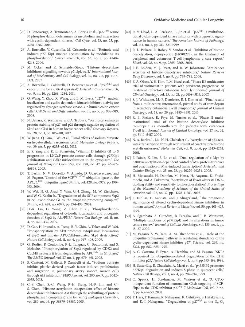
Figure 6: Mechanisms of BuA activity on Skp2 abundance. (a) K562 cells were incubated for 16 hours with 1mM BuA. Then, the nuclearand cytosolic compartments were prepared. Finally, content of Skp2 was determined by immunoblotting. Histone H3 and 𝛽-tubulin wereused as markers of nuclear and cytosolic fraction, respectively, and for equal loading evaluation. (b) EPN cells were transfected with threedifferent convalidated siRNAs directed against Skp2 mRNA. After 72 hours, the amount of Skp2, p27Kip1, and actin content was determinedby immunoblotting. In the brackets the catalog number of the siRNA is reported. (c) EPN cells were transfected with pcDNA3-Skp2 plasmidor treated with BuA (or both). After 48 hours, the amount of Skp2 and p27Kip1 (p27) content was determined by immunoblotting. Actin wasevaluated for confirming equal loading. (d) K562 cells were cultured with or without BuA for 16 hours. Then, cell extracts were preparedand analyzed by immunoblotting for GSK3𝛽 and phospho-GSK3𝛽 (pGSK3𝛽). Actin was also determined for confirming equal loading. (e)K562 cells were cultured with or without BuA for 16 hours. Then, cell extracts were prepared. CDK2 was immunoprecipitated from control(Con, untreated) and treated cells. On the top is showed CDK2 abundance in the input extracts, CDK2 remaining in the supernatants (toconfirm the total precipitation), and CDK2 in 1/10 of IP. An IP not related (NR) is also showed. On the bottom is reported the mixture ofassay. CDK2 immunoprecipitated from Con and BuA was employed to phosphorylate recombinant p27Kip1 on threonine 187. The mixture ofassay was analyzed for the content of p27Kip1 and phosphothreonine 187-p27Kip1 (pT187p27). The IP not related was also employed as controlof phosphorylation.
however, not noticeable (Figure 5(c)). We also evaluated theeffect of BuA on Skp2 gene transcription by quantitative PCR(Figure 5(d)). We observed a significant decrease only after24 hours of incubation while, at 48 hours of treatment, thetranscript reduction, with respect to untreated cells, was notstatistically significant. Conversely, as shown in Figure 5(e),Skp2 protein progressively diminishes in BuA-treated K562
cells starting from 16 hours. At 48 hours of BuA treatment,Skp2 signal was scarcely detectable (or undetectable).
The levels of Skp2 protein in both the nuclear andthe cytosolic compartments in response to BuA treatmentwere also investigated. Figure 6(a) shows that Skp2 is mostlylocalized in the nucleus and that it diminished in thiscompartment after 16 hours of treatment. No relocalization

12 Oxidative Medicine and Cellular Longevity
of the protein was detected. To further confirm the mean-ing of Skp2 downregulation in the increase of p27Kip1, wetreated EPN cells with different Skp2 siRNAs. As shown inFigure 6(b), the treated cells showed a complete silencing ofSkp2 and a significant upregulation of p27Kip1. Finally, weevaluated whether forced expression of Skp2 might causea decrease of p27Kip1 level. As shown in Figure 6(c), theaccumulation of Skp2 in EPN cells causes a significant down-regulation of p27Kip1 level. Moreover, the overexpression ofSkp2 reduced the effect of BuA on p27Kip1 cellular content.It is to underline that BuA might enhance the activity ofexpression vectors and this general effect might explain thehigh Skp2 abundance in cells transfected in the presence ofthe HDACI. Furthermore, the occurrence of various bandsin transfected Skp2 is probably due to different degree ofphosphorylation.
Since a remarkable difference between Skp2 transcriptdecrease and Skp2 protein reduction has been observed afterBuA treatment, we decided to take into consideration furthermechanisms of Skp2 regulation.
However, because NAM did not change p27Kip1 and Skp2abundance, an effect on Skp2 acetylation was discarded. Onthe other hand, Skp2 has been reported to be phosphorylatedand stabilized by Akt1 and CDK2 [35–37].Thus, we evaluatedwhether BuA treatment affected the activity of these twokinases. As shown in Figure 6(d), the treatment with the BuAreduced the amount of phosphorylated GSK3𝛽, a key Akt1substrate, suggesting that the HDACI inhibits the activityof Akt1 and confirming previous data [38, 39]. We alsoevaluated CDK2 activity after BuA treatment by investigatingthe capability of equal amount of the kinase to phosphorylatehuman recombinant p27Kip1 on T187 residue. As shown inFigure 6(e), after 16 hours of BuA treatment, theCDK2 kinaseactivity is strongly reduced.
3.4. Characterization of p27Kip1 Isoform Pattern after HDACIsTreatment. One of the most complex features of p27Kip1is its intricate metabolism. Indeed, a series of events, thatis, phosphorylation on different amino acid residues andthe nuclear/cytosol shuttling, controls its level and func-tion. The complexity of p27Kip1 metabolism investigationprompted us to develop a biochemical method based on aninitial separation of cell extracts by 2D-SDS PAGE, followedby immunoblotting detection with specific antibodies. Weemployed this analytical method to characterize in detailthe p27Kip1 isoforms [25, 26]. We also demonstrated thatthe p27Kip1 bidimensional electrophoretic pattern includes,in addition to the unmodified native form (form 0), threephosphorylated isoforms which contain one (forms 2, 3) ortwo (form 4) phosphate groups each (Figure 7(a)). Moreover,we identified a p27Kip1 isoform (form 1) focalizing betweenforms 0 and 2. This isoform does not carry a phosphategroup but a still unknown postsynthetic modification [27].The monophosphorylated (form 2) and the wild type (form0) forms are the most abundant isoforms, while the oth-ers are generally scarcely present. Furthermore, the ratio
between forms 0 and 2 is strongly dependent on the cel-lular localization (i.e., nucleus or cytosol) [25, 26]. Finally,isoform 2, which might include multiple p27Kip1 isoformsmonophosphorylated on different residues, is mainly formedby phosphoserine 10-p27Kip1 (pS10p27Kip1).
The analysis of p27Kip1 isoforms, performed separatelyon the nuclear and cytosolic fractions from 16-hour-treatedEPN cells with or without BuA, is reported in Figures 7(b)and 7(c). We observed that BuA induced in the nucleus thebuild-up of p27Kip1 monophosphoisoform 2. Similarly, inthe cytosol, the monophosphorylated isoform increases andbiphosphorylated forms appeared. Experiments of immuno-precipitation with anti-pS10p27Kip1 antibodies demonstrateda significant increase of this isoform (Figure 7(d)). Moreover,the bidimensional analysis of BuA-treated cell extracts beforeand after the removal of pS10p27Kip1 clearly showed that theincreased monophosphorylated isoform is essentially suchisoform (Figure 7(e)). This, in turn, suggested that, despiteSkp2 decrease, no upregulation of phosphothreonine 187-p27Kip1 (pT187p27Kip1) occurred confirming the strong BuA-dependent CDK2 activity inhibition.
4. Discussion
There is now ample evidence that cancers with low levelof p27Kip1 or its cytosolic relocalization show a negativeprognosis when compared to neoplasias of the same typeand stage, but with a higher content of nuclear p27Kip1.The finding, initially obtained in colon and breast tumors,has been subsequently confirmed in lung, prostate, gastric,and bladder tumors and other cancers [20, 21, 31, 32, 40].These burgeoning clinical data, supporting a role for p27Kip1dysregulation in cancer development, suggest that the CKImay be considered a target for therapy. In this context, theresults reported here provide support for the use of HDACIsas drugs for increasing p27Kip1 levels and might prove to beuseful for future anticancer therapies or preventive strategiesagainst human carcinogenesis.
HDACIs represent a class of compounds endowed withsignificant effects on proliferation, differentiation, and apop-tosis. These properties prompted their use in the treatmentof a number of pathologies, including cancer, erythropoieticdiseases, and, more recently, inflammatory and metabolicpathologies. Besides VPA, which has been for many years theonly HDACI introduced in therapy (mainly in the treatmentof epilepsy and bipolar disorder and prevention of migraineheadaches), vorinostat and romidepsin have been recentlyapproved by the FDA for the treatment of CTCL. Veryrecently also belinostat received approval for use in therapyof peripheral T cell lymphoma. Hundreds of clinical trials areinvestigating the clinical use of BuA, PBuA, SAHA, apicidin,MS-275, and other HDAC inhibitors for the treatment ofvarious human malignancies, both as single agents and incombination chemotherapeutic protocols [7, 41–45].
Themolecular characterization of HDACI mechanism(s)of action is a major goal of studies on these molecules in viewof identifying novel targets for therapy. It is generally acceptedthat HDACIs effects are mostly due to their capability of

Oxidative Medicine and Cellular Longevity 13
1P
1P
2P p27
4 2 0
3 1
?p27
p27isoforms
(a)
Con
+BuA
2
4 3 1
0
2 0
Nucleus p27
(b)
Con
+BuA
2
4 3 1
0
2 0
Cytosol p27
(c)
p27
IPpS10p27 IP IP
pS10p27supernatant Input NR
Con
BuA
Con
BuA
Con
BuA
BuA
(d)
2 0
2 0
1
IPpS10p27
supernatant
BuA-treatedinput
(e)
Figure 7: Effect of BuA on the various p27Kip1 isoforms. (a) EPN cells were treated with BuA 1mM for 24 hours.Then, the nuclear extract wasprepared and analyzed by bidimensional SDS PAGE and the isoforms were identified byMAb against p27Kip1 [25, 26]. Form 0 corresponds tothe wild type form and isoform 2 corresponds to the protein with 1 phosphate and isoform 4 corresponds to the CKI with 2 phosphate groups.Form 1 has not been identified and form 3 is the monophosphorylated derivative of form 1. Details on the identification are reported in [25].Note that the immunoblotting reported corresponds to that which is showed in (b) (BuA-treated nuclear extract) but is exposed to the filmfor a long time period (i.e., 10 minutes). (b) EPN cells were treated or not for 24 hours with 1mM BuA.Then, nuclear extracts were preparedand analyzed (1mg extracts) by bidimensional analysis for p27 isoforms. (c) Exactly as in (b) except that cytosolic extracts were analyzed.(d) EPN cells were treated or not for 24 hours with 1mM BuA. Then total extracts were prepared and immunoprecipitated with antibodiesagainst phosphoserine 10-p27Kip1 (pS10p27). The blot reports the input materials, the IP, the supernatant after pS10p27 removal, and an IPnot related (NR). (e) EPN cells were treated for 24 hours with 1mM BuA.Then cell extracts were prepared and divided into 2 equal aliquots.From one aliquot pS10p27 was removed by IP. Finally, the untreated aliquot and the aliquot from which pS10p27 was removed were analyzedby bidimensional immunoblotting.
reprogramming gene transcription by enhancing the acety-lation of either histones or nonhistone proteins, includingp53, GATA-1, FOXO proteins, and estrogen and androgenreceptors ([7, and references therein], [46–48]). However, thedefinite identification of HDACIs targets is still lacking.
We have focused our attention on the HDACI effects onp27Kip1, since this Cip/Kip inhibitor plays a critical role inmalignant transformation [20, 21, 40, 49–53] and, thus, itshandlingmight contribute to explain the putative antitumoralHDACIs activities.
p27Kip1 is a critical regulator of cell division cycle, sinceit interacts with and inhibits several cyclin/CDK complexes.The protein also modulates, in mid-G1, the assembly andnuclear import of cyclin Ds-CDK4/6 complexes. p27Kip1cellular content is regulated by intricate posttranslationalmechanisms and by proteasome removal ([20, 21] and refer-ences therein). p27Kip1 degradation is controlled by at leasttwo distinct ubiquitination processes which occur separatelyin the nuclear and in the cytosolic compartments. Nuclearubiquitin-dependent p27Kip1 degradation requires an initialphosphorylation on T187, catalyzed by active CDK2 in G1 →S phase transition [51, 52]. T187 phosphorylation permits
p27Kip1 recognition by Skp2, the recognition subunit of theE3 ligase of SCF-type complex, formed by Skp2 itself, togetherwith Cks1, Skp1, Cul1, and Roc1 [53, 54]. Conversely, cytosolicubiquitination occurs in a manner independent of T187phosphorylation and is required for the G1 reentry of cellsfrom G0 phase [55, 56]. The E3 complex responsible forthe cytosolic removal (named KPC) has been identified andcharacterized [57, 58]. Ubiquitinated p27Kip1 is degraded byproteasome activities both in the nucleus and in the cytosol.Additional degradation mechanisms have been putativelyidentified in transgenic mice harboring p27Kip1mutated gene[59].
We found that different HDACIs increase, in a numberof cellular models, p27Kip1 abundance. This effect, in spiteof the frequent HDACI capability to positively modulategene expression, is not due to an upregulation of CDKN1Btranscription. We also observed that p27Kip1 increase occursin both nuclear and cytosolic compartments, allowing theexclusion of protein relocalization as the cause of the CKIaccumulation.
When we investigated p27Kip1 turnover, we observed astabilization of the protein in BuA-treated cells, suggesting

14 Oxidative Medicine and Cellular Longevity
that at least one step of the CKI degradation pathway isdownregulated byHDACIs.Then, we ruled out the possibilitythat BuA acts as a general inhibitor of ubiquitination activity(Figure 4(c)), while we observed a strong HDACI-relateddownregulation of Skp2, an essential component of p27Kip1ubiquitination complex.
Various experiments demonstrate that Skp2 decreaseoccurs by using different HDACIs and in several distinctcellular models. Such data appear of importance, in that theeffect might be considered as a frequent response to thesedrugs, independently of the specific cellular context. Ourresults confirm previous data obtained employing specificHDACIs and distinct experimental models [29–31]. In onecellularmodel, that is, UT-7 cells, BuAdoes not downregulateSkp2. Preliminary data suggested to us that HDACIs alsodo not inhibit the proliferation of these cells (not reported).Future studies are necessary for clarifying the lack of effect inthis cellular model.
Although we did not identify definitely the mecha-nism(s) by which HDACIs decrease Skp2 protein, we initiallyobserved that these compounds reduce Skp2 mRNA. How-ever, Skp2 transcript reduction was statistically significantonly after 24 hours of treatment while itmight not be detectedafter 48 hours of BuA addition. On the other hand, Skp2protein reduction appears quantitativelymore significant andprecocious than that of the transcript, since it was statisticallydetectable at 16 hours of treatment. In addition, at 48 hoursof treatment very scarce Skp2 protein can be found byimmunoblotting. These results indicated the occurrence ofnontranscriptional mechanisms leading to BuA-dependentSkp2 decrease.
It has been demonstrated that Skp2 mRNA/protein levelare very low in G0 and early G1 [60]. Since HDACIs inducea rapid accumulation of cells in G1 phase (Figure 1(d)), weinitially hypothesized that Skp2 downregulation is a directconsequence of G1 phase arrest. However, cytofluorimetricstudies of BuA-, PBuA-, and VPA-treated K562 cells showa very rapid build-up of the cells in G1, corresponding toa fall-down of cells in S phase (a cell cycle phase in whichSkp2 abundance is at its maximum) persisting up to 24 hours;afterwards, among 24 and 48 hours of incubation, the S phasecell number rises at higher percentage while G1 phase cellsdecrease. These data suggest the occurrence of a complexeffect on the transitions between G1 → S and S → G2/M.
The FACS kinetics is clearly discordant with the analysisof Skp2 protein cellular content. Indeed, Skp2 continued todecrease and it almost disappeared at 48 hours of incubationwith BuA.These findings again suggest to us that if an effect ofcell cycle synchronizationmight influence Skp2 abundance, itis not the only phenomenon at the basis of Skp2 protein levelmodulation and that multiple mechanisms (translational andposttranslational) controlling Skp2 level are modulated byHDACIs.
As shown in Figure 4(a), the treatment with proteasomeinhibitor slightly increases Skp2. Analogously, the contem-poraneous addition of BuA and ALLN reduces the effect ofBuA alone. Both these findings suggest, in agreement withdata in literature, that Skp2 levels are regulated, at least in
part, by proteasome degradation. As for p27Kip1 and manyother proteins, Skp2 posttranslational modifications mightdrive itsmetabolism. Skp2 stability is regulated by acetylation.Particularly, the protein has been reported to be acetylated byp300 on K68 and K71, therefore becoming more stable [61].Analogously, inhibition of SIRT3 increases Skp2 acetylation,increasing the protein through impairment of its proteolysis[61]. However, NAM (a powerful inhibitor of SIRT3) does notaffect Skp2 and p27Kip1 abundance. Thus, acetylation shouldbe discarded as a mechanism for regulating p27Kip1 levels.
A further mechanism regulating Skp2 degradation is itsphosphorylation on S72 catalyzed by Akt/PKB [35, 36] or onS64 due to active CDK2/CDK1 [37]. Phosphorylation on S72induces twomajormolecular effects, that is, the relocalizationof Skp2 in the cytosol (and its binding to 14-3-3 protein) and aputative increased formation of SCF complex [35, 36]. In turn,the relocalization in the cytosol might stabilize the protein[35, 36]. Although we confirmed that HDACI might inhibitAkt1 and thus might reduce the phosphorylation of Skp2on S72, we did not evidence any remarkable change in thelocalization of Skp2 during the initial 24 hours of treatment.CDK2/CDK1-dependent Skp2 phosphorylation on S64 alsostabilizes the protein hampering its interaction with APC-Cdh1 (which targets Skp2 for degradation). Thus, the strongreduction of CDK2 activity due to BuA might contributeto Skp2 reduction. However, additional experiments arerequired for substantiating this hypothesis and for analyzingthe role of other putative Skp2-phosphorylating kinases.
The importance of Skp2 downregulation by HDACIsis not confined to its role in p27Kip1 metabolism. Indeed,the increase of Skp2 content has been reported as anindependent marker of cancer malignity and aggressiveness.In particular, Skp2 upregulation has been demonstratedin breast carcinoma, non-small cell lung cancer, colorectalcarcinoma, gastric carcinoma, renal cell carcinoma, earlyesophageal squamous cell carcinoma, malignant melanoma,prostate cancer, chronic myelogenous leukemia, malignantlymphoma, and several other human cancers [62, 63]. Thus,the discovery that HDACIs downregulate Skp2 levels hasimportant pharmacological implications in cancer develop-ment and prognosis. Skp2, as a part of the SCF-Skp2 E3ubiquitination complex, is required for the removal of severalkey proteins asMyc, FOXO1, cancer-derived Smad 4mutants,ISG15 isopeptidase UBP43, and other proteins.Therefore, theprotein downregulation should also have profound effects oncell phenotypes and might be important in explaining thenumerous additional activities of HDACIs. In this context,it is important to note that up to now there are scarcedata on the effect of HDACIs (i.e., BuA and VPA) on theubiquitination machinery [64]. It has been reported thatthe HDACIs downregulate HDAC2 content by inducing theexpression of E2 ubiquitin conjugase Ubc8 and, contem-poraneously, decreasing the level of the E3 ubiquitin ligaseRLIM, two components of the complex responsible for theubiquitination of HDAC2 [64].
In conclusion, our study reports that HDACIs increasep27Kip1 content by elongating its half-life. We also show thatthe effect is due, at least in part, to a reduced CKI degradation

Oxidative Medicine and Cellular Longevity 15
through a strong Skp2 downregulation. Finally, the decreaseof Skp2 abundance, due to the HDACIs treatment, appearsto be due to the convergence of translational and posttrans-lational mechanisms. Due to the importance of HDACIsin new therapeutic strategies, our data might contribute tounravelling their mechanism(s) of action, identifying p27Kip1as a major molecular target. Moreover, we demonstrate thatthese molecules might modulate the cellular protein profilescontrolling the activity of pivotal ubiquitination enginesincluding SCF-Skp2. This finding appears of importance inthe evaluation of the complex molecular effects of thesedrugs.
Conflict of Interests
The authors declare that there is no conflict of interestsregarding the publication of this paper.
Acknowledgments
The authors sincerely thank Dr. Germain (Department ofMedicine, Division of Hematology/Oncology, Mount SinaiSchool of Medicine, New York, USA) for pcDNA3-Skp2and pcDNA3.0-ubiquitin-cMyc. This work was supported bygrants from Associazione Italiana per la Ricerca sul Cancro(AIRC) (to Fulvio Della Ragione), Progetti di Rilevante Inter-esse Nazionale (PRIN 2010NFEB9L 007 to Antonio AgostinoSinisi), and FIRB (to Fulvio Della Ragione).
References
[1] S. L. Berger, “The complex language of chromatin regulationduring transcription,” Nature, vol. 447, no. 7143, pp. 407–412,2007.
[2] T. Kouzarides, “Chromatin modifications and their function,”Cell, vol. 128, no. 4, pp. 693–705, 2007.
[3] M. A. Dawson and T. Kouzarides, “Cancer epigenetics: frommechanism to therapy,” Cell, vol. 150, no. 1, pp. 12–27, 2012.
[4] K. J. Falkenberg and R.W. Johnstone, “Histone deacetylases andtheir inhibitors in cancer, neurological diseases and immunedisorders,” Nature Reviews Drug Discovery, vol. 13, no. 9, pp.673–691, 2014.
[5] H. A. Tissenbaum and L. Guarente, “Increased dosage of a sir-2 gene extends lifespan in Caenorhabditis elegans,” Nature, vol.410, no. 6825, pp. 227–230, 2001.
[6] M. A. Dawson, T. Kouzarides, and B. J. P. Huntly, “Targetingepigenetic readers in cancer,” The New England Journal ofMedicine, vol. 367, no. 7, pp. 647–657, 2012.
[7] A. C. West and R. W. Johnstone, “New and emerging HDACinhibitors for cancer treatment,”The Journal of Clinical Investi-gation, vol. 124, no. 1, pp. 30–39, 2014.
[8] L. Staiano-Coico, M. Steinberg, and P. J. Higgins, “Epider-mal cell-shape regulation and subpopulation kinetics duringbutyrate-induced terminal maturation of normal and SV40-transformed human keratinocytes: epithelial models of differ-entiation therapy,” International Journal of Cancer, vol. 46, no.4, pp. 733–738, 1990.
[9] A. Hague, A. M. Manning, K. A. Hanlon, L. I. Huschtscha, D.Hart, and C. Paraskeva, “Sodium butyrate induces apoptosis
in human colonic tumour cell lines in a p53-independentpathway: implications for the possible role of dietary fibre inthe prevention of large-bowel cancer,” International Journal ofCancer, vol. 55, no. 3, pp. 498–505, 1993.
[10] B. G. Heerdt, M. A. Houston, and L. H. Augenlicht, “Poten-tiation by specific short-chain fatty acids of differentiationand apoptosis in human colonic carcinoma cell lines,” CancerResearch, vol. 54, no. 12, pp. 3288–3293, 1994.
[11] E. C. Aguilar, A. J. Leonel, L. G. Teixeira et al., “Butyrate impairsatherogenesis by reducing plaque inflammation and vulnerabil-ity and decreasing NF𝜅B activation,”Nutrition, Metabolism andCardiovascular Diseases, vol. 24, no. 6, pp. 606–613, 2014.
[12] S. P. Perrine, M. F. Greene, and D. V. Faller, “Delay in the fetalglobin switch in infants of diabetic mothers,”The New EnglandJournal of Medicine, vol. 312, no. 6, pp. 334–338, 1985.
[13] S. P. Perrine, A. Rudolph, D. V. Faller et al., “Butyrate infusionsin the ovine fetus delay the biologic clock for globin geneswitching,” Proceedings of the National Academy of Sciences oftheUnited States of America, vol. 85, no. 22, pp. 8540–8542, 1988.
[14] S. P. Perrine, G. D. Ginder, D. V. Faller et al., “A short-term trialof butyrate to stimulate fetal-globin-gene expression in the 𝛽-globin disorders,”TheNewEngland Journal ofMedicine, vol. 328,no. 2, pp. 81–86, 1993.
[15] D. V. Faller and S. P. Perrine, “Butyrate in the treatment of sicklecell disease and𝛽-thalassemia,”Current Opinion inHematology,vol. 2, no. 2, pp. 109–117, 1995.
[16] S. P. Perrine, O. Hermine, T. Small et al., “A phase 1/2 trial ofarginine butyrate and ganciclovir in patients with Epstein-Barrvirus-associated lymphoid malignancies,” Blood, vol. 109, no. 6,pp. 2571–2578, 2007.
[17] V. M. Richon, Y. Webb, R. Merger et al., “Second generationhybrid polar compounds are potent inducers of transformedcell differentiation,” Proceedings of the National Academy ofSciences of the United States of America, vol. 93, no. 12, pp. 5705–5708, 1996.
[18] M. Gottlicher, S. Minucci, P. Zhu et al., “Valproic acid definesa novel class of HDAC inhibitors inducing differentiation oftransformed cells,”TheEMBO Journal, vol. 20, no. 24, pp. 6969–6978, 2001.
[19] A. Besson, S. F. Dowdy, and J. M. Roberts, “CDK inhibitors: cellcycle regulators and beyond,” Developmental Cell, vol. 14, no. 2,pp. 159–169, 2008.
[20] A. Borriello, V. Cucciolla, A. Oliva, V. Zappia, and F. DellaRagione, “p27Kip1 metabolism: a fascinating labyrinth,” CellCycle, vol. 6, no. 9, pp. 1053–1061, 2007.
[21] A. Borriello, D. Bencivenga, M. Criscuolo et al., “Targetingp27Kip1 protein: its relevance in the therapy of human cancer,”Expert Opinion on Therapeutic Targets, vol. 15, no. 6, pp. 677–693, 2011.
[22] F. Della Ragione, V. Cucciolla, V. Criniti, S. Indaco, A. Borriello,and V. Zappia, “𝑝21𝑐𝑖𝑝1 gene expression is modulated by Egr1:a novel regulatory mechanism involved in the resveratrolantiproliferative effect,” Journal of Biological Chemistry, vol. 278,no. 26, pp. 23360–23368, 2003.
[23] A. Borriello, V. Della Pietra, M. Criscuolo et al., “p27Kip1 accu-mulation is associated with retinoic-induced neuroblastomadifferentiation: evidence of a decreased proteasome-dependentdegradation,” Oncogene, vol. 19, no. 1, pp. 51–60, 2000.
[24] V. Cucciolla, A. Borriello, M. Criscuolo et al., “Histone deacety-lase inhibitors upregulate p57Kip2 level by enhancing its expres-sion through Sp1 transcription factor,” Carcinogenesis, vol. 29,no. 3, pp. 560–567, 2008.
16 Oxidative Medicine and Cellular Longevity
[25] D. Bencivenga, A. Tramontano, A. Borgia et al., “p27Kip1 serine10 phosphorylation determines its metabolism and interactionwith cyclin-dependent kinases,” Cell Cycle, vol. 13, no. 23, pp.3768–3782, 2014.
[26] A. Borriello, V. Cucciolla, M. Criscuolo et al., “Retinoic acidinduces p27 Kip1 nuclear accumulation by modulating itsphosphorylation,” Cancer Research, vol. 66, no. 8, pp. 4240–4248, 2006.
[27] M. Ocker and R. Schneider-Stock, “Histone deacetylaseinhibitors: signalling towards p21cip1/waf1,” International Jour-nal of Biochemistry and Cell Biology, vol. 39, no. 7-8, pp. 1367–1374, 2007.
[28] A. Borriello, I. Caldarelli, D. Bencivenga et al., “p57Kip2 andcancer: time for a critical appraisal,”Molecular Cancer Research,vol. 9, no. 10, pp. 1269–1284, 2011.
[29] Q. Wang, Y. Zhou, X. Wang, and B. M. Evers, “p27Kip1 nuclearlocalization and cyclin-dependent kinase inhibitory activity areregulated by glycogen synthase kinase-3 in human colon cancercells,” Cell Death and Differentiation, vol. 15, no. 5, pp. 908–919,2008.
[30] N. Uehara, K. Yoshizawa, and A. Tsubura, “Vorinostat enhancesprotein stability of p27 and p21 through negative regulation ofSkp2 and Cks1 in human breast cancer cells,” Oncology Reports,vol. 28, no. 1, pp. 105–110, 2012.
[31] W. Jiang, Q. Guo, J. Wu et al., “Dual effects of sodium butyrateon hepatocellular carcinoma cells,” Molecular Biology Reports,vol. 39, no. 5, pp. 6235–6242, 2012.
[32] E. S. Yang and K. L. Burnstein, “Vitamin D inhibits G1 to Sprogression in LNCaP prostate cancer cells through p27Kip1stabilization and Cdk2 mislocalization to the cytoplasm,” TheJournal of Biological Chemistry, vol. 278, no. 47, pp. 46862–46868, 2003.
[33] T. Bashir, N. V. Dorrello, V. Amado, D. Guardavaccaro, andM. Pagano, “Control of the SCFSkp2−Cks1 ubiquitin ligase by theAPC/CCdh1 ubiquitin ligase,”Nature, vol. 428, no. 6979, pp. 190–193, 2004.
[34] W. Wei, N. G. Ayad, Y. Wan, G.-J. Zhang, M. W. Kirschner,and W. G. Kaelin Jr., “Degradation of the SCF component Skp2in cell-cycle phase G1 by the anaphase-promoting complex,”Nature, vol. 428, no. 6979, pp. 194–198, 2004.
[35] H.-K. Lin, G. Wang, Z. Chen et al., “Phosphorylation-dependent regulation of cytosolic localization and oncogenicfunction of Skp2 by Akt/PKB,” Nature Cell Biology, vol. 11, no.4, pp. 420–432, 2009.
[36] D. Gao, H. Inuzuka, A. Tseng, R. Y. Chin, A. Toker, andW.Wei,“Phosphorylation by Akt1 promotes cytoplasmic localizationof Skp2 and impairs APCCdh1-mediated Skp2 destruction,”Nature Cell Biology, vol. 11, no. 4, pp. 397–408, 2009.
[37] G. Rodier, P. Coulombe, P.-L. Tanguay, C. Boutonnet, and S.Meloche, “Phosphorylation of Skp2 regulated by CDK2 andCdc14B protects it from degradation by APCCdh1 in G1 phase,”The EMBO Journal, vol. 27, no. 4, pp. 679–691, 2008.
[38] S. Cantoni, M. Galletti, F. Zambelli et al., “Sodium butyrateinhibits platelet-derived growth factor-induced proliferationand migration in pulmonary artery smooth muscle cellsthrough Akt inhibition,” FEBS Journal, vol. 280, no. 9, pp. 2042–2055, 2013.
[39] C.-S. Chen, S.-C. Weng, P.-H. Tseng, H.-P. Lin, and C.-S. Chen, “Histone acetylation-independent effect of histonedeacetylase inhibitors on Akt through the reshuffling of proteinphosphatase 1 complexes,” The Journal of Biological Chemistry,vol. 280, no. 46, pp. 38879–38887, 2005.
[40] R. V. Lloyd, L. A. Erickson, L. Jin et al., “p27kip1: a multifunc-tional cyclin-dependent kinase inhibitor with prognostic signif-icance in human cancers,” The American Journal of Pathology,vol. 154, no. 2, pp. 313–323, 1999.
[41] R. L. Piekarz, R. Robey, V. Sandor et al., “Inhibitor of histonedeacetylation, depsipeptide (FR901228), in the treatment ofperipheral and cutaneous T-cell lymphoma: a case report,”Blood, vol. 98, no. 9, pp. 2865–2868, 2001.
[42] J. E. Bolden, M. J. Peart, and R. W. Johnstone, “Anticanceractivities of histone deacetylase inhibitors,” Nature ReviewsDrug Discovery, vol. 5, no. 9, pp. 769–784, 2006.
[43] E. A. Olsen, Y. H. Kim, T.M. Kuzel et al., “Phase IIBmulticentertrial of vorinostat in patients with persistent, progressive, ortreatment refractory cutaneous t-cell lymphoma,” Journal ofClinical Oncology, vol. 25, no. 21, pp. 3109–3115, 2007.
[44] S. J. Whittaker, M.-F. Demierre, E. J. Kim et al., “Final resultsfrom a multicenter, international, pivotal study of romidepsinin refractory cutaneous T-cell lymphoma,” Journal of ClinicalOncology, vol. 28, no. 29, pp. 4485–4491, 2010.
[45] R. L. Piekarz, R. Frye, M. Turner et al., “Phase II multi-institutional trial of the histone deacetylase inhibitorromidepsin as monotherapy for patients with cutaneousT-cell lymphoma,” Journal of Clinical Oncology, vol. 27, no. 32,pp. 5410–5417, 2009.
[46] N. A. Barlev, L. Liu, N. H. Chehab et al., “Acetylation of p53 acti-vates transcription through recruitment of coactivators/histoneacetyltransferases,” Molecular Cell, vol. 8, no. 6, pp. 1243–1254,2001.
[47] F. Faiola, X. Liu, S. Lo et al., “Dual regulation of c-Myc byp300 via acetylation-dependent control ofMyc protein turnoverand coactivation of Myc-induced transcription,”Molecular andCellular Biology, vol. 25, no. 23, pp. 10220–10234, 2005.
[48] H. Matsuzaki, H. Daitoku, M. Hatta, H. Aoyama, K. Yoshi-mochi, and A. Fukamizu, “Acetylation of Foxo1 alters its DNA-binding ability and sensitivity to phosphorylation,” Proceedingsof the National Academy of Sciences of the United States ofAmerica, vol. 102, no. 32, pp. 11278–11283, 2005.
[49] J. Tsihlias, L. Kapusta, and J. Slingerland, “The prognosticsignificance of altered cyclin-dependent kinase inhibitors inhuman cancer,”Annual Review ofMedicine, vol. 50, pp. 401–423,1999.
[50] A. Sgambato, A. Cittadini, B. Faraglia, and I. B. Weinstein,“Multiple functions of p27(Kip1) and its alterations in tumorcells: a review,” Journal of Cellular Physiology, vol. 183, no. 1, pp.18–27, 2000.
[51] M. Pagano, S. W. Tam, A. M. Theodoras et al., “Role of theubiquitin-proteasome pathway in regulating abundance of thecyclin-dependent kinase inhibitor p27,” Science, vol. 269, no.5224, pp. 682–685, 1995.
[52] A. C. Carrano, E. Eytan, A. Hershko, and M. Pagano, “SKP2is required for ubiquitin-mediated degradation of the CDKinhibitor p27,”Nature Cell Biology, vol. 1, no. 4, pp. 193–199, 1999.
[53] H. Sutterluty, E. Chatelain, A. Marti et al., “p45SKP2 promotesp27Kip1 degradation and induces S phase in quiescent cells,”Nature Cell Biology, vol. 1, no. 4, pp. 207–214, 1999.
[54] C. Spruck, H. Strohmaier, M. Watson et al., “A CDK-independent function of mammalian Cks1: targeting of SCF-Skp2 to the CDK inhibitor p27Kip1,” Molecular Cell, vol. 7, no.3, pp. 639–650, 2001.
[55] T.Hara, T. Kamura, K.Nakayama, K.Oshikawa, S. Hatakeyama,and K.-I. Nakayama, “Degradation of p27Kip1 at the G
0-G1

Oxidative Medicine and Cellular Longevity 17
transition mediated by a Skp2-independent ubiquitinationpathway,” The Journal of Biological Chemistry, vol. 276, no. 52,pp. 48937–48943, 2001.
[56] N. P. Malek, H. Sundberg, S. McGrew, K. Nakayama, T. R.Kyriakidis, and J. M. Roberts, “A mouse knock-in modelexposes sequential proteolytic pathways that regulate p27Kip1 inG1 and S phase,” Nature, vol. 413, no. 6853, pp. 323–327, 2001.
[57] T. Kamura, T. Hara, M. Matsumoto et al., “Cytoplasmic ubiqui-tin ligase KPC regulates proteolysis of p27(Kip1) at G1 phase,”Nature Cell Biology, vol. 6, no. 12, pp. 1229–1235, 2004.
[58] S. Kotoshibai, T. Kamura, T. Hara, N. Ishida, and K. I.Nakayama, “Molecular dissection of the interaction betweenp27 and Kip1 ubiquitylation-promoting complex, the ubiquitinligase that regulates proteolysis of p27 in G1 phase,”The Journalof Biological Chemistry, vol. 280, no. 18, pp. 17694–17700, 2005.
[59] A. Besson, M. Gurian-West, X. Chen, K. S. Kelly-Spratt, C. J.Kemp, and J. M. Roberts, “A pathway in quiescent cells thatcontrols p27𝐾𝑖𝑝1 stability, subcellular localization, and tumorsuppression,” Genes and Development, vol. 20, no. 1, pp. 47–64,2006.
[60] H. Imaki, K. Nakayama, S. Delehouzee et al., “Cell cycle-dependent regulation of the Skp2 promoter by GA-bindingprotein,” Cancer Research, vol. 63, no. 15, pp. 4607–4613, 2003.
[61] H. Inuzuka, D. Gao, L. W. S. Finley et al., “Acetylation-dependent regulation of Skp2 function,” Cell, vol. 150, no. 1, pp.179–193, 2012.
[62] D. Frescas and M. Pagano, “Deregulated proteolysis by the F-box proteins SKP2 and 𝛽-TrCP: tipping the scales of cancer,”Nature Reviews Cancer, vol. 8, no. 6, pp. 438–449, 2008.
[63] D. D. Hershko, “Oncogenic properties and prognostic implica-tions of the ubiquitin ligase Skp2 in cancer,” Cancer, vol. 112, no.7, pp. 1415–1424, 2008.
[64] O. H. Kramer, P. Zhu, H. P. Ostendorff et al., “The histonedeacetylase inhibitor valproic acid selectively induces proteaso-mal degradation of HDAC2,”The EMBO Journal, vol. 22, no. 13,pp. 3411–3420, 2003.

Submit your manuscripts athttp://www.hindawi.com
Stem CellsInternational
Hindawi Publishing Corporationhttp://www.hindawi.com Volume 2014
Hindawi Publishing Corporationhttp://www.hindawi.com Volume 2014
MEDIATORSINFLAMMATION
of
Hindawi Publishing Corporationhttp://www.hindawi.com Volume 2014
Behavioural Neurology
EndocrinologyInternational Journal of
Hindawi Publishing Corporationhttp://www.hindawi.com Volume 2014
Hindawi Publishing Corporationhttp://www.hindawi.com Volume 2014
Disease Markers
Hindawi Publishing Corporationhttp://www.hindawi.com Volume 2014
BioMed Research International
OncologyJournal of
Hindawi Publishing Corporationhttp://www.hindawi.com Volume 2014
Hindawi Publishing Corporationhttp://www.hindawi.com Volume 2014
Oxidative Medicine and Cellular Longevity
Hindawi Publishing Corporationhttp://www.hindawi.com Volume 2014
PPAR Research
The Scientific World JournalHindawi Publishing Corporation http://www.hindawi.com Volume 2014
Immunology ResearchHindawi Publishing Corporationhttp://www.hindawi.com Volume 2014
Journal of
ObesityJournal of
Hindawi Publishing Corporationhttp://www.hindawi.com Volume 2014
Hindawi Publishing Corporationhttp://www.hindawi.com Volume 2014
Computational and Mathematical Methods in Medicine
OphthalmologyJournal of
Hindawi Publishing Corporationhttp://www.hindawi.com Volume 2014
Diabetes ResearchJournal of
Hindawi Publishing Corporationhttp://www.hindawi.com Volume 2014
Hindawi Publishing Corporationhttp://www.hindawi.com Volume 2014
Research and TreatmentAIDS
Hindawi Publishing Corporationhttp://www.hindawi.com Volume 2014
Gastroenterology Research and Practice
Hindawi Publishing Corporationhttp://www.hindawi.com Volume 2014
Parkinson’s Disease
Evidence-Based Complementary and Alternative Medicine
Volume 2014Hindawi Publishing Corporationhttp://www.hindawi.com
Related Documents