Histology Slides for the GI Track • Slides are presented in order of magnification • As you view the following slides make sure you can accomplish these goals. 1. Can you identify the organ from which the tissue sample was taken? 2. Can you identify the specific structures or layers indicates by the numbered arrows or brackets? • At the end of a sequence you will find the answers to the above for each organ.

Histology Slides for the GI Track Slides are presented in order of magnification As you view the following slides make sure you can accomplish these goals.
Dec 15, 2015
Welcome message from author
This document is posted to help you gain knowledge. Please leave a comment to let me know what you think about it! Share it to your friends and learn new things together.
Transcript

Histology Slides for the GI Track
• Slides are presented in order of magnification• As you view the following slides make sure
you can accomplish these goals.
1. Can you identify the organ from which the tissue sample was taken?
2. Can you identify the specific structures or layers indicates by the numbered arrows or brackets?
• At the end of a sequence you will find the answers to the above for each organ.
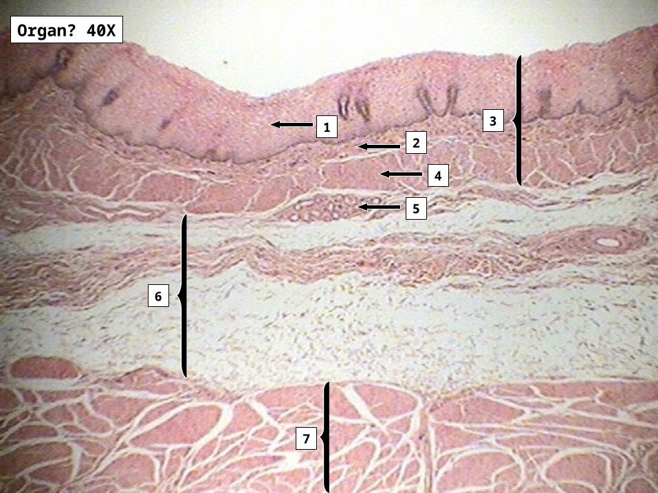
Organ? 40X
12
4
5
6
7
3

Organ 100X
1
2
3
4
5
Organ 100X
1
3
2
4
1
2
3
4
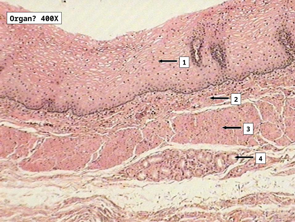
Organ? 400X
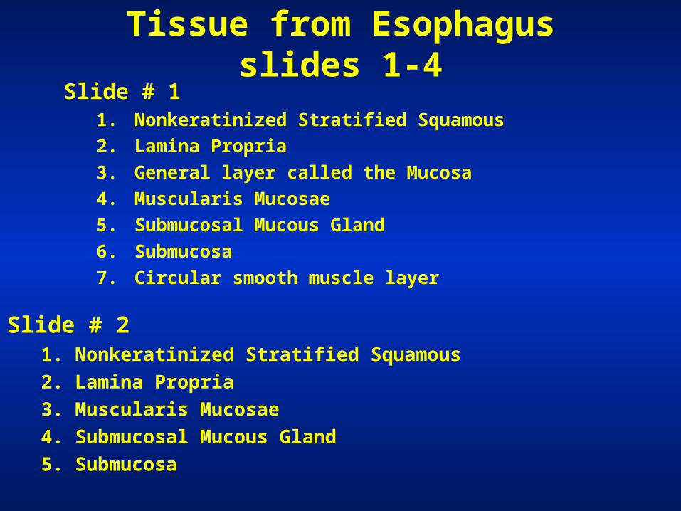
Tissue from Esophagus slides 1-4Slide # 1
1. Nonkeratinized Stratified Squamous
2. Lamina Propria
3. General layer called the Mucosa
4. Muscularis Mucosae
5. Submucosal Mucous Gland
6. Submucosa
7. Circular smooth muscle layer
Slide # 21. Nonkeratinized Stratified Squamous
2. Lamina Propria
3. Muscularis Mucosae
4. Submucosal Mucous Gland
5. Submucosa
Tissue from Esophagus slides 1-4
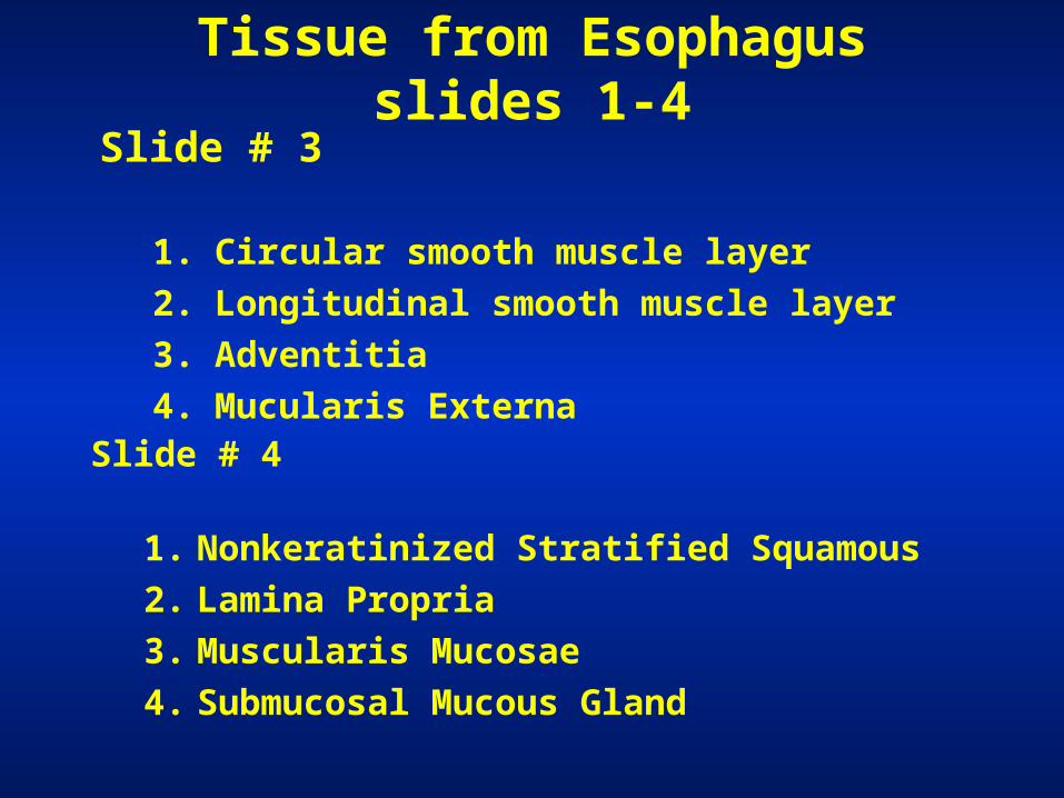
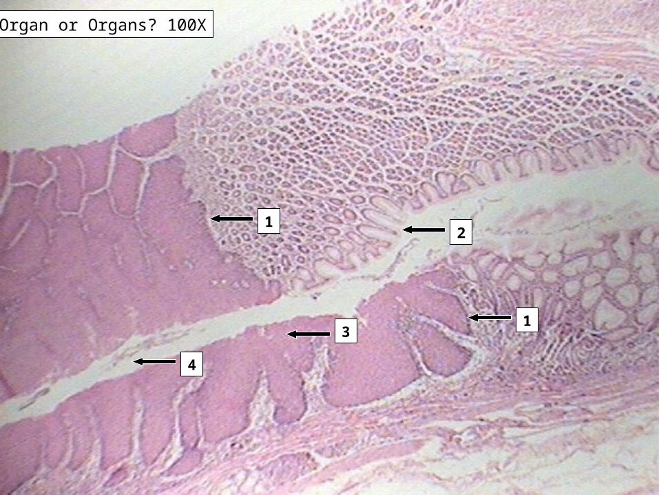
Slide # 31. Circular smooth muscle layer
2. Longitudinal smooth muscle layer
3. Adventitia
4. Mucularis Externa
Slide # 4
1. Nonkeratinized Stratified Squamous
2. Lamina Propria
3. Muscularis Mucosae
4. Submucosal Mucous Gland
4
3
21
1
Organ or Organs? 100X
1
2
Organ or Organs? 400X
Slides 8-9 Esophageal-Gastric Junction
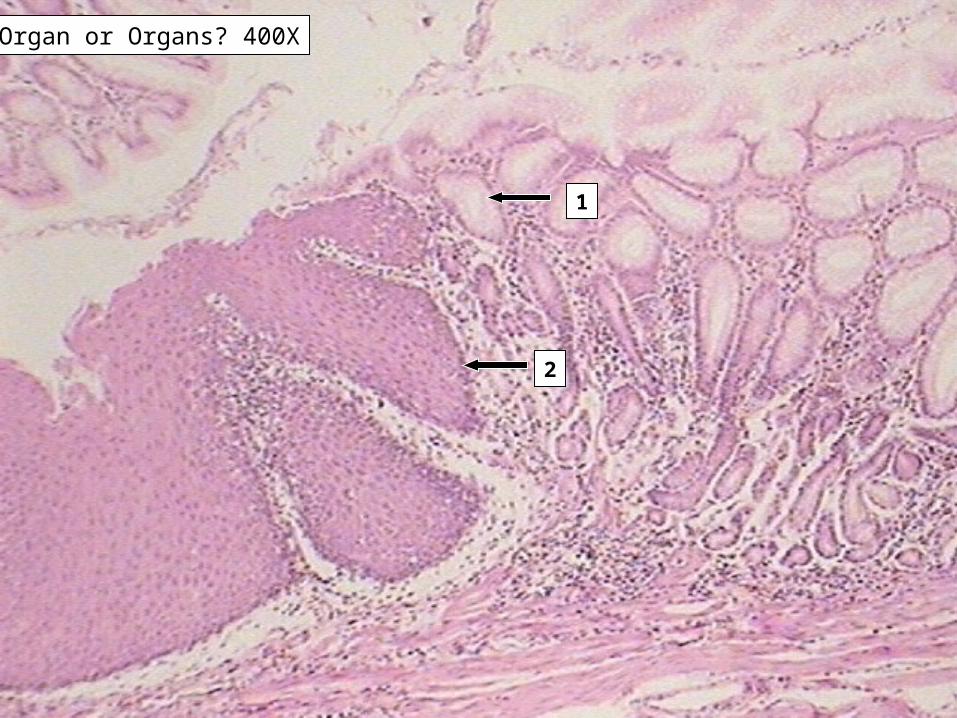
Slide # 81. Junction where epithelia changes from nonkeratinized
stratified squamous to simple columnar.
2. Simple columnar
3. Nonkeratinized stratified squamous
4. Lumen
Slide # 91. Simple columnar lining a gastric pit
2. Junction where epithelia changes from nonkeratinized
stratified squamous to simple columnar

Fold and organ? 40X1
5
4
3
2 1
6
organ? 100X
4
3
2
1
5
organ? 400X

1
3
65
4
2
9
8
7
organ? 400X
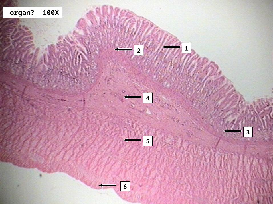
Slides 11-14 StomachSlide # 11
Large fold is a Rugae
• Gastric Mucosa
Slide # 121. Simple columnar lining a gastric pit
2. Muscularis Mucosae
3. Lamina Propria
4. Oblique smooth muscle layer
5. Circular smooth muscle layer
6. Longitudinal smooth muscle layer
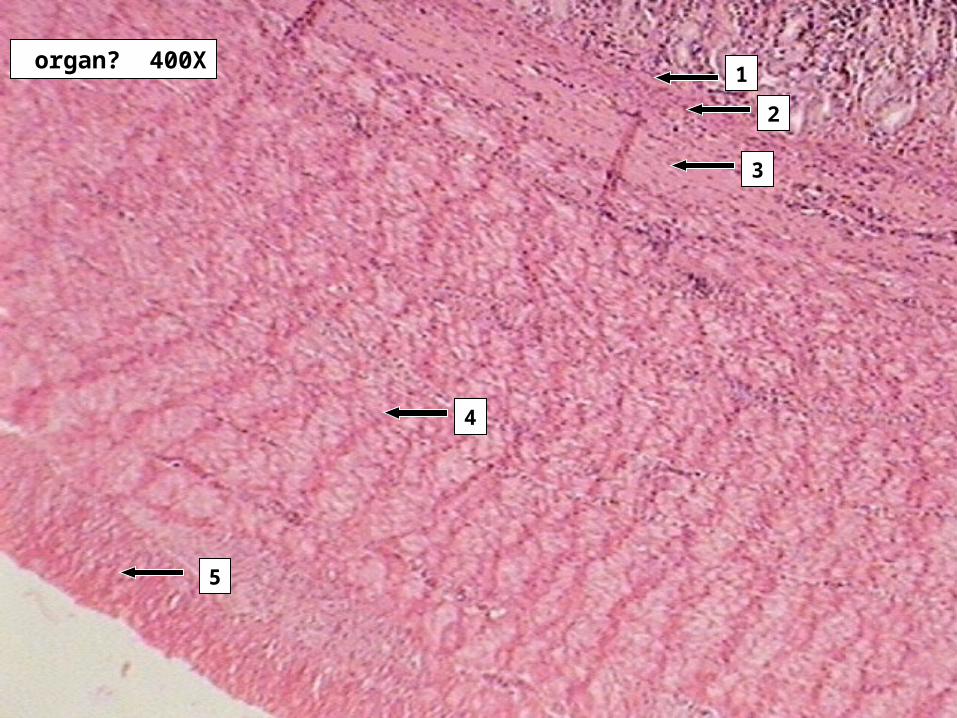
Slides 11-14 StomachSlide # 13
1. Muscularis Mucosae
2. Submucosa
3. Oblique smooth muscle layer
4. Circular smooth muscle layer
5. Longitudinal smooth muscle layer
Slide # 14
1. Muscularis Mucosae
2. G-cells at the bottom of a gastric pit
3. Submucosa
4. Lamina Propria
5. Parietal cells in a gastric pit
6. Chief cells in a gastric pit
7. Oblique smooth muscle layer
8. Circular smooth muscle layer
9. Longitudinal smooth muscle layer
organ? 40X
2
2
1
organ? 400X
1
2
3
organ? 400X
2
3
1
4
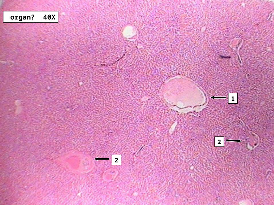
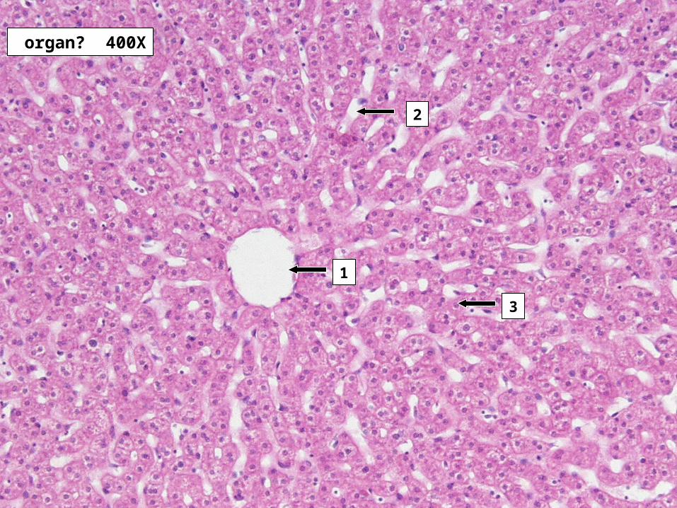
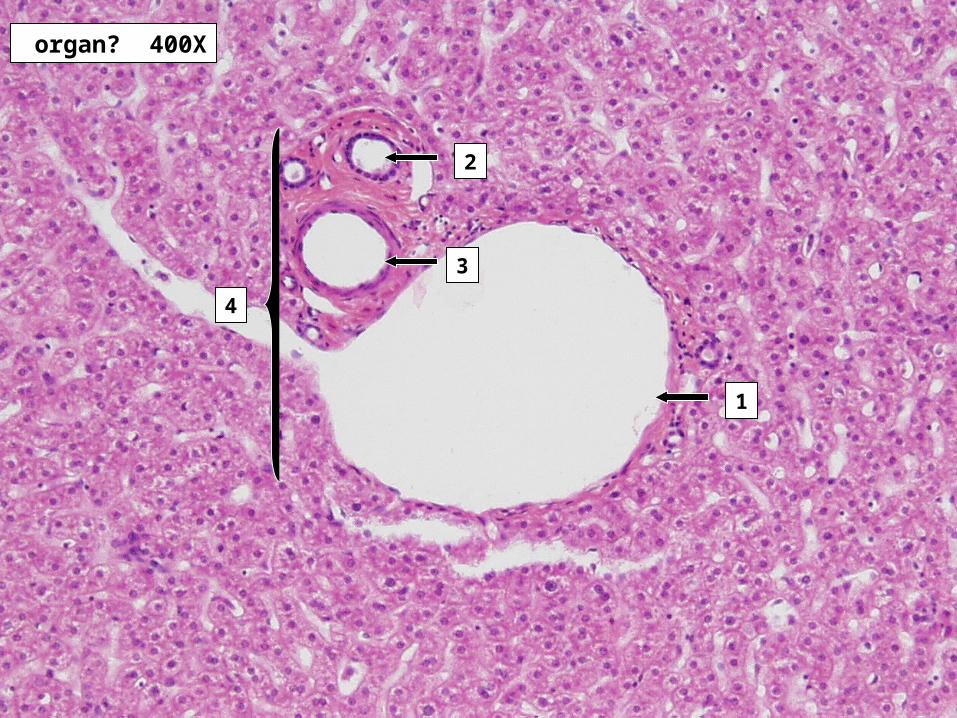
Slides 17-19 Liver TissueSlide # 17
1. Central Vein2. Portal triads demonstration the boundary of the lobule
Slide # 181. Central Vein
2. Sinusoids
3. Hepatocyte
Slide # 191. Branch of the Hepatic Portal Vein
2. Bile duct
3. Branch of the Hepatic Artery
4. Portal Triad
organ? 40X
1 2
organ? 100X
2
3
1
organ? 100X
2
1
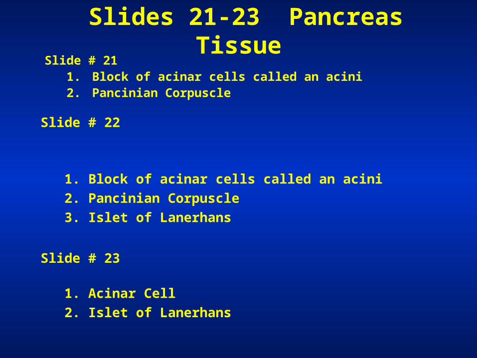
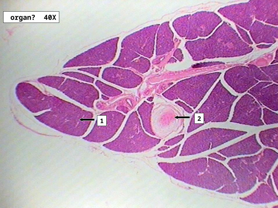
Slides 21-23 Pancreas TissueSlide # 21
1. Block of acinar cells called an acini2. Pancinian Corpuscle
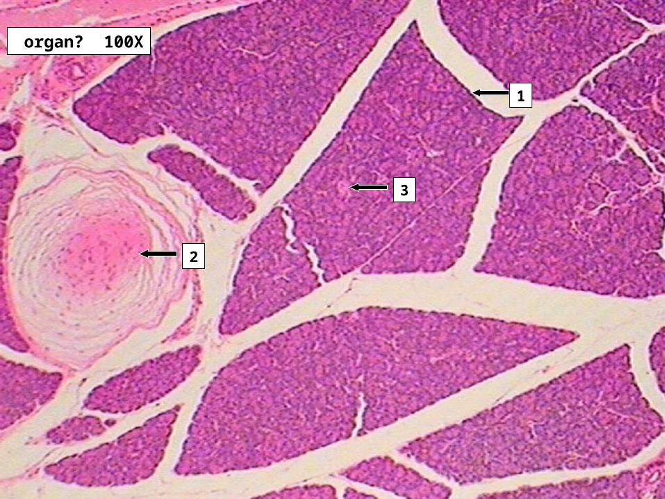
Slide # 22
1. Block of acinar cells called an acini
2. Pancinian Corpuscle
3. Islet of Lanerhans
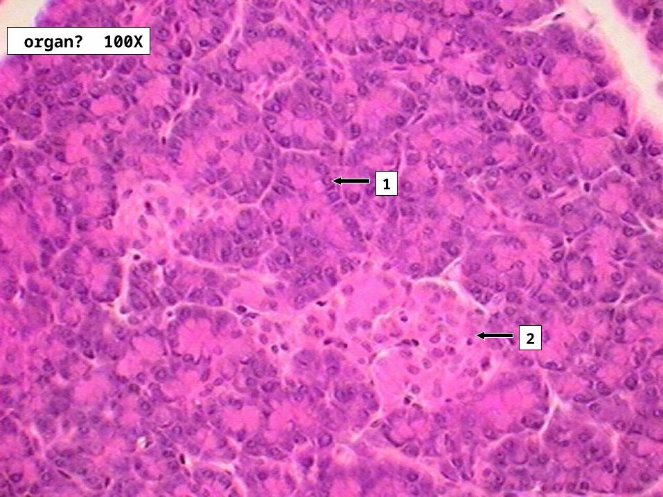
Slide # 23
1. Acinar Cell
2. Islet of Lanerhans
organ and folds? 40X
1
7
43
5
8
6
2
9
organ? 100X
4
32
1
5
6
organ? 400X
5
4
3
2
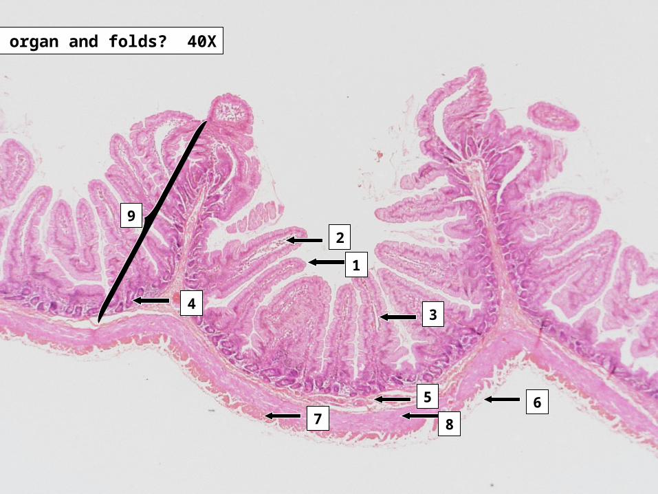
Slides 25-27 Small Intestine - Duodenum
Slide # 251. Villus2. Limina Propria3. Lacteal4. Crypt of Lieberkuhn5. Submucosa6. Visceral Peritoneum7. Longitudinal smooth muscle layer8. Circular smooth muscle layer9. Plicae Circularis
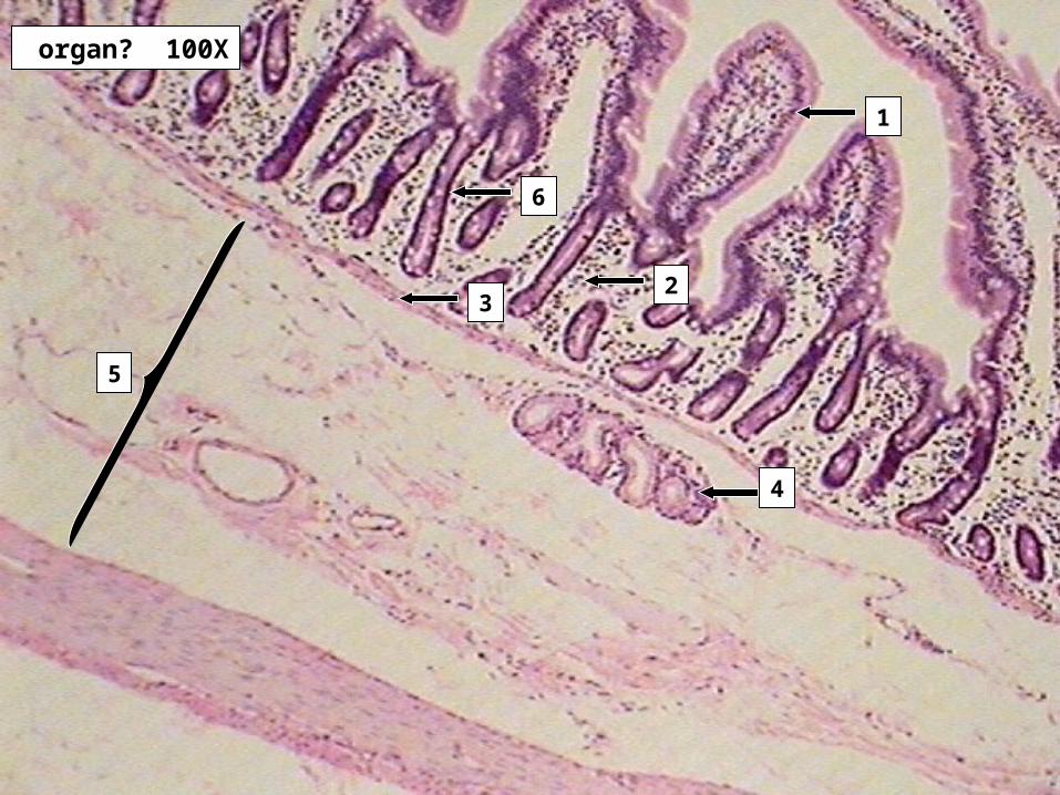
Slides 25-27 Small Intestine - Duodenum Slide # 26
1. Villus2. Limina Propria3. Muscularis Mucosae4. Brunner’s gland5. Submucosa6. Crypt of Lieberkuhn
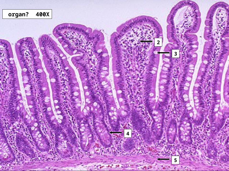
Slide # 271. Villus2. Limina Propria3. Goblet cells with mucous drop4. Crypt of Lieberkuhn 5. Muscularis Mucosae
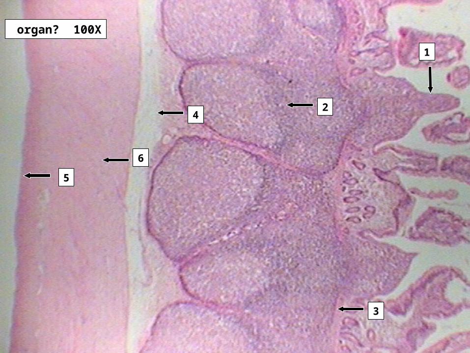
organ? 100X
1
6
42
5
3
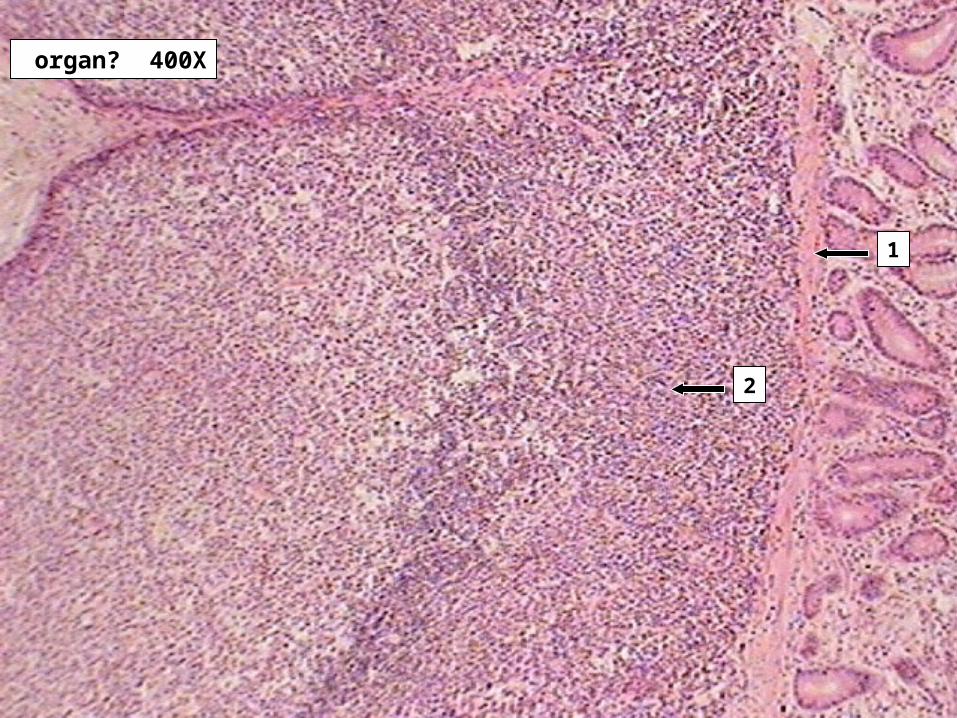
organ? 400X
2
1
Slides 30-31 Small Intestine - Ileum Slide # 30
1. Villus2. Peyer’s Patch3. Muscularis Mucosae4. Submucosa5. Longitudinal smooth muscle layer6. Circular smooth muscle layer
Slide # 311. Muscularis Mucosae2. Peyer’s Patch
organ? 40X
4
5
3
2
1
6
organ? 100X
2
1
3
5
4
organ? 400X
1
4
2
3
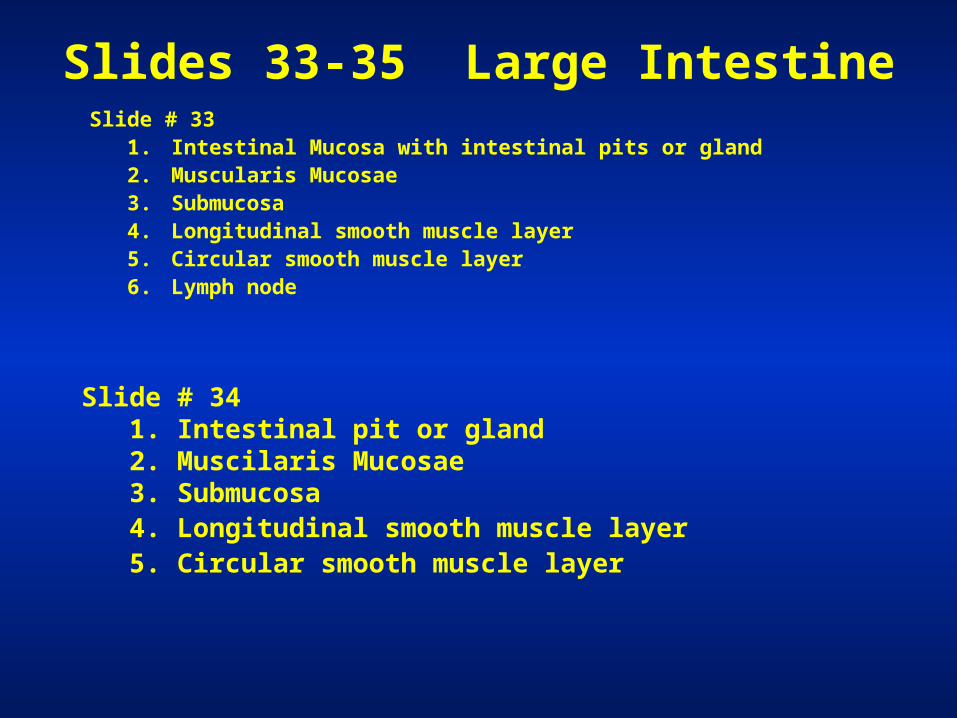
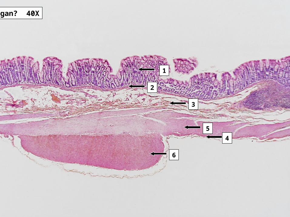
Slides 33-35 Large Intestine Slide # 33
1. Intestinal Mucosa with intestinal pits or gland2. Muscularis Mucosae3. Submucosa4. Longitudinal smooth muscle layer5. Circular smooth muscle layer6. Lymph node
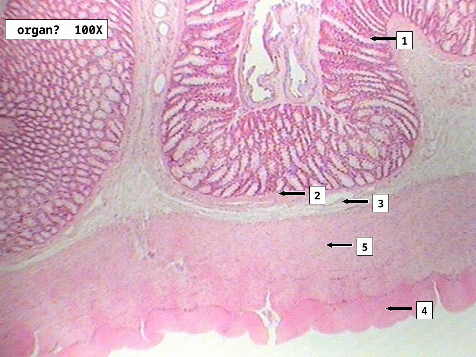
Slide # 341. Intestinal pit or gland2. Muscilaris Mucosae3. Submucosa4. Longitudinal smooth muscle layer5. Circular smooth muscle layer
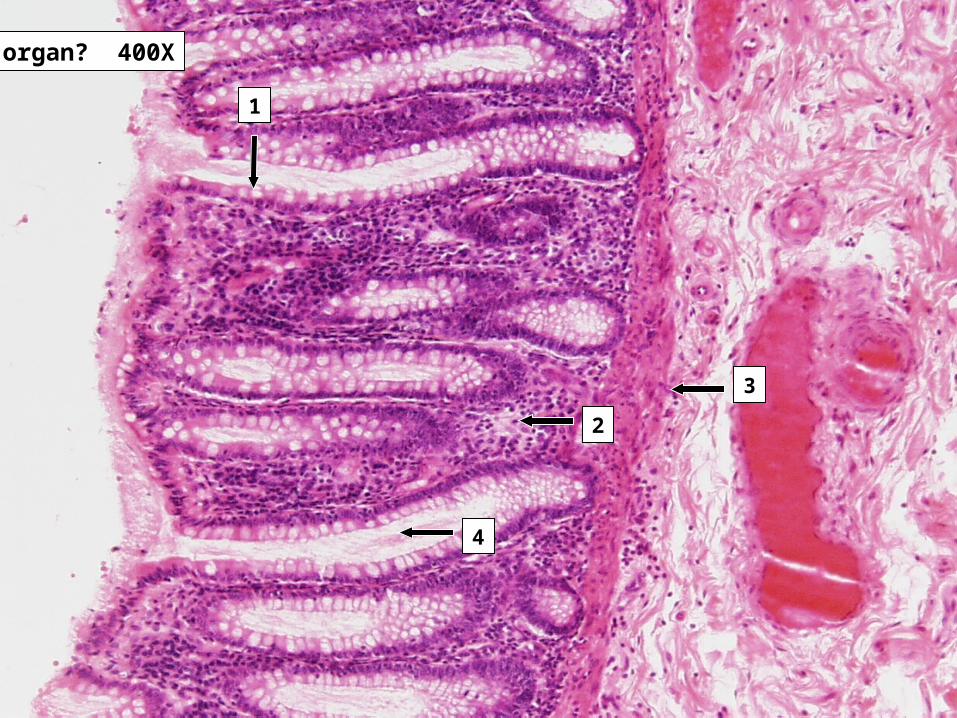
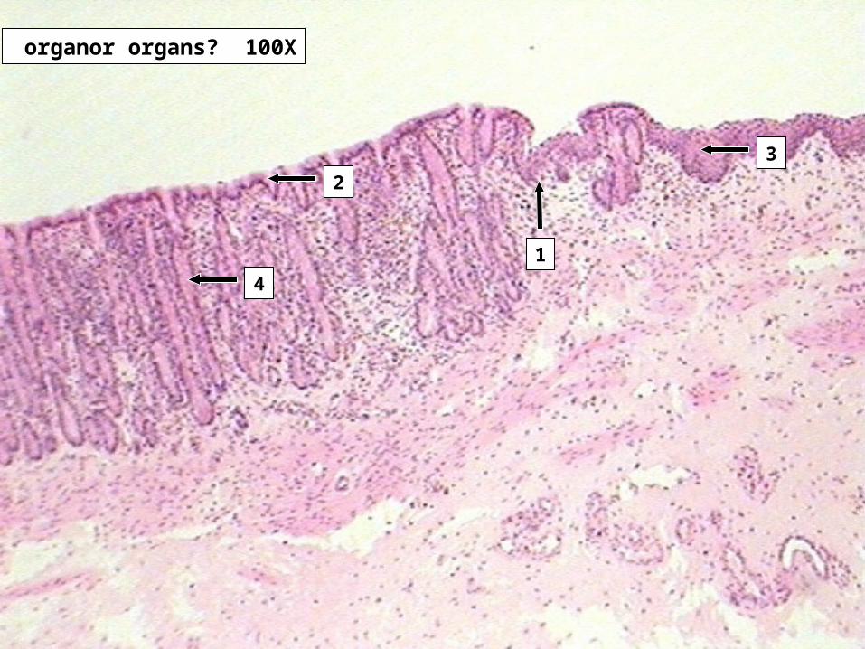
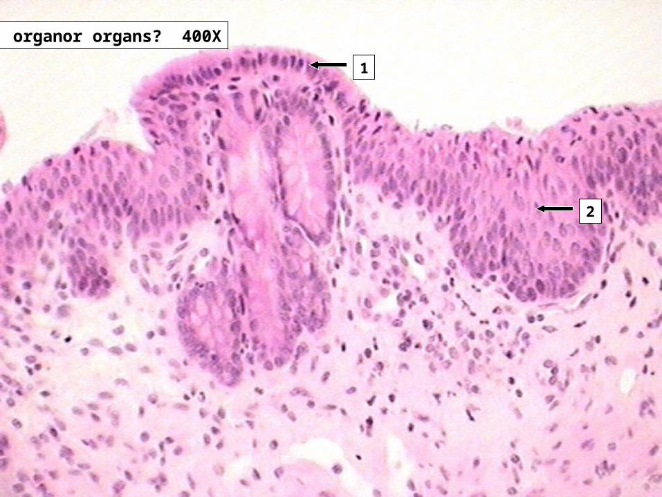
Slides 33-35 Large Intestine Slide # 35
1. Intestinal pits with goblet cells2. Lamina Propria3. Muscularis Mucosae4. Lumen of an intestinal pit or gland

organ or organs? 40X
1
organor organs? 100X
4
3
1
2
organor organs? 400X
1
2
Slides 38-40 Anorectal junction Slide # 38
1. Junction of the Rectum and Anus
Slide # 391. Junction of the rectum and Anus2. Simple Columnar cells of the Rectum3. nonkeratinized stratified squamous of the Anus4. Intestinal pit or gland
Slide # 401. Simple Columnar cells of the Rectum2. nonkeratinized stratified squamous of the Anus
Related Documents


