IJASR International Journal of Academic and Scientific Research Volume 1, Issue 1 (November-December 2013), PP 22-33 www.ijasrjournal.org www.ijasrjournal.org 22 | Page Histological Study of the Effect of Cisplatin on the Liver of Adult Male Albino Rat Hesham, A. Ahmed 1 and Mohamed M. Ghobara 2 1 (Department of Anatomy, Faculty of Medicine, Al-Azhar University, ARE) 2 (Department of Medical Laboratories Technology, Faculty of Applied Medical Sciences, Taibah University, KSA) ABSTRACT Objective: This study was designed to assess the toxic effect of Cisplatin drug on the histological structure of the liver of adult male rats and the degree of improvement after abstinence of the drug for 2 months for recovery as well as its effect on the body weight. Methods: Thirty adult male albino rats were divided into 3 groups 10 rats each. Animals of the control group (G1) received normal saline 0.9% intra-peritoneally, while those of the treated group (G2) received intra-peritoneal cisplatin in a dose of 0.2 mg/kg body weight (low dose) twice weekly for two months. Rats of recovery group (G3) received the same treatment as those of (G2) for two months and then were left for two months without treatment for recovery. The animals of (G1) and (G2) were sacrificed after two months from the start of the experiment while those of (G3) were left for two months more without treatment then sacrificed. Results: The body weights of cisplatin treated rats were significantly decreased in comparison to controls (P<0.001) Two months after withdrawal of the drug, there was a significant increase in body weights of the rats in the recovery group (P<0.01). Both light and electron microscopic studies revealed that low doses of cisplatin caused hepatotoxicity manifested by cytoplasmic vacuolization of hepatocytes, congested blood sinusoids, apparent increase in the number of Kupffer cells and focal mononuclear cellular infiltration. However, some specimens showed distortion of hepatocytes with rarified cytoplasm and focal necrosis while others revealed severely congested blood sinusoids. At ultrastructural level, hepatocytes showed cytoplasmic vacuoles (dilated cisternae of endoplasmic reticulum), irregular nuclei and many small mitochondria with ill distinct cristea. In some cells, the observed cytoplasmic vacuoles were remarkably large. Apparent increase in the number of Kupffer cells with many secondary lysosomes and lipofuscin deposits were noticed. Two months after withdrawal of the drug, lesser and smaller cytoplasmic vacuoles in the hepatocytes were observed, while others appeared more or less normal. Conclusion: Our results may provide histological evidence of hepatotoxicity caused by cisplatin and, on the other hand, the possibility of recovery. This can be used to consider administration of the lowest possible dose of this drug in order to ameliorate its hepatotoxic effects. Keywords: Cisplatin, Hepatotoxicity, Rat liver, Chemotherapy, Recovery. I. INTRODUCTION Cisplatin, cisplatinum, or cis-diamminedichloroplatinum (II) (CDDP) (trade names Platinol and Platinol- AQ) is a chemotherapy drug. It is used to treat various types of cancers, including sarcomas, some carcinomas (e.g. small cell lung cancer, and ovarian cancer), lymphomas, and germ cell tumors [1] . It was the first member of a class of platinum-containing anti-cancer drugs, which now also includes carboplatin and oxaliplatin. These platinum complexes react in vivo, binding to and causing crosslinking of DNA, which ultimately triggers apoptosis (programmed cell death) [2]. The nephrotoxicity of cisplatin is recognized as the most important dose-limiting factor, but high doses of cisplatin also produce hepatotoxicity. Many efforts have been made to improve the therapeutic index of cisplatin using pharmacological strategies, such as the administration of chemoprotectors [3]. Cisplatin is a coordination compound containing two chloro and two ammine ligands. Many other amine complexes of the platinum group metals have been evaluated for this application. Cisplatin is crosslinks DNA in several different ways, interfering with cell division by mitosis. The damaged DNA elicits DNA repair mechanisms, which in turn activate apoptosis when repair proves impossible. Recently it was shown that the apoptosis induced by

Histological Study of the Effect of Cisplatin on the Liver of Adult Male Albino Rat
Mar 17, 2016
Â
Welcome message from author
This document is posted to help you gain knowledge. Please leave a comment to let me know what you think about it! Share it to your friends and learn new things together.
Transcript
IJASR International Journal of Academic and Scientific Research
Volume 1, Issue 1 (November-December 2013), PP 22-33
www.ijasrjournal.org
www.ijasrjournal.org 22 | Page
Histological Study of the Effect of Cisplatin on the Liver of Adult
Male Albino Rat
Hesham, A. Ahmed1 and Mohamed M. Ghobara
2
1 (Department of Anatomy, Faculty of Medicine, Al-Azhar University, ARE)
2 (Department of Medical Laboratories Technology, Faculty of Applied Medical Sciences, Taibah University, KSA)
ABSTRACT
Objective: This study was designed to assess the toxic effect of Cisplatin drug on the histological structure of the
liver of adult male rats and the degree of improvement after abstinence of the drug for 2 months for recovery as well
as its effect on the body weight. Methods: Thirty adult male albino rats were divided into 3 groups 10 rats each. Animals of the control group (G1)
received normal saline 0.9% intra-peritoneally, while those of the treated group (G2) received intra-peritoneal
cisplatin in a dose of 0.2 mg/kg body weight (low dose) twice weekly for two months. Rats of recovery group (G3)
received the same treatment as those of (G2) for two months and then were left for two months without treatment for
recovery. The animals of (G1) and (G2) were sacrificed after two months from the start of the experiment while
those of (G3) were left for two months more without treatment then sacrificed.
Results: The body weights of cisplatin treated rats were significantly decreased in comparison to controls
(P<0.001) Two months after withdrawal of the drug, there was a significant increase in body weights of the rats in
the recovery group (P<0.01). Both light and electron microscopic studies revealed that low doses of cisplatin
caused hepatotoxicity manifested by cytoplasmic vacuolization of hepatocytes, congested blood sinusoids, apparent
increase in the number of Kupffer cells and focal mononuclear cellular infiltration. However, some specimens
showed distortion of hepatocytes with rarified cytoplasm and focal necrosis while others revealed severely
congested blood sinusoids. At ultrastructural level, hepatocytes showed cytoplasmic vacuoles (dilated cisternae of
endoplasmic reticulum), irregular nuclei and many small mitochondria with ill distinct cristea. In some cells, the
observed cytoplasmic vacuoles were remarkably large. Apparent increase in the number of Kupffer cells with many
secondary lysosomes and lipofuscin deposits were noticed. Two months after withdrawal of the drug, lesser and
smaller cytoplasmic vacuoles in the hepatocytes were observed, while others appeared more or less normal.
Conclusion: Our results may provide histological evidence of hepatotoxicity caused by cisplatin and, on the other
hand, the possibility of recovery. This can be used to consider administration of the lowest possible dose of this drug
in order to ameliorate its hepatotoxic effects.
Keywords: Cisplatin, Hepatotoxicity, Rat liver, Chemotherapy, Recovery.
I. INTRODUCTION
Cisplatin, cisplatinum, or cis-diamminedichloroplatinum (II) (CDDP) (trade names Platinol and Platinol-
AQ) is a chemotherapy drug. It is used to treat various types of cancers, including sarcomas, some carcinomas (e.g.
small cell lung cancer, and ovarian cancer), lymphomas, and germ cell tumors [1]
. It was the first member of a class
of platinum-containing anti-cancer drugs, which now also includes carboplatin and oxaliplatin. These platinum
complexes react in vivo, binding to and causing crosslinking of DNA, which ultimately triggers apoptosis
(programmed cell death) [2].
The nephrotoxicity of cisplatin is recognized as the most important dose-limiting factor, but high doses
of cisplatin also produce hepatotoxicity. Many efforts have been made to improve the therapeutic index of cisplatin
using pharmacological strategies, such as the administration of chemoprotectors [3].
Cisplatin is a coordination compound containing two chloro and two ammine ligands. Many other amine
complexes of the platinum group metals have been evaluated for this application. Cisplatin is crosslinks DNA in
several different ways, interfering with cell division by mitosis. The damaged DNA elicits DNA repair mechanisms,
which in turn activate apoptosis when repair proves impossible. Recently it was shown that the apoptosis induced by

International Journal of Academic and Scientific Research
Volume 1, Issue 1 (November-December 2013), PP 22-33
www.ijasrjournal.org 23 | Page
cisplatin on human colon cancer cells depends on the mitochondrial serine-protease Omi/Htra2. Since this was only
demonstrated for colon carcinoma cells, it remains an open question if the Omi/Htra2 protein participates in the
cisplatin-induced apoptosis in carcinomas from other tissues [4].
Sheets of connective tissue divide the liver into thousands of small units called lobules. A lobule is roughly
hexagonal in shape, with portal triads at the vertices and a central vein in the middle. The lobule is the structural unit
of the liver and rather easy to observe. In contrast, the hepatic acinus is more difficult to visualize, but represents a
unit that is of more relevance to hepatic function because it is oriented around the afferent vascular system. In the
case of the liver, the roads are connective tissue septae which convey vascular and biliary traffic, and the clusters of
houses are cord-like arrangements of hepatocytes, the parenchymal cell of the liver [5].
Hepatocytes are polygonal cells joined to one another in anastomosing plates, with borders that face either
the sinusoids or adjacent hepatocytes. The ultrastructure appearance of hepatocytes reflects their function as
metabolic superstars, with abundant rough and smooth endoplasmic reticulum, and Golgi membranes. Glycogen
granules and vesicles containing very low density lipoproteins are readily observed. Hepatocytes make contact with
blood in sinusoids, which are distensible vascular channels lined with highly fenestrated endothelial cells and
populated with phagocytic Kupffer cells. The space between endothelium and hepatocytes is called the Space of
Disse which collects lymph for delivery to lymphatic capillaries [6].
The aim of this work was to evaluate the toxic effect of cisplatin on the body weight and the liver of the
adult male rats and the degree of improvement after abstinence of the drug for 2 months for recovery.
II. MATERIALS ASND METHODS
Cisplatin was obtained as yellowish crystalline powder, soluble in physiological saline solution and
purchased from Sigma-Aldrich. The dose of cisplatin administered in this research was 0.2 mg/kg body weight (low
dose) [7]. Thirty adult male albino rats of Sprague-Dawley strain weighing (200-220g.) were divided into 3 groups:
The first (control) group (G1): consisted of 10 rats and received normal saline 0.9% serving as control.
The second (treated) group (G2): ten rats comprised the treated group and each of them received cisplatin
o.2 mg/kg B.WT. (Low dose) intraperitoneally, twice weekly for two months.
The third (recovery) group (G3): 10 rats received the same treatment as those of (G2) for two months and
then were left for two months after withdrawal of the drug.
The animals of (G1) and (G2) were sacrificed after two months from the start of the experiment while those
of (G3) were left for two month more without treatment then sacrificed. Body weights of the rats were measured
initially and after treatment in all groups, using an electronic analytic and precision balance.
At the appropriate time, the animals were sacrificed and their livers were carefully dissected out and cut
into small pieces, about 1mm3 each. The specimens were immediately fixed in 5% phosphate buffered
gluteraldehyde (Ph 7.4) for 2 hours at 4oC, post fixed in 1% osmium tetroxide for 2 hours, dehydrated in ascending
grades of ethyl alcohol and then embedded in Epoxy resin. Semithin sections (1µm) were cut, stained with toludine
blue and examined by light microscope. Ultrathin sections of 60 nm were cut with an LKB ultramicrotome using a
diamond Knife, mounted on copper grids and stained with uranyl acetate followed by lead citrate [8]. Examination
and photography were carried out using JEOL transmission electron microscope (JEM-1200 EXII) in faculty of
science, Ain shams University.
International Journal of Academic and Scientific Research
Volume 1, Issue 1 (November-December 2013), PP 22-33
www.ijasrjournal.org 24 | Page
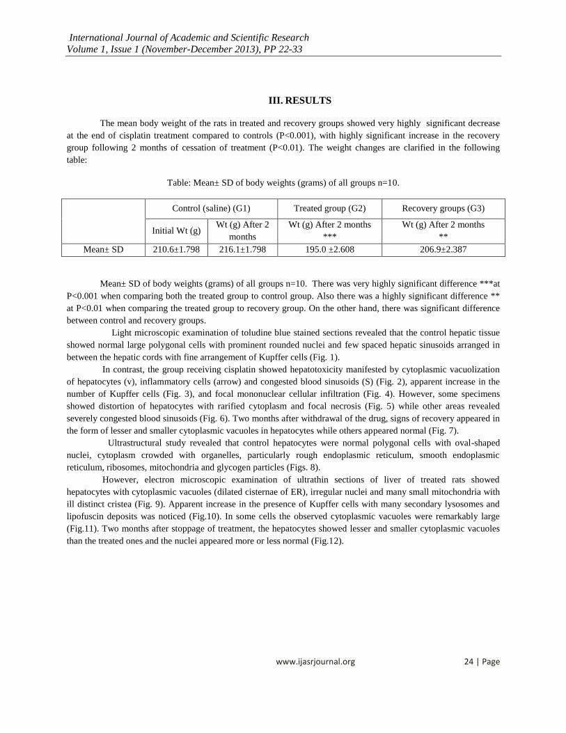
III. RESULTS
The mean body weight of the rats in treated and recovery groups showed very highly significant decrease
at the end of cisplatin treatment compared to controls (P<0.001), with highly significant increase in the recovery
group following 2 months of cessation of treatment (P<0.01). The weight changes are clarified in the following
table:
Table: Mean± SD of body weights (grams) of all groups n=10.
Control (saline) (G1) Treated group (G2) Recovery groups (G3)
Initial Wt (g) Wt (g) After 2
months
Wt (g) After 2 months
***
Wt (g) After 2 months
**
Mean± SD 210.6±1.798 216.1±1.798 195.0 ±2.608 206.9±2.387
Mean± SD of body weights (grams) of all groups n=10. There was very highly significant difference ***at
P<0.001 when comparing both the treated group to control group. Also there was a highly significant difference **
at P<0.01 when comparing the treated group to recovery group. On the other hand, there was significant difference
between control and recovery groups.
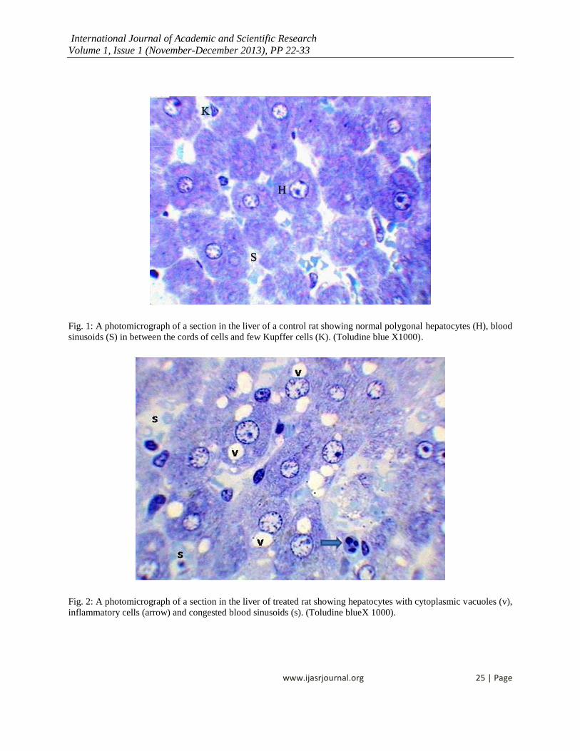
Light microscopic examination of toludine blue stained sections revealed that the control hepatic tissue
showed normal large polygonal cells with prominent rounded nuclei and few spaced hepatic sinusoids arranged in
between the hepatic cords with fine arrangement of Kupffer cells (Fig. 1).
In contrast, the group receiving cisplatin showed hepatotoxicity manifested by cytoplasmic vacuolization
of hepatocytes (v), inflammatory cells (arrow) and congested blood sinusoids (S) (Fig. 2), apparent increase in the
number of Kupffer cells (Fig. 3), and focal mononuclear cellular infiltration (Fig. 4). However, some specimens
showed distortion of hepatocytes with rarified cytoplasm and focal necrosis (Fig. 5) while other areas revealed
severely congested blood sinusoids (Fig. 6). Two months after withdrawal of the drug, signs of recovery appeared in
the form of lesser and smaller cytoplasmic vacuoles in hepatocytes while others appeared normal (Fig. 7). Ultrastructural study revealed that control hepatocytes were normal polygonal cells with oval-shaped
nuclei, cytoplasm crowded with organelles, particularly rough endoplasmic reticulum, smooth endoplasmic
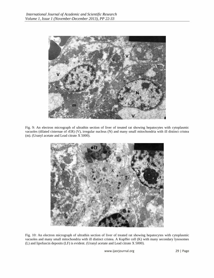
reticulum, ribosomes, mitochondria and glycogen particles (Figs. 8). However, electron microscopic examination of ultrathin sections of liver of treated rats showed
hepatocytes with cytoplasmic vacuoles (dilated cisternae of ER), irregular nuclei and many small mitochondria with
ill distinct cristea (Fig. 9). Apparent increase in the presence of Kupffer cells with many secondary lysosomes and
lipofuscin deposits was noticed (Fig.10). In some cells the observed cytoplasmic vacuoles were remarkably large
(Fig.11). Two months after stoppage of treatment, the hepatocytes showed lesser and smaller cytoplasmic vacuoles
than the treated ones and the nuclei appeared more or less normal (Fig.12).
International Journal of Academic and Scientific Research
Volume 1, Issue 1 (November-December 2013), PP 22-33
www.ijasrjournal.org 25 | Page
Fig. 1: A photomicrograph of a section in the liver of a control rat showing normal polygonal hepatocytes (H), blood
sinusoids (S) in between the cords of cells and few Kupffer cells (K). (Toludine blue X1000).
Fig. 2: A photomicrograph of a section in the liver of treated rat showing hepatocytes with cytoplasmic vacuoles (v),
inflammatory cells (arrow) and congested blood sinusoids (s). (Toludine blueX 1000).

International Journal of Academic and Scientific Research
Volume 1, Issue 1 (November-December 2013), PP 22-33
www.ijasrjournal.org 26 | Page
Fig. 3: A photomicrograph of a section in the liver of a treated rat showing a focal area of more cytoplasmic
vacuolation than the previous figure and many Kupffer cells (k). (Toludine blueX1000).
Fig. 4: A photomicrograph of a section in the liver of treated rat showing focal mononuclear cellular infiltration
(Inf), central vein (C.V.), cytoplasmic vacuoles (v). (Toludine blue X1000).

International Journal of Academic and Scientific Research
Volume 1, Issue 1 (November-December 2013), PP 22-33
www.ijasrjournal.org 27 | Page
Fig. 5: A photomicrograph of a section in the liver of treated rat showing a focal group of hepatocytes with distorted
and severely vacuolated cytoplasm , and some necrotic cells (n). Kuffer cells (K) were also seen. (Toludine blue X
1000).
Fig. 6: A photomicrograph of a section in the liver of treated rat showing severely congested blood sinusoids (S)
with many kuffer cells (K) (Toludine blue X 1000).

International Journal of Academic and Scientific Research
Volume 1, Issue 1 (November-December 2013), PP 22-33
www.ijasrjournal.org 28 | Page
Fig. 7: A photomicrograph of a section in the liver of experimental rat two months after stoppage of treatment
showing hepatocytes with lesser and smaller cytoplasmic vacuoles (v) and others apparently normal ones (N)
(Toludine blue X 1000).
Fig. 8: An electron micrograph of ultrathin section of liver control rat showing normal hepatocyte with oval nuclei
and cytoplasm crowded with organelles particularly endoplasmic reticulum (ER), glycogen particles (R),
mitochondria (m). (Uranyl acetate and Lead citrate X 2000).
International Journal of Academic and Scientific Research
Volume 1, Issue 1 (November-December 2013), PP 22-33
www.ijasrjournal.org 29 | Page
Fig. 9: An electron micrograph of ultrathin section of liver of treated rat showing hepatocytes with cytoplasmic
vacuoles (dilated cisternae of rER) (V), irregular nucleus (N) and many small mitochondria with ill distinct cristea
(m). (Uranyl acetate and Lead citrate X 5000).
Fig. 10: An electron micrograph of ultrathin section of liver of treated rat showing hepatocytes with cytoplasmic
vacuoles and many small mitochondria with ill distinct cristea. A Kupffer cell (K) with many secondary lysosomes
(L) and lipofuscin deposits (LF) is evident. (Uranyl acetate and Lead citrate X 5000).

International Journal of Academic and Scientific Research
Volume 1, Issue 1 (November-December 2013), PP 22-33
www.ijasrjournal.org 30 | Page
Fig. 11: An electron micrograph of ultrathin section of liver of treated rat showing hepatocytes with many large
cytoplasmic vacuoles (V) and many small mitochondria with ill distinct cristea (m). (Uranyl acetate and Lead citrate
X 7000).
Fig. 12: An electron micrograph of ultrathin section of the liver of experimental rat one months after stoppage of
treatment showing hepatocytes with lesser and smaller cytoplasmic vacuoles than the treated ones and regular nulei
(N). (Uranyl acetate and Lead citrate X 5000).
International Journal of Academic and Scientific Research
Volume 1, Issue 1 (November-December 2013), PP 22-33
www.ijasrjournal.org 31 | Page
IV. DISCUSSION
Cisplatin is one of the most commonly used potent antineoplastic agents for treatment of a wide range of
cancers ([9], [10]). Despite its excellent anticancer activity, the clinical use of cisplatin is often limited by its
undesirable sever toxic side effects that interfere with therapeutic efficacy ([11], [12], [13]).
The several toxicities and sides effects of cisplatin included hepatotoxicity ([14], [15]). and nephrotoxicity
[10]. Although, the precise mechanism for the cisplatin- induced toxicity is not well understood, many studies
documented that cisplatin is preferentially taken up and accumulated in the liver and kidney cells, resulting in the
enhancing production of reactive oxygen species ([16], [17]).. In their study, Mora et al., (2003)
[18] added that a
decrease in antioxidant enzymes resulted from cisplatin induced tissue toxicity.
Moreover, the development of therapies to prevent the appearance of cisplatin- induced tissue toxicities
has focused on administration of antioxidants along with cisplatin treatment. Thus, many studies for protective
effects against cisplatin induced tissue toxicities have been reported for extracts of natural products and dietary
antioxidant [19].
The current study was designed to evaluate the toxic effects of the low dose of cisplatin for two month on
the adult male rats, then after cutting of the treatment for two months. We found that cisplatin significantly
decreased the body weights in the treated rats compared to the controls in agreement with King and Berry, (2001)
[20], who suggested that hepatotoxicity might have contributed to this loss. Also Leite et al., (2009) [21] stated that
mice treated with high doses of free cisplatin showed a greater loss of body weight and more delayed recovery time.
Dissection of these animals indicated that the loss of the body weights was mostly due to loss of the mass of skeletal
muscles and adipose tissue as previously suggested by Devlin et al., (1997) [22]. Moreover, the reduction in body
weight of the animals in this study can be correlated with the decreased food intake by the animals of treated group
observed during the experimental period. However, in the current study the mean body weight was significantly
increased in the recovery group indicating evidence of the effect of abstinence of the drug.
In the present study the histological findings of the treated liver illustrated hepatotoxicity manifested by
cytoplasmic vacuolization of hepatocytes, inflammatory cells and congested blood sinusoids, apparent increase in
the number of kuffer cells and focal mononuclear infiltration.
El-Sayyad et al. (2009) [7] and Abdelmeguid et al., (2010) [23]Supporting our results also observed
histological abnormalities in the liver including inflammatory infiltration, dissolution of hepatic cords which
appeared as empty vacuoles and dilated blood sinusoids. In agreement with our study, Liao et al., (2008) [24]
mentioned that the liver is known to accumulate significant amounts of cisplatin, thus hepatotoxicity and its
histological abnormalities were associated with cisplatin treatment. In contrast, Leite et al., (2009) [21] stated that,
concerning hepatotoxicity, no histopathological alteration was observed after treatment by cisplatin.
In this work we found some specimens showing distortion of hepatocytes with rarified cytoplasm and focal
necrosis while other areas revealed severely congested blood sinusoids. El-Sayyad et al., (2009) [7] also revealed
that many hepatocytes showed marked degeneration in hepatic cords in addition to karyomegally and pyknotic
nuclei indicating apoptosis.
In the present work, ultrastructure examination showed hepatocytes with cytoplasmic vacuoles (dilated
cisternae of endoplasmic reticulum, irregular nuclei and many small mitochondria with ill distinct cristae. Apparent
increase in the presence of kuffer cells with many secondary lysosomes and lipofuscin deposits were noticed. In
some cells the observed cytoplasmic vacuoles were remarkably large. Stewart et al., (1982) [14] and El-Sayyad et
al., (2009) [7] demonstrated that the ultrastructure of liver sections showed dense collection of inflammatory cells
including macrophages and fibrocytes forming pattern of cirrhotic liver. Also they supported our study mentioning
that the cytoplasm contained atrophied mitochondria with ill defined cisternae and vesiculated rough endoplasmic
reticulum.

International Journal of Academic and Scientific Research
Volume 1, Issue 1 (November-December 2013), PP 22-33
www.ijasrjournal.org 32 | Page
Hae et al., (2009) [25]claimed that cisplatin accumulation shows toxicity to normal tissues and this is the
cause of hepatotoxicity. Nuria et al., (2008) [26] Moreover said that, cisplatin toxic side effects seemed to be
associated with mitochondrial injury both with in vivo treatment with the drug and in vitro exposure to it. They also
showed that cisplatin caused a direct and significant impairment of mitochondrial DNA and RNA synthesis.
In the current study, signs of recovery appeared in the form of lesser and smaller cytoplasmic vacuoles in
the hepatocytes while others appeared normal and the nuclei appeared more or less similar to those of the control. In
contrast, Herrera et al., (1998) [27]deducted that cisplatin inhibits liver regeneration.
V. CONCLUSION
In conclusion, our results may provide histological evidence of hepatotoxicity caused by cisplatin and, on
the other hand, the possibility of recovery. This can be used as the basis for determining the appropriate dose of this
drug in order to ameliorate its hepatotoxic effects. Administration of the lowest therapeutic dose of cisplatin with
some protective measures (antioxidants and others) can be recommended.
REFERENCES
[1] Ivana, S. T.; Branka,I. O.; Nataza, Z.; Ordevici, S.; Markovici, A.; Štajni, G.; Zorica, S. and Saicic, C. (2010): Effects of cisplatin on lipid
peroxidation and the glutathione redoxstatus in the liver of male rats.The protective role of selenium. Arch. Biol. Sci., Belgrade, 62 (1), 75-82.
[2] Lippard, S. J. and. Berg, J. M. (1994): “Principles of Bioinorganic Chemistry” University Science Books: Mill Valley, CA; ISBN 0-935702-73-
3. [3] Liu, J.; Liu, Y.; Habeebu, S.S. and Klaassen, C.D. (1998): Metallothionein (MT)-null mice are sensitive to cisplatin-induced hepatotoxicity.
Toxicol Appl Pharmacol; 149: 24-31.
[4] Pruefer, F.G.; Lizarraga, F.; Maldonado, V. and Melendez,-Z. J. (June 2008): "Participation of Omi Htra2 serine-protease activity in the apoptosis induced by cisplatin on SW480 colon cancer cells". J Chemother 20 (3): 348–54.
[5] Fawcet, D.W. and Jensh, R.P. (2001): Bloom& Fawcett: Concise Histology, Liver and Gall bladder. Chapter 18, 212 – 217.
[6] Ross, M.H. and Pawlina, W. (2005): Histology with correlated cell and molecular biology, Digestive System III, Liver. Chapter 18, 576- 589. [7] El- Sayyad, H.I.; Ismail, M.F.; Shalaby,F.M.; Abou-El-Magd, R.F. and Gaur,R.L. (2009): Histopathological effects of Cisplatin, Doxorubicin and
5- flurouracil (5-FU) on the liver of male albino rats.Int. J.Biol.Sci., 5: 466- 473.
[8] Hayat, M., (1986): Basic Techniques for Transmission Electron Microscopy. 2nd Edn. Academic Press, Baltimore. [9] Wang, G. Reed, E. and Li, Q.Q. (2004): Molecular basis of cellular response to cisplatin chemotherapy in non- small cell lung cancer (Review).
Oncol. Rep., 12: 955- 965.
[10] Park, H.R.; Ju, E.J.; Jo, S.K.; Kim,S.H. and Yee, S.T. (2009): Enhanced antitumour efficacy of cisplatin in combination with hemo HIM in tumour- bearing mice. BMC Cancer, 9: 85- 85.
[11] Aly, M.S.; Ashour, M.B.; El-Nahas and Abo-Zeid, (2003): Genotoxicity and cytotoxicity of the anticancer drugs gemcitabine and cisplatin,
separately and in combination: In vivo studies. J. Biol. Sci., 3: 961- 972. [12] Ajani, J.A. (2008): Optimizing docetaxel chemotherapy in patients with cancer of the gastric and gastroesophageal junction: Evolution of the
docetaxel, Cisplatin and 5-fluorouracil regimen. Cancer, 113: 945- 955.
[13] Dank, M. ; Zaluski, J.; Barone. C.; Valvere, V. and Yalcin, S. (2008): Randomized phase III study comparing irinotecan combined with 5- fluorouracil and folinic acid to cisplatin combined with 5- fluorouracil in chemotheraoy naïve patients with advanced adenocarcinoma of the
stomach oresophagogastric junction Ann. Oncol., 19: 1450- 1457.
[14] Mansour, H.H.; Hafez, H.F. and Fahmy, N.M. (2006): Silymarin modulates Cisplatin 10mg Kg-1platin induced oxidative stress and hepatotoxicity
in rats. J.Biochem. Mol. Biol., 39: 656- 661.
[15] Pratibha, R.; Sameer, R..; Rataboli, P.V; Bhiwgade, D.A. and Dhume, C.Y. (2006): Enzymatic studies of cisplatin induced oxidative stress in
hepatic tissue of rats. Eur. J. Pharmacol., 532: 290- 293. [16] H. El-Beshbishy, Bahashwan, S, Ali, H, and H. Fakher, "Abrogation of cisplatin-induced nephrotoxicity in mice by alpha lipoic acid through
ameliorating oxidative stress and enhancing gene expression of antioxidant enzymes", European Journal of Pharmacology, vol. 668, pp. 278-
284, 2011. [17] Stewart, D.J.; Bemjamin, R.S.; Luna, M.; Feun, L.; Caprioli, R.; Seifert, W. and Loo, T.L. (1982): Human tissue distribution of platinum after
cis-diamminedichloroplatinum. Cancer Chemother. Pharmacol. 10: 51- 54.
[18] Mora, L.O.; Antunes, L.M.; Francescato, H.D. and Bianchi, M. (2003): The effects of oral glutamine on cisplatin-induced nephrotoxicityin rats. Pharmacol. Res., 47:517- 522.
[19] Behling, E.B.; Sendao, M.C.; Francescato, H.D.; Antunes, L.M.; Costa,R.S. and Bianchi- Mde, L. (2006): Comparative study of multiple dosage of quercetin against cisplatin- induced nephrotoxicity and oxidative stress in rat kidneys. Pharmacol. Rep., 58:526- 532.
[20] King, P.D.and Berry, M.C. (2001): Hepatotoxicity of chemotherapy. Oncologist; 6:162-76.
[21] Leite, E.A.; Giuberti, C. S.; Wainstein, A.J.; Wainstein, A.P.; Coelho, L.G.; Lana, A.M.; Savassi, R. R. and De Oliveira, M.C. (2009): Acute toxicity of long-circulating and pH-sensitive liposomes containing cisplatin in mice after intraperitoneal administration. Life Sci. May 8;84(19-
20):641-9.

International Journal of Academic and Scientific Research
Volume 1, Issue 1 (November-December 2013), PP 22-33
www.ijasrjournal.org 33 | Page
[22] Devlin, T.M. (1997): Text book of biochemistry: with clinical correlation, 4th ed. New York: John Wiley and Sons Inc; 553.
[23] Abdelmeguid, N.E.; Chmaisse, H.N.; Abdou Zeinab, N.S. and Moharram,B. (2010): Silymarin ameliorates Cisplatin-induced hepatotoxicity in rats: Histopathological and Ultrastructure Studies. Pakistan journal of Biological Sciences, 13(10): 463- 479.
[24] Liao, Y.; Lu, X.; Lu, C.;Li, G.; Jin, Y. and Tang, H. (2008): Selection of agents for prevention of cisplatin-induced hepatotoxicity. Pharmacol Res; 57:125-31.
[25] Hae, R. P.; Eun, J. J.; Sung, K. o.; Uhee, J.; Sung, H. K. and Sung, T. Y. (2009): Enhanced antitumor efficacy of cisplatin in combination
with HemoHIM in tumor-bearing mice. BMC- Cancer; 17 March. [26] Nuria, G.; Acisclo, P.M.; Mercedes, F.; Jose, M.L.; Patricio, F. S. and Jose, A. E. (2008): Cisplatin- mediated impairment of mitochondrial DNA
metabolism inversely correlates with glutathione levels. Biochem. J.; 414, 93- 102.
[27] Herrera, M.G.; Palomero, M.F.; Macias, R.I.; Serrano, M.A. and Marin. J.J. (1998): Comparison of the effects of bischolyl glycinatecholoro-platinum (II) versus cisplatin on liver regeneration after partial hepatoctomy. Anticancer Res. Sep.- oct.; 18(5A): 3555-63.
Related Documents