The Egyptian Journal of Hospital Medicine (2007) Vol., 29: 492– 110 Histological, Scanning And Transmission Electron Microscopic Studies On The Possible Protective Role Of Ginger Extract Against Acrylamide Induced Intestinal Damage In Mice Hala Galal El-Tantawi Department Of Zoology –Faculty Of Science - Ain Shams University Abstract Objective: This study was carried out to evaluate the protective effect of ginger Zingeber officinale extract (ZOE) against the acrylamide (AC) which is an industrial chemical used in water treatment and it is synthesized during cooking of starch food at high temperature. Method: Thirty adult male albino mice, each weighs 20-25 g were divided into three groups (10 mice/group): (I)control group. (II)acrylamide treated group. (III) acrylamide & ginger group. Acrylamide was given to experimental animals in the drinking water at a non- lethal dose of 200 p.p.m for 10 weeks (3 days/week). Ginger extract was orally administrated at 50 mg/L (~5 ml/day) for 10 weeks (3 days/week). The ileum samples were collected for light microscope study and for scanning and transmission electron microscope examination. Results: This study revealed that acrylamide induces pathological changes of the ileum of the treated mice specially the absorptive epithelial cells. The scanning electron microscopic study revealed damage of the ileal villi, some red blood corpuscles appeared at the site of damage. The transmission electron microscopic examination clearly demonstrated degeneration of most cell organelles as mitochondria, deterioration and degranulation of the rough endoplasmic reticulum, dilatation of Golgi apparatus. Conclusion: The administration of ginger extract decreased the histological alterations and ensuring the anti-inflammatory, and antitoxic effects of ZOE at its chosen dosage level. Key words: Acrylamide – ginger- ileum. Introduction Acrylamide is an industrial chemical used in the synthesis of polyacrylamide and has multiple applications as additive for sewage and water treatment (Smith and Oehme, 1991). Individuals can be exposed to acrylamide either in their workplace or in the environment (Marsh et al., 1999). Recent findings of the presence of acrylamide in starch foods cooked at high temperature have refocused worldwide attention on its carcinogenicity (Tareke et al., 2002). In addition, acrylamide is used extensively in molecular laboratories for gel chromatography (LoPachin, 2004). According to WHO (2002), the average intake of 70μg of acrylamide per day for an adult would be associated with a lifetime cancer risk . Studies in animal models and humans are necessary to better understand the toxicity of acrylamide .It was shown that acrylamide might be formed through the Maillard reaction from amino acids (e.g asparagines) and reducing sugars (e.g glucose) (Mottram et al., 2002). Glycidamide, a metabolite of acryla- mide, binds to DNA and can cause genetic damage. Prolonged exposure to acrylamide has induced tumours in rats, but cancer in man has not been convincingly shown. The International Agency for Research on Cancer (IARC) has classified acrylamide as "probably carcinogenic to humans" (IARC, 1994). Barber et al. (2001) reported that the rate of acrylamide conversion to its epoxide metabolite glycidamide is higher during subchronic dosing conditions. On the other hand the ginger has been widely studied for its pharmacological acti- vities and has been reported to exhibit anti- inflammatory, antipyretic, antimicro-bial, hypoglycemic, antimigraine, antiox-idant, hepatoprotective, diuretic, hypocho-lestero- lemia (Langner et al.,1998; Mascolo et al.,1989) and antihypertensive activities (Ghayur and Gilani., 2005). Phytochemical studies showed the presence of pungent 492

Welcome message from author
This document is posted to help you gain knowledge. Please leave a comment to let me know what you think about it! Share it to your friends and learn new things together.
Transcript

The Egyptian Journal of Hospital Medicine (2007) Vol., 29: 492– 110
Histological, Scanning And Transmission Electron Microscopic Studies On
The Possible Protective Role Of Ginger Extract Against Acrylamide
Induced Intestinal Damage In Mice
Hala Galal El-Tantawi Department Of Zoology –Faculty Of Science - Ain Shams University
Abstract
Objective: This study was carried out to evaluate the protective effect of ginger Zingeber
officinale extract (ZOE) against the acrylamide (AC) which is an industrial chemical used in
water treatment and it is synthesized during cooking of starch food at high temperature. Method: Thirty adult male albino mice, each weighs 20-25 g were divided into three
groups (10 mice/group): (I)control group. (II)acrylamide treated group. (III) acrylamide &
ginger group. Acrylamide was given to experimental animals in the drinking water at a non-
lethal dose of 200 p.p.m for 10 weeks (3 days/week). Ginger extract was orally administrated at 50 mg/L (~5 ml/day) for 10 weeks (3 days/week). The ileum samples were collected for light
microscope study and for scanning and transmission electron microscope examination.
Results: This study revealed that acrylamide induces pathological changes of the ileum of the treated mice specially the absorptive epithelial cells. The scanning electron microscopic
study revealed damage of the ileal villi, some red blood corpuscles appeared at the site of
damage. The transmission electron microscopic examination clearly demonstrated degeneration
of most cell organelles as mitochondria, deterioration and degranulation of the rough endoplasmic reticulum, dilatation of Golgi apparatus.
Conclusion: The administration of ginger extract decreased the histological alterations
and ensuring the anti-inflammatory, and antitoxic effects of ZOE at its chosen dosage level. Key words: Acrylamide – ginger- ileum.
Introduction Acrylamide is an industrial chemical
used in the synthesis of polyacrylamide and
has multiple applications as additive for sewage and water treatment (Smith and
Oehme, 1991). Individuals can be exposed
to acrylamide either in their workplace or in the environment (Marsh et al., 1999).
Recent findings of the presence of
acrylamide in starch foods cooked at high temperature have refocused worldwide
attention on its carcinogenicity (Tareke et
al., 2002). In addition, acrylamide is used
extensively in molecular laboratories for gel chromatography (LoPachin, 2004).
According to WHO (2002), the
average intake of 70µg of acrylamide per day for an adult would be associated with a
lifetime cancer risk . Studies in animal
models and humans are necessary to better
understand the toxicity of acrylamide .It was shown that acrylamide might be
formed through the Maillard reaction from
amino acids (e.g asparagines) and reducing
sugars (e.g glucose) (Mottram et al., 2002).
Glycidamide, a metabolite of acryla-
mide, binds to DNA and can cause genetic damage. Prolonged exposure to acrylamide
has induced tumours in rats, but cancer in
man has not been convincingly shown. The International Agency for Research on
Cancer (IARC) has classified acrylamide as
"probably carcinogenic to humans" (IARC, 1994). Barber et al. (2001) reported that the
rate of acrylamide conversion to its epoxide
metabolite glycidamide is higher during
subchronic dosing conditions. On the other hand the ginger has been
widely studied for its pharmacological acti-
vities and has been reported to exhibit anti-inflammatory, antipyretic, antimicro-bial,
hypoglycemic, antimigraine, antiox-idant,
hepatoprotective, diuretic, hypocho-lestero-
lemia (Langner et al.,1998; Mascolo et al.,1989) and antihypertensive activities
(Ghayur and Gilani., 2005). Phytochemical
studies showed the presence of pungent
492

Hala Galal El-Tantawi
493
principles, such as gingerol, shogoal,
zingerone and paradol (Connell and McLachan., 1972).
The rhizome of the plant Zingiber
officinale roscoe, commonly known as
ginger, has been commonly used as a food additive and spice as well as phytomedicine
since ancient times. The typical use of
ginger in the kitchens as a condiment began in the 13
th century; which enhanced the
importance of this rhizome in european
markets (Langner et al.,1998). More comm-only, ginger has been traditionally used in
disorders of the gastrointestinal tract, as a
stomach laxative, sialogogue, gastric
emptying enhancer, appetizer, antiemetic and antidyspepsic and at the same time as
an antidiarrheal and anticolic agent (Ghayur
and Gilani., 2005; Nadkarni, 1976). Several studies conducted in animals
(Yamahara et al., 1990; Qian and Liu.,
1992) and humans (Mowerey and Clayson., 1982; Sharma and Gupta., 1998) showed
the prokinetic action of ginger; however,
the precise mechanism of its action is not
yet clear. On the contrary, some studies also reported the inability of ginger to impart
any stimulant effect on the bowel (Stewart
et al., 1991; Phillips et al.,1993); while others showed that ginger exhibits a
spasmolytic action but the precise mode of
action remains to be elucidated.
However, no study pointed out the presence of a combination of stimulatory
and inhibitory activities in ginger, because
of scarcity of information on acrylamide and its metabolites on the intestinal tissue.
This has opened up new avenues for
understanding the pathogenesis of intestinal degeneration induced by acrylamide and the
role of ginger in minimizing the toxicity
induced by it.
Material And Methods Materials 1-Acrylamide (AC) is patented by P.S Park
scientific limited, Northhampton,
United Kingdom. (AC) is a chemical
intermediate (monomer) used in synthesis of polyacrylamides.
Acrylamide monomer is a white
crystalline form, soluble in water, ethanol, methanol and acetone. Its chemical formula
is CH2CHCONH2. Synonyms of acrylamide
are: 2-propenamide, ethylene carboxamide,
acrylamide and vinylamide.
The experimental animals were given
acrylamide in drinking water at a dose of 200 p.p.m according to Ko et al. ( 1999).
2-Ginger or Zingeber officinale roscoe
(Family Zingeberaceae). Ginger
ethanolic extract was prepared from conc-entrated pure ginger powder. The
stock solution of the ginger extract at
conce-ntration 1g/l was prepared in 22% alcohol (200 mg of ginger exract
was dissolved in 44 ml of ethanol and
the volume was then adjusted to 200 ml with water). Drinking solution was
prepared freshly every 3 days, by
dilution of 25 ml of the ginger extract
stock solution into 500 ml of water, resulting in final concentration of 50
mg/l of ginger extract in 1.1 % alcohol
according to Bianca et al. (2000).
Experimental design
This study was carried out on 30 male
adult CD-1 mice, each weighs 20-25g; they were divided into three groups, 10 mice
each. The first group served as control and
received 1.1% alcohol (11 ml of alcohol in
1 l water) for 10 weeks and fed ad libitum. The second group was given acrylamide at
a dose 200 p.p.m in drinking water for 10
weeks (3 days/week) according to Ko et al. (1999). The third group received ginger
(ZOE) at 50 mg/L (~5 ml) 1 hour prior to
the administration of acrylamide (200 p.p.m) in drinking water for 10 weeks (3
days/ week) according to Bianca et al.
(2000).
Methods
Acrylamide was administered in
drinking water, and after 10 weeks (3 times
/week) the animals were decapitated; small parts of the ileum were immediately exci-
sed and fixed in alcoholic Bouin for light
microscopic study. Specimens were dehydr-ated, cleared and embedded in paraffin
wax. Sections of 5 µm in thickness were
stained with hematoxylin and eosin (Chayen et al., 1973). The cytoplasm appe-
ared reddish-pink and the nuclei acquired a
blue colour.
Other pieces of the ileum were fixed in 2% glutaraldehyde in 0.1 phosphate
buffer, postfixed in 1% osmium tetra oxide
for 2 hours at 4oC, dehydrated and embed-
ded in epon. The semithin sections were
stained with toluidine blue and the ultrathin

Histological, Scanning And Transmission Electron…………..
494
sections stained with uranyl acetate and
lead citrate and examined on transmission electron microscope (JEOL- Ex 1010
transmission electron microscope at Al-
Azhar University).
For scanning electron microscopic examination (SEM), small pieces of ileum
were washed several times in distilled
water. They were fixed in phosphate buffered 2.5% glutaraldehyde for 3-4 hr
and postfixed in phosphate buffered 1 %
osmium tetra oxide for 1 hr. The specimens were dehydrated in a graded series of
ethanol and dried at a critical point using
liquid CO2. The dried specimens were
attached to the stubs and then coated with gold by coating apparatus JFC-1100E Ion
sputter. These specimens were examined on
JEOL 1200EX II electron microscope at 28 KV at the Faculty of Science, Ain Shams
University, Cairo, Egypt.
Results
General observations Mice treated with acrylamide exhib-
ited restlessness and a marked increase of
body weight.
Light microscopic examination
a- Control animals
The ileal mucosa of the control mice is built up of numerous folds forming the
villi, through which the connective tissue of
the lamina propria containing the simple
tubular glands i.e; the crypts of Lieberkühn is found (Fig.1).
The lining epithelium of the villi is
composed of many cell types, such as the absorptive columnar epithelial cells (entero-
cytes), goblet cells and Paneth cell. The
predominant cell type is the enterocytes; they have striated borders, finely granular
cytoplasm and oval basally located nuclei
(Figs.2&3). The goblet cells are scattered
between the enterocytes, they have heavily chromatinated basal nuclei (Fig.2).
The epithelial layer lining the crypts
of Lieberkühn is continuous with that of the villi (Fig.4). The lamina propria of the
mucosa is formed of fine connective tissue
containing lymphocytes, fibroblasts and
blood capillaries (Fig.5).
b- Acrylamide treated animals
Mice treated with acrylamide for 10
weeks revealed marked histopathological alterations. The lining epithelium of the
mucosa showed marked discontinuity (Figs.
6&11). The absorptive columnar cells displayed signs of necrosis characterized by
distinct vacuolations and pyknotic nuclei
(Figs.6&7).
The mucus-secreting goblet cells were few (Figs. 6, 7&9). These changes resulted
in the formation of large clear spaces in this
material. In addition, lymphocytic infiltera-tion was prominent and numerous enlarged
lymph nodules appeared in the lamina
propria of the villi (Fig.8). The epithelial cells of the crypts of
Lieberkühn were faintly stained with ill-
defined cell boundaries, vacuolated cytopl-
asm and pyknotic nuclei (Figs. 9&10).
c-Animals received ginger together with
acrylamide
The histological structure of the ileal mucosa appeared more or less normal (Figs.
12&13). The absorptive columnar epithelial
cells lining the crypts of liberkühn and the villi were clearly demon-strated and were
more or less similar to that of the control
group (Figs.14&15). The lamina propria
showed less damage and alterations (Fig.16).
Scanning electron microscopic
examination
The filiform and foliate ileal villi of
the control animals appeared normal, with
non-damaged cover. Few mucous secret-ions were noticed, and normal submucosal
layer appeared as a base for the villi (Figs.
17&18). Administeration of acrylamide (AC)
for 10 weeks resulted in apparent changes
appeared in some surface areas of the villi (Figs.19&20). Also, mild bleeding appeared
as red blood corpuscles in the intervillar
spaces and facing the lumen, this bleeding
was accompanied with severe damage of the villi (Fig.21).
On the other hand, animal group
which had received the ginger (ZOE) before (AC), showed that the villi appeared
more or less normal. Also, some of mucous
secretions were observed but no bleeding or blood corpuscles were shown (Figs. 22, 23
& 24).
Transmission electron microscope
examination
a. Control animals Electron microscopic examination of
the ileal mucosa revealed that the ileal

Hala Galal El-Tantawi
495
mucosal epithelium is built up of one layer
containing many types of cells resting on a basement membrane.
The absorptive columnar cells have
ovoid basally located nuclei (Fig.25). Each
nucleus had a prominent nucleolus and heterochromatin clumps mostly adjacent to
the inner surface of the nuclear envelope.
The cells are provided with microvilli at their luminal surfaces. The microvilli
appeared as closely packed, long, parallel
projections on the apical surfaces of the epithelial cells (Fig. 28).
The cytoplasm of the absorptive cells
contained mitochondria of various shapes
with well developed cristae (Figs.26&27). Rough and smooth endoplasmic reticulum
(ER) were seen in the cytoplasm (Fig.27).
The endoplasmic reticulum ER is formed of a continuous network of canal-
iculi and saccules throughout the cell espec-
ially near the nucleus (Fig.26).The reticu-lum was predominantly rough (carrying
ribosomes), but smooth ER was also found
especially towards the apex (Fig.29).
At the cell periphery, especially near the apex, junctional complexes bound the
adjacent cells together (Fig.27). The lateral
cell interfaces showed interdigitations of adjacent plasmalemmae.
The Paneth cell (Fig.30) appeared
with oval, basal nucleus and prominent
nucleolus. Abundant rough endoplasmic reticulum and large secretory granules were
clearly shown.
Goblet cells were few among the abs-orptive cells in the ileum, without brush
border. They contained electron-lucent
mucus secreting granules and basal nuclei (Figs.31&32).
b. Acrylamide treated animals
The ileal epithelial cells of mice
recieved acrylamide for 10 weeks, displa-yed several changes in their ultrastructure.
In some cells, especially those in the villus
base, the microvilli forming the brush border were partially degenerated and
showed few plebs (Fig.33). In other cells,
especially those located at and near the villus tip, the microvilli appeared only
slightly affected (Fig.35). Severe
destructive changes in the absorptive
columnar epithelial cells were observed. Most nuclei of the cells of the treated
animals showed marked alterations
(Fig.33); some of them displayed few
chromatin and others seemed pyknotic and had highly irregular outlines, internal
cloudy appearance with the disappearance
of a prominent dense nucleolus (Fig.35).
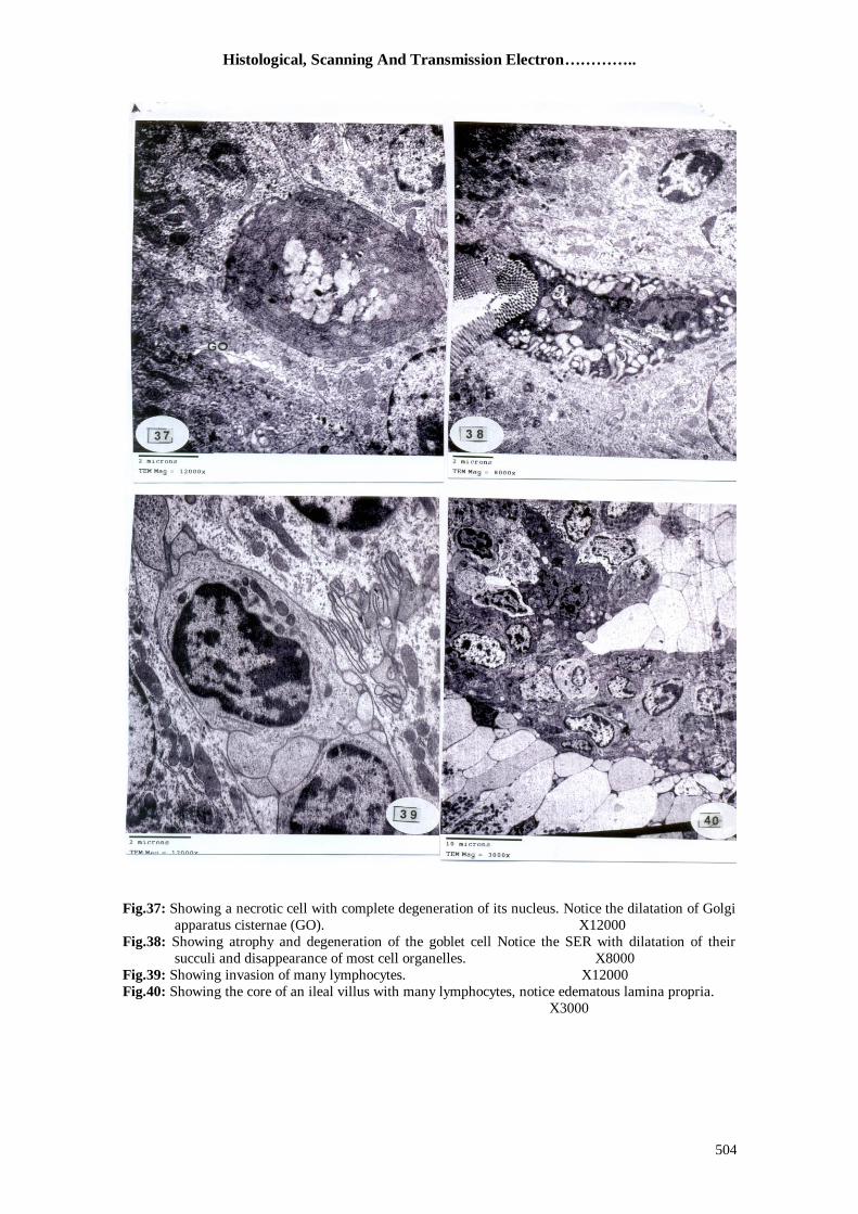
Moreover, in some highly injured cells, the nuclei were degenerated (Figs. 35, 36&37).
The Golgi apparatus of acrylamide-
treated animals cells seemed hypertrophied, so its cisternae were dilated (Fig. 34). The
arrangement of the rough ER cisternae was
somewhat disturbed; in some regions of the cytoplasm these cisternae seemed fragme-
nted (Fig.35). Many cells were severely
injured (Fig.37). Figure 36 represents two
cells in their way to degenerate. They appe-ared undergoing cloudy degeneration, and
the ER lumen was dilated and degenerated.
Acrylamide administration for 10 weeks revealed that the majority of the
mitochondria were slightly swollen, and the
cristae of some of them had lost their normal appearance and became difficult to
be distinguished (Figs. 34&35).
The goblet cells became few, tall,
more globular and many vacuoles were found within the cytoplasm. Their nuclei
showed pyknosis and became more basally
located. Smooth endoplasmic reticulum succuli became very dilated (Fig.38).
Figures 39 and 40 showed damage of
the lamina propria in the core of the ileal
villus. This damage was accompanied with the presence of many lymphocytes.
c. Animals reiceved ginger together with
acrylamide The electron microscopic examination
ileum of mice that received ginger (ZOE)
extract 1 hour before the acrylamide administration showed that the general
architecture of the ileal tissue was more or
less similar to that of the control group. The
nuclei of the absorptive cells appeared with normal basal position surrounded by
abundant cytoplasm, the microvilli of the
brush border were projecting in a regular and normal manner (Fig.41). Some ciste-
rnae of Golgi apparatus, and many lysoso-
mes were prominent (Fig.42). In addition, a plenty of mitochondria and the rough
endoplasmic reticulum were observed
(Fig.44). Different types of leukocytes with
normal nuclei and cytoplasm were clearly noticed (Fig.43).

Histological, Scanning And Transmission Electron…………..
496
Figures 1-5: Photomicrographs of transverse sections of the ileum of control mice.
Fig.1: Photomicrograph of transverse section of ileum of control mice showing the mucosal villi, and the
crypts of Lieberkuhn (arrows). (H&E)
Figures 2,3 &5: A magnified regions, illustrating the structure of ileal villi showing the simple columnar
epithelial cells with the brush border (arrow head) interspersed with few goblet cells (G) and interepithelial lymphocytes (arrows). (Figs.2 &5H&E) (Fig.3.TB)
Fig. 4: Showing the structure of the crypts of Lieberkuhn lined with columnar epithelial cells and few
goblet cells. (TB)

Hala Galal El-Tantawi
497
Figures 6-11: Photomicrographs of the ileum of mice treated with acrylamide for 10 weeks.
Figures 6,7 &11: Showing vacuolation and degeneration of lamina propria, apparent hyperplasia of the
surface columnar cells which appeared to be separated from each other at many sites(thin
arrow). (Figs .6&7H&E, Fig.11 TB)
Fig.8: A magnified part of the mucosal villus of ileum of mice revealed the loss of villus architecture and
increase of interepithelial lymphocytes with few goblet cells. (H&E)
Fig.9: A magnified part of the villus apical region, the cytoplasm showing vacuolation. (H&E)
Fig.10:Illustrating the crypts of Lieberkühn, faintly stained epithelial cells, Pyknotic nuclei and
vacuolation of the cytoplasm (arrow heads). (TB)
Fig. 11: Showing damage and vacuolation of lamina propria (right upper arrow), also notice discontinuity of surface columnar cells (left lower arrow). (TB)

Histological, Scanning And Transmission Electron…………..
498
Figures 12-16: Photomicrographs of the ileum of mice treated with acrylamide after receiving the
ginger extract ZOE.
Figures12&13: Showing normal histological structure of the columnar epithelial cells of the ileal villi
and lining of the crypts. (H&X)
Figures 14&15: Showing a magnified part of the inner regions of the crypts and columnar cells which
facing the lumen, the villi regained their normal structure, lamina propria appeared with more or
less normal structure in the core of the villi. (H&E)
Fig.16: Showing the mucosal villi with more or less normal lamina propria. Notice that the villi become
more thinner than of control villi, and very few goblet cells (G) are observed. (TB)

Hala Galal El-Tantawi
499
Figures 17&18: Scanning electron micrographs of the dorsal surfaces of different types of mucosal villi
(filiform & foliate) of ileum of control mice.
Figures 19-21: Scanning electron micrographs of the ileal villi of mice treated with the acrylamide, showing loss of the normal structure of the villi accompanied with damage and bleeding
which is shown in Fig.21 at the sites of red blood corpusles.

Histological, Scanning And Transmission Electron…………..
500
Figures 22-24: Scanning electron micrographs of the ileal villi of treated mice with acrylamide and
previously received ginger, showing the normal structure of the villi with presence of mucous
secretions (arrows) on the surface.

Hala Galal El-Tantawi
501
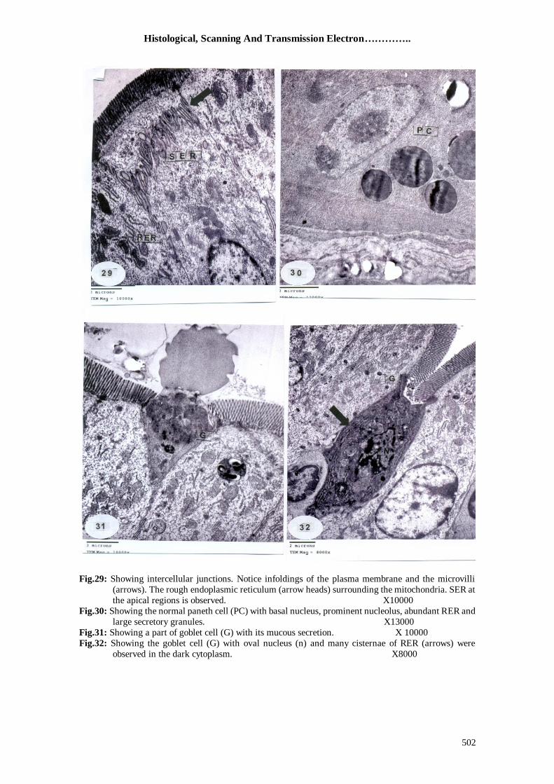
Figures 25-32: Transmission electron micrographs of the ileum of control mice.
Fig.25: Showing the columnar (absorptive) cells (c) with microvilli (arrow heads), different types of cells
such as endothelial cells (arrow), and lymphocytes (L) can be shown. ( X 3000) Fig.26: Showing a magnified part of absorptive cell illustrating the nucleus (N), different forms of
mitochondria (M) with well developed cristae. Note the smooth endoplasmic reticulum (SER)
and the free ribosomes (R). ( X 20000)
Fig.27: Showing the cellular junctions between the adjacent cells (arrows). Notice the rough endoplasmic
reticulum (RER). (X10000)
Fig.28: Showing the microvilli of control ileum. (X25000)
Histological, Scanning And Transmission Electron…………..
502
Fig.29: Showing intercellular junctions. Notice infoldings of the plasma membrane and the microvilli
(arrows). The rough endoplasmic reticulum (arrow heads) surrounding the mitochondria. SER at
the apical regions is observed. X10000
Fig.30: Showing the normal paneth cell (PC) with basal nucleus, prominent nucleolus, abundant RER and
large secretory granules. X13000
Fig.31: Showing a part of goblet cell (G) with its mucous secretion. X 10000
Fig.32: Showing the goblet cell (G) with oval nucleus (n) and many cisternae of RER (arrows) were
observed in the dark cytoplasm. X8000

Hala Galal El-Tantawi
503
Figures 33-40: Electron micrographs of ileum of mice treated with acry-lamide for 10 weeks at dose
200 p.p.m.
Fig.33: Showing highly damaged columnar absorptive cells, degeneration of the microvilli and formation
of blebs (arrow heads), pyknotic irregular nuclei (arrow). Note the hypertrophy of Golgi
elements (GO). X6000
Fig.34: Showing a magnified part of an absorptive columnar epithelial cell with dilated cisternae of Golgi apparatus (arrows).Notice the pyknotic nucleus (N). X20000
Fig.35: Showing highly damaged and degenerated epithelial cells. Notice the pyknotic nucleus (N),
fragmentation of RER, complete deterioration of the ground cytoplasm and mitochondria (M).
X15000
Fig.36: Showing complete degeneration and dissolution of the cytoplasm resulting in formation of large
vacuoles. Notice the karyolytic cells (arrows). X8000
Histological, Scanning And Transmission Electron…………..
504
Fig.37: Showing a necrotic cell with complete degeneration of its nucleus. Notice the dilatation of Golgi
apparatus cisternae (GO). X12000
Fig.38: Showing atrophy and degeneration of the goblet cell Notice the SER with dilatation of their
succuli and disappearance of most cell organelles. X8000
Fig.39: Showing invasion of many lymphocytes. X12000
Fig.40: Showing the core of an ileal villus with many lymphocytes, notice edematous lamina propria.
X3000

Hala Galal El-Tantawi
505
Figures 41-45: Electron micrographs of the ileum of a mouse treated with acrylamide and received
the ginger (ZOE) 1hr prior the acrylamide administration.
Fig.41: Showing the absorptive columnar epithelial cells appeared more or less normal. Lysosomes (LY)
were also noticed and mild dilatation of Golgi apparatus cisternae. (GO). X15000
Fig.42: Showing a magnified part of the absorptive columnar cell with dilated cisternae of Golgi
apparatus. The cytoplasm contains more or less normal cell organelles.
X 15000
Fig.43: Showing different types of leukocytes and the lamina propria displaying less damage and edema. X5000
Fig.44: Showing normal distribution of the mitochondria (M) normal appearance of RER which carries
many ribosome (arrow heads). X20000
Histological, Scanning And Transmission Electron…………..
506
Discussion
It is evident from the present study
that the light, scanning and transmission electron microscopic studies of ileal
epithelial cells of the mice adversely
responded to the administration of the
monomer crystalline substance acrylamide. Acrylamide is a water soluble organic
substance; it is absorbed by all routes and
easily distributed throughout the body organs (Barber et al., 2001). In the present
study, the mice were chosen for these
experiments due to their sensitivity to the toxic substances.
Paulsson et al. (2002) reported
neurotoxic and carcinogenic effect of
acrylamide in animals, and revealed the lower sensitivity of the rat than that of the
mouse to the carcinogenic action of this
substance. In 1995 Friedman and his associates concluded that the administration
of acrylamide in drinking water increased
the incidence of mammary tumours The gastrointestinal tract is lined with
a single layer of epithelium, which forms a
highly selection barrier designed to allow
the efficient transport of nutrients and water, while preventing the entery of
potentially toxic luminal pathogenic
organisms and their products or any toxic substances (Adam et al.,2006). According
to those authors, (as an interpretation of our
finding), acrylamide may have the ability to
destroy this intestinal barrier. At the beginning of the present study
after two to three weeks of administration
of acrylamide (200 p.p.m) in the drinking water, a marked increase of weight was
observed (Marsh et al., 2007).
Histopathologically the lymphocytes infilteration were noted, this finding come
in agree with the physiological explanation
which based on the pharmacokinetic model
for acrylamide and it was found that it is distributed within five compartments
(arterial blood, venous blood, liver, lung
and all other tissues lumped together) (Kirman et al., 2003). The present results
showed that the superficial luminal cells of
the ileal villi of the treated animals were affected; this agree with studies on aspirin
which have shown to induce similar
damage (Ivey et al., 1980). Vacuolar
degeneration, nuclear pyknosis, cloudy
swelling and necrosis were of the most marked signs of tissue impairment in the
present study. These destructive changes
were similarly reported in the colon,
nervous and reproductive systems and produce tumurs in certain hormonally
responsive tissues following treatment with
acrylamide (Barber et al., 2001). In the present study, the surface
mucosa of the ileum was studied by using
scanning electron microscope. After treatment with acrylamide, the cell surfaces
were damaged in some areas. Similar
results were observed by Specian and
Neutra (1980) who investigated the epithelial topography of the surface crypt
cell in rabbit and monkey colon. In
addition, Bonvicini et al. (1985) reported a clinical investigation of different
gastrointestinal diseases by using (SEM);
they found that the mucosal surface of the involved mucosa showed certain lesions as
bleeding and enlargement of the crypts.
These results agree with the present
findings where the bleeding appeared and was indicated by the presence of the red
blood corpuscles.
Kamel et al. (1985) investigated the structural changes in mouse intestinal villi
following lower body heating by using
SEM. Some villi were severely damaged at
their tips; these are similar to the damage described in the present study after
acrylamide treatment.
The ultrastructural study showed that the microvilli of the absorptive columnar
cells exhibited signs of degeneration. A
similar observation has been described in the small intestine of Tilapia nilotica (Sakr
, 1993), where signs of damage and
necrosis of the columnar epithelial cells
appeared, and then the cells revealed morphological alterations. These results
suggested that the columnar cells with their
brush borders are the main target cells and act as endocrine disruptors after treatment
with chemicals that manifacture plastic
products (Besaratinia and Pfeifer , 2004) who concluded that the mutagenicity of
acrylamide in human and mouse cells is
based on the capacity of its epoxide
Hala Galal El-Tantawi
507
metabolite glycidamide to form DNA
adducts. The mitochondrial destruction in the absorptive columnar cells of the ileum
of the mice was one of the important results
which has been illustrated under various
pathological conditions (Adams et al., 1977; EL-Beih et al., 1993).
The present results also showed that
the rough endoplasmic reticulum was broken-down, degranulated and fragmented
into small structures after treatment with
Acrylamide. Such damage is considered to be one of the essential factors responsible
for the lowered activity of the cell (Elewa et
al., 1999).
The Golgi apparatus of the cells of treated animals displayed damage. These
results confirm the findings of other
research workers in different pathological instances (Moussa et al., 1987; Winton and
Flaks, 1988; EL-Beih et al., 1994). The
observed increase of lysosomes in the cells of animals received ginger together with
acrylamide was confirmed by the reports
presented by Helman et al. (1985) which
revealed that the lysosomes undergo proliferation, abundance and finally rupture
at later stages of adverse conditions, with
the consequent release of their digestive hydrolases into the cytosol could
speculatively account for a considerable
proportion of damage produced in the cells
as observed in the present work. Moreover, the present study showed that the mucous
goblet cells were very few, became
elongated but with degenerated nucleus and cytoplasmic organelles; also the Paneth
cells were hardly detected. Furthermore,
many lymphocytes were seen in the lamina propria. These results explained the potent
immunologic function of the ileum, and
agree with the study that was carried out by
Mohamed et al. (2006). In the present study, the
administration of ginger (ZOE) 1 hr prior to
acrylamide in drinking water decreased the histopathological alterations which induced
by acrylamide. Where, the ethanolic ginger
extract was selected to apply in this study according to Mascolo et al. (1989) who
found that the ethanolic ginger extract (100-
300 mg/kg) contains compounds which
inhibit prostaglandin release by leukocytes, which may be responsible for ginger's
antipyretic activity and anti-inflammatory
effects.
Abdel-Ghaffar (2006) reported that
the animals treated with ZOE alone did not show any alteration in serum glucose. So,
this could ensure the protective role of
ginger against the oxidative hyperglycemia.
As far as we can throw more light on the beneficial effect of ginger which is often
touted for health benefits and how it was
able to reduce the ultrastructural changes caused by acrylamide in the ileal mucosa of
mice. Cotran et al. (1994) reported that the
mitochondrial dysfunction resulting from acrylamide treatment that would create lack
of ATP needed for normal function due to
its toxic effect {histotoxic hypoxia} and the
mitochondria were significantly protected after incorporating ginger.
In consistency with the result of the
present study Ghayur and Gilani (2006) found that the extract of ginger exhibits
species spasmogenicity in gut tissues, along
with a dormant no effect, mediated via the blockade of voltage-dependent Ca
2+
channels. Thereby, it maintains the integrity
of the plasma membrane and protects it
against apoptosis. Zhongguo (1992) reported that ginger
juice exhibits anticholinergic and
antihistaminic action. Ginger juice produces antimotion sickness action by central and
peripheral anticholinergic and antihistam-
inic effects. Since it was found that the
acrylamide substance is classified as carcinogenic and mutagenic substance that
can induce DNA damage in the cell (Barber
et al., 2001). Nakamura and Yamamoto. (1982&
1983) reported that ginger juice contains
antimutagenic components that suppress {6}-gigerol which is a potent mutagen. So,
it could induce DNA repair in damaged
cells and induce their apoptosis. In addition,
they revealed that the ginger has been repeatedly proven effective in the
gastrointestinal distress and is indicated as
safe to use in moderate amounts. Overall, studies have proved it to have some effect
on motion sickness and post-operative
nausea. However, the studies failed to indicate any CNS involvement.
Shulick. 1996; Williams. 1994 and
Mclntyre. (1995) concluded that ginger acts
as adaptogenic balance, antioxidant, antitoxic probiotic support, systemic
stimulant and cytoprotection .

Histological, Scanning And Transmission Electron…………..
508
In conclusion, the histopathological
changes of ileum may contribute to toxic effect of acrylamide, DNA changes which
lead to pyknosis of the nuclei and necrosis
of the cells. Hence, the application of
electron microscopy in the present study has the merit of bringing into vision
numerous details on the ginger protective
role which could be considered a good remedy in gastrointestinal problems that
counteracts the histopathological impair-
ments caused by acrylamide and provides improvement of the general health.
References 1. Abdel-Ghaffar, O. (2006): Protective role
of Zingiber officinale extract against
arsenic-induced toxicity in female albino
Rat. Egypt. J. Zool., 47: 183-201.
2. Adam J, Prashant K, Kathleen A,
RyanG,Wooten and AthonyT (2006): Prostaglandin mediated inhibition of
Na+/H+ exchanger isoform 2 stimulates
recovery of barrier function in ischemia-
injured intestine.Am.J.Physiol. Gastroin-
test. Liver, Physiology, 291:885-894.
3. Adams HR, Isacson EL and Masters BS
(1977): Inhibition of hepatic microsomal
enzymes by chloramphinecol. J. Pharmacol.
Exp.Ther., 203: 388-395.
4. Barber D S, Hunt J R, Ehrlisch M F,
Lehning E J and LoPachin R M (2001): Metabolism, toxokinetics and hemoglobin
adduct formation in rats following subacute
and subchronic acrylamide dosing.
Neurotoxicology, 22: 341-353.
5. Besaratinia A and Pfeifer G (2004): Genotoxicity of acrylamide and Glycidam-
ide. J.Nati.Cancer.Inst., 96 (13): 1023-9.
6. Bianca F, Mira R, Tony H, Raymond C
and Michael A (2000): Ginger extract
consumption reduces plasma cholesterol, inhibits LDL oxidation and attenuates
development of atherosclerotic, apolipo-
protein E-Deficient mice. Amirican Society
for Nutritional Science. 1124-1130.
7. Bonvincini F, Zoli G, Maltarello M C,
Bianchi D, Gasbarrini G and Iashi R
(1985): Clinical applications of scanning
electron microscopy in gastrointestinal
diseases. Scanning Electron Microscopy,
III: 1279-1294.
8. Borrelli F, Capasso R, Pinto A, Izzo A A
(2004): Inhibitory effect of ginger Zingiber officinale on rat ileal motility. Life Science,
74 (23): 2889-96.
9. Chayen J, Bitensky L and Butcher R
(1973): Practical histochemistry.Jhon
Wiley and sons.London.
10. Connel,DW.; McLachlan, R.(1972): Natural pungent compounds : examination
of gingerols , shogaols, paradols and related
compounds by thin layer and gas
chromatography.J. Chromatogr., 67:29-35.
11. Cotran R S, Kumar V and RobbinsS L (1994):Cellular injury and cellular death
.In. Robbins pathologic basis diseases.5th
edition. Saunders W.B.Company,
Philadelphia, London.
12. EL-Beih Z M, Amer M A and Ateia M A
(1994): Changes in the Golgi apparatus of
mammalian cells under the effect of
amphetamine and ethanol.Proc. Egypt.
Acad.Sci.,44: 109-118.
13. EL-Beih Z M, Amer M A and Elewa F H
(1993): Histochemical alterati-ons in
succinic dehydrogenase activity in the cells of mice administered erythromycin
.Proc.Egypt. Acad.Sci., 43:113-121.
14. Elewa F, Gabry M and Ibrahim M
(1999): Ultrastructural changes produced
by Diclofenac sodium (voltaren) in the liver
and duodenal epithelial cells of guinea pig.
Egypt.J.Zool., 33: 133-165.
15. Friedman M A, Dulak L H and Stedham
M A (1995): A life time oncogenicity study
in rats with acrylamide. Fundam.Appl.
Toxicol., 27(1):95-105. 16. Ghayur MN and Gilani AH (2006):
Species differences in the Prokinetic effects
of ginger. Int. J. Food.Sci.Nutr.,57(1-2):65-
73.
17. Ghayur M N and Gilani A H (2005): Ginger lowers blood pressure through bloc-
kade of voltage-dependent calcium chann-
els.J. Cardiovascular Pharmacol.,45: 74-80.
18. Helman R G, Adams L G, Deeric K R
and Bridges C H (1985): The role of
lysosomes in the pathogenesis of copper-
induced hepatoxicity.Morphological studies .J.Comp.Pathol.,95: 25-35.
19. International Agency for Research on
Cancer (IARC) (1994): Monogrph on the
evaluation of carcinogenic risks to human:
some industrial chemicals N0. 60. IARC,
Lyon, France.
20. Ivey KJ, Paone DB and rause WJ
(1980): Acute effect of systemic aspirin on
gastric mucosa in man.Dig.Dis.Sci.,25:97-
99.
21. Kamel H, Carr K, Kume S and Marigold J (1985):Structural changes in mouse small
intestinal villi following lower body hype-
rthermia.Scanning Electron Microscopy.,
II: 849-858.
22. Kirman C R, Gargas M L, Deskin R,
Tonner N L and Andersen ME (2003): A
physiologically bassed pharmacokinetic
model for acrylamide and its metabolite
glycidamide,in the rat.J.Toxicol. Environ.
Health, A., 66 (3): 253-274.

Hala Galal El-Tantawi
509
23. Ko M H, Chen W P, Lin-shiau S Y and
Hsieh S T (1999): Age- -dependent acry-
lamide neurotoxicity in mice:morphology,
Physiology and function. Exp.Neurol., 158
(1): 37-46.
24. Langner E, Greifenberg S and Gruenwald J (1998): Ginger:history and
use. Advances in Therapy.,15(1): 25-44.
25. LoPachin R M (2004):The changing view
of acrylamide neurotoxicity. Neurotoxico-
logy, 4: 617-630.
26. Marsh G M, Lucas L G, Youk AO and
Schall LC (1999):Mortality patterns among
workers exposed to acrylamide:1994 follow
up:Occup. Environ. Med., 56:181-190.
27. Marsh G M, Youk AO, Buchanich J M,
Kant IJ and Swaaen G (2007): Mortality
patterns among workers exposed to acrylamide: updated follow up.JOEM.,
49(1):82-95.
28. Mascolo N, Jain R, Jain S and Capasso F
(1989): Ethanopharmac-ologic investigat-
ion of ginger (Zingiber officinale) .J.
Ethano-pharmacol., 27: 129-140.
29. Mclntyre A (1995): The complete woman's
Herbal.New York :Henry Holt and C.
30. Mohamed S , Hindawy M, Sakara Z and
Soliman M (2006):Light and electron
microscopic study of the rabbit ileum at the region of Peyer"s patches.Egyption society
of histology and Cytology. The 30th
conference. (in print).
31. Mottram DS, Wedzicha BL and Dodson
A T (2002): Acrylamide is formed in the
Maillard reaction.Nature.,419:448-449.
32. Moussa T A, EL-Beih Z M and Amer M
A (1987): The Golgi apparat-us of the
gastric mucosal cells in normal and
organophosph-ate-treated guinea pigs.
Egypt. J.Histolo.,10 (1): 115-120.
33. Mowerey D B and Clayson D E (1982): Motion sickness , ginger and
psychophysics. Lancet, 1: 655-657.
34. Nadkarni K M (1976): Zingiber officin-
alle . In India Materia Medica Bombay,
Popular Prakashon, pp: 1308-1315.
35. Nakamura H and Yamamato T (1982): Mutagen and anti-mutagen in Ginger, zing-
iber pfficinalle.Mutation Res,103:119-126.
36. Nakamura H and Yamamato T (1983): The active part of the {6}- gingerol
molecule in mutagenesis.Mutation Res, 122: 87-94.
37. Paulsson B, Grawe J and Tornquist M
(2002): Hemoglobin adducts and
micronucleus frequencies in mouse and rat
after acrylamide or N-methylolacrylamide
treatment.Mutant. Res. 516(1-2):101-111.
38. Phillips S, Hutchinson S and Ruggier R
(1993): Zingiber officinale does not affect
gastric emptying rate.Anathesia ., 48 : 393-
95.
39. Qian D S and Liu Z S (1992): Pharmacol-
ogic studies of antimotion sickness actions of ginger.Chung Kuo Chung His Chieh Ho
Tsa Chih ., 12: 95-98.
40. Sakr S A (1993): Surface ultrastructure of
intestinal mucosa of Tilapia nilotica
exposed to diazinon.J.Egypt.Ger. Soc.Zool.,
12 (C):135-152.
41. Sharma S S and Gupta Y K (1998): Reversal of cisplatin-induced delay in
gastric emptying in rats by ginger. J.
Ethnopharmacol., 62: 49-55.
42. Shulick P (1996): Ginger common spice
and wonder drug. Vermont: Herbal free press, Ltd.
43. Smith E A and Oehme F W (1991). Acrylamide and polyacrylamide :a review
of production, uses, environmental fate and
neurotoxicity. Rev.Environ.Hlth., 9 (14):
215-228.
44. Specian R and Neutra M (1980): Mechanism of rapid mucous secretion in
goblet cells stimulated by acetylcholine.
J.Cell Biol., 85: 626-640.
45. Stewart J J, Wood M J, Wood CD and Mims ME (1991): Effects of ginger on
motion sickness susceptibility and gastric
function. Pharmacology, 42: 111-120.
46. Tareke E, Rydberg P, Karlson P,
Ericksson S and Tornquist M (2002). Analysis of acrylamide, a carcinogen
formed in heated foodstuffs.J.Agric. Food.
Chem., 50 (17): 4998-5006.
47. Williams JM (1994): Jude's herbal home
remedies.Minn:Llewellyn Publications .
48. Winton D J and Flaks B (1988): Effect of
fasting on aggregation of hepatocyte rough endoplasmic reticulum in adrenalctomized
and 3 ME DAB- treated rats. Quantitative
electron microscope Study .Br.J.Exp.
Pathol., 69: 877-889.
49. World Health Organization (WHO)
(2002): Additional research on Acrylamide
in food essential.Scientists declare.Geneva,
June 2002, Summary Report.
50. Yamahara J, Huang Q, Li L, Xu L and
Fujimura H (1990). Gastrointestinal
motility enhancing effect of ginger and its active constituents.Chem.Pharm.Bull., 38:
430-431.
51. Zhongguo zhong XI YI Jie He Za Zhi
(1992): Pharmacological studieof antim-
otion sickness actions of ginger. Lie Kx,
Ww wk, He E, Sunhl. Feb; 12 (2): 95-8,70.

Histological, Scanning And Transmission Electron…………..
510
دراسبت هستولوجيه و تركيبيه دقيقه للتأثير المحتمل لمستخلص الزنجبيل
المضبد لللأكريلاميذ المحذث لتلف الأمعبء الذقيقه في الفئران البيضبء
هبله جلال محمذ الطنطبوى عبهعخ عيي شوس -كليخ العلم –قسن علن الحياى
ف رخليق هابدح الجالأ ركشيلاهياذ كازل الأاكشيلاهيذ هبد كيويبئي صبعي رسزخذم
كواب رى الجاال اكشيلاهيااذ رساازخذم لزنيااخ هيااب , رسازخذم فاا العذيااذ هااي الزاجيناابد ال اابعيخ قذ رؤد الغشعبد الوزشاكوخ هي هبدح الاكشيلاهيذ غيش الوجلوشح ال حذس رضشاس ,الششة
شاء از الذساةاخ ثضاشم عوال هي رعل رل رن إع.عسيوخ للأعضبء الوخزلفخ ثغسن الإسبى
دساةاابد ساازلعيخ ثاابلوغش الضاائ كاال هااي الوغااش الإلوزشاا الوبةاا الوغااش
الإلوزشاا البفااز علاا الاهعاابء الذقينااخ للفاادس الأثاايو صاان دساةااخ الااذس القاابئ لوساازخل
.الضغجيل
, لا كحال هح 1.1الألا ضابثاخ هعبلغاخ ثا :رن رنسين الفئشاى ال صلاس هغوعابد
رةاابثي 10لوااذح ( عااضء فاا الوليااى 200)الضبيااخ علغااذ ثبللأكشيلاهيااذ فاا هيااب الشااشة
الوغوعاااخ الضبلضاااخ ااا الوغوعاااخ الوعبلغااا ثبلأكشيلاهياااذ ( صلاصاااخ ريااابم فااا الأةاااج )حيش رن اعابء الحيابد زا (لزش/هليغشام10)الضغجيل رعايذ هسزخل كحل للضغجيل
. قجل الأكشيلاهيذ ثسبعخ احذحالوسزخل
قذ عذ رى الأكشيلاهيذ رسجت ف حذس رلفب ف الأهعبء الذقينخ عل شاول رلاف فا
الخلايب الابئياخ للخوالاد الوعياخ إلا عبات حاذس رلاف فا سجناخ الأساغخ الضابهخ دا ال
اضاااشاة , لالائيااخا بريضااب لااحط رحااان رلااف الخواالاد الذقينااخ للخلاياا.الخواالاد الوعيااخ
الزظين العبم للسيظ ظشد ث الخلايب الإلزبثيخ الإسربشحيخ روذ رغابيف ةايزثلاصهيخ
رهاب .زا عل هسز الفحا ثابلوغش الضائ.ف الخلايب ظشد علاهبد الزحلل الشبهل الوغش الإلوزش الوبة فنذ رظش اى الخولاد هزحاوخ ه حذس ثعو الضف الوغش
, رحاواذ الويزكاذسيب. الأغشايخ الجلاصهياخ,البفز رظش رضيش هلحظ ف العضيبد الخليخ
رضيااش الزشكياات الااذقيق لغاابص عاالغ ظااش ريضااب اضااوحلا حااذس رفزااذ فاا الشااجوخ
ظااش ناا فاا الواابدح بالإذثلاصهيااخ كوااب حااذس رلااف فاا ريااخ الخلايااب هوااب غيااش شااول
.شد علاهبد الزذم الوبهلالوشهبرييخ ثذا لب ظ
رضااحذ الوعبهلااخ ثوساازخشط الضغجياال إلاا عباات الإكشيلاهيااذ رحسااب هلحظااب فاا ساايظ
قااذ رضااحذ الذساةاا دس الضغجياال كوشااشة سجيعاا قاابئأ فعااب ضااذ . الأهعاابء الذقينااخ
. الزدصيش الضبس لز الوبدح الز يووي رى رزظ هي سشينخ سجخ ثعو الأسعوخ الشيخ
Related Documents