HISTO LOGIC ® Technical Bulletin for Histotechnology Vol. XXXV, No. 1, May 2002 What happens inside a microwave? Have you ever stopped to think about how it’s possible to heat something in a chamber that remains at room temperature? It’s a fascinating process. Microwave devices use a power transformer to produce high voltage electricity (approximately 4000V). Through a complex process, the high voltage enables the magnetron in the instrument to produce microwaves, which are then channeled through a waveguide and delivered to the chamber. 1,2 A microwave (micro=small) is an electromagnetic wave with a frequency and wavelength that can be found about halfway between a radio wave and visible light in the electromagnetic spectrum. Most microwave devices operate at a frequency of 2.45 GHz (gigahertz) or 2,450,000,000 cycles per second. The microwaves generated have a wavelength of 12.2 cm in air but IN THIS ISSUE Microwave Technology in the Histology Laboratory ……………………………………1 Comparison of Seven Antibody Dilution Buffers for Immunohistochemistry ………5 Microwave-Enhanced Fixation for Rapid Preparation of Tissue Sections for Microscopic Evaluation ………………7 Prolonged Storage of Antibody Stock Solutions for Immunohistochemistry: A Three-Year Study ………………………12 Staining Artifact Associated with Bouin’s Substitute for Masson Trichrome Staining on Formalin-Fixed Liver Biopsies ………14 Living With Formaldehyde in Today’s Healthcare Workplace ……………………15 Helicobacter pylori Stain Using Diff-Quik Solution as a Counterstain ………………18 Use of Commercially Clarified Methyl Green in the Methyl Green-Pyronin Stain ………19 Making a List…Checking it Twice …………21 NSH to Offer New Programs in the Coming Year ……………………………22 Microwave Technology in the Histology Laboratory Donna Willis Harris Methodist Hospital Fort Worth, TX Jan Minshew President, Histology Consulting Services, Inc. Introduction Consumers first became familiar with microwave technology in the form of household microwave ovens that could cook or reheat foods in a fraction of the time required by conventional ovens. The use of microwave ovens in the histology laboratory started slowly, but today household microwave devices are commonly used to perform simple procedures such as specimen stabilization, staining, epitope retrieval, and some decalcification procedures. Laboratory-grade microwave devices are rapidly gaining popularity.They provide sophisticated systems for monitoring and controlling the energy, precise temperature control, agitation to prevent thermal layering, multiple safety features, and most importantly, appropriate ventilation. Laboratory microwave devices should be used for any technique that requires precise temperature control or involves the use of hazardous materials, especially toxic, flammable, or caustic reagents. Microwave technology is routinely used to accelerate basic epitope retrieval techniques; enhance and accelerate staining procedures; stabilize and harden gross specimens for easier thin section dissection; facilitate the identification of lymph nodes in gross specimens; fix small and large specimens; decalcify bone; and rapidly, yet gently, process both small and large specimens. These procedures can be accomplished without compromising specimen morphology or antigenicity. Managing Editor, Gilles Lefebvre Scientific Editor, Vinnie Della Speranza, MS, HTL (ASCP) HT, MT Fig. 1. Kidney 4 mm, microwave processed in 48 min. Masson trichrome, 40X 1

Welcome message from author
This document is posted to help you gain knowledge. Please leave a comment to let me know what you think about it! Share it to your friends and learn new things together.
Transcript
HISTOLOGIC ®
Te c h n i c a l B u l l e t i n f o r H i s t o t e c h n o l o g y Vol. XXXV, No. 1, May 2002
What happens inside a microwave?Have you ever stopped to thinkabout how it’s possible to heatsomething in a chamber thatremains at room temperature?It’s a fascinating process.
Microwave devices use a powertransformer to produce high voltageelectricity (approximately 4000V).Through a complex process, thehigh voltage enables the magnetronin the instrument to producemicrowaves, which are thenchanneled through a waveguide anddelivered to the chamber.1,2 Amicrowave (micro=small) is anelectromagnetic wave with afrequency and wavelength that canbe found about halfway between aradio wave and visible light in theelectromagnetic spectrum. Mostmicrowave devices operate at afrequency of 2.45 GHz (gigahertz)or 2,450,000,000 cycles per second.The microwaves generated have awavelength of 12.2 cm in air but
IN THIS ISSUE
Microwave Technology in the HistologyLaboratory ……………………………………1
Comparison of Seven Antibody DilutionBuffers for Immunohistochemistry ………5
Microwave-Enhanced Fixation forRapid Preparation of Tissue Sectionsfor Microscopic Evaluation ………………7
Prolonged Storage of Antibody StockSolutions for Immunohistochemistry:A Three-Year Study ………………………12
Staining Artifact Associated with Bouin’sSubstitute for Masson Trichrome Stainingon Formalin-Fixed Liver Biopsies ………14
Living With Formaldehyde in Today’sHealthcare Workplace ……………………15
Helicobacter pylori Stain Using Diff-QuikSolution as a Counterstain ………………18
Use of Commercially Clarified Methyl Greenin the Methyl Green-Pyronin Stain ………19
Making a List…Checking it Twice …………21NSH to Offer New Programs in
the Coming Year ……………………………22
Microwave Technology in theHistology Laboratory
Donna WillisHarris Methodist Hospital
Fort Worth, TX
Jan MinshewPresident, Histology Consulting Services, Inc.
IntroductionConsumers first became familiar with microwave technology in the form ofhousehold microwave ovens that could cook or reheat foods in a fraction ofthe time required by conventional ovens. The use of microwave ovens inthe histology laboratory started slowly, but today household microwavedevices are commonly used to perform simple procedures such as specimenstabilization, staining, epitope retrieval, and some decalcificationprocedures. Laboratory-grade microwave devices are rapidly gainingpopularity. They provide sophisticated systems for monitoring andcontrolling the energy, precise temperature control, agitation to preventthermal layering, multiple safety features, and most importantly,appropriate ventilation. Laboratory microwave devices should be used forany technique that requires precise temperature control or involves the useof hazardous materials, especially toxic, flammable, or caustic reagents.
Microwave technology is routinely used to accelerate basic epitoperetrieval techniques; enhance and accelerate staining procedures;stabilize and harden gross specimens for easier thin section dissection;facilitate the identification of lymph nodes in gross specimens; fix smalland large specimens; decalcify bone; and rapidly, yet gently, process bothsmall and large specimens. These procedures can be accomplishedwithout compromising specimen morphology or antigenicity.
Managing Editor, Gilles LefebvreScientific Editor, Vinnie Della Speranza,
MS, HTL (ASCP) HT, MT
Fig. 1. Kidney 4 mm, microwave processed in 48 min. Masson trichrome, 40X
1

2
this wavelength is reduced when itenters a substance of greaterdensity. The frequency of 2.45 GHzwas selected for household micro-wave ovens because it is thefrequency at which polar molecules,especially water molecules, respondstrongly and the microwavesmaintain good strength even at greatdepth. This capability is essential forcooking food and is also practical forhistology laboratory work. Higherfrequencies provide adequatepenetration and lower frequenciescause the microwaves to penetratewell but be absorbed very weakly.3
How do microwaves affecttissue specimens?Routine histology proceduresdepend on relatively slow infiltrationof solutions from the outer surfaces,and if heat is applied it must work itsway into the interior of the specimenby thermal conduction. Exposingthin sections of specimens to micro-wave energy affects the entirespecimen instantaneously andsimultaneously, facilitating theexchange of solutions andaccelerating reaction rates due tointernal heat.
Microwaves can either pass throughsomething with little or no effect, orthey can be reflected or absorbed.Some substances, such as mostplastics, glass, and paraffin pellets, areconsidered “microwave transparent”because they remain unaffectedwhen exposed to microwave energy.Other substances, such as metal, willreflect microwaves. When substancesabsorb microwave energy theybecome excited and generateinternal heat. It is widely acceptedthat as the microwave energy isabsorbed in tissues, it is converted tokinetic and chemical energy.
When small dipolar molecules, such aswater and the side chains of proteins,are exposed to microwave energy,they immediately attempt to alignthemselves within the electromagneticfield.They rotate rapidly through 180°at the rate of 2.45 billion cycles persecond.The rotation alone does notproduce heat.As the moleculescollide, they absorb the microwaves
and convert the energy to thermal orkinetic energy.The rotation ceasesimmediately when the exposurestops. Large molecules are not asresponsive because they cannotrotate as quickly in the electric field.
The chemical energy produced intissue specimens during microwaveexposure is less understood. Themicrowave energy packet (photon)can’t ionize molecules and is toosmall to break even weak molecularbonds, but it is believed that thehydrogen bonds may beredistributed. It is presumed thatmicrowaves affect the bound waterthat physically separates macro-molecules and electrically insulatesthem from one another, and that assome of the bound water is removed,the macromolecules unwind, formnew crosslinks, and then becomelocked into different configurations.4
In order to tie this informationtogether, let me give you an exampleof what happens during microwavetissue processing. Microwaves enterthe chamber; are reflected off themetal chamber walls; pass throughthe microwave transparent pro-cessing container and cassette; andexcite the polar molecules in thesolutions and the specimens.Theresulting kinetic and chemical energyexpedite the exchange of solutionswithin the specimens.This dramati-cally reduces the amount of timerequired for processing and elimi-nates the need for graded alcohols.Clearing solutions are not necessarybecause the alcohol is evaporatedfrom the paraffin after infiltration.The solutions commonly used forprocessing are absolute ethyl (orreagent) alcohol, 99% isopropylalcohol, and paraffin.The alcoholcan be used several times and theparaffin can be reused many times,possibly for months.The specimensproduced using this abbreviatedprocess have excellent sectioningcharacteristics and the morphologyand antigenicity are unaffected.
Incorporating the use ofmicrowaves in the lab The pathology laboratory at HarrisMethodist is a busy diagnostic
service that handles an annualcaseload of 20,000 cases, whichincludes the preparation of 60,000blocks, 24,000 special stains, andover 5,000 immunohistochemistrystains each year. In the 1980s, weexplored the use of a domesticmicrowave device to decrease ourspecial stains turnaround times. Wepurchased our first laboratorymicrowave device ten years later toreduce the time needed to processrush biopsy samples, selecting adevice specifically designed forlaboratory applications to enable usto avoid the ventilation problemsassociated with a householdmicrowave. Our program was sosuccessful that it became clear thatadditional devices would bebeneficial, allowing us to processeven larger samples. Today we havesix microwave devices in use forvarious histology proceduresthroughout the department.
As our department continued togrow, the need for rapid tissueprocessing became apparent. Ourearly efforts included a shortenedschedule on our conventional tissueprocessors which enabled same dayreporting if the sample wasreceived in the laboratory beforenoon. Despite this success, ourclinicians complained about therestriction of submitting biopsiesbefore noon in order to receive aquick turnaround. A furtherabbreviation of the schedule on theprocessor was not an option, andwe also realized that a lot of timewas wasted during solutionexchange on the instrument.
Our exploration of microwavetissue processing was done withsupport from our instrumentvendor. During the demonstration,we processed various tissue typesin parallel using both microwaveand routine processing. We foundthat the morphology we obtainedwas comparable for bothtechniques. We also performedimmunohistochemical staining toassure that stain quality was nothindered by microwave processing.This yielded results comparable toconventionally processed tissues.

Initially we worried aboutdamaging or cooking thespecimens. We learned a lot fromour experimentation. For example,we found that tissues were notdamaged if they were properlyfixed. We established that fixedbiopsy samples could be microwaveprocessed into paraffin in20 minutes (see Tables 1A, 1B).Specimens must be completelysubmerged in solution and theremust be sufficient space betweenthe cassettes to allow fluid transfer.Just as in conventional processing,encapsulated specimens like skintags or colon polyps must bebisected, and fatty samples requirelonger processing due to theamount of water in the sample.Microwave-processed samplessection with ease on the microtomeand the morphology andantigenicity are not affected.
The demonstration was a success forboth the lab and the vendor, and wepurchased our first laboratory devicefor processing in 1995. This enabledus to market our use of microwavetechnology to our clinicians bymaking it possible to turn aroundbiopsy samples from specimencollection to report charting in2 hours. In some instances results areavailable to the clinician before thepatient leaves the outpatientrecovery area. Cell blocks are alsomicrowave processed after an initialfixation for 30 minutes. No longer dowe have to wait until the next day togive the cell block slides to the cyto-technologist for reading. It hasbecome a routine procedure in ourlaboratory when we receive a biopsy,cell block, or fine needle aspiratesample in the morning, to microwaveprocess it, and even if special stainsor immunohistochemical stains arerequired, the case can still becompleted that same afternoon.
Our transition to microwavetechnology had an unexpectedbenefit. We found that by having ourslides completed by afternoon andearly evening for same-day surgeries,we could avoid the need to have athird shift perform this work.
Our success with rapid biopsyprocessing led us to consider micro-wave processing for larger tissues.Extra devices were purchased andwe began processing all of ourtissues in three microwave instru-ments. Tissue samples had to be nomore than 2 mm thick. Our initialattempt was successful and wecontinued this process for over8 months. Slide quality declinedwhen our pathologist’s assistantleft, and section thicknessrequirements were not followed.We placed the conventional tissueprocessors back into operation andbegan our research on how toimprove the process. We eventuallydeveloped a microwave processingschedule for each sample thickness(see Tables 2A, 2B, 2C).
Our lab receives small samples informalin from the operating room.Larger specimens are delivered tothe lab fresh. These samples aredissected and processed that eveningso that slides can be ready for thepathologist first thing the nextmorning. We struggle, as many labsdo, to adhere to our turnaround timerequirements while providingadequate fixation of our tissues. Ourstudies, using tissues of varyingthickness processed in parallel byboth conventional and microwave
procedures, have convinced us thatmicrowave technology can provideresults superior to conventionalprocessing irrespective of sectionthickness.
Results from the laboratorymicrowave-processed specimenswere remarkable. Not only didsection morphology improve butalso the morale of the microtomystaff. No longer did they have tostruggle to obtain superiormicroscopic sections. All of thishas led us to purchase anotherlaboratory microwave device forour surgical pathology area. We arecurrently performing research onwhole organ microwave fixationthat will allow the pathologist tohave a firm, well-fixed samplebefore dissection is performed.Sectioning 2-mm sections will notbe as much of a challenge if thesample is firm prior to dissection.We believe that this additionalinvestigation will permit us onceagain to abandon the use ofconventional tissue processing.
Bone decalcification procedures canalso be performed in a microwavedevice, decreasing turnaround time.Samples that can take days todecalcify with routine methods canbe completed in a few hours in
3
Table 1A*†
Solution Wattage Time Temp Comments
10% Formalin‡ 750W 20 min 50°C Loosely coverand agitate§
* These are sample procedures only. Procedures will need to be optimized in individual laboratories usingnondiagnostic specimens.
† Fixation of 1- to 3-mm thick specimens in cassettes before processing. Tables 1A and 1B represent the two currenttheories on microwave fixation. Both techniques have produced good results.
‡ Buffered formalin solution is not necessary as long as the solution is freshly prepared.§ Heated formalin solutions are very volatile and extremely hazardous. Do not allow fumes to enter the breathing
zone. Cover containers before removing from the microwave and do not open until under a ventilation hood.
Table 1B*†
Solution Wattage Time Temp Comments
10% Formalin‡ 350W 30-45 min 40°C Loosely coverand agitate§
10% Formalin‡ 650W 10-15 min 40°C Loosely coverand agitate§

such a unit. We are currentlyresearching various decal solutionsand using several different fixativesto obtain the best protocol for ourlab. Our goal is to no longer delayreports while waiting for a decalsample.
Today, with the availability of truewalk-away special staininginstruments, we are performingfewer stains in the microwave, atleast for those methods that havebeen successfully automated. Thispermits us to better utilize ourstaffing resources.
Epitope retrieval is a complexsubject beyond the scope of thisdiscussion. It should be stressed,however, that the retrieval methodemployed must be tailored to theantibody markers you are demon-strating and the detection systemyou are using. FDA-approvedmethods must be followedprecisely or a disclaimer must beincluded in the pathology reportindicating that the results reflect adeparture from the approvedmethod. Despite this, many havefound microwave technology tobe beneficial in achieving epitoperecovery in formalin-fixed tissuesfor some markers. Some laboratorymicrowave instruments will print acompleted run report that canprovide useful information for yourlaboratory’s quality controlprogram. Reports may include arecord of the temperaturesachieved and their durationduring antigen retrieval. It is notdifficult to imagine that laboratoryaccrediting agencies may requiresuch documentation in the future.
ConclusionBoth domestic and laboratorydevices can be used to performmany of the procedures in aroutine histology laboratory butsafety, reproducibility, and samplequality are important considerationswhen selecting the best device foryour operation. In order todetermine which device will bestmeet your needs, it is important toconsider which procedures will be
4
Table 2B. Processing FIXED 1-mm thick specimens(or bx specimens in sponges) that have been rinsed well in water*
Solution Time Temp Comments100% Ethyl or RT Rinse and discardReagent Alcohol100% Ethyl or 5 min 67°C Loosely coverReagent Alcohol and agitateEthanol/ 3 min 70°C Loosely coverIsopropanol(50/50) and agitateAbsolute 3 min 74°C Loosely coverIsopropanol and agitateParaffin (60°C) 2 min 65°C Uncovered†
(Use same solutionfor 2nd step)
Paraffin 5 min 80° to 84°C Uncovered†
Table 2A. Processing FIXED Biopsy specimens(1 mm or less in thickness) that have been rinsed well in water*
Solution Time Temp Comments100% Ethyl or RT Rinse and discardReagent Alcohol100% Ethyl or 5 min 67°C Loosely coverReagent Alcohol and agitateAbsolute 3 min 74°C Loosely coverIsopropanol and agitateParaffin (60°C) 2 min 65°C Uncovered†
(Use same solutionfor 2nd step)
Paraffin 5 min 80° to 84°C Uncovered†
* The sample processing protocols listed above have been run successfully on several 120V laboratory microwavemodels. Actual instrument wattage readings were between 650W and 1000W.
† During paraffin steps, stirring mechanisms may be used. Do not use air bubble agitation. Manually agitate specimenrack between paraffin steps.
Table 2C. Processing FIXED 3-mm thick specimensthat have been rinsed well in water*
Solution Time Temp Comments100% Ethyl or RT Rinse and discardReagent Alcohol100% Ethyl or 10 min 67°C Loosely coverReagent Alcohol and agitate100% Ethyl or 10 min 67°C Loosely coverReagent Alcohol and agitateAbsolute 10 min 74°C Loosely cover Isopropanol and agitateAbsolute 10 min 74°C Loosely cover Isopropanol and agitateParaffin (60°C) 10 min 65°C Uncovered†
(Use same solutionfor 2nd step)
Paraffin 10 min 80° to 84°C Uncovered†
5
performed; which chemicals/reagentswill be required; how thetemperature will be monitored andcontrolled; what wattage will benecessary for the desiredprocedures; and what the availabilityof power and ventilation will be inthe desired installation area. Keep inmind that reagent costs will decreasebecause you use fewer reagents andsmaller volumes than what is used ina routine processor. Paraffin costsalso will decrease since the alcohol isevaporated off in the paraffin whichmay be cooled and reused.
It is encouraging to see the growthof this beneficial technology in ourdiscipline. When used properly, itcan decrease turnaround time andreagent costs. Most tangible of all,perhaps, is the diminished wait bypatients for their diagnosis.
References1. Bloomfield LA. How Things Work.
Available at: http://howthingswork.virginia.edu/2. Minshew J. Turning up the heat: microwaves in the lab.
Advance Administrators Laboratory. September 2000;9(9):24-31.
3. Kok LP, Boon ME. Microwave Cookbook forMicroscopists: Art and Science of Visualization. 3rd ed.Leyden: Coulomb Press; 1992.
4. Dapson RW. Microwave fixation and processing.California Society for Histotechnology Meeting. 1999;workshop handout.
Suggested Reading1. Leong, Anthony S-Y. Microwave Technology for
Morphological Analysis. Cell Vision, Vol. 1, No. 4, 1994.
2. Leong, Anthony S-Y. Principles and Practice ofMedical Laboratory Science, Vol. 1, BasicHistotechnology. ISBN 0-443-05369-3. ChurchillLivingston.
3. Ted Pella, Inc. Breakthroughs in Microwave Processing:Techniques for Light and Electron Microscopy.Handout material from the 3rd annual UC BerkeleyMicrowave Processing Techniques Workshop,January 1999.
4. Giberson RT, Demaree RS, eds. Microwave Techniquesand Protocols. Humana Press Inc; 2001.
Web References1. http://www.gallawa.com/microtech/CH3.html
2. http://www.gallawa.com/microtech/CH5Pg1.htmlExcerpts from the book The Complete MicrowaveOven Service Handbook, Copyright © 1997-2000 byJ. Carlton Gallawa.
3. http://imagine.gsfc.nasa.gov/Imagine the Universe, High Energy AstrophysicsScience Archive Research Center (HEASARC),Dr. Nicholas E. White, (Director), within theLaboratory for High Energy Astrophysics (LHEA) atNASA/GSFC.
4. http://howthingswork.virginia.edu/How Things Work, Louis A. Bloomfield, Professor ofPhysics, The University of Virginia.
5. http://www.ebsciences.com/papers/mw_tech.htmScientific papers and techniques for microwave tissueprocessing.
Comparison of SevenAntibody Dilution
Buffers forImmunohistochemistry
Rosalba Tamayo, HT (ASCP)Karen Petrosyan, MD
Impath Inc.Los Angeles, CA
AbstractAntibody dilution buffer providesa suitable environment for antigen-antibody interaction.The diluentcan boost antibody affinity andenhance the quality ofimmunostaining.
The purpose of this experiment wasto compare various antibody diluentsfor their ability to support antigen-antibody interaction, as well as todetermine the shelf life of predilutedprimary antibodies before they beginlosing sensitivity against the epitope.One multitissue block was preparedusing various normal and cancertissues as a panel of cases to expressvarious antigens for adequatecomparison of antibodies. Wecompared seven buffers from varioussources. Nine primary antibodieswere diluted with each buffer andstored for further staining. The abilityof these diluents to sustain antibodyviability and to support antigen-antibody interaction was variable.Some dilution buffers showedselected advantage toward a specifictype of antibody.
Overall, the DAKO antibodydilution buffer and the VentanaChemMate antibody dilutionbuffer generated the strongestintensity of immunostaining andretained sensitivity of theantibodies relatively intact duringthe first 2-week period.
IntroductionImmunohistochemistry is a complexmethod of testing histological tissuepreparations with various antigen-specific antibodies. Antibody-antigen interaction is a delicateimmunological process which
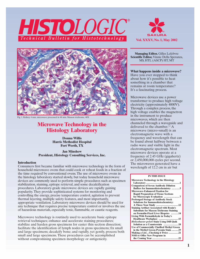
Fig. 2. Breast 1 mm, estrogen receptors stained with IHC.

6
depends on numerous equallyimportant factors for generatingadequate immunostaining results.
Choosing the right primaryantibody is perhaps the mostimportant determinant forsuccessful immunostaining.Meanwhile, choosing the rightantibody dilution buffer is equallyimportant, since antibodies requirea suitable environment foradequate interaction with the tissueantigen. It is also important forantibodies to be in the properenvironment for long-term storage.
Materials and MethodsTissues: The study included nineparaffin-embedded surgical biopsyspecimens. Tissues were pre-selected to express various types ofantigens for adequate comparisonof antibodies. One multitissueblock was prepared using variousnormal and cancer tissues: twonormal tonsils, one normal lymphnode, two mantle cell lymphomas,one normal pancreas, onechromogranin/synaptophysinpositive tissue, and two melanomas.
Primary Antibodies: Nine primaryantibodies were diluted in eachbuffer on day 1 and stored at 4ºCfor further staining and comparison.
Antibody Dilution Buffers: Thestudy included one homemade andsix commercially available antibodydilution buffers.
Immunohistochemistry: IHCstaining was performed onday 1, day 2, and 1 week, 2 weeks,4 weeks, and 8 weeks after dilutingthe antibodies. A standard immuno-peroxidase method was appliedaccording to the laboratory protocolwith overnight incubation. Theantigen retrieval procedure wasperformed in an autoclave at 105ºCfor 3 minutes.
Evaluation and AnalysisThe final analysis showed thatantibodies behaved differently ineach dilution buffer. A singlediluent could not equally support
all the antibodies for adequateimmunostaining. For example, actinshowed better immunostainingwith the buffer supplied byResearch Genetics, however, bcl-1,CD3, CK5/6 and S-100 did not stainas well with that same diluent.
Likewise, the performance of theCD3 antibody was unsurpassed withthe Biocare Blue buffer, however,actin, bcl-1, kappa, and lambdaantibodies stained inadequatelywith that buffer. This was probablybecause antibodies are highlysensitive and susceptible biologicalmolecules, and each one requires adiscrete chemical environment foroptimum behavior. Slightenvironmental changes caninfluence the immunoreaction,creating fluctuations in the results.
Considering the practicalinconveniences of using differentdiluents for different antibodies,we compared the overall achieve-
ment of all the antibodies obtainedwith each individual dilution buffer.In order to do that, we added thestaining intensity scores of allantibodies that were obtained withthe same antibody diluent andcompared the total scores ofdifferent antibody dilution buffers.
The results were analyzed bycomparing the sums of absoluteand relative sensitivity values of allantibodies using various dilutionbuffers on the same day, as well ason different days of immuno-staining (see Tables 1 and 2 on nextpage).
Ventana C dilution buffer waseliminated from the study due tohigh background and low reactivityobserved with this buffer.
Immunostaining results werecompared and evaluated semi-quantitatively by two independentinvestigators using light microscopy.
Primary Antibodies Evaluated
Antibody Clone Titer Source Actin HHF-35 1:500 DAKOBCL-1 DCS-6 1:400 Neomarkers CD3 (m) PSI 1:400 NovocastraCD4 1F6 1:200 NovacastraCHR (m/p) LK2H10(9)/poly 1:1K/1:32K BM/DAKOCK 5/6 D5/16B4 1:200 BMKappa (px) Polyclonal 1:200 DAKOLambda (px) Polyclonal 1:200 DAKOS-100 Polyclonal 1:8K DAKO
Antibody Dilution Buffers Evaluated
Antibody Dilution Buffers Cat # Source Antibody dilution buffer CD200082 DAKO ChemMate antibody dilution bufferVentana (A) ADB250 VentanaVentana (B) Sample Ventana Ventana (C) Sample Ventana PBS/BSA SOP Impath Inc.Primary Antibody Diluent 750104 Research Genetics Revival-Sky Blue PD901L Biocare Medical

In order to minimize day-to-daysubjective variations, results werecompared from different runs ofimmunostaining on different dayswith the same antibodies to ensurethat the same reference point wasused consistently for comparison ofantibody sensitivity. Each finalscore was recorded after twoinvestigators came to a consensusregarding the score.
Three variables were scored:sensitivity, specificity, and back-ground staining. The resultswere scored on a 0 to 6-plusscale. Each evaluation scorehad two components: A/B =sensitivity/background staining.
The results in Table 1 and Table 2demonstrate that DAKO antibodydilution buffer had higher overallscores throughout the 8-week periodwhen compared with the otherdilution buffers. DAKO diluentretained antibody sensitivityrelatively intact during the first2-week period. In addition, antibodiesthat were diluted in DAKO dilution
buffer showed higher relativesensitivity (less background staining)than with the others.
Conclusions1. DAKO antibody dilution buffer
generated the strongest intensityof immunostaining.
2.Most antibodies showed overalldecline in sensitivity on or afterweek 4 regardless of the antibodydilution buffer used.
3.All antibodies showed relativelyconsistent and stable results withthe same dilution buffer during the4-week study period.
4. All antibodies showed similardecline in sensitivity after the4-week study period.
5. All antibodies showed similarvariations during storage withthe same dilution buffer.
Microwave-EnhancedFixation for Rapid
Preparation of TissueSections forMicroscopicEvaluation
Carrie L. Schray, BA, HT(ASCP)Alan L. Metz, DVM, PhD, ACVP
Alex W. Gough, DVM, MSc, ACVPDrug Safety Evaluation
Pfizer Global Research & Development
Ann Arbor, MI
BackgroundMicrowave use in the histologylaboratory has increaseddramatically over the last 10 years.Microwave technology has beenapplied to rapid tissue fixation,special staining procedures, andantigen retrieval for immunocyto-chemical techniques. Addition ofheat to the fixation processincreases fixative penetration intotissue resulting in rapid cross-linkingof proteins and structuralstabilization. Variations oftemperature, tissue volume, andtime can make it difficult to controlfor consistent tissue fixation. Inaddition, different tissues undergothe fixation process at differentrates. Inadequate heat and length offixation can also result in poorfixation of inner tissue. Excessiveheat can result in shrinkage ofsmaller/thinner tissues. Differentfixatives can be used includingformalin, buffer solutions,commercial fixatives specified formicrowave use, and others. Eachcan present different hazards fromfumes and waste disposal. Thegeneration of fixative fumes isaccentuated with microwave useand requires adequate laboratoryventilation.
At Pfizer Global Research &Development, Ann Arbor, DrugSafety Evaluation, we haveexamined ways to utilizemicrowave technology to achievehigh-quality, rapid fixation of
7
Table 1. Comparison of Absolute Sensitivity of Antibodies*
Ventana Ventana Biocare Research PBS/DAKO A B Blue Genetics BSA
Day 2 46 45 32 37 39 32 Week 1 47 46 31 35 43 36 Week 2 33 36 24 23 31 29 Week 4 37 37 26 26 34 32 Week 8 36 30 23 23 32 31 Total 199 194 136 144 179 160
Table 2. Comparison of Relative Sensitivity of Antibodies†
Ventana Ventana Biocare Research PBS/DAKO A B Blue Genetics BSA
Day 2 44 43 32 33 37 29 Week 1 46 44 31 30 42 33 Week 2 33 35 24 17 31 28 Week 4 36 34 24 22 33 29 Week 8 32 27 23 16 31 30 Total 191 183 134 118 174 149
* Absolute sensitivity = intensity of specific staining.
† Relative sensitivity = absolute sensitivity – background staining results.
8
research animal tissues. Ourlaboratory provides histologysupport for a variety of pharma-ceutical preclinical safety studies inlaboratory animals. It is sometimesnecessary to evaluate tissuesmicroscopically within a short time.
Other laboratories are able toproduce histology slides rapidly,but often size and quality arecompromised. Our goal was toaccelerate the turnaround of ourstandard protocol tissue types andsizes while achieving optimalresults. By using microwave fixationof our tissues, we have been able toachieve completion of tissues aslarge as 22 mm x 13 mm x 4 mmthick within 24 hours, as opposed tothe four or five days that is typicalof our work when conventionalfixation and processing is used.
Materials and MethodsEquipmentThe PELCO laboratory microwave(PELCO Model 3451, Ted Pella, Inc.,Redding, CA) was chosen for ourlaboratory as it has many adjustablefeatures that are useful in achievingoptimal results. These includeadjustable settings for wattage,temperature, and incorporated waterload. Our unit was calibrated forspecific wattage (personal com-munication, Richard Giberson, TedPella, Inc.). It also has a temperatureprobe and bubbler for use within thesolutions placed inside the chamber.
Tissue Collection, Fixation,and ProcessingAll animal testing within the Pfizerfacility is conducted in accordancewith current guidelines for animalwelfare (Guide for the Care and
Use of Laboratory Animals, 1996)and reviewed by internalcommittees. Tissues were selectedbased on exploratory studyprotocols including spleen, colon,kidney, liver, pancreas, andmesenteric lymph nodes. Each wascollected and fixed the same way.
Cynomolgus monkey tissues werecollected during a necropsyprocedure. The tissues were placedin room temperature 10% NeutralBuffered Formalin (NBF) for30 minutes. All were trimmedand cassetted according to internalstandards, no larger than22 mm x 13 mm x 4 mm. Cassetteswere placed in fresh 10% NBF ina plastic PELCO cassette racksystem specifically designed formicrowaving, and placed intothe microwave chamber.
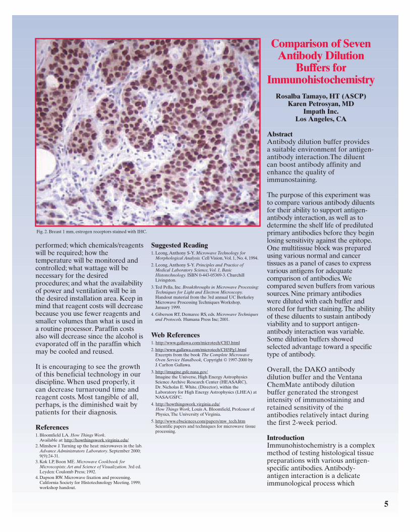
Fig. 1. Liver centrilobular hepatocytes: (A) immersion fixation; (B) microwave fixation. H&E, 20X
Fig. 2. Spleen white pulp with lymphoid follicle: (A) immersion fixation; (B) microwave fixation. H&E, 20X
A B
A B
PEACE OF MIND.A VIP THEN. A VIP NOW.
With the Tissue-Tek® VIP™ 5, Sakura brings today’shistology laboratories the fifth generation of an instrumentrecognized the world over as the laboratory standard for itsunmatched efficiency and reliability.
The VIP™ 5 both streamlines and speeds the processing oftissue samples, offering twenty different processingprograms, external reagent drain and fill, automatic reagenttransfer, and continuous mixing. Taking the lead ininnovation, quality, and reliability is nothing new for you—or the VIP™.
For more information, contact your Sakura Area Managertoday. Call 1-800-725-8723. Don’t delay.
©2001 Sakura Finetek U.S.A., Inc.
Tissue-Tek® VIP™ 5 Vacuum Infiltration Processor System
Sakura Finetek U.S.A., Inc.1750 West 214th Street
Torrance, CA 90501 U.S.A.Phone: (800) 725-8723
Visit our web site at www.sakuraus.com
ProvenReliability
The microwave was set at 45°C,wattage setting 3 (315 watts),medium-high power, for 70 minutes.Following the formalin, tissues wereplaced in Preserve™ fixative (EnergyBeam Sciences, Agawam, MA)
at 45°C, wattage setting 2(210 watts), medium-high power,for 40 minutes. Cassettes werethen rinsed in tap water brieflyand processed overnight in aconventional tissue processor
(Tissue-Tek® VIP™, SakuraFinetek, Torrance, CA) beginningwith 70% specially denatured ethylalcohol, through 100%, xylene, andparaffin (Paraplast X-tra, OxfordLabware, St. Louis, MO).
10
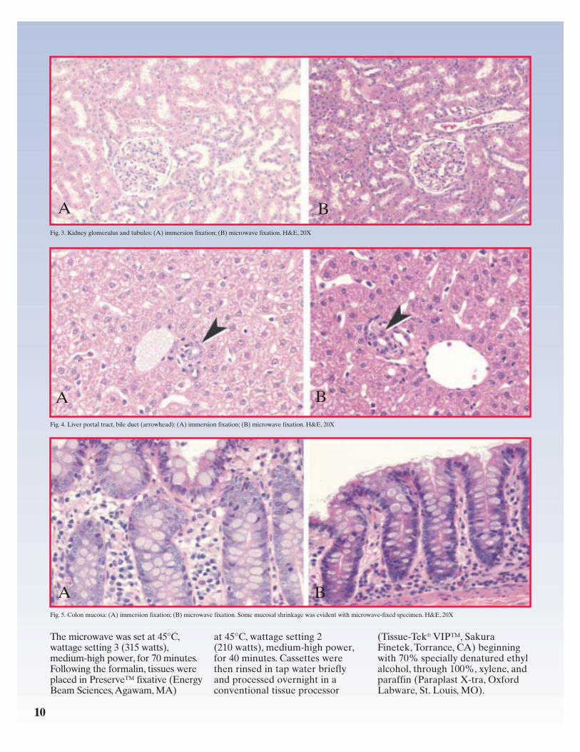
Fig. 5. Colon mucosa: (A) immersion fixation; (B) microwave fixation. Some mucosal shrinkage was evident with microwave-fixed specimen. H&E, 20X
Fig. 3. Kidney glomerulus and tubules: (A) immersion fixation; (B) microwave fixation. H&E, 20X
Fig. 4. Liver portal tract, bile duct (arrowhead): (A) immersion fixation; (B) microwave fixation. H&E, 20X
A B
A B
A B
Embedding, Sectioning,and StainingThe next morning the tissues wereembedded in Paraplast® embeddingmedium, (Oxford Labware,St. Louis, MO). Blocks weresectioned at 3µ and stained withHematoxylin & Eosin (H&E),Periodic Acid Schiff (PAS), and/orReticulin stain using standardprotocols.1
For comparison, tissue sectionsfrom the same tissue samples wereprepared using standard immersionfixation in 10% NBF for 2-4 daysand then processed using theovernight processor as describedabove. Embedding, sectioning, andstaining were also done asdescribed above, as well as perinternal standard operatingprocedures.
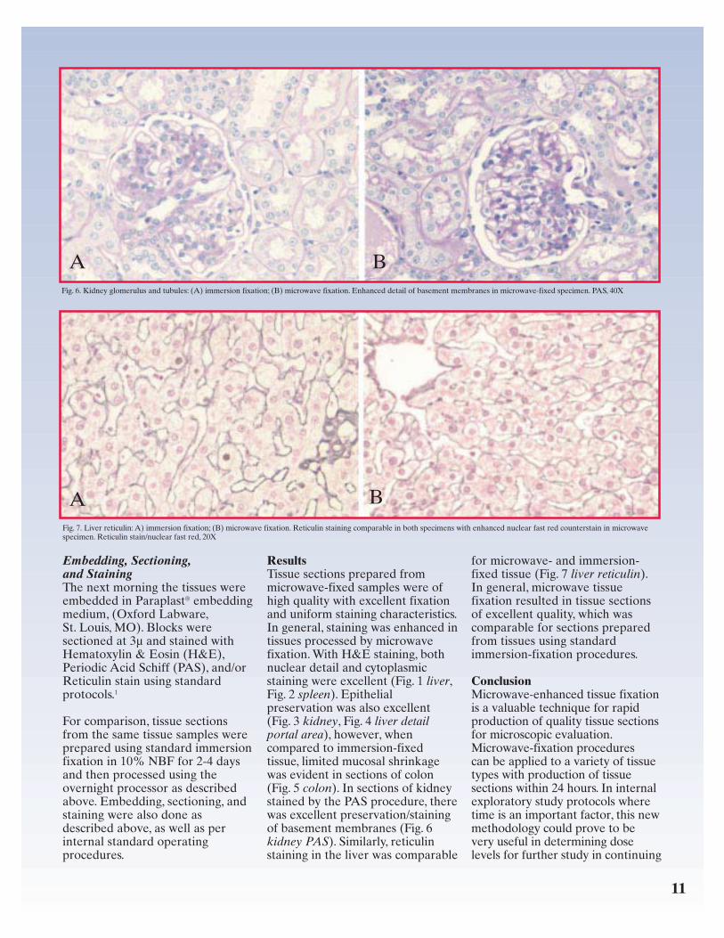
ResultsTissue sections prepared frommicrowave-fixed samples were ofhigh quality with excellent fixationand uniform staining characteristics.In general, staining was enhanced intissues processed by microwavefixation. With H&E staining, bothnuclear detail and cytoplasmicstaining were excellent (Fig. 1 liver,Fig. 2 spleen). Epithelialpreservation was also excellent(Fig. 3 kidney, Fig. 4 liver detailportal area), however, whencompared to immersion-fixedtissue, limited mucosal shrinkagewas evident in sections of colon(Fig. 5 colon). In sections of kidneystained by the PAS procedure, therewas excellent preservation/stainingof basement membranes (Fig. 6kidney PAS). Similarly, reticulinstaining in the liver was comparable
for microwave- and immersion-fixed tissue (Fig. 7 liver reticulin).In general, microwave tissuefixation resulted in tissue sectionsof excellent quality, which wascomparable for sections preparedfrom tissues using standardimmersion-fixation procedures.
ConclusionMicrowave-enhanced tissue fixationis a valuable technique for rapidproduction of quality tissue sectionsfor microscopic evaluation.Microwave-fixation procedurescan be applied to a variety of tissuetypes with production of tissuesections within 24 hours. In internalexploratory study protocols wheretime is an important factor, this newmethodology could prove to bevery useful in determining doselevels for further study in continuing
11
Fig. 6. Kidney glomerulus and tubules: (A) immersion fixation; (B) microwave fixation. Enhanced detail of basement membranes in microwave-fixed specimen. PAS, 40X
Fig. 7. Liver reticulin: A) immersion fixation; (B) microwave fixation. Reticulin staining comparable in both specimens with enhanced nuclear fast red counterstain in microwavespecimen. Reticulin stain/nuclear fast red, 20X
A B
A B

drug discovery. The use ofmicrowave technology in clinicaland industrial settings is now morecommonplace as laboratoriesbecome aware of its value inachieving quality histologicpreparations in less time.
Reference1. Sheehan DC, Hrapchak BB. Theory and Practiceof Histotechnology. 2nd ed. St. Louis, Mo:C.V. Mosby Co;1980.
Prolonged Storageof Antibody
Stock Solutions forImmunohistochemistry:A Three-Year Study
Marilyn E. Beissenherz, HT(ASCP)J. A. Ramos-Vara, DVM, PhD, ECVP
Veterinary MedicalDiagnostic Laboratory
University of Missouri-Columbia
IntroductionThe effect of storage of antibodystock solutions on the immuno-reactivity of these antibodies is notwell documented. We tested theimmunohistochemical reactivity ofstock solutions of antibodies storedfor several years at the University ofMissouri-Veterinary Medical Diag-nostic Laboratory (UM-VMDL)immunohistochemistry laboratory,using formalin-fixed, paraffin-embedded tissues. Determinationof immunoreactivity was qualitative(staining or no staining).
12
Glucagon 1998 Glucagon 2001
IgG 1998 IgG 2001
Insulin 1998 Insulin 2001
13
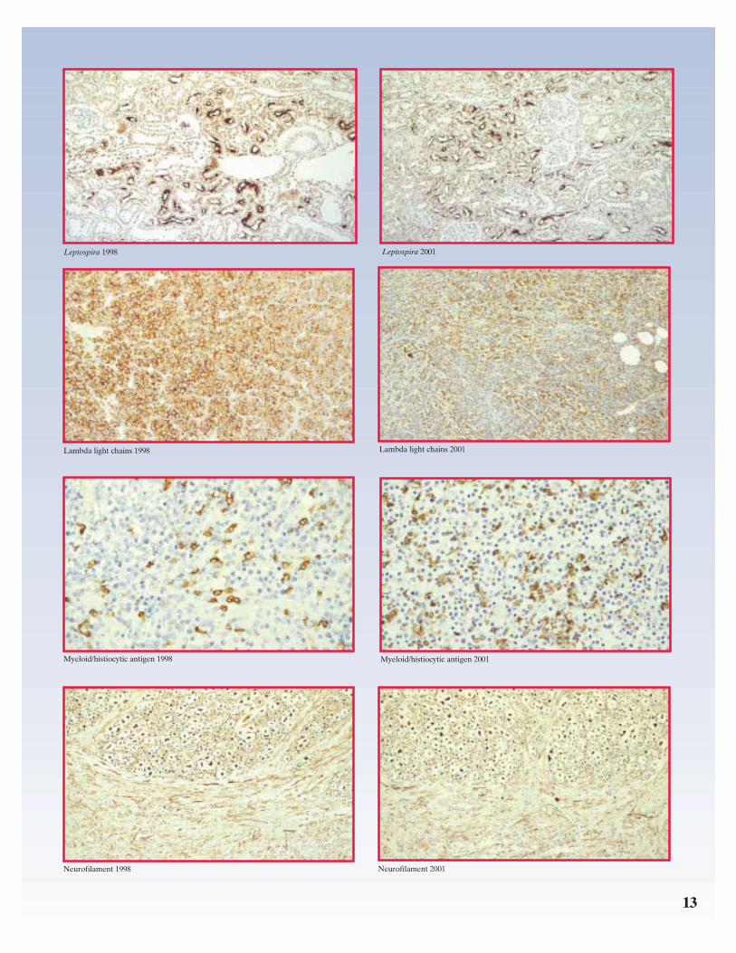
Leptospira 1998 Leptospira 2001
Lambda light chains 1998 Lambda light chains 2001
Myeloid/histiocytic antigen 1998 Myeloid/histiocytic antigen 2001
Neurofilament 1998 Neurofilament 2001

14
Materials and MethodsThe final dilution of each antibodywas made from a stock solution(1:100). Stock dilutions were pre-pared from concentrated (undiluted)antiserum using DAKO diluent,which was kept in plastic cappedvials and stored at 4°C. The finalvolume for these stock dilutionswas 5-10 mL. Immunohistochemicalmethods included labeledstreptavidin-peroxidase, ABC,and EnVision Plus-peroxidase.
ResultsOf 100 antibodies used at theUM-VMDL, stock solutions of15 antibodies have been in use withno apparent loss of staining sinceOctober 1998. These antibodies are:
• Calcitonin (DAKO; polyclonal) • Campylobacter sp. (KPL;
polyclonal)• GFAP (DAKO; polyclonal) • Glucagon (DAKO; polyclonal)• Canine IgG (Sigma;
monoclonal)• Insulin (Zymed; monoclonal)• Laminin (DAKO; polyclonal) • Leptospira sp.
(NVSL; polyclonal) • Listeria O antigen
(Difco; polyclonal) • Lambda light chain
(DAKO; polyclonal) • Myeloid/histiocytic antigen
(DAKO; monoclonal) • Mycobacterium bovis
(DAKO; polyclonal) • Myoglobin (DAKO; polyclonal)
• Neurofilament (SternbergerMonoclonals; monoclonal)
• Somatostatin(DAKO; polyclonal)
ConclusionThe reason for the variability in theimmunoreactivity of antibodiesafter prolonged storage is unknown.Although most of the antibodiesthat remained immunoreactive afterthree years of storage werepolyclonal, several weremonoclonal. Variations in theimmunoreactivity of antibodiesstored as stock solutions may bedue to diluent used, protein concen-tration, class and subclass ofimmunoglobulins, among others.
Staining ArtifactAssociated With
Bouin’s Substitute forMasson Trichrome
Staining on Formalin-Fixed Liver Biopsies
Nancy Miezin, BA, HT(ASCP)Terry Gramlich, MD
Department of AnatomicPathology
Cleveland Clinic FoundationCleveland, OH
IntroductionMasson trichrome staining iscommonly requested on formalin-fixed, paraffin-embedded liverbiopsies. Formalin-fixed tissue
sections stained with Massontrichrome require Bouin’s fixativeas a mordant. However, when usinga non–picric-acid-based Bouin’ssubstitute, microscopic examinationreveals an artifact demonstratingan overall pale red appearance andintense blue areas often around theperimeter of the section.
Materials and MethodsThe use of Bouin’s fixative (picricacid, formaldehyde, acetic acid, andwater) as a mordant in preparationfor Masson trichrome staining wascompared to the use of a Bouin’ssubstitute (chloroplatinic acid,formaldehyde, acetic acid).Duplicate paraffin-embedded,formalin-fixed needle biopsies ofliver were sectioned at 4 micronson twelve different liver biopsycases. One slide was stained with aMasson trichrome procedure usingBouin’s fixative (saturated picricacid) and the duplicate slide wasstained with the same Massontrichrome procedure, but with aBouin’s substitute (saturatedchloroplatinic acid). Afterward, theslides were compared for quality.
ResultsAll Masson trichrome-stainedslides using Bouin’s substitutedemonstrated diminished redstaining of the Biebrich scarlet acidfuchsin solution with a dark blueappearance as a result ofintensified aniline blue staining.All duplicate slides stained usingBouin’s fixative demonstratedhomogeneous Biebrich scarlet acidfuchsin and aniline blue staining.
Listeria 1998 Listeria 2001
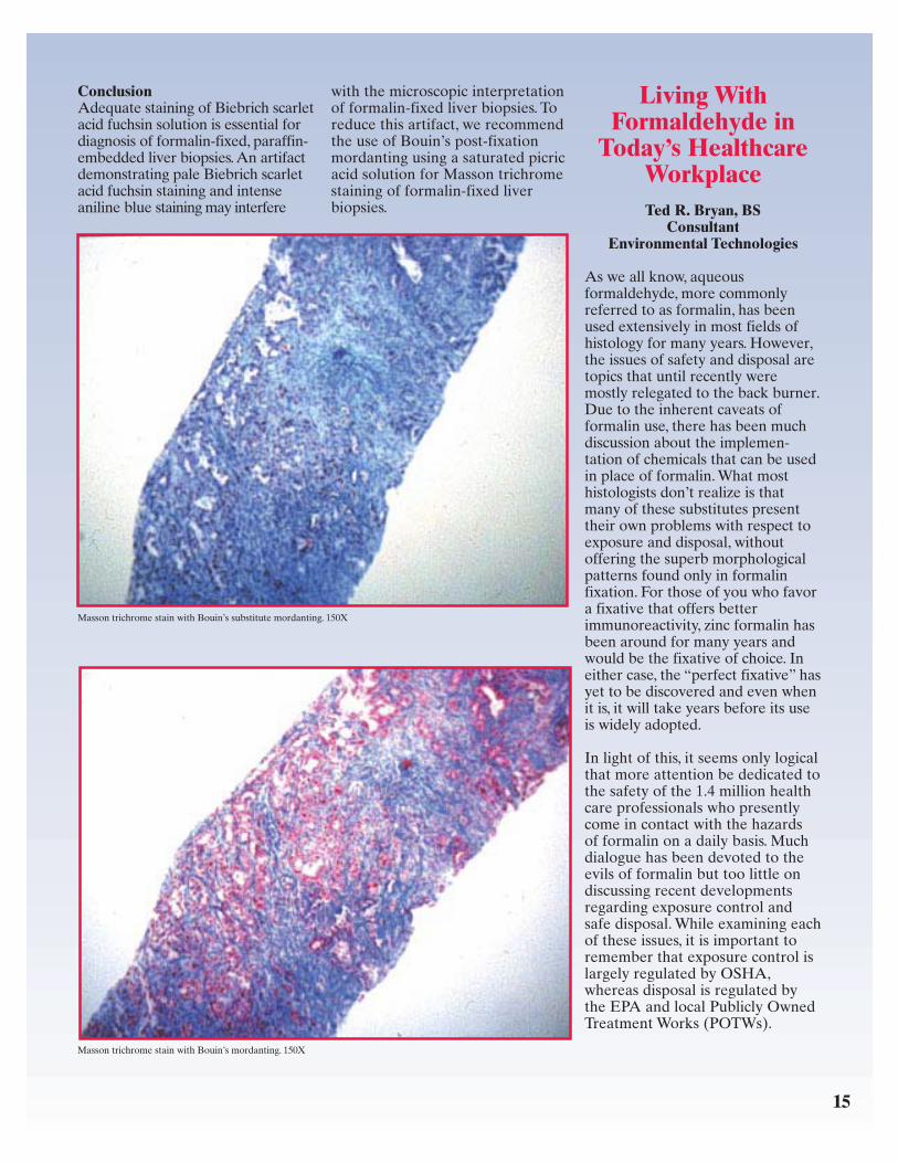
ConclusionAdequate staining of Biebrich scarletacid fuchsin solution is essential fordiagnosis of formalin-fixed, paraffin-embedded liver biopsies. An artifactdemonstrating pale Biebrich scarletacid fuchsin staining and intenseaniline blue staining may interfere
with the microscopic interpretationof formalin-fixed liver biopsies. Toreduce this artifact, we recommendthe use of Bouin’s post-fixationmordanting using a saturated picricacid solution for Masson trichromestaining of formalin-fixed liverbiopsies.
Living WithFormaldehyde in
Today’s HealthcareWorkplaceTed R. Bryan, BS
ConsultantEnvironmental Technologies
As we all know, aqueousformaldehyde, more commonlyreferred to as formalin, has beenused extensively in most fields ofhistology for many years. However,the issues of safety and disposal aretopics that until recently weremostly relegated to the back burner.Due to the inherent caveats offormalin use, there has been muchdiscussion about the implemen-tation of chemicals that can be usedin place of formalin. What mosthistologists don’t realize is thatmany of these substitutes presenttheir own problems with respect toexposure and disposal, withoutoffering the superb morphologicalpatterns found only in formalinfixation. For those of you who favora fixative that offers betterimmunoreactivity, zinc formalin hasbeen around for many years andwould be the fixative of choice. Ineither case, the “perfect fixative” hasyet to be discovered and even whenit is, it will take years before its useis widely adopted.
In light of this, it seems only logicalthat more attention be dedicated tothe safety of the 1.4 million healthcare professionals who presentlycome in contact with the hazardsof formalin on a daily basis. Muchdialogue has been devoted to theevils of formalin but too little ondiscussing recent developmentsregarding exposure control andsafe disposal. While examining eachof these issues, it is important toremember that exposure control islargely regulated by OSHA,whereas disposal is regulated bythe EPA and local Publicly OwnedTreatment Works (POTWs).
Masson trichrome stain with Bouin’s substitute mordanting. 150X
Masson trichrome stain with Bouin’s mordanting. 150X
15

Exposure ControlOver the last few years, advances intechnology have dramaticallydecreased the risk of exposure tohistotechnologists. Regulated byOSHA laws (29CFR 1910.134),formalin exposure can besubstantially mitigated byemploying general ventilation andsimple, low-cost work practicecontrols. By definition, workpractice controls “reduce thelikelihood of exposure by alteringthe manner in which a task isperformed.” For instance, ingrossing areas where ventilationmay not be adequate, employmentof appropriate respirators andneutralizing pads can dramaticallydecrease vapor exposure. Withrespect to the pads, it is importantto note that when implementingthe use of these types of controls,you are advised to thoroughlyinvestigate the product to insurethat it does actually have the abilityto neutralize the formalin. Readingthe MSDS and posing questions tothe manufacturer are both essentialin order to guarantee that theproduct functions as represented.Neutralization capabilities varydramatically and companies oftenmake performance claims withrespect to neutralization thatcannot be substantiated.
Formalin spills are another sourceof exposure which can be quicklyremedied if an appropriate workpractice control is in place. Forsmall spills, neutralizing cloths arefast and effective; large spills willrequire a spill control agent that iseasy to apply and not only absorbsthe spill rapidly, but completelyneutralizes it as well. Once again,buyer beware! Be sure to do yourhomework and investigate theproducts you use. Although somespill control agents may neutralizethe formalin, the resulting mixtureis at a pH that is below 1 and istherefore still consideredhazardous waste.
Formalin DisposalIn 1976, the US EnvironmentalProtection Agency (EPA) passedthe Resource Conservation andRecovery Act (RCRA) in order toprotect the environment fromimproper hazardous wastemanagement practices. This lawmandates “cradle to grave”responsibility for users ofhazardous waste material. As theawareness of our environmentalissues increases, the methods weemploy for disposal of hazardouschemicals becomes an issue eachof us must face. Since pollutiondoes not recognize the politicalboundaries separating states andnations, the responsibility for safedisposal belongs to all individualsunited in this cause. The decisionswe make regarding these issues willultimately affect the globalecosystem. For this reason it isimportant to set stringent standardsthat are both economically feasibleand relatively simple to follow.
With respect to formalin, we shouldall be thankful that the days ofdisposing untreated waste directlyinto a sink are fast coming to anend. Those institutions that stillpractice this will have to considerthe repercussions (see 40CFR 403)and chronic effects on aquaticdegradation, bioremediation, andhuman health. In the not toodistant future, all municipalPOTWs will be required to adhere
to regulatory laws and avoid theeffects of careless disposal practices.This is easier to accomplish than itsounds due to the availability ofdisposal options that are not only incompliance with hazardous wastedisposal guidelines, but areeconomical and easy to use as well.
One such option for waste disposalis hazardous waste hauling (off-sitedisposal). This remains a viableoption as long as it is provided byEPA-licensed, reputable firms.However, this method can beexpensive, labor intensive, and itcarries a potentially high liabilitydue to exposure and spill risks.
A viable alternative to off-sitedisposal is the use of a treatmenttechnology known asneutralization. In most cases,treatment can be trouble-free andconvenient because it takes placeat the site of waste generation anddoes not require transportation outof the department. However, inrecent years there has beenconsiderable controversy about theneutralization claims made by thevarious manufacturers of thesetreatment technologies. In responseto this, and long considered amodel for environmentalresponsibility, the California EPAhas developed its uniqueEnvironmental TechnologyCertification Program. The purposeof this program is to “provide anindependent technical evaluationof technologies to identify thosemeeting applicable qualitystandards, so as to facilitateregulatory and end-useracceptance..” In a nutshell, thisprogram certifies the scientificand engineering claims made bya manufacturer regarding theirtechnology. This is your guaranteethat the product claims have beensubstantiated by an independentsource other than themanufacturer. The program issignificant not just for those whoreside in California, but foreveryone in every state andcountry who values the importanceof safe waste disposal.
16
Personal protective equipment required when workingwith formaldehyde in a poorly ventilated area.
LOOKING GOOD.WORKING SMART. YEAR AFTER YEAR.
Sakura Finetek U.S.A., Inc.1750 West 214th Street
Torrance, CA 90501 U.S.A.Phone: (800) 725-8723
Visit our web site at www.sakuraus.com©2001 Sakura Finetek U.S.A., Inc.
Tissue-Tek® TEC™ 5 Tissue Embedding Console SystemThrough the years, each generation of the Tissue-Tek® TEC™
has maintained an outstanding record of innovation,quality, and reliability. The Tissue-Tek® TEC™ 5 issure to be no exception. Sakura has designed a fifth-
generation instrument that optimizes operator controland efficiency—and improves workflow—throughout
the tissue embedding process.
From a 4-liter paraffin chamber to ample hot and coldsurfaces to menu-driven programming, the TEC™ 5, like you,
is looking and working better than ever.
For more information, contact your Sakura Area Manager today.Call 1-800-725-8723. Don’t delay.
ProvenReliability

18
At one time there were threecertified formaldehyde treatmenttechnologies. Two of these wereoriginally certified under a pilotprogram that involved a veryrudimentary level of testing notinvolving 10% NBF wastes. Theother technology, Tissue-Tek®*Neutralex®†, was certified undera more stringent program thatincluded the treatment of actualformalin waste. When themanufacturers of the technologiescertified under the pilot programdecided to apply for recertification,the California EPA decided thatthey had to be held to the sametesting standards as wereperformed on Neutralex®. As aresult of this testing, both of theother two technologies were deniedrecertification. The data determinedthat after treatment in bothtechnologies “about one-half of thesamples in each batch were moretoxic after treatment than beforetreatment, and more toxic than thehazardous waste threshold.” As aresult of this denial, health carefacilities in California are no longerauthorized to treat their 10% NBFhazardous waste with either ofthese technologies. Neutralex® ispresently the only technologycertified by the California EPA.This is the type of regulatoryresponsibility that should also be amodel for every institution that hasa need to dispose of formalin waste.
ConclusionsRegulatory requirements affectingthe use of hazardous chemicalsboth domestically and globally arechanging as our awareness of theeffects of these substances becomesbetter understood. Contrary to thebelief of some, we can safely co-exist with formalin in theworkplace as well as protect theintegrity of our fragileenvironment. Through the use ofreliable engineering and workpractice controls, vapor exposurecan easily be maintained withinthe boundaries of OSHA laws.The California EPA EnvironmentalCertification Program will set astandard for safe disposal that can
be duplicated worldwide. We nowhave the technology to effectivelycontrol the integrity of ourworkplace and environment. Let’sface it — formalin will be a part ofmost histology departments foryears to come. With a concertedeffort by all affected, we cannavigate a course that is safe,economical, and effective.
* Tissue-Tek® is a registered trademark of SakuraFinetek U.S.A., Inc.
† Neutralex® is a registered trademark of SCIGEN Inc.
Helicobacter pyloriStain Using Diff-Quik
Solution as aCounterstain
Kevin ErnstSt. Vincent’s Medical Center
Jacksonville, FL
AbstractHelicobacter pylori is a spiral-shaped bacterium found in thegastrointestinal tract. It is nowknown to cause more than 90%of duodenal ulcers and up to 80%of gastric ulcers, hence itsdiagnostic significance. Our labuses the Diff-Quik procedure1
to identify H pylori. We recentlybegan to use a polyclonal H pyloriconcentrated antibody (DAKO),which aids in rapid visualizationof the bacteria, especially whenpresent in small numbers. Anotheradvantage is the specificity ofstaining which permits H pylorito be distinguished from non-Helicobacter organisms. We havefound that using Diff-QuikSolution II instead of hematoxylinas a counterstain results in a moreaesthetically pleasing stain which ispreferred by many pathologists.Histological tissue detail isimproved with no significant lossof contrast, which aids in findingthe organisms in tissue sections.Non-Helicobacter organisms arealso easier to identify with the
Diff-Quik counterstain. This isa fail-safe method to providechromogenic back-up stainingof H pylori in the rare event ofimmunoperoxidase technicalfailure. An added benefit may bethe visualization of Helicobacterstrains other than H pylori withthe Diff-Quik counterstain, suchas Gastrospirillum hominis(Helicobacter heilmannii)2 whichhas unknown antibody reactivity.3
Materials and ReagentsFixation: Hollande’s Fixativeor 10% buffered formalin
Technique: Paraffin sectionscut at 3-4 microns
Reagents:1. Tris Buffer with Tween 20 —
DAKO Corporation
2. Proteinase K —DAKO Corporation
3. H pylori concentrated polyclonalrabbit antibody —DAKO Corporation
4. EnVision Plus rabbit detectionsystem — DAKO Corporation
5. Diff-Quik Solution II —American Scientific Products
ResultsThe H pylori antibody stains theorganisms dark brown to black. TheDiff-Quik counterstain gives a brightblue background demonstratingimproved tissue detection overhematoxylin. Non-Helicobacterorganisms are stained blue by theDiff-Quik solution.
ConclusionImmunoperoxidase stain forHelicobacter pylori using DAKOpolyclonal antibody and Diff-Quikcounterstain offers the followingadvantages:
1. More rapid, lower magnificationidentification of H pylori.
2. Smaller numbers of H pyloriare more easily detected.
3. Specific antibody staining oforganisms is possible.
4. Fail-safe staining of bacteria

19
in the rare event of immuno-peroxidase technical failure.
5. Detection of bacteria other thanH pylori, which may be clinicallysignificant e.g., Gastrospirillumhominis (Helicobacter heilmannii)by the Diff-Quik counterstain.
References1. Skipper WR, Destephano DB. A rapid stain for
Campylobacter pylori in gastrointestinal tissuesections using Diff-Quik. J Histotechnol. 1989;12:303-304.
2. Andersen LP, Boye K, Blom J, Holck S, Norgaard A,Elsborg L. Characterization of a culturableGastrospirillum hominis (Helicobacter heilmannii)strain isolated from human gastric mucosa. J ClinMicrobiol. 1999;37(4)1069-1076.
3. Personal communication, Karen Atwood.DAKO Corporation.
Use of CommerciallyClarified Methyl
Green in the MethylGreen-Pyronin Stain
Joyce C. Hrad, HTLDepartment of Pathology& Laboratory MedicineMayo Medical Center200 First Street S.W.
Rochester, MN
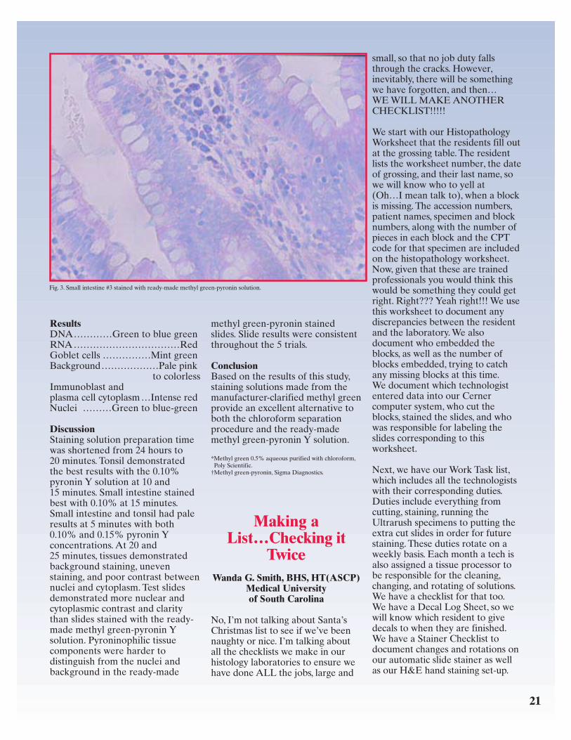
IntroductionMethyl green-pyronin is used inhistology labs to stain for DNA andRNA, which is especially beneficialfor demonstrating plasma cells,mast cells, and immunoblasts. Thepurpose of this study was to observethe staining results of methyl green-pyronin solution [when made withmethyl green 0.5% aqueous purifiedwith chloroform from Poly Scientific(Bay Shore, NY)] on formalin-fixedparaffin tissue sections. Observationswere made by comparing theseresults to the classical methodtouted by many texts whichrequires that the stain be purifiedusing a 24-hour chloroformseparation procedure to clarifythe methyl green. Slides were alsocompared to the staining results ofthe same tissue stained in a ready-made methyl green-pyronin solutionfrom a commercial source (SigmaDiagnostics).
IHC-stained H pylori with hematoxylin counterstain, oil immersion.
IHC-stained H pylori with Diff-Quick counterstain, oil immersion.
20
ProcedureTissue:• 3 paraffin blocks of small intestine
cut at 5 µm, 1 section per slide,51 slides per block
• 2 paraffin blocks of tonsil cut at4 µm, 1 section per slide, 51 slidesper block
Trials: 5 separate trials wereconducted• Each trial consisted of 10 sets of
5 slides, one slide of each of thetissues
• 5 sets were stained in 0.10%methyl green-pyronin Y solution;each set was stained for either5, 10, 15, 20, or 25 minutes
• 5 sets were stained in 0.15%methyl green-pyronin Y solution;each set was stained for either5, 10, 15, 20, or 25 minutes
• 1 set of slides was stained withready-made methyl green-pyronin Y solution forcomparison
Reagents0.10% Methyl Green-Pyronin Ystaining solution
140 ml 0.5% chloroform-clarifiedmethyl green*
0.14 g pyronin Y (CI 45005)(mixed thoroughly, filtered,and adjusted to pH 4.2 with3% acetic acid or 3% sodiumhydroxide)
0.15% Methyl Green-Pyronin Ystaining solution
140 ml 0.5% chloroform-clarifiedmethyl green*
0.21 g pyronin Y (CI 45005)(mixed thoroughly, filtered,and adjusted to pH 4.2 with3% acetic acid or 3% sodiumhydroxide)
Ready-made Methyl Green-Pyroninstaining solution† (adjusted topH 4.2 with 3% acetic acid or3% sodium hydroxide)
Method1. Deparaffinize slides and hydrate
to water.
2. Stain slides in filtered 0.10% and0.15% methyl green-pyronin Ysolutions at either 5, 10, 15, 20,or 25 minutes.
3. Briefly rinse the slides withdistilled water – 3 dips.
4. Blot sections dry with filterpaper.
5. Dip the slides in two changes ofacetone – 15 dips.
6. Dip slides in acetone/xylene –15 dips.
7. Dip in two changes of xylene –15 dips.
8. Place in xylene for 5 minutes.
9. Mount with synthetic resin.
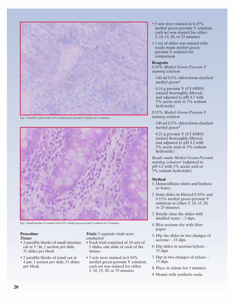
Fig. 1. Tonsil #1 stained with 0.10% methyl green-pyronin Y solution for 10 minutes.
Fig. 2. Small intestine #3 stained with 0.10% methyl green-pyronin Y solution for 15 minutes.
21
ResultsDNA…………Green to blue greenRNA……………………………RedGoblet cells ……………Mint greenBackground………………Pale pink
to colorlessImmunoblast andplasma cell cytoplasm…Intense redNuclei ………Green to blue-green
DiscussionStaining solution preparation timewas shortened from 24 hours to20 minutes. Tonsil demonstratedthe best results with the 0.10%pyronin Y solution at 10 and15 minutes. Small intestine stainedbest with 0.10% at 15 minutes.Small intestine and tonsil had paleresults at 5 minutes with both0.10% and 0.15% pyronin Yconcentrations. At 20 and25 minutes, tissues demonstratedbackground staining, unevenstaining, and poor contrast betweennuclei and cytoplasm. Test slidesdemonstrated more nuclear andcytoplasmic contrast and claritythan slides stained with the ready-made methyl green-pyronin Ysolution. Pyroninophilic tissuecomponents were harder todistinguish from the nuclei andbackground in the ready-made
methyl green-pyronin stainedslides. Slide results were consistentthroughout the 5 trials.
ConclusionBased on the results of this study,staining solutions made from themanufacturer-clarified methyl greenprovide an excellent alternative toboth the chloroform separationprocedure and the ready-mademethyl green-pyronin Y solution.
*Methyl green 0.5% aqueous purified with chloroform,Poly Scientific.
†Methyl green-pyronin, Sigma Diagnostics.
Making aList…Checking it
TwiceWanda G. Smith, BHS, HT(ASCP)
Medical Universityof South Carolina
No, I’m not talking about Santa’sChristmas list to see if we’ve beennaughty or nice. I’m talking aboutall the checklists we make in ourhistology laboratories to ensure wehave done ALL the jobs, large and
small, so that no job duty fallsthrough the cracks. However,inevitably, there will be somethingwe have forgotten, and then…WE WILL MAKE ANOTHERCHECKLIST!!!!!
We start with our HistopathologyWorksheet that the residents fill outat the grossing table. The residentlists the worksheet number, the dateof grossing, and their last name, sowe will know who to yell at(Oh…I mean talk to), when a blockis missing. The accession numbers,patient names, specimen and blocknumbers, along with the number ofpieces in each block and the CPTcode for that specimen are includedon the histopathology worksheet.Now, given that these are trainedprofessionals you would think thiswould be something they could getright. Right??? Yeah right!!! We usethis worksheet to document anydiscrepancies between the residentand the laboratory. We alsodocument who embedded theblocks, as well as the number ofblocks embedded, trying to catchany missing blocks at this time.We document which technologistentered data into our Cernercomputer system, who cut theblocks, stained the slides, and whowas responsible for labeling theslides corresponding to thisworksheet.
Next, we have our Work Task list,which includes all the technologistswith their corresponding duties.Duties include everything fromcutting, staining, running theUltrarush specimens to putting theextra cut slides in order for futurestaining. These duties rotate on aweekly basis. Each month a tech isalso assigned a tissue processor tobe responsible for the cleaning,changing, and rotating of solutions.We have a checklist for that too.We have a Decal Log Sheet, so wewill know which resident to givedecals to when they are finished.We have a Stainer Checklist todocument changes and rotations onour automatic slide stainer as wellas our H&E hand staining set-up.
Fig. 3. Small intestine #3 stained with ready-made methyl green-pyronin solution.

We have an On Call list for the year,and an Orientation and Trainingchecklist. We have a CompetencyAssessment checklist, which is usedat 6 months for new employees andyearly for the long-timers. We haveour EPMS checklist, which standsfor Employee PerformanceManagement System, also known asour evaluation forms. We have achecklist for when each CompetencyAssessment and EPMS is due.
When all of the slides are out forthe day, we tally the total numberof cases, blocks, and slides on theBlock Count Worksheet. We alsodocument mislabeled cases,blocks, and slides, and processormalfunctions (which we now seemto avoid since we got rid of oneparticularly “moody” processornamed Herman and we now haveall VIP tissue processors!). Wedocument any other notespertaining to what tissues shouldhave been decaled. (I’m sure no oneelse has THIS problem!!!), and anyother sectioning difficulties we haveencountered.
You would think after all thesechecklists we would not forgetANYTHING… well, we have anEvening Checklist for the lastperson leaving for the day. Thisperson does a walk-through tocheck off everything else includingthat the tissue processors are setcorrectly and running, no specimensare left out on the grossing tables,and the embedding centers andwaterbaths are turned off. We evencheck to make sure the radio andcordless telephone are locked up intheir cabinets.
Whew!!! I hope I have not forgottenanything. Oh yeah...
Dear Santa,I would really like one of thosereally fast motorized microtomesfor Christmas. I’ve been really goodthis year….
NSH to Offer NewPrograms in the
Coming YearVinnie Della Speranza
Scientific Editor
The National Society forHistotechnology has completednegotiations that will make two neweducational programs available topractitioners of the discipline. Thesenew initiatives will further enhancethe society’s efforts to reachpracticing histotechs who havebeen unable to attend educationalconferences due to cost.
In February, the College ofAmerican Pathologists Board ofGovernors voted to accept apartnership proposal proffered bythe NSH to market a proficiencytesting program to thehistotechnology community in theUnited States. The first of its kind,this program will make it possiblefor pathology laboratories tobenchmark the quality of theirstains and tissue sections againstthose from other participatingfacilities by completing periodicexercises.
The NSH program was developedas a result of the efforts of Sue Lewisof the University of Iowa, and BertDotson of Duke University whoconducted an NSH-funded pilotstudy in 1997. Twenty volunteerlaboratories participated in the pilotwhich helped to refine the programto offer the greatest benefit toparticipants. In its final form, theprogram is expected to offerguidance for improvement as well aseducational information; this willenable a laboratory’s staff to enhancetheir understanding of a method’stheoretical basis, a component oftenlacking in existing proficiency testingprograms that currently target clinicaltesting laboratories. A steeringcommittee composed of keyrepresentatives from eachorganization began meeting in lateApril to hammer out the final detailsfor an anticipated 2003 rollout.
In another exciting venture, theNSH Board of Directors met onMarch 22 to select a vendor to hostits new virtual library expected tobe made available later this year.This program will allow subscribersto experience a variety ofeducational topics that will beaccessible on the worldwide webfrom a link on the NSH homepage.This will make it possible forhistotechs to obtain their continuingeducation without ever leaving theirhomes. In contrast toteleconferences which can only beexperienced at the scheduled time,the virtual library can beexperienced whenever it isconvenient for the participant.Participants may complete aworkshop in installments, comingback as many times as necessary tocomplete the full experience. Theprogram will bookmark where youleft off, provide an online exam asevidence of completion, and track aparticipant’s continuing educationunits which may be printed outalong with any handout materialaccompanying the workshop.
The NSH anticipates high demandfor this product, especially fromthose unable to secure travel fundsfrom their employer. Employersmay find the program a usefuladjunct to their in-house continuingeducation program for staff. Furtherdetails are expected to becomeavailable by mid-summer.
Be sure to frequent the NSHwebsite at www.nsh.org for moreinformation on these excitingprograms.
22
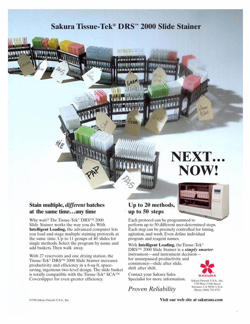
NEXT…NOW!
Stain multiple, different batchesat the same time…any timeWhy wait? The Tissue-Tek® DRS™ 2000Slide Stainer works the way you do. WithIntelligent Loading, the advanced computer letsyou load and stage multiple staining protocols atthe same time. Up to 11 groups of 40 slides forsingle methods. Select the program by name andadd baskets. Then walk away.
With 27 reservoirs and one drying station, theTissue-Tek® DRS™ 2000 Slide Stainer increasesproductivity and efficiency in a 6-sq-ft, space-saving, ingenious two-level design. The slide basketis totally compatible with the Tissue-Tek® SCA™Coverslipper for even greater efficiency.
Up to 20 methods,up to 50 stepsEach protocol can be programmed to perform up to 50 different user-determined steps.Each step can be precisely controlled for timing,agitation, and wash. Even define individualprogram and reagent names.
With Intelligent Loading, theTissue-Tek®
DRS™ 2000 Slide Stainer is a simply smarterinstrument—and instrument decision—for unsurpassed productivity andconsistency—slide after slide,shift after shift.
Contact your Sakura Sales Specialist for more information. Sakura Finetek U.S.A., Inc.
1750 West 214th StreetTorrance, CA 90501 U.S.A.
Phone: (800) 725-8723Proven Reliability
Sakura Tissue-Tek® DRS™ 2000 Slide Stainer
©1998 Sakura Finetek U.S.A., Inc. Visit our web site at sakuraus.com
To receive your own copy of HistoLogic,® or to have someone added to themailing list, submit home address to: Sakura Finetek U.S.A., Inc.,1750 West 214th Street, Torrance, CA 90501.
The editor wishes to solicit information, questions, and articles relating tohistotechnology. Submit these to: Vinnie Della Speranza, HistoLogic® Editor,165 Ashley Avenue, Suite 309, Charleston, SC 29425.Articles, photographs, etc,will not be returned unless requested in writing when they are submitted.
PRSRT STDU.S. POSTAGE
PAIDTAMPA, FL
PERMIT NO. 3513Sakura Finetek U.S.A., Inc.1750 West 214th StreetTorrance, California 90501
HISTOLOGIC ®
Te c h n i c a l B u l l e t i n f o r H i s t o t e c h n o l o g y
24
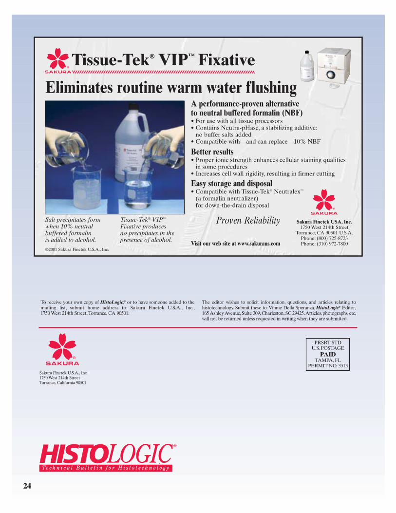
Tissue-Tek® VIP™ Fixative
Eliminates routine warm water flushingA performance-proven alternative to neutral buffered formalin (NBF)• For use with all tissue processors• Contains Neutra-pHase, a stabilizing additive:
no buffer salts added• Compatible with—and can replace—10% NBF
Better results• Proper ionic strength enhances cellular staining qualities
in some procedures• Increases cell wall rigidity, resulting in firmer cutting
Easy storage and disposal• Compatible with Tissue-Tek® Neutralex™
(a formalin neutralizer) for down-the-drain disposal
Sakura Finetek USA, Inc.1750 West 214th Street
Torrance, CA 90501 U.S.A.Phone: (800) 725-8723Phone: (310) 972-7800
Salt precipitates formwhen 10% neutralbuffered formalinis added to alcohol.
Tissue-Tek® VIP™
Fixative producesno precipitates in thepresence of alcohol.
Proven Reliability
©2001 Sakura Finetek U.S.A., Inc.Visit our web site at www.sakuraus.com
Related Documents