Histologic evaluation and immunohistochemical localization of STRO-1 and BMP-4 in rat immature teeth: A comparison between vital and induced pulp necrosis Daiana Elisabeth Bo ¨ ttcher a,d, *, Roberta Kochenborger Scarparo b,d , Eraldo Luiz Batista Jr. c,d , Anna Christina Medeiros Fossati c,d , Fabiana Soares Grecca b,d a Conservative Dentistry Department, Faculty of Dentistry, Federal University of Rio Grande do Sul (UFRGS), Brazil b Clinical Department, Faculty of Dentistry, Pontifical Catholic University of Rio Grande do Sul (PUCRS), Av. Ipiranga 6681 – Bld. 06, Porto Alegre, RS 90619-900, Brazil c Department of Diagnostics and Surgical Sciences, Faculty of Dentistry, University of Manitoba, D344B-790 Bannatyne Avenue, Winnipeg, MB R3E 0W2, Canada d Morphological Sciences Department, Faculty of Dentistry, Federal University of Rio Grande do Sul (UFRGS), Brazil 1. Introduction Dental caries or traumatic injuries to young permanent teeth can lead to pulp necrosis and tooth development arrest. Several reports 1–6 demonstrate apical healing and root completion after the treatment of necrotic immature teeth, suggesting that repair may occur even in adverse conditions. Some studies have identified the presence of stem cells (SC) on dental tissues by the immunodetection of markers such as STRO-1. 7–9 The existence of SC in the dental pulp (DP) has been established since the year 2000. 7 Nevertheless, their presence in the apical papilla has been verified only in the last years, being suggested as a probably source of undifferentiated cells, which could explain the clinical phenomenon of root formation in non-vital teeth. 8,9 In this regard Sonoyama et al. 10 stated that it a r c h i v e s o f o r a l b i o l o g y 5 8 ( 2 0 1 3 ) 1 1 7 4 – 1 1 7 9 a r t i c l e i n f o Article history: Accepted 2 April 2013 Keywords: Endodontics Dental pulp Immature necrotic teeth Mesenchymal stem cells a b s t r a c t Objective: To assess histological features and the expression of STRO-1 and BMP-4 in dental pulp and periapical tissues in vital or necrotic rat immature teeth. Design: The lower left first molars of male Wistar rats ageing four weeks (n = 24) had their pulps exposed to the oral environment for 3, 6, 9 and 12 weeks (animals ageing 7, 10, 13 and 16 weeks-old, respectively; n = 24). The right lower first molars served as control untouched teeth. After sample harvesting the jaws were dissected and processed for histology and immunodetection of STRO-1 and BMP-4. Results: Necrotic teeth had root development arrested, while control animals showed development of dental tissues. Immunohistochemistry showed that detection of BMP-4 was restricted to vital pulps. For both groups, STRO-1 expression was evident around blood vessels walls. Neither BMP-4 nor STRO-1 was observed in the apical papilla region. Conclusion: STRO-1-positive precursor cells were not detected in the apical papilla. BMP-4 expression has not been detected during infection. # 2013 Elsevier Ltd. All rights reserved. * Corresponding author at: Conservative Dentistry Department, Faculty of Dentistry, Federal University of Rio Grande do Sul (UFRGS), Av. Ramiro Barcelos, 2492 Porto Alegre, CEP: 90035-003, Brazil. Tel.: +55 51 84485602; fax: +55 51 33085002. E-mail addresses: [email protected], [email protected] (D.E. Bo ¨ ttcher). Available online at www.sciencedirect.com journal homepage: http://www.elsevier.com/locate/aob 0003–9969/$ – see front matter # 2013 Elsevier Ltd. All rights reserved. http://dx.doi.org/10.1016/j.archoralbio.2013.04.001

Welcome message from author
This document is posted to help you gain knowledge. Please leave a comment to let me know what you think about it! Share it to your friends and learn new things together.
Transcript
Histologic evaluation and immunohistochemical localizationof STRO-1 and BMP-4 in rat immature teeth: A comparisonbetween vital and induced pulp necrosis
Daiana Elisabeth Bottcher a,d,*, Roberta Kochenborger Scarparo b,d, Eraldo Luiz Batista Jr.c,d,Anna Christina Medeiros Fossati c,d, Fabiana Soares Grecca b,d
aConservative Dentistry Department, Faculty of Dentistry, Federal University of Rio Grande do Sul (UFRGS), BrazilbClinical Department, Faculty of Dentistry, Pontifical Catholic University of Rio Grande do Sul (PUCRS), Av. Ipiranga 6681 – Bld. 06,
Porto Alegre, RS 90619-900, BrazilcDepartment of Diagnostics and Surgical Sciences, Faculty of Dentistry, University of Manitoba, D344B-790 Bannatyne Avenue, Winnipeg,
MB R3E 0W2, CanadadMorphological Sciences Department, Faculty of Dentistry, Federal University of Rio Grande do Sul (UFRGS), Brazil
a r c h i v e s o f o r a l b i o l o g y 5 8 ( 2 0 1 3 ) 1 1 7 4 – 1 1 7 9
a r t i c l e i n f o
Article history:
Accepted 2 April 2013
Keywords:
Endodontics
Dental pulp
Immature necrotic teeth
Mesenchymal stem cells
a b s t r a c t
Objective: To assess histological features and the expression of STRO-1 and BMP-4 in dental
pulp and periapical tissues in vital or necrotic rat immature teeth.
Design: The lower left first molars of male Wistar rats ageing four weeks (n = 24) had their
pulps exposed to the oral environment for 3, 6, 9 and 12 weeks (animals ageing 7, 10, 13 and
16 weeks-old, respectively; n = 24). The right lower first molars served as control untouched
teeth. After sample harvesting the jaws were dissected and processed for histology and
immunodetection of STRO-1 and BMP-4.
Results: Necrotic teeth had root development arrested, while control animals showed
development of dental tissues. Immunohistochemistry showed that detection of BMP-4
was restricted to vital pulps. For both groups, STRO-1 expression was evident around blood
vessels walls. Neither BMP-4 nor STRO-1 was observed in the apical papilla region.
Conclusion: STRO-1-positive precursor cells were not detected in the apical papilla. BMP-4
expression has not been detected during infection.
# 2013 Elsevier Ltd. All rights reserved.
Available online at www.sciencedirect.com
journal homepage: http://www.elsevier.com/locate/aob
1. Introduction
Dental caries or traumatic injuries to young permanent teeth
can lead to pulp necrosis and tooth development arrest.
Several reports1–6 demonstrate apical healing and root
completion after the treatment of necrotic immature teeth,
suggesting that repair may occur even in adverse conditions.
* Corresponding author at: Conservative Dentistry Department, FacultyRamiro Barcelos, 2492 Porto Alegre, CEP: 90035-003, Brazil. Tel.: +55 5
E-mail addresses: [email protected], [email protected]
0003–9969/$ – see front matter # 2013 Elsevier Ltd. All rights reservehttp://dx.doi.org/10.1016/j.archoralbio.2013.04.001
Some studies have identified the presence of stem cells (SC)
on dental tissues by the immunodetection of markers such as
STRO-1.7–9 The existence of SC in the dental pulp (DP) has been
established since the year 2000.7 Nevertheless, their presence in
the apical papilla has been verified only in the last years, being
suggested as a probably source of undifferentiated cells, which
could explain the clinical phenomenon of root formation in
non-vital teeth.8,9 In this regard Sonoyama et al.10 stated that it
of Dentistry, Federal University of Rio Grande do Sul (UFRGS), Av.1 84485602; fax: +55 51 33085002.m.br (D.E. Bottcher).
d.
a r c h i v e s o f o r a l b i o l o g y 5 8 ( 2 0 1 3 ) 1 1 7 4 – 1 1 7 9 1175
is likely that stem cells from the apical papilla (SCAP) survive
infection because of their close proximity to periapical tissues.
Nevertheless, inflammatory mediators up-regulated in
injured tissues seem to interfere with the signals needed for
root development.11 Growth factors, like BMP-4, are among the
triggered signalling networks during dental morphogenesis
that may be negatively affected by inflammation.12 In
assessing the role of BMP-4 in Hertwig’s epithelial root sheath
(HERS) formation, Hosoya et al.13 verified that this protein
plays an important role in regulation of root formation,
preventing elongation and maintaining cell proliferation.
Few studies focused on alterations of tissues affected by
pulp necrosis in immature teeth.14,15 Moreover, in necrotic
teeth, the immunolocalization of precursor cells and growth
factors that have an influence on root development have yet to
be explored.
Thus, the purpose of the present study was to assess
histological features and the expression of STRO-1 and BMP-4
in dental pulp and periapical tissues of rat immature teeth
with vital or induced pulp necrosis.
2. Methods
2.1. Experimental protocols
This study was approved by the Institutional Review Board and
by the Research Ethics Committee of the School of Dentistry of
the Federal University of Rio Grande do Sul (Protocol #19001) and
is in accordance with the European Convention for the
Protection of Vertebrate Animals used for Experimental and
Other Scientific Purposes. The sample consisted of 24 Wistar
male rats, and a split-mouth experimental design was adopted.
Prior to the experimental procedures, the animals were
anesthetized intraperitoneally with ketamine 80 mg/kg and
xylazine 20 mg/kg (Virbac do Brasil, Juruatuba, SP, Brazil). The
procedures adopted herein were described elsewhere.15 Briefly,
right mandibular first molars were used for the observation of
natural morphogenesis (control group – CG). Pulp necrosis was
induced on the left mandibular first molars (test group – TG)
during the initial stage of root development (animals ageing 4
weeks-old). Dental pulps were exposed by drilling cavities on
the central portion of the occlusal surface with a 1011 HL round
bur in high speed (KG Sorensen, Cotia, SP, Brazil) to a depth
nearly equal to the bur diameter (1 mm). Teeth were left open to
the oral environment throughout the course of the experiment.
Time required for detection of periapical lesions was confirmed
by radiographs taken as previously reported.15,16 Animals were
euthanized by inhalation of CO2 at 3, 6, 9 or 12 weeks post pulp
exposure (animals ageing 7, 10, 13 and 16 weeks-old; n = 24).
Jaws were dissected for histologic and immunohistochemical
evaluation.
Immediately after euthanasia, samples were fixed in
buffered phosphate 10% paraformaldehyde for 24 h, decalci-
fied in 17% EDTA for 5 weeks, dehydrated in ascending
concentrations of ethanol, and embedded in paraffin. Five-
micrometre sections were cut and stained with haematoxylin–
eosin or processed for immunohistochemistry. Three sections
were selected per sample, so the central portion of the roots,
including the apex, was visible.
2.2. Histological evaluation
Histological evaluation (n = 35) was based on a descriptive
analysis on pulp tissue. The presence of inflammatory cells, as
well as features related to vascularity, odontoblast organiza-
tion, degeneration/destruction processes were considered.
Additionally, other phenomena including root resorptions and
closure of apical foramen were considered.
Periapical inflammation was classified by calibrated exam-
iners (k = 0.79, P < 0.001) according to the following scores: (0)
absence – absence of inflammatory reaction, (1) mild –
inflammatory cells restricted to the root canal space, (2)
intense – inflammatory cells in form of infiltrate in periapical
region, and (3) severe – inflammatory cells in form of infiltrate
in periapical region with abscess formation.
2.3. Immunohistochemistry
The sections were deparaffinized and immersed in 3.0%
hydrogen peroxide in absolute methanol (two incubations of
15 min each) in order to obtain the endogenous peroxidase
blocking, followed by three washing cycles in PBS, pH 7.2.
Sections were washed in PBS and incubated either with a
monoclonal mouse anti-human STRO-1 antibody (R&D Sys-
tems Inc., Minneapolis, MN, USA) or mouse monoclonal anti-
rat BMP-4 (Novocastra, Newcastle, Northumberland, Reino
Unido) at 4 8C overnight. The primary antibody was diluted to
1:100 (according to a pilot study) with antibody diluent and
background reducing agents as described by the manufacturer
(Dako, Carpinteria, CA, EUA) for both markers.
The sections were washed in PBS pH 7.2 and incubated with
a secondary conjugated antibody as recommended by the
manufacturer (Picture Max, HRP Polymer Conjugate Broad
Spectrum, Invitrogen, Carlsbad, CA, USA) during 30 min. The
sections were rinsed with PBS for 2 min, 3 times, and
immunoreactivity was visualized after incubation with 3,30-
diaminobenzidine (DAB) solution (Dako Liquid DAB Substrate
Chromogen System, Dako, Carpinteria, CA, EUA), and then
counterstained with Harry’s haematoxylin.
Immunohistochemical control was obtained by incubating
with nonimmune mouse IgM (Rockland Immunochemicals,
Gilbertsville, PA, USA) as the primary antibody. None of the
control sections were positive for the target markers (not
shown). Positive controls were alveolar bone for BMP-4 and
umbilical cord cells for STRO-1.
3. Results
3.1. Histological analysis
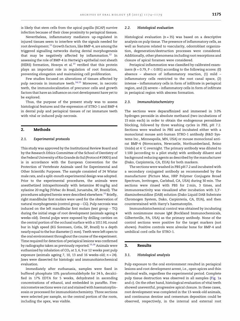
Pulp exposure to the oral environment resulted in periapical
lesions and root development arrest, i.e., open apices and thin
dentinal walls, regardless the experimental period. Complete
pulp tissue destruction was observed in all samples (Fig. 1a
and c). On the other hand, histological evaluation of vital teeth
showed uneventful, progressive apical closure. In these cases,
root development was completed in the 13-week-old animals,
and continuous dentine and cementum deposition could be
observed, respectively, in the internal and external root
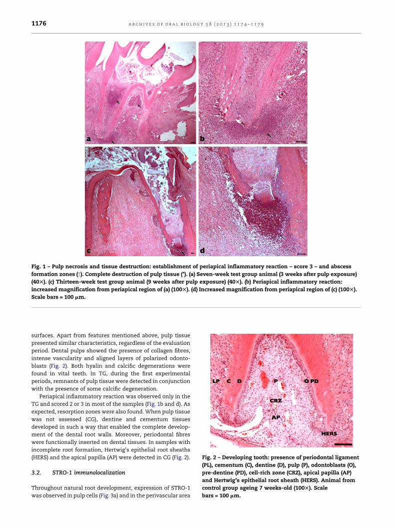
Fig. 1 – Pulp necrosis and tissue destruction: establishment of periapical inflammatory reaction – score 3 – and abscess
formation zones ("). Complete destruction of pulp tissue (*). (a) Seven-week test group animal (3 weeks after pulp exposure)
(40T). (c) Thirteen-week test group animal (9 weeks after pulp exposure) (40T). (b) Periapical inflammatory reaction:
increased magnification from periapical region of (a) (100T). (d) Increased magnification from periapical region of (c) (100T).
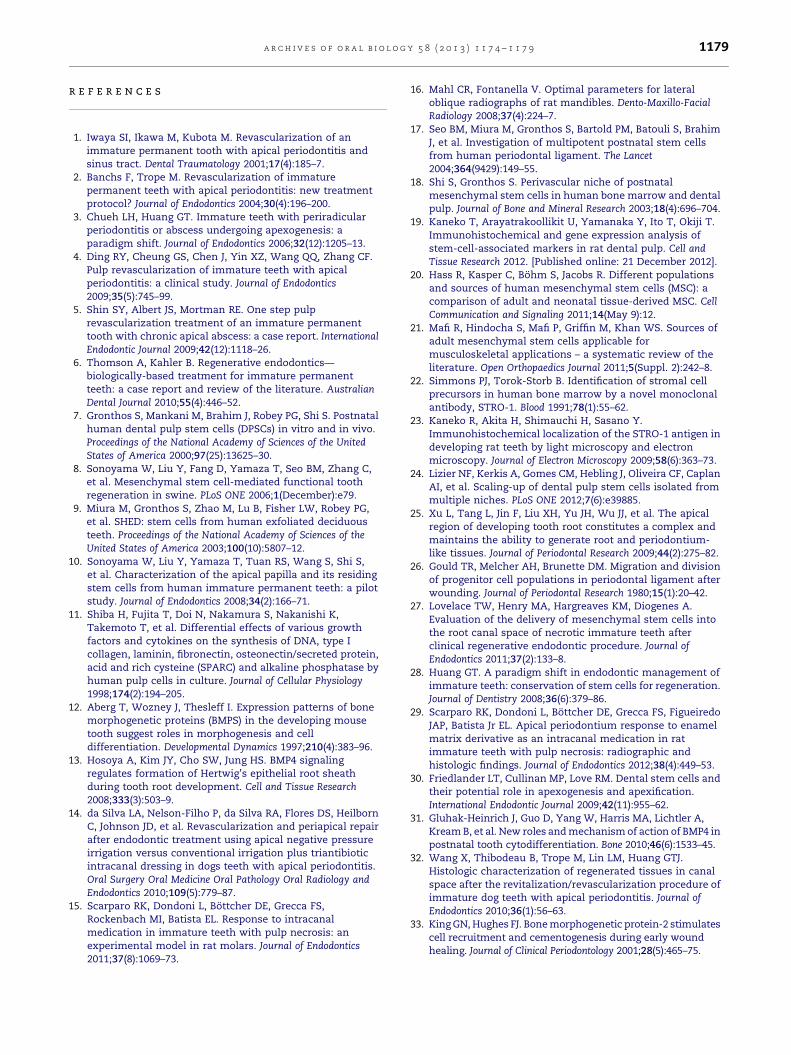
Scale bars = 100 mm.
Fig. 2 – Developing tooth: presence of periodontal ligament
(PL), cementum (C), dentine (D), pulp (P), odontoblasts (O),
pre-dentine (PD), cell-rich zone (CRZ), apical papilla (AP)
and Hertwig’s epithelial root sheath (HERS). Animal from
control group ageing 7 weeks-old (100T). Scale
bars = 100 mm.
a r c h i v e s o f o r a l b i o l o g y 5 8 ( 2 0 1 3 ) 1 1 7 4 – 1 1 7 91176
surfaces. Apart from features mentioned above, pulp tissue
presented similar characteristics, regardless of the evaluation
period. Dental pulps showed the presence of collagen fibres,
intense vascularity and aligned layers of polarized odonto-
blasts (Fig. 2). Both hyalin and calcific degenerations were
found in vital teeth. In TG, during the first experimental
periods, remnants of pulp tissue were detected in conjunction
with the presence of some calcific degeneration.
Periapical inflammatory reaction was observed only in the
TG and scored 2 or 3 in most of the samples (Fig. 1b and d). As
expected, resorption zones were also found. When pulp tissue
was not assessed (CG), dentine and cementum tissues
developed in such a way that enabled the complete develop-
ment of the dental root walls. Moreover, periodontal fibres
were functionally inserted on dental tissues. In samples with
incomplete root formation, Hertwig’s epithelial root sheaths
(HERS) and the apical papilla (AP) were detected in CG (Fig. 2).
3.2. STRO-1 immunolocalization
Throughout natural root development, expression of STRO-1
was observed in pulp cells (Fig. 3a) and in the perivascular area
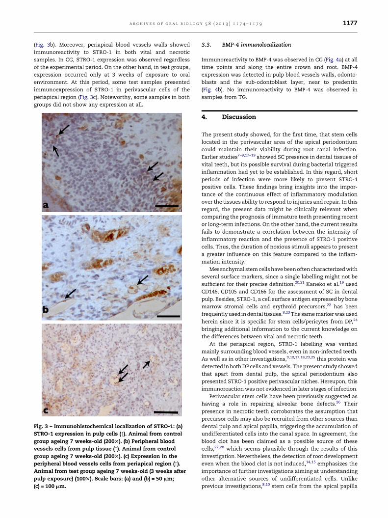
a r c h i v e s o f o r a l b i o l o g y 5 8 ( 2 0 1 3 ) 1 1 7 4 – 1 1 7 9 1177
(Fig. 3b). Moreover, periapical blood vessels walls showed
immunoreactivity to STRO-1 in both vital and necrotic
samples. In CG, STRO-1 expression was observed regardless
of the experimental period. On the other hand, in test groups,
expression occurred only at 3 weeks of exposure to oral
environment. At this period, some test samples presented
immunoexpression of STRO-1 in perivascular cells of the
periapical region (Fig. 3c). Noteworthy, some samples in both
groups did not show any expression at all.
Fig. 3 – Immunohistochemical localization of STRO-1: (a)
STRO-1 expression in pulp cells ("). Animal from control
group ageing 7 weeks-old (200T). (b) Peripheral blood
vessels cells from pulp tissue ("). Animal from control
group ageing 7 weeks-old (200T). (c) Expression in the
peripheral blood vessels cells from periapical region (").Animal from test group ageing 7 weeks-old (3 weeks after
pulp exposure) (100T). Scale bars: (a) and (b) = 50 mm;
(c) = 100 mm.
3.3. BMP-4 immunolocalization
Immunoreactivity to BMP-4 was observed in CG (Fig. 4a) at all
time points and along the entire crown and root. BMP-4
expression was detected in pulp blood vessels walls, odonto-
blasts and the sub-odontoblast layer, near to predentin
(Fig. 4b). No immunoreactivity to BMP-4 was observed in
samples from TG.
4. Discussion
The present study showed, for the first time, that stem cells
located in the perivascular area of the apical periodontium
could maintain their viability during root canal infection.
Earlier studies7–9,17–19 showed SC presence in dental tissues of
vital teeth, but its possible survival during bacterial triggered
inflammation had yet to be established. In this regard, short
periods of infection were more likely to present STRO-1
positive cells. These findings bring insights into the impor-
tance of the continuous effect of inflammatory modulation
over the tissues ability to respond to injuries and repair. In this
regard, the present data might be clinically relevant when
comparing the prognosis of immature teeth presenting recent
or long-term infections. On the other hand, the current results
fails to demonstrate a correlation between the intensity of
inflammatory reaction and the presence of STRO-1 positive
cells. Thus, the duration of noxious stimuli appears to present
a greater influence on this feature compared to the inflam-
mation intensity.
Mesenchymal stem cells have been often characterized with
several surface markers, since a single labelling might not be
sufficient for their precise definition.20,21 Kaneko et al.19 used
CD146, CD105 and CD166 for the assessment of SC in dental
pulp. Besides, STRO-1, a cell surface antigen expressed by bone
marrow stromal cells and erythroid precursors,22 has been
frequently used in dental tissues.8,23 The same marker was used
herein since it is specific for stem cells/pericytes from DP,24
bringing additional information to the current knowledge on
the differences between vital and necrotic teeth.
At the periapical region, STRO-1 labelling was verified
mainly surrounding blood vessels, even in non-infected teeth.
As well as in other investigations,9,10,17,18,23,25 this protein was
detected in both DP cells and vessels. The present study showed
that apart from dental pulp, the apical periodontium also
presented STRO-1 positive perivascular niches. Hereupon, this
immunoreaction was not evidenced in later stages of infection.
Perivascular stem cells have been previously suggested as
having a role in repairing alveolar bone defects.26 Their
presence in necrotic teeth corroborates the assumption that
precursor cells may also be recruited from other sources than
dental pulp and apical papilla, triggering the accumulation of
undifferentiated cells into the canal space. In agreement, the
blood clot has been claimed as a possible source of these
cells,27,28 which seems plausible through the results of this
investigation. Nevertheless, the detection of root development
even when the blood clot is not induced,14,15 emphasizes the
importance of further investigations aiming at understanding
other alternative sources of undifferentiated cells. Unlike
previous investigations,8,10 stem cells from the apical papilla
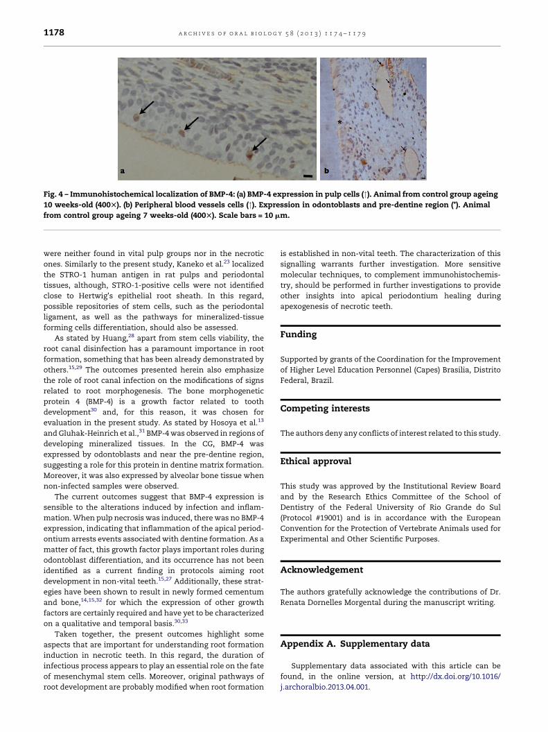
Fig. 4 – Immunohistochemical localization of BMP-4: (a) BMP-4 expression in pulp cells ("). Animal from control group ageing
10 weeks-old (400T). (b) Peripheral blood vessels cells ("). Expression in odontoblasts and pre-dentine region (*). Animal
from control group ageing 7 weeks-old (400T). Scale bars = 10 mm.
a r c h i v e s o f o r a l b i o l o g y 5 8 ( 2 0 1 3 ) 1 1 7 4 – 1 1 7 91178
were neither found in vital pulp groups nor in the necrotic
ones. Similarly to the present study, Kaneko et al.23 localized
the STRO-1 human antigen in rat pulps and periodontal
tissues, although, STRO-1-positive cells were not identified
close to Hertwig’s epithelial root sheath. In this regard,
possible repositories of stem cells, such as the periodontal
ligament, as well as the pathways for mineralized-tissue
forming cells differentiation, should also be assessed.
As stated by Huang,28 apart from stem cells viability, the
root canal disinfection has a paramount importance in root
formation, something that has been already demonstrated by
others.15,29 The outcomes presented herein also emphasize
the role of root canal infection on the modifications of signs
related to root morphogenesis. The bone morphogenetic
protein 4 (BMP-4) is a growth factor related to tooth
development30 and, for this reason, it was chosen for
evaluation in the present study. As stated by Hosoya et al.13
and Gluhak-Heinrich et al.,31 BMP-4 was observed in regions of
developing mineralized tissues. In the CG, BMP-4 was
expressed by odontoblasts and near the pre-dentine region,
suggesting a role for this protein in dentine matrix formation.
Moreover, it was also expressed by alveolar bone tissue when
non-infected samples were observed.
The current outcomes suggest that BMP-4 expression is
sensible to the alterations induced by infection and inflam-
mation. When pulp necrosis was induced, there was no BMP-4
expression, indicating that inflammation of the apical period-
ontium arrests events associated with dentine formation. As a
matter of fact, this growth factor plays important roles during
odontoblast differentiation, and its occurrence has not been
identified as a current finding in protocols aiming root
development in non-vital teeth.15,27 Additionally, these strat-
egies have been shown to result in newly formed cementum
and bone,14,15,32 for which the expression of other growth
factors are certainly required and have yet to be characterized
on a qualitative and temporal basis.30,33
Taken together, the present outcomes highlight some
aspects that are important for understanding root formation
induction in necrotic teeth. In this regard, the duration of
infectious process appears to play an essential role on the fate
of mesenchymal stem cells. Moreover, original pathways of
root development are probably modified when root formation
is established in non-vital teeth. The characterization of this
signalling warrants further investigation. More sensitive
molecular techniques, to complement immunohistochemis-
try, should be performed in further investigations to provide
other insights into apical periodontium healing during
apexogenesis of necrotic teeth.
Funding
Supported by grants of the Coordination for the Improvement
of Higher Level Education Personnel (Capes) Brasilia, Distrito
Federal, Brazil.
Competing interests
The authors deny any conflicts of interest related to this study.
Ethical approval
This study was approved by the Institutional Review Board
and by the Research Ethics Committee of the School of
Dentistry of the Federal University of Rio Grande do Sul
(Protocol #19001) and is in accordance with the European
Convention for the Protection of Vertebrate Animals used for
Experimental and Other Scientific Purposes.
Acknowledgement
The authors gratefully acknowledge the contributions of Dr.
Renata Dornelles Morgental during the manuscript writing.
Appendix A. Supplementary data
Supplementary data associated with this article can be
found, in the online version, at http://dx.doi.org/10.1016/
j.archoralbio.2013.04.001.
a r c h i v e s o f o r a l b i o l o g y 5 8 ( 2 0 1 3 ) 1 1 7 4 – 1 1 7 9 1179
r e f e r e n c e s
1. Iwaya SI, Ikawa M, Kubota M. Revascularization of animmature permanent tooth with apical periodontitis andsinus tract. Dental Traumatology 2001;17(4):185–7.
2. Banchs F, Trope M. Revascularization of immaturepermanent teeth with apical periodontitis: new treatmentprotocol? Journal of Endodontics 2004;30(4):196–200.
3. Chueh LH, Huang GT. Immature teeth with periradicularperiodontitis or abscess undergoing apexogenesis: aparadigm shift. Journal of Endodontics 2006;32(12):1205–13.
4. Ding RY, Cheung GS, Chen J, Yin XZ, Wang QQ, Zhang CF.Pulp revascularization of immature teeth with apicalperiodontitis: a clinical study. Journal of Endodontics2009;35(5):745–99.
5. Shin SY, Albert JS, Mortman RE. One step pulprevascularization treatment of an immature permanenttooth with chronic apical abscess: a case report. InternationalEndodontic Journal 2009;42(12):1118–26.
6. Thomson A, Kahler B. Regenerative endodontics—biologically-based treatment for immature permanentteeth: a case report and review of the literature. AustralianDental Journal 2010;55(4):446–52.
7. Gronthos S, Mankani M, Brahim J, Robey PG, Shi S. Postnatalhuman dental pulp stem cells (DPSCs) in vitro and in vivo.Proceedings of the National Academy of Sciences of the UnitedStates of America 2000;97(25):13625–30.
8. Sonoyama W, Liu Y, Fang D, Yamaza T, Seo BM, Zhang C,et al. Mesenchymal stem cell-mediated functional toothregeneration in swine. PLoS ONE 2006;1(December):e79.
9. Miura M, Gronthos S, Zhao M, Lu B, Fisher LW, Robey PG,et al. SHED: stem cells from human exfoliated deciduousteeth. Proceedings of the National Academy of Sciences of theUnited States of America 2003;100(10):5807–12.
10. Sonoyama W, Liu Y, Yamaza T, Tuan RS, Wang S, Shi S,et al. Characterization of the apical papilla and its residingstem cells from human immature permanent teeth: a pilotstudy. Journal of Endodontics 2008;34(2):166–71.
11. Shiba H, Fujita T, Doi N, Nakamura S, Nakanishi K,Takemoto T, et al. Differential effects of various growthfactors and cytokines on the synthesis of DNA, type Icollagen, laminin, fibronectin, osteonectin/secreted protein,acid and rich cysteine (SPARC) and alkaline phosphatase byhuman pulp cells in culture. Journal of Cellular Physiology1998;174(2):194–205.
12. Aberg T, Wozney J, Thesleff I. Expression patterns of bonemorphogenetic proteins (BMPS) in the developing mousetooth suggest roles in morphogenesis and celldifferentiation. Developmental Dynamics 1997;210(4):383–96.
13. Hosoya A, Kim JY, Cho SW, Jung HS. BMP4 signalingregulates formation of Hertwig’s epithelial root sheathduring tooth root development. Cell and Tissue Research2008;333(3):503–9.
14. da Silva LA, Nelson-Filho P, da Silva RA, Flores DS, HeilbornC, Johnson JD, et al. Revascularization and periapical repairafter endodontic treatment using apical negative pressureirrigation versus conventional irrigation plus triantibioticintracanal dressing in dogs teeth with apical periodontitis.Oral Surgery Oral Medicine Oral Pathology Oral Radiology andEndodontics 2010;109(5):779–87.
15. Scarparo RK, Dondoni L, Bottcher DE, Grecca FS,Rockenbach MI, Batista EL. Response to intracanalmedication in immature teeth with pulp necrosis: anexperimental model in rat molars. Journal of Endodontics2011;37(8):1069–73.
16. Mahl CR, Fontanella V. Optimal parameters for lateraloblique radiographs of rat mandibles. Dento-Maxillo-FacialRadiology 2008;37(4):224–7.
17. Seo BM, Miura M, Gronthos S, Bartold PM, Batouli S, BrahimJ, et al. Investigation of multipotent postnatal stem cellsfrom human periodontal ligament. The Lancet2004;364(9429):149–55.
18. Shi S, Gronthos S. Perivascular niche of postnatalmesenchymal stem cells in human bone marrow and dentalpulp. Journal of Bone and Mineral Research 2003;18(4):696–704.
19. Kaneko T, Arayatrakoollikit U, Yamanaka Y, Ito T, Okiji T.Immunohistochemical and gene expression analysis ofstem-cell-associated markers in rat dental pulp. Cell andTissue Research 2012. [Published online: 21 December 2012].
20. Hass R, Kasper C, Bohm S, Jacobs R. Different populationsand sources of human mesenchymal stem cells (MSC): acomparison of adult and neonatal tissue-derived MSC. CellCommunication and Signaling 2011;14(May 9):12.
21. Mafi R, Hindocha S, Mafi P, Griffin M, Khan WS. Sources ofadult mesenchymal stem cells applicable formusculoskeletal applications – a systematic review of theliterature. Open Orthopaedics Journal 2011;5(Suppl. 2):242–8.
22. Simmons PJ, Torok-Storb B. Identification of stromal cellprecursors in human bone marrow by a novel monoclonalantibody, STRO-1. Blood 1991;78(1):55–62.
23. Kaneko R, Akita H, Shimauchi H, Sasano Y.Immunohistochemical localization of the STRO-1 antigen indeveloping rat teeth by light microscopy and electronmicroscopy. Journal of Electron Microscopy 2009;58(6):363–73.
24. Lizier NF, Kerkis A, Gomes CM, Hebling J, Oliveira CF, CaplanAI, et al. Scaling-up of dental pulp stem cells isolated frommultiple niches. PLoS ONE 2012;7(6):e39885.
25. Xu L, Tang L, Jin F, Liu XH, Yu JH, Wu JJ, et al. The apicalregion of developing tooth root constitutes a complex andmaintains the ability to generate root and periodontium-like tissues. Journal of Periodontal Research 2009;44(2):275–82.
26. Gould TR, Melcher AH, Brunette DM. Migration and divisionof progenitor cell populations in periodontal ligament afterwounding. Journal of Periodontal Research 1980;15(1):20–42.
27. Lovelace TW, Henry MA, Hargreaves KM, Diogenes A.Evaluation of the delivery of mesenchymal stem cells intothe root canal space of necrotic immature teeth afterclinical regenerative endodontic procedure. Journal ofEndodontics 2011;37(2):133–8.
28. Huang GT. A paradigm shift in endodontic management ofimmature teeth: conservation of stem cells for regeneration.Journal of Dentistry 2008;36(6):379–86.
29. Scarparo RK, Dondoni L, Bottcher DE, Grecca FS, FigueiredoJAP, Batista Jr EL. Apical periodontium response to enamelmatrix derivative as an intracanal medication in ratimmature teeth with pulp necrosis: radiographic andhistologic findings. Journal of Endodontics 2012;38(4):449–53.
30. Friedlander LT, Cullinan MP, Love RM. Dental stem cells andtheir potential role in apexogenesis and apexification.International Endodontic Journal 2009;42(11):955–62.
31. Gluhak-Heinrich J, Guo D, Yang W, Harris MA, Lichtler A,Kream B, et al. New roles and mechanism of action of BMP4 inpostnatal tooth cytodifferentiation. Bone 2010;46(6):1533–45.
32. Wang X, Thibodeau B, Trope M, Lin LM, Huang GTJ.Histologic characterization of regenerated tissues in canalspace after the revitalization/revascularization procedure ofimmature dog teeth with apical periodontitis. Journal ofEndodontics 2010;36(1):56–63.
33. King GN, Hughes FJ. Bone morphogenetic protein-2 stimulatescell recruitment and cementogenesis during early woundhealing. Journal of Clinical Periodontology 2001;28(5):465–75.
Related Documents