Commentary Histamine in the brain: Beyond sleep and memory Maria Beatrice Passani a, *, Patrizia Giannoni a , Corrado Bucherelli b , Elisabetta Baldi b , Patrizio Blandina a a Dipartimento di Farmacologia Preclinica e Clinica Viale Pieraccini 6, 50139 Firenze, Italy b Dipartimento di Fisiologia, Viale Morgagni 63, 50134 Firenze, Italy 1. Interactions among histaminergic and other neurotransmitter systems regulate the sleep–wake cycle Several experimental observations support the hypothesis that the histaminergic system constitutes a major wake- promoting system, as its terminals influence neuronal excitability in several brain areas [1,2]. Direct electrophysio- logical recordings from freely moving cats showed that the activity of histaminergic neurons is high during waking and low or absent during sleep [3], and their firing rate changes with the behavioral state [4]. The importance of histaminergic biochemical pharmacology 73 (2007) 1113–1122 article info Keywords: Endocannabinoids Acetylcholine Hypothalamus Microdialysis Feeding Fear conditioning abstract A few decades elapsed between the attribution of unwanted side effects of classic anti- histamine compounds to the blockade of central H 1 receptors, and the acceptance of the concept that the histaminergic system commands general states of metabolism and con- sciousness. In the early 80s, two laboratories discovered independently that histaminergic neurons are located in the posterior hypothalamus and project to the whole CNS [Panula P, Yang HY, Costa E. Histamine-containing neurons in the rat hypothalamus. Proc Natl Acad Sci 1984;81:2572–76, Watanabe T, Taguchi Y, Hayashi H, Tanaka J, Shiosaka S, Tohyama M, Kubota H, Terano Y, Wada H. Evidence for the presence of a histaminergic neuron system in the rat brain: an immunohistochemical analysis. Neurosci Lett 1983;39:249–54], suggesting a global nature of histamine regulatory effects. Recently, functional studies demonstrated that activation of the central histaminergic system alters CNS functions in both behavioral and homeostatic contexts, which include sleep and wakefulness, learning and memory, anxiety, locomotion, feeding and drinking, and neuroendocrine regulation. These actions are achieved through interactions with other neurotransmitter systems, and the interplay between histaminergic neurons and other neurotransmitter systems are becoming clear. Hence, numerous laboratories are pursuing novel compounds targeting the three known histamine receptors found in the brain for various therapeutic indications. Preclinical studies are focusing on three major areas of interest and intense research is mainly oriented towards providing drugs for the treatment of sleep, cognitive and feeding disorders. This commentary is intended to summarize some of the latest findings that suggest functional roles for the interplay between histamine and other neurotransmitter systems, and to propose novel interactions as physiological substrates that may partially underlie some of the behavioral changes observed following manipulation of the histaminergic system. # 2006 Elsevier Inc. All rights reserved. * Corresponding author. Tel.: +39 055 4271237; fax: +39 055 4271280. E-mail address: beatrice.passani@unifi.it (M.B. Passani). available at www.sciencedirect.com journal homepage: www.elsevier.com/locate/biochempharm 0006-2952/$ – see front matter # 2006 Elsevier Inc. All rights reserved. doi:10.1016/j.bcp.2006.12.002

Welcome message from author
This document is posted to help you gain knowledge. Please leave a comment to let me know what you think about it! Share it to your friends and learn new things together.
Transcript

b i o c h e m i c a l p h a r m a c o l o g y 7 3 ( 2 0 0 7 ) 1 1 1 3 – 1 1 2 2
Commentary
Histamine in the brain: Beyond sleep and memory
Maria Beatrice Passani a,*, Patrizia Giannoni a, Corrado Bucherelli b,Elisabetta Baldi b, Patrizio Blandina a
aDipartimento di Farmacologia Preclinica e Clinica Viale Pieraccini 6, 50139 Firenze, ItalybDipartimento di Fisiologia, Viale Morgagni 63, 50134 Firenze, Italy
a r t i c l e i n f o
Keywords:
Endocannabinoids
Acetylcholine
Hypothalamus
Microdialysis
Feeding
Fear conditioning
a b s t r a c t
A few decades elapsed between the attribution of unwanted side effects of classic anti-
histamine compounds to the blockade of central H1 receptors, and the acceptance of the
concept that the histaminergic system commands general states of metabolism and con-
sciousness. In the early 80s, two laboratories discovered independently that histaminergic
neurons are located in the posterior hypothalamus and project to the whole CNS [Panula P,
Yang HY, Costa E. Histamine-containing neurons in the rat hypothalamus. Proc Natl Acad
Sci 1984;81:2572–76, Watanabe T, Taguchi Y, Hayashi H, Tanaka J, Shiosaka S, Tohyama M,
Kubota H, Terano Y, Wada H. Evidence for the presence of a histaminergic neuron system in
the rat brain: an immunohistochemical analysis. Neurosci Lett 1983;39:249–54], suggesting a
global nature of histamine regulatory effects. Recently, functional studies demonstrated
that activation of the central histaminergic system alters CNS functions in both behavioral
and homeostatic contexts, which include sleep and wakefulness, learning and memory,
anxiety, locomotion, feeding and drinking, and neuroendocrine regulation. These actions
are achieved through interactions with other neurotransmitter systems, and the interplay
between histaminergic neurons and other neurotransmitter systems are becoming clear.
Hence, numerous laboratories are pursuing novel compounds targeting the three known
histamine receptors found in the brain for various therapeutic indications. Preclinical
studies are focusing on three major areas of interest and intense research is mainly oriented
towards providing drugs for the treatment of sleep, cognitive and feeding disorders. This
commentary is intended to summarize some of the latest findings that suggest functional
roles for the interplay between histamine and other neurotransmitter systems, and to
propose novel interactions as physiological substrates that may partially underlie some of
the behavioral changes observed following manipulation of the histaminergic system.
# 2006 Elsevier Inc. All rights reserved.
avai lab le at www.sc iencedi rec t .com
journal homepage: www.e lsev ier .com/ locate /b iochempharm
1. Interactions among histaminergic andother neurotransmitter systems regulate thesleep–wake cycle
Several experimental observations support the hypothesis
that the histaminergic system constitutes a major wake-
* Corresponding author. Tel.: +39 055 4271237; fax: +39 055 4271280.E-mail address: [email protected] (M.B. Passani).
0006-2952/$ – see front matter # 2006 Elsevier Inc. All rights reserveddoi:10.1016/j.bcp.2006.12.002
promoting system, as its terminals influence neuronal
excitability in several brain areas [1,2]. Direct electrophysio-
logical recordings from freely moving cats showed that the
activity of histaminergic neurons is high during waking and
low or absent during sleep [3], and their firing rate changes
with the behavioral state [4]. The importance of histaminergic
.

b i o c h e m i c a l p h a r m a c o l o g y 7 3 ( 2 0 0 7 ) 1 1 1 3 – 1 1 2 21114
neurons in maintaining the brain in an awake state when
challenged by environmental demands was demonstrated in
mice lacking either histidine decarboxylase (HDC, the hista-
mine synthesizing enzyme) [5], or the histamine H1 receptor
[6]. Indeed, the abolition of histamine synthesis or one of its
effector mechanisms impairs the cortical electroencephalo-
gram (EEG) and deteriorates both sleep and waking quality,
thus causing somnolence and behavioral deficits.
Wakefulness is maintained by the interactions among, or
coordinated action of different chemical neurotransmitters,
such as the histaminergic, cholinergic, serotonergic, adrener-
gic, and orexinergic cells (Fig. 1). Collectively, these wake
promoting neurons are named the ascending arousal system
[7], and each contributes in a unique way to the onset or
maintenance of wakefulness [8]. The histaminergic effects on
arousal are likely mediated by stimulation of cholinergic
Fig. 1 – Simplified circuitry representing the hypothetical wirin
wake cycle, cognition and feeding behavior. Multiple interactio
tuberomamillary nucleus (TMN) and the other components of the
activation during wakefulness. Histaminergic neurons can also
projections. The TMN is under the inhibitory control of GABAer
H3 receptor ligands influence learning and memory by modulati
that receives cholinergic projections from the nucleus basalis m
BLA, septum and NBM increases histamine release locally. In the
2 phosphorylation and improves the expression of fear memory
scheduled feeding, and by infralimbic (ILCx) inputs during the
hypothalamic, feeding related nuclei are not clear. Endocannab
role is not known.
nuclei. Acetylcholine-containing neurons discharge during
waking, decrease firing during slow-wave sleep (SWS) and fire
at high rates during paradoxical sleep (PS) in association with
fast cortical activity. The histaminergic system achieves
cortical activation through excitatory interactions with the
cholinergic corticopetal neurons originating in the basal
forebrain [9], as well as with the cholinergic mesopontine
tegmentum projecting to the thalamus and hypothalamus
which in turn affect cortical excitability [10]. Activation of H1
receptors is responsible for the wake promoting effect of
histamine in cats [10]; furthermore, H1 receptor agonists cause
excitation of cholinergic neurons in the nucleus basalis
magnocellularis (NBM) that project to the cortex [11]. Close
partners of the histaminergic system in the regulation of
wakefulness are the noradrenergic and serotonergic neurons,
which together with the histaminergic neurons are minimally
g of histaminergic neurons and their impact on the sleep–
ns between histamine-containing neurons in the
ascending arousal system have a major role in neocortical
activate the cortex directly by diffuse hypothalamo–cortical
gic projections from the ventrolateral preoptic area (VLPO).
ng ACh release in the basolateral amygdala (BLA), a region
agnocellularis (NBM). Blockade of H3 autoreceptors in the
hippocampus, H2 and H3 receptors activation triggers erk-
. The TMN is engaged by circadian signals entrained during
appetitive behavior. The interactions between TMN and
inoids (EndoCB) activate TMN neurons, but the functional

b i o c h e m i c a l p h a r m a c o l o g y 7 3 ( 2 0 0 7 ) 1 1 1 3 – 1 1 2 2 1115
active during PS [12,13]. Noradrenaline-, serotonin- and
histamine-containing neurons are active during waking with
behavioral arousal, decrease firing during SWS and cease firing
during PS (reviewed in Ref. [8]). However, very little is known
on the interplay between histaminergic neurons and the other
aminergic cells in the regulation of wakefulness. Noteworthy,
a striking difference among the activity of these monoami-
nergic systems was reported in genetically narcoleptic Dober-
man dogs during cataplexy, a state in which muscle tone is
suddenly lost, but awareness continues as in alert waking.
Whereas noradrenergic cells of the locus coeruleus and
serotonergic cells cease or reduce discharge during cataplexy
[14,15], histaminergic cells maintain waking levels of activity
[16], reinforcing the concept of brain histamine being tightly
linked to forebrain arousal, whereas serotonergic and nora-
drenergic cells controlling muscle tone during waking.
Histamine is also involved in regulating the maintenance of
the circadian rhythm; indeed, histamine deficiency leads to a
lowered activity level, disrupted circadian rhythm of the clock
genes mPer1 and mPer2 expression in the neocortex and
striatum, but not in the circadian pacemaker suprachiasmatic
nucleus, suggesting that histamine modulates the output
behavior of the circadian pacemaker [17].
Hypocretin/orexin neurons integrate circadian-photic and
nutritional-metabolic influences and coordinate the activity of
the aminergic nuclei [18]. They fire maximally during active
waking and are quiescent during PS, like the histaminergic
cells [8]. One of the major outputs of the orexinergic system
that likely promotes wakefulness is the direct activation of
histaminergic neurons in the tuberomamillary nucleus (TMN)
where all histaminergic cell bodies are located [19]. The first
demonstration of a functional interaction between the
histaminergic and orexin systems was provided by Huang
and collaborators. They showed that i.c.v. administration of
orexin causes increased wakefulness in rats, and this effect is
dampened when histamine neurotransmission is blocked [20].
A further proof of the tight link between the two systems in
controlling wakefulness is the altered histamine content in
the brain of orexin receptor-deficient, narcoleptic dogs [21]
and in the cerebrospinal fluid of orexin-deficient narcoleptic
patients [22].
Histamine-containing neurons, therefore, participate to a
complex neuronal network that promotes wakefulness. The
histaminergic system participate in the generation and
maintenance of wakefulness, and recent data indicate that
the activity of histaminergic neurons is mostly linked to
behavioral arousal, for instance, in food anticipating arousal
(see below).
2. Interactions among histaminergic andother neurotransmitter systems affect cognition
It is known that manipulation of the histaminergic central
system during several learning paradigms modifies animal
behavior; however, the results are often contradictory, as both
facilitatory and inhibitory effects of histamine on memory
have been described (see review [23]). This is not too
surprising, as memory is a complex process that consists of
related but dissociable events, involving, in the elaboration of
disparate learning situations, distinct brain regions activated
to different degrees and at different times. The specificity of
action of histamine depends on the localization of histami-
nergic receptor subtypes, the brain region and the nature of
the cognitive task involved, and the activation of specific
intracellular pathways. Furthermore, intracerebral pharma-
cological manipulations may help elucidate the role of small
brain regions in certain behavioral responses, but do not
necessarily predict general conclusions on the effect of the
same compounds when administered systemically.
We proposed that the controversial role of histamine can
be partially reconciled with the observation that histamine
modulates the cholinergic function differently in discrete
brain regions that are known to be devoted to the acquisition
and/or expression of specific behaviors [23,24]. In our
laboratory, we are interested in emotional memory, which
we study using an adversely motivated training task,
contextual fear conditioning. In this test, the experimental
animal learns to associate a mild electrical foot-shock with the
environment where it receives the punishment. Re-exposure
to the same environment will induce, even in the absence of
the punishment, a stereotyped behavior named freezing that
is characterized by the complete absence of voluntary move-
ments. The time spent freezing during recall is correlated with
animal memory ability, since an amnesic animal will spend
less time freezing during recall, than a normal one. Fear
memories become stabilized through a time-dependent
process known as consolidation, during which they are labile
and can be disrupted by a number of interfering events,
including electroconvulsive shock, trauma and several drugs,
such as protein synthesis inhibitors or receptor blockers. A
critical event for emotional memory consolidation is the
stimulation of muscarinic receptors within the basolateral
amygdala (BLA) [25–27]. Also, administration of H3 receptor
ligands into the BLA modifies the expression of fear memories
in a bimodal fashion, and modulates the cholinergic tone
within the amygdala accordingly. Local perfusion with H3-
antagonists/inverse agonists moderates acetylcholine (ACh)
release from the BLA, as measured with microdialysis, and
decreases the freezing time of trained rats compared to saline
injected controls [26], thus causing an amnesic effect (Fig. 2A).
Conversely, intra-BLA administration of H3 receptor agonists
augments the freezing time, which is an indication of
procognitive effects, and increases ACh release from the
BLA [27].
Moreover, systemic administration of ABT-239, a selective
H3-receptor antagonist, improves social memory and the
acquisition of a five trial, inhibitory avoidance test, and
increases ACh release from the hippocampus and the cortex
[28]. This effect may be relevant for the observed behavioral
changes. Since local administration of histamine in the
hippocampus does not modify ACh release [29], likely, H3
receptor antagonists facilitate ACh release in the hippocam-
pus interacting with histaminergic H3 autoreceptors in the
septum where the cholinergic cell bodies that project to the
hippocampus are located (see Fig. 1). Indeed, using the dual-
probe microdialysis technique, it was shown that the blockade
of H3 receptors in the septum with selective antagonists/
inverse agonists, such as thioperamide or ciproxifan,
increases ACh release from the hippocampus [29]. However,

Fig. 2 – Antagonists of CB1-r, muscarinic and histamine H3 receptors have different effects on consolidation and
reconsolidation of contextual fear memory. (A) Effects on contextual fear conditioning of immediate post-training bilateral
injection of AM251, scopolamine, or thioperamide into the amygdala. (B) Effects on contextual fear conditioning of bilateral
injection of AM251, scopolamine, or thioperamide into the amygdala administered 4 days after training and immediately
after re-exposure to TC. In both paradigms, freezing was measured seven days after training during the 6 min test period in
saline-injected controls (n = 9), rats injected with 280 pg AM251 (n = 9), 50 mg scopolamine (n = 8) or 44 pg thioperamide
(n = 9). Means W S.E.M. are shown. *P < 0.05 vs. saline (ANOVA and post hoc ‘‘All Pairs Tukey–Kramer’’ test). US:
unconditioned stimulus; TC: training context. Data were obtained from Ref. [35].
b i o c h e m i c a l p h a r m a c o l o g y 7 3 ( 2 0 0 7 ) 1 1 1 3 – 1 1 2 21116
hippocampal histamine may influence cognition also inde-
pendently of ACh modulation. For instance, local administra-
tion of histamine in the hippocampus improves the
expression of fear memory through activation of H2 and H3
receptors [30]. This effect is a direct consequence of the H2 and
H3 receptor-elicited activation of erk-2 in hippocampal CA3
pyramidal neurons [30].
Consolidation of fear memory is affected by other systems
as well [31]. We have begun to explore the possibility that
endocannabinoids and histamine modulate the behavior
associated with fear memory in a concerted manner in the
amygdala. Endocannabinoids are unorthodox neuromodula-
tors, as supposedly they are produced and travel from a
postsynaptic to a presynaptic site. A well-established physio-
logical role of the endocannabinoid system is the regulation of
neurotransmitter release at a wide variety of synapses
throughout the CNS [32]. Endocannabinoids underlie associa-
tive plasticity in the amygdala [33] and cannabinoid CB1
receptors are involved in long-term depression of GABA-
mediated inhibitory currents in the amygdala [34]. We recently
showed that blockade of the CB1 receptor immediately after
training, impairs consolidation of fear memory in a manner
similar to H3 receptor antagonists and scopolamine (Fig. 2A),
suggesting that after contextual fear conditioning endocan-
nabinoids are produced in the amygdala and participate in the
consolidation of emotional memories [35].
Evidence suggests that a consolidated memory is not
permanent, because retrieval renders a consolidated memory
susceptible to amnesic treatments [36]. Retrieval may be
induced by briefly re-exposing the experimental animal to
one of the elements (e.g. context) initially associated with the
punishment (footshock). Another consolidation process,
named ‘reconsolidation’, has been hypothesized to keep
the original memory persistent [37]. Results, though, appear
conflicting, as some authors failed to demonstrate ‘reconso-
lidation’. Indeed, although the protein synthesis inhibitor
anisomycin impairs memory when given after retrieval [36],
this amnesia reverses with time [38–40]. Thus, a temporary
deficit argues against the ‘reconsolidation’ hypothesis. To
unravel this controversy, much effort has focused on
determining whether reconsolidation and consolidation
share the same mechanisms. The question is not of merely
academic interest, since erasing stubborn memories reacti-
vated with neutral stimuli may have promising clinical
applications in the treatment of mood disorders, for instance
post-traumatic stress disorders. In clinical practice, one way
to overcome inappropriate panic is to expose the patient to an
element of the disturbing situation. Then, one might envisage
the association of exposure with drug treatment to disrupt
the reactivated memory. Similarly, the treatment of drug
abuse may benefit from disrupting reconsolidation of
memory associated drugs of abuse to reduce drug seeking
behavior, as demonstrated in rats [41]. We recently showed
that in the amygdala, the neuronal systems engaged in
consolidation and reconsolidation, do not completely overlap
[35]. As shown in Fig. 2B, the endocannabinoid system
appears to participate in both memory-consolidation and -
maintenance after reactivation, as the intra-amygdala
administration of the selective CB1 receptor antagonist
AM251 (used at a concentration that do not block Na+
conductance [42]) has detrimental effects on both. Conver-
sely, cholinergic and histaminergic neurotransmission
b i o c h e m i c a l p h a r m a c o l o g y 7 3 ( 2 0 0 7 ) 1 1 1 3 – 1 1 2 2 1117
appears involved only in consolidation [35]. Conceivably,
these findings fit well with the ‘reconsolidation’ hypothesis,
and it appears that in the amygdala the neuronal circuitries
engaged during ‘reconsolidation’ partially recapitulate the
activity during consolidation. This is presumably a more
reliable and economic way to maintain fear-associated
memories after reactivation. A caveat is, however, appro-
priate: a 3-day interval might be too short to rule out that the
observed amnesia is temporary. Nevertheless, further inves-
tigation is required to elucidate whether the endocannabi-
noid and histaminergic systems modulate these processes in
a concerted manner within the amygdala. Furthermore,
systemic administrations of compounds should clarify the
potential clinical application of cannabinoid and histami-
nergic ligands. In fact, one cautionary note should be drawn
from these studies. Intracerebral administration of com-
pounds is an important protocol to elucidate specific
mechanisms underlying biological events such as learning
and memory and to pinpoint possible unwanted side effects.
Caution should be used when making general conclusions on
the effect of systemic administrations of the same com-
pounds.
One concluding remark should be spent addressing the
issue of the difficulty in dissociating the arousal from the
cognitive effects of histaminergic compounds. It is known that
the level of arousal affects retention and consolidation of
memories [43]. Thus, it is conceivable that neuronal histamine
affects cognitive processes by modulating neuronal functions
throughout the brain, according to the animal state. Never-
theless, our data provide one piece of evidence indicating that
the histaminergic system influences directly neurobiological
processes underlying learning and memory. Infact, in our
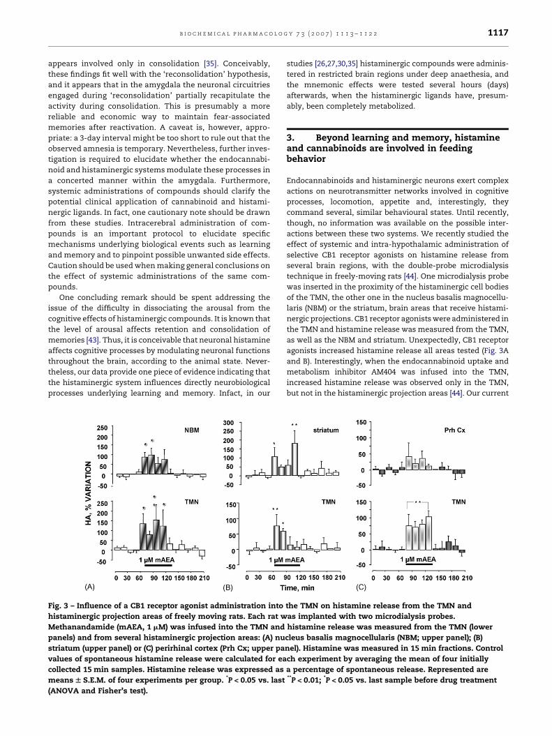
Fig. 3 – Influence of a CB1 receptor agonist administration into
histaminergic projection areas of freely moving rats. Each rat w
Methanandamide (mAEA, 1 mM) was infused into the TMN and
panels) and from several histaminergic projection areas: (A) nu
striatum (upper panel) or (C) perirhinal cortex (Prh Cx; upper pa
values of spontaneous histamine release were calculated for ea
collected 15 min samples. Histamine release was expressed as
means W S.E.M. of four experiments per group. *P < 0.05 vs. last
(ANOVA and Fisher’s test).
studies [26,27,30,35] histaminergic compounds were adminis-
tered in restricted brain regions under deep anaethesia, and
the mnemonic effects were tested several hours (days)
afterwards, when the histaminergic ligands have, presum-
ably, been completely metabolized.
3. Beyond learning and memory, histamineand cannabinoids are involved in feedingbehavior
Endocannabinoids and histaminergic neurons exert complex
actions on neurotransmitter networks involved in cognitive
processes, locomotion, appetite and, interestingly, they
command several, similar behavioural states. Until recently,
though, no information was available on the possible inter-
actions between these two systems. We recently studied the
effect of systemic and intra-hypothalamic administration of
selective CB1 receptor agonists on histamine release from
several brain regions, with the double-probe microdialysis
technique in freely-moving rats [44]. One microdialysis probe
was inserted in the proximity of the histaminergic cell bodies
of the TMN, the other one in the nucleus basalis magnocellu-
laris (NBM) or the striatum, brain areas that receive histami-
nergic projections. CB1 receptor agonists were administered in
the TMN and histamine release was measured from the TMN,
as well as the NBM and striatum. Unexpectedly, CB1 receptor
agonists increased histamine release all areas tested (Fig. 3A
and B). Interestingly, when the endocannabinoid uptake and
metabolism inhibitor AM404 was infused into the TMN,
increased histamine release was observed only in the TMN,
but not in the histaminergic projection areas [44]. Our current
the TMN on histamine release from the TMN and
as implanted with two microdialysis probes.
histamine release was measured from the TMN (lower
cleus basalis magnocellularis (NBM; upper panel); (B)
nel). Histamine was measured in 15 min fractions. Control
ch experiment by averaging the mean of four initially
a percentage of spontaneous release. Represented are**P < 0.01; *P < 0.05 vs. last sample before drug treatment

b i o c h e m i c a l p h a r m a c o l o g y 7 3 ( 2 0 0 7 ) 1 1 1 3 – 1 1 2 21118
interpretation is that the increased endocannabinoid tone
produced by AM404 augments histamine release only in the
TMN, presumably by activating a more restricted, or different
population of CB1 receptors than those activated by the
administration of direct acting CB1 receptor agonists. Relevant
to our study is that our results are in agreement with in vivo
studies demonstrating that the administration of AM404 or
URB597, an inhibitor of the metabolic pathway of the
endocannabinoid anandamide, do not mimic the full spec-
trum of pharmacological responses produced by classical CB1
receptor agonists (see Refs. [45,46] for a review). Hence,
understanding in what circumstances endocannabinoids are
released and activate histaminergic cells warrants further
investigations and may provide interesting hints to develop
new therapeutic strategies in the treatment, for instance, of
food intake disorders. In this regard, the role of brain
histamine and endocannabinoids is gaining increasing atten-
tion, because of the prominent role that they play in regulating
appetite.
4. Brain histamine and feeding behavior
The association of histamine with feeding behavior became
clear when it was observed that antidepressants and
antipsychotics stimulate appetite and induce weight gain
and that these drugs are potent H1 receptor blockers [47]. We
now know that brain histamine is involved in feeding
physiology by modulating the release of neurotransmitters
and hormones that drive or inhibit feeding [48] (Fig. 1). A large
body of literature links the histaminergic system with
consumption of food, as a satiety signal: (a) intracerebroven-
tricular injections of histamine suppress appetite, whereas
depletion of histamine stimulates feeding [49]; (b) hypotha-
lamic neuronal histamine has been implicated in the regula-
tion of feeding behavior and body adiposity through activation
of postsynaptic histamine H1-receptor (H1-R) in the ventro-
medial hypothalamic (VMH) and paraventricular (PVN)
nucleus [50,51], two brain areas that secrete neuroactive
peptides crucially involved in the regulation of feeding
behavior [52]; (c) histaminergic neurons are the targets of
leptin in the brain, and central administration of leptin
increases histamine turnover in the hypothalamus [53,54]; (d)
blockade of the histaminergic H3 autoreceptor increases
extracellular histamine levels in the hypothalamus and
reduces food intake [55]. Indeed, antagonists of the H3
receptors are being developed as anti obesity drugs [55,56].
However, a recent study suggested that in mice H3 receptor
agonists have an antiobesity effect with a mechanism
apparently independent of histaminergic tone modulation
[57]. Nowadays, though, it is generally accepted that histamine
does more than only mediating satiety. Recently, it has
become clear that food-anticipatory activity exhibits the
characteristics of a circadian rhythm, although determining
the anatomic location of the food-entrainable oscillator (FEO)
has been very difficult. Histamine drives feeding anticipating
arousal, an important actor in the FEO, and is probably
involved in numerous other feeding related processes [58]. In
fact, while the effects of the histaminergic system in the
modulation of the consummatory phase of feeding have been
shown to be robust, the role of histamine in the appetitive
phase and underlying behavioral mechanisms remains
unclear. The appetitive phase of motivated behaviors has
distinctive preparatory physiological changes, such as
increases in behavioral arousal [59] and core temperature
[58]. Therefore, the histaminergic system is a good candidate
to promote arousal during the appetitive state (Fig. 1). Indeed,
histamine-containing neurons are the only aminergic neurons
related to arousal that become active in anticipation of an
upcoming meal, as rats rendered motivated for food by 24 h
fasting and enticed with food that they cannot obtain, show a
significant increase in c-Fos immunoreactivity in the TMN,
much earlier than in other brain regions [60]. Recent reports
demonstrated that the infralimbic cortex that receives visceral
information [61] and coordinates motivated behavior [62], and
the TMN are activated in a coordinated temporal way during
the appetitive responses to food enticing [63].
Histamine neurons may participate in the appetitive
aspects of feeding also modulating reward processes involved
in the motivation to feed. The shell of the nucleus accumbens
(NAcc, a reward related brain area) receives a dense
histaminergic innervation [64] and local administration of
histamine into the NAcc enhances dopamine release [65]. The
role of histamine in reward-related processes, though, is
controversial, as both inhibitory and facilitatory effects have
been described [66,67], and nothing is known on the involve-
ment of this pathway during feeding behavior.
5. Endocannabinoids and feeding behavior
There is convincing evidence that both exogenous cannabi-
noids and the endogenous cannabinoids anandamide and 2-
arachidonoylglicerol (2-AG) stimulate feeding. Their action is
mediated by activation of CB1 receptors distributed in all brain
areas and peripheral tissues involved in the control of energy
intake, including the hypothalamus and NAcc (reviewed in
Ref. [68]). This effect is of therapeutic relevance, as cannabi-
noid agonists are currently used to alleviate anorexia and
nausea in AIDS patients, whereas the CB1 antagonist
rimonabant (SR141716A) is effective in the treatment of
obesity [69].
It has been suggested that brain endocannabinoids control
energy balance both in the appetitive phase, increasing the
incentive to find food, and during the consummatory phase,
increasing appetite, but the mechanisms involved remain to
be elucidated. Recent evidence, though, indicates that both
the mesolimbic reward mechanism and the homeostatic
hypothalamic nuclei are involved in these two aspects of
feeding behavior. Indeed, endocannabinoid levels vary in the
hypothalamus and limbic forebrain with different nutritional
manipulations, with levels being the highest with food
deprivation and lowest during food consumption [70].
Furthermore, either 2-AG injections in the shell of the NAcc
[70], or anandamide administration into the VMH induce
hyperfagia [71]. In some aspect, the histaminergic and
endocannabinoid systems seem to be regulated in an
opposing fashion: for instance, systemic administration of
leptin that signals to the hypothalamus the nutritional state
and reduces food intake, facilitates histamine release from

b i o c h e m i c a l p h a r m a c o l o g y 7 3 ( 2 0 0 7 ) 1 1 1 3 – 1 1 2 2 1119
the hypothalamus [72], whereas it downregulates endocan-
nabinoids levels in the same region [73]. Furthermore,
concentrations of hypothalamic histamine and tele-methyl-
histamine, a major histamine metabolite, are significantly
lower in obese (ob/ob) and diabetic (db/db) mice, and fatty ( fa/
fa) rats, leptin-deficient and leptin-receptor defective ani-
mals, respectively, relative to lean littermates [54]. On the
other hand, defective leptin signalling is associated with
elevated hypothalamic levels of enocannabinoids in obesedb/
db and ob/ob mice and Zucker rats. These data made it clear
that histamine controls heterogeneous aspects of feeding.
Presumably, histamine drives food intake by increasing the
arousal state of the animal [58]. Secondary to arousing the
animal, brain histamine seems to coordinate satiety and the
consolidation of temporal information associated with food
consumption [74,75].
In conclusion, for both histamine and endocannabinoids
the mechanisms involved in regulating food intake are not
fully understood and nothing is known about the temporal
and causal relationship between these two systems in
controlling the appetitive behavior. The question arises then,
if and where in the brain the endocannabinoids and the
histaminergic system interact and whether these interactions
are involved in the consummatory and/or appetitive behavior.
6. Histamine, cannabinoids and otherfunctional implications
The deleterious effects that cannabinoids have on cognitive
processes are well known [76]. Therefore it may seem
counterintuitive that cannabinoids facilitate histamine
release from the NBM, given that activation of histamine H1
receptors in the NBM increases cortical ACh release [11] and
improves rat performance in the object recognition test
[77,78]. However, augmented histamine release is also an
indicator of stress [79] and it is conceivable that protracted
occupancy of CB1 receptors, as produced by administering
cannabinoid agonists, disrupts the spatiotemporal specificity
of histamine release in different brain regions, contributing to
maladaptive behavioral responses.
Administration of CB1 receptor agonists in the TMN
facilitates histamine release from the striatum as well
(Fig. 3B), a brain region that provides the anatomical substrate
for the integration of movements [80], and participates in
learning and executing adequate behavioral responses to
environmental stimuli [81]. Histamine induces hypokinetic
effects that are accompanied by altered dopaminergic
transmission in the striatum [82], whereas systemic admin-
istration of CB1 receptor agonists reduces locomotion [83]. It is
conceivable that the augmented histamine release in the
dorsal striatum following cannabinoid administration in the
TMN may contribute to the direct actions of cannabinoids on
striatal neurones [84–86], worsening locomotor activity.
Taken together, these results show that the administration
of cannabinoids is associated with a hyperhistaminergic state.
Whether this is important in controlling food related behavior,
in contributing to the cannabinoid detrimental effects on
cognitive and locomotor performance and to drug-motivated
habits that are crucial for the establishment of addiction are all
open questions. Answering these queries may provide hints
for potential therapeutics targets to treat motivated behaviors
such as obesity or drug addiction.
7. Are histaminergic neurons aheterogeneous cell population?
The major obstacle in identifying the histaminergic system as
a target for specific therapeutic applications is the global
nature of its function. Tracing studies failed to reveal any
topographical organization of the histaminergic projections
arising from the TMN, however, two recent studies suggest
that histamine neurons are functionally heterogeneous, based
on differential activation by acute stress [87], and on the
expression of different g-subunits that confer different
sensitivity to exogenous GABA [88]. As reported previously
[44], infusions of the CB1 receptor agonist methanandamide
(mAEA) in the posterior hypothalamus in the proximity of the
histaminergic cell bodies, increase histamine release from
histaminergic projection areas such as the NBM and striatum
(Fig. 3A and B). However, during perfusion of the posterior
hypothalamus with mAEA, histamine release from the
perirhinal cortex (Prh Cx) does not change significantly
(Fig. 3C), despite the profuse histaminergic innervation of
the perhinal cortex [64] and the presence of histaminergic
receptors [89]. Therefore, mAEA-induced excitation of hista-
minergic neurons might not necessarily produce a broad
activation of all histaminergic projections, as subpopulations
of histaminergic cells projecting to different brain regions
respond differently to the same pharmacological manipula-
tion. In addition, preliminary results from our laboratory using
the double-probe microdialysis technique in freely moving
rats suggest that subpopulations of histaminergic cells
projecting to different brain regions respond differently to
bicuculline or thioperamide [90].
The observation that histaminergic neurons are not a
homogenous neuronal population may have relevant con-
sequences in the development of target specific drugs that
affect only subset of histaminergic cells, and in reducing the
occurrence of collateral or undesired effects.
8. Concluding remarks
Our knowledge of the functional roles of brain histamine is far
from complete. For as much as it may seem that the role of the
histaminergic system is redundant in modulating the sleep–
wake cycle, it is becoming clear that histamine in the brain
finely orchestrates diverse aspects of behavioral responses that
require an aroused state. For example, histamine supposedly
drives food intake by increasing the arousal state of the animal
[58], and secondary to arousing the animal, histamine coordi-
nates satiety and the consolidation of temporal information
associated with food consumption [74,75]. Augmented hista-
mine release is also an indicator of stress and disrupting the
spatiotemporal specificity of histamine release may contribute
to maladaptive behavioral responses.
The many actions of the histaminergic system are achieved
through interactions with other neurotransmitter systems,

b i o c h e m i c a l p h a r m a c o l o g y 7 3 ( 2 0 0 7 ) 1 1 1 3 – 1 1 2 21120
and some of the interplay between histaminergic neurons and
other neurotransmitter system has been described (Fig. 1). For
instance, the sleep–wake cycle and learning are presumably
influenced by the control that histamine exerts on the
forebrain cholinergic neurons. On the other hand, the
unexpected excitatory effect that cannabinoids exert on
histaminergic cells is still orphan of a functional explanation.
Obviously, new discoveries create great expectations and
great effort is being channeled into developing ever more
selective histaminergic compounds for the treatment of
neuropsychiatric disorders and metabolic dysfunctions. This
will be a great challenge in the years to come.
r e f e r e n c e s
[1] Panula P, Yang HY, Costa E. Histamine-containing neuronsin the rat hypothalamus. Proc Natl Acad Sci 1984;81:2572–6.
[2] Watanabe T, Taguchi Y, Hayashi H, Tanaka J, Shiosaka S,Tohyama M, et al. Evidence for the presence of ahistaminergic neuron system in the rat brain: animmunohistochemical analysis. Neurosci Lett1983;39:249–54.
[3] Lin JS, Sakai K, Jouvet M. Evidence for histaminergic arousalmechanisms in the hypothalamus of cat.Neuropharmacology 1988;27:111–22.
[4] Weiler HT, Hasenohrl RU, Landeghem AALV, LandeghemMV, Brankack J, Huston JP, et al. Differential modulation ofhippocampal signal transfer by tuberomammillary nucleusstimulation in freely moving rats dependent on behavioralstate. Synapse 1998;28:294–301.
[5] Parmentier R, Ohtsu H, Djebarra-Hannas Z, Valtx J-L,Watanabe T, Lin J-S. Anatomical, physiological andpharmacological characteristics of histidine decarboxylaseknock-out mice: evidence for the role of brain histamine inbehavioral and sleep–wake control. J Neurosci2002;22:7695–711.
[6] Parmentier R, Anaclet C, Watanabe T, Lin J-S.Characteristics of cortical EEG and sleep–wake cycle inhistamine H1-receptor knock out mice. Delphi: EuropeanHistamine Research Society; 2006 [Ed. Athens MSotUo].
[7] Saper C, Chou T, Scammell T. The sleep switch:hypothalamic control of sleep and wakefulness. TrendsNeurosci 2001;24:726–31.
[8] Jones BE. From waking to sleeping: neuronal and chemicalsubstrates. Trends Pharmacol Sci 2005;26:578–86.
[9] Lin J, Sakai K, Jouvet M. Hypothalamo-preoptichistaminergic projections in sleep–wake control in the cat.Eur J Neurosci 1994;6:618–25.
[10] Lin JS, Hou Y, Sakai K, Jouvet M. Histaminergic descendinginputs to the mesopontine tegmentum and their role in thecontrol of cortical activation and wakefulness in the cat. JNeurosci 1996;16:1523–2137.
[11] Cecchi M, Passani MB, Bacciottini L, Mannaioni PF,Blandina P. Cortical acetylcholine release elicited bystimulation of histamine H1 receptors in the nucleusbasalis magnocellularis: a dual probe microdialysisstudy in the freely moving rat. Eur J Neurosci 2001;13:68–78.
[12] Aston-Jones G, Bloom FE. Activity of norepinephrine-containing locus coeruleus neurons in behaving ratsanticipates fluctuations in the sleep–waking cycle. JNeurosci 1981;1:876–86.
[13] Sakai K, Mansari ME, Lin J, Zhang J, Mercier GV. Theposterior hypothalamus in the regulation of wakefulnessand paradoxical sleep. In: Mancia M, editor. The
diencephalon and sleep. New York: Raven Press; 1990. p.171–98.
[14] Wu MF, Gulyani SA, Yau E, Mignot E, Phan B, Siegel JM.Locus coeruleus neurons: cessation of activity duringcataplexy. Neuroscience 1999;91:1389–99.
[15] Wu MF, John J, Boehmer LN, Yau D, Nguyen GB, Siegel JM.Activity of dorsal raphe cells across the sleep–waking cycleand during cataplexy in narcoleptic dogs. J Physiol2004;554:202–15.
[16] John J, Wu MF, Boehmer LN, Siegel JM. Cataplexy-activeneurons in the hypothalamus: implications for the role ofhistamine in sleep and waking behavior. Neuron2004;42:619–34.
[17] Abe H, Honma S, Ohtsu H, Honma K. Circadian rhythms inbehavior and clock gene expressions in the brain of micelacking histidine decarboxylase. Brain Res Mol Brain Res2004;124:178–87.
[18] Selbach O, Haas HL. Hypocretins: the timing of sleep andwaking. Chronobiol Int 2006;23:63–70.
[19] Eriksson K, Sergeeva O, Brown R, Haas H. Orexin/hypocretin excites the histaminergic neurons of thetuberomammillary nucleus. J Neurosci 2001;21:9273–9.
[20] Huang Z-L, Qu W-M, Li W-D, Mochizuki T, Eguchi N,Watanabe T, et al. Arousal effect of orexin A depends onactivation of the histaminergic system. Proc Natl Acad Sci2001;98:9965–70.
[21] Nishino S, Fujiki N, Ripley B, Sakurai E, Kato M, WatanabeT, et al. Decreased brain histamine content in hypocretin/orexin receptor-2 mutated narcoleptic dogs. Neurosci Lett2001;313:125–8.
[22] Kanbayashi T, Yano T, Ishiguro H, Kawanishi K, Chiba S,Aizawa R, et al. Hypocretin-1 (orexin-A) levels in humanlumbar CSF in different age groups: infants to elderlypersons. Sleep 2002;25:337–9.
[23] Blandina P, Efoudebe M, Cenni G, Mannaioni PF, PassaniMB. Acetylcholine, histamine and cognition: two sides ofthe same coin. Learn Mem 2004;11(1):1–8.
[24] Passani M, Blandina P. The neuronal histaminergic systemin cognition. Curr Med Chem 2004;4:17–26.
[25] Vazdarjanova A, McGaugh JL. Basolateral amygdala isinvolved in modulating consolidation of memory forclassical conditioning. J Neurosci 1999;19:6615–22.
[26] Passani MB, Cangioli I, Baldi E, Bucherelli C, Mannaioni PF,Blandina P. Histamine H3 receptor-mediated impairment ofcontextual fear conditioning, and in-vivo inhibition ofcholinergic transmission in the rat basolateral amygdala.Eur J Neurosci 2001;14:1522–32.
[27] Cangioli I, Baldi E, Mannaioni PF, Bucherelli C, Blandina P,Passani MB. Activation of histaminergic H3 receptors in therat basolateral amygdala improves expression of fearmemory and enhances acetylcholine release. Eur J Neurosci2002;16:521–8.
[28] Fox GB, Esbenshade TA, Pan JB, Radek RJ, Krueger KM, YaoBB, et al. Pharmacological properties of ABT-239 [4-(2-{2-[(2R)-2-methylpyrrolidinyl]ethyl}-benzofuran-5-yl)benzonitrile]: II. Neurophysiological characterization andbroad preclinical efficacy in cognition and schizophrenia ofa potent and selective histamine H3 receptor antagonist. JPharmacol Exp Ther 2005;313:176–90.
[29] Bacciottini L, Passani M, Giovannelli L, Cangioli I,Mannaioni P, Schunack W, et al. Endogenous histamine inthe medial septum-diagonal band complex increases therelease of acetylcholine from the hippocampus: a dual-probe microdialysis study in the freely moving animal. Eur JNeurosci 2002;15:1669–80.
[30] Giovannini M, Efoudebe M, Passani M, Baldi E, Bucherelli C,Giachi F, et al. Improvement in fear memory by histamineelicited erk2 activation in hippocampal CA3 cells. JNeurosci 2003;23:9016–23.

b i o c h e m i c a l p h a r m a c o l o g y 7 3 ( 2 0 0 7 ) 1 1 1 3 – 1 1 2 2 1121
[31] McGaugh JL, Cahill L, Roozendaal B. Involvement of theamygdala in memory storage: Interaction with other brainsystems. Proc Natl Acad Sci 1996;93:13508–14.
[32] Freund TF, Katona I, Piomelli D. Role of endogenouscannabinoids in synaptic signalling. Physiol Rev2003;83:1017–66.
[33] Azad SC, Monory K, Marsicano G, Cravatt BF, Lutz B,Zieglgansberger W, et al. Circuitry for associative plasticityin the amygdala involves endocannabinoids signaling. JNeurosci 2004;24:9953–61.
[34] Marsicano G, Wotjak CT, Azad S, Bisogno T, Rammes G,Cascio MG, et al. The endogenous cannabinoid systemcontrols extinction of aversive memories. Nature2002;418:530–4.
[35] Bucherelli C, Baldi E, Mariottini C, Passani MB, Blandina P.Aversive memory reactivation engages in the amygdalaonly some neurotransmitters involved in consolidation.Learn Mem 2006;13:426–30.
[36] Nader K, Schafe GE, LeDoux JE. Fear memories requireprotein synthesis in the amygdala for reconsolidation afterretrieval. Nature 2000;406:722–6.
[37] Alberini CM. Mechanisms of memory stabilization: areconsolidation and reconsolidation similar or distinctprocesses? Trends Neurosci 2005;28:51–6.
[38] Vianna M, Szapiro G, McGaugh J, Medina J, Izquierdo I.Retrieval of memory for fear-motivated training initiatesextinction requiring protein synthesis in the rathippocampus. Proc Natl Acad Sci 2001;98:12251–4.
[39] Lattal K, Abel T. Behavioral impairments caused byinjections of the protein synthesis inhibitor anisomycinafter contextual retrieval reverse with time. Proc Natl AcadSci 2004;101:4667–72.
[40] Power AE, Berlau DJ, McGaugh JL, Steward O. Anisomycininfused into the hippocampus fails to block‘‘reconsolidation’’ but impairs extinction: The role of re-exposure duration. Learn Mem 2006;13:27–34.
[41] Lee JL, Ciano PD, Thomas KL, Everitt BJ. Disruptingreconsolidation of drug memories reduces cocaine-seekingbehavior. Neuron 2005;47:795–801.
[42] Liao C, Zheng J, David LS, Nicholson RA. Inhibition ofvoltage-sensitive sodium channels by the cannabinoid 1receptor antagonist AM 251 in mammalian brain.Pharmacol Toxicol 2004;94:73–8.
[43] Cahill L, McGaugh J. A novel demonstration of enhancedmemory associated with emotional arousal. ConsciousCogn 1995;4:410–21.
[44] Cenni G, Blandina P, Mackie K, Nosi D, Formigli L, GiannoniP, et al. Differential effect of cannabinoid agonists andendocannabinoids on histamine release from distinctregions of the rat brain. Eur J Neurosci 2006;24:1633–44.
[45] Solinas M, Panlilio LV, Tanda G, Makriyannis A, MatthewsSA, Goldberg SR. Cannabinoid agonists but not inhibitors ofendogenous cannabinoid transport or metabolism enhancethe reinforcing efficacy of heroin in rats.Neuropsychopharmacology 2005;30:2046–57.
[46] Gaetani S, Cuomo V, Piomelli D. Anandamide hydrolysis: anew target for anti-anxiety drugs? Trends Mol Med2003;9:474–8.
[47] Russ MJ, Ackerman SH. Antidepressants and weight gain.Appetite 1988;10:103–17.
[48] Toftegaard CL, Knigge U, Kjaer A, Warberg J. The role ofhypothalamic histamine in leptin-induced suppression ofshort-term food intake in fasted rats. Regul Pept2003;111:83–90.
[49] Tuomisto L, Yamatodani A, Jokkonen J, Sainio E, AiraksinenM. Inhibition of brain histamine synthesis increases foodintake and inhibits vasopressin response to salt loading inrats. Methods Find Exp Clin Pharmacol 1994;16:355–9.
[50] Fukagawa K. Neuronal histamine modulates feedingbehavior through H1-receptor in rat hypothalamus. Am JPhysiol 1989;256:R605–11.
[51] Okuma K, Sakata T, Fukagawa K, et al. Neuronal histaminein the hypothalamus suppresses food intake in rats. BrainRes 1994;628:235–42.
[52] Hillebrand J, Wied D, Adan R. Neuropeptides, food intakeand body weight regulation: a hypothalamic focus. Peptides2002;23:2283–306.
[53] Yoshimatsu H, Itateyama E, Kondou S, Hidaka S, Tajima D,Kurokawa M. Hypothalamic neuronal histamine as a targetof leptin action on feeding behavior in the central nervoussystem. Diabetes 1999;48:1342–6.
[54] Itateyama E, Chiba S, Sakata T, Yoshimatsu H.Hypothalamic neuronal histamine in genetically obeseanimals: its implication of leptin action in the brain. ExpBiol Med 2003;228:1132–7.
[55] Malmlof K, Zaragoza F, Golozoubova V, Refsgaard HH,Cremers T, Raun K, et al. Influence of a selective histamineH3 receptor antagonist on hypothalamic neural activity,food intake and body weight. Int J Obes 2005;29:1402–12.
[56] Esbenshade T, Hancock A, Bitner R, Krueger K, Otte S,Nikkel A, Fey T, Bush E, Dickinson R, Shapiro R, Knourek-Segel V, Droz B, Brune M, Jacobson P, Cowart M.Distinctions and contradistinctions between antiobesityhistamine H(3) receptor (H (3)R) antagonists compared tocognition-enhancing H(3) receptor antagonists. InflammRes 2006; Epub ahead of print.
[57] Yoshimoto R, Miyamoto Y, Shimamura K, Ishihara A,Takahashi K, Kotani H, et al. Therapeutic potential ofhistamine H3 receptor agonist for the treatment of obesityand diabetes mellitus. Proc Nat Acad Sci 2006;103:13866–71.
[58] Valdes J, Farıas P, Ocampo-Garce A, Cortes N, Seron-FerreM, Torrealba F. Arousal and differential Fos expression inhistaminergic neurons of the ascending arousal systemduring a feeding-related motivated behaviour. Eur JNeurosci 2005;21:1931–42.
[59] Pfaff D, Frohlich J, Morgan M. Hormonal and geneticinfluences on arousal–sexual and otherwise. TrendsNeurosci 2002;25:45–50.
[60] Meynard MM, Valdes JL, Recabarren M, Seron-Ferre M,Torrealba F. Specific activation of histaminergic neuronsduring daily feeding anticipatory behavior in rats. BehavBrain Res 2005;158:311–9.
[61] Recabarren M, Valdes J, Farıas P, Seron-Ferre M, TorrealbaF. Differential effects of infralimbic cortical lesions ontemperature and locomotor activity responses to feeding inrats. Neuroscience 2005;134:1413–22.
[62] Hok V, Save E, Lenck-Santini P, Poucet B. Coding for spatialgoals in the prelimbic/infralimbic area of the rat frontalcortex. Proc Natl Acad Sci 2005;102:4602–7.
[63] Valdes J, Maldonado P, Recabarren M, Fuentes R, TorrealbaF. The infralimbic cortical area commands the behavioraland vegetative arousal during appetitive behavior in therat. Eur J Neurosci 2006;23:1352–64.
[64] Panula P, Pirvola U, Auvinen S, Airaksinen M. Histamine-immunoreactive nerve fibers in the rat brain. Neuroscience1989;28:585–610.
[65] Galosi R, Lenard L, Knoche A, Haas H, Huston J, SchwartingR. Dopaminergic effects of histamine administration in thenucleus accumbens and the impact of H1-receptorblockade. Neuropharmacology 2001;40:624–33.
[66] Wagner U, Segura-Torres P, Weiler T, Huston J. Thetuberomammillary nucleus region as a reinforcementinhibiting substrate: facilitation of ipsihypothalamic self-stimulation by unilateral ibotenic acid lesions. Brain Res1993;613:269–74.
[67] Hasenohrl R, Kuhlen A, Frisch C, Galosi R, Brandao M,Huston H. Comparison of intra-accumbens injection of

b i o c h e m i c a l p h a r m a c o l o g y 7 3 ( 2 0 0 7 ) 1 1 1 3 – 1 1 2 21122
histamine with histamine H1-receptor antagonistchlorpheniramine in effects on reinforcement and memoryparameters. Behav Brain Res 2001;124:203–11.
[68] Matias I, Bisogno T, Marzo VD. Endogenous cannabinoids inthe brain and peripheral tissues: regulation of their levelsand control of food intake. Int J Obest 2006;(Suppl 1):S7–12.
[69] Berry EM, Mechoulam R. Tetrahydrocannabinol andendocannabinoids in feeding and appetite. Pharmacol Ther2002;95:185–90.
[70] Kirkham TC, Williams CM, Fezza F, Marzo VD.Endocannabinoid levels in rat limbic forebrain andhypothalamus in relation to fasting, feeding and satiation:stimulation of eating by 2-arachidonoyl glycerol. Br JPharmacol 2002;136:550–7.
[71] Jamshidi N, Taylor D. Anandamide administration into theventromedial hypothalamus stimulates appetite in rats. BrJ Pharmacol 2001;134:1151–4.
[72] Morimoto T, Yamamoto Y, Mobarakeh IJ, Yanai K,Watanabe T, Yamatodani A. Involvement of thehistaminergic system in leptin-induced suppression offood intake. Physiol Behav 1999;67:679–83.
[73] DiMarzo V, Goparaju S, Wang L, Liu J, Batkai S, Jarai Z, et al.Leptin-regulated endocannabinoids are involved inmaintaining food intake. Nature 2001;410:822–5.
[74] Sakata T, Fukagawa K, Ookuma K, Fujimoto K, YoshimatsuH, Yamatodani A, et al. Hypothalamic neuronal histaminemodulates ad libitum feeding by rats. Brain Res1990;537:303–6.
[75] Angeles-Castellanos M, Mendoza J, Diaz-Munoz M, EscobarC. Food entrainment modifies the c-Fos expression patternin brain stem nuclei of rats. Am J Physiol Regul Integr CompPhysiol 2005;288:R678–84.
[76] Schneider M, Koch M. The cannabinoid agonist WIN 55,212-2 reduces sensorimotor gating and recognition memory inrats. Behav Pharmacol 2002;13:29–37.
[77] Orsetti M, Ferretti C, Gamalero R, Ghi P. Histamine H3-receptor blockade in the rat nucleus basalismagnocellularis improves place recognition memory.Psychopharmacology 2002;159:133–7.
[78] Malmberg-Aiello P, Ipponi A, Blandina P, Bartolini L,Schunack W. Pro-cognitive effect of a selective H1 receptoragonist, 2-(3-trifluoromethylphenyl)histamine, in the ratobject recognition test. Inflam Res 2003;52:S33–4.
[79] Westerink BH, Cremers TI, Vries JBD, Liefers H, Tran N, BoerPD. Evidence for activation of histamine H3 autoreceptors
during handling stress in the prefrontal cortex of the rat.Synapse 2002;15:238–43.
[80] Brown L. Somatotopic organization in the rat striatum:evidence for a combinatorial map. Proc Nat Acad Sci1992;89:7403–7.
[81] Hyman SE, Malenka RC. Addiction and the brain: theneurobiology of compulsion and its persistence. Nature RevNeurosci 2001;2:695–703.
[82] Chiavegatto S, Nasello AG, Bernardi MM. Histamine andspontaneous motor activity: biphasic changes, receptorsinvolved and participation of the striatal dopamine system.Life Sci 1998;62:1875–88.
[83] McLaughlin PJ, Lu D, Winston KM, Thakur G, Swezey LA,Makriyannis A, et al. Behavioral effects of the novelcannabinoid full agonist AM 411. Pharmacol BiochemBehav 2005;81:78–88.
[84] Sanudo-Pena MC, Tsou K, Walker JM. Motor actions ofcannabinoids in the basal ganglia output nuclei. Life Sci1999;65:703–13.
[85] Huang C-C, Lo S-W, Hsu K-S. Presynaptic mechanismsunderlying cannabinoid inhibition of excitatory synaptictransmission in rat striatal neurons. J Physiol2001;532.3:731–48.
[86] Kofalvi A, Rodrigues RJ, Ledent C, Mackie K, Vizi ES, CunhaRA, et al. Involvement of cannabinoid receptors in theregulation of neurotransmitter release in the rodentstriatum: a combined immunochemical andpharmacological analysis. J Neurosci 2005;25:2874–84.
[87] Miklos I, Kovacs K. Functional heterogeneity of theresponses of histaminergic neuron subpopulations tovarious stress challenges. Eur J Neurosci 2003;18:3069–79.
[88] Sergeeva OA, Eriksson KS, Sharonova IN, Vorobjev VS, HaasHL. GABA(A) receptor heterogeneity in histaminergicneurons. Eur J Neurosci 2002;16:1472–82.
[89] Pillot C, Heron A, Cochois V, Tardivel-Lacombe J, Ligneau X,Schwartz J-C, et al. A detailed mapping of the histamine H3receptor and its gene transcript in rat brain. Neuroscience2002;114:173–93.
[90] Giannoni P, Cenni G, Passani M, Mannaioni P, Medhurst A,Blandina P. Different responses to GABAA or H3antagonists suggest the existence of distinctsubpopulations among histaminergic neurons. In:Proceedings of the XXXV European histamine researchsociety; 2006. p. 52 [Ed. Athens MSotUo].
Related Documents