Welcome message from author
This document is posted to help you gain knowledge. Please leave a comment to let me know what you think about it! Share it to your friends and learn new things together.
Transcript

Proceedings of 6th Hiroshima Conference on Education and Science in DentistryOctober 23-25, 2015, in Hiroshima, Japan
Hiroshima University Faculty of Dentistry
1965 - 201550th Anniversary Commemoration
01_目次 15.9.25 1:44 PM ページ 1
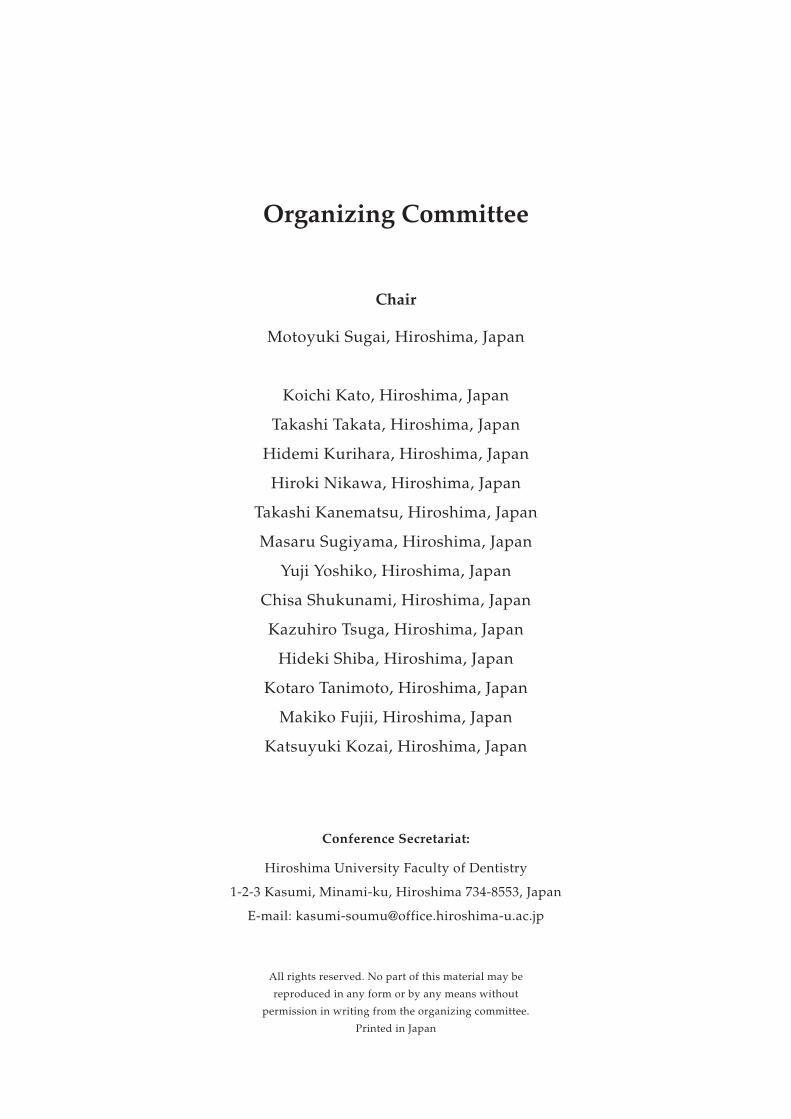
Organizing Committee
Chair
Motoyuki Sugai, Hiroshima, Japan
Koichi Kato, Hiroshima, Japan
Takashi Takata, Hiroshima, Japan
Hidemi Kurihara, Hiroshima, Japan
Hiroki Nikawa, Hiroshima, Japan
Takashi Kanematsu, Hiroshima, Japan
Masaru Sugiyama, Hiroshima, Japan
Yuji Yoshiko, Hiroshima, Japan
Chisa Shukunami, Hiroshima, Japan
Kazuhiro Tsuga, Hiroshima, Japan
Hideki Shiba, Hiroshima, Japan
Kotaro Tanimoto, Hiroshima, Japan
Makiko Fujii, Hiroshima, Japan
Katsuyuki Kozai, Hiroshima, Japan
Conference Secretariat:
Hiroshima University Faculty of Dentistry
1-2-3 Kasumi, Minami-ku, Hiroshima 734-8553, Japan
E-mail: [email protected]
All rights reserved. No part of this material may be
reproduced in any form or by any means without
permission in writing from the organizing committee.
Printed in Japan
01_目次 15.9.25 1:44 PM ページ 2
PREFACE i
PREFACE
On behalf of organizing committee members of Hiroshima Conference and Hiroshima University Faculty of
Dentistry, it is my great pleasure of extending to you an invitation to participate in 6th Hiroshima Conference on
Education and Science in Dentistry with the theme, “BioDental Education and Research towards the Next 50 years - 50th
Anniversary Commemoration” to be held in Hiroshima, Japan on October 23-25, 2015.
We held the 1st Hiroshima Conference on Education and Science in Dentistry to commemorate the 40th anniversary
of the founding of the Faculty of Dentistry in January 2006. Time flies so fast and I am now filled with deep emotion
when I look back the previous 10 years that have passed since the 1st Hiroshima Conference. Ten years ago, coinciden-
tally, Dental technician school and Hygienist school (2-year college) were reorganized as one school and it started as
School of Oral Health Sciences as 4-year university, which is doubling the joy of this commemorable occasion.
One of the important characteristics and uniqueness of this conference is its organization: you may notice education
sessions and science sessions together in this as well as previous Hiroshima Conference. This idea was originated from
the belief of the founder of Hiroshima Conference, Prof. Kurihara, “the advanced research is indispensable to the
advanced education”. Within these 10 years, our school has significantly grown and transformed under the key concept,
BioDental education and research.
I strongly wish all of you to enjoy state of the art special lectures on science and education, presentations of young
investigators from various countries, stimulate discussions, develop international and inter-school collaborations, and
think together what we should do in dental education and research towards next 50 years! And please plan to join
Hiroshima University Faculty of Dentistry 50th Anniversary Cerebration at the room Sunflower on Oct 24 afternoon with
our distinguished guests, international alumni and friends.
All the best,
Motoyuki Sugai, DDS, PhD
President, 6th Hiroshima Conference on Education and Science in Dentistry
Dean, Faculty of Dentistry Hiroshima University
01_目次 15.9.25 1:44 PM ページ i

01_目次 15.9.25 1:44 PM ページ ii
Congratulatory Address iii
Congratulatory Address on the 50th Anniversary of the HU Faculty of Dentistry
cine.
In 2013, Hiroshima University was elected to be part
of the “Program for Promoting the Enhancement of
Research Universities”, together with 21 other institu-
tions, and in 2014, our university became one of 13 mem-
bers to take part in the “Top Global University Project” as
a “Type A (Top type)” university.
Participating in these two projects means that
Hiroshima University has the potential to “be ranked in
the world’s top 100 universities within the next ten
years”, which comprises one of the milestones along the
road towards our long-term goal.
Looking ahead, Hiroshima University aims to
become a “university with sustained world-wide fame
and splendor even after 100 years”, by continuously fos-
tering “peace-pursuing, cultured personnel with interna-
tional experience”, amidst close cooperation with the
Faculty of Dentistry, the Graduate School of Biomedical
& Health Sciences and other departments.
I expect that the Faculty of Dentistry will continue to
dedicate itself to producing excellent dental professionals
who are active in global and local communities, always
considering the perspective of their patients.
In conclusion, I would like to ask all of you here
today for your continued understanding and support,
and I would also like to offer a prayer for the continuing
growth and prosperity of the Faculty.
Sincerely,
Mitsuo Ochi
President, Hiroshima University
Today, on the occasion of the 50-year anniversary of
the Hiroshima University Faculty of Dentistry, I would
like to say a few words of congratulations.
The origins of Hiroshima University’s Faculty of
Dentistry can be traced back to April 1st, 1965, when it
was established as the third national university’s dental
school after Tokyo Medical and Dental University and
Osaka University’s Faculty of Dentistry. The establish-
ment of the Faculty met the demands of a broad range of
Hiroshima Prefecture’s population, and it was the
Hiroshima Prefecture Dental Association which first pro-
posed that the Faculty of Dentistry become an official
part of the university.
In 2005, the School of Oral Health Science was
founded, and since its inception it has greatly contributed
to the creation of excellent dentists, dental technicians
and dental hygienists, active throughout Japan.
The 21st century is an international era of low birth
rates and longevity, and globalism extends to every part
of the world. Also in the field of dentistry, global-stan-
dard knowledge and technical skills are required.
The Hiroshima University Faculty of Dentistry pro-
vides high level dental medicine, medical services and
oral health science, based on life sciences. It also fosters
dental specialists, able to be active in an aging society
and globalized environment.
Additionally, in 2012 an “International Dental
Course” was established, resulting in Hiroshima
University accepting international students from all over
Asia, and becoming a nationwide pioneer in teaching lec-
tures both in English and Japanese. Thus, via this multi-
cultural educational environment, our university is also
making efforts to foster the future leaders of dental medi-
01_目次 15.9.25 1:44 PM ページ iii
iv
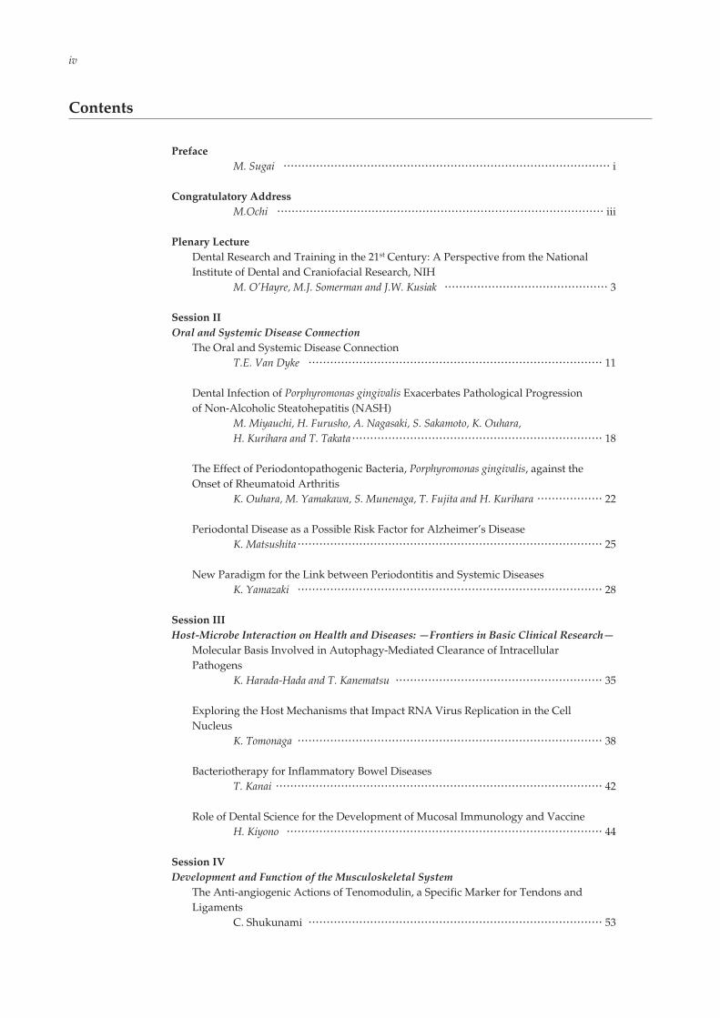
PrefaceM. Sugai ⋯⋯⋯⋯⋯⋯⋯⋯⋯⋯⋯⋯⋯⋯⋯⋯⋯⋯⋯⋯⋯⋯⋯⋯⋯⋯⋯⋯⋯⋯ i
Congratulatory AddressM.Ochi ⋯⋯⋯⋯⋯⋯⋯⋯⋯⋯⋯⋯⋯⋯⋯⋯⋯⋯⋯⋯⋯⋯⋯⋯⋯⋯⋯⋯⋯⋯ iii
Plenary LectureDental Research and Training in the 21st Century: A Perspective from the NationalInstitute of Dental and Craniofacial Research, NIH
M. O’Hayre, M.J. Somerman and J.W. Kusiak ⋯⋯⋯⋯⋯⋯⋯⋯⋯⋯⋯⋯⋯⋯⋯ 3
Session IIOral and Systemic Disease Connection
The Oral and Systemic Disease ConnectionT.E. Van Dyke ⋯⋯⋯⋯⋯⋯⋯⋯⋯⋯⋯⋯⋯⋯⋯⋯⋯⋯⋯⋯⋯⋯⋯⋯⋯⋯⋯ 11
Dental Infection of Porphyromonas gingivalis Exacerbates Pathological Progression of Non-Alcoholic Steatohepatitis (NASH)
M. Miyauchi, H. Furusho, A. Nagasaki, S. Sakamoto, K. Ouhara, H. Kurihara and T. Takata⋯⋯⋯⋯⋯⋯⋯⋯⋯⋯⋯⋯⋯⋯⋯⋯⋯⋯⋯⋯⋯⋯⋯ 18
The Effect of Periodontopathogenic Bacteria, Porphyromonas gingivalis, against theOnset of Rheumatoid Arthritis
K. Ouhara, M. Yamakawa, S. Munenaga, T. Fujita and H. Kurihara ⋯⋯⋯⋯⋯⋯ 22
Periodontal Disease as a Possible Risk Factor for Alzheimer’s DiseaseK. Matsushita⋯⋯⋯⋯⋯⋯⋯⋯⋯⋯⋯⋯⋯⋯⋯⋯⋯⋯⋯⋯⋯⋯⋯⋯⋯⋯⋯⋯ 25
New Paradigm for the Link between Periodontitis and Systemic DiseasesK. Yamazaki ⋯⋯⋯⋯⋯⋯⋯⋯⋯⋯⋯⋯⋯⋯⋯⋯⋯⋯⋯⋯⋯⋯⋯⋯⋯⋯⋯⋯ 28
Session IIIHost-Microbe Interaction on Health and Diseases: —Frontiers in Basic Clinical Research—
Molecular Basis Involved in Autophagy-Mediated Clearance of IntracellularPathogens
K. Harada-Hada and T. Kanematsu ⋯⋯⋯⋯⋯⋯⋯⋯⋯⋯⋯⋯⋯⋯⋯⋯⋯⋯⋯ 35
Exploring the Host Mechanisms that Impact RNA Virus Replication in the CellNucleus
K. Tomonaga ⋯⋯⋯⋯⋯⋯⋯⋯⋯⋯⋯⋯⋯⋯⋯⋯⋯⋯⋯⋯⋯⋯⋯⋯⋯⋯⋯⋯ 38
Bacteriotherapy for Inflammatory Bowel DiseasesT. Kanai ⋯⋯⋯⋯⋯⋯⋯⋯⋯⋯⋯⋯⋯⋯⋯⋯⋯⋯⋯⋯⋯⋯⋯⋯⋯⋯⋯⋯⋯⋯ 42
Role of Dental Science for the Development of Mucosal Immunology and VaccineH. Kiyono ⋯⋯⋯⋯⋯⋯⋯⋯⋯⋯⋯⋯⋯⋯⋯⋯⋯⋯⋯⋯⋯⋯⋯⋯⋯⋯⋯⋯⋯ 44
Session IVDevelopment and Function of the Musculoskeletal System
The Anti-angiogenic Actions of Tenomodulin, a Specific Marker for Tendons andLigaments
C. Shukunami ⋯⋯⋯⋯⋯⋯⋯⋯⋯⋯⋯⋯⋯⋯⋯⋯⋯⋯⋯⋯⋯⋯⋯⋯⋯⋯⋯ 53
Contents
01_目次 15.9.25 1:44 PM ページ iv
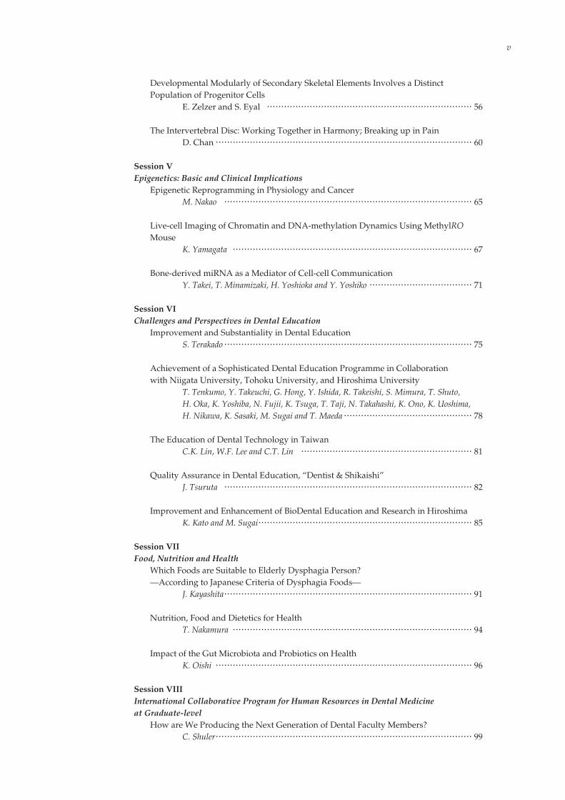
v
Developmental Modularly of Secondary Skeletal Elements Involves a DistinctPopulation of Progenitor Cells
E. Zelzer and S. Eyal ⋯⋯⋯⋯⋯⋯⋯⋯⋯⋯⋯⋯⋯⋯⋯⋯⋯⋯⋯⋯⋯⋯⋯⋯ 56
The Intervertebral Disc: Working Together in Harmony; Breaking up in PainD. Chan ⋯⋯⋯⋯⋯⋯⋯⋯⋯⋯⋯⋯⋯⋯⋯⋯⋯⋯⋯⋯⋯⋯⋯⋯⋯⋯⋯⋯⋯⋯ 60
Session VEpigenetics: Basic and Clinical Implications
Epigenetic Reprogramming in Physiology and CancerM. Nakao ⋯⋯⋯⋯⋯⋯⋯⋯⋯⋯⋯⋯⋯⋯⋯⋯⋯⋯⋯⋯⋯⋯⋯⋯⋯⋯⋯⋯⋯ 65
Live-cell Imaging of Chromatin and DNA-methylation Dynamics Using MethylROMouse
K. Yamagata ⋯⋯⋯⋯⋯⋯⋯⋯⋯⋯⋯⋯⋯⋯⋯⋯⋯⋯⋯⋯⋯⋯⋯⋯⋯⋯⋯⋯ 67
Bone-derived miRNA as a Mediator of Cell-cell CommunicationY. Takei, T. Minamizaki, H. Yoshioka and Y. Yoshiko ⋯⋯⋯⋯⋯⋯⋯⋯⋯⋯⋯⋯ 71
Session VIChallenges and Perspectives in Dental Education
Improvement and Substantiality in Dental EducationS. Terakado ⋯⋯⋯⋯⋯⋯⋯⋯⋯⋯⋯⋯⋯⋯⋯⋯⋯⋯⋯⋯⋯⋯⋯⋯⋯⋯⋯⋯⋯ 75
Achievement of a Sophisticated Dental Education Programme in Collaboration with Niigata University, Tohoku University, and Hiroshima University
T. Tenkumo, Y. Takeuchi, G. Hong, Y. Ishida, R. Takeishi, S. Mimura, T. Shuto, H. Oka, K. Yoshiba, N. Fujii, K. Tsuga, T. Taji, N. Takahashi, K. Ono, K. Uoshima, H. Nikawa, K. Sasaki, M. Sugai and T. Maeda ⋯⋯⋯⋯⋯⋯⋯⋯⋯⋯⋯⋯⋯⋯⋯ 78
The Education of Dental Technology in TaiwanC.K. Lin, W.F. Lee and C.T. Lin ⋯⋯⋯⋯⋯⋯⋯⋯⋯⋯⋯⋯⋯⋯⋯⋯⋯⋯⋯⋯ 81
Quality Assurance in Dental Education, “Dentist & Shikaishi”J. Tsuruta ⋯⋯⋯⋯⋯⋯⋯⋯⋯⋯⋯⋯⋯⋯⋯⋯⋯⋯⋯⋯⋯⋯⋯⋯⋯⋯⋯⋯⋯ 82
Improvement and Enhancement of BioDental Education and Research in HiroshimaK. Kato and M. Sugai⋯⋯⋯⋯⋯⋯⋯⋯⋯⋯⋯⋯⋯⋯⋯⋯⋯⋯⋯⋯⋯⋯⋯⋯⋯ 85
Session VIIFood, Nutrition and Health
Which Foods are Suitable to Elderly Dysphagia Person?—According to Japanese Criteria of Dysphagia Foods—
J. Kayashita⋯⋯⋯⋯⋯⋯⋯⋯⋯⋯⋯⋯⋯⋯⋯⋯⋯⋯⋯⋯⋯⋯⋯⋯⋯⋯⋯⋯⋯ 91
Nutrition, Food and Dietetics for HealthT. Nakamura ⋯⋯⋯⋯⋯⋯⋯⋯⋯⋯⋯⋯⋯⋯⋯⋯⋯⋯⋯⋯⋯⋯⋯⋯⋯⋯⋯⋯ 94
Impact of the Gut Microbiota and Probiotics on HealthK. Oishi ⋯⋯⋯⋯⋯⋯⋯⋯⋯⋯⋯⋯⋯⋯⋯⋯⋯⋯⋯⋯⋯⋯⋯⋯⋯⋯⋯⋯⋯⋯ 96
Session VIIIInternational Collaborative Program for Human Resources in Dental Medicine at Graduate-level
How are We Producing the Next Generation of Dental Faculty Members?C. Shuler⋯⋯⋯⋯⋯⋯⋯⋯⋯⋯⋯⋯⋯⋯⋯⋯⋯⋯⋯⋯⋯⋯⋯⋯⋯⋯⋯⋯⋯⋯ 99
01_目次 15.9.25 1:44 PM ページ v
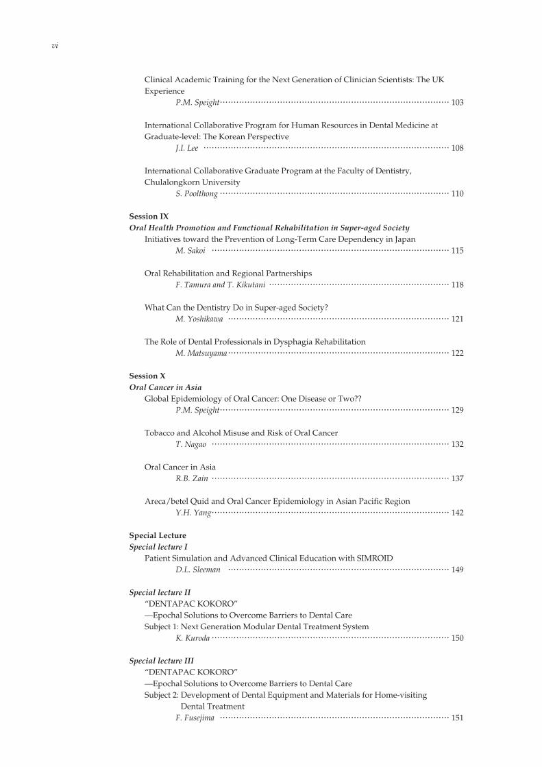
vi
Clinical Academic Training for the Next Generation of Clinician Scientists: The UKExperience
P.M. Speight⋯⋯⋯⋯⋯⋯⋯⋯⋯⋯⋯⋯⋯⋯⋯⋯⋯⋯⋯⋯⋯⋯⋯⋯⋯⋯⋯⋯ 103
International Collaborative Program for Human Resources in Dental Medicine atGraduate-level: The Korean Perspective
J.I. Lee ⋯⋯⋯⋯⋯⋯⋯⋯⋯⋯⋯⋯⋯⋯⋯⋯⋯⋯⋯⋯⋯⋯⋯⋯⋯⋯⋯⋯⋯⋯ 108
International Collaborative Graduate Program at the Faculty of Dentistry,Chulalongkorn University
S. Poolthong ⋯⋯⋯⋯⋯⋯⋯⋯⋯⋯⋯⋯⋯⋯⋯⋯⋯⋯⋯⋯⋯⋯⋯⋯⋯⋯⋯⋯ 110
Session IXOral Health Promotion and Functional Rehabilitation in Super-aged Society
Initiatives toward the Prevention of Long-Term Care Dependency in JapanM. Sakoi ⋯⋯⋯⋯⋯⋯⋯⋯⋯⋯⋯⋯⋯⋯⋯⋯⋯⋯⋯⋯⋯⋯⋯⋯⋯⋯⋯⋯⋯ 115
Oral Rehabilitation and Regional PartnershipsF. Tamura and T. Kikutani ⋯⋯⋯⋯⋯⋯⋯⋯⋯⋯⋯⋯⋯⋯⋯⋯⋯⋯⋯⋯⋯⋯ 118
What Can the Dentistry Do in Super-aged Society?M. Yoshikawa ⋯⋯⋯⋯⋯⋯⋯⋯⋯⋯⋯⋯⋯⋯⋯⋯⋯⋯⋯⋯⋯⋯⋯⋯⋯⋯⋯ 121
The Role of Dental Professionals in Dysphagia RehabilitationM. Matsuyama⋯⋯⋯⋯⋯⋯⋯⋯⋯⋯⋯⋯⋯⋯⋯⋯⋯⋯⋯⋯⋯⋯⋯⋯⋯⋯⋯ 122
Session XOral Cancer in Asia
Global Epidemiology of Oral Cancer: One Disease or Two??P.M. Speight⋯⋯⋯⋯⋯⋯⋯⋯⋯⋯⋯⋯⋯⋯⋯⋯⋯⋯⋯⋯⋯⋯⋯⋯⋯⋯⋯⋯ 129
Tobacco and Alcohol Misuse and Risk of Oral CancerT. Nagao ⋯⋯⋯⋯⋯⋯⋯⋯⋯⋯⋯⋯⋯⋯⋯⋯⋯⋯⋯⋯⋯⋯⋯⋯⋯⋯⋯⋯⋯ 132
Oral Cancer in AsiaR.B. Zain ⋯⋯⋯⋯⋯⋯⋯⋯⋯⋯⋯⋯⋯⋯⋯⋯⋯⋯⋯⋯⋯⋯⋯⋯⋯⋯⋯⋯⋯ 137
Areca/betel Quid and Oral Cancer Epidemiology in Asian Pacific RegionY.H. Yang⋯⋯⋯⋯⋯⋯⋯⋯⋯⋯⋯⋯⋯⋯⋯⋯⋯⋯⋯⋯⋯⋯⋯⋯⋯⋯⋯⋯⋯ 142
Special LectureSpecial lecture I
Patient Simulation and Advanced Clinical Education with SIMROIDD.L. Sleeman ⋯⋯⋯⋯⋯⋯⋯⋯⋯⋯⋯⋯⋯⋯⋯⋯⋯⋯⋯⋯⋯⋯⋯⋯⋯⋯⋯ 149
Special lecture II“DENTAPAC KOKORO”—Epochal Solutions to Overcome Barriers to Dental CareSubject 1: Next Generation Modular Dental Treatment System
K. Kuroda ⋯⋯⋯⋯⋯⋯⋯⋯⋯⋯⋯⋯⋯⋯⋯⋯⋯⋯⋯⋯⋯⋯⋯⋯⋯⋯⋯⋯⋯ 150
Special lecture III“DENTAPAC KOKORO”—Epochal Solutions to Overcome Barriers to Dental CareSubject 2: Development of Dental Equipment and Materials for Home-visiting
Dental TreatmentF. Fusejima ⋯⋯⋯⋯⋯⋯⋯⋯⋯⋯⋯⋯⋯⋯⋯⋯⋯⋯⋯⋯⋯⋯⋯⋯⋯⋯⋯⋯ 151
01_目次 15.9.25 1:44 PM ページ vi

vii
Special lecture IVThe Evaluation of Human Pluripotent Stem Cell Culture by Advanced CellMorphological Analysis
M. Kusuda Furue⋯⋯⋯⋯⋯⋯⋯⋯⋯⋯⋯⋯⋯⋯⋯⋯⋯⋯⋯⋯⋯⋯⋯⋯⋯⋯ 152
Poster PresentationA. Dental Education ⋯⋯⋯⋯⋯⋯⋯⋯⋯⋯⋯⋯⋯⋯⋯⋯⋯⋯⋯⋯⋯⋯⋯⋯⋯⋯⋯⋯ 163
B. Frontiers of Biological Science in Dentistry ⋯⋯⋯⋯⋯⋯⋯⋯⋯⋯⋯⋯⋯⋯⋯⋯⋯ 167
C. Latest Trends in BioDental Engineering ⋯⋯⋯⋯⋯⋯⋯⋯⋯⋯⋯⋯⋯⋯⋯⋯⋯⋯ 182
D. Oral Health and Clinical Treatments ⋯⋯⋯⋯⋯⋯⋯⋯⋯⋯⋯⋯⋯⋯⋯⋯⋯⋯⋯ 185
Auther Index
01_目次 15.9.25 1:44 PM ページ vii

01_目次 15.9.25 1:44 PM ページ viii

Plen
ary Lectu
re
Plenary LectureDental Research and Training in the 21st Century: A Perspective from the National Institute of
Dental and Craniofacial Research, NIH
National Institutes of Health
M. O’Hayre, M.J. Somerman and J.W. Kusiak
Plenary Lecture
02_本文1027(web用追加) 15.10.27 9:04 AM ページ 1

02_本文1027(web用追加) 15.10.27 9:04 AM ページ 2
Plenary Lecture 3
Plen
ary Lectu
re
ABSTRACTThe Sixth Hiroshima Conference on Education and
Science in Dentistry provides an opportunity to describethe research and training efforts of the National Instituteof Dental and Craniofacial Research, National Institutesof Health, USA, which will guide our research endeavorsas we look forward to the next 50 years of dental science.As highlighted below, major advances in research toolsand technologies have resulted in marked insights intodrivers modulating health and disease and some of thesediscoveries are transforming the quality of health of allcommunities. Selected topics covered include: a) NIDCRinvestments in tools and technologies; b) examples ofbasic research that have led to products; and c) NIDCRnetworks and consortia.
INTRODUCTIONThe mission of the National Institutes of Health
(NIH) is to seek fundamental knowledge about thenature and behavior of living systems and to apply thatknowledge to enhance health, lengthen life and reduceillness and disability. The NIH is the nation’s medicalresearch agency and the world’s largest source of fund-ing for biomedical research. The NIH is made up of 27Institutes, Centers and Offices each supporting a broadarray of basic, translational, clinical and social andbehavioral research. The NIH also funds research train-ing to help grow and strengthen our national researchcapacity. Most of the NIH budget supports academicresearch laboratories, private research institutions andsmall businesses as well as international research organi-zations. Approximately 10% of the NIH budget supportsan intramural research program located mostly on ourcampus in Bethesda, Maryland. This intramural fundingincludes support for the NIH Clinical Center, the world’slargest hospital dedicated to clinical research. Most NIHresearch support goes to projects that are investigator ini-tiated, reviewed by peers for their meritorious science,and funded by the various Institutes based on theirresearch priorities. A portion of the NIH budget sup-ports programs that are of high risk or are cross-cuttingthroughout the NIH involving topics of interest to mostInstitutes and Centers. Such initiatives currently includeprojects focused on the human microbiome, epigenetics,glycomics, undiagnosed diseases, health care systemsresearch collaboratory, and big data.
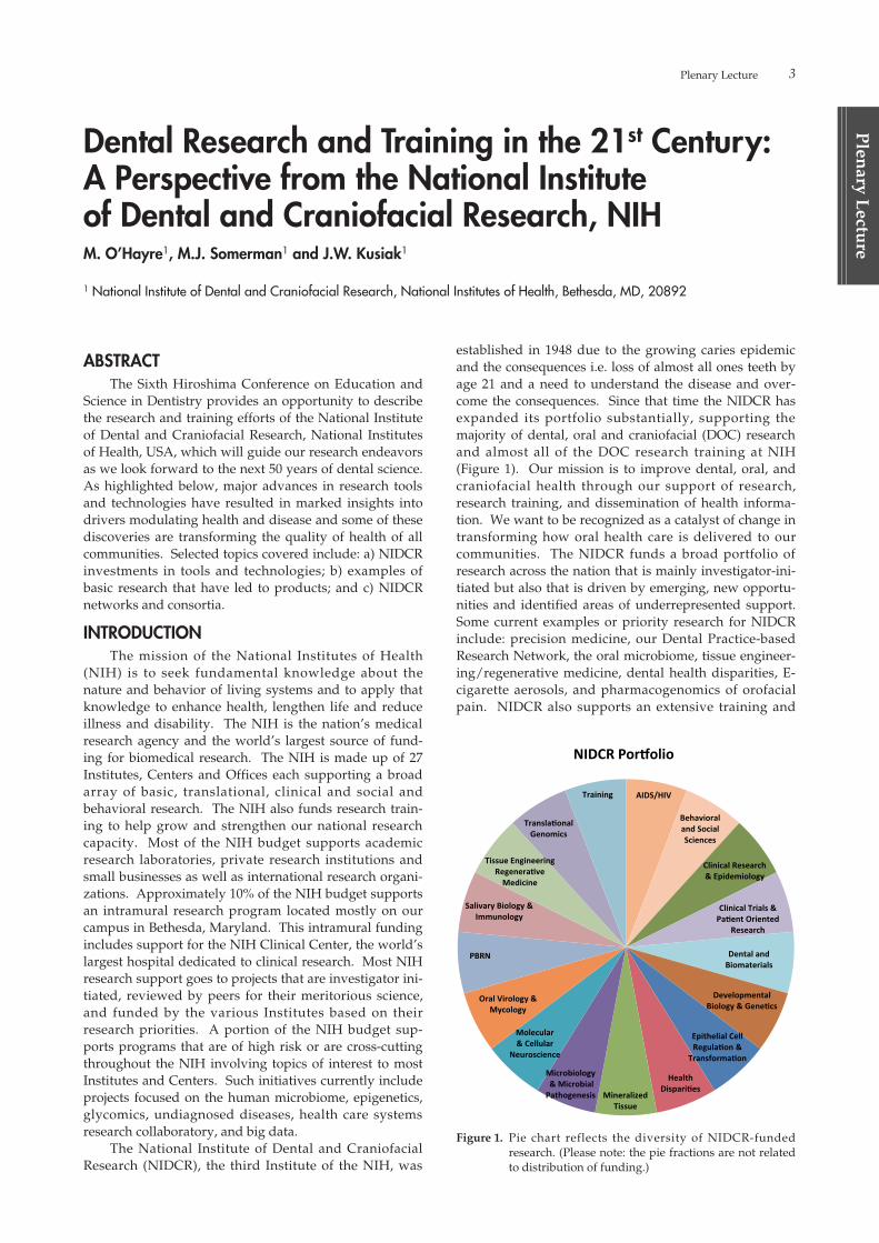
The National Institute of Dental and CraniofacialResearch (NIDCR), the third Institute of the NIH, was
established in 1948 due to the growing caries epidemicand the consequences i.e. loss of almost all ones teeth byage 21 and a need to understand the disease and over-come the consequences. Since that time the NIDCR hasexpanded its portfolio substantially, supporting themajority of dental, oral and craniofacial (DOC) researchand almost all of the DOC research training at NIH(Figure 1). Our mission is to improve dental, oral, andcraniofacial health through our support of research,research training, and dissemination of health informa-tion. We want to be recognized as a catalyst of change intransforming how oral health care is delivered to ourcommunities. The NIDCR funds a broad portfolio ofresearch across the nation that is mainly investigator-ini-tiated but also that is driven by emerging, new opportu-nities and identified areas of underrepresented support.Some current examples or priority research for NIDCRinclude: precision medicine, our Dental Practice-basedResearch Network, the oral microbiome, tissue engineer-ing/regenerative medicine, dental health disparities, E-cigarette aerosols, and pharmacogenomics of orofacialpain. NIDCR also supports an extensive training and
Dental Research and Training in the 21st Century: A Perspective from the National Institute of Dental and Craniofacial Research, NIHM. O’Hayre1, M.J. Somerman1 and J.W. Kusiak1
1 National Institute of Dental and Craniofacial Research, National Institutes of Health, Bethesda, MD, 20892
Figure 1. Pie chart reflects the diversity of NIDCR-fundedresearch. (Please note: the pie fractions are not relatedto distribution of funding.)
02_本文1027(web用追加) 15.10.27 9:04 AM ページ 3
4
Ple
nar
y L
ectu
re
career development program ranging from curiousyoung high school students to established senior investi-gators seeking advanced training in new methods of con-temporary biomedical science. In addition, NIDCR pro-vides formal training in a dental public health residencyprogram focused on oral and craniofacial health-relatedepidemiologic research. Through these mechanisms,NIDCR strives to establish a trained and diverse work-force of scientists and clinicians at the forefront ofresearch to advance oral health care and reduce healthdisparities.
NIDCR Research: Advancing Tools and TechnologiesNIDCR encourages the development and application
of new tools and technologies to advance scientificknowledge and improve oral health. Rapid advance-ments in technologies have enabled the collection of largedata sets including genome-wide sequencing, pro-teomics, epigenetics, microbiome analyses, and patientcohort data. However, to be most useful, this informa-tion requires considerable resources to store, analyze,and disseminate in a manner that allows for effectivecross-disciplinary use. Towards this goal, NIH has insti-tuted a data sharing policy requiring that all genomicdata are shared, established an NIH Office of DataScience, and launched a trans-NIH Big Data toKnowledge (BD2K) initiative. The BD2K initiative aimsto facilitate broad use of digital biomedical data, improvethe tools needed to analyze biomedical big data, supportcenters and systems for big data discovery and collection,and enhance training in biomedical big data science.Thus, the resources provided by BD2K should enhanceaccess to and use of a range of big data collected inNIDCR’s research portfolio, including a number ofgenome-wide association studies (GWAS).
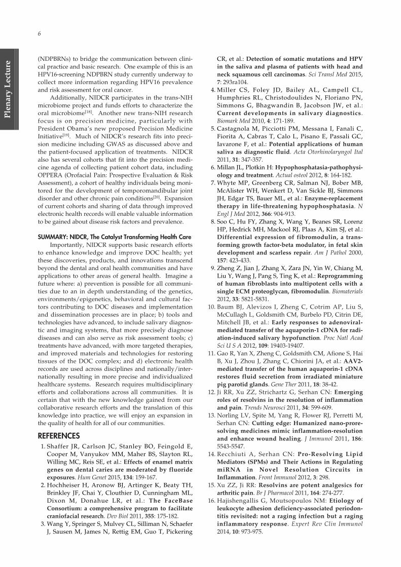
NIDCR supports GWAS and next-generationsequencing efforts to better understand the underlyinggenetic factors contributing to DOC diseases and disor-ders including oral cancer, temporomandibular joint dis-order (TMD), craniofacial development and disorders,caries and periodontal disease, and Sjögren’s syndrome(SS). For example, NIDCR-supported scientists recentlyreported on the effects of variations in enamel matrixgenes and fluoride exposure on dental caries[1]. Theirdata indicated that the effects of two identified geneticvariants are modulated by fluoride exposure, as partici-pants with the risk genotype only exhibited greater inci-dence of dental caries if they were exposed to less than0.7 ppm fluoride in drinking water[1]. Additionally,genetics can greatly influence an individual’s responsesto drugs and other therapeutic agents. As such, pharma-cogenomics will enable a better understanding of thegenetic variations that may influence drug metabolism,therapeutic response, and side effects. To encouragemore research in this area, NIDCR has proposed a newinitiative for pharmacogenomics of orofacial pain man-agement. Additionally, by combining genetic, molecular,and 3D imaging technologies, the NIDCR-fundedFaceBase consortium and our intramural clinical centerstaff are collecting genomic and gene expression dataalong with human facial imagery to help identify thegenetic changes underlying facial development[2]. Theseresources provide a unique opportunity for craniofacialresearchers to better understand craniofacial develop-ment and disorders. Overall, NIDCR has a broad portfo-lio of research to investigate how genetic and epigeneticvariations influence health and disease and to evaluatethe interplay between environmental factors and genetics(see Table 1 for a list of various Data Sharing resourcesand corresponding websites).
Table 1. Data Sharing Resources
Database Sharing Resource Web address
NHANES NIDCR and NIH partner with other Federal agencies http://www.cdc.gov/nchs/nhanes.htm
The Cancer Genome Atlas (TCGA) and International Cancer Genomics Consortium http://cancergenome.nih.gov/https://icgc.org/
Global Alliance for Chronic Diseases http://www.gacd.org/
Human Microbiome Project and the Human Oral Microbiome Database http://hmpdacc.org/http://www.homd.org/
The Sjögren’s Syndrome International Collaborative Clinical Alliance (SICCA) https://sicca-online.ucsf.edu/
MD Anderson Salivary Gland biorepository https://research.mdacc.tmc.edu/Salivary_DB/
Oral Cancer genome database http://www.tumor-gene.org/Oral/oral.html
FaceBase https://www.facebase.org/
Salivary proteome wiki http://salivaryproteome.nidcr.nih.gov/
NIH’s database of Genotypes and Phenotypes (dbGaP) http://www.ncbi.nlm.nih.gov/gap
Big Data 2 Knowledge (BD2K) https://datascience.nih.gov/bd2k
The National Dental Practice-Based Research Network (NDPBRN) http://www.nationaldentalpbrn.org/
GTEx consortium http://www.gtexportal.org/home/
Orofacial Pain: Prospective Evaluation and Risk Assessment (OPPERA) https://www.oppera2.org/OPPERAII/FAQs
02_本文1027(web用追加) 15.10.27 9:04 AM ページ 4
Plenary Lecture 5
Plen
ary Lectu
re
In addition to better understanding genetic factorsunderlying disease, NIDCR invests in research to identifybiomarkers and to develop salivary diagnostics for earlydisease detection and prevention. One recent example ofthis includes an NIDCR-supported study to detect tumorDNA in saliva, using somatic mutations and HPV genesas tumor DNA biomarkers[3]. Tumor DNA detection insaliva was 100% effective for oral cancer detection and47-70% effective for detecting other cancers tested.Moreover, tumor DNA could be detected in patients withcancer recurrence, but not in patients without diseaserecurrence[3]; therefore, tumor DNA detection in salivamay present an effective and non-invasive screeningstrategy for cancer recurrence following chemotherapyand/or surgery. In addition to salivary diagnostic appli-cations in the DOC region, studies are also underway todetermine if saliva may be effective for the detection ofautism spectrum disorder and for identifying biomarkerspredictive for maladaptive stress reactions such as post-traumatic stress disorder. Therefore, salivary diagnosticshave the potential for broad cross-disciplinary applica-tions[4,5]. The oral cavity also presents an ideal location touse biosensors for monitoring health and disease. Whilesmall wireless biosensors are still in early stages of devel-opment, they have the potential to provide valuabledynamic, real-time diagnostic and physiologic informa-tion; thus, oral biosensors are an area of interest for cur-rent and future NIDCR research.
NIDCR Research: Basic Research to Product DevelopmentBasic science research funded by NIDCR has not
only enhanced knowledge in the field, but also led toapplied improvements in DOC health with the develop-ment of novel and improved treatments for a variety ofdiseases and conditions. One such innovation developedby NIDCR researchers is alkaline phosphatase replace-ment therapy for skeletal and dental defects related tohypophosphatasia (HPP). HPP is a disease of brittlebones and tooth loss that is caused by deficiency in theenzyme, alkaline phosphatase[6]. Basic research on thisenzyme was initiated in the 1990s and continues to befunded by NIDCR, and the translation to product hasbeen through public-private partnerships. Clinical trialsfor enzyme therapy to treat skeletal dysplasia were com-pleted in 2010, with very positive outcomes[7], and thistreatment was granted Breakthrough Therapy designa-tion by the Food and Drug Administration (FDA) in 2013to expedite the approval and development process.
Another example of basic research leading to innov-ative treatment involves the application of the fibromod-ulin peptide to reduce scarring and correct birth defectsincluding cleft lip. The role of fibromodulin in promot-ing scarless wound repair was discovered while investi-gating the transition from fetal wound repair, which isscarless, to wound healing in adults, which results inscars and fibrosis, in rodent models. Researchers discov-ered that fibromdulin was induced in fetal wound repairbut not after the transition to adult-type repair, indicatingthis molecule could be important for scarless woundhealing[8]. Starting with an exploratory basic scienceresearch grant, development of a therapeutic peptidederived from native fibromodulin[9] into a product for
scar treatment has progressed through two phases ofNIDCR-funded small business grants and is anticipatedto enter into a clinical trial in 2016.
Research from NIDCR’s intramural program led tothe proposed use of aquaporin-1 (AQP1) gene therapyfor treatment of dry mouth induced by head and neckcancer radiation treatment[10,11]. Approximately 85% ofhead and neck cancer patients undergoing radiationtreatment experience complications from dry mouthincluding dental caries, enamel erosion, oral infections,weight loss and malnutrition, and difficulty talking,chewing, swallowing. While many will recover somegland activity over time, at least half will not, resulting ina long term chronic condition with no conventional ther-apy. Aquaporins are key proteins important for salivaproduction and salivary gland function[10,11]; thus, AQP1gene therapy is being tested to improve saliva productionto mitigate the effects of dry mouth and has completed aPhase I clinical trial. In this trial about half the patientsexperienced a reduction in symptoms related to the radi-ation induced xerostomia. These responses were depen-dent on the dose of the vector used to deliver the AQP1gene, with participants who did not respond having vec-tor related inflammation. Currently a second trial isbeing planned using a new vector which was shown tobe less immunogenic and can result in long term activityin preclinical studies.
Another product currently in clinical trials that wasinitiated from NIDCR research funding is ClinRinse, amouthwash for gingivitis. The effective molecule inClinRinse is a lipoxin-like drug, which derives from aclass of lipids that were shown to be potent modulatorsof inflammation and associated pain and tissue destruc-tion[12-15]. Beyond applications to gingivitis, lipoxin couldhave therapeutic benefits for a variety of other inflamma-tory diseases to reduce tissue damage and bone loss andresolve inflammation. Lipoxin also provides a valuablealternative to the use of antibiotics. Limiting the use ofantibiotics and identifying therapeutic alternatives willreduce the burden of antibiotic resistance the medicalfield is currently facing. An additional example of thisinvolves NIDCR intramural researchers, collaboratingwith extramural researchers, to discover anti-IL-17/IL-21treatment as a potential effective therapy for leukocyteadhesion deficiency 1 (LAD-1)[16,17]. LAD-1 is a raregenetic immune system disorder that causes severe peri-odontitis and other systemic inflammatory reactions.LAD-1 patients frequently receive broad spectrum antibi-otics for life and are treated with steroids and antibioticsduring inflammatory reactions, with limited success;more directed treatments could improve the healthcareand quality of life for these patients.
NIDCR: Developing Networks and ConsortiaResearch progress will come at the interface of dif-
ferent disciplines and through networks and collabora-tions to collect, analyze, and disseminate valuable infor-mation. As such, communication between researchersand dental practitioners, as well as other health profes-sionals, is essential to ensure directed efforts for improv-ing DOC health. Towards this goal, NIDCR supports theNational Dental Practice Based Research Networks
02_本文1027(web用追加) 15.10.27 9:04 AM ページ 5
6
Ple
nar
y L
ectu
re
(NDPBRNs) to bridge the communication between clini-cal practice and basic research. One example of this is anHPV16-screening NDPBRN study currently underway tocollect more information regarding HPV16 prevalenceand risk assessment for oral cancer.
Additionally, NIDCR participates in the trans-NIHmicrobiome project and funds efforts to characterize theoral microbiome[18]. Another new trans-NIH researchfocus is on precision medicine, particularly withPresident Obama’s new proposed Precision MedicineInitiative[19]. Much of NIDCR’s research fits into preci-sion medicine including GWAS as discussed above andthe patient-focused application of treatments. NIDCRalso has several cohorts that fit into the precision medi-cine agenda of collecting patient cohort data, includingOPPERA (Orofacial Pain: Prospective Evaluation & RiskAssessment), a cohort of healthy individuals being moni-tored for the development of temporomandibular jointdisorder and other chronic pain conditions[20]. Expansionof current cohorts and sharing of data through improvedelectronic health records will enable valuable informationto be gained about disease risk factors and prevalence.
SUMMARY: NIDCR, The Catalyst Transforming Health CareImportantly, NIDCR supports basic research efforts
to enhance knowledge and improve DOC health; yetthese discoveries, products, and innovations transcendbeyond the dental and oral health communities and haveapplications to other areas of general health. Imagine afuture where: a) prevention is possible for all communi-ties due to an in depth understanding of the genetics,environments/epigenetics, behavioral and cultural fac-tors contributing to DOC diseases and implementationand dissemination processes are in place; b) tools andtechnologies have advanced, to include salivary diagnos-tic and imaging systems, that more precisely diagnosediseases and can also serve as risk assessment tools; c)treatments have advanced, with more targeted therapies,and improved materials and technologies for restoringtissues of the DOC complex; and d) electronic healthrecords are used across disciplines and nationally/inter-nationally resulting in more precise and individualizedhealthcare systems. Research requires multidisciplinaryefforts and collaborations across all communities. It iscertain that with the new knowledge gained from ourcollaborative research efforts and the translation of thisknowledge into practice, we will enjoy an expansion inthe quality of health for all of our communities.
REFERENCES01. Shaffer JR, Carlson JC, Stanley BO, Feingold E,
Cooper M, Vanyukov MM, Maher BS, Slayton RL,Willing MC, Reis SE, et al.: Effects of enamel matrixgenes on dental caries are moderated by fluorideexposures. Hum Genet 2015, 134: 159-167.
02. Hochheiser H, Aronow BJ, Artinger K, Beaty TH,Brinkley JF, Chai Y, Clouthier D, Cunningham ML,Dixon M, Donahue LR, et al.: The FaceBaseConsortium: a comprehensive program to facilitatecraniofacial research. Dev Biol 2011, 355: 175-182.
03. Wang Y, Springer S, Mulvey CL, Silliman N, SchaeferJ, Sausen M, James N, Rettig EM, Guo T, Pickering
CR, et al.: Detection of somatic mutations and HPVin the saliva and plasma of patients with head andneck squamous cell carcinomas. Sci Transl Med 2015,7: 293ra104.
04. Miller CS, Foley JD, Bailey AL, Campell CL,Humphries RL, Christodoulides N, Floriano PN,Simmons G, Bhagwandin B, Jacobson JW, et al.:Current developments in salivary diagnostics .Biomark Med 2010, 4: 171-189.
05. Castagnola M, Picciotti PM, Messana I, Fanali C,Fiorita A, Cabras T, Calo L, Pisano E, Passali GC,Iavarone F, et al.: Potential applications of humansaliva as diagnostic fluid. Acta Otorhinolaryngol Ital2011, 31: 347-357.
06. Millan JL, Plotkin H: Hypophosphatasia-pathophysi-ology and treatment. Actual osteol 2012, 8: 164-182.
07. Whyte MP, Greenberg CR, Salman NJ, Bober MB,McAlister WH, Wenkert D, Van Sickle BJ, SimmonsJH, Edgar TS, Bauer ML, et al.: Enzyme-replacementtherapy in life-threatening hypophosphatasia. NEngl J Med 2012, 366: 904-913.
08. Soo C, Hu FY, Zhang X, Wang Y, Beanes SR, LorenzHP, Hedrick MH, Mackool RJ, Plaas A, Kim SJ, et al.:Differential expression of fibromodulin, a trans-forming growth factor-beta modulator, in fetal skindevelopment and scarless repair. Am J Pathol 2000,157: 423-433.
09. Zheng Z, Jian J, Zhang X, Zara JN, Yin W, Chiang M,Liu Y, Wang J, Pang S, Ting K, et al.: Reprogrammingof human fibroblasts into multipotent cells with asingle ECM proteoglycan, fibromodulin. Biomaterials2012, 33: 5821-5831.
10. Baum BJ, Alevizos I, Zheng C, Cotrim AP, Liu S,McCullagh L, Goldsmith CM, Burbelo PD, Citrin DE,Mitchell JB, et al.: Early responses to adenoviral-mediated transfer of the aquaporin-1 cDNA for radi-ation-induced salivary hypofunction. Proc Natl AcadSci U S A 2012, 109: 19403-19407.
11. Gao R, Yan X, Zheng C, Goldsmith CM, Afione S, HaiB, Xu J, Zhou J, Zhang C, Chiorini JA, et al.: AAV2-mediated transfer of the human aquaporin-1 cDNArestores fluid secretion from irradiated miniaturepig parotid glands. Gene Ther 2011, 18: 38-42.
12. Ji RR, Xu ZZ, Strichartz G, Serhan CN: Emergingroles of resolvins in the resolution of inflammationand pain. Trends Neurosci 2011, 34: 599-609.
13. Norling LV, Spite M, Yang R, Flower RJ, Perretti M,Serhan CN: Cutting edge: Humanized nano-prore-solving medicines mimic inflammation-resolutionand enhance wound healing. J Immunol 2011, 186:5543-5547.
14. Recchiuti A, Serhan CN: Pro-Resolving LipidMediators (SPMs) and Their Actions in RegulatingmiRNA in Novel Resolution Circuits inInflammation. Front Immunol 2012, 3: 298.
15. Xu ZZ, Ji RR: Resolvins are potent analgesics forarthritic pain. Br J Pharmacol 2011, 164: 274-277.
16. Hajishengallis G, Moutsopoulos NM: Etiology ofleukocyte adhesion deficiency-associated periodon-titis revisited: not a raging infection but a raginginflammatory response. Expert Rev Clin Immunol2014, 10: 973-975.
02_本文1027(web用追加) 15.10.27 9:04 AM ページ 6

Plenary Lecture 7
Plen
ary Lectu
re
17. Moutsopoulos NM, Konkel J, Sarmadi M, Eskan MA,Wild T, Dutzan N, Abusleme L, Zenobia C, Hosur KB,Abe T, et al.: Defective neutrophil recruitment inleukocyte adhesion deficiency type I disease causeslocal IL-17-driven inflammatory bone loss. Sci TranslMed 2014, 6: 229ra240.
18. Chen T, Yu WH, Izard J, Baranova OV, LakshmananA, Dewhirst FE: The Human Oral MicrobiomeDatabase: a web accessible resource for investigat-
ing oral microbe taxonomic and genomic informa-tion. Database (Oxford) 2010, 2010: baq013.
19. Collins FS, Varmus H: A new initiative on precisionmedicine. N Engl J Med 2015, 372: 793-795.
20. Fillingim RB, Slade GD, Diatchenko L, Dubner R,Greenspan JD, Knott C, Ohrbach R, Maixner W:Summary of findings from the OPPERA baselinecase-control study: implications and future direc-tions. J Pain 2011, 12: T102-107.
02_本文1027(web用追加) 15.10.27 9:04 AM ページ 7

02_本文1027(web用追加) 15.10.27 9:04 AM ページ 8
Session
II
Oral and Systemic Disease ConnectionThe Oral and Systemic Disease Connection
Forsyth Institute
T.E. Van Dyke
Dental Infection of Porphyromonas gingivalis Exacerbates Pathological Progression
of Non-Alcoholic Steatohepatitis (NASH)
Hiroshima University
M. Miyauchi, H. Furusho, A. Nagasaki, S. Sakamoto, K. Ouhara,
H. Kurihara and T. Takata
The Effect of Periodontopathogenic Bacteria, Porphyromonas gingivalis, against the Onset
of Rheumatoid Arthritis
Hiroshima University
K. Ouhara, M. Yamakawa, S. Munenaga, T. Fujita and H. Kurihara
Periodontal Disease as a Possible Risk Factor for Alzheimer’s Disease
National Center for Geriatrics and Gerontology
K. Matsushita
New Paradigm for the Link between Periodontitis and Systemic Diseases
Niigata University
K. Yamazaki
Session II
02_本文1027(web用追加) 15.10.27 9:04 AM ページ 9

02_本文1027(web用追加) 15.10.27 9:04 AM ページ 10

Session II 11
Session
IIABSTRACTThis introductory paper examines the relationship
between periodontitis and systemic diseases that are con-comitantly expressed in people with periodontitis. Anoverview of current understanding of the associationsbetween periodontitis and cardiovascular disease, dia-betes mellitus, pre-term low birth weight, pulmonaryinfections, rheumatoid arthritis and kidney disease willbe presented. The potential mechanisms and supportingevidence will then be examined. Three mechanisms havebeen proposed, including spread of local infections toremote sites, inflammatory mechanisms and the complexinteractions of both. The same mechanisms do notappear to be involved in all systemic diseases and theinteractions are often disease specific. Data from animalmodels and human studies will be presented focusing onnew evidence for the role of inflammation in the patho-genesis of periodontitis, diabetes and cardiovascular dis-ease. The data support an interaction between periodon-titis and the occurrence and pathogenesis of several sys-temic diseases. The strength of the evidence variesbetween systemic diseases, but as more data accumu-lates, the importance of periodontal health in overallhealth is becoming more apparent.
INTRODUCTIONTwo or more diseases can occur simultaneously or
sequentially in an individual where the course or severityof one disease can have an adverse impact on the other(s). A large body of evidence now exists showing anassociation of periodontitis with diabetes mellitus, car-diovascular disease, low birth-weight and prematureinfants, rheumatoid arthritis, pulmonary infections, andto a lesser degree chronic kidney disease (for currentreview, see J. Periodontology 84: 4 supplement, 2013).Three mechanisms have been suggested to play a role innon-oral manifestations of periodontitis (Thoden vanVelzen, Abraham-Inpijn et al. 1984). These include dis-semination of infection, bacterial toxins and immuno-inflammatory injury. In the case of periodontitis, clearlythe dissemination of the etiologic bacteria or their toxinsfrom the biofilm is possible. Likewise, since the patho-genesis of periodontitis is now known to be inflammato-ry (Van Dyke and Serhan 2003, Serhan, Chiang et al.2008), immuno-inflammatory tissue damage is possiblealso.
However, the systemic disease associated with peri-odontitis in any given individual is also a variable and toassume that the mechanism in each case is the same isprobably naive. In this introductory paper, each diseaselisted above will be considered separately. The strengthof the evidence for the association with periodontitis willbe considered, as will the likely mechanism.
Pulmonary DiseaseChronic Obstructive Pulmonary Disease (COPD) : COPD ischaracterized by progressive obstruction of airflow andinflammation of the airways. The link between peri-odontitis and COPD was first identified in epidemiologicanalyses of NHANES and Veterans Administration data(Hayes, Sparrow et al. 1998, Scannapieco, Papandonatoset al. 1998, Scannapieco and Ho 2001). Smoking is thesingle greatest risk factor for COPD, but the associationin NHANES data remains even after adjusting for smok-ing (Garcia, Nunn et al. 2001). No data are availablerelating to the mechanism of action although increasedairway inflammation stemming from periodontal inflam-mation has been suggested. Systematic reviews byScannapieco and Azarpazhooh (Scannapieco, Bush et al.2003, Azarpazhooh and Leake 2006) concluded that evi-dence was poor supporting a weak association and morestudies are needed.
Pneumonia : Pneumonia can be acquired in the communi-ty, but it is also a frequent nosocomial infection. In hos-pitals and nursing homes, patients often have poor oralhygiene and the oral cavity can be colonized by organ-isms that cause pneumonia, including periodontal organ-isms (Scannapieco, Stewart et al. 1992). While there is noapparent association between periodontitis and commu-nity acquired pneumonia, there is fairly strong evidencethat nosocomial pneumonias result from oral organismsand that oral hygiene preventive measures have a signifi-cant impact reducing risk up to 11.7% (Sjogren, Nilssonet al. 2008).
The mechanism of action in the case of pneumoniahas been clearly demonstrated. Aspiration of bacteriafrom the oral cavity to the lower respiratory tract is adirect infectious process. Importantly, it has also beendocumented that proper preventive care can have amajor impact.
The Oral and Systemic Disease ConnectionT.E. Van Dyke
Vice President for Clinical and Translational Research, Chair, Department of Applied Oral Sciences, Senior Member of the Staff, Forsyth Institute, Cambridge, MA 02142, USA
Key words: Periodontitis, Cardiovascular Disease, Diabetes, Rheumatoid Arthritis, Pregnancy, Pulmonary Infection, Kidney Disease
02_本文1027(web用追加) 15.10.27 9:04 AM ページ 11
12
Ses
sion
II
Chronic Kidney DiseaseKidney damage with decrease glomerular filtration
rate of more than 3 months is a significant public healthproblem worldwide associated with aging, diabetes,hypertension, obesity and cardiovascular disease (Leveyand Coresh 2012). The Atherosclerosis Risk inCommunities (ARIC study found a significant associa-tion between periodontitis and chronic kidney disease(Kshirsagar, Moss et al. 2005, Kshirsagar, Offenbacher etal. 2007), when adjusting for all of the above con-founders. Importantly, a prospective study of Type 2diabetes in the Pima Indian population found that peri-odontal disease predicted overt end stage renal disease ina dose dependent manner (Shultis, Weil et al. 2007).Nevertheless, the complexity of chronic kidney diseasepathogenesis and its association with diabetes and car-diovascular disease makes implication for a role of chron-ic periodontitis challenging. There are no data implicat-ing dissemination of infection vs. disseminating inflam-mation that may stem from periodontitis in chronic kid-ney disease.
Rheumatoid ArthritisRheumatoid arthritis (RA) is a classic inflammatory
disease of unknown etiology with persistent synovialinflammation that damages articular cartilage and under-lying bone (Scott, Wolfe et al. 2010). There have beenreports from small studies of associations between peri-odontitis and RA and the similarities in the pathogenesisof both disease makes the potential association veryattractive (de Pablo, Dietrich et al. 2008), especially theassociation with citrulinated peptides (Klareskog, Catrinaet al. 2009). The common mechanism would appear to bepurely inflammatory, but citrulination of peptides hasbeen suggested to be a property of the periodontalpathogen, Porphyromas gingivalis (Marchant, Smith et al.2013). More detailed discussion of the relationshipbetween periodontitis and RA follows in later presenta-tions.
Adverse Pregnancy OutcomesPregnancy is a normal, healthy physiologic process
that sometimes has adverse outcomes, including lowbirth-weight, pre-term birth (prematurity), growthrestriction, preeclampsia, pneumonia and miscarriage(still-birth). Adverse pregnancy outcomes are associatedwith local and systemic inflammation and intra-uterineinfections. Numerous epidemiologic and animal studiessupport the relationship between periodontitis andadverse pregnancy outcomes; however, few prospectiveintervention trials have supported the association (Sanz,Kornman et al. 2013). The mechanism of action probablyinvolves both direct infection and inflammation. Ourunderstanding of the pathogenesis suggests that inflam-mation cause the adverse outcomes. However, sinceintra-uterine infections are involved, there is reasonablesupport for direct infection causing the inflammation.Most adverse outcomes originate from ascending vaginalinfection or blood born infections from known orunknown non-genital sites. Maternal periodontitis is asource of microorganisms in the blood and oral organ-isms such as Fusobacterium nucleatum have been found in
amniotic fluid, placenta and chorioamniotic membranesof women delivering prematurely (Han, Redline et al.2004).
DiabetesBoth type 2 diabetes and periodontitis are complex
chronic inflammatory diseases. Type 1 diabetes, anautoimmune disorder resulting in loss of beta cells andinsulin, is also associated with periodontitis. Here, wewill focus on type 2. It is clear from epidemiologic stud-ies that severe periodontitis directly impacts glycemiccontrol in diabetes as well as other diabetic complications(Chapple, Genco et al. 2013). Likewise, poor glycemiccontrol impacts periodontal outcomes (Chapple, Genco etal. 2013).
The Role of Inflammation : In recent years, it has becomeclear that control of inflammation prevents and reversesperiodontitis. Periodontal infections are commensalinfections characterized by dysbiosis of the biofilm(Hajishengallis, Liang et al. 2011, Hajishengallis, Darveauet al. 2012). Importantly, inflammation drives the dysbio-sis and pharmacological control of inflammation reversesdysbiosis (Hasturk, Kantarci et al. 2007). Lipid mediatorsof inflammation (eicosanoids) have long been known toprovide the acute inflammatory stimulus; more recently,pathways of resolution of inflammation have also beenuncovered (Table 1). Importantly, natural resolution ofinflammation and pharmacological antiinflammation(inhibition, antagonism) are not the same thing. Naturalmediators of resolution of inflammation have providednew pharmacologic tools to determine the role of inflam-
Table 1. Lipid mediators of inflammation.
Lipid Mediators of Inflammation
Proinflammatory• Arachidonic acid derived:
• Prostaglandins• Leukotrienes
Proresolution• Arachidonic acid derived:
• Lipoxins• Aspirin triggered Lipoxins*
• ω-3 fatty acid derived:• E-series resolvins (EPA)• D-series resolvins (DHA)
* The role of Aspirin: Aspirin enhances lipoxin and resolvinactivity by producing longer acting isomers of native com-pounds
The pro- and anti-inflammatory eicosanoids derived fromarachidonic acid and omega-3 fatty acids are identified asProinflammatory and Proresolution. Proinflammatoryeicosanoids derived from arachidonic acid are the products ofcyclooxygenases (1 and 2) and lipoxygenases (5, 12 and 15).Proresolution eicosanoids are products of lipoxygenase: lipoxy-genase interactions; for review see (Serhan, Chiang et al. 2008).Depending on the substrate, lipoxins (arachidonic acid) orresolvins (omega-3 fatty acids) are produced. Aspirin has theunique property of inhibiting COX-2 by changing its activity to a15R-lipoxygenase. The resulting proresolution products arelonger acting isomers of the lipoxins and resolvins with longerhalf-life and greater potency.
02_本文1027(web用追加) 15.10.27 9:04 AM ページ 12
Session II 13
Session
II
mation in the pathogenesis of disease and in the contextof this paper, the relationship of inflammation to associa-tions between periodontitis and systemic diseases.
Obesity and Metabolic Syndrome : Obesity (body massindex >30 kg/m2) and metabolic syndrome (a clusteringof interrelated atherosclerotic risk factors, includingabdominal obesity, dyslipidemia, hyperglycemia andhypertension) are known proinflammatory modifiers.Obesity and metabolic syndrome are both associatedwith periodontitis (Grundy 2005, Chaffee and Weston2010). These conditions can eventually lead to type 2 dia-betes by increasing inflammation and insulin resistance.
Inflammation Links Periodontitis and Type 2 Diabetes :Type 2 diabetes is preceded by systemic inflammationthat leads to insulin resistance, reduced β cell function inthe pancreas with eventual apoptosis of β cells and lackof insulin production (Chapple, Genco et al. 2013,Chapple, Borgnakke et al. 2014). Acute phase and oxida-tive stress biomarkers further support the role of inflam-mation. Inflammation can be induced by oral bacteriathat get access to the circulation (Genco, Grossi et al.2005). Sustained elevations of blood glucose levels leadsto non-enzymatic glycation of proteins called advancedglycation endproducts (AGE) (Schmidt, Weidman et al.1996) that bind to their receptor (RAGE) on inflammatorycells to increase inflammation (Lalla, Lamster et al. 2000,Lalla, Lamster et al. 2000, Lalla, Lamster et al. 2001).Thus, there are both direct and indirect proinflammatorystimuli.
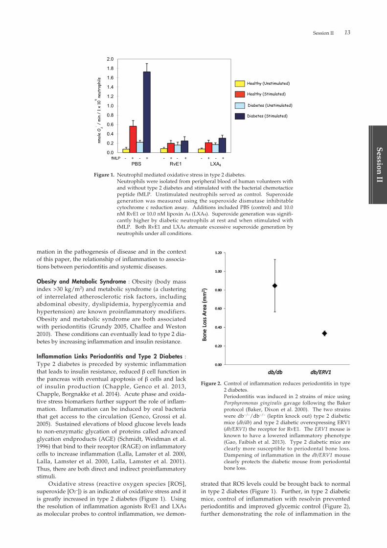
Oxidative stress (reactive oxygen species [ROS],superoxide [O2–]) is an indicator of oxidative stress and itis greatly increased in type 2 diabetes (Figure 1). Usingthe resolution of inflammation agonists RvE1 and LXA4
as molecular probes to control inflammation, we demon-
strated that ROS levels could be brought back to normalin type 2 diabetes (Figure 1). Further, in type 2 diabeticmice, control of inflammation with resolvin preventedperiodontitis and improved glycemic control (Figure 2),further demonstrating the role of inflammation in the
Figure 1. Neutrophil mediated oxidative stress in type 2 diabetes.Neutrophils were isolated from peripheral blood of human volunteers withand without type 2 diabetes and stimulated with the bacterial chemotacticepeptide fMLP. Unstimulated neutrophils served as control. Superoxidegeneration was measured using the superoxide dismutase inhibitablecytochrome c reduction assay. Additions included PBS (control) and 10.0nM RvE1 or 10.0 nM lipoxin A4 (LXA4). Superoxide generation was signifi-cantly higher by diabetic neutrophils at rest and when stimulated withfMLP. Both RvE1 and LXA4 atenuate excessive superoxide generation byneutrophils under all conditions.
Figure 2. Control of inflammation reduces periodontitis in type2 diabetes.Periodontitis was induced in 2 strains of mice usingPorphyromonas gingivalis gavage following the Bakerprotocol (Baker, Dixon et al. 2000). The two strainswere db–/–/db–/– (leptin knock out) type 2 diabeticmice (db/db) and type 2 diabetic overexpressing ERV1(db/ERV1) the receptor for RvE1. The ERV1 mouse isknown to have a lowered inflammatory phenotype(Gao, Faibish et al. 2013). Type 2 diabetic mice areclearly more susceptible to periodontal bone loss.Dampening of inflammation in the db/ERV1 mouseclearly protects the diabetic mouse from periodontalbone loss.
02_本文1027(web用追加) 15.10.27 9:04 AM ページ 13
14
Ses
sion
II
Periodontitis/type 2 diabetes connection.
Cardiovascular DiseasePeriodontitis is a risk factor for systemic inflammato-
ry diseases, including atherosclerosis, myocardial infarc-tion, and stroke (Dietrich, Sharma et al. 2013). Recentsystematic reviews suggest a significant risk association.Clinical studies also demonstrate that people with peri-odontitis have elevated C-reactive protein (CRP), inter-leukin 6, haptoglobin, and fibrinogen. People who havehad a myocardial infarction with periodontitis have sig-nificantly higher CRP than those with myocardial infarc-tion alone, suggesting that periodontal disease is an inde-pendent contributor to systemic inflammation(Kodovazenitis, Pitsavos et al. 2011, Schenkein and Loos2013).
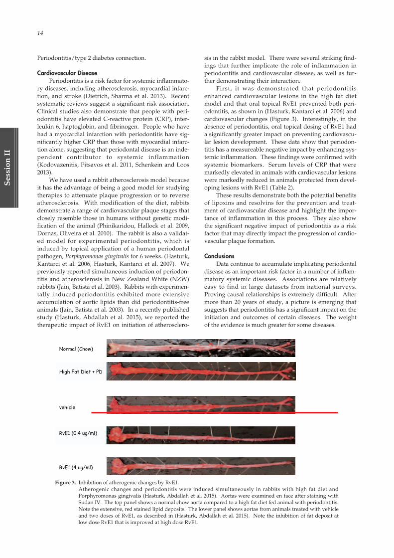
We have used a rabbit atherosclerosis model becauseit has the advantage of being a good model for studyingtherapies to attenuate plaque progression or to reverseatherosclerosis. With modification of the diet, rabbitsdemonstrate a range of cardiovascular plaque stages thatclosely resemble those in humans without genetic modi-fication of the animal (Phinikaridou, Hallock et al. 2009,Dornas, Oliveira et al. 2010). The rabbit is also a validat-ed model for experimental periodontitis, which isinduced by topical application of a human periodontalpathogen, Porphyromonas gingivalis for 6 weeks. (Hasturk,Kantarci et al. 2006, Hasturk, Kantarci et al. 2007). Wepreviously reported simultaneous induction of periodon-titis and atherosclerosis in New Zealand White (NZW)rabbits (Jain, Batista et al. 2003). Rabbits with experimen-tally induced periodontitis exhibited more extensiveaccumulation of aortic lipids than did periodontitis-freeanimals (Jain, Batista et al. 2003). In a recently publishedstudy (Hasturk, Abdallah et al. 2015), we reported thetherapeutic impact of RvE1 on initiation of atherosclero-
sis in the rabbit model. There were several striking find-ings that further implicate the role of inflammation inperiodontitis and cardiovascular disease, as well as fur-ther demonstrating their interaction.
First, it was demonstrated that periodontitisenhanced cardiovascular lesions in the high fat dietmodel and that oral topical RvE1 prevented both peri-odontitis, as shown in (Hasturk, Kantarci et al. 2006) andcardiovascular changes (Figure 3). Interestingly, in theabsence of periodontitis, oral topical dosing of RvE1 hada significantly greater impact on preventing cardiovascu-lar lesion development. These data show that periodon-titis has a measureable negative impact by enhancing sys-temic inflammation. These findings were confirmed withsystemic biomarkers. Serum levels of CRP that weremarkedly elevated in animals with cardiovascular lesionswere markedly reduced in animals protected from devel-oping lesions with RvE1 (Table 2).
These results demonstrate both the potential benefitsof lipoxins and resolvins for the prevention and treat-ment of cardiovascular disease and highlight the impor-tance of inflammation in this process. They also showthe significant negative impact of periodontitis as a riskfactor that may directly impact the progression of cardio-vascular plaque formation.
ConclusionsData continue to accumulate implicating periodontal
disease as an important risk factor in a number of inflam-matory systemic diseases. Associations are relativelyeasy to find in large datasets from national surveys.Proving causal relationships is extremely difficult. Aftermore than 20 years of study, a picture is emerging thatsuggests that periodontitis has a significant impact on theinitiation and outcomes of certain diseases. The weightof the evidence is much greater for some diseases.
Figure 3. Inhibition of atherogenic changes by RvE1.Atherogenic changes and periodontitis were induced simultaneously in rabbits with high fat diet andPorphyromonas gingivalis (Hasturk, Abdallah et al. 2015). Aortas were examined en face after staining withSudan IV. The top panel shows a normal chow aorta compared to a high fat diet fed animal with periodontitis.Note the extensive, red stained lipid deposits. The lower panel shows aortas from animals treated with vehicleand two doses of RvE1, as described in (Hasturk, Abdallah et al. 2015). Note the inhibition of fat deposit atlow dose RvE1 that is improved at high dose RvE1.
02_本文1027(web用追加) 15.10.27 9:04 AM ページ 14
Session II 15
Session
II
The mechanism of action for the interaction varieswith disease, but in most cases, it is related to inflamma-tion. In some instances (pulmonary disease), the interac-tion is directly bacterial seeding the lungs with oral bac-teria. However, in most diseases, such as diabetes andcardiovascular disease, the role of bacteria is less clear. Itis thought that bacteremia plays a role in initiation of sys-temic inflammation, but it is the inflammation that seemsto drive the interaction.
The other aspect of the periodontal disease-systemicdisease interaction for which we have little data (with theexception of diabetes) is directionality. Most studies aredesigned to show, or assume, that periodontitis impactsthe systemic disease. Is the converse also true? It is indiabetes (Chapple, Genco et al. 2013), but we know littleabout increased susceptibility to periodontitis in peoplewith cardiovascular diseases or rheumatoid arthritis.
Clearly, there is much work to be done. However,the promise of new methods to control inflammation tocontrol both periodontitis and inflammatory systemicdisease will likely lead to new therapeutic approaches.
REFERENCESAzarpazhooh, A. and J.L. Leake (2006). “Systematic
review of the association between respiratory dis-eases and oral health.” J Periodontol 77 (9): 1465-1482.
Baker, P.J., M. Dixon and D.C. Roopenian (2000).“Genetic control of susceptibility to Porphyromonasgingivalis-induced alveolar bone loss in mice.” InfectImmun 68 (10): 5864-5868.
Chaffee, B.W. and S.J. Weston (2010). “Associationbetween chronic periodontal disease and obesity: asystematic review and meta-analysis.” J Periodontol81 (12): 1708-1724.
Chapple, I.L., W.S. Borgnakke and R.J. Genco (2014).“Hemoglobin A1c levels among patients with dia-betes receiving nonsurgical periodontal treatment.”JAMA 311 (18): 1919-1920.
Chapple, I.L., R. Genco and E.F.P.A.A.P.w. workinggroup 2 of the joint (2013). “Diabetes and periodon-tal diseases: consensus report of the Joint EFP/AAPWorkshop on Periodontitis and Systemic Diseases.” J
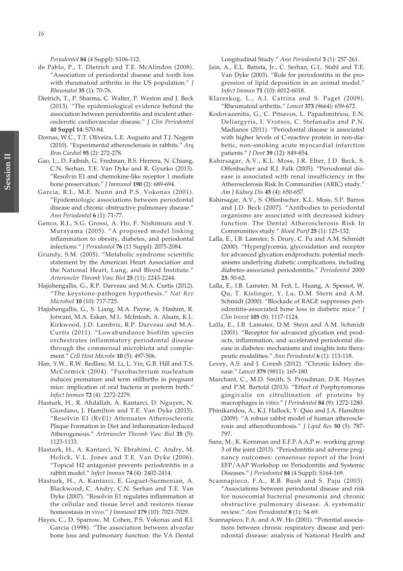
Figure 4. Atherogenic changes compared to high fat diet alone.Changes in the area of aorta covered by lipid deposits were quantified as percent changes from highfat diet alone (horizontal line). PD increased the area by over 10%, which was greater after vehicletreatment. Low dose treatment of PD with RvE1 brought the area back to diet alone values and highdose RvE1 showed a 20% improvement (PD + 12% to -8%). In the absence of PD, oral topical RvE1reduced lipid covered area 40-50% suggesting that PD induced inflammation has a direct impact onseverity of atherogenic changes in the model.
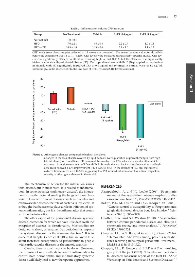
Table 2. Inflammation induces CRP in serum.
Group No Treatment Vehicle RvE1 (0.4 µg/ml) RvE1 (4.0 µg/ml)
Normal diet 1.0 ± 0.1HFD 7.0 ± 2.1 8.6 ± 0.8 2.2 ± 0.7 1.0 ± 0.5HFD + PD 14.9 ± 1.8 11.9 ± 0.6 3.1 ± 1.0 1.1 ± 0.7
CRP levels from blood samples collected at 13 weeks are presented. The mean baseline value for all rabbitsbefore the experiment was 1.5 ± 1.1. Rabbit CRP levels were measured using a rabbit-specific ELISA. CRP lev-els were significantly elevated in all rabbit receiving high fat diet (HFD), but the elevation was significantlyhigher in animals with periodontal disease (PD). Oral topical treatment with RvE1 (10 µl applied to the gingiva)in animals with PD significantly improved CRP at 0.4 µg/ml and returned to normal levels at 4.0 µg/ml.Interestingly, in the absence of PD, the low dose of RvE1 returned CRP levels to normal.
02_本文1027(web用追加) 15.10.27 9:04 AM ページ 15
16
Ses
sion
II
Periodontol 84 (4 Suppl): S106-112.de Pablo, P., T. Dietrich and T.E. McAlindon (2008).
“Association of periodontal disease and tooth losswith rheumatoid arthritis in the US population.” JRheumatol 35 (1): 70-76.
Dietrich, T., P. Sharma, C. Walter, P. Weston and J. Beck(2013). “The epidemiological evidence behind theassociation between periodontitis and incident ather-osclerotic cardiovascular disease.” J Clin Periodontol40 Suppl 14: S70-84.
Dornas, W.C., T.T. Oliveira, L.E. Augusto and T.J. Nagem(2010). “Experimental atherosclerosis in rabbits.” ArqBras Cardiol 95 (2): 272-278.
Gao, L., D. Faibish, G. Fredman, B.S. Herrera, N. Chiang,C.N. Serhan, T.E. Van Dyke and R. Gyurko (2013).“Resolvin E1 and chemokine-like receptor 1 mediatebone preservation.” J Immunol 190 (2): 689-694.
Garcia, R.I., M.E. Nunn and P.S. Vokonas (2001).“Epidemiologic associations between periodontaldisease and chronic obstructive pulmonary disease.”Ann Periodontol 6 (1): 71-77.
Genco, R.J., S.G. Grossi, A. Ho, F. Nishimura and Y.Murayama (2005). “A proposed model linkinginflammation to obesity, diabetes, and periodontalinfections.” J Periodontol 76 (11 Suppl): 2075-2084.
Grundy, S.M. (2005). “Metabolic syndrome scientificstatement by the American Heart Association andthe National Heart, Lung, and Blood Institute.”Arterioscler Thromb Vasc Biol 25 (11): 2243-2244.
Hajishengallis, G., R.P. Darveau and M.A. Curtis (2012).“The keystone-pathogen hypothesis.” Nat RevMicrobiol 10 (10): 717-725.
Hajishengallis, G., S. Liang, M.A. Payne, A. Hashim, R.Jotwani, M.A. Eskan, M.L. McIntosh, A. Alsam, K.L.Kirkwood, J.D. Lambris, R.P. Darveau and M.A.Curtis (2011). “Lowabundance biofilm speciesorchestrates inflammatory periodontal diseasethrough the commensal microbiota and comple-ment.” Cell Host Microbe 10 (5): 497-506.
Han, Y.W., R.W. Redline, M. Li, L. Yin, G.B. Hill and T.S.McCormick (2004). “Fusobacterium nucleatuminduces premature and term stillbirths in pregnantmice: implication of oral bacteria in preterm birth.”Infect Immun 72 (4): 2272-2279.
Hasturk, H., R. Abdallah, A. Kantarci, D. Nguyen, N.Giordano, J. Hamilton and T.E. Van Dyke (2015).“Resolvin E1 (RvE1) Attenuates AtheroscleroticPlaque Formation in Diet and Inflammation-InducedAtherogenesis.” Arterioscler Thromb Vasc Biol 35 (5):1123-1133.
Hasturk, H., A. Kantarci, N. Ebrahimi, C. Andry, M.Holick, V.L. Jones and T.E. Van Dyke (2006).“Topical H2 antagonist prevents periodontitis in arabbit model.” Infect Immun 74 (4): 2402-2414.
Hasturk, H., A. Kantarci, E. Goguet-Surmenian, A.Blackwood, C. Andry, C.N. Serhan and T.E. VanDyke (2007). “Resolvin E1 regulates inflammation atthe cellular and tissue level and restores tissuehomeostasis in vivo.” J Immunol 179 (10): 7021-7029.
Hayes, C., D. Sparrow, M. Cohen, P.S. Vokonas and R.I.Garcia (1998). “The association between alveolarbone loss and pulmonary function: the VA Dental
Longitudinal Study.” Ann Periodontol 3 (1): 257-261.Jain, A., E.L. Batista, Jr., C. Serhan, G.L. Stahl and T.E.
Van Dyke (2003). “Role for periodontitis in the pro-gression of lipid deposition in an animal model.”Infect Immun 71 (10): 6012-6018.
Klareskog, L., A.I. Catrina and S. Paget (2009).“Rheumatoid arthritis.” Lancet 373 (9664): 659-672.
Kodovazenitis, G., C. Pitsavos, L. Papadimitriou, E.N.Deliargyris, I. Vrotsos, C. Stefanadis and P.N.Madianos (2011). “Periodontal disease is associatedwith higher levels of C-reactive protein in non-dia-betic, non-smoking acute myocardial infarctionpatients.” J Dent 39 (12): 849-854.
Kshirsagar, A.V., K.L. Moss, J.R. Elter, J.D. Beck, S.Offenbacher and R.J. Falk (2005). “Periodontal dis-ease is associated with renal insufficiency in theAtherosclerosis Risk In Communities (ARIC) study.”Am J Kidney Dis 45 (4): 650-657.
Kshirsagar, A.V., S. Offenbacher, K.L. Moss, S.P. Barrosand J.D. Beck (2007). “Antibodies to periodontalorganisms are associated with decreased kidneyfunction. The Dental Atherosclerosis Risk InCommunities study.” Blood Purif 25 (1): 125-132.
Lalla, E., I.B. Lamster, S. Drury, C. Fu and A.M. Schmidt(2000). “Hyperglycemia, glycoxidation and receptorfor advanced glycation endproducts: potential mech-anisms underlying diabetic complications, includingdiabetes-associated periodontitis.” Periodontol 200023: 50-62.
Lalla, E., I.B. Lamster, M. Feit, L. Huang, A. Spessot, W.Qu, T. Kislinger, Y. Lu, D.M. Stern and A.M.Schmidt (2000). “Blockade of RAGE suppresses peri-odontitis-associated bone loss in diabetic mice.” JClin Invest 105 (8): 1117-1124.
Lalla, E., I.B. Lamster, D.M. Stern and A.M. Schmidt(2001). “Receptor for advanced glycation end prod-ucts, inflammation, and accelerated periodontal dis-ease in diabetes: mechanisms and insights into thera-peutic modalities.” Ann Periodontol 6 (1): 113-118.
Levey, A.S. and J. Coresh (2012). “Chronic kidney dis-ease.” Lancet 379 (9811): 165-180.
Marchant, C., M.D. Smith, S. Proudman, D.R. Haynesand P.M. Bartold (2013). “Effect of Porphyromonasgingivalis on citrullination of proteins bymacrophages in vitro.” J Periodontol 84 (9): 1272-1280.
Phinikaridou, A., K.J. Hallock, Y. Qiao and J.A. Hamilton(2009). “A robust rabbit model of human atheroscle-rosis and atherothrombosis.” J Lipid Res 50 (5): 787-797.
Sanz, M., K. Kornman and E.F.P.A.A.P.w. working group3 of the joint (2013). “Periodontitis and adverse preg-nancy outcomes: consensus report of the JointEFP/AAP Workshop on Periodontitis and SystemicDiseases.” J Periodontol 84 (4 Suppl): S164-169.
Scannapieco, F.A., R.B. Bush and S. Paju (2003).“Associations between periodontal disease and riskfor nosocomial bacterial pneumonia and chronicobstructive pulmonary disease. A systematicreview.” Ann Periodontol 8 (1): 54-69.
Scannapieco, F.A. and A.W. Ho (2001). “Potential associa-tions between chronic respiratory disease and peri-odontal disease: analysis of National Health and
02_本文1027(web用追加) 15.10.27 9:04 AM ページ 16
Session II 17
Session
II
Nutrition Examination Survey III.” J Periodontol 72(1): 50-56.
Scannapieco, F.A., G.D. Papandonatos and R.G. Dunford(1998). “Associations between oral conditions andrespiratory disease in a national sample survey pop-ulation.” Ann Periodontol 3 (1): 251-256.
Scannapieco, F.A., E.M. Stewart and J.M. Mylotte (1992).“Colonization of dental plaque by respiratorypathogens in medical intensive care patients.” CritCare Med 20 (6): 740-745.
Schenkein, H.A. and B.G. Loos (2013). “Inflammatorymechanisms linking periodontal diseases to cardio-vascular diseases.” J Periodontol 84 (4 Suppl): S51-69.
Schmidt, A.M., E. Weidman, E. Lalla, S.D. Yan, O. Hori,R. Cao, J.G. Brett and I.B. Lamster (1996). “Advancedglycation endproducts (AGEs) induce oxidant stressin the gingiva: a potential mechanism underlyingaccelerated periodontal disease associated with dia-betes.” J Periodontal Res 31 (7): 508-515.
Scott, D.L., F. Wolfe and T.W. Huizinga (2010).“Rheumatoid arthritis.” Lancet 376 (9746): 1094-1108.
Serhan, C.N., N. Chiang and T.E. Van Dyke (2008).
“Resolving inflammation: dual antiinflammatoryand pro-resolution lipid mediators.” Nat RevImmunol 8 (5): 349-361.
Shultis, W.A., E.J. Weil, H.C. Looker, J.M. Curtis, M.Shlossman, R.J. Genco, W.C. Knowler and R.G.Nelson (2007). “Effect of periodontitis on overtnephropathy and end-stage renal disease in type 2diabetes.” Diabetes Care 30 (2): 306-311.
Sjogren, P., E. Nilsson, M. Forsell, O. Johansson and J.Hoogstraate (2008). “A systematic review of the pre-ventive effect of oral hygiene on pneumonia and res-piratory tract infection in elderly people in hospitalsand nursing homes: effect estimates and method-ological quality of randomized controlled trials.” JAm Geriatr Soc 56 (11): 2124-2130.
Thoden van Velzen, S.K., L. Abraham-Inpijn and W.R.Moorer (1984). “Plaque and systemic disease: a reap-praisal of the focal infection concept.” J ClinPeriodontol 11 (4): 209-220.
Van Dyke, T.E. and C.N. Serhan (2003). “Resolution ofinflammation: a new paradigm for the pathogenesisof periodontal diseases.” J Dent Res 82 (2): 82-90.
02_本文1027(web用追加) 15.10.27 9:04 AM ページ 17
18
Ses
sion
II
ABSTRACTObesity is becoming a worldwide epidemic.
Especially abdominal obesity closely relates to metabolicsyndrome including diabetes mellitus, high cholesteroland high blood pressure, which are risk factors for themost dangerous heart attack. It is well accepted that thestress responses caused by “slight chronic inflammation”contribute to the development/progression of metabolicsyndrome. Periodontitis is chronic infectious disease.“Slight chronic inflammation” caused by periodontalpathogen like Porphyromonas gingivalis (P.g.) may deterio-rate systemic diseases such as cardiovascular disease,diabetes mellitus and preterm birth. In addition, P.g. isdetected in atheromatous plaque and placenta withpreterm birth, indicating that P.g. can enter the circula-tion and disseminate throughout the body. Howeverthere is little study which has reported a relationshipbetween P.g. and liver diseases. Fatty liver and non-alco-holic steatohepatitis (NASH) are liver phenotypes ofmetabolic syndrome. The prevalence is increasingbecause of the epidemic rise in obesity. Although fattyliver generally has a benign prognosis, it has the potentialto progress to NASH, cirrhosis and eventually hepatocel-lular carcinoma. Therefore, NASH is critical health prob-lem, which is required appropriate prevention and earlyintervention. Recent our data showed that P.g. exacerbat-ed diet-induced NASH via the induction of inflamma-some and inflammatory cytokines in the liver. We alsodemonstrated that detection of P.g. in liver from NASHpatients were related with advanced fibrosis.
Conclusion : Dental infection of P.g. may play an impor-tant role in NASH progression. Therefore, preventingand/or eliminating P.g. infection by dental therapy mayhave a beneficial impact on management of NASH.
INTRODUCTIONObesity is becoming a worldwide epidemic. Today,
2.1 billion people—nearly 30% of the world’s popula-
tion—are either obese or overweight. Especially abdomi-nal obesity closely relates to metabolic syndrome includ-ing diabetes, high cholesterol and high blood pressure,which are risk factors for the most dangerous heartattack[1]. Recent studies have focused on “slight chronicinflammation” as common pathological mechanism ofthe metabolic syndrome. The long-term stress responsescaused by “slight chronic inflammation” establish vis-cous cycle between abnormal metabolism and tissueinjury resulting in the development/progression of meta-bolic syndrome[2].
Periodontitis is chronic infectious disease, which issymptomless in early stage. It is reported that “slightchronic inflammation” caused by periodontal pathogenmay deteriorate systemic diseases such as cardiovasculardisease, diabetes mellitus, preterm birth and rheumatoidarthritis[3-5]. Porphyromonas gingivalis (P.g.); one of the mostimportant dental pathogens, is related to both of chronicmarginal periodontitis and periapical periodontitis[6,7]. P.g.is known to enter the blood circulation and is disseminat-ed throughout the body. P.g. DNA is detected in athero-sclerotic plaque. However there is little information show-ing a relationship between P.g. and liver diseases[3-5].
Non-alcoholic Steatohepatitis (NASH)Fatty liver is liver phenotype of metabolic syndrome
in adults and children, affecting over 30% of the popula-tion in Western countries[8,9]. In Japan, its prevalence is10%-30% in adults and increasing because of the epidem-ic rise in obesity and diabetes mellitus[9] It is well knownthat alcoholic liver injury has pathological progressionfrom fatty liver, steatohepatitis, cirrhosis and hepatocel-lular carcinoma. In 1980, Ludwig et al[10]. first describednon-alcoholic steatohepatitis (NASH) in a series ofpatients whose liver histology mimicked alcoholic steato-hepatitis without history of alcohol abuse. Althoughfatty liver generally has a benign prognosis, it has thepotential to progress to NASH, cirrhosis and eventuallyhepatocellular carcinoma[8,9,10]. Therefore, NASH is criti-cal health problem, which requires appropriate preven-
Dental Infection of Porphyromonas gingivalisExacerbates Pathological Progression of Non-Alcoholic Steatohepatitis (NASH)M. Miyauchi1, H. Furusho1, A. Nagasaki1, S. Sakamoto1, K. Ouhara2, H. Kurihara2 and T. Takata1
Department of 1 Oral and Maxillofacial Pathobiology,2 Periodontal Medicine, Hiroshima University Institutes of Biomedical & Health Sciences, 1-2-3, Kasumi, Minami-ku, Hiroshima 734-8553, Japan E-Mail: [email protected]
Key words: Dental infection, Porphyromonas gingivalis, non-alcoholic steatohepatitis, TLR2, inflammasome, Oral SystemicDisease Connection
02_本文1027(web用追加) 15.10.27 9:04 AM ページ 18
Session II 19
Session
II
tion and early intervention. In 1998, the two-hit hypothe-sis of NASH pathogenesis was proposed[11]. The first hitinvolves fat accumulation in the liver as a result of exces-sive delivery of free fatty acids and imbalance of lipidsynthesis and export in hepatocytes. The second hitinvolves oxidative stress caused by factors that enhancethe production of reactive oxygen species[11,12]. Growingevidence indicates that lipopolysaccharides (LPS) origi-nating from the enteric bacteria can act as a secondhit[13,14]. P.g. is gram negative bacteria and also possessLPS similar to enteric bacteria. However a relationshipbetween P.g. and NASH is not well understood.Recently, we demonstrated that P.g. exacerbated diet-induced steatohepatitis via the induction of inflamma-some and inflammatory cytokines in the liver.Furthermore, the infection of P.g. was demonstrated forthe first time in the liver[15].
Effects of Dental Infection of P.g. on High Fat Diet InducedSreatohepatitis in Mice
5-week-old male C57BL/6J mice were randomlydivided into two groups. One fed a high fat diet (HFDgroup), the other fed a chow-diet (CD group). Afterdevelopment of fatty liver for 12 weeks of HFD feeding,mice were divided into two subgroups, with and withoutdental infection of P.g., named HFD-P.g. (+) and HFD-P.g. (–), respectively. CD-P.g. (+) and CD-P.g. (–) werealso prepared to serve as control. P.g. is detected notonly in biofilm in periodontal pockets but also as a majorbacterium in infected pulp chambers with periapicalperiodontal diseases[6,7]. Therefore, we applied P.g. frompulp chamber where anaerobic conditions suitable forP.g. growth were easily established.
In contrast to normal periodontal tissues, all the ani-mals of CD-P.g. (+) and HFD-P.g. (+) showed total pulpnecrosis and periapical granuloma with infiltration ofneutrophils and macrophages. P.g. was immunodetected
in the pulp chamber and in neutrophils and macrophagesin the periapical granuloma. Serum LPS was significant-ly upregulated. These observations indicate that the peri-apical granuloma is a stable and persistent supply sourceof the P.g. and its products.
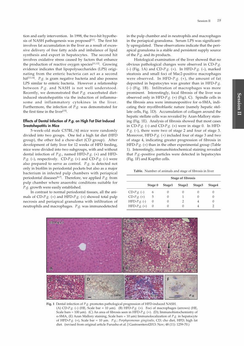
Histological examination of the liver showed that noobvious pathological changes were observed in CD-P.g.(–) (Fig. 1A) and CD-P.g. (+). In HFD-P.g. (–), markedsteatosis and small foci of Mac2-positive macrophageswere observed. In HFD-P.g. (+), the amount of fatdeposited in hepatocytes was greater than in HFD-P.g.(–) (Fig. 1B). Infiltration of macrophages was moreprominent. Interestingly, focal fibrosis of the liver wasobserved only in HFD-P.g. (+) (Fig1. C). Spindle cells inthe fibrosis area were immunopositive for α-SMA, indi-cating their myofibroblastic nature (namely hepatic stel-late cells, Fig. 1D). Accumulation of collagen around thehepatic stellate cells was revealed by Azan-Mallory stain-ing (Fig. 1E). Analysis of fibrosis showed that most casesin CD-P.g. (–) and CD-P.g. (+) were in stage 0. In HFD-P.g. (–), there were two of stage 2 and four of stage 3.Moreover, HFD-P.g. (+) included four of stage 3 and twoof stage 4, indicating greater progression of fibrosis inHFD-P.g. (+) than in the other experimental group (Table1). Interestingly, immunohistochemical staining revealedthat P.g.-positive particles were detected in hepatocytes(Fig. 1F) and Kupffer cells.
Fig. 1 Dental infection of P.g. promotes pathological progression of HFD-induced NASH.(A) CD-P.g. (–) (HE, Scale bar = 10 µm). (B) HFD-P.g. (+). Foci of macrophages (arrows) (HE,Scale bars = 100 µm). (C) An area of fibrosis seen in HFD-P.g. (+). (D); Immunohistochemistry ofα-SMA, (E) Azan Mallory staining, Scale bars = 10 µm) Immunolocalization of P.g. in hepatocyteof HFD-P.g. (+), Scale bar = 10 µm. P.g.; Porphyromonas gingivalis, CD; cho diet, HFD; high fatdiet. (revised from original article Furusho et al. J Gastroenterol2013: Nov; 48 (11): 1259-70.)
Table. Number of animals and stage of fibrosis in liver
Stage of fibrosis
Stage 0 Stage1 Stage2 Stage3 Stage4
CD-P.g. (–) 6 0 0 0 0CD-P.g. (+) 5 0 1 0 0HFD-P.g. (-) 0 0 2 4 0HFD-P.g. (+) 0 0 0 4 2
02_本文1027(web用追加) 15.10.27 9:04 AM ページ 19
20
Ses
sion
II
These findings indicate that dentally applied P.g.enters the blood circulation, translocates into the liverand accelerates pathological progression of NASH.
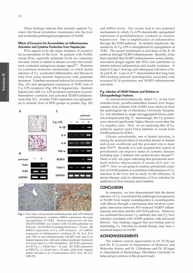
Effects of Excessive Fat Accumulation on InflammasomeActivation and Cytokine Production from Hepatocytes
FFAs appear to be the major mediator of excessivefat accumulation in the liver. In patients with NASH,serum FFAs, especially palmitate levels are commonlyelevated, which is related to disease severity and consid-ered a potential endogenous danger signal[16]. Thereforewe examined molecular mechanisms, in which dentalinfection of P.g. accelerated inflammation and fibrosis infatty liver using steatotic hepatocytes with palmitatetreatment. Palmitate treatment induced fat accumulation(Fig. 2A) and upregulated expression of TLR2 (one ofP.g.-LPS receptors) (Fig. 2B) in hepatocytes. Steatotichepatocytes with P.g.-LPS promoted expression of proin-flammatory cytokines and activated NLRP3-inflamma-some (Fig. 2C). Actually TLR2 expression was upregulat-ed in steatotic liver of HFD groups at protein (Fig. 2D)
and mRNA levels. Our results lead to two potentialmechanisms in which P.g.-LPS dramatically upregulatedexpression of proinflammatory cytokines in steatotichepatocytes. One is amplification of inflammationthrough the TLR2-pathway. In steatotic hepatocyte, thesensitivity to P.g.-LPS is strengthened by upregulation ofTLR2. The second mechanism is activation of the IL-1βpathway through NLRP3 inflammasome. Recently, it hasbeen reported that NLRP3 inflammasome senses obesity-associated danger signals like FFAs and contributes toobesity-induced inflammation and insulin resistance. Itleads to Casp-1 cleavage and subsequent activation of IL-1β and IL-18. Csak et al.[17] demonstrated that long termHFD-feeding induced steatohepatitis associated withincreased IL-1β production and NLRP3 inflammasomeactivation.
P.g. Infection of NASH Patients and Relation toClinicopathologic Features.
To immunohistochemically detect P.g. in the liverformalin-fixed, paraffin-embedded human liver biopsysamples from patients with NASH were retrieved fromthe pathological file of Hiroshima University Hospital.P.g. was identified as single and aggregated brown parti-cles in hepatocytes (fig. 3). Interestingly, the P.g.-positivecases showed significantly higher fibrosis scores than theP.g.-negative cases. Now, we are analyzing titer of P.g.antibody against type4 FimA fimbriae in serum fromNASH patients by ELISA.
Chronic periodontitis, one of dental infection, isamong the most prevalent of microbial disease in humanand occurs worldwide and the prevalent rate is morethan 70%[18]. Recently it is well accepted that control ofperiodontitis can improve status of systemic diseasesincluding type 2 diabetes and cardiovascular disease[3,4].There is only one paper indicating that periodontal treat-ment induces improvement of serum ALT and /orAST[19]. Now we are going to examine periodontal condi-tion of NASH patients in correlation with presence of P.g.infection in the liver and to study on the efficiency ofdental therapy and/or elimination of liver infection byantibiotics in liver function and condition.
CONCLUSIONIn summary, we first demonstrated that the dental
infection of P.g. exacerbated the pathological progressionof NASH from simple steatohepatitis to steatohepatitiswith fibrosis through a mechanism that involves syner-gistic interaction between FFA-induced NLRP3 inflam-masome activation and the LPS-TLR pathway. Moreoverwe confirmed that serum P.g. antibody titer and P.g. liverinfection correlated with NASH patients with advancedfibrosis. Our findings suggest that preventing and/oreliminating P.g. infection by dental therapy may have abeneficial impact on NASH.
ACKNOWLEDGEMENTSThe authors express appreciation to Dr. H Hyogo
and Dr. K Cyayama in Department of Medicine andMolecular Science and Dr. J Hisatsune and Dr. M Sugaiin Department of Bacteriology, Hiroshima University tohelp and give advices in the present study.
Fig. 2 Free fatty acid promotes inflammasome and LPS-inducedproinflammatory cytokine mRNA expression throughupregulation of TLR2. Human hepatocyte cell line(Hc3716-hTERT) was treated with 0.4 mM palmitate for18 hours. (A) Oil Red O staining (Scale bars = 10 µm). (B)mRNA expression of P.g.-LPS receptors. (C) mRNAexpression of inflammatory cytokines (IL-1β, IL-6, IL-8and TNF-α) and inflammasome (NLRP3 and casp-1) inhuman hepatocytes with and without palmitate treatmentat 6 hours after P.g.-LPS stimulation. (D) TLR2 expressionin CD-P.g. (–). (Scale bars = 10 µm). (E) TLR2 expressionin HFD-P.g. (+) (Scale bars = 10 µm). (cited from originalarticle Furusho et al. J Gastroenterol 2013: Nov; 48 (11):1259-70.)
02_本文1027(web用追加) 15.10.27 9:04 AM ページ 20
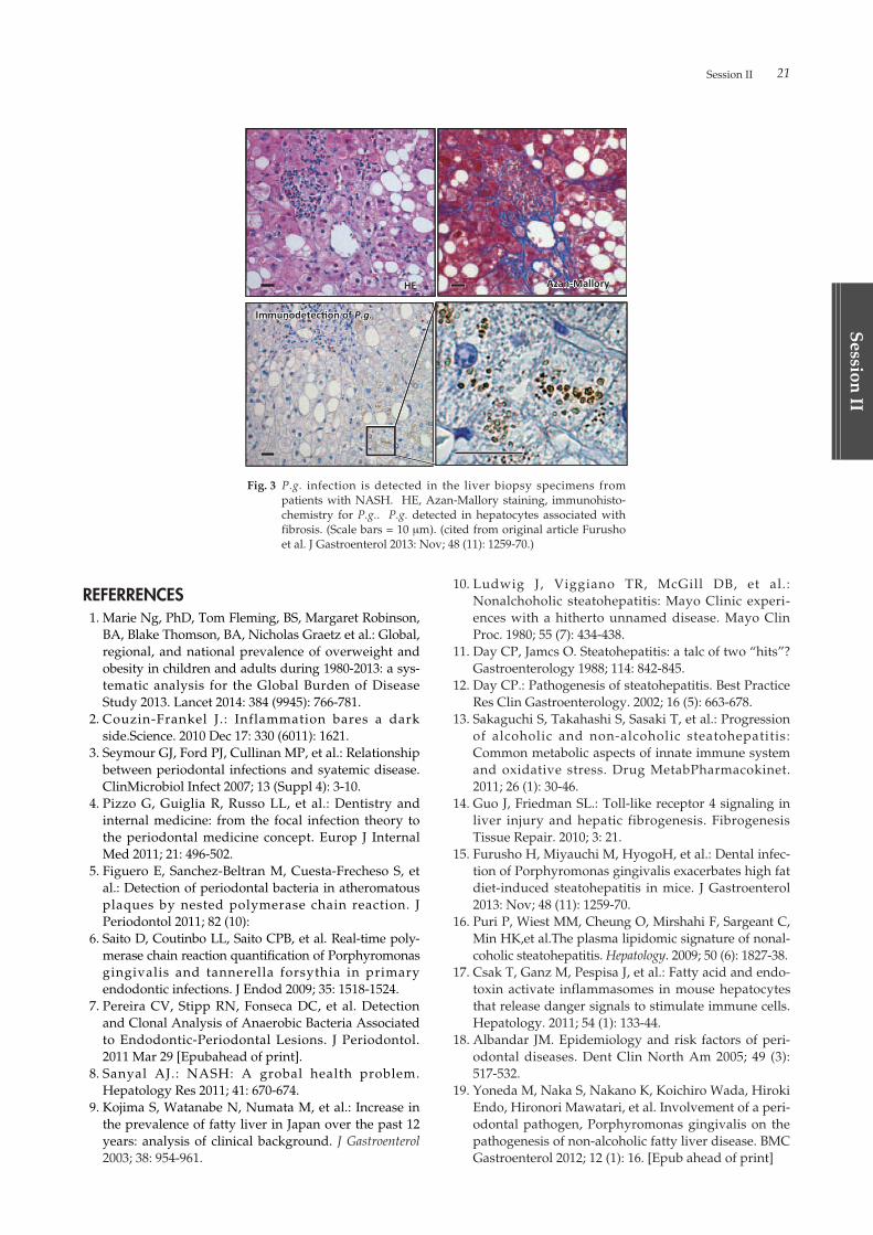
Session II 21
Session
II
REFERRENCES01. Marie Ng, PhD, Tom Fleming, BS, Margaret Robinson,
BA, Blake Thomson, BA, Nicholas Graetz et al.: Global,regional, and national prevalence of overweight andobesity in children and adults during 1980-2013: a sys-tematic analysis for the Global Burden of DiseaseStudy 2013. Lancet 2014: 384 (9945): 766-781.
02. Couzin-Frankel J.: Inflammation bares a darkside.Science. 2010 Dec 17: 330 (6011): 1621.
03. Seymour GJ, Ford PJ, Cullinan MP, et al.: Relationshipbetween periodontal infections and syatemic disease.ClinMicrobiol Infect 2007; 13 (Suppl 4): 3-10.
04. Pizzo G, Guiglia R, Russo LL, et al.: Dentistry andinternal medicine: from the focal infection theory tothe periodontal medicine concept. Europ J InternalMed 2011; 21: 496-502.
05. Figuero E, Sanchez-Beltran M, Cuesta-Frecheso S, etal.: Detection of periodontal bacteria in atheromatousplaques by nested polymerase chain reaction. JPeriodontol 2011; 82 (10):
06. Saito D, Coutinbo LL, Saito CPB, et al. Real-time poly-merase chain reaction quantification of Porphyromonasgingivalis and tannerella forsythia in primaryendodontic infections. J Endod 2009; 35: 1518-1524.
07. Pereira CV, Stipp RN, Fonseca DC, et al. Detectionand Clonal Analysis of Anaerobic Bacteria Associatedto Endodontic-Periodontal Lesions. J Periodontol.2011 Mar 29 [Epubahead of print].
08. Sanyal AJ.: NASH: A grobal health problem.Hepatology Res 2011; 41: 670-674.
09. Kojima S, Watanabe N, Numata M, et al.: Increase inthe prevalence of fatty liver in Japan over the past 12years: analysis of clinical background. J Gastroenterol2003; 38: 954-961.
10. Ludwig J, Viggiano TR, McGill DB, et al.:Nonalchoholic steatohepatitis: Mayo Clinic experi-ences with a hitherto unnamed disease. Mayo ClinProc. 1980; 55 (7): 434-438.
11. Day CP, Jamcs O. Steatohepatitis: a talc of two “hits”?Gastroenterology 1988; 114: 842-845.
12. Day CP.: Pathogenesis of steatohepatitis. Best PracticeRes Clin Gastroenterology. 2002; 16 (5): 663-678.
13. Sakaguchi S, Takahashi S, Sasaki T, et al.: Progressionof alcoholic and non-alcoholic steatohepatitis:Common metabolic aspects of innate immune systemand oxidative stress. Drug MetabPharmacokinet.2011; 26 (1): 30-46.
14. Guo J, Friedman SL.: Toll-like receptor 4 signaling inliver injury and hepatic fibrogenesis. FibrogenesisTissue Repair. 2010; 3: 21.
15. Furusho H, Miyauchi M, HyogoH, et al.: Dental infec-tion of Porphyromonas gingivalis exacerbates high fatdiet-induced steatohepatitis in mice. J Gastroenterol2013: Nov; 48 (11): 1259-70.
16. Puri P, Wiest MM, Cheung O, Mirshahi F, Sargeant C,Min HK,et al.The plasma lipidomic signature of nonal-coholic steatohepatitis. Hepatology. 2009; 50 (6): 1827-38.
17. Csak T, Ganz M, Pespisa J, et al.: Fatty acid and endo-toxin activate inflammasomes in mouse hepatocytesthat release danger signals to stimulate immune cells.Hepatology. 2011; 54 (1): 133-44.
18. Albandar JM. Epidemiology and risk factors of peri-odontal diseases. Dent Clin North Am 2005; 49 (3):517-532.
19. Yoneda M, Naka S, Nakano K, Koichiro Wada, HirokiEndo, Hironori Mawatari, et al. Involvement of a peri-odontal pathogen, Porphyromonas gingivalis on thepathogenesis of non-alcoholic fatty liver disease. BMCGastroenterol 2012; 12 (1): 16. [Epub ahead of print]
Fig. 3 P.g. infection is detected in the liver biopsy specimens frompatients with NASH. HE, Azan-Mallory staining, immunohisto-chemistry for P.g.. P.g. detected in hepatocytes associated withfibrosis. (Scale bars = 10 µm). (cited from original article Furushoet al. J Gastroenterol 2013: Nov; 48 (11): 1259-70.)
02_本文1027(web用追加) 15.10.27 9:04 AM ページ 21
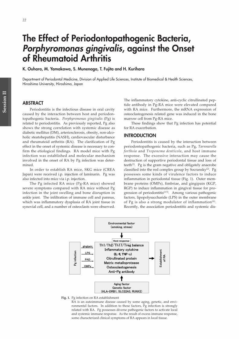
22
Ses
sion
II
ABSTRACTPeriodontitis is the infectious disease in oral cavity
caused by the interaction between host and periodon-topathogenic bacteria. Porphyromonas gingivalis (Pg) isrelated to periodontitis. As previously reported, Pg alsoshows the strong correlation with systemic disease asdiabetic mellitus (DM), arteriosclerosis, obesity, non-alco-holic steatohepatitis (NASH), cardiovascular disturbanceand rheumatoid arthritis (RA). The clarification of Pgeffect in the onset of systemic disease is necessary to con-firm the etiological findings. RA model mice with Pginfection was established and molecular mechanisminvolved in the onset of RA by Pg infection was deter-mined.
In order to establish RA mice, SKG mice (CREAJapan) were received i.p. injection of laminarin. Pg wasalso infected into mice via i.p. injection.
The Pg infected RA mice (Pg-RA mice) showedsevere symptoms compared with RA mice without Pginfection in the joint swelling and bone disruption inankle joint. The infiltration of immune cell and pannus,which was inflammatory dysplasia of RA joint tissue insynovial cell, and a number of osteoclasts were observed.
The inflammatory cytokine, anti-cyclic citrullinated pep-tide antibody in Pg-RA mice were elevated comparedwith RA mice. Furthermore, the mRNA expression ofosteoclastogenesis related gene was induced in the bonemarrow cell from Pg-RA mice.
These findings show that Pg infection has potentialfor RA exacerbation.
INTRODUCTIONPeriodontitis is caused by the interaction between
periodontopathogenic bacteria, such as Pg, Tarnnarellaforthsia and Treponema denticola, and host immuneresponse. The excessive interaction may cause thedestruction of supportive periodontal tissue and loss ofteeth(1). Pg is the gram negative and obligately anaerobeclassified into the red complex group by Socransky(2). Pgpossesses some kinds of virulence factors to induceinflammation in periodontal tissue (Fig. 1). Outer mem-brane proteins (OMPs), fimbriae, and gingipain (KGP,RGP) to induce inflammation in gingival tissue for pro-gression of periodontitis(3-5). Among various pathogenicfactors, lipopolysaccharide (LPS) in the outer membraneof Pg is also a strong modulator of inflammation(6).Recently, the association periodontitis and systemic dis-
The Effect of Periodontopathogenic Bacteria, Porphyromonas gingivalis, against the Onset of Rheumatoid ArthritisK. Ouhara, M. Yamakawa, S. Munenaga, T. Fujita and H. Kurihara
Department of Periodontal Medicine, Division of Applied Life Sciences, Institute of Biomedical & Health Sciences, Hiroshima University, Hiroshima, Japan
Fig. 1. Pg infection on RA establishmentRA is an autoimmune disease caused by some aging, genetic, and envi-ronmental factors. In addition to these factors, Pg infection is stronglyrelated with RA. Pg possesses diverse pathogenic factors to activate localand systemic immune response. As the result of excess immune response,some characterized clinical symptoms of RA appears in local tissue.
02_本文1027(web用追加) 15.10.27 9:04 AM ページ 22

Session II 23
Session
II
eases such as diabetes mellitus (DM), arterial sclerosis,nonalcoholic steatohepatitis (NASH), low weight birthand rheumatoid arthritis (RA) were reported(7-11). Thelocally induced inflammatory cytokine caused by Pgdirect challenge or Pg infection into blood stream, bac-teremia, may exacerbate the onset of systemic disease(12).RA is a systemic autoimmune disease characterized bychronic inflammation of the diarthrodial joints leading tobone erosions and progressive disability(13). There is ageneral agreement of two hit theory that RA develops asa result of interaction between susceptibility genes, hor-monal and environmental factors, such as smoking, sili-ca, female hormone, and periodontitis(14). Most of thegenetic studies on RA have been focused on the HumanLeucocyte Antigen (HLA) region, and PADI4 gene thatcodifies for the peptidyl arginine deiminase type IVenzyme(15). As the result of the mutation of HLA andPADI4, the elevation of anti-citrullinated protein anti-body (ACPA) occurs following the increase of citrullinat-ed protein(16). The production of ACPA in serum can bethe important diagnostic marker of RA in common withrheumatoid factor which is the autoantibody of IgMagainst Fc portion of IgG. There is the report about thecorrelation RA and periodontitis. Especially, productionof citrullinated protein by peptidylarginine deiminases(PAD) derived from Pg (PgPAD) shows the stronginvolvement in the onset of RA(17). The citrullination is apost-translational modification of arginine residues,mediated by the family of PAD. The mechanism of gen-eration of CP by PgPAD is different from endogenouslyproduced CP. The fragmented target protein, such as fib-rinogen and alpha-enolase, after digestion by Pg pro-duced arginine specific enzyme, gingipain (RGP) is tar-geted the citrullination of c-terminal arginine(18). Theincrease of CP and ACPA by PgPAD also can be thecause of RA. Therefore, there is a common issue to coop-erate and overcome for the treatment of RA and peri-odontitis. As for the correlation between periodontitisand RA in clinical study, the non-surgical treatment ofperiodontitis reduces the severity of RA in patients treat-ed with or without tumor necrosis factor inhibitors(14).The elevation of inflammatory cytokine, such as IL-6 andTNF-α, and matrix metalloprotease (MMP) 3 are themajor clinical parameter of RA. These factors may com-municate the progression of periodontitis via bloodstream. Therefore, the use of cytokine inhibitor,Infliximab (anti-TNF-α neutralization antibody),Etanercept, (anti-TNF-α receptor antibody), Adalimumab(anti-TNF-α neutralization antibody), Tocilizumab, (anti-IL-6 receptor antibody), IL-6 receptor, results in theimprovement of periodontitis condition by suppressingthe inflammation in periodontal tissue(19). Previously, toexamine whether induction of periodontitis affects theprogression of experimental arthritis in vivo, mice immu-nized with CII received orally inoculations of Pg everyother day. Interestingly, periodontitis induced by Pgaccelerated the onset of arthritis in addition to periodon-tal bone loss in mice. The analysis of T cell population injoint-draining lymph node with Pg infection showed theactivation of Th17. Furthermore, the Pg stimulation acti-vated the bone marrow derived dendritic cells (BMDCs)to produce cytokine affecting T cell differentiation, such
as IL-1β, IL-6, TNF-α via toll like receptor 2 signaling(20).Although there are some reports focused on the immuneresponse against periodontopathogenic bacteria in RAmodel mice, the effect of Pg infection in the bone resorp-tion as one of the progressed RA symptom. However, itis important to clarify the involvement of Pg infection inthe osteoclastogenesis in advanced RA for treatment ofsystemic disease. Here, we aimed to elucidate the localand systemic immune response in the progression of RAby establishing Pg infected SKG mice as RA model.
RESULTFirstly, RA model mice was established by injecting
laminarin, which was the group of β-glucan fromLaminaria digitata. The joint swelling was monitoredestablished by Sakaguchi’s arthritis score(21). As shownin Fig. 2, the single injection of laminarin induced RAsymptom in 20 weeks. The Pg infection induced the jointswelling in laminarin injected RA mice. The sever tissuedestruction and forming of pannus were observed bymicroCT and immunohistochemical staining in Pg inject-ed RA mice. The production of MMP- 3 and anti-CCPIgG in serum were statistically increased in the Pg inject-ed RA mice. The increase of CP and Pg derived proteinwere detected in ankle joint tissue of Pg injected RAmice. The production of IL-1β, IL-2, IL-6, and CXCL1 inserum of Pg injected RA mice was increased comparedwith RA induced mice. The osteoclast differentiation ofbone marrow derived mononuclear cell was stronglyinduced in Pg injected RA mice. Furthermore, the induc-tion of osteoclastogenesis was supported by the induc-tion of mRNA expression of RANK, OSCAR, TRAF6, c-Fos, NFATc1, CatK, MMP-9, and DC-STAMP.
CONCLUSIONThese findings might the involvement of Pg in the
exacerbation of RA. Further studies will be needed inorder to clarify the detail of the correlation between peri-odontitis and RA.
ACKNOWLEDGEMENTThis research was supported by a Grant-in-Aid for
the Encouragement of Young Scientists (B) (22792085)from the Japan Society for the Promotion of Science.
Fig. 2. The assessment of joint swelling after 20 weeks.The SKG mice were received i.p. injection of laminarin(Sigma, 10 µg/mouse) at day 0. Mice were divided into 2groups (no injection, laminrin single injection). The jointswelling was assessed by Sakaguchi’s arthritis score after20 weeks of LA injection. Three independent experimentsof all groups were analyzed by Mann-Whitney U test.
02_本文1027(web用追加) 15.10.27 9:04 AM ページ 23
24
Ses
sion
II
REFERENCES01. Socransky, S.S., and Haffajee, A.D. (1994) Implications
of periodontal microbiology for the treatment of peri-odontal infections. Compend Suppl, S684-685, 688-693;quiz S714-687
02. Ximenez-Fyvie, L.A., Haffajee, A.D., and Socransky,S.S. (2000) Comparison of the microbiota of supra-and subgingival plaque in health and periodontitis. JClin Periodontol 27, 648-657
03. Amano, A., Nakagawa, I., Okahashi, N., and Hamada,N. (2004) Variations of Porphyromonas gingivalisfimbriae in relation to microbial pathogenesis. JPeriodontal Res 39, 136-142
04. Kadowaki, T., Nakayama, K., Okamoto, K., Abe, N.,Baba, A., Shi, Y., Ratnayake, D.B., and Yamamoto, K.(2000) Porphyromonas gingivalis proteinases as viru-lence determinants in progression of periodontal dis-eases. J Biochem 128, 153-159
05. Abe, N., Baba, A., Takii, R., Nakayama, K.,Kamaguchi, A., Shibata, Y., Abiko, Y., Okamoto, K.,Kadowaki, T., and Yamamoto, K. (2004) Roles of Arg-and Lys-gingipains in coaggregation ofPorphyromonas gingivalis: identification of itsresponsible molecules in translation products ofrgpA, kgp, and hagA genes. Biol Chem 385, 1041-1047
06. Barksby, H.E., Nile, C.J., Jaedicke, K.M., Taylor, J.J.,and Preshaw, P.M. (2009) Differential expression ofimmunoregulatory genes in monocytes in response toPorphyromonas gingivalis and Escherichia colilipopolysaccharide. Clin Exp Immunol 156, 479-487
07. Munenaga, Y., Hiroshima Study, G., Yamashina, T.,Tanaka, J., and Nishimura, F. (2013) Improvement ofglycated hemoglobin in Japanese subjects with type 2diabetes by resolution of periodontal inflammationusing adjunct topical antibiotics: results from theHiroshima Study. Diabetes Res Clin Pract 100, 53-60
08. Yoneda, M., Naka, S., Nakano, K., Wada, K., Endo, H.,Mawatari, H., Imajo, K., Nomura, R., Hokamura, K.,Ono, M., Murata, S., Tohnai, I., Sumida, Y., Shima, T.,Kuboniwa, M., Umemura, K., Kamisaki, Y., Amano,A., Okanoue, T., Ooshima, T., and Nakajima, A. (2012)Involvement of a periodontal pathogen,Porphyromonas gingivalis on the pathogenesis ofnon-alcoholic fatty liver disease. BMC Gastroenterol 12,16
09. Offenbacher, S., Jared, H.L., O’Reilly, P.G., Wells, S.R.,Salvi, G.E., Lawrence, H.P., Socransky, S.S., and Beck,J.D. (1998) Potential pathogenic mechanisms of peri-odontitis associated pregnancy complications. AnnPeriodontol 3, 233-250
10. Soto-Barreras, U., Olvera-Rubio, J.O., Loyola-Rodriguez, J.P., Reyes-Macias, J.F., Martinez-Martinez, R.E., Patino-Marin, N., Martinez-Castanon,G.A., Aradillas-Garcia, C., and Little, J.W. (2013)Peripheral arterial disease associated with caries andperiodontal disease. Journal of periodontology 84, 486-494
11. Okada, M., Kobayashi, T., Ito, S., Yokoyama, T.,Komatsu, Y., Abe, A., Murasawa, A., and Yoshie, H.(2011) Antibody responses to periodontopathic bacte-ria in relation to rheumatoid arthritis in Japanese
adults. Journal of periodontology 82, 1433-144112. Perez-Chaparro, P.J., Gracieux, P., Lafaurie, G.I.,
Donnio, P.Y., and Bonnaure-Mallet, M. (2008)Genotypic characterization of Porphyromonas gingi-valis isolated from subgingival plaque and bloodsample in positive bacteremia subjects with periodon-titis. J Clin Periodontol 35, 748-753
13. van Venrooij, W.J., van Beers, J.J., and Pruijn, G.J.(2008) Anti-CCP Antibody, a Marker for the EarlyDetection of Rheumatoid Arthritis. Ann N Y Acad Sci1143, 268-285
14. Ortiz, P., Bissada, N.F., Palomo, L., Han, Y.W., Al-Zahrani, M.S., Panneerselvam, A., and Askari, A.(2009) Periodontal therapy reduces the severity ofactive rheumatoid arthritis in patients treated with orwithout tumor necrosis factor inhibitors. Journal ofperiodontology 80, 535-540
15. Goronzy, J.J., and Weyand, C.M. (2009) Developmentsin the scientific understanding of rheumatoid arthri-tis. Arthritis Res Ther 11, 249
16. Saeki, Y., Kudo-Tanaka, E., Ohshima, S., Matsushita,M., Tsuji, S., Maeda, Y., Yoshimura, M., Watanabe, A.,Katada, Y., Harada, Y., Ichikawa, K., Suenaga, Y.,Ohta, Y., Tohma, S., and Group, N.H.O.i.-n.S. (2013)Baseline anti-citrullinated peptide antibody (ACPA)titers and serum interleukin-6 (IL-6) levels possiblypredict progression of bone destruction in early stagesof rheumatoid arthritis (ERA). Rheumatol Int 33, 451-456
17. Montgomery, A.B., Kopec, J., Shrestha, L., Thezenas,M.L., Burgess-Brown, N.A., Fischer, R., Yue, W.W.,and Venables, P.J. (2015) Crystal structure ofPorphyromonas gingivalis peptidylarginine deimi-nase: implications for autoimmunity in rheumatoidarthritis. Ann Rheum Dis
18. Wegner, N., Wait, R., Sroka, A., Eick, S., Nguyen,K.A., Lundberg, K., Kinloch, A., Culshaw, S., Potempa,J., and Venables, P.J. (2010) Peptidylarginine deimi-nase from Porphyromonas gingivalis citrullinateshuman fibrinogen and alpha-enolase: implications forautoimmunity in rheumatoid arthritis. Arthritis Rheum62, 2662-2672
19. Golub, L.M., Payne, J.B., Reinhardt, R.A., andNieman, G. (2006) Can systemic diseases co-induce(not just exacerbate) periodontitis? A hypothetical“two-hit” model. Journal of dental research 85, 102-105
20. de Aquino, S.G., Abdollahi-Roodsaz, S., Koenders,M.I., van de Loo, F.A., Pruijn, G.J., Marijnissen, R.J.,Walgreen, B., Helsen, M.M., van den Bersselaar, L.A.,de Molon, R.S., Avila Campos, M.J., Cunha, F.Q.,Cirelli, J.A., and van den Berg, W.B. (2014)Periodontal pathogens directly promote autoimmuneexperimental arthritis by inducing a TLR2- and IL-1-driven Th17 response. Journal of immunology 192, 4103-4111
21. Yoshitomi, H., Sakaguchi, N., Kobayashi, K., Brown,G.D., Tagami, T., Sakihama, T., Hirota, K., Tanaka, S.,Nomura, T., Miki, I., Gordon, S., Akira, S., Nakamura,T., and Sakaguchi, S. (2005) A role for fungal {beta}-glucans and their receptor Dectin-1 in the induction ofautoimmune arthritis in genetically susceptible mice. JExp Med 201, 949-960
02_本文1027(web用追加) 15.10.27 9:04 AM ページ 24
Session II 25
Session
II
ABSTRACTPeriodontal disease is known to cause systemic
spread of chronic inflammation and exacerbate lifestyle-related diseases such as ischemic heart disease, diabetesmellitus, and obesity, while the inflammatory responseplays a major role in the development of neurodegenera-tive conditions such as Alzheimer’s disease (AD). Mildsystemic inflammation has been reported to increase therisk of AD. Chronic inflammation in periodontal diseaseand periodontal disease-related bacteria are transmittedto the brain via an unknown mechanism, possibly exacer-bating AD. In this super-aging society, measures for pre-vention of periodontal disease will become increasinglyimportant.
INTRODUCTIONRather than systemic diseases being risk factors for
periodontal disease, periodontal disease has been shownto cause systemic diseases, including lifestyle-related dis-eases. To date, periodontal disease has been reported tobe a risk factor for diabetes mellitus (DM), cardio- andcerebrovascular diseases, aspiration pneumonia, prema-ture and low birth weight infants, bacterial endocarditis,glomerulonephritis, arthritis, and palmoplantar pustulo-sis(1-5). The following three pathways are assumed to bethe mechanisms of the systemic spread of periodontaldisease: direct action of bacterial bodies and toxins ofperiodontal disease-related bacteria at a local periodontalsite that spread to target organs through a hematogenousroute or the respiratory tract(4); actions of inflammation-inducing substances such as cytokines, produced by theinflammatory response within periodontal tissue or bythe immune response, that spread hematogenously to thetarget organs(5); and a pathway that results in intracere-bral spread through the nervous system(6,7). There arevarious data on intravascular infiltration of periodontaldisease-related bacteria and their spread to target organs,but the mechanism by which they affect diseases is notfully understood. On the other hand, the inflammatoryresponse is known to play a major role in the progressionof cerebrovascular disorders and dementia, conditionsthat often occur in the elderly(8-10); however, the effect ofperiodontal disease is not fully understood.
Taking results of other studies into consideration, wediscuss here our most recent results of analysis of the cor-relation between periodontal disease and Alzheimer’sdisease (AD) in a mouse model in this study.
Current status of AD in JapanMore than 30 million people in Japan are over 65
years of age. A study group of the Ministry of Health,Labour and Welfare (2013) showed that the number ofpeople with dementia was 4,620,000, with another 4 mil-lion people in the general population estimated to havedementia. This number of people with dementia isexpected to increase in the future. Sixty to seventy per-cent of people with dementia have AD, which is a seriousproblem in an advancing aging society such as that inJapan. There is still no effective method for prevention ofAD or a fundamental method for treatment(11).
Inflammation and ADIn addition to aging and genetic mutation, AD is
caused by accumulation of amyloid β protein (Aβ) due tointracerebral inflammation(12). Aβ deposits also causeinflammation, which results in the progression ofsynapse disorders and neuronopathy. In recent years,mutations of the TREM2 gene, which controls the inflam-matory response, have been found in patients with AD,renewing the importance of the inflammatory responsein the development of AD(13). Chronic inflammation isalso thought to play an important role in the develop-ment of central nervous system (CNS) diseases. Thelong-term use of non-steroidal anti-inflammatory drugsis known to prevent the occurrence of neurodegenerativedisease. The effectiveness of these drugs for delaying theprogression of neurodegenerative disease has beenshown by an epidemiologic study and an animal experi-ment(14). The immune system of the CNS is extremelysimple and is not acquired. Accordingly, the immuneresponse is served by the innate immune system.
Microglia are cells of the macrophage system thathave a phagocytic capacity that plays a central role in theintracerebral innate immune response. Microglia digestAβ that has accumulated in the brain and remove it fromthe brain. These cells produce cytokines such as activeoxygen, interleukin (IL)-1, IL-6, tumor necrosis factor-α(TNF-α), i.e., an inflammatory response producing mole-cules that are known to promote neurodegeneration inAD(15,16). On the other hand, they also produce anti-inflammatory molecules such as IL-4 and IL-10, whichare thought to have a neuroprotective role in addition tocontrolling the inflammatory response(17). Therefore,microglia are important cells in the control of AD status.
Apart from the exacerbation of AD status due tointracerebral inflammation, mild systemic inflammationhas been reported to reduce cognitive function and hip-pocampal capacity and increase the risk of AD(18,19,20).Inflammation has been thought to spread through the cir-culatory system and CNS. Increased amounts of inflam-matory mediators in the blood are transmitted to the
Periodontal Disease as a Possible Risk Factor for Alzheimer’s DiseaseK. Matsushita
DDS, PhD, Department of Oral Disease Research, National Center for Geriatrics and Gerontology, Obu, Aichi 474-8511, Japan
02_本文1027(web用追加) 15.10.27 9:04 AM ページ 25

26
Ses
sion
II
brain and may activate microglia in the brain. TNF-αlevel is increased in the blood of patients with AD andreportedly correlates with reduced cognitive func-tion(21,22).
Periodontal disease and ADAs mentioned earlier, chronic inflammation within
peripheral organs might play a role in exacerbation of themolecular pathogenesis of AD. One such inflammatorycondition is periodontal disease. The inflammatoryresponse that occurs in periodontal disease is known tobe involve in the development of various diseases, suchas arteriosclerotic disease, DM, and obesity, and the inci-dence of premature and low birth weightinfants(1,2,23,3,24,4,5). Periodontal disease has also beenreported to be involved in cerebral abscess formation(25).Periodontal disease-related bacteria spread systemicallythrough the blood vessels and respiratory tract, suggest-ing their possible direct effects on target organs. In addi-tion, inflammatory mediators such as cytokines, whichare produced in local periodontal tissue, are carriedhematogenously to target organ and are thought to wors-en the inflammatory response.
There have been interesting reports on the correla-tion between AD and periodontal disease. Porphyromonasgingivalis, a periodontal disease-related bacterium, wasfound at high frequency in autopsied brain tissues ofpatients who died of AD; however, it was not found innormal human brain tissues(26). These finding suggestthat said the bacteria spread hematogenously into thebrain. P. gingivalis is a gram-negative anaerobic bacillusthat possesses various toxins including lipopolysaccha-ride. Accordingly, it is known to cause a strong inflam-matory response. In addition, an interesting finding isthat periodontal disease-related bacteria of the Treponemagenus were found in the trigeminal ganglion, brainstem,and cerebral cortex: with a particularly high frequency inpatients with AD(6). These findings suggest that peri-odontal disease-related bacteria can be directly transmit-ted to the brain and cause inflammation. The mechanism(hypothesis) of AD exacerbation due to periodontal dis-ease is shown in Figure 1. The mechanism should beanalyzed in detail in the future. However, it is unlikelythat AD is induced only by an inflammatory response
due to periodontal disease and periodontal disease-relat-ed bacteria. Inflammatory responses are thought toaggravate the molecular level of AD, cause an earlieronset, worsen the degree of cognitive disorders, andcause more rapid progression, suggesting its action inmodifying the disease status. The long-term use of anti-inflammatory drugs has been suggested to reduce therisk of AD onset(27).
Periodontal disease is the main cause of tooth loss;however, some reports have shown a correlation betweentooth loss and AD. Tooth loss may be a risk factor forAD(28,29). Tooth loss reduces chewing function, resultingin reduced cerebral blood flow and possibly leading toreduced cognitive function. However, tooth loss itself isoften not accompanied by a chronic inflammatoryresponse, suggesting that the effects of tooth loss are notnecessarily the same as those of periodontal disease. Oueet al. found that cognitive function was reduced by toothremoval in AAP transgenic mice, but that there was noeffect of tooth removal on the molecular pathology ofAD(30). On the other hand, when we induced periodontaldisease in the same mice, we found that intracerebral Aβdeposits increased and that the intracerebral inflammato-ry response was enhanced in addition to reduced cogni-tive function. Both periodontal disease and tooth lossreduce cognitive function, but their molecular mecha-nisms are thought to differ.
CONCLUSIONAll organisms survive by consuming food; chewing
function is therefore very important. This function notonly supports life but also might be important for main-tenance of cognitive function. In this modern aging soci-ety, prevention of periodontal disease and maintenanceof oral cavity function will become increasingly impor-tant.
REFERENCES01. Chapple IL, Genco R, Working group 2 of joint
EFPAAPw. Diabetes and periodontal diseases: con-sensus report of the Joint EFP/AAP Workshop onPeriodontitis and Systemic Diseases. Journal of clini-cal periodontology. 2013; 40 Suppl 14: S106-12.
02. Cullinan MP, Seymour GJ. Periodontal disease and
Fig. 1 Possible mechanisms by which periodontitis induced by bacterial infectionexacerbate features of Alzheimer’s disease.
02_本文1027(web用追加) 15.10.27 9:04 AM ページ 26
Session II 27
Session
II
systemic illness: will the evidence ever be enough?Periodontology 2000. 2013; 62 (1): 271-86.
03. Linden GJ, Herzberg MC, working group 4 of thejoint EFPAAPw. Periodontitis and systemic diseases:a record of discussions of working group 4 of the JointEFP/AAP Workshop on Periodontitis and SystemicDiseases. Journal of periodontology. 2013; 84 (4Suppl): S20-3.
04. Reyes L, Herrera D, Kozarov E, Rolda S, Progulske-Fox A. Periodontal bacterial invasion and infection:contribution to atherosclerotic pathology. Journal ofperiodontology. 2013; 84 (4 Suppl): S30-50.
05. Schenkein HA, Loos BG. Inflammatory mechanismslinking periodontal diseases to cardiovascular dis-eases. Journal of clinical periodontology. 2013; 40Suppl 14: S51-69.
06. Riviere GR, Riviere KH, Smith KS. Molecular andimmunological evidence of oral Treponema in thehuman brain and their association with Alzheimer’sdisease. Oral microbiology and immunology. 2002; 17(2): 113-8.
07. Kamer AR. Systemic inflammation and disease pro-gression in Alzheimer disease. Neurology. 2010; 74(14): 1157; author reply -8.
08. Galea J, Brough D. The role of inflammation andinterleukin-1 in acute cerebrovascular disease. Journalof inflammation research. 2013; 6: 121-8.
09. Liu L, Chan C. The role of inflammasome inAlzheimer’s disease. Ageing research reviews. 2014;15C: 6-15.
10. Serpente M, Bonsi R, Scarpini E, Galimberti D. Innateimmune system and inflammation in Alzheimer’s disease: from pathogenesis to treatment.Neuroimmunomodulation. 2014; 21 (2-3): 79-87.
11. Fettelschoss A, Zabel F, Bachmann MF. Vaccinationagainst Alzheimer disease: An update on futurestrategies. Human vaccines & immunotherapeutics.2014; 10 (4).
12. Shankar GM, Li S, Mehta TH, Garcia-Munoz A,Shepardson NE, Smith I, et al. Amyloid-beta proteindimers isolated directly from Alzheimer’s brainsimpair synaptic plasticity and memory. Nature medi-cine. 2008; 14 (8): 837-42.
13. Jonsson T, Stefansson H, Steinberg S, Jonsdottir I,Jonsson PV, Snaedal J, et al. Variant of TREM2 associ-ated with the risk of Alzheimer’s disease. The NewEngland journal of medicine. 2013; 368 (2): 107-16.
14. Cudaback E, Jorstad NL, Yang Y, Montine TJ, KeeneCD. Therapeutic implications of the prostaglandinpathway in Alzheimer’s disease. Biochemical pharma-cology. 2014; 88 (4): 565-72.
15. Akiyama H, Arai T, Kondo H, Tanno E, Haga C,Ikeda K. Cell mediators of inflammation in theAlzheimer disease brain. Alzheimer disease and asso-ciated disorders. 2000; 14 Suppl 1: S47-53.
16. Ridolfi E, Barone C, Scarpini E, Galimberti D. The roleof the innate immune system in Alzheimer’s diseaseand frontotemporal lobar degeneration: an eye onmicroglia. Clinical & developmental immunology.
2013; 2013: 939786.17. Kloss CU, Kreutzberg GW, Raivich G. Proliferation of
ramified microglia on an astrocyte monolayer: charac-terization of stimulatory and inhibitory cytokines.Journal of neuroscience research. 1997; 49 (2): 248-54.
18. Marsland AL, Gianaros PJ, Abramowitch SM,Manuck SB, Hariri AR. Interleukin-6 covaries inverse-ly with hippocampal grey matter volume in middle-aged adults. Biological psychiatry. 2008; 64 (6): 484-90.
19. Engelhart MJ, Geerlings MI, Meijer J, Kiliaan A,Ruitenberg A, van Swieten JC, et al. Inflammatoryproteins in plasma and the risk of dementia: the rot-terdam study. Archives of neurology. 2004; 61 (5):668-72.
20. Tobinick EL. Re: Inflammatory markers and the riskof Alzheimer disease: the Framingham Study.Neurology. 2008; 70 (14): 1222-3; author reply 3.
21. Alvarez A, Cacabelos R, Sanpedro C, Garcia-FantiniM, Aleixandre M. Serum TNF-alpha levels areincreased and correlate negatively with free IGF-I inAlzheimer disease. Neurobiology of aging. 2007; 28(4): 533-6.
22. Holmes C, Cunningham C, Zotova E, Woolford J,Dean C, Kerr S, et al. Systemic inflammation and dis-ease progression in Alzheimer disease. Neurology.2009; 73 (10): 768-74.
23. Genco RJ, Borgnakke WS. Risk factors for periodontaldisease. Periodontology 2000. 2013; 62 (1): 59-94.
24. Linden GJ, Lyons A, Scannapieco FA. Periodontal sys-temic associations: review of the evidence. Journal ofperiodontology. 2013; 84 (4 Suppl): S8-S19.
25. Zijlstra EE, Swart GR, Godfroy FJ, Degener JE.Pericarditis, pneumonia and brain abscess due to acombined Actinomyces—Actinobacillus actino-mycetemcomitans infection. The Journal of infection.1992; 25 (1): 83-7.
26. Poole S, Singhrao SK, Kesavalu L, Curtis MA, CreanS. Determining the presence of periodontopathic viru-lence factors in short-term postmortem Alzheimer’sdisease brain tissue. Journal of Alzheimer’s disease:JAD. 2013; 36 (4): 665-77.
27. Heneka MT, Kummer MP, Weggen S, Bulic B,Multhaup G, Munter L, et al. Molecular mechanismsand therapeutic application of NSAIDs and derivedcompounds in Alzheimer’s disease. CurrentAlzheimer research. 2011; 8 (2): 115-31.
28. Kondo K, Niino M, Shido K. A case-control study ofAlzheimer’s disease in Japan—significance of life-styles. Dementia. 1994; 5 (6): 314-26.
29. Gatz M, Mortimer JA, Fratiglioni L, Johansson B, BergS, Reynolds CA, et al. Potentially modifiable risk fac-tors for dementia in identical twins. Alzheimer’s &dementia: the journal of the Alzheimer’s Association.2006; 2 (2): 110-7.
30. Oue H, Miyamoto Y, Okada S, Koretake K, Jung CG,Michikawa M, et al. Tooth loss induces memoryimpairment and neuronal cell loss in APP transgenicmice. Behavioural brain research. 2013; 252: 318-25.
02_本文1027(web用追加) 15.10.27 9:04 AM ページ 27
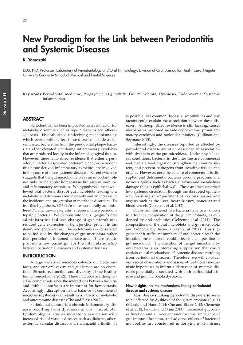
28
Ses
sion
II
ABSTRACTPeriodontitis has been implicated as a risk factor for
metabolic disorders such as type 2 diabetes and athero-sclerosis. Hypothesized underlying mechanisms bywhich periodontitis affect these diseases include a dis-seminated bacteremia from the periodontal plaque bacte-ria and/or elevated circulating inflammatory cytokinesthat are produced locally in the inflamed gingival tissues.However, there is no direct evidence that either a peri-odontal bacteria-associated bacteremia and/or periodon-titis tissue-derived inflammatory cytokines are involvedin the course of these systemic diseases. Recent evidencesuggests that the gut microbiome plays an important rolenot only in metabolic homeostasis but also in immuneand inflammatory responses. We hypothesize that swal-lowed oral bacteria disrupt gut microbiota leading to ametabolic endotoxemia seen in obesity and an increase inthe incidence and progression of metabolic disorders. Totest this hypothesis, C57BL/6 mice were orally adminis-tered Porphyromonas gingivalis, a representative periodon-topathic bacteria. We demonstrate that P. gingivalis oraladministration induces change of gut microbiota,reduced gene expression of tight junction protein in theileum, and endotoxemia. The endotoxemia is consideredto be induced by the changes of gut microbiota ratherthan periodontal inflamed surface area. These resultsprovide a new paradigm for the interrelationshipbetween periodontal diseases and systemic diseases.
INTRODUCTIONA large variety of microbes colonize our body sur-
faces, and our oral cavity and gut lumen are no excep-tions (Structure, function and diversity of the healthyhuman microbiome 2012). These microbes are designat-ed as commensals since the interactions between bacteriaand epithelial surfaces are important for homeostasis.Accordingly, disruption in the balance of commensalmicrobes (dysbiosis) can result in a variety of metabolicand autoimmune diseases (Cho and Blaser 2012).
Periodontal disease is a chronic inflammatory dis-ease resulting from dysbiosis of oral microbiota.Epidemiological studies indicate its association withincreased risk of various diseases such as diabetes, ather-osclerotic vascular diseases and rheumatoid arthritis. It
is possible that common disease susceptibilities and riskfactors could explain the association between these dis-eases. Although direct evidence is still lacking, causalmechanisms proposed include endotoxemia, proinflam-matory cytokines and molecular mimicry (Cullinan andSeymour 2013).
Interestingly, the diseases reported as affected byperiodontal disease are often described in associationwith dysbiosis of the gut microbiota. Under physiologi-cal conditions, bacteria in the intestine are commensaland mediate food digestion, strengthen the immune sys-tem, and prevent pathogens from invading tissues andorgans. However, once the balance of commensals is dis-rupted and detrimental bacteria become predominant,noxious agents such as bacterial toxins and metabolitesdamage the gut epithelial wall. These are then absorbedinto systemic circulation through the disrupted epitheli-um, resulting in impairment of various tissues andorgans such as the liver, heart, kidney, pancreas andblood vessels (Clemente et al. 2012).
Orally administered live bacteria have been shownto affect the composition of the gut microbiota, as evi-denced by oral probiotics (Delzenne et al. 2011). Thecompositions of the oral microbiota and gut microbiotaare taxonomically distinct (Koren et al. 2011). This sug-gests that if sufficient numbers of oral bacteria reach theintestine, these bacteria could affect the composition ofgut microbiota. The alteration of the gut microbiota byoral bacteria is an interesting supposition that couldexplain causal mechanisms of systemic diseases resultingfrom periodontal diseases. Therefore, we will considerour recent observations and issues of traditional mecha-nistic hypotheses to inform a discussion of systemic dis-eases potentially associated with both periodontal dis-ease and gut microbiota dysbiosis.
New insights into the mechanisms linking periodontal disease and systemic disease
Most diseases linking periodontal disease also seemto be affected by dysbiosis of the gut microbiota (Fig. 1)(Belkaid and Hand 2014; Cho and Blaser 2012; Clementeet al. 2012; Fukuda and Ohno 2014). Decreased gut barri-er function and subsequent endotoxemia, imbalance ofgut immune function, and adverse effects of bacterialmetabolites are considered underlying mechanisms,
New Paradigm for the Link between Periodontitis and Systemic DiseasesK. Yamazaki
DDS, PhD, Professor, Laboratory of Periodontology and Oral Immunology, Division of Oral Science for Health Care, NiigataUniversity Graduate School of Medical and Dental Sciences
Key words: Periodontal medicine, Porphyromonas gingivalis, Gut microbiota, Dysbiosis, Endotoxemia, Systemic inflammation
02_本文1027(web用追加) 15.10.27 9:04 AM ページ 28
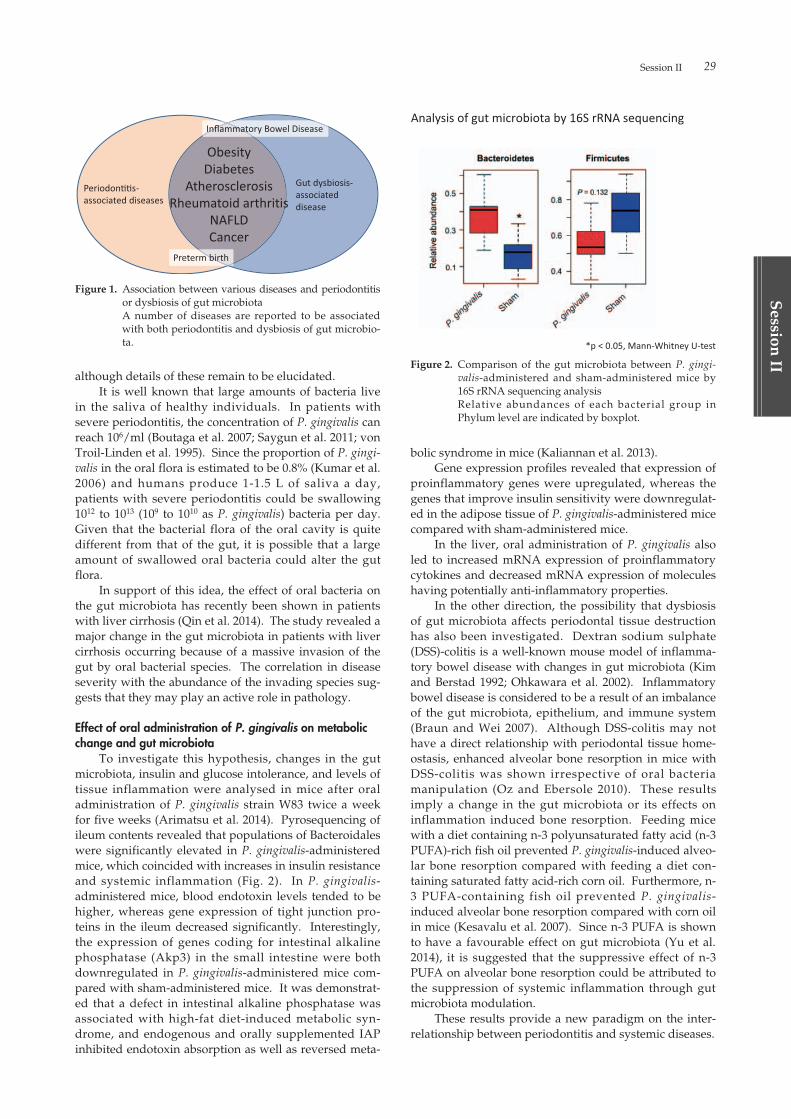
Session II 29
Session
II
although details of these remain to be elucidated.It is well known that large amounts of bacteria live
in the saliva of healthy individuals. In patients withsevere periodontitis, the concentration of P. gingivalis canreach 106/ml (Boutaga et al. 2007; Saygun et al. 2011; vonTroil-Linden et al. 1995). Since the proportion of P. gingi-valis in the oral flora is estimated to be 0.8% (Kumar et al.2006) and humans produce 1-1.5 L of saliva a day,patients with severe periodontitis could be swallowing1012 to 1013 (109 to 1010 as P. gingivalis) bacteria per day.Given that the bacterial flora of the oral cavity is quitedifferent from that of the gut, it is possible that a largeamount of swallowed oral bacteria could alter the gutflora.
In support of this idea, the effect of oral bacteria onthe gut microbiota has recently been shown in patientswith liver cirrhosis (Qin et al. 2014). The study revealed amajor change in the gut microbiota in patients with livercirrhosis occurring because of a massive invasion of thegut by oral bacterial species. The correlation in diseaseseverity with the abundance of the invading species sug-gests that they may play an active role in pathology.
Effect of oral administration of P. gingivalis on metabolicchange and gut microbiota
To investigate this hypothesis, changes in the gutmicrobiota, insulin and glucose intolerance, and levels oftissue inflammation were analysed in mice after oraladministration of P. gingivalis strain W83 twice a weekfor five weeks (Arimatsu et al. 2014). Pyrosequencing ofileum contents revealed that populations of Bacteroidaleswere significantly elevated in P. gingivalis-administeredmice, which coincided with increases in insulin resistanceand systemic inflammation (Fig. 2). In P. gingivalis-administered mice, blood endotoxin levels tended to behigher, whereas gene expression of tight junction pro-teins in the ileum decreased significantly. Interestingly,the expression of genes coding for intestinal alkalinephosphatase (Akp3) in the small intestine were bothdownregulated in P. gingivalis-administered mice com-pared with sham-administered mice. It was demonstrat-ed that a defect in intestinal alkaline phosphatase wasassociated with high-fat diet-induced metabolic syn-drome, and endogenous and orally supplemented IAPinhibited endotoxin absorption as well as reversed meta-
bolic syndrome in mice (Kaliannan et al. 2013).Gene expression profiles revealed that expression of
proinflammatory genes were upregulated, whereas thegenes that improve insulin sensitivity were downregulat-ed in the adipose tissue of P. gingivalis-administered micecompared with sham-administered mice.
In the liver, oral administration of P. gingivalis alsoled to increased mRNA expression of proinflammatorycytokines and decreased mRNA expression of moleculeshaving potentially anti-inflammatory properties.
In the other direction, the possibility that dysbiosisof gut microbiota affects periodontal tissue destructionhas also been investigated. Dextran sodium sulphate(DSS)-colitis is a well-known mouse model of inflamma-tory bowel disease with changes in gut microbiota (Kimand Berstad 1992; Ohkawara et al. 2002). Inflammatorybowel disease is considered to be a result of an imbalanceof the gut microbiota, epithelium, and immune system(Braun and Wei 2007). Although DSS-colitis may nothave a direct relationship with periodontal tissue home-ostasis, enhanced alveolar bone resorption in mice withDSS-colitis was shown irrespective of oral bacteriamanipulation (Oz and Ebersole 2010). These resultsimply a change in the gut microbiota or its effects oninflammation induced bone resorption. Feeding micewith a diet containing n-3 polyunsaturated fatty acid (n-3PUFA)-rich fish oil prevented P. gingivalis-induced alveo-lar bone resorption compared with feeding a diet con-taining saturated fatty acid-rich corn oil. Furthermore, n-3 PUFA-containing fish oil prevented P. gingivalis-induced alveolar bone resorption compared with corn oilin mice (Kesavalu et al. 2007). Since n-3 PUFA is shownto have a favourable effect on gut microbiota (Yu et al.2014), it is suggested that the suppressive effect of n-3PUFA on alveolar bone resorption could be attributed tothe suppression of systemic inflammation through gutmicrobiota modulation.
These results provide a new paradigm on the inter-relationship between periodontitis and systemic diseases.
Figure 1. Association between various diseases and periodontitisor dysbiosis of gut microbiotaA number of diseases are reported to be associatedwith both periodontitis and dysbiosis of gut microbio-ta.
Figure 2. Comparison of the gut microbiota between P. gingi-valis-administered and sham-administered mice by16S rRNA sequencing analysisRelative abundances of each bacterial group inPhylum level are indicated by boxplot.
02_本文1027(web用追加) 15.10.27 9:04 AM ページ 29
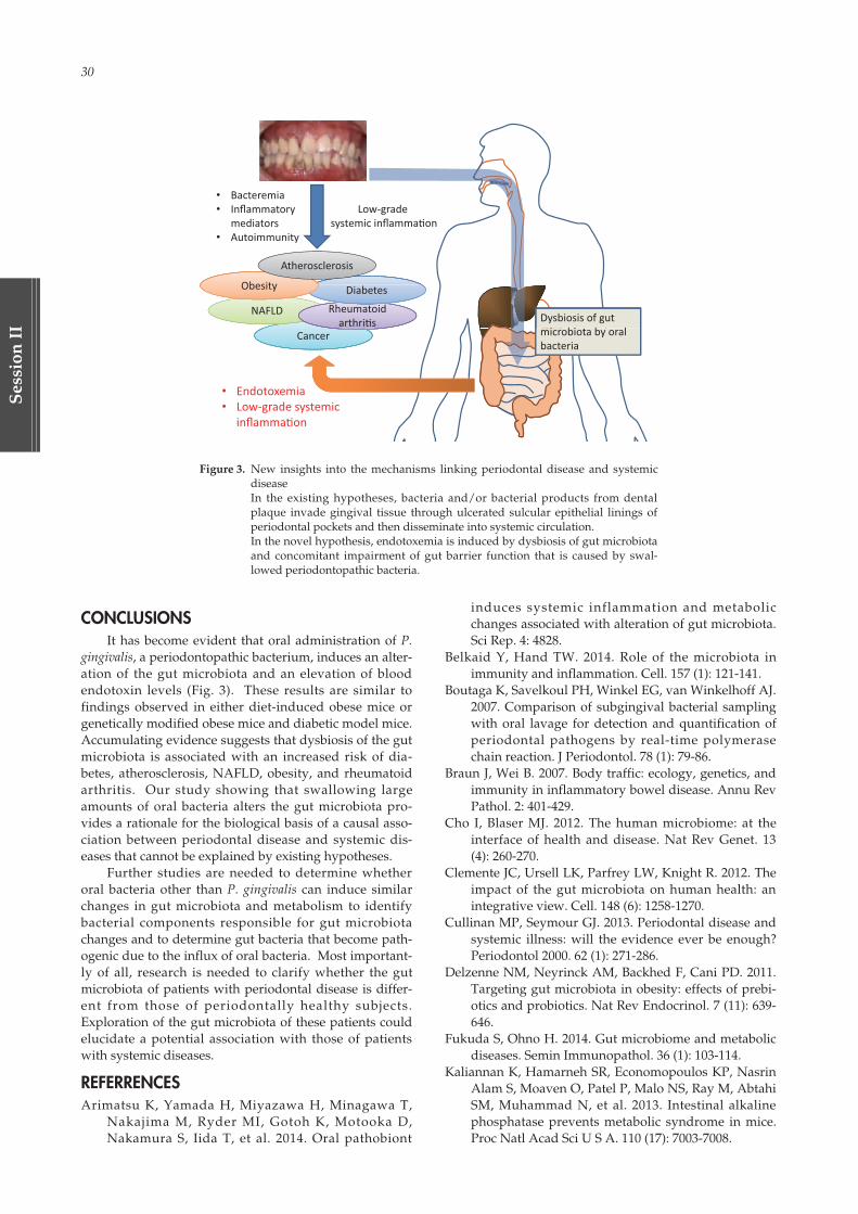
30
Ses
sion
II
CONCLUSIONSIt has become evident that oral administration of P.
gingivalis, a periodontopathic bacterium, induces an alter-ation of the gut microbiota and an elevation of bloodendotoxin levels (Fig. 3). These results are similar tofindings observed in either diet-induced obese mice orgenetically modified obese mice and diabetic model mice.Accumulating evidence suggests that dysbiosis of the gutmicrobiota is associated with an increased risk of dia-betes, atherosclerosis, NAFLD, obesity, and rheumatoidarthritis. Our study showing that swallowing largeamounts of oral bacteria alters the gut microbiota pro-vides a rationale for the biological basis of a causal asso-ciation between periodontal disease and systemic dis-eases that cannot be explained by existing hypotheses.
Further studies are needed to determine whetheroral bacteria other than P. gingivalis can induce similarchanges in gut microbiota and metabolism to identifybacterial components responsible for gut microbiotachanges and to determine gut bacteria that become path-ogenic due to the influx of oral bacteria. Most important-ly of all, research is needed to clarify whether the gutmicrobiota of patients with periodontal disease is differ-ent from those of periodontally healthy subjects.Exploration of the gut microbiota of these patients couldelucidate a potential association with those of patientswith systemic diseases.
REFERRENCESArimatsu K, Yamada H, Miyazawa H, Minagawa T,
Nakajima M, Ryder MI, Gotoh K, Motooka D,Nakamura S, Iida T, et al. 2014. Oral pathobiont
induces systemic inflammation and metabolicchanges associated with alteration of gut microbiota.Sci Rep. 4: 4828.
Belkaid Y, Hand TW. 2014. Role of the microbiota inimmunity and inflammation. Cell. 157 (1): 121-141.
Boutaga K, Savelkoul PH, Winkel EG, van Winkelhoff AJ.2007. Comparison of subgingival bacterial samplingwith oral lavage for detection and quantification ofperiodontal pathogens by real-time polymerasechain reaction. J Periodontol. 78 (1): 79-86.
Braun J, Wei B. 2007. Body traffic: ecology, genetics, andimmunity in inflammatory bowel disease. Annu RevPathol. 2: 401-429.
Cho I, Blaser MJ. 2012. The human microbiome: at theinterface of health and disease. Nat Rev Genet. 13(4): 260-270.
Clemente JC, Ursell LK, Parfrey LW, Knight R. 2012. Theimpact of the gut microbiota on human health: anintegrative view. Cell. 148 (6): 1258-1270.
Cullinan MP, Seymour GJ. 2013. Periodontal disease andsystemic illness: will the evidence ever be enough?Periodontol 2000. 62 (1): 271-286.
Delzenne NM, Neyrinck AM, Backhed F, Cani PD. 2011.Targeting gut microbiota in obesity: effects of prebi-otics and probiotics. Nat Rev Endocrinol. 7 (11): 639-646.
Fukuda S, Ohno H. 2014. Gut microbiome and metabolicdiseases. Semin Immunopathol. 36 (1): 103-114.
Kaliannan K, Hamarneh SR, Economopoulos KP, NasrinAlam S, Moaven O, Patel P, Malo NS, Ray M, AbtahiSM, Muhammad N, et al. 2013. Intestinal alkalinephosphatase prevents metabolic syndrome in mice.Proc Natl Acad Sci U S A. 110 (17): 7003-7008.
Figure 3. New insights into the mechanisms linking periodontal disease and systemicdiseaseIn the existing hypotheses, bacteria and/or bacterial products from dentalplaque invade gingival tissue through ulcerated sulcular epithelial linings ofperiodontal pockets and then disseminate into systemic circulation.In the novel hypothesis, endotoxemia is induced by dysbiosis of gut microbiotaand concomitant impairment of gut barrier function that is caused by swal-lowed periodontopathic bacteria.
02_本文1027(web用追加) 15.10.27 9:04 AM ページ 30
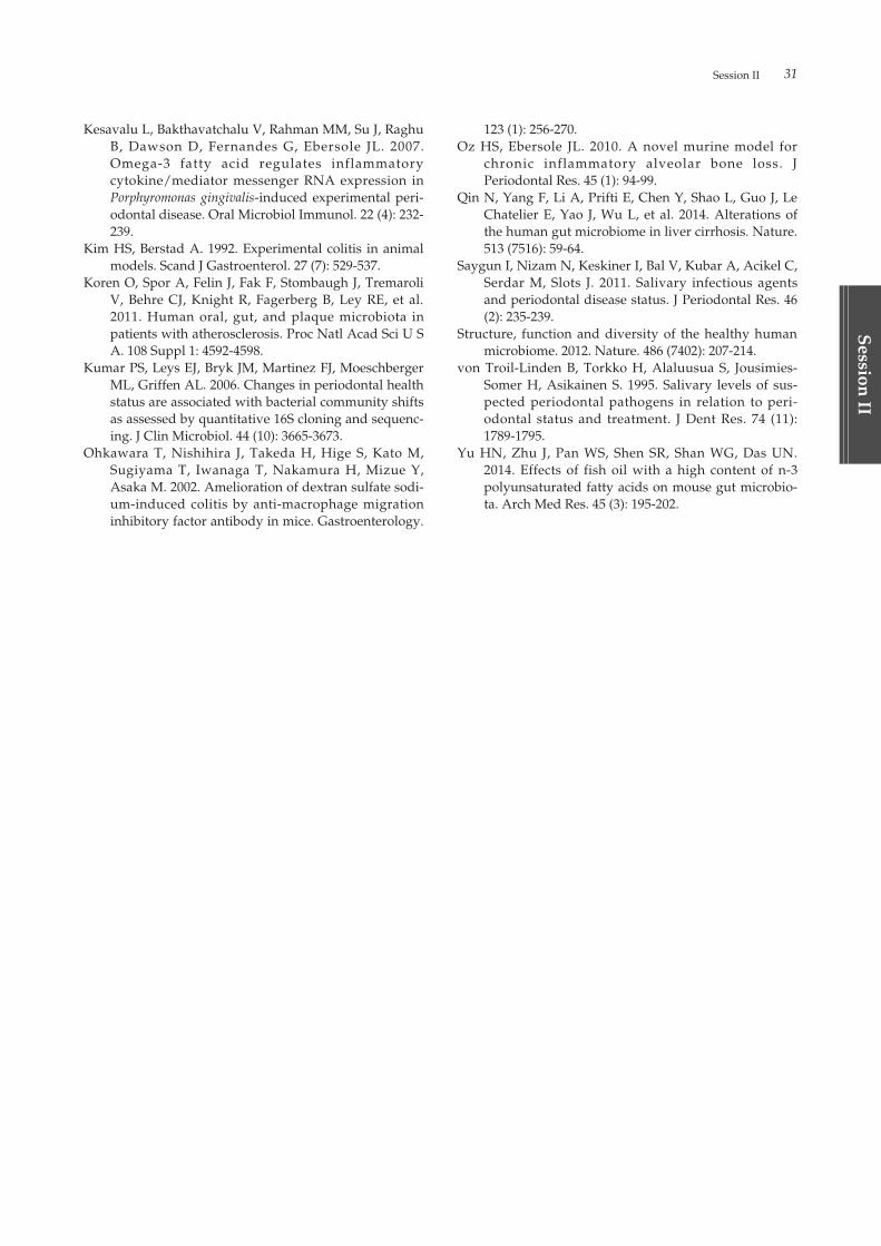
Session II 31
Session
II
Kesavalu L, Bakthavatchalu V, Rahman MM, Su J, RaghuB, Dawson D, Fernandes G, Ebersole JL. 2007.Omega-3 fatty acid regulates inflammatorycytokine/mediator messenger RNA expression inPorphyromonas gingivalis-induced experimental peri-odontal disease. Oral Microbiol Immunol. 22 (4): 232-239.
Kim HS, Berstad A. 1992. Experimental colitis in animalmodels. Scand J Gastroenterol. 27 (7): 529-537.
Koren O, Spor A, Felin J, Fak F, Stombaugh J, TremaroliV, Behre CJ, Knight R, Fagerberg B, Ley RE, et al.2011. Human oral, gut, and plaque microbiota inpatients with atherosclerosis. Proc Natl Acad Sci U SA. 108 Suppl 1: 4592-4598.
Kumar PS, Leys EJ, Bryk JM, Martinez FJ, MoeschbergerML, Griffen AL. 2006. Changes in periodontal healthstatus are associated with bacterial community shiftsas assessed by quantitative 16S cloning and sequenc-ing. J Clin Microbiol. 44 (10): 3665-3673.
Ohkawara T, Nishihira J, Takeda H, Hige S, Kato M,Sugiyama T, Iwanaga T, Nakamura H, Mizue Y,Asaka M. 2002. Amelioration of dextran sulfate sodi-um-induced colitis by anti-macrophage migrationinhibitory factor antibody in mice. Gastroenterology.
123 (1): 256-270.Oz HS, Ebersole JL. 2010. A novel murine model for
chronic inflammatory alveolar bone loss. JPeriodontal Res. 45 (1): 94-99.
Qin N, Yang F, Li A, Prifti E, Chen Y, Shao L, Guo J, LeChatelier E, Yao J, Wu L, et al. 2014. Alterations ofthe human gut microbiome in liver cirrhosis. Nature.513 (7516): 59-64.
Saygun I, Nizam N, Keskiner I, Bal V, Kubar A, Acikel C,Serdar M, Slots J. 2011. Salivary infectious agentsand periodontal disease status. J Periodontal Res. 46(2): 235-239.
Structure, function and diversity of the healthy humanmicrobiome. 2012. Nature. 486 (7402): 207-214.
von Troil-Linden B, Torkko H, Alaluusua S, Jousimies-Somer H, Asikainen S. 1995. Salivary levels of sus-pected periodontal pathogens in relation to peri-odontal status and treatment. J Dent Res. 74 (11):1789-1795.
Yu HN, Zhu J, Pan WS, Shen SR, Shan WG, Das UN.2014. Effects of fish oil with a high content of n-3polyunsaturated fatty acids on mouse gut microbio-ta. Arch Med Res. 45 (3): 195-202.
02_本文1027(web用追加) 15.10.27 9:04 AM ページ 31

02_本文1027(web用追加) 15.10.27 9:04 AM ページ 32
Session
III
Host-Microbe Interaction on Health and Diseases: —Frontiers in Basic Clinical Research—
Molecular Basis Involved in Autophagy-Mediated Clearance of Intracellular Pathogens
Hiroshima University
K. Harada-Hada and T. Kanematsu
Exploring the Host Mechanisms that Impact RNA Virus Replication in the Cell Nucleus
Kyoto University
K. Tomonaga
Bacteriotherapy for Inflammatory Bowel Diseases
Keio University School of Medicine
T. Kanai
Role of Dental Science for the Development of Mucosal Immunology and Vaccine
The University of Tokyo
H. Kiyono
Session III
02_本文1027(web用追加) 15.10.27 9:04 AM ページ 33

02_本文1027(web用追加) 15.10.27 9:04 AM ページ 34
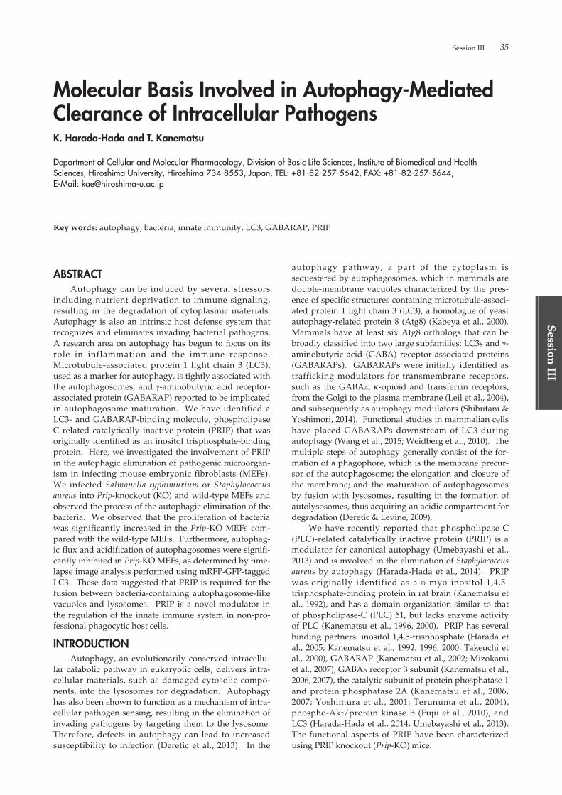
Session III 35
Session
III
ABSTRACTAutophagy can be induced by several stressors
including nutrient deprivation to immune signaling,resulting in the degradation of cytoplasmic materials.Autophagy is also an intrinsic host defense system thatrecognizes and eliminates invading bacterial pathogens.A research area on autophagy has begun to focus on itsrole in inflammation and the immune response.Microtubule-associated protein 1 light chain 3 (LC3),used as a marker for autophagy, is tightly associated withthe autophagosomes, and γ-aminobutyric acid receptor-associated protein (GABARAP) reported to be implicatedin autophagosome maturation. We have identified aLC3- and GABARAP-binding molecule, phospholipaseC-related catalytically inactive protein (PRIP) that wasoriginally identified as an inositol trisphosphate-bindingprotein. Here, we investigated the involvement of PRIPin the autophagic elimination of pathogenic microorgan-ism in infecting mouse embryonic fibroblasts (MEFs).We infected Salmonella typhimurium or Staphylococcusaureus into Prip-knockout (KO) and wild-type MEFs andobserved the process of the autophagic elimination of thebacteria. We observed that the proliferation of bacteriawas significantly increased in the Prip-KO MEFs com-pared with the wild-type MEFs. Furthermore, autophag-ic flux and acidification of autophagosomes were signifi-cantly inhibited in Prip-KO MEFs, as determined by time-lapse image analysis performed using mRFP-GFP-taggedLC3. These data suggested that PRIP is required for thefusion between bacteria-containing autophagosome-likevacuoles and lysosomes. PRIP is a novel modulator inthe regulation of the innate immune system in non-pro-fessional phagocytic host cells.
INTRODUCTIONAutophagy, an evolutionarily conserved intracellu-
lar catabolic pathway in eukaryotic cells, delivers intra-cellular materials, such as damaged cytosolic compo-nents, into the lysosomes for degradation. Autophagyhas also been shown to function as a mechanism of intra-cellular pathogen sensing, resulting in the elimination ofinvading pathogens by targeting them to the lysosome.Therefore, defects in autophagy can lead to increasedsusceptibility to infection (Deretic et al., 2013). In the
autophagy pathway, a part of the cytoplasm issequestered by autophagosomes, which in mammals aredouble-membrane vacuoles characterized by the pres-ence of specific structures containing microtubule-associ-ated protein 1 light chain 3 (LC3), a homologue of yeastautophagy-related protein 8 (Atg8) (Kabeya et al., 2000).Mammals have at least six Atg8 orthologs that can bebroadly classified into two large subfamilies: LC3s and γ-aminobutyric acid (GABA) receptor-associated proteins(GABARAPs). GABARAPs were initially identified astrafficking modulators for transmembrane receptors,such as the GABAA, κ-opioid and transferrin receptors,from the Golgi to the plasma membrane (Leil et al., 2004),and subsequently as autophagy modulators (Shibutani &Yoshimori, 2014). Functional studies in mammalian cellshave placed GABARAPs downstream of LC3 duringautophagy (Wang et al., 2015; Weidberg et al., 2010). Themultiple steps of autophagy generally consist of the for-mation of a phagophore, which is the membrane precur-sor of the autophagosome; the elongation and closure ofthe membrane; and the maturation of autophagosomesby fusion with lysosomes, resulting in the formation ofautolysosomes, thus acquiring an acidic compartment fordegradation (Deretic & Levine, 2009).
We have recently reported that phospholipase C(PLC)-related catalytically inactive protein (PRIP) is amodulator for canonical autophagy (Umebayashi et al.,2013) and is involved in the elimination of Staphylococcusaureus by autophagy (Harada-Hada et al., 2014). PRIPwas originally identified as a D-myo-inositol 1,4,5-trisphosphate-binding protein in rat brain (Kanematsu etal., 1992), and has a domain organization similar to thatof phospholipase-C (PLC) δ1, but lacks enzyme activityof PLC (Kanematsu et al., 1996, 2000). PRIP has severalbinding partners: inositol 1,4,5-trisphosphate (Harada etal., 2005; Kanematsu et al., 1992, 1996, 2000; Takeuchi etal., 2000), GABARAP (Kanematsu et al., 2002; Mizokamiet al., 2007), GABAA receptor β subunit (Kanematsu et al.,2006, 2007), the catalytic subunit of protein phosphatase 1and protein phosphatase 2A (Kanematsu et al., 2006,2007; Yoshimura et al., 2001; Terunuma et al., 2004),phospho-Akt/protein kinase B (Fujii et al., 2010), andLC3 (Harada-Hada et al., 2014; Umebayashi et al., 2013).The functional aspects of PRIP have been characterizedusing PRIP knockout (Prip-KO) mice.
Molecular Basis Involved in Autophagy-MediatedClearance of Intracellular PathogensK. Harada-Hada and T. Kanematsu
Department of Cellular and Molecular Pharmacology, Division of Basic Life Sciences, Institute of Biomedical and HealthSciences, Hiroshima University, Hiroshima 734-8553, Japan, TEL: +81-82-257-5642, FAX: +81-82-257-5644, E-Mail: [email protected]
Key words: autophagy, bacteria, innate immunity, LC3, GABARAP, PRIP
02_本文1027(web用追加) 15.10.27 9:04 AM ページ 35
36
Ses
sion
III
Accumulation of S. typhimurium and S. aureus in LC3-positive autophagosome-like vacuoles are observed in Prip-KO MEFs compared with wild-type MEFs
To investigate whether PRIP affects proliferation ofbacteria in infecting host cells, we used MEFs preparedfrom Prip-KO and wild-type mice to examine mecha-nisms of PRIP-mediated elimination of Salmonellatyphimurium, a gram-negative bacterial pathogen thatcauses human foodborne illness worldwide by infectingboth mammalian intestinal epithelial cells andmacrophages, and S. aureus, a gram-positive bacterialpathogen that causes serious diseases including pneumo-nia, endocarditis, and osteomyelitis, in addition towound infection, by autophagic pathway. Either S.typhimurium or S. aureus were infected into MEFs thatwere transfected with mRFP-LC3. The number of S.typhimurium or S. aureus was increased in the LC3-posi-tive autophagosome-like vacuoles in Prip-KO MEFs com-pared with wild-type MEFs.
Inhibition of autophagic flux and acidification in Prip-KOMEFs
We next monitored the autophagy maturation usinga tandem fluorescent-tagged LC3 (RFP-GFP-LC3), a con-venient tool to monitor autophagic flux based on the dif-ferent pH stabilities of the EGFP and mRFP fluorescentproteins. The red signals (i.e., green fluorescence-quenched vesicles) appeared in wild-type MEFs within 3h, whereas red signals in Prip-KO cells were infrequentlyobserved in both experiments using S. typhimurium andS. aureus. To confirm the existence of autolysosome-likeacidic vacuoles, we then analyzed the acidification of S.aureus-containing autophagosome-like vesicles (SAcVs)in MEFs using LysoTracker blue DND-22, a probe foracidic compartments. LC3-positive SAcVs stained withLysoTracker were observed 3 h post-infection in wild-type MEFs, but there were fewer apparent in Prip-KOMEFs.
PRIP is a novel modulator of autophagy We show here that PRIP is involved in the entrap-
ment of pathogens by LC3-positive autophagosome-likevacuoles and contributes to the autophagic clearance ofbacterial pathogens as part of an innate defense system ineukaryotic non-professional phagocytic cells. FollowingS. typhimurium invasion, most bacteria reside and repli-cate within intracellular compartments termedSalmonella-containing vacuoles (SCVs). However, a sig-nificant fraction of S. typhimurium, which colocalizes withp62 that binds both polyubiquitinated proteins and LC3,is targeted by autophagy, and thus autophagy restrictsSalmonella replication (Zheng et al., 2009).Phosphatidylinositol 3-phosphate [PI(3)P], and doubleFYVE domain-containing protein 1 (DFCP1) and WDrepeat domain phosphoinositide-interacting proteins(WIPI), binding proteins of PI(3)P, mediates autophagicelimination of intracellular bacteria (Huang et al., 2011;Mauthe et al. 2012; Mostowy et al., 2014). In Prip-KOcells, S. typhimurium and S. aureus can escape the hostdefense system via autophagy due to the autophago-some/lysosome fusion process being disabled. A possi-ble mechanism in Prip-mediated autophagic eliminationof bacteria is shown in Fig. 1. The PRIP-mediated elimi-nation of bacteria from infected host cells may give usnew insight into potentially effective treatments for infec-tious diseases.
AKNOWLEDGMENTSK.H. and T.K. conceived and designed the experi-
ments. K.H. chiefly performed the experiments. Thiswork was supported by the Funding Program for NextGeneration World-Leading Researchers [LS087 to TK],and by grants from JSPS KAKENHI [Grant numbers25861757 and 15K11039 to K.H.]. The authors have nocompeting interests to declare.
REFERENCESDeretic V, Levine B. Autophagy, immunity, and micro-
bial adaptations. Cell Host Microbe, 2009: 5: 527-549.Deretic V, Saitoh T, Akira S. Autophagy in infection,
inflammation and immunity. Nat Rev Immunol,2013: 13: 722-737.
Figure 1. A possible mechanism in PRIP-mediated autophagic elimination of bacteriaInvading bacterial pathogens are sequestered by autophagosome-like vacuoles decorated with phos-phatidylinositol 3-phosphate [PI(3)P], double FYVE domain-containing protein 1 (DFCP1), and WDrepeat domain phosphoinositide-interacting proteins (WIPI). The LC3-positive autophagosome-likevacuoles are fused with lysosome, and autolysosomes are formed, resulting in elimination of bacteria.PRIP may regulate this maturation process of autophagy, and GABARAP may be involved in theenclosing and/or maturation process together with PRIP, a binding partner of GABARAP.
02_本文1027(web用追加) 15.10.27 9:04 AM ページ 36
Session III 37
Session
III
Fujii M, Kanematsu T, Ishibashi H, Fukami K, et al.Phospholipase C-related but catalytically inactiveprotein is required for insulin-induced cell surfaceexpression of γ-aminobutyric acid type A receptors, JBiol Chem, 2010: 285: 4837-4846.
Harada K, Takeuchi H, Oike M, Matsuda M, et al. Role ofPRIP-1, a novel Ins(1,4,5)P3 binding protein, inIns(1,4,5)P3-mediated Ca2+ signaling. J Cell Physiol,2005: 202: 422-433.
Harada-Hada K, Harada K, Kato F, Hisatsune J, et al.Phospholipase C-related catalytically inactive pro-tein participates in the autophagic elimination ofStaphylococcus aureus infecting mouse embryonicfibroblasts. PLoS One, 2014: 9 (5): e98285
Huang J, Birmingham CL, Shahnazari S, Shiu J, et al.Antibacterial autophagy occurs at PI(3)P-enricheddomains of the endoplasmic reticulum and requiresRab1 GTPase. Autophagy, 2011: 7 (1): 17-26.
Kabeya Y, Mizushima N, Uero T, Yamamoto A, KirisakoT, et al. LC3, a mammalian homologue of yeastApg8p, is localized in autophagosome membranesafter processing. EMBO J, 2000: 19: 5720-5728.
Kanematsu T, Takeya H, Watanabe Y, Ozaki S, et al.Putative inositol 1,4,5-trisphosphate binding proteinsin rat brain cytosol. J Biol Chem, 1992: 267: 6518-6525.
Kanematsu T, Misumi Y, Watanabe Y, Ozaki S, et al. Anew inositol 1,4,5-trisphosphate binding protein sim-ilar to phospholipase C-δ1. Biochem J, 1996: 313 (Pt1): 319-325.
Kanematsu T, Yoshimura K, Hidaka K, Takeuchi H, et al.Domain organization of p130, PLC-related catalyti-cally inactive protein, and structural basis for thelack of enzyme activity. Eur J Biochem, 2000: 267:2731-2737.
Kanematsu T, Jang IS, Yamaguchi T, Nagahama H, et al.Role of the PLC-related, catalytically inactive proteinp130 in GABAA receptor function. EMBO J, 2002: 21:1004-1011.
Kanematsu T, Yasunaga A, Mizoguchi Y, Kuratani A, etal. Modulation of GABAA receptor phosphorylationand membrane trafficking by phospholipase C-relat-ed inactive protein/protein phosphatase 1 and 2Asignaling complex underlying BDNF-dependent reg-ulation of GABAergic inhibition, J Biol Chem, 2006:281: 22180-22189.
Kanematsu T, Fujii M, Mizokami A, Kittler JT, et al.Phospholipase C-related inactive protein is implicat-ed in the constitutive internalization of GABAA
receptors mediated by clathrin and AP2 adaptorcomplex, J Neurochem, 2007: 101: 898-905.
Leil TA, Chen Z-W, Chang C-SS, Olsen RW. GABAA
receptor-associated protein traffics GABAA receptors
to the plasma membrane in neurons. J Neurosci,2004: 24 (50): 11429-11438.
Mauthe M, Yu W, Krut O, Krönke M, et al. WIPI-1 posi-tive autophagosome-like vesicles entrap pathogenicStaphylococcus aureus for lysosomal degradation. Int JCell Biol, 2012: 179207.
Mizokami A, Kanematsu T, Ishibashi H, Yamaguchi T, etal. Phospholipase C-related but catalytically inactiveprotein is involved in trafficking of γ2 subunit-con-taining GABAA receptors in the cell surface, JNeurosci, 2007: 27: 1692-1701.
Mostowy S. Multiple Roles of the Cytoskeleton inBacterial Autophagy. PLoS Pathog, 2014: 10 (11):e1004409.
Schnaith A, Kashkar H, Leggio SA, Addicks K, et al.Staphylococcus aureus subvert autophagy for induc-tion of caspase-independent host cell death. J BiolChem, 2007: 282: 2695-2706.
Shibutani ST, Yoshimori T. A current perspective ofautophagosome biogenesis. Cell Res, 2014: 24 (1): 58-68.
Takeuchi H, Oike M, Paterson HF, Allen V, et al.Inhibition of Ca2+ signalling by p130, a phospholi-pase-C-related catalytically inactive protein: criticalrole of the p130 pleckstrin homology domain.Biochem J, 2000: 349: 357-368.
Terunuma M, Jang IS, Ha SH, Kittler JT, et al. GABAA
receptor phospho-dependent modulation is regulat-ed by phospholipase C-related inactive protein type1, a novel protein phosphatase 1 anchoring protein, JNeurosci, 2004: 24: 7074-7084.
Umebayashi H, Mizokami A, Matsuda M, Harada K, etal. PLC-related catalytically inactive protein (PRIP),a novel microtubule-associated protein 1 light chain3 (LC3)-binding protein, negatively regulatesautophagosome formation. Biochem Biophys ResCommun, 2013: 432: 268-274.
Wang H, Sun HQ, Zhu X, Zhang L, et al. GABARAPsregulate PI4P-dependent autophagosome: lysosomefusion. Proc Natl Acad Sci USA, 2015: 112 (22): 7015-7020.
Weidberg H, Shvets E, Shpilka T, Shimron F, et al. LC3and GATE-16/GABARAP subfamilies are bothessential yet act differently in autophagosome bio-genesis. EMBO J, 2010: 29 (11): 1792-1802.
Yoshimura K, Takeuchi H, Sato O, Hidaka K, et al.Interaction of p130 with, and consequent inhibitionof, the catalytic subunit of protein phosphatase 1α. JBiol Chem, 2001: 276: 17908-17913.
Zheng YT, Shahnazari S, Brech A, Lamark T, et al. Theadaptor protein p62/SQSTM1 targets invading bac-teria to the autophagy pathway. J Immunol, 2009: 83(9): 5909-5916.
02_本文1027(web用追加) 15.10.27 9:04 AM ページ 37
38
Ses
sion
III
ABSTRACTBornavirus, a nonsegmented, negative-strand RNA
virus, is characterized by highly neurotropic and noncy-topathic infection and has several unique features amonganimal-derived RNA viruses. The most striking featureof this virus is that they readily establish a long-lastingpersistent infection in the cell nucleus without causingapparent cell damage. This characteristic makes bor-navirus the only animal RNA virus capable of intranu-clear parasitism. Therefore, study of this virus allows usto uncover previously unknown interactions betweenRNA virus and host cells. Recently, we demonstratedthat ribonucleoprotein of a mammalian bornavirus,Borna disease virus (BDV), interacts directly with thehost chromosome throughout cell cycle and that a hostchromatin-binding protein, HMGB1, influences BDVreplication and persistent infection. To investigate theroles of HMGB1 in BDV replication, we also identifiedHMGB1-binding proteins (HBP) in the nucleus of BDV-infected cells. We demonstrated that HBP-1 may play arole in the intranuclear sensing of viral RNP, leading tothe repression of BDV replication. In this talk, I will pre-sent recent our data on the molecular mechanisms of thepersistent infection of BDV and the RNA sensing of hostcells in the nucleus, providing novel insights into theinteraction between RNA viruses and the host cells.
INTRODUCTIONThe cell nucleus is a dynamic organelle that may be
considered an inconvenient site for virus infection.Dissolution of the nuclear membrane during mitosis andnucleocytoplasmic trafficking through nuclear porescould lead to the loss or dilution of viral genomes fromthe nucleus. Alteration of the intranuclear environmentby gene expression may also affect virus stability. Forpersistent DNA viruses, such as gammaherpesviruses[Kaposi’s sarcoma-associated herpesvirus (KSHV) andEpstein-Barr virus] and papillomaviruses, association ofa low copy number of viral episomal DNA with cellularchromosomes ensures their stable retention over longperiods of time in such a dynamic environment[1-5].Residence in the nucleus, however, would not be an easy
task for other types of viruses, in particular viruses usingRNA as the template for replication.
Among vertebrate RNA viruses, except for thereverse-transcribing retrovirus, only a few families,Bornaviridae, Nyamiviridae and Orthomyxoviridae, enter thenucleus for replication and transcription. However, thereis a fundamental difference between these viruses.Bornaviruses show a non-cytopathic replication andreadily establish a long-lasting, persistent infection in thenuclei of cultured cells and various tissues[6,7], whereasinfections by the viruses of the Nyamiviridae andOrthomyxoviridae, seem to induce cytopathic effects.These features indicate that bornaviruses are the onlyknown RNA virus that can survive for a long-time withinthe nuclear membrane. However, the mechanism of howbornaviruses can achieve such a stable intranuclear infec-tion has not been fully understood[8,9].
During viral infection, invading foreign DNA andRNA potently activate the innate and subsequent adap-tive immune responses[10,11]. Nucleic acid sensors playcritical roles in the induction of immune responses, suchas the induction of interferon (IFN), proinflammatorycytokines and chemokines. Toll-like receptor (TLR) 3,TLR7, RIG-I, and MDA5 are best known as RNA sensorsin the cytoplasm[10-12]. In addition, DNA sensors, such asTLR9, DNA-dependent activator of IRFs (DAI), IFN-inducible protein 16 (IFI16), and absent in melanoma 2(AIM2) are known to induce innate and adaptiveimmune responses[13-16]. It has been believed that thecytoplasm is the center of nucleic acid sensing, becauseall of these nucleic acid sensors are mainly located in thecytoplasm. On the other hand, IFI16 has been recentlyshown to act as a nuclear pathogenic DNA sensor inresponse to KSHV infection[17]. It has been still unclear,however, whether the host cell nucleus has the machiner-ies for sensing the invading viral RNAs.
In this report, with a unique model of intranuclearinfection, bornaviruses, we provide new insights into themolecular mechanisms of how the host nucleus standsagainst invading RNA molecules and how a particularRNA virus can evade such host sensing mechanism inthe nucleus.
Exploring the Host Mechanisms that Impact RNA Virus Replication in the Cell NucleusK. Tomonaga*
Department of Viral Oncology, Institute for Virus Research (IVR), Department of Mammalian Regulatory Network, Graduate School of Biostudies, and Department of Tumor Viruses, Graduate School of Medicine, Kyoto University, Kyoto 606-8507, Japan* To whom correspondence should be addressed. Keizo Tomonaga: Department of Viral Oncology, Institute for Virus Research,
Kyoto University, 53 Kawahara-cho, Shogoin, Sakyo-ku, Kyoto 606-8507, Japan. [email protected]
Key words: RNA viruses, Bornavirus, persistent infection, cell nucleus, innate immune responses
02_本文1027(web用追加) 15.10.27 9:04 AM ページ 38
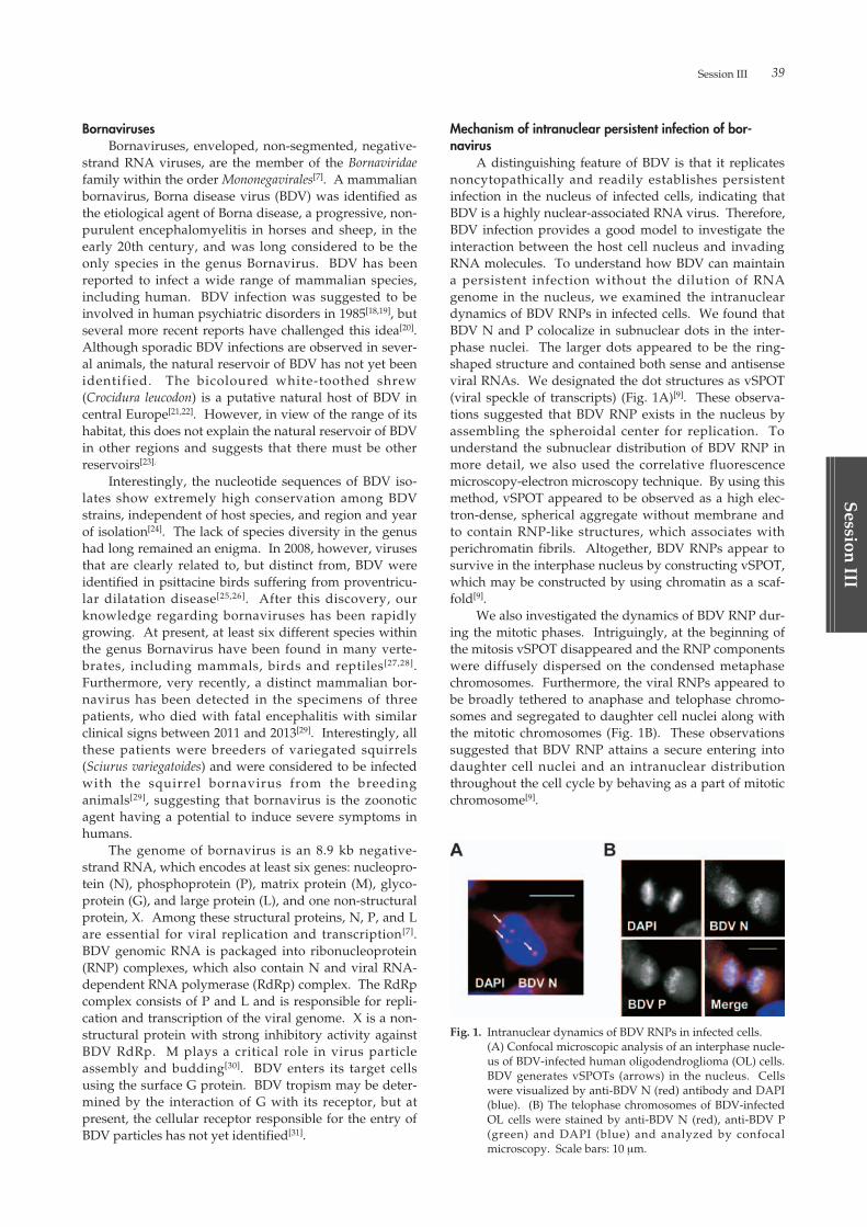
Session III 39
Session
III
BornavirusesBornaviruses, enveloped, non-segmented, negative-
strand RNA viruses, are the member of the Bornaviridaefamily within the order Mononegavirales[7]. A mammalianbornavirus, Borna disease virus (BDV) was identified asthe etiological agent of Borna disease, a progressive, non-purulent encephalomyelitis in horses and sheep, in theearly 20th century, and was long considered to be theonly species in the genus Bornavirus. BDV has beenreported to infect a wide range of mammalian species,including human. BDV infection was suggested to beinvolved in human psychiatric disorders in 1985[18,19], butseveral more recent reports have challenged this idea[20].Although sporadic BDV infections are observed in sever-al animals, the natural reservoir of BDV has not yet beenidentified. The bicoloured white-toothed shrew(Crocidura leucodon) is a putative natural host of BDV incentral Europe[21,22]. However, in view of the range of itshabitat, this does not explain the natural reservoir of BDVin other regions and suggests that there must be otherreservoirs[23].
Interestingly, the nucleotide sequences of BDV iso-lates show extremely high conservation among BDVstrains, independent of host species, and region and yearof isolation[24]. The lack of species diversity in the genushad long remained an enigma. In 2008, however, virusesthat are clearly related to, but distinct from, BDV wereidentified in psittacine birds suffering from proventricu-lar dilatation disease[25,26]. After this discovery, ourknowledge regarding bornaviruses has been rapidlygrowing. At present, at least six different species withinthe genus Bornavirus have been found in many verte-brates, including mammals, birds and reptiles[27,28].Furthermore, very recently, a distinct mammalian bor-navirus has been detected in the specimens of threepatients, who died with fatal encephalitis with similarclinical signs between 2011 and 2013[29]. Interestingly, allthese patients were breeders of variegated squirrels(Sciurus variegatoides) and were considered to be infectedwith the squirrel bornavirus from the breedinganimals[29], suggesting that bornavirus is the zoonoticagent having a potential to induce severe symptoms inhumans.
The genome of bornavirus is an 8.9 kb negative-strand RNA, which encodes at least six genes: nucleopro-tein (N), phosphoprotein (P), matrix protein (M), glyco-protein (G), and large protein (L), and one non-structuralprotein, X. Among these structural proteins, N, P, and Lare essential for viral replication and transcription[7].BDV genomic RNA is packaged into ribonucleoprotein(RNP) complexes, which also contain N and viral RNA-dependent RNA polymerase (RdRp) complex. The RdRpcomplex consists of P and L and is responsible for repli-cation and transcription of the viral genome. X is a non-structural protein with strong inhibitory activity againstBDV RdRp. M plays a critical role in virus particleassembly and budding[30]. BDV enters its target cellsusing the surface G protein. BDV tropism may be deter-mined by the interaction of G with its receptor, but atpresent, the cellular receptor responsible for the entry ofBDV particles has not yet identified[31].
Mechanism of intranuclear persistent infection of bor-navirus
A distinguishing feature of BDV is that it replicatesnoncytopathically and readily establishes persistentinfection in the nucleus of infected cells, indicating thatBDV is a highly nuclear-associated RNA virus. Therefore,BDV infection provides a good model to investigate theinteraction between the host cell nucleus and invadingRNA molecules. To understand how BDV can maintaina persistent infection without the dilution of RNAgenome in the nucleus, we examined the intranucleardynamics of BDV RNPs in infected cells. We found thatBDV N and P colocalize in subnuclear dots in the inter-phase nuclei. The larger dots appeared to be the ring-shaped structure and contained both sense and antisenseviral RNAs. We designated the dot structures as vSPOT(viral speckle of transcripts) (Fig. 1A)[9]. These observa-tions suggested that BDV RNP exists in the nucleus byassembling the spheroidal center for replication. Tounderstand the subnuclear distribution of BDV RNP inmore detail, we also used the correlative fluorescencemicroscopy-electron microscopy technique. By using thismethod, vSPOT appeared to be observed as a high elec-tron-dense, spherical aggregate without membrane andto contain RNP-like structures, which associates withperichromatin fibrils. Altogether, BDV RNPs appear tosurvive in the interphase nucleus by constructing vSPOT,which may be constructed by using chromatin as a scaf-fold[9].
We also investigated the dynamics of BDV RNP dur-ing the mitotic phases. Intriguingly, at the beginning ofthe mitosis vSPOT disappeared and the RNP componentswere diffusely dispersed on the condensed metaphasechromosomes. Furthermore, the viral RNPs appeared tobe broadly tethered to anaphase and telophase chromo-somes and segregated to daughter cell nuclei along withthe mitotic chromosomes (Fig. 1B). These observationssuggested that BDV RNP attains a secure entering intodaughter cell nuclei and an intranuclear distributionthroughout the cell cycle by behaving as a part of mitoticchromosome[9].
Fig. 1. Intranuclear dynamics of BDV RNPs in infected cells.(A) Confocal microscopic analysis of an interphase nucle-us of BDV-infected human oligodendroglioma (OL) cells.BDV generates vSPOTs (arrows) in the nucleus. Cellswere visualized by anti-BDV N (red) antibody and DAPI(blue). (B) The telophase chromosomes of BDV-infectedOL cells were stained by anti-BDV N (red), anti-BDV P(green) and DAPI (blue) and analyzed by confocalmicroscopy. Scale bars: 10 µm.
02_本文1027(web用追加) 15.10.27 9:04 AM ページ 39
40
Ses
sion
III
We have previously demonstrated that the chro-matin-remodeling, DNA-binding protein, HMGB1 (highmobility group box protein 1) binds to BDV RNPs[32]. Wealso found that HMGB1 is involved in the stability andtranscription of BDV on chromatin by increasing the sta-bility of the P with chromatin-bound RNPs[9], suggestingthat bornavirus establishes a life cycle highly associatedwith the cellular chromosome and takes advantage ofchromosomal stability and dynamics to ensure theintegrity of its RNP in the infected nucleus.
BDV can establish a long-lasting persistent infectionin the nucleus and induce sustained chemokine synthesisin the absence of IFNs during its persistent infection[33],indicating that the study of BDV may allow us to uncoverintranuclear machineries for sensing pathogenic RNAs.Intriguingly, HMGB1 is also reported to function as auniversal sensor for nucleic acids, triggering the innateimmune responses[34]. The binding of nucleic acids toHMGB1 is a precondition for the more efficient, subse-quent recognition by discriminative sensors [34].Therefore, it is conceivable that BDV RNPs may be recog-nized by HMGB1 and unidentified discriminative sensor(s) in the nucleus by the host to induce innate immuneresponses. To evaluate this possibility, we now investi-gating the intranuclear sensing mechanisms for non-selfRNAs by using BDV as an infection model of RNA virus-es in the nucleus. Our studies could provide a new para-digm of infection of non-reverse transcribing RNA virus-es in eukaryotic cells.
ACKNOWLEDGMENTThis study was supported in part by KAKENHI
Grant Numbers 26253027, 26670225, 15H01259 (KT), theCore-to-Core Program A, Advanced Research Networks(KT) from the Japan Society for the Promotion of Science(JSPS), Basic Science and Platform Technology Programfor Innovative Biological Medicine from the JapanAgency for Medical Research and Development (AMED)(KT).
REFERENCES01. Ballestas ME, Chatis PA, Kaye KM. Efficient persis-
tence of extrachromosomal KSHV DNA mediated bylatency-associated nuclear antigen. Science 1999: 284:641-4.
02. Deutsch MJ, Ott E, Papior P, Schepers A. The latentorigin of replication of Epstein-Barr virus directs viralgenomes to active regions of the nucleus. J Virol 2010:84: 2533-46.
03. Feeney KM, Parish JL, Targeting mitotic chromo-somes: a conserved mechanism to ensure viralgenome persistence. Proc Biol Sci 2009: 276: 1535-44.
04. Lehman CW, Botchan MR. Segregation of viral plas-mids depends on tethering to chromosomes and isregulated by phosphorylation. Proc Natl Acad Sci U SA 1998: 95: 4338-43.
05. Piolot T, Tramier M, Coppey M, Nicolas JC, MarechalV. Close but distinct regions of human herpesvirus 8latency-associated nuclear antigen 1 are responsiblefor nuclear targeting and binding to human mitoticchromosomes. J Virol 2001: 75: 3948-59.
06. Schneider U, Martin A, Schwemmle M, Staeheli P.
Genome trimming by Borna disease viruses: viralreplication control or escape from cellular surveil-lance? Cell Mol Life Sci 2007: 64: 1038-42.
07. Tomonaga K, Kobayashi T, Ikuta K. Molecular andcellular biology of Borna disease virus infection.Microbes Infect 2002: 4: 491-500.
08. Horie M, Honda T, Suzuki Y, Kobayashi Y, Daito T,Oshida T, Ikuta K, Jern P, Gojobori T, Coffin JM,Tomonaga K. Endogenous non-retroviral RNA viruselements in mammalian genomes. Nature 2010: 463:84-7.
09. Matsumoto Y, Hayashi Y, Omori H, Honda T, DaitoT, Horie M, Ikuta K, Fujino K, Nakamura S, SchneiderU, Chase G, Yoshimori T, Schwemmle M, TomonagaK. Bornavirus closely associates and segregates withhost chromosomes to ensure persistent intranuclearinfection. Cell Host Microbe 2012: 11: 492-503.
10. Medzhitov R, Horng T. Transcriptional control of theinflammatory response. Nat Rev Immunol 2009: 9:692-703.
11. Akira S, Uematsu S, Takeuchi O. Pathogen recogni-tion and innate immunity. Cell 2006: 124: 783-801.
12. Yoneyama M, Fujita T. Recognition of viral nucleicacids in innate immunity. Rev Med Virol 2010: 20: 4-22.
13. Schroder K, Muruve DA, Tschopp J. Innate immunity:cytoplasmic DNA sensing by the AIM2 inflamma-some. Curr Biol 2009: 19: R262-5.
14. Unterholzner L, Keating SE, Baran M, Horan KA,Jensen SB, Sharma S, Sirois CM, Jin T, Latz E, Xiao TS,Fitzgerald KA, Paludan SR, Bowie AG. IFI16 is aninnate immune sensor for intracellular DNA. NatImmunol 2010: 11: 997-1004.
15. Takaoka A, Wang Z, Choi MK, Yanai H, Negishi H,Ban T, Lu Y, Miyagishi M, Kodama T, Honda K, OhbaY, Taniguchi T. DAI (DLM-1/ZBP1) is a cytosolicDNA sensor and an activator of innate immuneresponse. Nature 2007: 448: 501-5.
16. Ishii KJ, Kawagoe T, Koyama S, Matsui K, Kumar H,Kawai T, Uematsu S, Takeuchi O, Takeshita F, CobanC, Akira S. TANK-binding kinase-1 delineates innateand adaptive immune responses to DNA vaccines.Nature 2008: 451: 725-9.
17. Kerur N, Veettil MV, Sharma-Walia N, Bottero V,Sadagopan S, Otageri P, Chandran B. IFI16 acts as anuclear pathogen sensor to induce the inflammasomein response to Kaposi Sarcoma-associated herpesvirusinfection. Cell Host Microbe 2011: 9: 363-75.
18. Amsterdam JD, Winokur A, Dyson W, Herzog S,Gonzalez F, Rott R, Koprowski H. Borna diseasevirus. A possible etiologic factor in human affectivedisorders? Arch Gen Psychiatry 1985: 42: 1093-6.
19. Rott R, Herzog S, Fleischer B, Winokur A, AmsterdamJ, Dyson W, Koprowski H. Detection of serum anti-bodies to Borna disease virus in patients with psychi-atric disorders. Science 1985: 228: 755-6.
20. Hornig M, Briese T, Licinio J, Khabbaz RF, AltshulerLL, Potkin SG, Schwemmle M, Siemetzki U, Mintz J,Honkavuori K, Kraemer HC, Egan MF, Whybrow PC,Bunney WE, Lipkin WI. Absence of evidence for bor-navirus infection in schizophrenia, bipolar disorderand major depressive disorder. Mol Psychiatry 2012:
02_本文1027(web用追加) 15.10.27 9:04 AM ページ 40
Session III 41
Session
III
17: 486-93.21. Hilbe M, Herrsche R, Kolodziejek J, Nowotny N,
Zlinszky K, Ehrensperger F. Shrews as reservoir hostsof borna disease virus. Emerg Infect Dis 2006: 12: 675-7.
22. Puorger ME, Hilbe M, Muller JP, Kolodziejek J,Nowotny N, Zlinszky K, Ehrensperger F. Distributionof Borna disease virus antigen and RNA in tissues ofnaturally infected bicolored white-toothed shrews,Crocidura leucodon, supporting their role as reservoirhost species. Vet Pathol 2010: 47: 236-44.
23. Kinnunen PM, Palva A, Vaheri A, Vapalahti O.Epidemiology and host spectrum of Borna diseasevirus infections. J Gen Virol 2013: 94: 247-62.
24. Nowotny N, Kolodziejek J, Jehle CO, Suchy A,Staeheli P, Schwemmle M. Isolation and characteriza-tion of a new subtype of Borna disease virus. J Virol2000: 74: 5655-8.
25. Honkavuori KS, Shivaprasad HL, Williams BL, QuanPL, Hornig M, Street C, Palacios G, Hutchison SK,Franca M, Egholm M, Briese T, Lipkin WI. Novelborna virus in psittacine birds with proventriculardilatation disease. Emerg Infect Dis 2008: 14: 1883-6.
26. Kistler AL, Gancz A, Clubb S, Skewes-Cox P, FischerK, Sorber K, Chiu CY, Lublin A, Mechani S, FarnoushiY, Greninger A, Wen CC, Karlene SB, Ganem D,DeRisi JL. Recovery of divergent avian bornavirusesfrom cases of proventricular dilatation disease: identi-fication of a candidate etiologic agent. Virol J 2008: 5:88.
27. Stenglein MD, Leavitt EB, Abramovitch MA, McGuireJA, DeRisi JL. Genome Sequence of a BornavirusRecovered from an African Garter Snake (Elapsoidealoveridgei). Genome Announc 2014: 2.
28. Kuhn JH, Durrwald R, Bao Y, Briese T, Carbone K,Clawson AN, deRisi JL, Garten W, Jahrling PB,
Kolodziejek J, Rubbenstroth D, Schwemmle M,Stenglein M, Tomonaga K, Weissenbock H, NowotnyN. Taxonomic reorganization of the familyBornaviridae. Arch Virol 2015: 160: 621-32.
29. Hoffmann B, Tappe D, Hoper D, Herden C, Boldt A,Mawrin C, Niederstrasser O, Muller T, Jenckel M, vander Grinten E, Lutter C, Abendroth B, Teifke JP,Cadar D, Schmidt-Chanasit J, Ulrich RG, Beer MA.Variegated Squirrel Bornavirus Associated with FatalHuman Encephalitis. N Engl J Med 2015: 373: 154-62.
30. Chase G, Mayer D, Hildebrand A, Frank R, HayashiY, Tomonaga K, Schwemmle M. Borna disease virusmatrix protein is an integral component of the viralribonucleoprotein complex that does not interferewith polymerase activity. J Virol 2007: 81: 743-9.
31. Honda T, Horie M, Daito T, Ikuta K, Tomonaga K.Molecular chaperone BiP interacts with Borna diseasevirus glycoprotein at the cell surface. J Virol 2009: 83:12622-5.
32. Kamitani W, Shoya Y, Kobayashi T, Watanabe M, LeeBJ, Zhang G, Tomonaga K, Ikuta K. Borna diseasevirus phosphoprotein binds a neurite outgrowth fac-tor, amphoterin/HMG-1. J Virol 2001: 75: 8742-51.
33. Sauder C, Hallensleben W, Pagenstecher A,Schneckenburger S, Biro L, Pertlik D, Hausmann J,Suter M, Staeheli P. Chemokine gene expression inastrocytes of Borna disease virus-infected rats andmice in the absence of inflammation. J Virol 2000: 74:9267-80.
34. Yanai H, Ban T, Wang Z, Choi MK, Kawamura T,Negishi H, Nakasato M, Lu Y, Hangai S, Koshiba R,Ronfani L, Akira S, Bianchi ME, Honda K, Tamura T,Kodama T, Taniguchi T. HMGB proteins function asuniversal sentinels for nucleic-acid-mediated innateimmune responses. Nature 2009: 462: 99-103.
02_本文1027(web用追加) 15.10.27 9:04 AM ページ 41
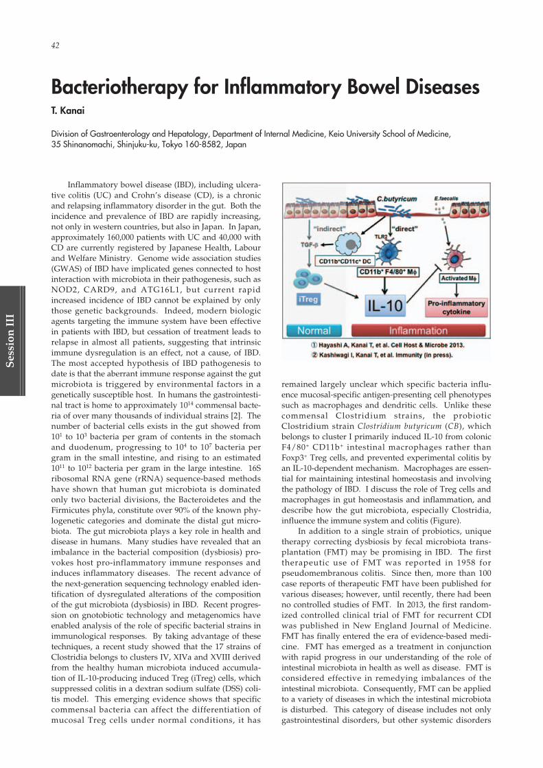
42
Ses
sion
III
Inflammatory bowel disease (IBD), including ulcera-tive colitis (UC) and Crohn’s disease (CD), is a chronicand relapsing inflammatory disorder in the gut. Both theincidence and prevalence of IBD are rapidly increasing,not only in western countries, but also in Japan. In Japan,approximately 160,000 patients with UC and 40,000 withCD are currently registered by Japanese Health, Labourand Welfare Ministry. Genome wide association studies(GWAS) of IBD have implicated genes connected to hostinteraction with microbiota in their pathogenesis, such asNOD2, CARD9, and ATG16L1, but current rapidincreased incidence of IBD cannot be explained by onlythose genetic backgrounds. Indeed, modern biologicagents targeting the immune system have been effectivein patients with IBD, but cessation of treatment leads torelapse in almost all patients, suggesting that intrinsicimmune dysregulation is an effect, not a cause, of IBD.The most accepted hypothesis of IBD pathogenesis todate is that the aberrant immune response against the gutmicrobiota is triggered by environmental factors in agenetically susceptible host. In humans the gastrointesti-nal tract is home to approximately 1014 commensal bacte-ria of over many thousands of individual strains [2]. Thenumber of bacterial cells exists in the gut showed from101 to 103 bacteria per gram of contents in the stomachand duodenum, progressing to 104 to 107 bacteria pergram in the small intestine, and rising to an estimated1011 to 1012 bacteria per gram in the large intestine. 16Sribosomal RNA gene (rRNA) sequence-based methodshave shown that human gut microbiota is dominatedonly two bacterial divisions, the Bacteroidetes and theFirmicutes phyla, constitute over 90% of the known phy-logenetic categories and dominate the distal gut micro-biota. The gut microbiota plays a key role in health anddisease in humans. Many studies have revealed that animbalance in the bacterial composition (dysbiosis) pro-vokes host pro-inflammatory immune responses andinduces inflammatory diseases. The recent advance ofthe next-generation sequencing technology enabled iden-tification of dysregulated alterations of the compositionof the gut microbiota (dysbiosis) in IBD. Recent progres-sion on gnotobiotic technology and metagenomics haveenabled analysis of the role of specific bacterial strains inimmunological responses. By taking advantage of thesetechniques, a recent study showed that the 17 strains ofClostridia belongs to clusters IV, XIVa and XVIII derivedfrom the healthy human microbiota induced accumula-tion of IL-10-producing induced Treg (iTreg) cells, whichsuppressed colitis in a dextran sodium sulfate (DSS) coli-tis model. This emerging evidence shows that specificcommensal bacteria can affect the differentiation ofmucosal Treg cells under normal conditions, it has
remained largely unclear which specific bacteria influ-ence mucosal-specific antigen-presenting cell phenotypessuch as macrophages and dendritic cells. Unlike thesecommensal Clostridium strains, the probioticClostridium strain Clostridium butyricum (CB), whichbelongs to cluster I primarily induced IL-10 from colonicF4/80+ CD11b+ intestinal macrophages rather thanFoxp3+ Treg cells, and prevented experimental colitis byan IL-10-dependent mechanism. Macrophages are essen-tial for maintaining intestinal homeostasis and involvingthe pathology of IBD. I discuss the role of Treg cells andmacrophages in gut homeostasis and inflammation, anddescribe how the gut microbiota, especially Clostridia,influence the immune system and colitis (Figure).
In addition to a single strain of probiotics, uniquetherapy correcting dysbiosis by fecal microbiota trans-plantation (FMT) may be promising in IBD. The firsttherapeutic use of FMT was reported in 1958 forpseudomembranous colitis. Since then, more than 100case reports of therapeutic FMT have been published forvarious diseases; however, until recently, there had beenno controlled studies of FMT. In 2013, the first random-ized controlled clinical trial of FMT for recurrent CDIwas published in New England Journal of Medicine.FMT has finally entered the era of evidence-based medi-cine. FMT has emerged as a treatment in conjunctionwith rapid progress in our understanding of the role ofintestinal microbiota in health as well as disease. FMT isconsidered effective in remedying imbalances of theintestinal microbiota. Consequently, FMT can be appliedto a variety of diseases in which the intestinal microbiotais disturbed. This category of disease includes not onlygastrointestinal disorders, but other systemic disorders
Bacteriotherapy for Inflammatory Bowel DiseasesT. Kanai
Division of Gastroenterology and Hepatology, Department of Internal Medicine, Keio University School of Medicine, 35 Shinanomachi, Shinjuku-ku, Tokyo 160-8582, Japan
02_本文1027(web用追加) 15.10.27 9:04 AM ページ 42

Session III 43
Session
III
such as metabolic syndrome, diabetes mellitus, non-alco-holic steatohepatitis, autoimmune diseases, and cardio-vascular diseases. Although FMT has huge potential inthe treatment of various diseases, there remain many
unanswered questions on FMT regarding proper indica-tions, optimal protocols, and patient selection, and moreresearch is required. I here introduce such new thera-peutic approach for the cure of IBD.
02_本文1027(web用追加) 15.10.27 9:04 AM ページ 43
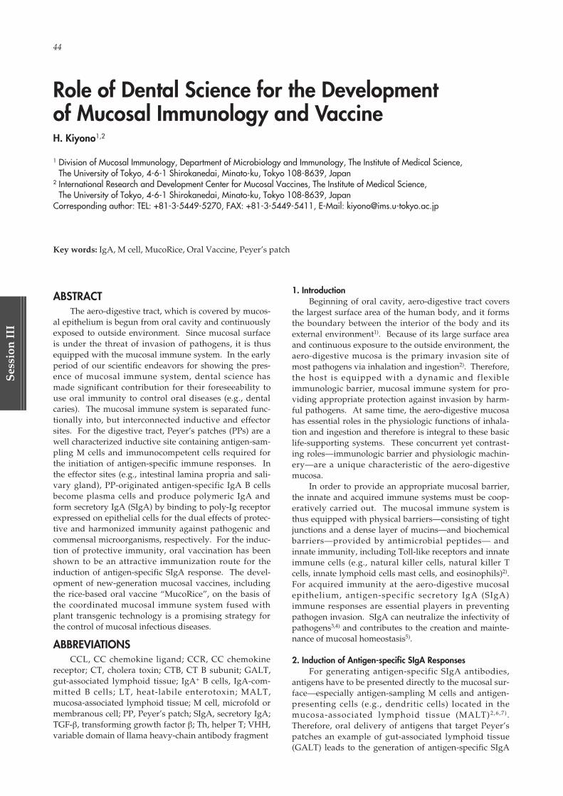
44
Ses
sion
III
ABSTRACTThe aero-digestive tract, which is covered by mucos-
al epithelium is begun from oral cavity and continuouslyexposed to outside environment. Since mucosal surfaceis under the threat of invasion of pathogens, it is thusequipped with the mucosal immune system. In the earlyperiod of our scientific endeavors for showing the pres-ence of mucosal immune system, dental science hasmade significant contribution for their foreseeability touse oral immunity to control oral diseases (e.g., dentalcaries). The mucosal immune system is separated func-tionally into, but interconnected inductive and effectorsites. For the digestive tract, Peyer’s patches (PPs) are awell characterized inductive site containing antigen-sam-pling M cells and immunocompetent cells required forthe initiation of antigen-specific immune responses. Inthe effector sites (e.g., intestinal lamina propria and sali-vary gland), PP-originated antigen-specific IgA B cellsbecome plasma cells and produce polymeric IgA andform secretory IgA (SIgA) by binding to poly-Ig receptorexpressed on epithelial cells for the dual effects of protec-tive and harmonized immunity against pathogenic andcommensal microorganisms, respectively. For the induc-tion of protective immunity, oral vaccination has beenshown to be an attractive immunization route for theinduction of antigen-specific SIgA response. The devel-opment of new-generation mucosal vaccines, includingthe rice-based oral vaccine “MucoRice”, on the basis ofthe coordinated mucosal immune system fused withplant transgenic technology is a promising strategy forthe control of mucosal infectious diseases.
ABBREVIATIONSCCL, CC chemokine ligand; CCR, CC chemokine
receptor; CT, cholera toxin; CTB, CT B subunit; GALT,gut-associated lymphoid tissue; IgA+ B cells, IgA-com-mitted B cells; LT, heat-labile enterotoxin; MALT,mucosa-associated lymphoid tissue; M cell, microfold ormembranous cell; PP, Peyer’s patch; SIgA, secretory IgA;TGF-β, transforming growth factor β; Th, helper T; VHH,variable domain of llama heavy-chain antibody fragment
1. IntroductionBeginning of oral cavity, aero-digestive tract covers
the largest surface area of the human body, and it formsthe boundary between the interior of the body and itsexternal environment1). Because of its large surface areaand continuous exposure to the outside environment, theaero-digestive mucosa is the primary invasion site ofmost pathogens via inhalation and ingestion2). Therefore,the host is equipped with a dynamic and flexibleimmunologic barrier, mucosal immune system for pro-viding appropriate protection against invasion by harm-ful pathogens. At same time, the aero-digestive mucosahas essential roles in the physiologic functions of inhala-tion and ingestion and therefore is integral to these basiclife-supporting systems. These concurrent yet contrast-ing roles—immunologic barrier and physiologic machin-ery—are a unique characteristic of the aero-digestivemucosa.
In order to provide an appropriate mucosal barrier,the innate and acquired immune systems must be coop-eratively carried out. The mucosal immune system isthus equipped with physical barriers—consisting of tightjunctions and a dense layer of mucins—and biochemicalbarriers—provided by antimicrobial peptides— andinnate immunity, including Toll-like receptors and innateimmune cells (e.g., natural killer cells, natural killer Tcells, innate lymphoid cells mast cells, and eosinophils)2).For acquired immunity at the aero-digestive mucosalepithelium, antigen-specific secretory IgA (SIgA)immune responses are essential players in preventingpathogen invasion. SIgA can neutralize the infectivity ofpathogens3,4) and contributes to the creation and mainte-nance of mucosal homeostasis5).
2. Induction of Antigen-specific SIgA ResponsesFor generating antigen-specific SIgA antibodies,
antigens have to be presented directly to the mucosal sur-face—especially antigen-sampling M cells and antigen-presenting cells (e.g., dendritic cells) located in themucosa-associated lymphoid tissue (MALT)2,6 ,7 ) .Therefore, oral delivery of antigens that target Peyer’spatches an example of gut-associated lymphoid tissue(GALT) leads to the generation of antigen-specific SIgA
Role of Dental Science for the Development of Mucosal Immunology and VaccineH. Kiyono1,2
1 Division of Mucosal Immunology, Department of Microbiology and Immunology, The Institute of Medical Science, The University of Tokyo, 4-6-1 Shirokanedai, Minato-ku, Tokyo 108-8639, Japan
2 International Research and Development Center for Mucosal Vaccines, The Institute of Medical Science, The University of Tokyo, 4-6-1 Shirokanedai, Minato-ku, Tokyo 108-8639, Japan
Corresponding author: TEL: +81-3-5449-5270, FAX: +81-3-5449-5411, E-Mail: [email protected]
Key words: IgA, M cell, MucoRice, Oral Vaccine, Peyer’s patch
02_本文1027(web用追加) 15.10.27 9:04 AM ページ 44
Session III 45
Session
III
antibodies in the secretions of the oral cavity and intes-tine6). In contrast, although very effective in the induc-tion of systemic immunity (e.g., serum IgG antibody), theinjection of antigen, apparently does not induce antigen-specific SIgA antibody responses7,8). These facts indicatethat new-generation vaccines must be rationally designedto efficiently induce antigen-specific mucosal immunityat the entry sites of mucosal pathogens.
The mucosal immune system consists of coordinatedinductive and effector tissues (Fig. 1). As inductive sites,MALTs are furnished with all of the necessary antigen-sampling presenting and immunocompetent cells for theinitiation of antigen-specific immune responses6-8). Forexample, at Peyer’s patches (PPs), orally administeredantigens are taken up by M cells located in the follicle-associated epithelium. M cells then deliver antigens toantigen-presenting cells, such as dendritic cells andmacrophages, that lie beneath the follicle-associatedepithelium of PPs for the subsequent presentation ofprocessed antigen leading to the generation of Th1 cells,Th2 cells, Th17 cells, follicular helper T cells or cytotoxicT cells and the production of IgA-committed B cells (IgA+
B cells)6). Concurrent with antigen presentation, dendrit-ic cells located in PPs induce gut-imprinting molecules(e.g., CC chemokine receptor [CCR]9, CCR10, α4β7 inte-grin) on antigen-specific lymphocytes through the
retinoic acid cascade for their subsequent migration tothe effector tissues (e.g., intestinal lamina propria)7,8) (Fig.1). The lipid mediator system, which includes sphingo-sine-1-phosphate and its receptor, sphingosine-1-phos-phate receptor type 1, plays a critical role in the egressfrom PPs of antigen-specific lymphocytes that carry gut-imprinting molecules and in their subsequent immuno-logic journey to distant effector sites9,10).
At the effector sites such as salivary gland and intes-tine, PP-originated antigen-specific Th2 cells provide theIgA-enhancing cytokines (including IL-5, IL-6, and IL-10)required for the final differentiation of IgA+ B cells intoplasma cells that produce dimeric or polymeric forms ofIgA6,11,12). These IgA antibodies then bind to poly-Igreceptors expressed on the basal membrane of epithelialcells, where they form SIgA and are transported to secre-tions13,14) (Fig. 1). This interconnected and well-orches-trated process between the inductive (e.g., PPs) and effec-tor (e.g., oral cavity and intestine) sites provides theimmunologic basis for the induction and regulation ofantigen-specific immune responses (e.g., SIgA produc-tion) at the mucosal surface2,6-8).
3. Dental Science Contributed for the Creation of MucosalImmunology
In past several years, mucosal immunology has
Fig. 1. The Mucosal immune system for the induction and regulation of antigen-specific immuneresponsesOrally delivered antigens in the lumens of the gastrointestinal tract are endocytosed by M cellslocated on the follicle-associated epithelium (FAE) of gut-associated lymphoid tissue (GALT), orPeyer’s patches, M cells located in the FAE form the subepithelial dome structure, and antigen-presenting cells (APCs) such as dendritic cells (DCs) lie immediately beneath the FAE. M-cell-endocytosed antigens are immediately processed by DCs, which transport antigens to underly-ing T and B cell zones for priming of antigen-specific T cells and IgA-committed B cells (IgA+ Bcells). These antigen-specific antigen-specific lymphocytes acquire mucosal-imprinting mole-cules, such as CCR9, CCR10, and α4β7 integrin, and subsequently migrate to the effector sites.At the effector sites (e.g., salivary gland and intestinal lamina propria), IgA+ B cells differentiateinto plasma cells after stimulation by the IgA-enhancing cytokines IL-5, IL-6, and IL-10, whichare secreted by antigen-specific Th2 cells. Dimeric or polymeric IgA secreted from plasma cellsis transported to the mucosal surface as secretory IgA (SIgA) through the binding to polymericIg receptor expressed on the basal membrane of epithelial cells.
02_本文1027(web用追加) 15.10.27 9:04 AM ページ 45
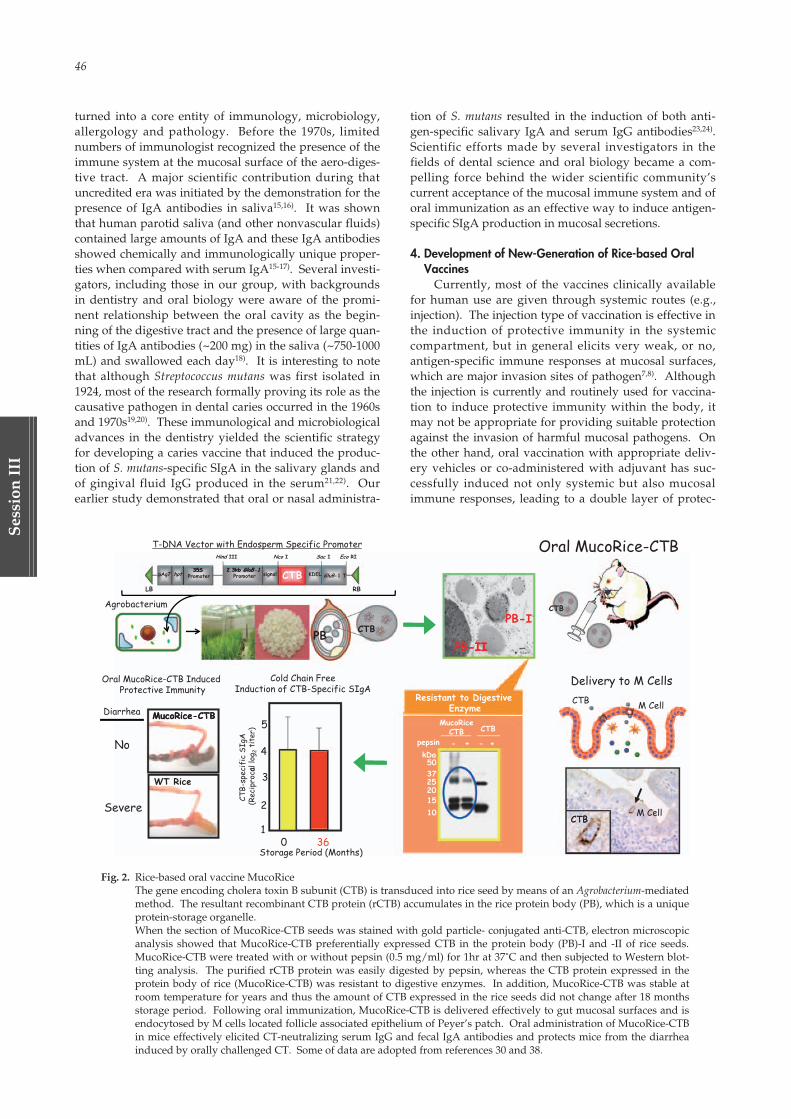
46
Ses
sion
III
turned into a core entity of immunology, microbiology,allergology and pathology. Before the 1970s, limitednumbers of immunologist recognized the presence of theimmune system at the mucosal surface of the aero-diges-tive tract. A major scientific contribution during thatuncredited era was initiated by the demonstration for thepresence of IgA antibodies in saliva15,16). It was shownthat human parotid saliva (and other nonvascular fluids)contained large amounts of IgA and these IgA antibodiesshowed chemically and immunologically unique proper-ties when compared with serum IgA15-17). Several investi-gators, including those in our group, with backgroundsin dentistry and oral biology were aware of the promi-nent relationship between the oral cavity as the begin-ning of the digestive tract and the presence of large quan-tities of IgA antibodies (~200 mg) in the saliva (~750-1000mL) and swallowed each day18). It is interesting to notethat although Streptococcus mutans was first isolated in1924, most of the research formally proving its role as thecausative pathogen in dental caries occurred in the 1960sand 1970s19,20). These immunological and microbiologicaladvances in the dentistry yielded the scientific strategyfor developing a caries vaccine that induced the produc-tion of S. mutans-specific SIgA in the salivary glands andof gingival fluid IgG produced in the serum21,22). Ourearlier study demonstrated that oral or nasal administra-
tion of S. mutans resulted in the induction of both anti-gen-specific salivary IgA and serum IgG antibodies23,24).Scientific efforts made by several investigators in thefields of dental science and oral biology became a com-pelling force behind the wider scientific community’scurrent acceptance of the mucosal immune system and oforal immunization as an effective way to induce antigen-specific SIgA production in mucosal secretions.
4. Development of New-Generation of Rice-based OralVaccines
Currently, most of the vaccines clinically availablefor human use are given through systemic routes (e.g.,injection). The injection type of vaccination is effective inthe induction of protective immunity in the systemiccompartment, but in general elicits very weak, or no,antigen-specific immune responses at mucosal surfaces,which are major invasion sites of pathogen7,8). Althoughthe injection is currently and routinely used for vaccina-tion to induce protective immunity within the body, itmay not be appropriate for providing suitable protectionagainst the invasion of harmful mucosal pathogens. Onthe other hand, oral vaccination with appropriate deliv-ery vehicles or co-administered with adjuvant has suc-cessfully induced not only systemic but also mucosalimmune responses, leading to a double layer of protec-
Oral MucoRice-CTB2 3kb GluB 135S
Hind III Nco I Sac I Eco RI
T-DNA Vector with Endosperm Specific Promoter
signal2.3kb GluB-1
Promoter KDEL GluB-1 TpAg7 hpt35S
Promoter
Agrobacterium CTB
PB CTB
Delivery to M Cells
M CellCTBMucoRice-CTBDiarrhea
Cold Chain FreeInduction of CTB-Specific SIgA
Oral MucoRice-CTB InducedProtective Immunity
MucoRice CTB
No 4
5
ific
SIgA
allo
g 2ti
ter)
CTBM Cell
WT Rice
Severe
3
2
CTB-
spec
(Rec
ipro
ca
CTB1
Storage Period (Months)0
PB-I
PB--IIII
36
pepsin
50kDa
3725201510
Resistant to Digestive Enzyme
MucoRiceCTB CTB
- + - +
CTBBL BR
TMPTMP--IMSUTIMSUT
Fig. 2. Rice-based oral vaccine MucoRiceThe gene encoding cholera toxin B subunit (CTB) is transduced into rice seed by means of an Agrobacterium-mediatedmethod. The resultant recombinant CTB protein (rCTB) accumulates in the rice protein body (PB), which is a uniqueprotein-storage organelle.When the section of MucoRice-CTB seeds was stained with gold particle- conjugated anti-CTB, electron microscopicanalysis showed that MucoRice-CTB preferentially expressed CTB in the protein body (PB)-I and -II of rice seeds.MucoRice-CTB were treated with or without pepsin (0.5 mg/ml) for 1hr at 37˚C and then subjected to Western blot-ting analysis. The purified rCTB protein was easily digested by pepsin, whereas the CTB protein expressed in theprotein body of rice (MucoRice-CTB) was resistant to digestive enzymes. In addition, MucoRice-CTB was stable atroom temperature for years and thus the amount of CTB expressed in the rice seeds did not change after 18 monthsstorage period. Following oral immunization, MucoRice-CTB is delivered effectively to gut mucosal surfaces and isendocytosed by M cells located follicle associated epithelium of Peyer’s patch. Oral administration of MucoRice-CTBin mice effectively elicited CT-neutralizing serum IgG and fecal IgA antibodies and protects mice from the diarrheainduced by orally challenged CT. Some of data are adopted from references 30 and 38.
02_本文1027(web用追加) 15.10.27 9:04 AM ページ 46
Session III 47
Session
III
tion against mucosal pathogens25). Further, mucosal vac-cines provide other advantages over injectable vaccinesincluding increased ease of administration, less physicaland psychologic discomfort, and avoidance of needle-stick injuries and transmission of blood-borne diseases7,8).
In spite of these noticeable advantages, only a fewmucosal vaccines have been approved for clinical use inhumans: vaccines against poliovirus, rotavirus,Salmonella typhi, and Vibrio cholerae are administered oral-ly, and vaccines against influenza virus are givenintranasally8). Most of these currently available oral andnasal vaccines involve either attenuated or gene-modi-fied live or killed forms of whole microorganisms8). Amucosal vaccine that delivers a component (subunit) orpurified form is not yet available for clinical use. Oralimmunization of a protein based vaccine antigen alonefails to effectively induce antigen-specific immuneresponses due to intrinsic physiologic mechanisms of thedigestive tract, namely degradation of vaccine antigen bydigestive enzymes (e.g., pepsin26)); clearance mechanisms(e.g., peristalsis action and mucus secretion); and physio-logic and biologic barriers (e.g., gastric acids, mucins, andtight junctions) to the access of vaccine antigen to intesti-nal inductive tissue (e.g., Peyer’s patches)6,8). Althoughvarious obstacles remain, an oral vaccination strategyappears attractive and beneficial for the next generationof vaccines. Furthermore, recent scientific advancesachieved through the sharing of knowledge and tech-nologies among different fields of science are helping toovercome various hurdles in the development of oralvaccines.
For the development of oral vaccine, transgenicplant-based vaccines are spotlighted because of theirpracticality, safety, and low cost7,27-29). When comparedwith injectable vaccines, oral vaccines require additionaltechnological advances in antigen-delivery techniques,because the antigens must cross intestinal barriers com-prising digestive enzymes, mucus, and physiologicmechanisms to be presented to gut inductive site (e.g.,Peyer’s patches). Among the diffrent plant candidates(e.g., carrot, potato, rice, soybean, tobacco, and tomato)for transgenic vaccines composed of a bacterial compo-nent of heat-labile enterotoxin [LT] B subunit, choleratoxin B subunit [CTB], or Yersinia pestis and a viral com-ponent of hepatitis B virus, rotavirus, or norovirus30-37),rice may be the most suitable antigen-expressing plantbecause rice seed (especially the protein body) is resistantto digestion by gastric acid8,27,30). Furthermore, proteinexpressed in rice seeds is stable for a prolonged time inthe absence of refrigerated storage30). Rice can thus beconsidered a viable candidate for the creation of cold-chain-free and needle-free vaccine.
MucoRice-CTB, which consists of CTB expressed intransgenic rice, is one of the most potent plant-based vac-cines (Figure 2)30). The CTB gene was inserted into riceseed by use of an Agrobacterium-transformation system(Figure 2) which resulted in the production of recombi-nant CTB protein reaches 15-30 µg per transgenic riceseed30). These CTB proteins are accumulated in the riceprotein body, a unique protein-storage organelle in riceseed (Figure 2)30). The CTB proteins expressed in the pro-tein body are stable at room temperature for more than 3
years without loss of immunogenicity and also resistantto intestinal digestive enzymes (Figure 2)30,38). Theseresults suggest that the rice protein bodies are acting as anatural capsule for the oral delivery of vaccine antigensto the gut immune system. Thus, oral delivery ofMucoRice-CTB leads to its subsequent uptake by M cellsand transport to the inside of Peyer’s patches (Figure2)8,27).
In addition to its advantages of refrigeration-freestorage and effective oral delivery, MucoRice-CTB effec-tively induces antigen-specific protective immuneresponses in both the systemic and mucosal compart-ments (Figure 2). Oral vaccination of MucoRice-CTB inmice induces CT-neutralizing serum IgG and fecal IgAantibodies and protects mice from diarrhea induced byorally challenged CT (Figure 2)30,38). MucoRice-CTB alsoinduces CT-specific neutralizing antibody production innon-human primates39). Orally administered MucoRice-CTB induces cross-protective immunity against LT38).The cross-protective immunity against LT inducedthrough vaccination with MucoRice-CTB is thought toreflect the close similarity between the CTB and LTB sub-units38,40). Therefore, MucoRice-CTB may be a potentialvaccine against the toxin produced by enterotoxic E. coli,which is a major cause of severe diarrhea in children indeveloping countries38,41).
The MucoRice system is also applicable to oral pas-sive immunotherapy because of its advantages of bothstability in the absence of refrigeration and effective oraldelivery42). MucoRice-ARP1 expresses the anti-rotavirusspecific variable domain of llama heavy-chain antibodyfragment (VHH) in rice seeds and is a candidate for anew oral passive immunotherapy against rotavirus infec-tion42). VHH, which is composed of heavy-chain dimersand is devoid of a light chain, possesses an extensiveantigen-binding repertoire43), and because rotavirus-spe-cific VHH (ARP1) has been efficiently produced in yeastand reduces morbidity from rotaviral diarrhea in mice44),its application to passive immunotherapy for rotaviralinfection offers an attractive strategy for the control ofrotavirus infection, in addition to the currently availablerotavirus oral vaccines. MucoRice-ARP1 comprises the12-kDa ARP1 protein expressed in transgenic rice (0.85%of seed weight) and has all the advantages described forMucoRice-CTB, such as stability at room temperature42).Further, oral administration of MucoRice-ARP1 attenu-ates rotaviral infection in both immunocompetent andimmunodeficient mice, suggesting that MucoRice-ARP1can be used for the immunocompromised population inaddition to healthy subjects. The combined technologiesof the MucoRice expression system and VHH are expect-ed to evolve into oral passive immunotherapy options fordiverse mucosal infectious diseases42).
5. ConclusionThe mucosal surface of aero-digestive tract harbors a
dynamic and flexible immune network that protects thehost against external pathogens. The digestive tractbegin with oral cavity has its own immune inductive tis-sue—the so-called GALT including Peyer’s patches. Theinductive tissue possess unique functional characteristics,including antigen sampling by M cells and subsequent
02_本文1027(web用追加) 15.10.27 9:04 AM ページ 47
48
Ses
sion
III
processing and presentation by antigen-presenting cells,resulting in the initiation of antigen-specific mucosalimmune responses. Th1, Th2, Th17, and follicular helperT cells and cytotoxic T cells, as well as IgA+ B cells, arethus generated in PPs and then migrate to the distantintestine and oral cavity to provide the first line ofdefense (e.g., SIgA).
The importance of mucosal immunity is well known,however, traditional systemic vaccines, which typicallyare administered by injection, fail to induce antigen-spe-cific mucosal immune responses and thus cannot provideprotection against the invasion of pathogens at epithelialsurfaces. Despite our increased knowledge of the need tomaximize the effective induction of both antigen-specificsystemic and mucosal immune responses, few mucosalvaccines are currently available. However, scientific andtechnological fusions among the fields of mucosalimmunology, bioengineering, agricultural science, plantbiology and plant factory have led to the creation of new-generation rice-based oral vaccine (e.g., MucoRice).Continued concomitant progress in characterizing theunderlying aspects of the mucosal immune system andtheir application to mucosal vaccines relies not only onthe scientific advancement of mucosal immunology butalso on novel mergers of modern concepts and technolo-gies from both related and unrelated fields.
ACKNOWLEDGEMENTSOur studies were supported by grants from the
Ministry of Education, Culture, Sports, Science, andTechnology of Japan (Scientific Research S); the GlobalCenter of Excellence Program of the Center of Educationand Research for Advanced Genome-based Medicine;MEXT Translational Research Network Seed C; and theCore Research for Evolutional Science and TechnologyProgram of the Japan Science and Technology Agency. Ithank Dr. T. Azegami for helping the preparation of thisreview article.
REFERRENCES[01] Takahashi, I., Fujihashi, K. and Kiyono, H.: Mucosal
regulatory cells in the gastrointestinal tract and peri-odontium. Periodontol 2000 54: 247-256, 2010.
[02] Kiyono, H., Kunisawa, J., McGhee, J.R. andMestecky, J. (2008) The mucosal immune system. InFundamental Immunology (eds. W. E. Paul).Lippincott Williams & Wilkins, Philadelphia, pp.983-1030.
[03] Williams, R.C. and Gibbons, R.J.: Inhibition of bacter-ial adherence by secretory immunoglobulin A: amechanism of antigen disposal. Science 177: 697-699,1972.
[04] Taylor, H.P. and Dimmock, N.J.: Mechanism of neu-tralization of influenza virus by secretory IgA is dif-ferent from that of monomeric IgA or IgG. J Exp Med161: 198-209, 1985.
[05] Johansen, F.E., Pekna, M., Norderhaug, I.N.,Haneberg, B., Hietala, M.A., et al.: Absence of epithe-lial immunoglobulin A transport, with increasedmucosal leakiness, in polymeric immunoglobulinreceptor/secretory component-deficient mice. J ExpMed 190: 915-922, 1999.
[06] Kunisawa, J., Kurashima, Y. and Kiyono, H.: Gut-associated lymphoid tissues for the development oforal vaccines. Adv Drug Deliv Rev 64: 523-530, 2012.
[07] Yuki, Y. and Kiyono, H.: Mucosal vaccines: noveladvances in technology and delivery. Expert RevVaccines 8: 1083-1097, 2009.
[08] Azegami, T., Yuki, Y. and Kiyono, H.: Challenges inmucosal vaccines for the control of infectious dis-eases. Int Immunol 26: 517-528, 2014.
[09] Gohda, M., Kunisawa, J., Miura, F., Kagiyama, Y.,Kurashima, Y., et al.: Sphingosine 1-phosphate regu-lates the egress of IgA plasmablasts from Peyer'spatches for intestinal IgA responses. J Immunol 180:5335-5343, 2008.
[10] Kunisawa, J. and Kiyono, H.: Immunological func-tion of sphingosine 1-phosphate in the intestine.Nutrients 4: 154-166, 2012.
[11] Mestecky, J. and McGhee, J.R.: Immunoglobulin A(IgA): molecular and cellular interactions involved inIgA biosynthesis and immune response. AdvImmunol 40: 153-245, 1987.
[12] Cerutti, A.: The regulation of IgA class switching.Nat Rev Immunol 8: 421-434, 2008.
[13] Mostov, K.E., Kraehenbuhl, J.P. and Blobel, G.:Receptor-mediated transcellular transport ofimmunoglobulin: synthesis of secretory componentas multiple and larger transmembrane forms. ProcNatl Acad Sci U S A 77: 7257-7261, 1980.
[14] Mostov, K.E., Friedlander, M. and Blobel, G.: Thereceptor for transepithelial transport of IgA and IgMcontains multiple immunoglobulin-like domains.Nature 308: 37-43, 1984.
[15] Tomasi, T.B., Jr. and Zigelbaum, S.: The selectiveoccurence of gamma-1a globulins in certain body flu-ids. J Clin Invest 42: 1552-1560, 1963.
[16] Chodirker, W.B. and Tomasi, T.B., Jr.: Gamma-globu-lins: quantitative relationships in human serum andnonvascular fluids. Science 142: 1080-1081, 1963.
[17] Tomasi, T.B., Jr., Tan, E.M., Solomon, A. andPrendergast, R.A.: Characteristics of an immune sys-tem common to certain external secretions. J ExpMed 121: 101-124, 1965.
[18] Fujihashi, K. and Kiyono, H. (2012) The Nasopharyngealand oral immune system. In Principles of MucosalImmunology (eds. Smith, P.D. MacDonald, T.T. andBlumberg, R.S.). Garland Science, Danvers, pp. 293-307.
[19] Hamada, S. (1986) Overview of the biology ofStreptococcus mutans. In Molecular Microbiology andImmunology of Streptococcus mutans (eds. Hamada,S., Michalek, S.M., Kiyono, H., Menaker, S.L. andMcGhee, J.R.). Elsevier, Amsterdam, pp. 7-20.
[20] Hirasawa, M., Kiyono, H., Babb, J.L., Shiota, T.,Michalek, S.M., et al.: Virulence of Streptococcusmutans: in vivo reversion of a low-virulence mutantresults in partial displacement and pathogenesis.Infect Immun 27: 1003-1011, 1980.
[21] Kiyono, H., Czerkinsky, C., Koga, T., Kimura, S.,Kurita, T., et al. (1986) Induction and T cell regula-tion of IgA responses to Streptococcus mutans anti-gens.. In The Borderland between Caries andPeriodontal Diseases III (eds. G. Cimasoni and T.
02_本文1027(web用追加) 15.10.27 9:04 AM ページ 48
Session III 49
Session
III
Lehner). Editions Medecine et Hygiene, Geneva, pp.249-267.
[22] Hamada, S. and Kodama, Y.: Passive immunity forprotection against mucosak infections and vaccina-tion for dental caries. (1996) In Mucosal Vaccines.(eds. Kiyono, H., Ogra, P.L. and McGhee, J.R.)Academic Press, San Diego, pp. 187-197.
[23] Kiyono, H., Michalek, S.M., Mosteller, L.M., Torii,M., Hamada, S., et al.: Enhancement of murineimmune responses to orally administered haptenat-ed Streptococcus mutans. Scand J Immunol 16: 455-463, 1982.
[24] Saito, M., Otake, S., Ohmura, M., Hirasawa, M., Takada,K., et al.: Protective immunity to Streptococcus mutansinduced by nasal vaccination with surface proteinantigen and mutant cholera toxin adjuvant. J InfectDis 183: 823-826, 2001.
[25] Lamichhane, A., Azegami, T. and Kiyono, H.: Themucosal immune system for vaccine development.Vaccine 32: 6711-6723, 2014.
[26] Brenneman, K.E., Willingham, C., Kilbourne, J.A.,Curtiss, R., 3rd and Roland, K.L.: A low gastric pHmouse model to evaluate live attenuated bacterialvaccines. PLoS One 9: e87411, 2014.
[27] Azegami, T., Itoh, H., Kiyono, H. and Yuki, Y.: NovelTransgenic rice-based vaccines. Arch Immunol TherExp (Warsz) 63: 87-99, 2015.
[28] Fukuyama, Y., Tokuhara, D., Kataoka, K., Gilbert,R.S., McGhee, J.R., et al.: Novel vaccine developmentstrategies for inducing mucosal immunity. ExpertRev Vaccines 11: 367-379, 2012.
[29] Takahashi, I., Nochi, T., Yuki, Y. and Kiyono, H.:New horizon of mucosal immunity and vaccines.Curr Opin Immunol 21: 352-358, 2009.
[30] Nochi, T., Takagi, H., Yuki, Y., Yang, L., Masumura,T., et al.: Rice-based mucosal vaccine as a globalstrategy for cold-chain- and needle-free vaccination.Proc Natl Acad Sci U S A 104: 10986-10991, 2007.
[31] Thanavala, Y., Mahoney, M., Pal, S., Scott, A.,Richter, L., et al.: Immunogenicity in humans of anedible vaccine for hepatitis B. Proc Natl Acad Sci U SA 102: 3378-3382, 2005.
[32] Li, J.T., Fei, L., Mou, Z.R., Wei, J., Tang, Y., et al.:Immunogenicity of a plant-derived edible rotavirussubunit vaccine transformed over fifty generations.Virology 356: 171-178, 2006.
[33] Zhang, X., Buehner, N.A., Hutson, A.M., Estes, M.K.and Mason, H.S.: Tomato is a highly effective vehiclefor expression and oral immunization with Norwalkvirus capsid protein. Plant Biotechnol J 4: 419-432,2006.
[34] Santi, L., Giritch, A., Roy, C.J., Marillonnet, S.,
Klimyuk, V., et al.: Protection conferred by recombi-nant Yersinia pestis antigens produced by a rapid andhighly scalable plant expression system. Proc NatlAcad Sci U S A 103: 861-866, 2006.
[35] Moravec, T., Schmidt, M.A., Herman, E.M. andWoodford-Thomas, T.: Production of Escherichia coliheat labile toxin (LT) B subunit in soybean seed andanalysis of its immunogenicity as an oral vaccine.Vaccine 25: 1647-1657, 2007.
[36] Jiang, X.L., He, Z.M., Peng, Z.Q., Qi, Y., Chen, Q., etal.: Cholera toxin B protein in transgenic tomato fruitinduces systemic immune response in mice.Transgenic Res 16: 169-175, 2007.
[37] Rosales-Mendoza, S., Soria-Guerra, R.E., Lopez-Revilla, R., Moreno-Fierros, L. and Alpuche-Solis,A.G.: Ingestion of transgenic carrots expressing theEscherichia coli heat-labile enterotoxin B subunit pro-tects mice against cholera toxin challenge. Plant CellRep 27: 79-84, 2008.
[38] Tokuhara, D., Yuki, Y., Nochi, T., Kodama, T.,Mejima, M., et al.: Secretory IgA-mediated protectionagainst V. cholerae and heat-labile enterotoxin-pro-ducing enterotoxigenic Escherichia coli by rice-basedvaccine. Proc Natl Acad Sci U S A 107: 8794-8799,2010.
[39] Nochi, T., Yuki, Y., Katakai, Y., Shibata, H.,Tokuhara, D., et al.: A rice-based oral cholera vaccineinduces macaque-specific systemic neutralizing anti-bodies but does not influence pre-existing intestinalimmunity. J Immunol 183: 6538-6544, 2009.
[40] Peltola, H., Siitonen, A., Kyronseppa, H., Simula, I.,Mattila, L., et al.: Prevention of travellers' diarrhoeaby oral B-subunit/whole-cell cholera vaccine. Lancet338: 1285-1289, 1991.
[41] Abba, K., Sinfield, R., Hart, C.A. and Garner, P.:Pathogens associated with persistent diarrhoea inchildren in low and middle income countries: sys-tematic review. BMC Infect Dis 9: 88, 2009.
[42] Tokuhara, D., Alvarez, B., Mejima, M., Hiroiwa, T.,Takahashi, Y., et al.: Rice-based oral antibody frag-ment prophylaxis and therapy against rotavirusinfection. J Clin Invest 123: 3829-3838, 2013.
[43] Hamers-Casterman, C., Atarhouch, T., Muyldermans,S., Robinson, G., Hamers, C., et al.: Naturally occur-ring antibodies devoid of light chains. Nature 363:446-448, 1993.
[44] van der Vaart, J.M., Pant, N., Wolvers, D., Bezemer,S., Hermans, P.W., et al.: Reduction in morbidity ofrotavirus induced diarrhoea in mice by yeast pro-duced monovalent llama-derived antibody frag-ments. Vaccine 24: 4130-4137, 2006.
02_本文1027(web用追加) 15.10.27 9:04 AM ページ 49

02_本文1027(web用追加) 15.10.27 9:04 AM ページ 50
Session
IV
Development and Function of the Musculoskeletal SystemThe Anti-angiogenic Actions of Tenomodulin, a Specific Marker for Tendons and Ligaments
Hiroshima University
C. Shukunami
Developmental Modularly of Secondary Skeletal Elements Involves a Distinct Population
of Progenitor Cells
Weizmann Institute of Science
E. Zelzer and S. Eyal
The Intervertebral Disc: Working Together in Harmony; Breaking up in Pain
The University of Hong Kong
D. Chan
Session IV
02_本文1027(web用追加) 15.10.27 9:04 AM ページ 51

02_本文1027(web用追加) 15.10.27 9:04 AM ページ 52
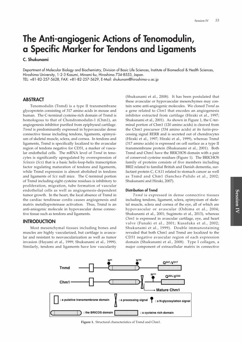
Session IV 53
Session
IV
ABSTRACTTenomodulin (Tnmd) is a type II transmembrane
glycoprotein consisting of 317 amino acids in mouse andhuman. The C-terminal cysteine-rich domain of Tnmd ishomologous to that of Chondromodulin-1 (Chm1), anangiogenesis inhibitor purified from epiphyseal cartilage.Tnmd is predominantly expressed in hypovascular denseconnective tissue including tendons, ligaments, epimysi-um of skeletal muscle, sclera, and cornea. In tendons andligaments, Tnmd is specifically localized to the avascularregion of tendons negative for CD31, a marker of vascu-lar endothelial cells. The mRNA level of Tnmd in teno-cytes is significantly upregulated by overexpression ofScleaxis (Scx) that is a basic helix-loop-helix transcriptionfactor regulating maturation of tendons and ligaments,while Tnmd expression is almost abolished in tendonsand ligaments of Scx null mice. The C-terminal portionof Tnmd including eight cysteine residues is inhibitory toproliferation, migration, tube formation of vascularendothelial cells as well as angiogenesis-dependenttumor growth. In the heart, the local absence of Tnmd inthe cardiac tendineae cordis causes angiogenesis andmatrix metalloproteinase activation. Thus, Tnmd is ananti-aniogenic molecule in hypovascular dense connec-tive tissue such as tendons and ligaments.
INTRODUCTIONMost mesenchymal tissues including bones and
muscles are highly vascularized, but cartilage is avascu-lar and resistant to neovascularization as well as tumorinvasion (Hayami et al., 1999; Shukunami et al., 1999).Similarly, tendons and ligaments have low vascularity
(Shukunami et al., 2008). It has been postulated thatthese avascular or hypovascular mesenchymes may con-tain some anti-angiogenic molecules. We cloned Tnmd asa gene related to Chm1 that encodes an angiogenesisinhibitor extracted from cartilage (Hiraki et al., 1997;Shukunami et al., 2001). As shown in Figure 1, the C-ter-minal portion of Chm1 (120 amino acids) is cleaved fromthe Chm1 precursor (334 amino acids) at its furin-pro-cessing signal RERR and is secreted out of chondrocytes(Hiraki et al., 1997; Hiraki et al., 1999), whereas Tnmd(317 amino acids) is expressed on cell surface as a type IItransmembrane protein (Shukunami et al., 2001). BothTnmd and Chm1 have the BRICHOS domain with a pairof conserved cysteine residues (Figure 1). The BRICHOSfamily of proteins consists of five members includingBRI2 related to familial British and Danish dementia, sur-factant protein C, CA11 related to stomach cancer as wellas Tnmd and Chm1 (Sanchez-Pulido et al., 2002;Shukunami and Hiraki, 2007).
Distribution of TnmdTnmd is expressed in dense connective tissues
including tendons, ligament, sclera, epimysium of skele-tal muscle, sclera and cornea of the eye, all of which arehypovascular or avascular (Oshima et al., 2004;Shukunami et al., 2001; Sugimoto et al., 2013), whereasChm1 is expressed in avascular cartilage, eye, and heartvalve (Funaki et al., 2001; Kusafuka et al., 2002;Shukunami et al., 1999). Double immunostainingrevealed that both Chm1 and Tnmd are localized to theCD31 negative avascular region of each expressiondomain (Shukunami et al., 2008). Type I collagen, amajor component of extracellular matrix in connective
The Anti-angiogenic Actions of Tenomodulin, a Specific Marker for Tendons and LigamentsC. Shukunami
Department of Molecular Biology and Biochemistry, Division of Basic Life Sciences, Institute of Biomedical & Health Sciences,Hiroshima University, 1-2-3 Kasumi, Minami-ku, Hiroshima 734-8553, Japan. TEL: +81-82-257-5628, FAX: +81-82-257-5629, E-Mail: [email protected]
Figure 1. Structural characteristics of Tnmd and Chm1.
02_本文1027(web用追加) 15.10.27 9:04 AM ページ 53

54
Ses
sion
IV
tissues, is distributed in dense and loose connective tis-sues, but Tnmd is only localized to dense connective tis-sues where thick bundles of aligned collagen fibers arefound (Shukunami et al., 2006). In tendons, elongatedtenocytes (or tendon fibroblasts) are aligned in parallel tothick mature collagen fibers to form the regular layer,whereas oval tenocytes are randomly distributed in theinterlaminar spaces. Tnmd expression is not detected inoval tenocytes but in elongated tenocytes (Shukunami etal., 2006), suggesting that Tnmd is a good marker ofmature tenocytes in vivo.
Anti-angiogenic action of TnmBoth Tnmd and Chm1 precursor are type II trans-
membrane glycoproteins that have the anti-angiogenicdomain at the C-terminus. The C-terminal domain haseight cysteine residues to form four disulfide bridges(Miura et al., 2010; Miura et al., 2012; Shukunami et al.,2006) and is well conserved across species (Sachdev et al.,2001; Shukunami et al., 2006). Overexpression of matureChm1 and the C-terminal portion of Tnmd (116 aminoacids) inhibited proliferation, migration, and tube forma-tion of human umbilical vein endothelial cells as well ashuman retinal vascular endothelial cells (Oshima et al.,2003; Oshima et al., 2004). When malignant melanomacells overexpressing either of these molecules are inocu-lated into syngeneic mice, tumor growth was significant-ly suppressed due to inhibition of angiogenesis (Oshimaet al., 2004). In the heart, the local absence of Tnmd in thecardiac tendineae cordis induces angiogenesis and matrixmetalloproteinase activation (Kimura et al., 2008), where-as loss of Chm1 caused enhanced vascular endothelialgrowth factor (VEGF)-A expression, lipid deposition, andmineralization in the cardiac valve in aged mice(Yoshioka et al., 2006).
Regulation of Tnmd expression by ScxScx is induced in the early stages of tendon/ligament
development, while Tnmd expression is more associatedwith maturation of tendons and ligaments (Shukunami etal., 2006; Yukata et al., 2010). Tnmd is coexpressed withScx in mature tendons and ligaments during develop-ment and postnatal growth (Shukunami et al., 2006;Komiyama et al., 2013). Tnmd expression is upregulatedin both tenocytes and periodontal ligament (PDL) cells byScx (Alberton et al., 2012; Shukunami et al., 2006;Takimoto et al., 2015). Conversely, knockdown of Scx inPDL cells downregulated the expression of Tnmd(Takimoto et al., 2015). Consistent with these results,Tnmd expression is abolished in hypoplastic tendons andligaments of Scx knockout newborn mice. Thus, Scx is animportant regulator for Tnmd during tendon and liga-ment development.
CONCLUSIONTnmd is a good cell surface marker for tenocytes and
ligamentocytes both in vivo and in vitro. The cysteine-richChm1 like domain of Tnmd has the anti-angiogenic andanti-tumorigenic activities. Scx is indispensable for theexpression of Tnmd during development and postnatalgrowth.
REFERENCESAlberton P, Popov C, Pragert M, Kohler J, Shukunami C,
Schieker M et al. (2012). Conversion of human bonemarrow-derived mesenchymal stem cells into ten-don progenitor cells by ectopic expression of sclerax-is. Stem Cells and Development 21 (6): 846-858.
Funaki H, Sawaguchi S, Yaoeda K, Koyama Y, Yaoita E,Funaki S et al. (2001). Expression and localization ofangiogenic inhibitory factor, chondromodulin-I, inadult rat eye. Investigative Ophthalmology & VisualScience 42 (6): 1193-1200.
Hayami T, Shukunami C, Mitsui K, Endo N, Tokunaga K,Kondo J et al. (1999). Specific loss of chondromod-ulin-I gene expression in chondrosarcoma and thesuppression of tumor angiogenesis and growth byits recombinant protein in vivo. FEBS Letters 458 (3):436-440.
Hiraki Y, Inoue H, Iyama K, Kamizono A, Ochiai M,Shukunami C et al. (1997). Identification of chondro-modulin I as a novel endothelial cell growthinhibitor. Purification and its localization in the avas-cular zone of epiphyseal cartilage. The Journal ofBiological Chemistry 272 (51): 32419-32426.
Hiraki Y, Mitsui K, Endo N, Takahashi K, Hayami T,Inoue H et al. (1999). Molecular cloning of humanchondromodulin-I, a cartilage-derived growth mod-ulating factor, and its expression in Chinese hamsterovary cells. European Journal of Biochemistry/FEBS 260 (3): 869-878.
Kimura N, Shukunami C, Hakuno D, Yoshioka M, MiuraS, Docheva D et al. (2008). Local tenomodulinabsence, angiogenesis, and matrix metalloproteinaseactivation are associated with the rupture of thechordae tendineae cordis. Circulation 118 (17): 1737-1747.
Komiyama Y, Ohba S, Shimohata N, Nakajima K, HojoH, Yano F et al. (2013). Tenomodulin expression inthe periodontal ligament enhances cellular adhesion.PloS One 8 (4): e60203.
Kusafuka K, Hiraki Y, Shukunami C, Kayano T,Takemura T (2002). Cartilage-specific matrix protein,chondromodulin-I (ChM-I), is a strong angio-inhibitor in endochondral ossification of humanneonatal vertebral tissues in vivo: relationship withangiogenic factors in the cartilage. Acta Histochemica104 (2): 167-175.
Miura S, Mitsui K, Heishi T, Shukunami C, Sekiguchi K,Kondo J et al. (2010). Impairment of VEGF-A-stimu-lated lamellipodial extensions and motility of vascu-lar endothelial cells by chondromodulin-I, a carti-lage-derived angiogenesis inhibitor. Experimental CellResearch 316 (5): 775-788.
Miura S, Kondo J, Kawakami T, Shukunami C, Aimoto S,Tanaka H et al. (2012). Synthetic disulfide-bridgedcyclic peptides mimic the anti-angiogenic actions ofchondromodulin-I. Cancer Science 103 (7): 1311-1318.
Oshima Y, Shukunami C, Honda J, Nishida K, Tashiro F,Miyazaki J et al. (2003). Expression and localizationof tenomodulin, a transmembrane type chondro-modulin-I-related angiogenesis inhibitor, in mouseeyes. Investigative Ophthalmology & Visual Science 44
02_本文1027(web用追加) 15.10.27 9:04 AM ページ 54
Session IV 55
Session
IV
(5): 1814-1823.Oshima Y, Sato K, Tashiro F, Miyazaki J, Nishida K,
Hiraki Y et al. (2004). Anti-angiogenic action of theC-terminal domain of tenomodulin that shareshomology with chondromodulin-I. Journal of CellScience 117 (Pt 13): 2731-2744.
Sachdev SW, Dietz UH, Oshima Y, Lang MR, KnapikEW, Hiraki Y et al. (2001). Sequence analysis ofzebrafish chondromodulin-1 and expression profilein the notochord and chondrogenic regions duringcartilage morphogenesis. Mechanisms of Development105 (1-2): 157-162.
Sanchez-Pulido L, Devos D, Valencia A (2002). BRI-CHOS: a conserved domain in proteins associatedwith dementia, respiratory distress and cancer.Trends in Biochemical Sciences 27 (7): 329-332.
Shukunami C, Iyama K, Inoue H, Hiraki Y (1999).Spatiotemporal pattern of the mouse chondromod-ulin-I gene expression and its regulatory role in vas-cular invasion into cartilage during endochondralbone formation. The International Journal ofDevelopmental Biology 43 (1): 39-49.
Shukunami C, Oshima Y, Hiraki Y (2001). Molecularcloning of tenomodulin, a novel chondromodulin-Irelated gene. Biochemical and Biophysical ResearchCommunications 280 (5): 1323-1327.
Shukunami C, Takimoto A, Oro M, Hiraki Y (2006).Scleraxis positively regulates the expression oftenomodulin, a differentiation marker of tenocytes.Developmental Biology 298 (1): 234-247.
Shukunami C, Hiraki Y (2007). Chondromodulin-I andtenomodulin: the negative control of angiogenesis inconnective tissue. Current Pharmaceutical Design 13(20): 2101-2112.
Shukunami C, Takimoto A, Miura S, Nishizaki Y, HirakiY (2008). Chondromodulin-I and tenomodulin aredifferentially expressed in the avascular mes-enchyme during mouse and chick development. Celland Tissue Research 332 (1): 111-122.
Sugimoto Y, Takimoto A, Akiyama H, Kist R, Scherer G,Nakamura T et al. (2013). Scx+/Sox9+ progenitorscontribute to the establishment of the junctionbetween cartilage and tendon/ligament. Development140 (11): 2280-2288.
Takimoto A, Kawatsu M, Yoshimoto Y, Kawamoto T,Seiryu M, Takano-Yamamoto T et al. (2015). Scleraxisand osterix antagonistically regulate tensile force-responsive remodeling of the periodontal ligamentand alveolar bone. Development 142 (4): 787-796.
Yoshioka M, Yuasa S, Matsumura K, Kimura K, ShiomiT, Kimura N et al. (2006). Chondromodulin-I main-tains cardiac valvular function by preventing angio-genesis. Nature Medicine 12 (10): 1151-1159.
Yukata K, Matsui Y, Shukunami C, Takimoto A,Hirohashi N, Ohtani O et al. (2010). Differentialexpression of Tenomodulin and Chondromodulin-1at the insertion site of the tendon reflects a pheno-typic transition of the resident cells. Tissue & Cell 42(2): 116-120.
02_本文1027(web用追加) 15.10.27 9:04 AM ページ 55
56
Ses
sion
IV
ABSTRACTSecondary elements such as bone eminences and
sesamoid bones are essential for the function of the mus-culoskeletal system. Nevertheless, their development hasbeen largely understudies. Recently, we reported that inmouse embryos, both bone eminences and the patella,which is the largest sesamoid bone in the human body,originate in a distinct population of progenitor cells.These progenitors uniquely express both Sox9, a chon-drogenic marker in developing bones, and scleraxis,which is typically expressed by all tendon cells and prog-enitors. This progenitor pool was also shown to be regu-lated separately, as TGFβ was necessary for its specifica-tion and BMP4 signaling was required for the differentia-tion of progenitor cells to chondrocytes. These findingreveal a common mechanism for modular developmentof secondary skeletal elements, which enhances the plas-ticity in skeletal development and evolution.
INTRODUCTIONOver the past two centuries, the development of the
skeletal system has been extensively studied (Lefebvreand Bhattaram; Olsen et al., 2000), resulting in a substan-tial body of knowledge. Nevertheless, several importantaspects of this developmental process have remainedlargely neglected. Among these is the development ofauxiliary elements such as bone eminences and sesamoidbones.
A key step in the assembly of the musculoskeletalsystem is the attachment of muscles via tendons to bones.Tendons are often inserted into bone eminences, super-structures that protrude from the bone surface and exhib-it a variety of shapes and sizes (Gray, 1918; Hill, 1964).Bone eminences provide stable anchoring points for ten-dons and dissipate stress concentrated at the insertionsite, which improves the mechanical resilience of muscleattachment and facilitates movement (Benjamin et al.,2002; Biewener et al., 1996; Thomopoulos, 2011;Thomopoulos et al., 2010). Because of their prominencein the bone landscape, these structures also contribute tothe unique three-dimensional morphology of each bone(Gray, 1918).
The current view of skeletal patterning fails to
explain the formation of sesamoid bones. Named aftertheir morphological resemblance to the sesame seed,these small and flat bones share the unique property ofbeing superficially embedded within tendons, notablytendons that wrap around joints. The most recognizedand studied sesamoid bone is the patella, also known asthe kneecap. The patella, which is the largest sesamoidin the human body, is part of the patellofemoral joint,one of two joints composing the knee (Pearson andDavin, 1921a). It has a crucial effect on the mechanicsand stability of the knee, increasing the distance betweenthe quadriceps muscle and the knee and thereby themoment arm of the muscle (Schindler and Scott, 2011).Until recently, sesamoids had been thought to developinside tendons in response to mechanical signals from theattaching muscles. However, this widely accepted modellacked substantiation.
In recent years, we have investigated bone eminenceand sesamoid bone development in two separate studies.Our surprising results have uncovered a common devel-opmental strategy, based on modularity and a previouslyunknown progenitor population.
Bone Eminences Are Formed Modularly by a Distinct Poolof Scx-Sox9 Double-Positive Progenitors
Several studies have provided a histological and, tosome extent, molecular description of the mature tendon-to-bone insertion site, known as the enthesis (Benjamin etal., 2002; Biewener et al., 1996; Thomopoulos et al., 2011;Thomopoulos et al., 2010). To address the fundamentalquestion of how tendons are attached to bones duringmusculoskeletal assembly, we have focused on the devel-opment of the bone eminence. Previously, we showedthat the development of these superstructures is mediat-ed by a miniature growth plate, under regulation of sig-nals from tendons cells and, subsequently, of muscle-induced mechanical signals (Blitz, 2009; Seo and Serra,2007; Spagnoli et al., 2007). Focusing on early patterningevents, we recently discovered a unique pool of progeni-tor cells that give rise to bone eminences (Blitz et al.,2013). Long bones develop by endochondral ossification,during which a cartilaginous template of the future boneis formed first and is then replace by ossified tissue. Weshow that two distinct pools of progenitors form the car-
Developmental Modularly of Secondary Skeletal Elements Involves a Distinct Population of Progenitor CellsE. Zelzer and S. Eyal
Department of Molecular Genetics, Weizmann Institute of Science, Rehovot 76100, Israel.
Key words: Skeletogenesis, Modularity, Progenitor cell, Patterning, Morphogenesis, Bone eminence, Sesamoid bone,Sox9, Scleraxis, TGFβ, BMP4
02_本文1027(web用追加) 15.10.27 9:04 AM ページ 56
Session IV 57
Session
IV
tilaginous template in a modular fashion. Whereas prog-enitors expressing the transcription factor SRY-box 9(Sox9) form the primary, cylindrical structure of the carti-laginous anlage, a previously unknown second pool ofSox9 and scleraxis (Scx) double-positive progenitors isdedicated to the formation of bone eminences. Later indevelopment, the expression of the basic helix-loop-helix(bHLH) transcription factor Scx is restricted to formingtendons (Schweitzer et al., 2001). However, previousworks reported early and extensive expression of Scx inthe limb bud mesenchyme (Asou et al., 2002; Cserjesi etal., 1995).
Intriguingly, while our work provides the first indi-cation for developmental modularity in bones, this strate-gy has previously been reported in the mammalian heart.During development, the heart is formed by cells fromtwo distinct pools, known as the first and second heartfields. Initially, the heart tube is formed by cells of thefirst field. Then, cells that originate in the second heartfield contribute to its elongation and to the formation ofthe outflow tract (Kelly et al., 2001; Mjaatvedt et al., 2001;Waldo et al., 2001). This similarity highlights the use ofmodularity in organogenesis in order to allow morpho-logical and functional complexity.
The discovery of a second pool of progenitor cellsthat form bone eminences suggests that a different regu-latory mechanism controls the specification and differen-tiation of these progenitors. Indeed, by blocking theexpression of Tgf-βRII in limb mesenchyme we show thatTGFβ signaling exclusively regulate the specification ofbone eminence progenitors. Previously, tendon cellshave been implicated in the initiation of bone eminenceformation (Blitz, 2009; Seo and Serra, 2007; Spagnoli etal., 2007). It is tempting to speculate that by controllingboth tendon and bone eminence formation, TGFβ signal-ing is a key regulator of tendon-bone attachment.Indeed, TGFβ was suggested to coordinate cartilage andtendon differentiation during limb development (Lorda-Diez et al., 2009). However, the mechanism that under-lies the role of TGFβ signaling in specification of emi-nence progenitors is still unclear, especially in light of thebroad expression of Tgf-βRII and TGFβ ligands in thedeveloping limb (Pryce et al., 2009).
Previously, we showed that the SCX-BMP4 pathwayinduces eminence formation (Blitz, 2009). Subsequently,we showed that this pathway regulates the differentia-tion of eminence progenitors into chondrocytes. Theexpression of Scx by eminence progenitors raises a newhypothesis that Bmp4 expression may be regulated bySCX within the attachment unit, thus ruling out a non-autonomous role for tendons. Gene expression analysisrevealed an overlap in the expression domains of Sox9and Scx in the forming attachment unit between E11.5and 12.5. Yet, by E13.5 the domains were separated, asScx expression became restricted to tendon cells. At thatexact stage, SCX drives Bmp4 expression in cells at the tipof the tendon (Blitz et al., 2009) and Sox9-positive cellsundergo differentiation to chondrocytes. This suggeststhat in the forming tendon, SCX drives Bmp4 expressionto regulate non-autonomously the differentiation of Sox9-positive cells to Sox9/Col2a1-positive chondrocytes, whichform the bone eminence. Because TGFβ signaling was
previously shown to regulate Scx expression (Murchisonet al., 2007; Pryce et al., 2009), it is possible that TGFβ andBMP4 signaling together form a hierarchical mechanismthat coordinates specification and differentiation of boneeminence progenitors.
On the Development of the PatellaTraditionally, the developmental program of
sesamoid bones has been regarded as an exception, sinceno canonical skeletogenic program could explain theirunique property of being separate from the main skele-ton and integrated with a tendon. Studying the develop-ment of the mouse patella as a model system, we recentlyshowed that similarly to bone eminences, the patellaforms by a distinct pool of progenitors expressing bothSox9 and Scx under the regulation of TGFβ and BMP sig-naling (Eyal et al, 2015). Previously, it was reported thatpatella cells are descendants of Scx-positive cells and thatpatella development is arrested when Sox9 expression isblocked in Scx-positive cells (Sugimoto et al., 2013). Wealso showed that the patella progenitor population firstconstitutes an integral part of the anteriodistal surface ofthe femur. Later in development, the patella separatesfrom the femur by a process of joint formation, which ismechanically regulated.
Comparative anatomy studies dating back to theearly 19th century have resulted in the postulation of twoopposing models for the development of sesamoid bones.The first model suggested that sesamoids develop asintratendinous cartilage in response to mechanical stim-uli applied to the functional tendons by muscles(Parsons, 1904; Parsons, 1908). On the other hand, the“detachment” model suggested that sesamoids arisefrom bony processes that are separated from the skeletonthrough a process that was then unknown (Pearson andDavin, 1921a; Pearson and Davin, 1921b). Biomechanicalstudies tilted the balance towards the intratendinousmodel, which has been widely adopted (Hall, 2005). Ourfinding that the patella initially forms as part of thefemur agrees with the theoretical model raised byPearson. Moreover, cellular and molecular indicationsfor joint formation between the rudimentary patella andfemur provide an explanation for the detachment phase.Analysis of paralyzed embryos revealed that muscle con-traction was necessary for joint cavitation during the sep-aration of the patella from the femur. Consistently, wepreviously showed that muscle contraction is necessaryfor maintenance of joint progenitors and for proper cavi-tation (Kahn et al., 2009).
A paradigm for plasticity in skeletal developmentThe findings of our two studies suggest a common
developmental mechanism for bone eminences andsesamoid bones such as the patella. The foundations ofthis mechanism are a distinct pool of progenitors thatuniquely express both Sox9 and Scx, development of theelement as a separate module that is either added ontothe bone shaft or detached from it, and a regulatorymechanism involving TGFβ and BMP signaling. From anevolutionary viewpoint, modular pools of progenitorsincrease the plasticity in skeletal patterning and morpho-genesis. Modularity also provides an efficient mecha-
02_本文1027(web用追加) 15.10.27 9:04 AM ページ 57
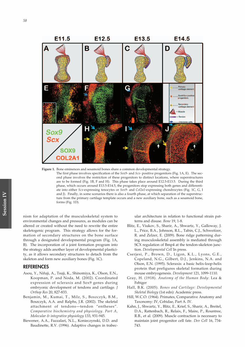
58
Ses
sion
IV
nism for adaptation of the musculoskeletal system toenvironmental changes and pressures, as modules can bealtered or created without the need to rewrite the entireskeletogenic program. This strategy allows for the for-mation of secondary structures on the bone surfacethrough a designated developmental program (Fig. 1A,B). The incorporation of a joint formation program intothe strategy adds another layer of developmental plastici-ty, as it allows secondary structures to detach from theskeleton and form new auxiliary bones (Fig. 1C).
REFERENCESAsou, Y., Nifuji, A., Tsuji, K., Shinomiya, K., Olson, E.N.,
Koopman, P. and Noda, M. (2002). Coordinatedexpression of scleraxis and Sox9 genes duringembryonic development of tendons and cartilage. JOrthop Res 20, 827-833.
Benjamin, M., Kumai, T., Milz, S., Boszczyk, B.M.,Boszczyk, A.A. and Ralphs, J.R. (2002). The skeletalattachment of tendons—tendon “entheses”.Comparative biochemistry and physiology. Part A,Molecular & integrative physiology 133, 931-945.
Biewener, A.A., Fazzalari, N.L., Konieczynski, D.D. andBaudinette, R.V. (1996). Adaptive changes in trabec-
ular architecture in relation to functional strain pat-terns and disuse. Bone 19, 1-8.
Blitz, E., Viukov, S., Sharir, A., Shwartz, Y., Galloway, J.L., Price, B.A., Johnson, R.L., Tabin, C.J., Schweitzer,R. and Zelzer, E. (2009). Bone ridge patterning dur-ing musculoskeletal assembly is mediated throughSCX regulation of Bmp4 at the tendon-skeleton junc-tion. Developmental Cell In press.
Cserjesi, P., Brown, D., Ligon, K.L., Lyons, G.E.,Copeland, N.G., Gilbert, D.J., Jenkins, N.A. andOlson, E.N. (1995). Scleraxis: a basic helix-loop-helixprotein that prefigures skeletal formation duringmouse embryogenesis. Development 121, 1099-1110.
Gray, H. (1918). Anatomy of the Human Body: Lea &Febiger
Hall, B.K. (2005). Bones and Cartilage: DevelopmentalSkeletal Biology (1st edn): Academic press.
Hill, W.C.O. (1964). Primates, Comparative Anatomy andTaxonomy: IV, Cebidae, Part A. IV.
Kahn, J., Shwartz, Y., Blitz, E., Krief, S., Sharir, A., Breitel,D.A., Rattenbach, R., Relaix, F., Maire, P., Rountree,R.B., et al. (2009). Muscle contraction is necessary tomaintain joint progenitor cell fate. Dev Cell 16, 734-743.
Figure 1. Bone eminences and sesamoid bones share a common developmental strategy.The first phase involves specification of the Sox9- and Scx- positive progenitors (Fig. 1A, E). The sec-ond phase involves the restriction of these progenitors to distinct locations, where superstructuresare to be formed (Fig. 1B, F and H). This phase takes place around E12.5-E13.5. During the thirdphase, which occurs around E13.5-E14.5, the progenitors stop expressing both genes and differenti-ate into either Scx-expressing tenocytes or Sox9- and Col2a1-expressing chondrocytes (Fig. 1C, G, Iand J). Finally, in some scenarios there is also a fourth phase, at which separation of the superstruc-ture from the primary cartilage template occurs and a new auxiliary bone, such as a sesamoid bone,forms (Fig. 1D).
02_本文1027(web用追加) 15.10.27 9:04 AM ページ 58
Session IV 59
Session
IV
Kelly, R.G., Brown, N.A. and Buckingham, M.E. (2001).The arterial pole of the mouse heart forms fromFgf10-expressing cells in pharyngeal mesoderm. DevCell 1, 435-440.
Lefebvre, V. and Bhattaram, P. Vertebrate skeletogenesis.Curr Top Dev Biol 90, 291-317.
Lorda-Diez, C.I., Montero, J.A., Martinez-Cue, C., Garcia-Porrero, J.A. and Hurle, J.M. (2009). Transforminggrowth factors beta coordinate cartilage and tendondifferentiation in the developing limb mesenchyme. JBiol Chem 284, 29988-29996.
Mjaatvedt, C.H., Nakaoka, T., Moreno-Rodriguez, R.,Norris, R.A., Kern, M.J., Eisenberg, C.A., Turner, D.and Markwald, R.R. (2001). The outflow tract of theheart is recruited from a novel heart-forming field.Dev Biol 238, 97-109.
Murchison, N.D., Price, B.A., Conner, D.A., Keene, D.R.,Olson, E.N., Tabin, C.J. and Schweitzer, R. (2007).Regulation of tendon differentiation by scleraxis dis-tinguishes force-transmitting tendons from muscle-anchoring tendons. Development 134, 2697-2708.
Olsen, B.R., Reginato, A.M. and Wang, W. (2000). Bonedevelopment. Annu Rev Cell Dev Biol 16, 191-220.
Parsons, F.G. (1904). Observations on Traction Epiphyses.J Anat Physiol 38, 248-258.
Parsons, F.G. (1908). Further Remarks on TractionEpiphyses. J Anat Physiol 42, 388-396.
Pearson, K. and Davin, A.G. (1921a). On the sesamoids ofthe knee-joint: Part I. Man. Biometrika 13, 133-175.
Pearson, K. and Davin, A.G. (1921b). On the Sesamoids ofthe Knee-Joint: Part II. Evolution of the Sesamoids.Biometrika 13, 350-400.
Pryce, B.A., Watson, S.S., Murchison, N.D., Staverosky,J.A., Dunker, N. and Schweitzer, R. (2009).Recruitment and maintenance of tendon progenitorsby TGFbeta signaling are essential for tendon forma-tion. Development 136, 1351-1361.
Schindler, O.S. and Scott, W.N. (2011). Basic kinematicsand biomechanics of the patello-femoral joint. Part 1:
The native patella. Acta Orthop Belg 77, 421-431.Schweitzer, R., Chyung, J.H., Murtaugh, L.C., Brent, A.E.,
Rosen, V., Olson, E.N., Lassar, A. and Tabin, C.J.(2001). Analysis of the tendon cell fate usingScleraxis, a specific marker for tendons and liga-ments. Development 128, 3855-3866.
Seo, H. S. and Serra, R. (2007). Deletion of Tgfbr2 in Prx1-cre expressing mesenchyme results in defects indevelopment of the long bones and joints. Dev Biol310, 304-316.
Spagnoli, A., O’Rear, L., Chandler, R.L., Granero-Molto,F., Mortlock, D.P., Gorska, A.E., Weis, J.A.,Longobardi, L., Chytil, A., Shimer, K., et al. (2007).TGF-beta signaling is essential for joint morphogene-sis. J Cell Biol 177, 1105-1117.
Sugimoto, Y., Takimoto, A., Akiyama, H., Kist, R., Scherer,G., Nakamura, T., Hiraki, Y. and Shukunami, C.(2013). Scx+/Sox9+ progenitors contribute to theestablishment of the junction between cartilage andtendon/ligament. Development 140, 2280-2288.
Thomopoulos, S. (2011). The role of mechanobiology inthe attachment of tendon to bone. IBMS BoneKEy 8,271-285.
Thomopoulos, S., Das, R., Birman, V., Smith, L., Ku, K.,Elson, E.L., Pryse, K.M., Marquez, J.P. and Genin,G.M. (2011). Fibrocartilage tissue engineering: therole of the stress environment on cell morphologyand matrix expression. Tissue Eng Part A 17, 1039-1053.
Thomopoulos, S., Genin, G.M. and Galatz, L.M. (2010).The development and morphogenesis of the tendon-to-bone insertion - what development can teach usabout healing. J Musculoskelet Neuronal Interact 10,35-45.
Waldo, K.L., Kumiski, D.H., Wallis, K.T., Stadt, H.A.,Hutson, M.R., Platt, D.H. and Kirby, M.L. (2001).Conotruncal myocardium arises from a secondaryheart field. Development 128, 3179-3188.
02_本文1027(web用追加) 15.10.27 9:04 AM ページ 59
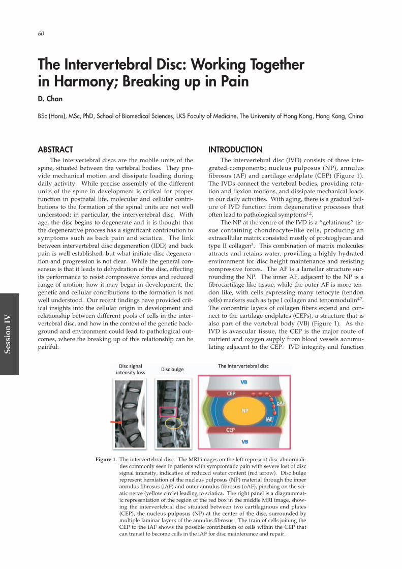
60
Ses
sion
IV
The Intervertebral Disc: Working Together in Harmony; Breaking up in PainD. Chan
BSc (Hons), MSc, PhD, School of Biomedical Sciences, LKS Faculty of Medicine, The University of Hong Kong, Hong Kong, China
ABSTRACTThe intervertebral discs are the mobile units of the
spine, situated between the vertebral bodies. They pro-vide mechanical motion and dissipate loading duringdaily activity. While precise assembly of the differentunits of the spine in development is critical for properfunction in postnatal life, molecular and cellular contri-butions to the formation of the spinal units are not wellunderstood; in particular, the intervertebral disc. Withage, the disc begins to degenerate and it is thought thatthe degenerative process has a significant contribution tosymptoms such as back pain and sciatica. The linkbetween intervertebral disc degeneration (IDD) and backpain is well established, but what initiate disc degenera-tion and progression is not clear. While the general con-sensus is that it leads to dehydration of the disc, affectingits performance to resist compressive forces and reducedrange of motion; how it may begin in development, thegenetic and cellular contributions to the formation is notwell understood. Our recent findings have provided crit-ical insights into the cellular origin in development andrelationship between different pools of cells in the inter-vertebral disc, and how in the context of the genetic back-ground and environment could lead to pathological out-comes, where the breaking up of this relationship can bepainful.
INTRODUCTIONThe intervertebral disc (IVD) consists of three inte-
grated components; nucleus pulposus (NP), annulusfibrosus (AF) and cartilage endplate (CEP) (Figure 1).The IVDs connect the vertebral bodies, providing rota-tion and flexion motions, and dissipate mechanical loadsin our daily activities. With aging, there is a gradual fail-ure of IVD function from degenerative processes thatoften lead to pathological symptoms1,2.
The NP at the centre of the IVD is a “gelatinous” tis-sue containing chondrocyte-like cells, producing anextracellular matrix consisted mostly of proteoglycan andtype II collagen3. This combination of matrix moleculesattracts and retains water, providing a highly hydratedenvironment for disc height maintenance and resistingcompressive forces. The AF is a lamellar structure sur-rounding the NP. The inner AF, adjacent to the NP is afibrocartilage-like tissue, while the outer AF is more ten-don like, with cells expressing many tenocyte (tendoncells) markers such as type I collagen and tenonmodulin4-7.The concentric layers of collagen fibers extend and con-nect to the cartilage endplates (CEPs), a structure that isalso part of the vertebral body (VB) (Figure 1). As theIVD is avascular tissue, the CEP is the major route ofnutrient and oxygen supply from blood vessels accumu-lating adjacent to the CEP. IVD integrity and function
Figure 1. The intervertebral disc. The MRI images on the left represent disc abnormali-ties commonly seen in patients with symptomatic pain with severe lost of discsignal intensity, indicative of reduced water content (red arrow). Disc bulgerepresent herniation of the nucleus pulposus (NP) material through the innerannulus fibrosus (iAF) and outer annulus fibrosus (oAF), pinching on the sci-atic nerve (yellow circle) leading to sciatica. The right panel is a diagrammat-ic representation of the region of the red box in the middle MRI image, show-ing the intervertebral disc situated between two cartilaginous end plates(CEP), the nucleus pulposus (NP) at the center of the disc, surrounded bymultiple laminar layers of the annulus fibrosus. The train of cells joining theCEP to the iAF shows the possible contribution of cells within the CEP thatcan transit to become cells in the iAF for disc maintenance and repair.
02_本文1027(web用追加) 15.10.27 9:04 AM ページ 60
Session IV 61
Session
IV
rely on the proper cellular function of the disc as a whole.The failure of one is sufficient to induce the breakdownof function as a whole with painful consequences3.Understanding the cellular origin, differentiation, andgenetic and environmental influences on tissue mainte-nance are of high clinical relevance in treating sympto-matic outcomes.
Cellular origin and differentiation of the nucleus pulposuscells
In embryonic development, while the exact cellularorigin of the notochord is unresolved since it originatesfrom cells in the epiblast migrating through the primitivestreak, in which the mesendoderm cells are bipotential,the NP is clearly derived from cells of the notochord8,9.Using genetic tools in mice to tag and trace notochordalcells with notochord-specific Cre drivers, Shh-CreERT2 10
and Noto-Cre11, showed that destiny of notochordal cellsin the mouse adult spine is indeed the NP. Furthermore,these cells are long lasting in the NP with notochordaldescendent cells residing in the adult NP for study peri-ods of up to 9 months11 and 19 months10. However, it isstill unclear whether other cells contribute to the NP inpostnatal life, as this possibility cannot be excluded fromthe present studies. Some studies have suggested othercell types are present in aged or degenerated discs withfibroblastic features and the possible presence ofmacrophages, suggesting other cells can infiltrate into theNP12-14.
Recently, a detailed analysis of human NP cells showedthe presence of distinct pools of cells with specific cellsurface markers that could define possible progenitorcells in the NP, and the progressive differentiation towardsmature NP cells15. Using the tyrosine kinase receptor 2(Tie2) and disialoganglioside 2 (GD2) as new cell mark-ers, it was shown that Tie2+/GD2-/CD24- cells havecharacteristics of dormant stem cells; Tie2+/GD2+/CD24-cells have self-renewal potentials, Tie2-/GD2+/CD24- asNP cell progenitors; Tie2-/GD2+/CD24+ as committedNP progenitors; and Tie2-/GD2-/CD24+ as mature cellsin the NP15. Of clinical relevance is a correlation of pro-gressive loss of these stem/progenitor cell pools withonset/severity of disc degeneration in human15. We havefurther shown that similar correlations exist in mice, ininbreed strains with genetic backgrounds that are moresusceptible to intervertebral disc degeneration (unpub-lished data). Significantly, we propose that maintainingTie2-/GD2+/CD24+ cells correlates with better outcomethat can protect against disc degeneration (unpublisheddata).
Cellular contribution to the formation and maintenance ofthe annulus fibrosus
Anatomical and histological analyses suggest the AFis derived from the sclerotome16-18, supported by a recentgenetic study in mice tagging sclerotome cells using theTbx18-Cre19, that traced cells to the anterior part of the AFand the vertebral bodies, but not in the NP compartment.Involvement of other cell types in development againcannot be excluded.
A mature AF consists of inner (IAF) and outer (OAF)compartments with distinct cell morphology and extra-
cellular matrix composition (Figure 1). How these areformed or partitioned is not clear. In mouse develop-ment, the lamellar structures of the AF are not welldefined at E13.5-E14.5, but cells begin to orientate con-centrically around the NP as formation of the IVD pro-gresses20. With growth, the number of lamellae and cellswithin the AF increase. However, neither the cellularcontribution nor the processes of lamellae increment areknown.
The presence of progenitor cells identified in IVDs ofvarious animal models21 suggest these progenitors maycontribute to growth, maintenance and repair of the IVD.Of interest, a recent study identified possible “migrationroutes” of progenitor cells in the outer AF and theinvolvement of epithelial to mesenchymal transitionprocess (EMT)22. As such, this pool of cells is suggestedto be a source of progenitor cells for the maintenance ofthe AF during adult life.
The CEP and AF are tightly connected, analogous toa tendon insertion into bone, forming a “continuous tis-sue”. There is also evidence of “Sharpey’s fibers” bridg-ing the two tissues23,24. The inner AF inserts directly intothe CEP. The junction where the two structures intersectis described as the AF-VB interface25. At this interface,there are histological evidence showing column of cellsbridging the interface with gradually morphologicalchange from round chondrocyte-like cells to elongatedfibroblastic like cells aligned along the laminar structureof the AF25.
To assess whether these two types of cells are relatedor share a common origin, we used a Cola10a1-Cremouse to tag and trace hypertrophic chondrocytes at thecartilaginous junction of the AF-CEP interface, and showthat indeed they can become cells of the inner AF(unpublished data). We also showed that this chondro-cyte to inner AF transition is enhanced in the experimen-tal model to induce degeneration, suggesting that thisreplenishment of inner AF cells may play a role in IVDmaintenance and repair (unpublished data).
CONCLUSIONWhile it is clear that there are anatomical and
mechanical loading differences between mouse andhuman; at the cellular level, it is likely to have many sim-ilarities in developmental processes, cellular differentia-tion, and regulatory mechanisms. Furthermore, the enor-mous repertoire of inbred mice with distinct geneticbackgrounds allows the possibility of studying differ-ences in intervertebral disc maintenance and degenera-tion. The concept of varying healing potentials in differ-ent strains of mice can be exploited. In particular, micewith varying healing potential of cartilaginous tissues,which have been reported26, and some genetic studiesperformed aiming to identify risk and protective loci27-30.Indeed, our analysis of four strains of mice, C57Black andSM/J (poor healers), and MRL and LG/J (good healers)showed clear differences in IVD maintenance and repairpotentials (unpublished data). Dissecting the geneticvariations between these mice will enable the identifica-tion of potential risk and protective factors for discdegeneration, and analyses of the molecular and cellularchanges in relation to the progression of NP cell differen-
02_本文1027(web用追加) 15.10.27 9:04 AM ページ 61
62
Ses
sion
IV
tiation and AF cell maintenance will provide the relation-ship between the function and contribution of the differ-ent cells in the IVD structure, enabling the disc to work inharmony in the good healer mice, and their failure inpoor healer mice leading to destruction and “pain”.
REFERENCES01. Antoniou J, Steffen T, Nelson F, et al. The human lum-
bar intervertebral disc: evidence for changes in thebiosynthesis and denaturation of the extracellularmatrix with growth, maturation, ageing, and degener-ation. J Clin Invest. 1996 Aug 15; 98 (4): 996-1003.
02. Luoma K, Riihimaki H, Luukkonen R, et al. Low backpain in relation to lumbar disc degeneration. Spine.2000 Feb 15; 25 (4): 487-92.
03. Chan WC, Au TY, Tam V, et al. Coming together is abeginning: the making of an intervertebral disc. BirthDefects Res C Embryo Today. 2014 Mar; 102 (1): 83-100.
04. Peacock A. Observations on the prenatal developmentof the intervertebral disc in man. J Anat. 1951 Jul; 85(3): 260-74.
05. Walmsley R. The development and growth of theintervertebral disc. Edinb Med J. 1953 Aug; 60 (8):341-64.
06. Humzah MD, Soames RW. Human intervertebraldisc: structure and function. Anat Rec. 1988 Apr; 220(4): 337-56.
07. Rufai A, Benjamin M, Ralphs JR. The development offibrocartilage in the rat intervertebral disc. Anatomyand embryology. 1995 Jul; 192 (1): 53-62.
08. Aszodi A, Chan D, Hunziker E, et al. Collagen II isessential for the removal of the notochord and the for-mation of intervertebral discs. The Journal of cell biol-ogy. 1998 Nov 30; 143 (5): 1399-412.
09. Theiler K. Vertebral malformations. Advances inanatomy, embryology, and cell biology. 1988; 112: 1-99.
10. Choi KS, Cohn MJ, Harfe BD. Identification of nucleuspulposus precursor cells and notochordal remnants inthe mouse: implications for disk degeneration andchordoma formation. Developmental dynamics: anofficial publication of the American Association ofAnatomists. 2008 Dec; 237 (12): 3953-8.
11. McCann MR, Tamplin OJ, Rossant J, Seguin CA.Tracing notochord-derived cells using a Noto-cremouse: implications for intervertebral disc develop-ment. Disease models & mechanisms. 2012 Jan; 5 (1):73-82.
12. Boos N, Weissbach S, Rohrbach H, et al. Classificationof age-related changes in lumbar intervertebral discs:2002 Volvo Award in basic science. Spine. 2002 Dec 1;27 (23): 2631-44.
13. Liebscher T, Haefeli M, Wuertz K, et al. Age-relatedvariation in cell density of human lumbar interverte-bral disc. Spine. 2011 Jan 15; 36 (2): 153-9.
14. Pazzaglia UE, Salisbury JR, Byers PD. Developmentand involution of the notochord in the human spine.Journal of the Royal Society of Medicine. 1989 Jul; 82(7): 413-5.
15. Sakai D, Nakamura Y, Nakai T, et al. Exhaustion ofnucleus pulposus progenitor cells with ageing and
degeneration of the intervertebral disc. Nature com-munications. 2012; 3: 1264.
16. Bagnall KM, Higgins SJ, Sanders EJ. The contributionmade by cells from a single somite to tissues within abody segment and assessment of their integrationwith similar cells from adjacent segments.Development. 1989 Dec; 107 (4): 931-43.
17. Goldstein RS, Kalcheim C. Determination of epithelialhalf-somites in skeletal morphogenesis. Development.1992 Oct; 116 (2): 441-5.
18. Huang R, Zhi Q, Wilting J, Christ B. The fate of somi-tocoele cells in avian embryos. Anatomy and embry-ology. 1994 Sep; 190 (3): 243-50.
19. Bruggeman BJ, Maier JA, Mohiuddin YS, et al. Avianintervertebral disc arises from rostral sclerotome andlacks a nucleus pulposus: implications for evolutionof the vertebrate disc. Developmental dynamics: anofficial publication of the American Association ofAnatomists. 2012 Apr; 241 (4): 675-83.
20. Smith LJ, Nerurkar NL, Choi KS, et al. Degenerationand regeneration of the intervertebral disc: lessonsfrom development. Dis Model Mech. 2011 Jan; 4 (1):31-41.
21. Henriksson H, Thornemo M, Karlsson C, et al .Identification of cell proliferation zones, progenitorcells and a potential stem cell niche in the interverte-bral disc region: a study in four species. Spine. 2009Oct 1; 34 (21): 2278-87.
22. Henriksson HB, Svala E, Skioldebrand E, et al .Support of concept that migrating progenitor cellsfrom stem cell niches contribute to normal regenera-tion of the adult mammal intervertebral disc: adescriptive study in the New Zealand white rabbit.Spine. 2012 Apr 20; 37 (9): 722-32.
23. Inoue H. Three-dimensional architecture of lumbarintervertebral discs. Spine. 1981 Mar-Apr; 6 (2): 139-46.
24. Hashizume H. Three-dimensional architecture anddevelopment of lumber intervertebral discs. Actamedica Okayama. 1980 Nov; 34 (5): 301-14.
25. Nosikova YS, Santerre JP, Grynpas M, et al .Characterization of the annulus fibrosus-vertebralbody interface: identification of new structural fea-tures. Journal of anatomy. 2012 Dec; 221 (6): 577-89.
26. Fitzgerald J, Rich C, Burkhardt D, et al. Evidence forarticular cartilage regeneration in MRL/MpJ mice.Osteoarthritis Cartilage. 2008 Nov; 16 (11): 1319-26.
27. Rai MF, Hashimoto S, Johnson EE, et al. Heritability ofarticular cartilage regeneration and its associationwith ear wound healing in mice. Arthritis Rheum.2012 Jul; 64 (7): 2300-10.
28. Rai MF, Sandell LJ. Regeneration of articular cartilagein healer and non-healer mice. Matrix Biol. 2014 Oct;39: 50-5.
29. Rai MF, Sandell LJ. Genetic Variability in theResponse to Injury. Post-Traumatic Arthritis: SpringerUS; 2015. p. 149-64.
30. Rai MF, Schmidt EJ, McAlinden A, et al. Molecularinsight into the association between cartilage regener-ation and ear wound healing in genetic mouse mod-els: targeting new genes in regeneration. G3(Bethesda). 2013 Nov; 3 (11): 1881-91.
02_本文1027(web用追加) 15.10.27 9:04 AM ページ 62

Session
V
Epigenetics: Basic and Clinical ImplicationsEpigenetic Reprogramming in Physiology and Cancer
Kumamoto University
M. Nakao
Live-cell Imaging of Chromatin and DNA-methylation Dynamics Using MethylRO Mouse
Kinki University
K. Yamagata
Bone-derived miRNA as a Mediator of Cell-cell Communication
Hiroshima University
Y. Takei, T. Minamizaki, H. Yoshioka and Y. Yoshiko
Session V
02_本文1027(web用追加) 15.10.27 9:04 AM ページ 63

02_本文1027(web用追加) 15.10.27 9:04 AM ページ 64
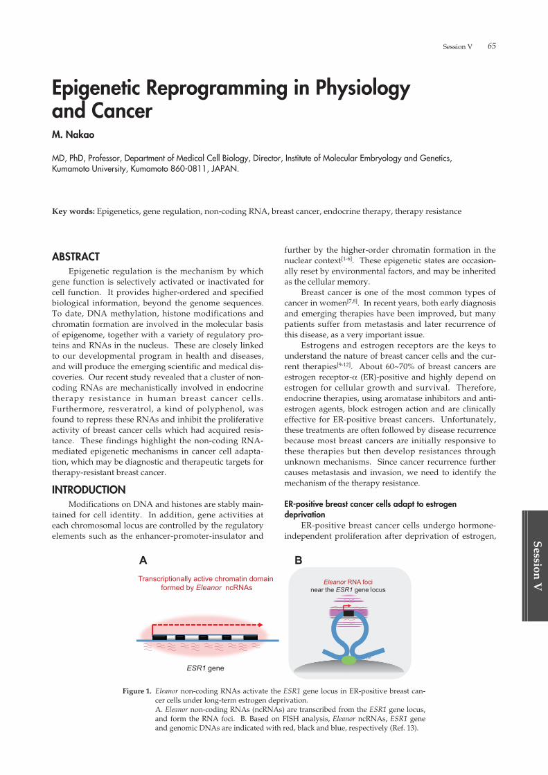
Session V 65
Session
V
ABSTRACTEpigenetic regulation is the mechanism by which
gene function is selectively activated or inactivated forcell function. It provides higher-ordered and specifiedbiological information, beyond the genome sequences.To date, DNA methylation, histone modifications andchromatin formation are involved in the molecular basisof epigenome, together with a variety of regulatory pro-teins and RNAs in the nucleus. These are closely linkedto our developmental program in health and diseases,and will produce the emerging scientific and medical dis-coveries. Our recent study revealed that a cluster of non-coding RNAs are mechanistically involved in endocrinetherapy resistance in human breast cancer cells.Furthermore, resveratrol, a kind of polyphenol, wasfound to repress these RNAs and inhibit the proliferativeactivity of breast cancer cells which had acquired resis-tance. These findings highlight the non-coding RNA-mediated epigenetic mechanisms in cancer cell adapta-tion, which may be diagnostic and therapeutic targets fortherapy-resistant breast cancer.
INTRODUCTIONModifications on DNA and histones are stably main-
tained for cell identity. In addition, gene activities ateach chromosomal locus are controlled by the regulatoryelements such as the enhancer-promoter-insulator and
further by the higher-order chromatin formation in thenuclear context[1-6]. These epigenetic states are occasion-ally reset by environmental factors, and may be inheritedas the cellular memory.
Breast cancer is one of the most common types ofcancer in women[7,8]. In recent years, both early diagnosisand emerging therapies have been improved, but manypatients suffer from metastasis and later recurrence ofthis disease, as a very important issue.
Estrogens and estrogen receptors are the keys tounderstand the nature of breast cancer cells and the cur-rent therapies[9-12]. About 60~70% of breast cancers areestrogen receptor-α (ER)-positive and highly depend onestrogen for cellular growth and survival. Therefore,endocrine therapies, using aromatase inhibitors and anti-estrogen agents, block estrogen action and are clinicallyeffective for ER-positive breast cancers. Unfortunately,these treatments are often followed by disease recurrencebecause most breast cancers are initially responsive tothese therapies but then develop resistances throughunknown mechanisms. Since cancer recurrence furthercauses metastasis and invasion, we need to identify themechanism of the therapy resistance.
ER-positive breast cancer cells adapt to estrogen deprivation
ER-positive breast cancer cells undergo hormone-independent proliferation after deprivation of estrogen,
Epigenetic Reprogramming in Physiology and CancerM. Nakao
MD, PhD, Professor, Department of Medical Cell Biology, Director, Institute of Molecular Embryology and Genetics, Kumamoto University, Kumamoto 860-0811, JAPAN.
Key words: Epigenetics, gene regulation, non-coding RNA, breast cancer, endocrine therapy, therapy resistance
Figure 1. Eleanor non-coding RNAs activate the ESR1 gene locus in ER-positive breast can-cer cells under long-term estrogen deprivation.A. Eleanor non-coding RNAs (ncRNAs) are transcribed from the ESR1 gene locus,and form the RNA foci. B. Based on FISH analysis, Eleanor ncRNAs, ESR1 geneand genomic DNAs are indicated with red, black and blue, respectively (Ref. 13).
02_本文1027(web用追加) 15.10.27 9:04 AM ページ 65
66
Ses
sion
V
leading to endocrine therapy resistance. Up-regulationof the ESR1 gene is critical for this process, but the under-lying mechanisms remained unclear. To investigateprocesses of the cancer cell adaptation, we used thehuman breast cancer cell line MCF7 which was ER-posi-tive[13]. These cells can acquire regrowth after long termestrogen deprivation (LTED), similar to cancer cells thatbecome resistant to endocrine therapy. The expression ofthe ER gene (ESR1) was increased during this adaptationin the LTED cells.
At this time, we performed the combinations of tran-scriptome and fluorescence in situ hybridization (FISH)analyses[13]. We found that estrogen deprivation induceda cluster of non-coding RNAs (ncRNAs), which wereexpressed from a large chromatin domain containing theESR1 locus. We named these RNAs as Eleanors (ESR1locus enhancing and activating non-coding RNAs).Interestingly, Eleanor ncRNAs were produced from theESR1 gene locus and were localized at the site of activelytranscribed ESR1 locus, resulting in the formation of dis-tinct RNA foci in the nucleus (Figure 1).
The effects of Eleanors inhibition and resveratrol in ER-overexpressing LTED cells
Depletion of Eleanors that were transcribed from theputative enhancer of the ESR1 gene, upstream (u)-Eleanor,impaired transcription of intragenic Eleanors and ESR1mRNA, indicating that Eleanors cis-activate the ESR1gene[13]. The u-Eleanor depletion significantly reducedcell growth. Furthermore, resveratrol, a kind of polyphe-nol with estrogen-like activity, was found to repressthese RNAs and inhibit the proliferative activity of breastcancer cells which had acquired estrogen-independentgrowth.
These findings uncovered the molecular basis forendocrine therapy-resistant breast cancer, which showedthe significant role of a new type of ncRNA-mediatedregulation of the ESR1 gene locus. Thus, Eleanor ncRNAsare actively involved in the epigenetic adaptation of ER-positive breast cancer cells via high expression of theESR1 gene. Our study emphasizes that the ncRNA-medi-ated mechanisms in cancer cell adaptation may be diag-nostic and therapeutic targets for endocrine therapy-resistant breast cancer.
In addition, Eleanors-mediated gene activation repre-sents a new type of locus control mechanism and playsan essential role in the adaptation of breast cancer cells.These findings exemplify one of the novel epigeneticmechanisms involving in cancer cell adaptation to envi-ronmental factors through the alteration of chromatinand nuclear context.
CONCLUSIONThis study revealed that a cluster of Eleanor non-cod-
ing RNAs are mechanistically involved in endocrine ther-apy resistance in human breast cancer cells.Furthermore, resveratrol was found to repress theseRNAs and inhibit the proliferative activity of breast can-cer cells which had acquired resistance.
ACKNOWLEDGEMENTS & CONFLICT OFINTEREST
This work was supported by grants from theMinistry of Education, Culture, Sports, Science andTechnology of Japan and the Japan Science andTechnology Agency (CREST). There is no conflict ofinterest in this study.
REFERENCES01. Nakao M. Epigenetics: interactions of DNA methyla-
tion and chromatin. Gene 2001: 278: 25-31.02. Ohki I, Shimotake N, Fujita N, Jee JG, Ikegami T,
Nakao M, and Shirakawa M. Solution structure of themethyl-CpG-binding domain of human MBD1 incomplex with a methylated DNA. Cell 2001: 105: 487-497.
03. Hino S, Sakamoto A, Nagaoka K, Anan K, Wang Y,Mimasu S, Umehara T, Yokoyama S, Kosai K, andNakao M. FAD-dependent lysine demethylase LSD1regulates cellular energy expenditure. Nat. Commun.2012: 3: 758.
04. Ishihara K, Oshimura M, and Nakao M. CTCF-depen-dent chromatin insulator is linked to epigeneticremodeling. Mol. Cell 2006: 23: 733-742.
05. Wendt KS, Yoshida K, Itoh T, Bando M, Koch B,Schirghuber E, Tsutsumi S, Nagae G, Ishihara K,Mishiro T, Yahata K, Imamoto F, Aburatani H, NakaoM, Imamoto N, Maeshima K, Shirahige K, and PetersJM. Cohesin mediates transcriptional insulation byCCCTC-binding factor. Nature 2008: 451: 796-803.
06. Mishiro T, Ishihara K, Hino S, Tsutsumi S, AburataniH, Shirahige K, Kinoshita Y, and Nakao M.Architectural roles of multiple chromatin insulators atthe human apolipoprotein gene cluster. EMBO J.2009: 28: 1234-1245.
07. Sotiriou C and Piccart MJ. Taking gene-expressionprofiling to the clinic: when will molecular signaturesbecome relevant to patient care? Nat. Rev. Cancer2007: 7: 545-553.
08. Ali S and Coombes RC. Endocrine-responsive breastcancer and strategies for combating resistance. Nat.Rev. Cancer 2002: 2: 101-112.
09. Yager JD and Davidson NE. Estrogen carcinogenesisin breast cancer. N. Engl. J. Med. 2006: 354: 270-282.
10. Musgrove EA and Sutherland RL. Biological determi-nants of endocrine resistance in breast cancer. Nat.Rev. Cancer 2009: 9: 631-643.
11. Katzenellenbogen BS, Kendra KL, Norman MJ, andBerthois Y. Proliferation, hormonal responsiveness,and estrogen receptor content of MCF-7 human breastcancer cells grown in the short-term and long-termabsence of estrogens. Cancer Res. 1987: 47: 4355-4360.
12. Welshons WV and Jordan VC. Adaptation of estro-gen-dependent MCF-7 cells to low estrogen (phenolred-free) culture. Eur. J. Cancer Clin. Oncol. 1987: 23:1935-1939.
13. Tomita S, Abdalla MO, Fujiwara S, Matsumori H,Maehara K, Ohkawa Y, Iwase H, Saitoh N, and NakaoM. A cluster of non-coding RNAs activates the ESR1locus during breast cancer adaptation to hormonedeprivation. Nat. Commun. 2015: 6: 6966.
02_本文1027(web用追加) 15.10.27 9:04 AM ページ 66
Session V 67
Session
V
ABSTRACTIn mammals, DNA is methylated at CpG sites, which
play pivotal roles in gene silencing and chromatin orga-nization. Furthermore, DNA methylation undergoesdynamic changes during development, differentiation,and in pathological processes. The conventional methodsrepresent snapshots; therefore, the dynamics of thismarker within living organisms remains unclear. Totrack this dynamics, we made a knockin mouse thatexpresses a red fluorescent protein (RFP)-fused methyl-CpG-binding domain (MBD) protein from the ROSA26locus ubiquitously; we named it MethylRO (methylationprobe in ROSA26 locus). Using this mouse, we per-formed RFP-mediated methylated DNA immunoprecipi-tation sequencing (MeDIP-seq), whole-body sectionanalysis, and live-cell imaging. We discovered thatmobility and pattern of heterochromatin as well as DNAmethylation signal intensity inside the nuclei can bemarkers for cellular differentiation status. Thus, theMethylRO mouse represents a powerful bioresource andtechnique for DNA methylation dynamics studies indevelopmental biology, stem cell biology, as well as indisease states.
TEXTIn mammals, DNA is covalently modified by methy-
lation of the cytosine base in the CpG dinucleotides andthese sites are frequently concentrated in repetitivesequences, such as pericentromeric regions and transpos-able elements[1,2]. Once these CpG sites are methylated,these are recognized by methyl-CpG-binding domain(MBD) family proteins[3], which then recruit additionalprotein complexes to these methylated DNA regions torepress gene expression and/or to constitute higherorder condensed chromatin structure called heterochro-matin to stabilize chromosome structures[3]. Previousstudies using DNA methyltransferase knockout micehave revealed that loss of DNA methylation leads toearly embryonic lethality (around E9.0), implicating theimportance of this epigenetic mark during develop-ment[4], and recent studies have indicated that DNAmethylation undergoes dynamic changes during mouseembryonic development as well as in tumorigenic
processes[5,6]. Although there is a strong need for study-ing the dynamics of DNA methylation status, there is noappropriate way at present to study this dynamics in liv-ing cells or in living organisms. Currently, the majorways to study DNA methylation are bisulfite sequenc-ing[7], methylated DNA immunoprecipitation (MeDIP)combined either with microarray or next-generationsequencing (MeDIP-seq)[8], and immunohistochem-istry[9,10]. However, all techniques require fixation of thecells and hence make it almost impossible to study thedynamics of this pivotal epigenetic mark. Previously,Yamagata and colleagues have succeeded to visualizeDNA methylation status by injecting green fluorescentprotein fused MBD and nuclear localizing signal ofMBD1 protein (EGFP-MBD-NLS) mRNA to living micezygote. By use of this method, they have succeeded tovisualize the DNA methylation status in preimplantationmouse embryos and found that DNA hypomethylationas a key signature which distinguishes germ cells fromsomatic cells[1,11]. Importantly, this method not only cantrace epigenetic changes in real time but also in single-cell resolution which was impossible with conventionalmethods. To apply this methodology to other cellulardifferentiation processes, we have knocked-in RFP (redfluorescent protein) fused MBD and NLS reporter probe(mCherry-MBD-NLS) gene into ROSA26 locus and gener-ated a mouse strain which captures global DNA methyla-tion status in living condition.
Generation of MethylRO mouseWe chose ROSA26 locus because this locus is well
known for its ubiquitous and uniform expression inmice[12,13] and widely used in reporter gene expres-sions[14,15]. We decided to fuse RFP because they havebetter signal to noise ratio compared to GFP. By usingconventional gene targeting method, mCherry-MBD-NLScDNA was successfully knocked into ROSA26 locus withhigh efficiency (28 out of 96 clones). In excellent agree-ment with previous studies[1,11,16], mCherry-MBD-NLSprobe localized to pericentromeric regions and formedfoci in interphase nucleus in ES cells (Figure 1A).Importantly, mCherry-MBD-NLS probe expressed fromROSA26 locus was uniform compared to probe expressedfrom other promoters, such as CMV and CAG. We then
Live-cell Imaging of Chromatin and DNA-methylation Dynamics Using MethylRO MouseK. Yamagata
Department of Genetic Engineering, Faculty of Biology-Oriented Science and Technology, Kinki University, 930 Nishimitani, Kinokawa, Wakayama 649-6493, Japan
Key words: MethylRO, DNA methylation, Epigenetic Dynamics, Heterochromatin, Live-cell Imaging
02_本文1027(web用追加) 15.10.27 9:04 AM ページ 67
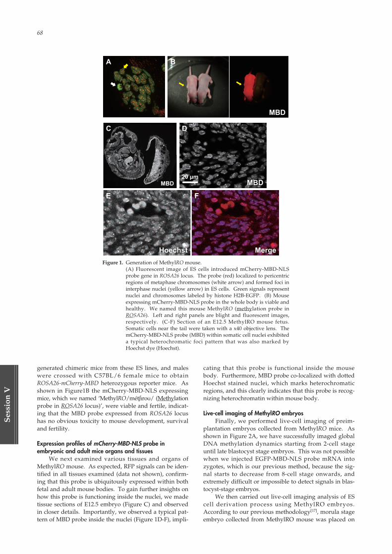
68
Ses
sion
V
generated chimeric mice from these ES lines, and maleswere crossed with C57BL/6 female mice to obtainROSA26-mCherry-MBD heterozygous reporter mice. Asshown in Figure1B the mCherry-MBD-NLS expressingmice, which we named ‘MethylRO/mét∫ıroU/ (Methylationprobe in ROSA26 locus)’, were viable and fertile, indicat-ing that the MBD probe expressed from ROSA26 locushas no obvious toxicity to mouse development, survivaland fertility.
Expression profiles of mCherry-MBD-NLS probe in embryonic and adult mice organs and tissues
We next examined various tissues and organs ofMethylRO mouse. As expected, RFP signals can be iden-tified in all tissues examined (data not shown), confirm-ing that this probe is ubiquitously expressed within bothfetal and adult mouse bodies. To gain further insights onhow this probe is functioning inside the nuclei, we madetissue sections of E12.5 embryo (Figure C) and observedin closer details. Importantly, we observed a typical pat-tern of MBD probe inside the nuclei (Figure 1D-F), impli-
cating that this probe is functional inside the mousebody. Furthermore, MBD probe co-localized with dottedHoechst stained nuclei, which marks heterochromaticregions, and this clearly indicates that this probe is recog-nizing heterochromatin within mouse body.
Live-cell imaging of MethylRO embryosFinally, we performed live-cell imaging of preim-
plantation embryos collected from MethylRO mice. Asshown in Figure 2A, we have successfully imaged globalDNA methylation dynamics starting from 2-cell stageuntil late blastocyst stage embryos. This was not possiblewhen we injected EGFP-MBD-NLS probe mRNA intozygotes, which is our previous method, because the sig-nal starts to decrease from 8-cell stage onwards, andextremely difficult or impossible to detect signals in blas-tocyst-stage embryos.
We then carried out live-cell imaging analysis of EScell derivation process using MethylRO embryos.According to our previous methodology[17], morula stageembryo collected from MethylRO mouse was placed on
Figure 1. Generation of MethylRO mouse.(A) Fluorescent image of ES cells introduced mCherry-MBD-NLSprobe gene in ROSA26 locus. The probe (red) localized to pericentricregions of metaphase chromosomes (white arrow) and formed foci ininterphase nuclei (yellow arrow) in ES cells. Green signals representnuclei and chromosomes labeled by histone H2B-EGFP. (B) Mouseexpressing mCherry-MBD-NLS probe in the whole body is viable andhealthy. We named this mouse MethylRO (methylation probe inROSA26). Left and right panels are blight and fluorescent images,respectively. (C-F) Section of an E12.5 MethylRO mouse fetus.Somatic cells near the tail were taken with a x40 objective lens. ThemCherry-MBD-NLS probe (MBD) within somatic cell nuclei exhibiteda typical heterochromatic foci pattern that was also marked byHoechst dye (Hoechst).
02_本文1027(web用追加) 15.10.27 9:04 AM ページ 68
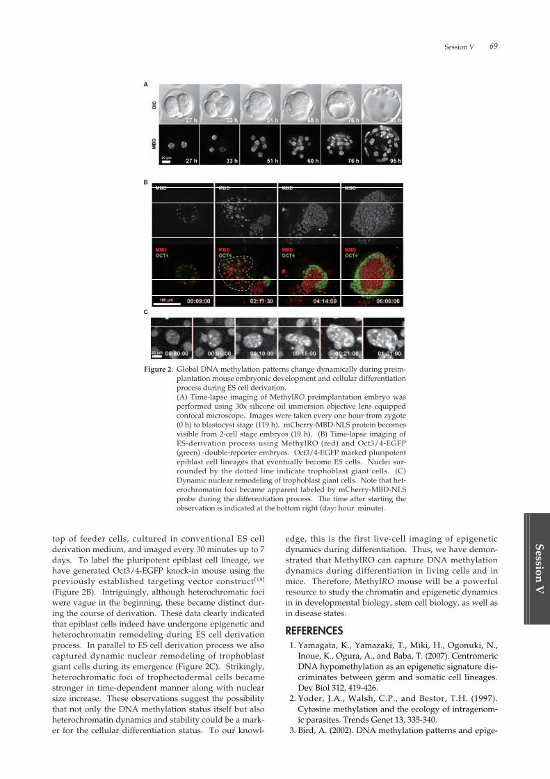
Session V 69
Session
V
top of feeder cells, cultured in conventional ES cellderivation medium, and imaged every 30 minutes up to 7days. To label the pluripotent epiblast cell lineage, wehave generated Oct3/4-EGFP knock-in mouse using thepreviously established targeting vector construct[18]
(Figure 2B). Intriguingly, although heterochromatic fociwere vague in the beginning, these became distinct dur-ing the course of derivation. These data clearly indicatedthat epiblast cells indeed have undergone epigenetic andheterochromatin remodeling during ES cell derivationprocess. In parallel to ES cell derivation process we alsocaptured dynamic nuclear remodeling of trophoblastgiant cells during its emergence (Figure 2C). Strikingly,heterochromatic foci of trophectodermal cells becamestronger in time-dependent manner along with nuclearsize increase. These observations suggest the possibilitythat not only the DNA methylation status itself but alsoheterochromatin dynamics and stability could be a mark-er for the cellular differentiation status. To our knowl-
edge, this is the first live-cell imaging of epigeneticdynamics during differentiation. Thus, we have demon-strated that MethylRO can capture DNA methylationdynamics during differentiation in living cells and inmice. Therefore, MethylRO mouse will be a powerfulresource to study the chromatin and epigenetic dynamicsin in developmental biology, stem cell biology, as well asin disease states.
REFERENCES01. Yamagata, K., Yamazaki, T., Miki, H., Ogonuki, N.,
Inoue, K., Ogura, A., and Baba, T. (2007). CentromericDNA hypomethylation as an epigenetic signature dis-criminates between germ and somatic cell lineages.Dev Biol 312, 419-426.
02. Yoder, J.A., Walsh, C.P., and Bestor, T.H. (1997).Cytosine methylation and the ecology of intragenom-ic parasites. Trends Genet 13, 335-340.
03. Bird, A. (2002). DNA methylation patterns and epige-
Figure 2. Global DNA methylation patterns change dynamically during preim-plantation mouse embryonic development and cellular differentiationprocess during ES cell derivation.(A) Time-lapse imaging of MethylRO preimplantation embryo wasperformed using 30x silicone oil immersion objective lens equippedconfocal microscope. Images were taken every one hour from zygote(0 h) to blastocyst stage (119 h). mCherry-MBD-NLS protein becomesvisible from 2-cell stage embryos (19 h). (B) Time-lapse imaging ofES-derivation process using MethylRO (red) and Oct3/4-EGFP(green) -double-reporter embryos. Oct3/4-EGFP marked pluripotentepiblast cell lineages that eventually become ES cells. Nuclei sur-rounded by the dotted line indicate trophoblast giant cells. (C)Dynamic nuclear remodeling of trophoblast giant cells. Note that het-erochromatin foci became apparent labeled by mCherry-MBD-NLSprobe during the differentiation process. The time after starting theobservation is indicated at the bottom right (day: hour: minute).
02_本文1027(web用追加) 15.10.27 9:05 AM ページ 69
70
Ses
sion
V
netic memory. Genes Dev 16, 6-21.04. Li, E., Bestor, T.H., and Jaenisch, R. (1992). Targeted
mutation of the DNA methyltransferase gene resultsin embryonic lethality. Cell 69, 915-926.
05. Ehrlich, M. (2009). DNA hypomethylation in cancercells. Epigenomics 1, 239-259.
06. Reik, W. (2007). Stability and flexibility of epigeneticgene regulation in mammalian development. Nature447, 425-432.
07. Frommer, M., McDonald, L.E., Millar, D.S., Collis,C.M., Watt, F., Grigg, G.W., Molloy, P.L., and Paul,C.L. (1992). A genomic sequencing protocol thatyields a positive display of 5-methylcytosine residuesin individual DNA strands. Proc Natl Acad Sci U S A89, 1827-1831.
08. Clark, C., Palta, P., Joyce, C.J., Scott, C., Grundberg,E., Deloukas, P., Palotie, A., and Coffey, A.J. (2012). Acomparison of the whole genome approach of MeDIP-seq to the targeted approach of the InfiniumHumanMethylation450 BeadChip ((R)) for methylomeprofiling. PLoS One 7, e50233.
09. Ito, S., D’Alessio, A.C., Taranova, O.V., Hong, K.,Sowers, L.C., and Zhang, Y. (2010). Role of Tet pro-teins in 5mC to 5hmC conversion, ES-cell self-renewaland inner cell mass specification. Nature 466, 1129-1133.
10. Jorgensen, H.F., Adie, K., Chaubert, P., and Bird, A.P.(2006). Engineering a high-affinity methyl-CpG-bind-ing protein. Nucleic Acids Res 34, e96.
11. Yamazaki, T., Yamagata, K., and Baba, T. (2007).Time-lapse and retrospective analysis of DNA methy-lation in mouse preimplantation embryos by live cell
imaging. Dev Biol 304, 409-419.12. Soriano, P. (1999). Generalized lacZ expression with
the ROSA26 Cre reporter strain. Nat Genet 21, 70-71.13. Srinivas, S., Watanabe, T., Lin, C.S., William, C.M.,
Tanabe, Y., Jessell, T.M., and Costantini, F. (2001). Crereporter strains produced by targeted insertion ofEYFP and ECFP into the ROSA26 locus. BMC DevBiol 1, 4.
14. Abe, T., Sakaue-Sawano, A., Kiyonari, H., Shioi, G.,Inoue, K., Horiuchi, T., Nakao, K., Miyawaki, A.,Aizawa, S., and Fujimori, T. (2013). Visualization ofcell cycle in mouse embryos with Fucci2 reporterdirected by Rosa26 promoter. Development 140, 237-246.
15. Shioi, G., Kiyonari, H., Abe, T., Nakao, K., Fujimori,T., Jang, C.W., Huang, C.C., Akiyama, H., Behringer,R.R., and Aizawa, S. (2011). A mouse reporter line toconditionally mark nuclei and cell membranes for invivo live-imaging. Genesis 49, 570-578.
16. Kobayakawa, S., Miike, K., Nakao, M., and Abe, K.(2007). Dynamic changes in the epigenomic state andnuclear organization of differentiating mouse embry-onic stem cells. Genes Cells 12, 447-460.
17. Yamagata, K., Ueda, J., Mizutani, E., Saitou, M., andWakayama, T. (2010). Survival and death of epiblastcells during embryonic stem cell derivation revealedby long-term live-cell imaging with an Oct4 reportersystem. Dev Biol. 346 (1): 90-101.
18. Toyooka, Y., Shimosato, D., Murakami, K., Takahashi,K., and Niwa, H. (2008). Identification and characteri-zation of subpopulations in undifferentiated ES cellculture. Development. 135 (5): 909-18.
02_本文1027(web用追加) 15.10.27 9:05 AM ページ 70
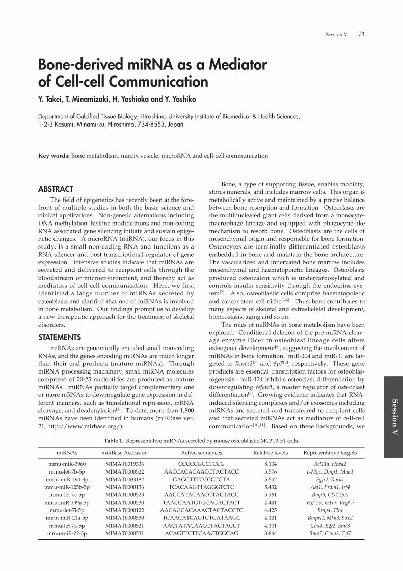
Session V 71
Session
V
ABSTRACTThe field of epigenetics has recently been at the fore-
front of multiple studies in both the basic science andclinical applications. Non-genetic alternations includingDNA methylation, histone modifications and non-codingRNA associated gene silencing initiate and sustain epige-netic changes. A microRNA (miRNA), our focus in thisstudy, is a small non-coding RNA and functions as aRNA silencer and post-transcriptional regulator of geneexpression. Intensive studies indicate that miRNAs aresecreted and delivered to recipient cells through thebloodstream or microenvironment, and thereby act asmediators of cell-cell communication. Here, we firstidentified a large number of miRNAs secreted byosteoblasts and clarified that one of miRNAs is involvedin bone metabolism. Our findings prompt us to developa new therapeutic approach for the treatment of skeletaldisorders.
STATEMENTSmiRNAs are genomically encoded small non-coding
RNAs, and the genes encoding miRNAs are much longerthan their end products (mature miRNAs). ThroughmiRNA processing machinery, small miRNA moleculescomprised of 20-25 nucleotides are produced as maturemiRNAs. miRNAs partially target complementary oneor more mRNAs to downregulate gene expression in dif-ferent manners, such as translational repression, mRNAcleavage, and deadenylation[1]. To date, more than 1,800miRNAs have been identified in humans (miRBase ver.21, http://www.mirbase.org/).
Bone, a type of supporting tissue, enables mobility,stores minerals, and includes marrow cells. This organ ismetabolically active and maintained by a precise balancebetween bone resorption and formation. Osteoclasts arethe multinucleated giant cells derived from a monocyte-macrophage lineage and equipped with phagocytic-likemechanism to resorb bone. Osteoblasts are the cells ofmesenchymal origin and responsible for bone formation.Osteocytes are terminally differentiated osteoblastsembedded in bone and maintain the bone architecture.The vascularized and innervated bone marrow includesmesenchymal and haematopoietic lineages. Osteoblastsproduced osteocalcin which is undercarboxylated andcontrols insulin sensitivity through the endocrine sys-tem[2]. Also, osteoblastic cells comprise haematopoieticand cancer stem cell niche[3-5]. Thus, bone contributes tomany aspects of skeletal and extraskeletal development,homeostasis, aging and so on.
The roles of miRNAs in bone metabolism have beenexplored. Conditional deletion of the pre-miRNA cleav-age enzyme Dicer in osteoblast lineage cells altersosteogenic development[6], suggesting the involvement ofmiRNAs in bone formation. miR-204 and miR-31 are tar-geted to Runx2[7] and Sp7[8], respectively. These geneproducts are essential transcription factors for osteoblas-togenesis. miR-124 inhibits osteoclast differentiation bydownregulating Nfatc1, a master regulator of osteoclastdifferentiation[9]. Growing evidence indicates that RNA-induced silencing complexes and/or exosomes includingmiRNAs are secreted and transferred to recipient cellsand that secreted miRNAs act as mediators of cell-cellcommunication[10,11]. Based on these backgrounds, we
Bone-derived miRNA as a Mediator of Cell-cell CommunicationY. Takei, T. Minamizaki, H. Yoshioka and Y. Yoshiko
Department of Calcified Tissue Biology, Hiroshima University Institute of Biomedical & Health Sciences, 1-2-3 Kasumi, Minami-ku, Hiroshima, 734-8553, Japan
Key words: Bone metabolism, matrix vesicle, microRNA and cell-cell communication
Table 1. Representative miRNAs secreted by mouse osteoblastic MC3T3-E1 cells.
miRNAs miRBase Accession Active sequences Relative levels Representative targets
mmu-miR-3960 MIMAT0019336 CCCCCGCCTCCG 8.104 Bcl11a, Hoxa2mmu-let-7b-5p MIMAT0000522 AACCACACAACCTACTACC 5.576 c-Myc, Dmp1, Muc1
mmu-miR-494-3p MIMAT0003182 GAGGTTTCCCGTGTA 5.542 Fgfr2, Rock1mmu-miR-125b-5p MIMAT0000136 TCACAAGTTAGGGTCTC 5.432 Akt1, Prdm1, Irf4
mmu-let-7c-5p MIMAT0000523 AACCATACAACCTACTACC 5.161 Bmp5, CDC25Ammu-miR-199a-3p MIMAT0000230 TAACCAATGTGCAGACTACT 4.441 Hif-1α, mTor, Vegf-a
mmu-let-7i-5p MIMAT0000122 AACAGCACAAACTACTACCTC 4.425 Bmp4, Tlr4mmu-miR-21a-5p MIMAT0000530 TCAACATCAGTCTGATAAGC 4.121 BmprII, Mkk3, Sox2
mmu-let-7a-5p MIMAT0000521 AACTATACAACCTACTACCT 4.101 Chd4, E2f2, Stat3mmu-miR-22-3p MIMAT0000531 ACAGTTCTTCAACTGGCAG 3.864 Bmp7, Ccna2, Tcf7
02_本文1027(web用追加) 15.10.27 9:05 AM ページ 71
72
Ses
sion
V
provide evidence for a new form of intracellular commu-nication in bone.
We performed miRNA microarray analysis on extra-cellular components from mouse osteoblastic MC3T3-E1cells. Of miRNAs identified, over 50 miRNAs were iden-tical to human counterparts. We chose 10 miRNAs withabundant amounts and validated target genes (Table 1).As expected, one of miRNAs was selectively transferredto recipient cells and downregulated target gene, result-ing in defective cell activities. We will provide experi-mental data on the roles of this miRNA in bone both invivo and in vitro and discuss in detail.
REMARKSBone act as an endocrine organ to orchestrate human
body. Thus, the appropriate interventions to cell-cellcommunication in bone may be necessary to integratetherapies for both the skeletal and extraskeletal conse-quences. We expect that this approach is greatly facilitat-ed by our findings in bone-derived secreted miRNAs.
REFERENCES01. Ambros V. The functions of animal microRNAs.
Nature. 2004; 431 (7006): 350-5.02. Ferron M, McKee MD, Levine RL, Ducy P, Karsenty
G. Intermittent injections of osteocalcin improve glu-cose metabolism and prevent type 2 diabetes in mice.Bone. 2012; 50 (2): 568-575.
03. Calvi LM, Adams GB, Weibrecht KW, Weber JM,Olson DP, Knight MC, Martin RP, Schipani E, DivietiP, Bringhurst FR, Milner LA, Kronenberg HM,Scadden DT. Osteoblastic cells regulate thehaematopoietic stem cell niche. Nature. 2003; 425(6960): 841-846.
04. Zhang J, Niu C, Ye L, Huang H, He X, Tong WG, RossJ, Haug J, Johnson T, Feng JQ, Harris S, Wiedemann
LM, Mishina Y, Li L. Identification of the haematopoi-etic stem cell niche and control of the niche size.Nature. 2003; 425 (6960): 836-841.
05. Iwasaki H, Suda T. Cancer stem cells and their niche.Cancer Sci. 2009; 100 (7): 1166-1172.
06. Gaur T, Hussain S, Mudhasani R, Parulkar I, ColbyJL, Frederick D, Kream BE, van Wijnen AJ, Stein JL,Stein GS, Jones SN, Lian JB. Dicer inactivation inosteoprogenitor cells compromises fetal survival andbone formation, while excision in differentiatedosteoblasts increases bone mass in the adult mouse.Dev Biol. 2010; 340 (1): 10-21.
07. Huang J, Zhao L, Xing L, Chen D. MicroRNA-204 reg-ulates Runx2 protein expression and mesenchymalprogenitor cell differentiation. Stem Cells. 2010; 28 (2):357-364.
08. Baglío SR, Devescovi V, Granchi D, Baldini N.MicroRNA expression profiling of human bone mar-row mesenchymal stem cells during osteogenic differ-entiation reveals Osterix regulation by miR-31. Gene.2013; 527 (1): 321-331.
09. Lee Y, Kim HJ, Park CK, Kim YG, Lee HJ, Kim JY,Kim HH. MicroRNA-124 regulates osteoclast differen-tiation. Bone. 2013; 56 (2): 383-389.
10. Valadi H, Ekström K, Bossios A, Sjöstrand M, Lee JJ,Lötvall JO. Exosome-mediated transfer of mRNAsand microRNAs is a novel mechanism of geneticexchange between cells. Nat Cell Biol. 2007; 9 (6): 654-659.
11. Hergenreider E, Heydt S, Tréguer K, Boettger T,Horrevoets AJ, Zeiher AM, Scheffer MP, FrangakisAS, Yin X, Mayr M, Braun T, Urbich C, Boon RA,Dimmeler S. Atheroprotective communicationbetween endothelial cells and smooth muscle cellsthrough miRNAs. Nat Cell Biol. 2012; 14 (3): 249-256.
02_本文1027(web用追加) 15.10.27 9:05 AM ページ 72
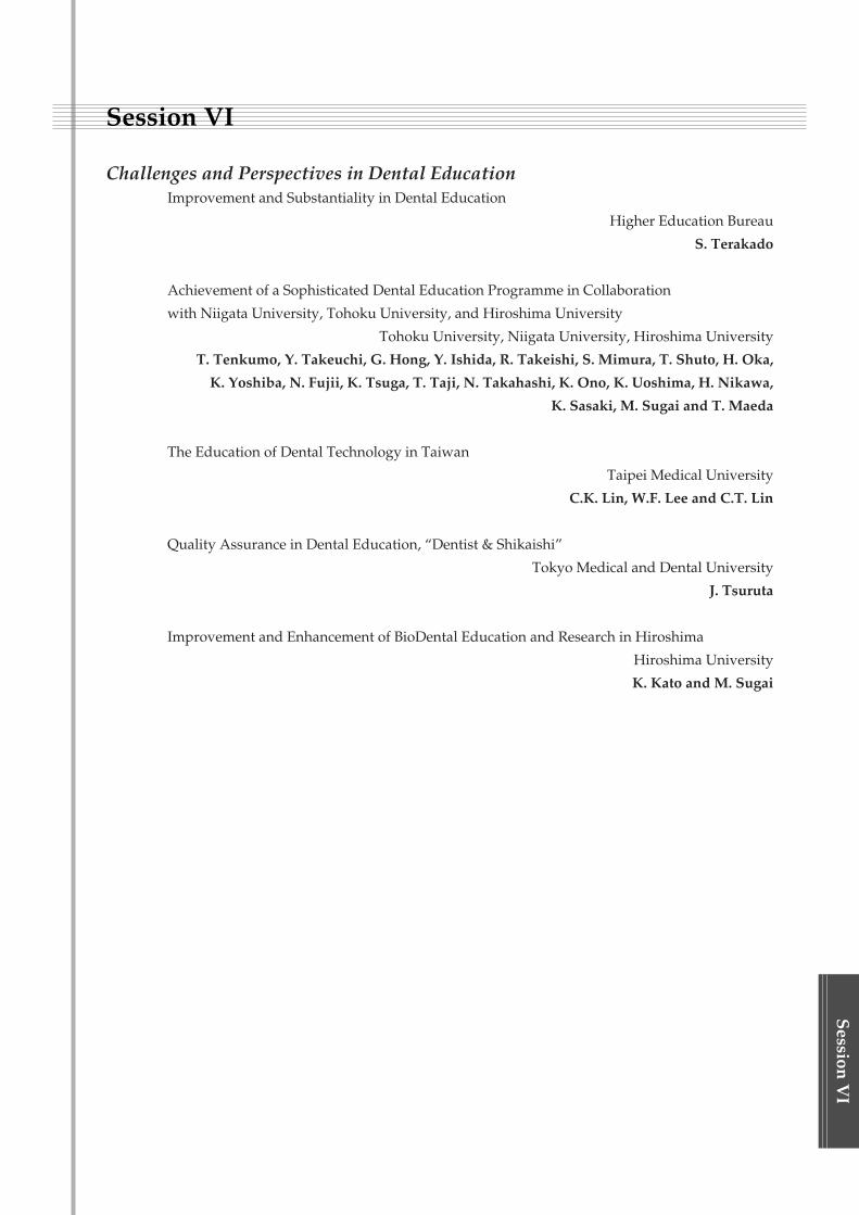
Session
VI
Challenges and Perspectives in Dental EducationImprovement and Substantiality in Dental Education
Higher Education Bureau
S. Terakado
Achievement of a Sophisticated Dental Education Programme in Collaboration
with Niigata University, Tohoku University, and Hiroshima University
Tohoku University, Niigata University, Hiroshima University
T. Tenkumo, Y. Takeuchi, G. Hong, Y. Ishida, R. Takeishi, S. Mimura, T. Shuto, H. Oka,
K. Yoshiba, N. Fujii, K. Tsuga, T. Taji, N. Takahashi, K. Ono, K. Uoshima, H. Nikawa,
K. Sasaki, M. Sugai and T. Maeda
The Education of Dental Technology in Taiwan
Taipei Medical University
C.K. Lin, W.F. Lee and C.T. Lin
Quality Assurance in Dental Education, “Dentist & Shikaishi”
Tokyo Medical and Dental University
J. Tsuruta
Improvement and Enhancement of BioDental Education and Research in Hiroshima
Hiroshima University
K. Kato and M. Sugai
Session VI
02_本文1027(web用追加) 15.10.27 9:05 AM ページ 73

02_本文1027(web用追加) 15.10.27 9:05 AM ページ 74
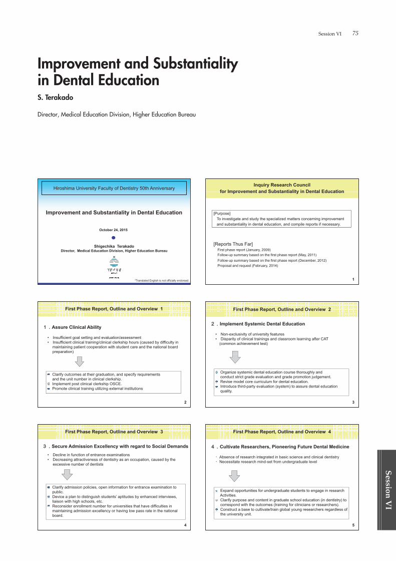
Session VI 75
Session
VI
Hiroshima University Faculty of Dentistry 50th Anniversary
Improvement and Substantiality in Dental Education
October 24, 2015
Shi hik T k d
,
Shigechika TerakadoDirector, Medical Education Division, Higher Education Bureau
*Translated English is not officially endorsed.
First Phase Report, Outline and Overview 1
Assure Clinical AbilityAssure Clinical Ability
p.p
2
First Phase Report, Outline and Overview 3
Secure Admission Excellency with regard to Social Demands
4
Inquiry Research Council for Improvement and Substantiality in Dental Educationfor Improvement and Substantiality in Dental Education
[Purpose]To investigate and study the specialized matters concerning improvementTo investigate and study the specialized matters concerning improvementand substantiality in dental education, and compile reports if necessary.
First phase report (January, 2009)Follow-up summary based on the first phase report (May, 2011) Follow-up summary based on the first phase report (December, 2012) Proposal and request (February, 2014)
1
First Phase Report, Outline and Overview 2
Implement Systemic Dental Education
3
First Phase Report, Outline and Overview 4
Cultivate Researchers, Pioneering Future Dental Medicine
Absence of research integrated in basic science and clinical dentistryNecessitate research mind-set from undergraduate level
Expand opportunities for undergraduate students to engage in researchExpand opportunities for undergraduate students to engage in researchActivities.Clarify purpose and content in graduate school education (in dentistry) to
d ith th t (t i i f li i i h )correspond with the outcomes (training for clinicians or researchers).Construct a base to cultivate/train global young researchers regardless ofthe university unit.
5
Improvement and Substantiality in Dental EducationS. Terakado
Director, Medical Education Division, Higher Education Bureau
02_本文1027(web用追加) 15.10.27 9:05 AM ページ 75
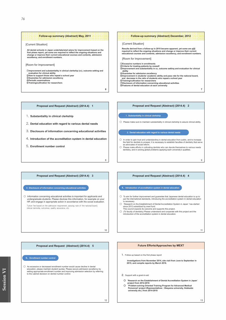
76
Ses
sion
VI
Follow-up summary (Abstract) May, 2011
[Current Situation]
All dental schools in Japan undertake/start plans for improvement based on the first phase report, yet some are required to reflect the ongoing situations and
h i th i t d ti l d t t d i ichange or improve their current educational courses and contents, admissionexcellency, and enrollment numbers.
[Room for Improvement]
Improvement and substantiality in clinical clerkship (cc), outcome setting and evaluation for clinical abilityHow to support those who repeat a school yearGuarantee for admission excellencyPeriodic examinationsTraining/cultivation for researchers
6
Proposal and Request (Abstract) (2014.4) 1
Substantiality in clinical clerkshipSubstantiality in clinical clerkship
Dental education with regard to various dental needsDental education with regard to various dental needs
Disclosure of information concerning educational activitiesDisclosure of information concerning educational activities
Introduction of the accreditation system in dental educationIntroduction of the accreditation system in dental education
Enrollment number controlEnrollment number control
8
Proposal and Request (Abstract) (2014.4) 3
Disclosure of information concerning educational activities
Information concerning educational activities is important for applicants and undergraduate students. Please disclose this information, for example on yourHP and engage in appropriate action in accordance with the social evaluationHP, and engage in appropriate action in accordance with the social evaluation.
10
Proposal and Request (Abstract) (2014.4) 5
Enrollment number control
As excessive or decreased enrollment number would cause decline in dental education, please maintain student quotas. Please secure admission excellency bysetting appropriate enrollment number and improving admission selection by referringg pp p p g y gto the cabinet decision on dentist number control.
12
Follow-up summary (Abstract) December, 2012
[Current Situation]
Results derived from a follow-up in 2010 became apparent, yet some are still required to reflect the ongoing situations and change or improve their current educational courses and contents, admission excellency, and enrollment numbers., y,
[Room for Improvement]
Excessive numbers in enrollmentsCriteria for treating patients by oneselfImprovement and substantiality in cc, outcome setting and evaluation for clinicalp y gabilityGuarantee for admission excellencyImprovement in students’ academic ability and pass rate for the national board, and decrease in the ratio of students who repeat a school yearTraining/cultivation for researchersDisclosure of information concerning educational activities Features of dental education at each university
7
Proposal and Request (Abstract) (2014.4) 2
Substantiality in clinical clerkship
Please make sure to maintain substantiality in clinical clerkship to assure clinical ability.
D t l d ti ith d t i d t l dDental education with regard to various dental needs
In order to gain trust and understanding in dental education from public and to increaseIn order to gain trust and understanding in dental education from public, and to increasethe field for dentists to prosper, it is necessary to establish faculties of dentistry that serveas advocates of social reform.Please make efforts in cultivating dentists who can devote themselves to various needsPlease make efforts in cultivating dentists who can devote themselves to various needsdentistry, and in solving global problems applying each university’s qualities.
9
Proposal and Request (Abstract) (2014.4) 4
Introduction of accreditation system in dental education
To aim for further improvement and guarantee that Japanese dental education is up to par the international standards, introducing the accreditation system in dental educationis necessaryy`Research on the Establishment of Dental Accreditation System in Japan` has started since 2012 subsidized by mext fund. This council expects its outcome and supports this project.p pp p j(To faculty of dentistry) Please understand and cooperate with this project and the introduction of the accreditation system in dental education.
11
Future Efforts/Approaches by MEXT
Follow-up based on the first phase report
Investigations from November 2014, site visit from June to September in 2015, and compile reports by March 2016.
Support with a grant-in-aid
` f S ``Research on the Establishment of Dental Accreditation System in Japan`project from 2012-2016‘Problem-solving Oriented Training Program for Advanced Medical P l’ j t (R t ti Ok i it H kk idPersonnel’ project (Representatives : Okayama university, Hokkaidouniversity etc.) from 2014-2018
13
02_本文1027(web用追加) 15.10.27 9:05 AM ページ 76

Session VI 77
Session
VI
Thank you
02_本文1027(web用追加) 15.10.27 9:05 AM ページ 77
78
Ses
sion
VI
INTRODUCTIONCurrently, the improvement of the quality of the
undergraduate education system is a main issue inJapanese dental education. Especially, the decline ofundergraduate students’ dental clinical competency dueto the shortage of educational resources for current den-tal education and the resultant inefficiency of ensuringexcellent students or leaders for the next generation, aswell as the disparity between dental universities in termsof their performance on accredited assessments, arepointed out as urgent issues. Niigata University, TohokuUniversity, and Hiroshima University have collaboratedto work on these issues as the basis of the programme‘Sophistication of Dental Education ProgrammesUtilising Inter-School Relationships’, supported by aMEXT grant since 2012. In this programme, we aim toassure the quality of dental clinical competency and toadapt the Japanese dental education system to globalstandards thorough the following programme missions:1) to promote dental clinical competency for currentneeds, 2) to foster promising dental researchers, 3) tocultivate internationality and sociality, and 4) to assessthe educational system for undergraduate dental stu-dents. To achieve these missions, we developed original,individual education sources for the purposes, sharedthem amongst the three universities, and attempted thestandardisation of dental education. In the followingchapters, we will describe the achievements of these mis-sions and our further prospect.
1. FOSTERING DENTISTS WITH CLINICAL COMPETENCYFOR CURRENT NEEDS
The increase of the average life expectancy hasinduced a rise in the number of elderly patients visiting
dental hospitals with complicated oral and systemic dis-orders. This change has promptly demanded a highquality of dental treatment, which has made it difficult toensure there are patients available who are suitable fortaking part in the clinical education of undergraduatedental students in Japan and resulted in a shortage of(volunteer) patients for clinical education. Therefore,more effective clinical education programmes are neededto compensate for the dental clinical competency ofundergraduate students and to deliver a high quality ofdental services to the public.
· Clinical simulation training utilising an integrated dentaltraining model (Tohoku University)
Tohoku University has developed clinical simulationtraining by using a dental training jaw model, based onan original model provided from Niigata University. Themodel can be used for comprehensive clinical treatments,including endodontic, periodontic, and prosthodonticprocedures using a mannequin. We prepared scenariossuch as the patient’s information and history, X-rayimages, periodontal chart, etc., for the clinical statementof the model. An original scoring sheet based on a rubricassessment (rubric sheet) and a questionnaire concerningthis training course were used to evaluate students’ prod-ucts and this course, respectively. The training coursewas conducted by six departments in the school andoffered for preclinical undergraduate students. Therubric sheet and questionnaire were collected to assessthe validity. This training course could promote theeffective compilation and organisation of previouslylearned knowledge and skills, and it is considered to beappropriate for the guidance of basic clinical training.On the other hand, there was a wide gap in scores amongthe instructors in some t.asks, suggesting that further
Achievement of a Sophisticated Dental Education Programme in Collaboration with Niigata University, Tohoku University, and Hiroshima UniversityT. Tenkumo1, Y. Takeuchi1, G. Hong1, Y. Ishida2, R. Takeishi2, S. Mimura3, T. Shuto3, H. Oka3, K. Yoshiba2,N. Fujii2, K. Tsuga3, T. Taji3, N. Takahashi1, K. Ono2, K. Uoshima2, H. Nikawa3, K. Sasaki1, M. Sugai3 and T. Maeda2
1 Tohoku University Graduate School of Dentistry, 4-1 Seiryo-machi, Aoba-ku, Sendai, Miyagi, 980-8575, Japan2 Niigata University Graduate School of Medical and Dental Sciences, 2-5274, Gakkocho-dori, Chuo-ku, Niigata,
951-8514, Japan3 Hiroshima University Graduate School of Biomedical and Health Sciences, Kasumi 1-2-3, Minami-ku, Hiroshima,
734-8553, JapanCorrespondence to: TEL: +81-25-227-2818, FAX: +81-25-223-6499, E-Mail: [email protected]
Key words: inter-university collaboration, undergraduate clinical education, clinical competency, cultivation of competent dentist
02_本文1027(web用追加) 15.10.27 9:05 AM ページ 78
Session VI 79
Session
VI
modification of the rubric score sheet is needed, and theinstructors should adopt more uniform views.
· Development of software for dental diagnosis simulation(Tohoku University)
We developed the original diagnosis simulation soft-ware programme, which incorporates a practical methodof thinking, to improve the students’ diagnostic compe-tence. The participants selected some exploration itemsthat they thought were necessary for a clinical site, thendiagnosed patients using the collected information anddrew up treatment plans. The participants could learnthe comprehensive process of clinical necessary explo-rations and dental diagnosis by selecting the explorationsby themselves and establishing the diagnosis and treat-ment plan on the software. We evaluated the validity ofthe software through a questionnaire given to the partici-pants. The questionnaire results showed that the pro-gramme was effective for improving the undergraduatestudents’ competency in diagnosis and treatment plans.We are now increasing the number of cases and trying tofind an appropriate degree of difficulty for students’effective learning.
· Development of an objective-structured evaluationmethod for clinical competency utilising a standardiseddental model (Niigata University, Tohoku University, andHiroshima University)
The evaluation system for the dental clinical tech-nique of undergraduate students is considered necessaryfor the national examination for a dental license.However, there is no standardised evaluation system yet.Therefore, we developed an objective-structured evalua-tion method using a standardised dental model and thespecial evaluation method, beginning in 2013. The modelhas teeth with dental calculus and dental caries, whichrequire composite resin restoration, endodontic treat-ment, or extraction. A special evaluation sheet for thesuccessful treatment of dental morbidity was prepared.Trial tests were carried out twice. In each test, approxi-mately 30 students and residents (10 fifth-year students,10 sixth-year students, and 10 residents) at each universi-ty completed the specified dental tasks like compositeresin restoration, endodontic treatment, or tooth extrac-tion, etc., within a limited time (total of three hours). Aprepared evaluation sheet was used for self-evaluationand evaluation by instructors who had more than eightyears of experience as dentists. The evaluation was per-formed within five minutes per subject. Meanwhile, thevalidity of the developed evaluation system was assessedthrough statistical analysis and a questionnaire. Theresults of the statistical analysis showed that the resi-dents received higher scores on the evaluation comparedto the students. The questionnaire results showed thatthe developed dental model was well accepted by morethan 90% of students and residents. These results sug-gest that the evaluation system has adequate validity andcan be useful in the national examination for a dentallicense. We are planning to introduce a digital assess-ment to increase the objectivity of the evaluation as thenext step.
2. CULTIVATION OF PROMISING DENTAL RESEARCHERS· Lectures on the latest dentistry (Niigata University,Tohoku University, and Hiroshima University)
Each university has original and special departmentssuch as swallowing rehabilitation at Niigata University,disaster dental medicine and dentistry-engineering col-laboration at Tohoku University, and dental globalisationeducation and new dental technical education atHiroshima University. While these departments providevaluable lectures within the university, the lectures arenot systematically available at other universities. In thisproject, collaboration lectures were carried out for dentalstudents to acquire the latest dental knowledge. A totalof 26 collaboration lectures (6 lectures in 2014 and 10 lec-tures in 2015) were provided for fifth-year students, andthey were video-recorded for (video) on demand releasein the future. The evaluation was carried out via a ques-tionnaire filled out by the students, 86.9% of whom gavepositive answers about the lectures. Our goal in this pro-ject is to set up these collaborative dental educationcourses to be regular courses for culturing competentdentists.
· BioDental programme at Hiroshima University (HiroshimaUniversity)
Hiroshima University has had a BioDental educationprogramme based on biology since 2008 to cultivate bio-dentists, oral health managers, and oral engineers for thedevelopment of promising dental professionals andresearchers. The BioDental programme was held for stu-dents at Niigata University and Tohoku University whowere past their fourth year of studies, to standardise theeducation programme at the three universities. In theBioDental education, students conducted the basic prac-tice for cell culture and clinical practice for the evaluationof oral function using ME machines, and techniques fordigital dentistry using the CAD/CAM system and 3Dprinter, over a period of four days. The evaluation wascarried out through a questionnaire and comparativeanalysis of self-evaluations between the pre- and post-mission. This evaluation by external students stronglyfacilitated the standardisation of the BioDental pro-gramme.
3. CULTIVATION OF INTERNATIONALITY AND SOCIALITYIn the rapidly globalising era, the acquisition of
internationality and sociality is prerequisite for cliniciansand researchers to be active in the world. Fostering den-tists who hold an international view is an important issuein dental education for the next generation.
A short stay programme was carried out, in whichapproximately 15 students (in their first to sixth year ofstudy) visited the Faculty of Dentistry, Khon KaenUniversity, Thailand, and stayed there for 10 days. Thestudents were divided in three groups and observed thedental clinic in accordance with the prepared schedule.They communicated directly with instructors and stu-dents of the host university in English and experiencedthe dental education and cultural differences. The stu-dents discussed points that were similar and differentfrom Japan as well as the reasons. The achievements ofthis short stay were presented at the student session of
02_本文1027(web用追加) 15.10.27 9:05 AM ページ 79
80
Ses
sion
VI
JFDA (2015).
4. ASSESSMENT OF THE EDUCATIONAL COMPETENCIESOF UNDERGRADUATE DENTAL STUDENTS (NiigataUniversity, Tohoku University, and Hiroshima University)
· The organisation of the education programme and quality assurance system by instructors at each universityusing faculty development (FD)
To provide high quality education to undergraduatestudents, instructors have to resolve differences in view-points among instructors and collaborate on currentissues in dental education. We have held faculty devel-opment (FD) once a year to improve quality assurance ata personal level and to introduce the education techniquebased on advanced education theory. These FDs havedealt with common themes at the three universities asfollows: ‘The evaluation of learning achievements’ in2014 and ‘Issues and directions in the accreditation evalu-ation of dental education in Japan’ in 2015.
· The organisation of the systematic education programmeand quality assurance system: Platform committee andexternal evaluation committee
We have periodically held platform meetings andreceived external reviews by a commission composed ofstakeholder commissioners (JEDA) and external expertsfor the assurance of the quality of this project. In addi-
tion, we held the external evaluation committee (Tokyo)(http://www.dent.niigata-u.ac.jp/renkei/report/2014/1003/) and the symposium of the interim report for stan-dardisation in Japanese dental education (Niigata)(http://www.dent.niigata-.ac.jp/renkei/report/2014/1003/). The reviews for this project were edited and pub-lished in a book. Furthermore, platform committee meet-ings were held to promote collaborative education at thethree universities three or four times per year, and themanagement and results of each mission were evaluatedand discussed. This self-evaluation in the platform com-mittee meeting and reviews from external experts orstakeholders efficiently facilitated the PDCA cycle func-tion and promoted our education programme and quali-ty assurance system.
· Development of the standardisation of dental educationwith information transmission
The achievements of these projects have been pub-lished in international and national conferences such asJEDA and ADEE. The leaflet was published in Englishand distributed to ADEE, King’s College University, UK,and dental schools in Southeast Asia. Furthermore, weset up the homepage of Niigata University (http://www.dent.niigata-u.ac.jp/renkei/), where the contents of thiseducational programme and its achievements are avail-able.
02_本文1027(web用追加) 15.10.27 9:05 AM ページ 80
Session VI 81
Session
VI
ABSTRACTThe “The Dental Technicians Law” was passed on
23th January, 2009 certifying the national licensing ofdental technologists. In order to implement the dentaltechnicians act, regulations for senior professional techni-cian examinations, and the regulations for special of fordental technicians was released on 29th March, 2010. Thedental technician examination is divided into 2 parts: awritten test and an on-site test. There are two subjects forthe on-site test: dental morphology and carving, and fullmouth removable denture arrangement. A new era forthe education of dental technology in Taiwan has begun.
There are four schools of dental technology locatedin Taiwan, one in the north, one in central Taiwan, andtwo in the south. In southern Taiwan are found theDepartment of Dental Laboratory Technology at Min-Hwei College of Health Care Management (programstarted from September 2007) and Shu-Zen JuniorCollege of Medicine and Management (program startedfrom September 2006). These two schools have five-yearprograms consisting of three years of high school andtwo years of professional dental laboratory technologyeducation. These schools focus on traditional dental tech-nology skills.
The Department of Dental Technology and MaterialsScience at Central Taiwan University of Science andTechnology is the oldest dental technology school inTaiwan. The program here started in 1981 and was ini-tially a five-year program similar to the two schools men-tioned above. Currently it has a four year undergraduateprogram and a master’s course. Most dental techniciansin Taiwan graduated from this school.
While these three dental technology schools aresupervised by the technological and vocational educationsystem under the Ministry of Education, the School ofDental Technology at Taipei Medical University, foundedin 2007, is the only dental technology school within amedical university and is supervised by the higher edu-cation system under the Ministry of Education. TheCollege of Oral Medicine at Taipei Medical University(COM-TMU) is one of the largest dental colleges inTaiwan. COM-TMU offers innovative educational, clini-cal, research, community service, and industrial connec-tions, and is recognized by various field professionals.
We have resources from three adjunct universityhospitals: TMU hospital, Wan Fang hospital, andShuangho hospital. Currently we have a four year under-graduate program and are applying for a master’s course.COM-TMU was established to determine the necessaryfacilities, equipment, and all-inclusive digital patientrecords to facilitate comprehensive treatment planningand efficient delivery of oral health care at the highestlevel of quality using digital dental technologies.
The School of Dental Technology at Taipei MedicalUniversity (SDT-TMU), aims for a combination of acade-mics, art, science, and technology. SDT-TMU instructsstudents in professional knowledge of dental technologyand guides them to graduate school. Concerning profes-sional skills and certificates, SDT-TMU educates studentsstarting from basic dental technology and finishes withnew digital oral engineering. This includes biomechanics,dental materials, oral function and esthetics, and anunderstanding of design and manufacture using rapidlyexpanding digital processes and new dental materials.We encourage international and domestic dental practice.In addition, SDT-TMU expects students to performresearch and pursue higher degrees. There are threecourse tracks that have been developed to enable stu-dents to know what they can learn from SDT-TMU pro-grams: traditional dental skills training, preparation forgraduate school, and improving digital oral engineeringability.
Many students from SDT-TMU take the studentresearch course and propose their projects to the Ministryof Science and Technology. During this course, the stu-dents learn how to find research articles, design experi-ments, and perform research, allowing them to pursuehigher degrees. Currently, six students are running theirstudent research projects and will present their findingsearly next year.
In order to continue improving the education qualityof SDT-TMU, comments from alumni, dental technicians,students, faculties, and friends from sister schools arediscussed. The accreditation processes for SDT-TMU is inprogress. Excellent performances from alumni, students,and faculty are the key to ensure the SDT-TMU main-tains its status as one of the best dental technologyschools in the world.
The Education of Dental Technology in TaiwanC.K. Lin1, W.F. Lee1 and C.T. Lin2,3*
1 School of Dental Technology, Taipei Medical University, Taipei, Taiwan2 School of Dentistry, Taipei Medical University, Taipei, Taiwan3 Department of Dentistry, Taipei Medical University Hospital, Taipei, Taiwan
02_本文1027(web用追加) 15.10.27 9:05 AM ページ 81
82
Ses
sion
VI
ABSTRACT“Quality Assurance” is a communication tool
between the public and our dental education. In thebeginning of 21st century, dental educators and otherstakeholders in dentistry took active efforts to establish ascheme to assure quality of dental education in Japan.The environment surrounding dental education has beenchanged as a result of globalization. The accreditationscheme is a key for the mutual recognition of dentallicense. In the EU, free movement of dentists are guaran-teed by the Directives. The Association of DentalEducation in Europe has a role to harmonize and con-verge dental education to create desirable environmentfor students, patients and the public. As a result of theADEE activities, the “Profile and Competences for thegraduating European dentist—update 2009” has beenissued for over 200 dental schools in Europe. In ASEAN,the Ministers signed the Mutual RecognitionArrangement on Dental Practitioner to facilitate themobility of dental service professions in 2009. Followingthis arrangement, The South East Asian Association forDental Education started to discuss their common corecompetencies for ASEAN dentist. Sharing common com-petencies and profiles of the dentist can be a basic ele-ment for mutual recognition. In medical education,ECFMG in U.S. announced their policy change in 2010, itis that only international medical graduates from accred-ited medical schools can apply for ECFMG certification.This requirement will be in effective in 2023, however,those medical schools, which has not had the appropriateaccreditation started to take a plan to change their cur-riculum to meet the standard. The accreditation schemeand the standard have been an internal tool for qualityassurance in a country until today. If the appropriateaccreditation scheme and the standard are implementedto Japanese dental education, a graduate dentist“Shikaishi” may become “Dentist” in other countries inthe future.
INTORODUTIONIn our society, “Quality Assurance” is getting popu-
lar and it influences our life very much. For the lastdozen years or so, dental educators and other stakehold-ers in dentistry took active efforts and contributed toestablish a scheme to assure quality of dental educationin Japan. For that purpose, the Japanese Model CoreCurriculum for Dental Education (JMCCDE), theCommon Achievement Test (CAT) which is based oncontents of JMCCDE are successfully implemented to
dental undergraduate curriculum and they broughtmeaningful outcomes for the public[1]. It became possiblefor dental schools to design their dental curriculum withJMCCDE as the standard for Japanese dentist “Shikaishi”and to evaluate dental students’ competencies for patienttreatment by CAT, CBT & OSCE. Nowadays, a schemeof JMCCDE and CAT becomes fundamental elementslike the “Air” for us to spend our life in a field of dentaleducation. A main objective of implementing JMCCDEwas to promote more chances for students to have theirown patients during a clinical period. It meant that theobjective was to strengthen students’ clinical competen-cies at graduation time and to foster a qualified Japanesedentist “Shikaishi” who can work for public and patientssafely and independently. For a decade, a lot of activitieshave been done to introduce new schemes for dentaleducation. Recently, a new accreditation scheme is dis-cussed to improve further quality assurance.
Quality Assurance for dental programs in JapanIn a field of dental education in Japan, especially in
undergraduate dental education, an educational institu-tion is one of the most important targets for qualityassurance. Dental schools in Japan provide dental pro-grams to foster future dentists “Shikaishi” licensed by theMinistry of Health and Labour, Welfare (MHLW). Theoutcome of 6-year program is regarded as the competen-cies and the profile of a new graduate dentist, and dentalschools are required to show the evidence how much stu-dents learn and perform their skill and behavior in theirscheduled programs. In 2001, the Ministry of Education,Culture, Sports, Science and Technology (MEXT) pub-lished the Japanese Model Core Curriculum for medicaland dental education and it suggested a way of reform-ing conventional dental curriculum to foster talented stu-dents with clinical competencies. JMCCDE will be a kindof the standard for dental education in Japan. A dentalschool designs a program for 6 years by following itemsin JMCCDE. And all dental students have to pass theCommon Achievement Test, CBT & OSCE, to start treat-ment of their patients in the clinical period. It is placed atthe end of 4th year or the middle of 5th year in the dentalprograms. CAT can be one of the quality assuranceschemes by evaluating students’ clinical readiness. Inthis point, CAT has a role of a summative examinationsimilar to the National Examinations for DentalPractitioner, which is held in the end of 6th year by theMHLW. In recent years, a pass rate of the NationalExaminations for Dental Practitioner, is around 70%, itseems to be low rate as qualification examination though
Quality Assurance in Dental Education, “Dentist & Shikaishi”J. Tsuruta
DDS, Ph. D. Associate Professor, Center for Interprofessional Education, Tokyo Medical and Dental University, Graduate Schoolof Medical and Dental Sciences, Professional Development in Health Sciences, Tokyo Medical and Dental University
02_本文1027(web用追加) 15.10.27 9:05 AM ページ 82
Session VI 83
Session
VI
the pass rate varies among 29 dental schools and somedental schools seem to have some problems of graduate’scompetencies. To control this circumstance, the accredi-tation scheme would be a strong tool for quality assur-ance, however, we have only institutional certified evalu-ation and accreditation scheme by independent organiza-tions certified by the MEXT as an accreditation organiza-tion. It requires 7-year cycle mandatory accreditation forall higher educational institutions based on a self-assess-ment report. From 2012, a research team supported bythe MEXT grants studies and discusses a new scheme forprofessional (subject) evaluation for dental programs toenhance a role of accreditation in dental education.Following these movements, Japanese dental schools willprovide dental programs that satisfy these elements toreach the goal to foster Japanese dentist, “Shikaishi” inthe next few years.
Movement of professionals and Quality Assurance of professional educationUndergraduate dental curriculum in EU
“Quality Assurance” is a word for communicationbetween the public and our dental society. The publicmeans not only “Internal society” but also “Global soci-ety” in some cases. Across the European Union (EU), sin-gle social and economic area, mutual recognition of qual-ification and free movement of dentists are prescribed inEU Directive 2005/36/EC. License was basically for acountry, an internal society, citizens and patients, howev-er, qualified dentists from each member country canpractice in other member country with their originallicense in the EU and EEA. It also affected undergradu-ate dental programs and the mobility of students forexchange programs. In 1999, ministers of Education ofthe European countries had signed Bologna Declarationto converge the higher educational systems. In dentaleducation, the Association for Dental Education inEuropa (ADEE), which has over 200 member schoolsthroughout European countries, led many activities andprojects called DentEd, DentEd Evolves, DentEd III toharmonize and converge dental education in Europeancountries[2,3]. In 2009, the “Profile and Competences forthe graduating European dentist—update 2009” waspublished to harmonize the curriculum in European den-tal schools as a result of a consultation on “Profile andCompetencies for the European Dentist” published in2005. It is consisted of 12 domains and each domain issupported by major competences and supporting compe-tences[4].
ASEAN Mutual Recognition Arrangement on DentalPractitioner
The Association of Southeast Asian Nations(ASEAN) was established in 1967. ASEAN enhanced thecooperation among member countries to accelerate theeconomic growth, social progress and cultural develop-ment in the region. And member countries had agreedwith the Mutual Recognition Arrangement to facilitatethe movement of professional services in the ASEANarea. In professional services, the Economic Ministers at14th ASEAN Summit in Thailand signed MRA on DentalPractitioner (MRA on DP), 2009[5]. The MRA on DP is
discussed at the ASEAN Joint Coordinating Committeeon Dental Practitioners (AJCCD). By the information ofAJCCD, this MRA facilitates “the mobility of dental ser-vices professionals within ASEAN, enhance exchange ofinformation and expertise on standards and qualifica-tions, promote adoption of best practice for professionaldental services and provide opportunities for capacitybuilding and training of dental practitioners”[6].Following this MRA on DP, the South East Associationfor Dental Education (SEAADE) has started to discusstheir role for the MRA and has discussed core competen-cies in the workshop “Identifying Dental ProfessionsCore Competencies of the SEAADE member countries”at the annual academic meeting in Bali, 2015. SEAADEwill have some more discussion regarding this MRA.These movements will be affect on dental education inASEAN countries in the next few years[7].
Movement of future dentistsPeople can move globally with the Free Trade
Agreement and Economic Partnership Agreement. Inother cases, a dentist can move to the other country ifhe/she fulfills some conditions to get a license in thecountry of destination. There is another way to assessgraduates’ competences by accrediting dental programsinternationally. International accreditation and the glob-al standard will be an important key to facilitate qualityassurance in the context of global dental education.Dental educators and dental programs will cross the bor-der in educational fields. In some cases, students crossthe border.
The Commission on Dental Accreditation is anaccreditation organization which accredits dental schoolsand programs in the U.S.. CODA has the Accreditationscheme and the Standard for Dental Education Programsfor U.S.. And beside the U.S. accreditation scheme,CODA has a scheme for international accreditation[8].There is a way to be a dental school accredited by CODA.
In Malaysia, The International Medical University(IMU), School of Dentistry has a special program, whichprovide international pathway for students and the pro-gram meets international standards[9]. The dental schoolhas 4 partner dental schools, the University of Adelaide,The University of Queensland, the University of Otagoand the University of Glasgow. In the IMU program, stu-dents study basic dental and medical sciences, introduc-tion to the basic clinical skills at Phase I for the first 2.5years. After that, students who choose partner dentalschools will come to other countries to accomplish fur-ther 2~2.5 years and they will get respective degrees thatare recognized internationally and locally by theMalaysian Dental Council. IMU designs their dental cur-riculum to meet international standards and share basiccompetencies of a Malaysian dentist with a dentist inother countries.
New accreditation Requirement in the U.S. medical education
International medical graduates who want to prac-tice in the U.S. have to get a certification from theEducational Commission for Foreign Medical Graduate(ECFMG) to enter programs accredited by the
02_本文1027(web用追加) 15.10.27 9:05 AM ページ 83
84
Ses
sion
VI
Accreditation Council for Graduate Medical Education(ACGME). ECFMG certification is a key for internationalmedical graduate to get into the U.S. health care system.In 2010, ECFMG announced that only international med-ical graduates from accredited medical schools couldapply for ECFMG certification[10]. This requirement willbe in effective in 2023, however, those medical schools,which has not had the appropriate accreditation startedto take a plan to change their curriculum to meet theaccreditation. In response to this announcement,Japanese medical schools decided to establish the JapanAccreditation Council for Medical Education (JACME) in2013 and will try to be recognized by the WorldFederation for Medical Education (WFME) as the accredi-tation organization in Japan, so that JACME is expectedto have a role of accreditation organization international-ly. In a plan, JACME will start the accreditation schemefor medical schools after 2017, and those medical schoolsthat are accredited by JACME will be registered to theFoundation for Advancement of International MedicalEducation and Research (FAMER). WFME has a stan-dard for medical education and this document is essen-tial for accreditation of medical education[11]. Japanesemedical schools followed the content of Model CoreCurriculum for Medical Education however, the “BasicMedical Education, WFME Global Standards for Qualityimprovement”, has been regarded as one of standards formedical curriculum and some of Japanese medicalschools need to change their curriculum to enhance theclinical period and environment where students learn inreal clinical procedures. It seems that it is not so impor-tant for those medical schools where their graduate don’tintend to come to the U.S., on the other hand, this move-ment becomes a turning point for medical schools inJapan to assure the quality of medical education.
CONCLUSIONThe relationships among countries are changing by
the minute. To meet the global standard in dental educa-tion, we need to catch all information regarding dentaleducation, especially about the accreditation. In thispoint, we need to look carefully not only undergraduateeducation but also Continuing Professional Developmentas postgraduate education. Establishing professional(subject) evaluation scheme is a key for internal qualityassurance for the public, and also it will be a key forinternational recognition of Japanese Dentist “Shikaishi”in the world. “Shikaishi” and “Dentist”, it is same mean-ing when we translate “Shikaishi” into English, however,Japanese dental educators have to reconsider our role forthe society and the profile and competencies of“Shikaishi” for the coming future without translating itinto English. “Dentist & Shikaishi”, are they the same?
Conflicts of InterestThis investigation was supported in part by Health
Labour Sciences Research Grant, Japan No. 201101008B
REFERENCES01. 森尾郁子, 鶴田 潤, 江藤一洋, モデル・コア・カリキュラムと共用試験 歯学教育モデル・コア・カリキュラムその意義と課題, 日本歯科医学教育学会雑誌, 18巻1号,p43-46, 2002
02. 森尾郁子, 鶴田 潤, 江藤一洋, 最近の歯学 医歯総合教育開発 歯学教育と世界標準, 口腔病学会雑誌, 69巻3号,p228, 2002
03. 森尾郁子, 鶴田 潤, 魚島勝美, 谷口弘江, 江藤一洋, 欧州の歯科医学教育の動向 教育の質の向上を目指すプロジェクトの背景と概略, 日本歯科医学教育学会雑誌, 20巻2号, p405-411, 2005
04. Cowpe J1, Plasschaert A, Et al, Profile and compe-tences for the graduating European dentist - update.,Eur J Dent Educ. Nov; 14 (4): 193-202., 2010
05. ASEAN MUTUAL RECOGNITION ARRANGE-MENT ON DENTAL PRACTITIONERS, http://www.asean.org/communities/asean-economic-community/item/asean-mutual-recognit ion-arrangement-on-dental-practitioners-2, accessed on1st, August, 2015
06. Dental practitioners (AJCCD) http://www.asean.org/communities/asean-economic-community/category/dental-practitioners-ajccd, accessed on 1st,August, 2015
07. Yoshifumi FUKUNAGA, Assessing the Progress ofASEAN MRAs on Professional Services, ERIADiscussion Paper Series, 2015, http://www.eria.org/ERIA-DP-2015-21.pdf, accessed on 1st, August, 2015
08. Joint Advisory Committee on InternationalAccreditation, Guidelines for InternationalConsultation and Preliminary AccreditationConsultation Visit (PACV) Survey, http://www.ada.org/~/media/CODA/Files/internatl_policies_fees.ashx, accessed on 1st, August, 2015
09. International Medical University, School of Dentistry,About the dentistry degree, http://www.imu.edu.my/imu/education/undergraduate/dentistry/overview, accessed on 1st, August, 2015
10. Educational Commission for Foreign medical gradu-ates (ECFMG), Medical School AccreditationRequierement for ECFMG Certification, http://www.ecfmg.org/about/initiatives-accreditation-requirement.html accessed on 1st, August, 2015
11. Basic Medical Education: Japanese SpecificationsWFME Global Standards for Quality Improvement,http://edu.med.hokudai.ac.jp/cme/wp-content/uploads/sites/2/2015/02/WFMEグローバルスタンダード.pdf, accessed on 1st, August, 2015
02_本文1027(web用追加) 15.10.27 9:05 AM ページ 84
Session VI 85
Session
VI
Improvement and Enhancement of BioDental Education and Research in HiroshimaK. Kato1 and M. Sugai2
1 Center for International Collaboration Development for Dentistry, Faculty of Dentistry, Hiroshima University, Hiroshima 734-8553, Japan, TEL: +81-82-257-5645, FAX: +81-82-257-5649, E-Mail: [email protected]
2 Dean, Faculty of Dentistry, Hiroshima University, Hiroshima 734-8553, Japan
Key words: Globalization, BioDentistry, Dual linguistic education system, International Dental Course, Short-term pro-grams
ABSTRACTFor more than a decade, Hiroshima University
Faculty of Dentistry has been involved in the globaliza-tion of our education and research, driven by our per-spective that the conventional dental education systemwould not be compliant to the imminent situation sur-rounding clinical dental care communities in a globaliza-tion era. On the other hand, we have a fundamental andstrong belief that contemporary dental medicine must begrounded primarily on scientific knowledge consistentwith modern biology, as represented by a new term“BioDentistry”. These perspectives have been the dri-ving force for the reformation that we made on dentaleducation provided at our faculty. The most importantchallenge has been the use of English in our undergradu-ate dental program. Consequently this reformation hasbrought us with increasing opportunities to acceptinbound international students staying in Hiroshima for10 days to 4 years and send outbound Japanese studentsto sister universities around the world. We believe thatthese dynamic changes we exerted for the last few yearshave facilitated to cultivate global leaders playing anactive part in worldwide dental medicine in future,extending the idea of BioDentistry.
COMMUNITIES FOR CLINICAL DENTAL CARE IN GLOBALIZATION ERA
“Globalization” is defined as a process throughwhich people, money, information, and culture are inter-acted and integrated among different countries.Currently this process is a trend not only in the socio-eco-nomic world but also biomedical communities includingthose of dental medicine. This is well exemplified by therecent efforts made by ASEAN member states for theirunification.
ASEAN countries are making steady and tremen-dous progress toward their economic unification by 2015.Associated with this reformation, mutual recognitionagreement has been ratified to enhance the mobility ofdental practitioners over the borders within the associat-ed nations. Mutually recognizing dental practitionersrequires a universal framework for dental education aswell as healthcare systems. In fact, these countries are
currently in discussions to establish standard competen-cies necessary for dentists who will be able to moveabroad within the community.
We do not believe that such a revolution in theASEAN community is just an affair of our neighbors. Itmay be supposed that dental medicine will necessarily beglobalized in the near future over the entire Asian-Pacificarea, including Japan. We can say with confidence thatwe cannot afford to be left out. The Trans-PacificPartnership, an issue of current political argument, willalso serve to accelerate mutual relationships in dentalmedicine as well in the course of economic unification.We should note that similar systems have already beenlaid out in the US/Canada, EU, and UK to accredit dentaland dental-related education programs, so that dentiststrained through compliant programs overseas can workas dental practitioners in these regions.
What would happen in our country as a result of fur-ther Asian globalization? This is a question that we havecontinuously asked by ourselves for the last decade. Iguess a society will significantly change so that studentsand skilled labors such as dentists will be able to movemore frequently within the Asian economic area, includ-ing Japan. In such a mobile society, the place of studyand work will not matter any longer, and people willmove in search for better environments and conditions.Consequently, the mobilization will make societies muchmore international and competitive. Accordingly we allwill need to cooperate or compete with others in multina-tional classes, teams, and societies.
How can we prepare for the trend of the time inadvance to survive as a dentist with global competen-cies? There is no doubt that improving our English skillsis of primary importance because common languageinfrastructure is the basis for our international activities.However, I think for sure this is clearly not enough.More importantly, we need to precisely and deeplyunderstand what is going on in other countries throughdirect experiences in a wide perspective, and then todevelop new values and strategies, extending profession-al networks, for dynamically playing an active part inworldwide dental medicine.
The background mentioned above brought us withurgent concern that traditional dental education would
02_本文1027(web用追加) 15.10.27 9:05 AM ページ 85
86
Ses
sion
VI
not been compliant any more for building dentists capa-ble of surviving imminent global environments. Suchperspective was our principal motivation for the drasticreformation that we have made in the last several yearsfor the dental education system provided by the Facultyof Dentistry Hiroshima University. It should be notedthat this revolution is quite coherent to our scope as aSuper Global University (Hiroshima University has beenselected as one of the Super Global Universities by thegovernment under the national project aiming at inten-sively enhancing the globalization of Japanese universi-ties). In addition, the Hiroshima Conference onEducation and Science in Dentistry is one of the mostimportant activities in which we have been involved formore than a decade toward globalization.
INITIATIVE IN BIODENTAL EDUCATION AND RESEARCH“BioDentistry” is a coined word that we initially cre-
ated almost a decade ago for representing the basic con-cept universal in scope of our dental education andresearch. The word BioDentistry denotes our strongbelief that contemporary dental medicine must begrounded primarily on scientific knowledge consistentwith modern biology.
We are quite sure that conventional dental educationhad exclusively stuck for long time to improve technicalproficiency of dental students. As a matter of course, thisis still important for warranting the quality of dentaltreatments, yet it is a time to reconsider that oral/max-illofacial malfunctions might be regarded as biologicalconsequences, as the most of oral disorders such as cariesand periodontitis are initially caused by bacterial infec-tion. It should be stressed that we need to understandthese conditions in the context of molecular and cellularbiology. It is not until we logically understand thebehaviors of bacteria and the responses of host defensesystems that we are able to properly make strategies forprevention, diagnosis, treatments and improvement ofprognosis. It seems that this aspect had been more or lessoverlooked in the conventional dental education.
Such a new paradigm in dental education facilitatesto position dentistry as a part of contemporary biologicalsciences, which serves to provide multidisciplinary envi-ronments, activating scientific research in dentistry on aninternational level. This is demonstrated by the fact thatmost of recent progresses contributed by our research inHiroshima, for instance, technological advances in regen-erative dentistry and deeper pathological understandingof oral cancer, have always been made on the basis ofcurrent biological science and engineering.
Thus, initiatives for BioDental education andresearch are closely connected each other. We now knowthat such a circumstance in Hiroshima has great impactson the manner of logical thinking by dental students aswell as academic staffs. In addition, the advantage asso-ciated with BioDental education and research shouldbenefit not only Japanese students but also internationalscholars. This is the reason why we strongly promoteaccepting international scholars as described below.
BIODENTISTRY LECTURES IN TRULY BORDERLESS CLASSROOMS
We are eager to promote students’ competency tohave a sense of purpose and behave more on their owninitiative in global environments. We believe that one ofthe most effective ways to realize them is to expose stu-dents to really international circumstances.
Taking above into consideration, our School ofDentistry has just started simultaneous distribution of aninternational BioDentistry class using a teleconferencesystem to the School of Dentistry at Airlangga Universityand University of Medicine and Pharmacy, Ho Chi MinhCity. Distinguished lecturers from these three universi-ties as well as University of Sheffield (in this case, 4 uni-versities are connected each other) contribute to the pro-gram, sharing their own expertise from the viewpoint ofboth fundamental and applied aspects of advanced den-tistry. Totally more than 230 undergraduate students atthree classrooms in Japan, Indonesia, and Vietnam jointhis class all at once.
This provides an amazing opportunity for all stu-dents to extend their human networks, gaining deeperinsights into cutting-edge BioDental research.Importantly, this highly international and interactiveclass makes students for sure aware of being the mem-bers of a contemporary Asian community.
DENTAL EDUCATION THROUGH ENGLISHOne of the most critical and important challenges we
exerted ourselves during these years was the introduc-tion of a new teaching method in 2012 to the dental edu-cation at the School of Dentistry, Hiroshima University.In the new method, English is thoroughly used in everyclass of lectures, exercises and practical trainings provid-ed in the undergraduate program for 2nd - 5th grades.
English is obviously the language that is used inmost international environments and therefore webelieve that teaching dentistry through English serves forstudents to improve their communication skill, gain pro-fessional knowledge in English, and thus remarkablyenhance students’ international competency.
It should be noted that this is in fact a quite big chal-lenge for Japanese schools not only in the field of den-tistry but also any other disciplines, because education atJapanese universities has been traditionally and exclu-sively given in Japanese. Moreover, a linguistic problemis always a big issue for most of Japanese people andsocieties. Nevertheless, we have adopted the new teach-ing method to the education at the School of Dentistry forthe first time in our country.
The reformation of a teaching method has requiredour great efforts. To optimize our education method, wehave offered several faculty development workshopsduring years. In addition, we have promoted teachingstaffs to take development programs for efficiently teach-ing in English, while individually consulting those whoare not familiar with teaching in English. We have seenthat all these efforts have served to gradually but certain-ly improve the quality of our education.
Another concern associated with this new method isquite specific to our country. Because our national boarddental examination is done in Japanese, students are also
02_本文1027(web用追加) 15.10.27 9:05 AM ページ 86
Session VI 87
Session
VI
required to gain sufficient knowledge of dentistry withJapanese technical terms. That is why, even in the newteaching method, we need to teach students not only inEnglish but also in Japanese. Therefore, we use both lan-guages in every single class. Accordingly the system iscalled “dual linguistic education system (DLES)”.
Our assessment after 4-year experiences with DLEStells us that the majority of Japanese students have grad-ually been getting used to the system, steadily gainingknowledge necessary for dental professionals. This iswell evidenced by the successful result that we have seenin the recent CBT (computer-based testing conducted inJapan to assess if students’ are sufficiently competent forstarting clinical training at the university hospital) for the5th-grade Japanese students who leaned dentistry for 4years by means of DLES.
What is more important is that an increasing numberof Japanese students are now interested in internationalactivities. In fact, more than 40 Japanese students joinedoutbound programs during a year. We sent 2 students ina research track to University of Sheffield and ForsythInstitute. During last years many students participatedin outstanding short-visit programs provided byUniversity of British Colombia, University ofWashington, Airlangga University, University ofMedicine and Pharmacy at Ho Chi Minh City, KhonKaen University, Taipei Medical University, WonkwangUniversity, and Pusan Catholic University. A dentalcamp in Cambodia, organized every year under collabo-ration with NGO Hiroshima, the Society to Protect theTeeth of Asian Children, also attracts an increase numberof Japanese students.
ESTABLISHMENT OF INTERNATIONAL DENTAL COURSEThanks to the DLES, we are able to accept interna-
tional undergraduate students all the time to the schoolof dentistry and invite them to our regular dentistryclasses with approximately 50 Japanese students. Thisbrought us with a great opportunity to create much inter-national environments in a classroom, efficiently facilitat-ing to cultivate international awareness of Japanese stu-dents as well as the international scholars.
The most obvious and significant outcome is that wehave a possibility of inviting inbound students to beenrolled in the 4-year undergraduate dentistry program.Namely, in 2012 we established an International DentalCourse (IDC), collaborating with our sister universitiesincluding Airlangga University, Indonesia, University ofMedicine and Pharmacy, Ho Chi Minh City, Vietnam,and University of Health Sciences, Cambodia, with atremendous support from the Japanese government aswell as our alumni association. In the IDC program, weaccept 3 students a year, each from the three schools. Thestudents spend 4 years in Hiroshima to complete almostall the subjects necessary for reaching the level equivalentto a bachelor. At 4th to 5th grades, they also join one ofthe research laboratories to conduct cutting-edgeBioDental research. Finally, credits granted to the stu-dents in Hiroshima are transferred based on our mutualaccreditation.
After 4 years in Hiroshima, the students return totheir mother countries and then start their clinical train-
ing at hospitals affiliated to their universities beforelicensed in their country as a dental practitioner.Actually, three IDC students of the 1st generation havejust completed the program with great success andreturned to their mother countries. We believe that theywill act as global leaders in oral health communities infuture. This year we have just selected three new IDCstudents of the 5th generation.
INCREASING CAPACITY FOR ACCEPTING INTERNATIONAL STUDENTS
The reformation we made on our education systemhas brought us with further opportunities: We became tobe able to provide several new short-term stay programsfor international inbound students from sister universi-ties. The programs we are providing are available forundergraduate dental students, and the duration of theirstay is ranging from 10 days to 1 year. These programsare also partially supported by the Japanese government.
In practice, we accept every year around 20 inboundstudents for the 10-day program. These students are ableto gain many valuable experiences from the speciallydesigned short-tem program that includes lectures, prac-tical training, and clinical observation at the universityhospital. Cultural excursions and intimate interactionswith Japanese and other international students are alsoimportant elements of this program. Last year 18 stu-dents joined from Asian countries and one from UK.
In the 6-month (1 semester) program we normallyaccept 6 students every year. They are able to join a den-tistry class at either grade from 2nd to 4th depending onthe subjects they have already completed at their ownuniversities. Similar to this, inbound students enrolled inthe 1-year program (2 semesters) study dentistry for ayear in either class with Japanese students. Last year weaccepted totally 8 students from Indonesia, Vietnam,Thailand, Taiwan, and Brazil to the 6-month and 1-yearprograms.
Accordingly, a lot of international inbound studentsstudy at our school during a year. It is amazing to some-times see that more than 20% of students in a single class-room are international inbound students. Such a quiteinternational environment has been created inHiroshima, most likely owing to the DLES.
CONCLUSIONSAs demonstrated in this paper, our globalization
strategy is yielding a lot of success in cultivating futureBioDentists in the world. We owe our success entirely toour sister universities and are obliged to the fact that wecould conclude an academic exchange agreement with anincreasing number of outstanding universities abroad.
It may be an indicative of our leadership in the glob-alization of dental education and research that we arecommitted to develop and implement globalized educa-tion programs for dentistry, dental technology, and den-tal hygiene under the national projects, such as “Programfor Raising Standards of Dental Education UtilizingIntegrative Functions, Program for Promoting Inter-University Collaborative Education (collaborating withNiigata University and Tohoku University)” and “Projectfor Developing a Program for Cultivating Global Dental
02_本文1027(web用追加) 15.10.27 9:05 AM ページ 87

88
Ses
sion
VI
Care Professionals, Strategic Promotional Program forCultivation of Core Professionals in Targeted GrowthFields”, both driven by the Ministry of Education,Culture, Sports, Science and Technology.
What should we go further for taking a step towardmore globalized BioDental education and research? We
know for sure that one of the most important directionsis to strategically strengthen our contribution toBioDental research through functional enhancement atthe postgraduate level. Apparently our efforts to acquirea continuous and sufficient budget are critical for furtherprogress in globalized BioDental education and research.
02_本文1027(web用追加) 15.10.27 9:05 AM ページ 88
Session
VII
Food, Nutrition and HealthWhich Foods are Suitable to Elderly Dysphagia Person?
—According to Japanese Criteria of Dysphagia Foods—
Prefectural University of Hiroshima
J. Kayashita
Nutrition, Food and Dietetics for Health
Kanagawa University of Human Services
T. Nakamura
Impact of the Gut Microbiota and Probiotics on Health
Yakult Central Institute
K. Oishi
Session VII
02_本文1027(web用追加) 15.10.27 9:05 AM ページ 89

02_本文1027(web用追加) 15.10.27 9:05 AM ページ 90
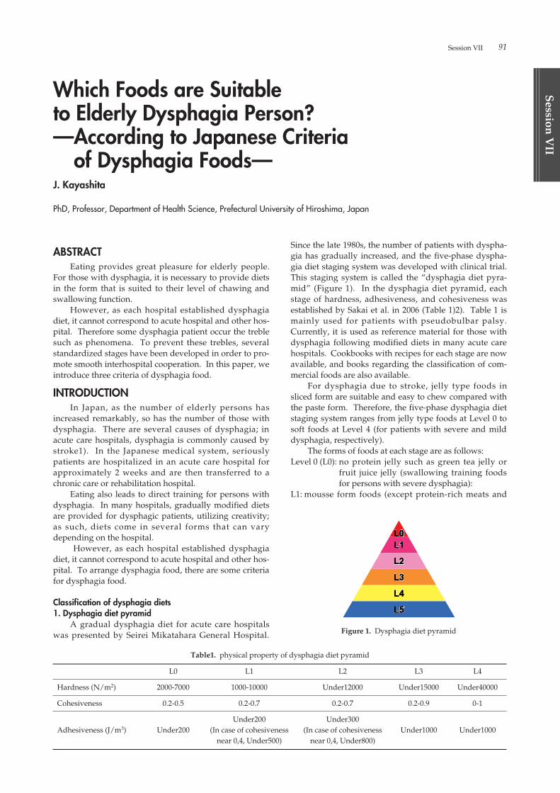
Session VII 91
Session
VII
ABSTRACTEating provides great pleasure for elderly people.
For those with dysphagia, it is necessary to provide dietsin the form that is suited to their level of chawing andswallowing function.
However, as each hospital established dysphagiadiet, it cannot correspond to acute hospital and other hos-pital. Therefore some dysphagia patient occur the treblesuch as phenomena. To prevent these trebles, severalstandardized stages have been developed in order to pro-mote smooth interhospital cooperation. In this paper, weintroduce three criteria of dysphagia food.
INTRODUCTIONIn Japan, as the number of elderly persons has
increased remarkably, so has the number of those withdysphagia. There are several causes of dysphagia; inacute care hospitals, dysphagia is commonly caused bystroke1). In the Japanese medical system, seriouslypatients are hospitalized in an acute care hospital forapproximately 2 weeks and are then transferred to achronic care or rehabilitation hospital.
Eating also leads to direct training for persons withdysphagia. In many hospitals, gradually modified dietsare provided for dysphagic patients, utilizing creativity;as such, diets come in several forms that can varydepending on the hospital.
However, as each hospital established dysphagiadiet, it cannot correspond to acute hospital and other hos-pital. To arrange dysphagia food, there are some criteriafor dysphagia food.
Classification of dysphagia diets1. Dysphagia diet pyramid
A gradual dysphagia diet for acute care hospitalswas presented by Seirei Mikatahara General Hospital.
Since the late 1980s, the number of patients with dyspha-gia has gradually increased, and the five-phase dyspha-gia diet staging system was developed with clinical trial.This staging system is called the “dysphagia diet pyra-mid” (Figure 1). In the dysphagia diet pyramid, eachstage of hardness, adhesiveness, and cohesiveness wasestablished by Sakai et al. in 2006 (Table 1)2). Table 1 ismainly used for patients with pseudobulbar palsy.Currently, it is used as reference material for those withdysphagia following modified diets in many acute carehospitals. Cookbooks with recipes for each stage are nowavailable, and books regarding the classification of com-mercial foods are also available.
For dysphagia due to stroke, jelly type foods insliced form are suitable and easy to chew compared withthe paste form. Therefore, the five-phase dysphagia dietstaging system ranges from jelly type foods at Level 0 tosoft foods at Level 4 (for patients with severe and milddysphagia, respectively).
The forms of foods at each stage are as follows:Level 0 (L0): no protein jelly such as green tea jelly or
fruit juice jelly (swallowing training foodsfor persons with severe dysphagia):
L1: mousse form foods (except protein-rich meats and
Which Foods are Suitable to Elderly Dysphagia Person?—According to Japanese Criteria
of Dysphagia Foods—J. Kayashita
PhD, Professor, Department of Health Science, Prefectural University of Hiroshima, Japan
Figure 1. Dysphagia diet pyramid
Table1. physical property of dysphagia diet pyramid
L0 L1 L2 L3 L4
Hardness (N/m2) 2000-7000 1000-10000 Under12000 Under15000 Under40000
Cohesiveness 0.2-0.5 0.2-0.7 0.2-0.7 0.2-0.9 0-1
Under200 Under300Adhesiveness (J/m3) Under200 (In case of cohesiveness (In case of cohesiveness Under1000 Under1000
near 0,4, Under500) near 0,4, Under800)
02_本文1027(web用追加) 15.10.27 9:05 AM ページ 91
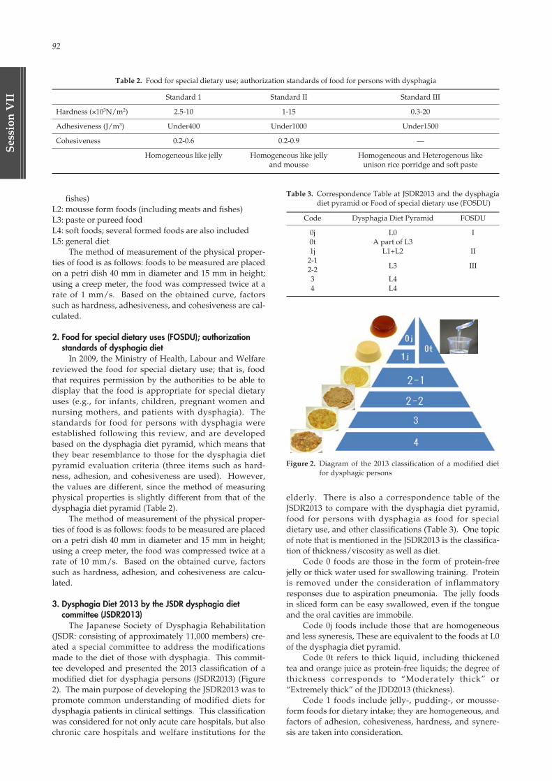
92
Ses
sion
VII
fishes)L2: mousse form foods (including meats and fishes)L3: paste or pureed foodL4: soft foods; several formed foods are also includedL5: general diet
The method of measurement of the physical proper-ties of food is as follows: foods to be measured are placedon a petri dish 40 mm in diameter and 15 mm in height;using a creep meter, the food was compressed twice at arate of 1 mm/s. Based on the obtained curve, factorssuch as hardness, adhesiveness, and cohesiveness are cal-culated.
2. Food for special dietary uses (FOSDU); authorizationstandards of dysphagia diet
In 2009, the Ministry of Health, Labour and Welfarereviewed the food for special dietary use; that is, foodthat requires permission by the authorities to be able todisplay that the food is appropriate for special dietaryuses (e.g., for infants, children, pregnant women andnursing mothers, and patients with dysphagia). Thestandards for food for persons with dysphagia wereestablished following this review, and are developedbased on the dysphagia diet pyramid, which means thatthey bear resemblance to those for the dysphagia dietpyramid evaluation criteria (three items such as hard-ness, adhesion, and cohesiveness are used). However,the values are different, since the method of measuringphysical properties is slightly different from that of thedysphagia diet pyramid (Table 2).
The method of measurement of the physical proper-ties of food is as follows: foods to be measured are placedon a petri dish 40 mm in diameter and 15 mm in height;using a creep meter, the food was compressed twice at arate of 10 mm/s. Based on the obtained curve, factorssuch as hardness, adhesion, and cohesiveness are calcu-lated.
3. Dysphagia Diet 2013 by the JSDR dysphagia diet committee (JSDR2013)
The Japanese Society of Dysphagia Rehabilitation(JSDR: consisting of approximately 11,000 members) cre-ated a special committee to address the modificationsmade to the diet of those with dysphagia. This commit-tee developed and presented the 2013 classification of amodified diet for dysphagia persons (JSDR2013) (Figure2). The main purpose of developing the JSDR2013 was topromote common understanding of modified diets fordysphagia patients in clinical settings. This classificationwas considered for not only acute care hospitals, but alsochronic care hospitals and welfare institutions for the
elderly. There is also a correspondence table of theJSDR2013 to compare with the dysphagia diet pyramid,food for persons with dysphagia as food for specialdietary use, and other classifications (Table 3). One topicof note that is mentioned in the JSDR2013 is the classifica-tion of thickness/viscosity as well as diet.
Code 0 foods are those in the form of protein-freejelly or thick water used for swallowing training. Proteinis removed under the consideration of inflammatoryresponses due to aspiration pneumonia. The jelly foodsin sliced form can be easy swallowed, even if the tongueand the oral cavities are immobile.
Code 0j foods include those that are homogeneousand less syneresis, These are equivalent to the foods at L0of the dysphagia diet pyramid.
Code 0t refers to thick liquid, including thickenedtea and orange juice as protein-free liquids; the degree ofthickness corresponds to “Moderately thick” or“Extremely thick” of the JDD2013 (thickness).
Code 1 foods include jelly-, pudding-, or mousse-form foods for dietary intake; they are homogeneous, andfactors of adhesion, cohesiveness, hardness, and synere-sis are taken into consideration.
Table 2. Food for special dietary use; authorization standards of food for persons with dysphagia
Standard 1 Standard II Standard III
Hardness (×103N/m2) 2.5-10 1-15 0.3-20
Adhesiveness (J/m3) Under400 Under1000 Under1500
Cohesiveness 0.2-0.6 0.2-0.9 —
Homogeneous like jelly Homogeneous like jelly Homogeneous and Heterogenous like and mousse unison rice porridge and soft paste
Table 3. Correspondence Table at JSDR2013 and the dysphagiadiet pyramid or Food of special dietary use (FOSDU)
Code Dysphagia Diet Pyramid FOSDU
0j L0 I0t A part of L31j L1+L2 II
2-1L3 III2-2
3 L44 L4
Figure 2. Diagram of the 2013 classification of a modified dietfor dysphagic persons
02_本文1027(web用追加) 15.10.27 9:05 AM ページ 92
Session VII 93
Session
VII
Code 2 foods are purees, pastes, and blended foods.They have a fluid quality and are smooth, not sticky, andeasily mixed; in spite of this fluidity, Code 2 foods are notfluids that can be passed through a tube. They can basi-cally be eaten using a spoon, forming a bolus of food inthe mouth by simple operation; consideration is given forneither residues nor incorrect swallowing at the pharynx.According to homogeneity, Code 2 is subdivided intohomogeneous 2-1 and heterogeneous 2-2. Even if thefood is categorized as heterogeneous, it should be softenough that the drop can simply be swallowed withoutneeding to be crushed by the tongue.
Code 3 foods are formed-foods that can be crushedeasily by the tongue and palate; a bolus of food can beformed and transferred easily, with consideration toavoid being scattered. There is not a lot of syneresiscaused during chewing and transferring in the oral cavi-ty.
Code 4 foods are formed-foods that can be easily cutusing chopsticks and a spoon, but some mandatory forceis required because such foods are difficult to crush using
the tongue and palate; after chewing, these foods are eas-ily gathered up as a bolus without breaking up the com-ponents.
SUMMARYWe introduce Japanese dysphagia diet categories
such as Dysphagia diet, FOSDU and JSDR2013. The jellytype food located most easy for dysphagia patient inthese categories. Main cause of dysphagia in acute hospi-tal is pseudobulbar paralysis of stroke. The jelly typefood is suitable compare to puree type food in this case.The jelly is not necessary to chewing and that is notspread in the mouse. Medical stuffs serve the jelly tomake slice type and go into mouse in patient.
REFERENCES1) S. Teramoto et.al. Journal of the American Geriatric
Society 56, 577-579, 20082) M. Sakai et. al. The Japanese Journal of Dysphagia
Rehabilitation 10, 239-248, 2006
02_本文1027(web用追加) 15.10.27 9:05 AM ページ 93
94
Ses
sion
VII
1. Diets of humanHomo sapiens who was our ancestors got the curios-
ity and creativity by making development the cerebrumdepend on the evolution of bipedalism. By that, we gotthe omnivorous enough not found in other animals todistinguish between food and poison by anything eatexperience in the severe environmental changes. Thediversity of diet and the stability of the food supply fur-ther increase by the development of agriculture andcooking.
However, animals and plants which were selecteddo not exist intended to be the original food for human.Individual food supply energy and nutrients to partiallyto human, but not intend to us fully guarantee the humanhealth by supplying the necessary amount of all nutri-ents. We can not live to eat only bamboo leaves like thepanda.
In other words, human evolved omnivorous tosecure the required amount of energy and all the nutri-ents in the diet which is a combination of food to comple-ment the problems of individual foods. The human canlive everywhere on the earth by such the way, and thenthe agricultural development has enabled an increase inthe population. Furthermore, since not using more ener-gy in the digestion as herbivores, it was possible to devel-op the cerebrum.
However, in order to maintain the omnivorous, theingenuity and wisdom to be able to properly select andto eat an appropriate amount from a number of foodsbecame necessary. Nutrition science was born and devel-oped as a method of obtaining the scientific basis. Sohuman experienced the food shortages by disaster andwar and inadequate food selection by the eating habitsmany times.
In other words, human have been saddled the fate of“eat to think nutrition” by the omnivorous.
For example in Japan, there were high infant mortal-ity rates and infectious diseases such as tuberculoss bymalnutrition which was caused with modest contents ofthe rice the staple food.
By malnutrition to overlap excessive intake of salt,high blood pressure, stroke, and even people who die ofstomach cancer were many. After all, the Japanese wasshort-lived. The low nutritional meal with traditionalJapanese and post-war food shortages of the pre-warwith the introduction and nutrition education of post-war Western diet, the current excellent Japanese foodthat balanced nutrition is formed.
2. Over nutrition and Under nutrition are mixedJapanese diets is a balance of nutrition on average
and good for health. But in recent years, what is dis-cussed, it is the extension of healthy life expectancy
rather than the average life span. To prolong the healthylife expectancy, it is necessary to prevent lifestyle dis-eases and senile syndrome. Lifestyle related disease likediabetes mellitus, arteriosclerosis, ischemic heart disease,for the prevention of stroke, visceral fat made with theserisk factors, hyperglycemia, lipids, and improvement oflifestyle to reduce blood pressure, etc. and is the target.
Prevention point of one of the geriatric syndrome is“flail”. The Flail, by a decrease in various functionsdecline and spare capacity associated with aging, it refersto a state in which increasing vulnerable with respect todisease onset and physical dysfunction. First cause offalling to flail is to fall into the low nutritional status.
If the amount is gradually decreased continuedshortage state of energy and protein intake, it becomessarcopenia, activities force, decreased muscle strength,and physical function, energy consumption is reduced, asa result, appetite is reduced, furthermore intake isreduced, if malnutrition is Kore with serious flail isformed.
So the present nutritional problem in Japan is thatthe excess nutrients with lifestyle related diseases andmalnutrition are mixed.
3. Healthy diet and how to eatIn “Japanese Dietary Reference Intakes 2015 edition”
recommended amount and recommended dose to avoidfrom the nutrient deficiency, tolerable upper intake toavoid from the nutrient excess diseases, target amounthas been set to avoid risk of lifestyle-related diseases. Inother words, nutrition is to clarify the proper intake ofenergy and nutrients needed in a day, by the “FoodGuide”, these values in terms of the intake of foods andfood groups, after all, what eat habitually on the averagehave been solved almost.
However, in the 21st century, there is excess nutri-tion and malnutrition are mixed “Double burden of mal-nutrition DBM”, nutrition problem is becoming diversi-fied and complexity. In addition, the biological rhythmfrom the viewpoint of environmental adaptation, “Howto eat” it is becoming a challenge in recent years.
The authors have cleared that diet induced thermo-genesis (DIT) decrease on revealed the by skippingbreakfast on the evening type meal in the same nutrition-al content of the day. In addition, DIT varies with thespeed to eat meal time, furthermore due to the differenceof the combination of order and food intake, metabolismof energy and nutrients has been observed to change.
The ministry, in June 2013, launched the “StudyGroup on the way of” Healthy Eating “to support thelongevity of the Japanese.” Its purpose is to make theconcept organizing of a healthy diet, and create a mecha-nism to ensure the standards and their quality based on a
Nutrition, Food and Dietetics for HealthT. Nakamura
PhD, RD, President of Kanagawa University of Human Services
02_本文1027(web用追加) 15.10.27 9:05 AM ページ 94

Session VII 95
Session
VII
scientific basis, to promote health maintenance and dis-ease prevention, and creating the associated healthindustry, a “health it was to promote the development ofmeal” to practice a friendly environment.
About the results of the study from the various fieldsof up to one year, after all, it has been the definition thatthe “Healthy Diets” mean “the state of which is based ona nutritional balance that is required for maintenance andpromotion of a healthy mind and body is sustainablewithout difficulty”.
Then, it is considered necessary the following condi-tions in order to achieve a healthy diet.1) along with the take over the goodness of Japanese food
culture, it is important to be accompanied by a deli-cious and fun. Taste and fun, ingenuity of ingredientsand cooking, the formation of the food tastes and dietviews, such as the selection of the food scene, and awide range of elements.
2) people, makes it possible to routinely access to appro-priate food social, economic and cultural conditions
are in place.3) it has been carried out initiatives such as education
and experience activities related to food of stable sup-ply of secure and diet that takes advantage of localcharacteristics.
We do not ingest nutrients itself directly, but cookfoods, a menu that is a combination of food is ingested ata certain eating. In other words, maintain a healthy state,and a healthy diet that can improve, nutrients intake fallin the range that has been set from the Dietary ReferenceIntakes, the best and the possible food and cooking tak-ing of Japanese, suitable for biological and environmentalit is necessary to be consumed in the process.
This process is constructed from a wide range ofcomponents.
REFERENCES1) Ministry of Health, Labour and Welfare, A Study on
the way of “supporting the longevity of Japanese”Healthy Eating "Meeting Report ", October 2014
02_本文1027(web用追加) 15.10.27 9:05 AM ページ 95
96
Ses
sion
VII
ABSTRACTAn adult human gastrointestinal tract is inhabited by
up to 1014 indigenous bacteria. These bacteria belong toover 1,000 different species and interact with each otheras well as with their host (Qin et al, 2010). The gut micro-biota plays an important role in human health by provid-ing a barrier against a colonization of pathogens. It isaccomplished by stimulating the development of theimmune system and by utilizing indigestible fiber to pro-duce short-chain fatty acids that act as energy substratesfor the host (Guarner and Malagelada, 2003). A humanoral cavity also has microbiota which is composed ofover 700 different species (Aas et al, 2005) and its distur-bance causes oral disorders such as periodontal diseaseand dental caries (Do et al, 2013).
Probiotics are defined as living microorganisms thatconfer a health benefit on the host when administered inadequate amounts (FAO/WHO, 2002). Lactobacillus caseiShirota (LcS) is a well-known probiotic strain and itinduces health benefits such as improvement of bowelfunction (Sakai et al, 2015) and reduction of infection inci-dence (Gleeson et al, 2011).
During my talk, I will briefly summarize thegut/oral microbiota and introduce the properties of pro-biotics by showing the data about LcS. Our recent find-ings with regard to the probiotic candidates in humanoral bacteria for the prevention of dental disease will alsobe presented.
REFERENCESAas JA, Paster BJ, Stokes LN, Olsen I, Dewhirst FE. 2005.
Defining the normal bacterial flora of the oral cavity.
J. Clin. Microbiol., 43: 5721-5732.Do T, Devine D, Marsh PD. 2013. Oral biofilms: molecu-
lar analysis, challenges, and future prospects in den-tal diagnostics. Clin. Cosmet. Investig. Dent., 28: 11-19.
Gleeson M, Bishop NC, Oliveira M, Tauller P. 2011. Dailyprobiotic’s (Lactobacillus casei Shirota) reduction ofinfection incidence in athletes. Int. J. Sport Nutr.Exerc. Metab. 21: 55-64.
Guarner F, Malagelada JR. 2003. Gut flora in health anddisease. Lancet 361: 512-519.
Joint FAO/WHO Working Group Report on DraftingGuidelines for the Evaluation of Probiotics in Food.2002. Guidelines for the evaluation of probiotics infood. PP 1-11.
Qin J, Li R, Raes J, Arumugam M, Burgdorf KS,Manichanh C, Nielsen T, Pons N, Levenez F,Yamada T, Mende DR, Li Junhua, Xu Junming,Dongfang Li, Cao Jianjum, Wang Bo, Liang H,Zheng H, Xie Yinlong, Tap J, Lepage P, Bertalan M,Batto JM, Hansen T, Le Paslier D, Linneberg A,Nielsen B, Pelletier E, Renault P, Sicheritz-Ponten,Turner K, Zhang X, Li Songgang, Qin N, Yang H,Wang J, Brunak S, Dore J, Guarner F, Kristiansen K,Pedersen O, Parkhill J, Weissenbach J, Bork P,Ehrilich SD, Wang J. 2010. A human gut microbialgene catalogue established by metagenomicsequencing. Nature 464: 59-65.
Sakai T, Kubota H, Gawad A, Gheyle L, Ramael S, OishiK. 2015. Effect of fermented milk containingLactobacillus casei strain Shirota on constipation-relat-ed symptoms and haemorrhoids in women duringpuerperium. Benef. Microbes 6: 253-262.
Impact of the Gut Microbiota and Probiotics on HealthK. Oishi
PhD, Manager for Applied Microbiology Laboratory, Microbiological Research Department, Yakult Central Institute, 5-11 Izumi, Kunitachi-shi, Tokyo 186-8650, Japan. TEL: +81-42-577-8960, FAX: +81-42-577-3020, E-Mail: [email protected]
Key words: microbiota, gastrointestinal tract, oral cavity, probiotics, Lactobacillus casei Shirota, lactobacilli, bifidobacteria.
02_本文1027(web用追加) 15.10.27 9:05 AM ページ 96
Session
VIII
International Collaborative Program for Human Resources in Dental Medicine at Graduate-level
How are We Producing the Next Generation of Dental Faculty Members?
University of British Columbia
C. Shuler
Clinical Academic Training for the Next Generation of Clinician Scientists: The UK Experience
University of Sheffield
P.M. Speight
International Collaborative Program for Human Resources in Dental Medicine
at Graduate-level: The Korean Perspective
Seoul National University
J.I. Lee
International Collaborative Graduate Program at the Faculty of Dentistry,
Chulalongkorn University
Chulalongkorn University
S. Poolthong
Session VIII
02_本文1027(web用追加) 15.10.27 9:05 AM ページ 97

02_本文1027(web用追加) 15.10.27 9:05 AM ページ 98
Session VIII 99
Session
VIII
ABSTRACTUniversity research is critical to pushing the enve-
lope of knowledge forward to enable improvements inpatient care to occur. Dental schools/Faculties are thecritical element to generate new knowledge for oralhealth care. Faculty members academic roles includeaccomplishment of scholarly activities so it is critical thatwe have individuals with strong research preparationand novel research ideas. There is now a shortage ofdental faculty members and in the United States theremay be as many as 400 faculty positions that remainunfilled. Often the reason that no new faculty memberwas hired is due to a lack of candidates with the appro-priate experience to complete research, teaching and ser-vice at the levels expected by the university. This situa-tion is not limited to the US but also occurs in other coun-tries, including Canada. The unfilled faculty slots havebeen reported by numerous authors and a key factor fora position remaining unfilled is that the applicants do nothave sufficient preparation to complete original scholar-ship. The pipeline for these new faculty members hastraditionally been dental school graduate programs thatcombine clinical specialty training with original researchprojects. However, the numbers of students choosing aneducational program that combines advanced dental spe-cialty and fundamental research training has dwindled.There are multiple reasons to explain the decline in grad-uate education in dentistry. In some cases this may bethe result of the large amount of educational debt thatdental students accrue to complete their basic dentaldegree. In other cases it may be that the income of den-tists in private practice is much higher than the salaries ofdental faculty members. There are other instances wherethe inability to secure grant funding to support researchleads a new faculty member away from an academiccareer. The result is fewer dentists receiving the type ofeducational experience that would result in strong prepa-ration for a faculty position. The consequence is thatdental research has fewer scientists pursuing questionsthat could ultimately improve the oral health of patients.One solution is that dental schools must encourage theirbrightest students to consider an academic career anddevelop programs that mentor their career developmentto insure that there is another generation of scholarsentering dental academics. It is a critical time in thedevelopment of the next generation of dental scientists.
A brief History of Dentistry as an Academic Discipline inUniversities
The Gies Report in 1926 provided a foundation forexamining dental curricula at schools in the United Statesand Canada(1). The results of that report led to dentalschools increasingly becoming integrated with major uni-versities, which led to dental schools adopting theresearch, teaching and service cultures of their universi-ties. This was a major step forward in dentistry becom-ing a learned profession. One major responsibility of anydental school should be a commitment to research pro-ducing the outcomes that will lead the profession for-ward. Gies made a prophetic quote in 1926 when hesaid, “Each dental school should encourage and supportresearch by its teachers and should arouse and developcreative capacity and professional leadership among itsmost gifted students.” Dental faculty members didbecome more active in research and several major find-ings helped to improve oral health. The importance oforal health research was recognized by the US Congressin 1948 when the National Institute of Dental Researchwas founded as one of the six original institutes(2). TheNIDR quickly became one of the primary supporters ofdental research and the research initiatives in dentalschools grew rapidly and became aligned with their par-ent universities. As the quantity of dental researchexpanded it was necessary to have increasing numbers offaculty members qualified to conduct this research at thehighest levels. Dentist-scientists and PhD-scientists col-laborated to grow the research capacity of dental schools.In the 1960’s until the 1980’s there was increasing supportfor dental research, training grants to produce the nextgeneration of scientists and a commitment by dentalschools to a robust research program. Unfortunately theresearch support began to diminish due to fiscal con-straints in government and the investment in oral healthresearch declined. In 1995 the Institute of Medicine of theUS National Academy of Science again reviewed dentaleducation, in ways similar to those Gies followed in 1926.Their report was published, Dental Education at theCrossroads: Challenges and Change(3), and the conclusionswere of interest both in comparison to the Gies reportand to the status of dentistry as a learned profession.Specific recommendations related to the research missionof dental schools included #9, “To expand oral healthknowledge and to affirm the importance of research andscholarship,” #10 “all dental schools should develop andpursue collaborative research strategies that start withthe academic health center or the university and extendto industry, government, dental societies and other insti-
How are We Producing the Next Generation of Dental Faculty Members?C. Shuler
DMD PhD, University of British Columbia, Faculty of Dentistry, IRC 345-2194 Health Sciences Mall, Vancouver, British Columbia V6T 1Z3, Canada, TEL: 604-822-5773, E-Mail: [email protected]
02_本文1027(web用追加) 15.10.27 9:05 AM ページ 99
100
Ses
sion
VII
I
tutions able to support or assist basic science, clinical orhealth services research,” and #11 “to strengthen theresearch capacity of dental schools and faculty.” It wasrecognized that it would be critical for academic den-tistry to prepare new scientists who would emphasizeoral health problems in their research. We are now 20years after the IOM report and still discussing ways toachieve the recommendations generated by that report.One current major deficiency is the number of dental fac-ulty members prepared to conduct scientific investiga-tions at the highest levels.
A deficiency in the number of academic dental facultymembers
There have been many publications reporting thenumber of faculty positions open at US dental schoolsand the reasons that these positions have not been filled(4-12). It has been reported that there may be as manyas 400 faculty positions that are open in US dentalschools. As the number of US dental schools has grownthe lack of academically prepared faculty members hasgrown. There have been many reasons used to explainthe open positions, which include expectations for thefaculty member that exceed their education/experience,expectations for promotion and tenure that exceedachievement, faculty salaries that are lower that cliniciansin the private sector and a diminishing pool of grantfunding to support research programs(7,8,9,10). The resultof these obstacles for faculty members is that dental stu-dents learn that an academic career is difficult, time-intensive and not economically rewarding. So some ofthe best students with potential to be outstanding facultymembers are being led away from academic careers. Soif our best and brightest students are not being encour-aged to follow a career development pathway that wouldprepare them for success as an academic faculty memberthen where will we find the faculty members that areneeded? Currently, many US and Canadian schools aredependent on immigration to fill open positions, bring-ing dental educators and dental scientists from interna-tional locations(5). The other pool of new faculty mem-bers is coming from dental private practice, dentists retir-ing from practice and becoming faculty members(4). Isthis a sustainable model? If outstanding people are leav-ing other countries for US positions, what happens to thefaculty profile in the dental schools in their home coun-tries? If a person leaves private practice after 25 years tobecome a faculty member what preparation do they haveto complete a full academic profile of research, teachingand service? What are our current dental students learn-ing about potential career opportunities as a dental facul-ty member? The best sustainable approach to maintain-ing a robust dental faculty pool is to follow the recom-mendation that Gies made in 1926, “arouse and developcreative capacity and professional leadership among itsmost gifted students.” The lack of a commitment of den-tal students to an academic career has resulted in theinability to fill open faculty positions.
Approaches to address the shortage of faculty membersCurrently the majority of new faculty members
appointed in dental schools are coming from private
practice. These individuals are most often appointed inclinical stream faculty positions since their career profilewould not be consistent with a tenure stream appoint-ment(12). While these hirings fill positions and bring inclinicians able to teach in the clinical disciplines it doesnot address the deficiency in dental scholars who wouldconduct the research necessary to push the professionforward. Since many of these individuals coming fromprivate practice have already had a 20-25 year career theywill be available as faculty members for a relatively shortperiod of time and it is unlikely that they will add acareer development pathway linked to research. Somemay pursue additional education in education-relatedareas that will benefit the dental school but not researchproductivity. There could be some scholarship of teach-ing and learning (SOTL) productivity if the environmentsupported it. There is the potential to engage these indi-viduals in clinical research projects that utilize theirexpertise in patient care, however these projects will like-ly need to be developed and funding obtained by fulltime tenure stream faculty members. It will be critical tobalance dental school faculty hiring to insure both thecapacity to deliver the dental curriculum and to engagein cutting edge dental research.
Another approach to dental school hiring for facultymembers to engage in research is to recruit productivefaculty members from another dental school(9). This willincrease the quantity of research occurring in the“recruiting” school but decreases the amount of researchat the “recruited from” dental school. The result is nogain overall in the amount of dental research and likelyan overall increase in costs (salary, start-up, etc) thus anactual decrease in the research return on investment.This may provide short term benefits for the “recruiting”school but this is not a sustainable process. If the personwho is recruited is a “builder” or “magnet” then the cul-ture at the recruiting school may change and a newcohort of trainees develop based on the research profileof the recruited faculty member. A new cohort oftrainees is the most productive approach to generateadditional dental scientists.
New dental scientists could come from PhD gradu-ates of basic science programs recruited to do research indental schools, however these individuals may not bewell versed in the types of problems that exist in den-tistry; diagnosis, treatment, management, prevention.New young research-focused dental faculty membersshould optimally come from dental school graduate edu-cation programs. Importantly, linking a dental specialtywith the research training would be optimal since dentalschools tend to recruit faculty members into the specialtydisciplines. These individuals would also be quite famil-iar with the clinical problems that need to be investigatedat the basic biomedical level that could eventually lead totranslation into new therapeutic approaches. This wouldrequire dental graduate programs to emphasize bothdental specialty clinical education as well as a stronggraduate research education. There are many programsworld-wide that have such an educational profile howev-er there appears to be a decline in the numbers of gradu-ate students interested in this type of career develop-ment, which is focused on a dental academic position. It
02_本文1027(web用追加) 15.10.27 9:05 AM ページ 100
Session VIII 101
Session
VIII
is critical for dental faculty members to mentor dentalstudents who are inclined towards an academic careerand model the best practices of a faculty member. A cul-ture where faculty members complain about beingunderpaid, overworked and underappreciated is not onethat will incubate the interest of a dental student in anacademic career. Mentorship is the critical componentand that requires dental faculty members with the careerprofile that is desired to reach out to the inclined dentalstudents and continue to nurture their interest and pro-vide guidance into educational pathways that will beoptimal preparation for a faculty career.
Changes that could increase the numbers of potential faculty members
In order to increase the pipeline of potential dentalschool faculty members several things will need to bedone. It may not be possible for all of them to be done atevery dental school but if all dental schools collectivelycan achieve these goals then there should be more poten-tial new faculty members for all the dental schools.
1. Encourage dental students to include research intheir preparationMany dental schools have student research groupsthat provide a forum for students interested inresearch to pursue that interest. It is important thatthe students commit to an in depth research experi-ence and not have as a goal to answer “Yes” to agraduate admissions question, “do you have anyresearch experience?” Dental student researchshould be suitable for publication and the role of adental student in published research clearly identi-fied. Faculty members with a research-intensiveprofile should be encouraged to participate withthese students and engage their interest in researchas a potential career profile.
2. Develop DDS/DMD- PhD curriculaThe PhD is the entry level degree for scientists in allother biomedical departments and to compete on aneven footing with scientists in other disciplines itwill be important for dental-scientists to have thepreferred entry level preparation. These combinedprograms will need to integrate the clinical andresearch curricula to avoid duplication of learningand to provide the student with the incentive to pur-sue two degrees. There will also likely be a need toprovide some tuition support to avoid dental schooldebt from becoming an impediment to an academiccareer. The graduates of these programs should alsobe encouraged to continue their research preparationwith a postdoctoral research position, which is alsotypical of research faculty members in other depart-ments.
3. Develop dental specialty-postdoctoral research fel-lowship programsFor an individual with a DDS/DMD-PhD prepara-tion a postdoctoral research experience is critical totheir success as a scientist. Additionally dentalschools typically recruit faculty members into disci-pline specific departments. Thus a successful facultyrecruit would need both a dental specialty and apostdoctoral research experience. For an individual
with a PhD the postdoctoral research experiencecould substitute for the graduate research educationcurrently linked to dental specialty graduate pro-grams. A person completing this type of programwould be very well prepared for success as a facultymember.
4. Mentoring programs for new junior faculty membersA new faculty member has considerable preparationfor their clinical discipline and their area of researchhowever they have little preparation for negotiatingthe academic pathway of a tenure-track facultymember(12). This preparation can be accomplishedthrough a formal mentoring program by more seniorfaculty members who have demonstrated success intheir academic career. These mentoring programsshould provide assistance in research grant prepara-tion/management, introductions to those individu-als with important roles in research both on campusand with national funding agencies, clearly definedgoals for the accomplishments necessary to achievepromotion and tenure, and defined protected time toachieve their research objectives. There should beoversight from department heads and school admin-istration to insure that the mentoring occurs andachieves the intended outcomes.
5. Writing programs for junior faculty membersOne area that most new faculty members have hadlimited experience is scientific writing. This type ofwriting is essential to prepare manuscripts for publi-cation, grants to funding agencies and communica-tions with collaborators. Most often this is an areathat is not featured in graduate education and manyPhD mentors provide a major editing role in docu-ments but not a way to enhance the scientific writingabilities of trainees. A formal program with a skilledprofessional in scientific writing could enhance suc-cess with grant applications and facilitate publica-tion of scientific findings. Both of these are critical tothe success of a junior faculty member and develop-ing a program to help achieve that success would bea significant resource.
CONCLUSIONThere is clearly a lack of individuals optimally pre-
pared for a successful career as a dental academic.Current strategies have provided some short termanswers to address the problem. However a sustainablelong term solution is necessary to insure that the profes-sion of dentistry continues to achieve the scholarly objec-tives. More people are needed in the dental faculty mem-ber pipeline and it is essential for dentaleducators/administrators to develop the programs toaddress this requirement.
REFERENCES01. Gies, W.J. Dental Education in the U.S. and Canada: A
Report to the Carnegie Foundation for the Advancement ofTeaching. New York: Carnegie Foundation for theAdvancement of Teaching, 1926.
02. Harris, R.R. Dental Science in a New Age: A History ofthe National Institute of Dental Research, MontrosePress, Rockville, MD, 1989.
02_本文1027(web用追加) 15.10.27 9:05 AM ページ 101
102
Ses
sion
VII
I
03. Field, M.J. editor, Dental Education at the Crossroads:Challenges and Change, National Academy Press,Washington, D.C., 1995.
04. Knechtel, M.E. A Proposal to Alleviate FacultyShortages in Dental Schools, JCDA 73: 815-817, 2007.
05. Flores-Mir, C. Dental Faculty Shortage in the UnitedStates and Canada: Are there solutions? JCDA, 72:725-6, 2006.
06. Haden, N.K, Hendricson, W.B, Killip, J.W, O’Neill,PN, Reed M.J. Weinstein, G, Williams, J.N.Valachovic, R.W., Developing Dental Faculty for theFuture: ADEA/AAL Institute for Teaching andLearning 2006-09. J Dent Educ 73: 1320-1335, 2009.
07. John, V, Papageorge, M, Jahangiri, L, Wheater, M,Cappelli, D, Frazer, R, Sohn, W. Recruitment,Development, and Retention of Dental Faculty in aChanging Environment, J Dent Educ 75: 82-89, 2011.
08. Haden, N.K. Weaver, R.G. Valachovic, R.W. Meetingthe Demand for Future Dental School Faculty: Trends,Challenges, and Responses, J Dent Educ 66: 1102-1113, 2002.
09. Bertolami, C.N. Creating the Dental School Faculty ofthe Future: A guide for the Perplexed. J Dent Educ 71:1267-1280.
10. Trower, C.A. Making Academic Dentistry MoreAttractive to New Teacher-Scholars, J Dent Educ 71:601-605, 2007
11. Trotman, C-A. Haden, N.K. Hendricson, W. Does theDental School Work Environment Promote SuccessfulAcademic Careers? J Dent Educ 71: 713-725, 2007.
12. Slayton, R.K. Kachalia, P.R. Lozano-Pineda, J. Rolf,D.D. Kovarik, R.E. Dillon, J.A. Rethinking Tenure inDental Education, J Dent Educ 76: 574-583, 2012.
02_本文1027(web用追加) 15.10.27 9:05 AM ページ 102
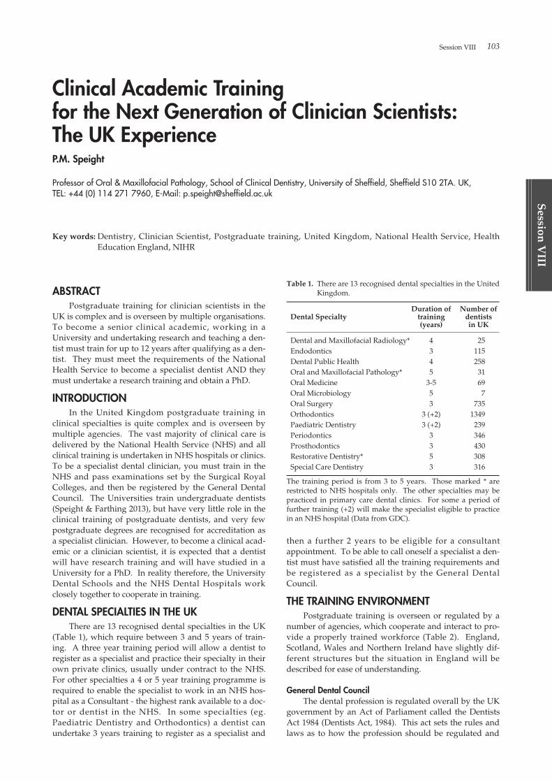
Session VIII 103
Session
VIII
ABSTRACTPostgraduate training for clinician scientists in the
UK is complex and is overseen by multiple organisations.To become a senior clinical academic, working in aUniversity and undertaking research and teaching a den-tist must train for up to 12 years after qualifying as a den-tist. They must meet the requirements of the NationalHealth Service to become a specialist dentist AND theymust undertake a research training and obtain a PhD.
INTRODUCTIONIn the United Kingdom postgraduate training in
clinical specialties is quite complex and is overseen bymultiple agencies. The vast majority of clinical care isdelivered by the National Health Service (NHS) and allclinical training is undertaken in NHS hospitals or clinics.To be a specialist dental clinician, you must train in theNHS and pass examinations set by the Surgical RoyalColleges, and then be registered by the General DentalCouncil. The Universities train undergraduate dentists(Speight & Farthing 2013), but have very little role in theclinical training of postgraduate dentists, and very fewpostgraduate degrees are recognised for accreditation asa specialist clinician. However, to become a clinical acad-emic or a clinician scientist, it is expected that a dentistwill have research training and will have studied in aUniversity for a PhD. In reality therefore, the UniversityDental Schools and the NHS Dental Hospitals workclosely together to cooperate in training.
DENTAL SPECIALTIES IN THE UKThere are 13 recognised dental specialties in the UK
(Table 1), which require between 3 and 5 years of train-ing. A three year training period will allow a dentist toregister as a specialist and practice their specialty in theirown private clinics, usually under contract to the NHS.For other specialties a 4 or 5 year training programme isrequired to enable the specialist to work in an NHS hos-pital as a Consultant - the highest rank available to a doc-tor or dentist in the NHS. In some specialties (eg.Paediatric Dentistry and Orthodontics) a dentist canundertake 3 years training to register as a specialist and
then a further 2 years to be eligible for a consultantappointment. To be able to call oneself a specialist a den-tist must have satisfied all the training requirements andbe registered as a specialist by the General DentalCouncil.
THE TRAINING ENVIRONMENTPostgraduate training is overseen or regulated by a
number of agencies, which cooperate and interact to pro-vide a properly trained workforce (Table 2). England,Scotland, Wales and Northern Ireland have slightly dif-ferent structures but the situation in England will bedescribed for ease of understanding.
General Dental CouncilThe dental profession is regulated overall by the UK
government by an Act of Parliament called the DentistsAct 1984 (Dentists Act, 1984). This act sets the rules andlaws as to how the profession should be regulated and
Clinical Academic Training for the Next Generation of Clinician Scientists: The UK ExperienceP.M. Speight
Professor of Oral & Maxillofacial Pathology, School of Clinical Dentistry, University of Sheffield, Sheffield S10 2TA. UK, TEL: +44 (0) 114 271 7960, E-Mail: [email protected]
Key words: Dentistry, Clinician Scientist, Postgraduate training, United Kingdom, National Health Service, HealthEducation England, NIHR
Table 1. There are 13 recognised dental specialties in the UnitedKingdom.
Duration of Number of Dental Specialty training dentists
(years) in UK
Dental and Maxillofacial Radiology* 4 25Endodontics 3 115Dental Public Health 4 258Oral and Maxillofacial Pathology* 5 31Oral Medicine 3-5 69Oral Microbiology 5 7Oral Surgery 3 735Orthodontics 3 (+2) 1349Paediatric Dentistry 3 (+2) 239Periodontics 3 346Prosthodontics 3 430Restorative Dentistry* 5 308Special Care Dentistry 3 316
The training period is from 3 to 5 years. Those marked * arerestricted to NHS hospitals only. The other specialties may bepracticed in primary care dental clinics. For some a period offurther training (+2) will make the specialist eligible to practicein an NHS hospital (Data from GDC).
02_本文1027(web用追加) 15.10.27 9:05 AM ページ 103
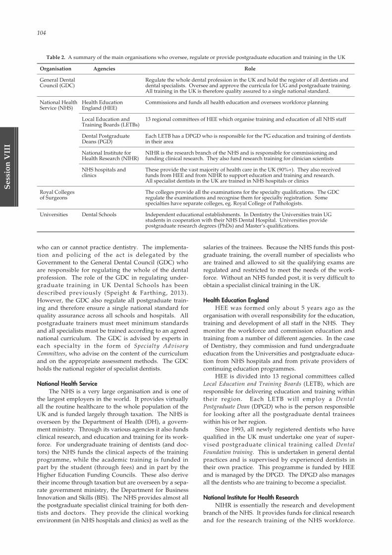
104
Ses
sion
VII
I
who can or cannot practice dentistry. The implementa-tion and policing of the act is delegated by theGovernment to the General Dental Council (GDC) whoare responsible for regulating the whole of the dentalprofession. The role of the GDC in regulating under-graduate training in UK Dental Schools has beendescribed previously (Speight & Farthing, 2013).However, the GDC also regulate all postgraduate train-ing and therefore ensure a single national standard forquality assurance across all schools and hospitals. Allpostgraduate trainers must meet minimum standardsand all specialists must be trained according to an agreednational curriculum. The GDC is advised by experts ineach specialty in the form of Specialty AdvisoryCommittees, who advise on the content of the curriculumand on the appropriate assessment methods. The GDCholds the national register of specialist dentists.
National Health ServiceThe NHS is a very large organisation and is one of
the largest employers in the world. It provides virtuallyall the routine healthcare to the whole population of theUK and is funded largely through taxation. The NHS isoverseen by the Department of Health (DH), a govern-ment ministry. Through its various agencies it also fundsclinical research, and education and training for its work-force. For undergraduate training of dentists (and doc-tors) the NHS funds the clinical aspects of the trainingprogramme, while the academic training is funded inpart by the student (through fees) and in part by theHigher Education Funding Councils. These also derivetheir income through taxation but are overseen by a sepa-rate government ministry, the Department for BusinessInnovation and Skills (BIS). The NHS provides almost allthe postgraduate specialist clinical training for both den-tists and doctors. They provide the clinical workingenvironment (in NHS hospitals and clinics) as well as the
salaries of the trainees. Because the NHS funds this post-graduate training, the overall number of specialists whoare trained and allowed to sit the qualifying exams areregulated and restricted to meet the needs of the work-force. Without an NHS funded post, it is very difficult toobtain a specialist clinical training in the UK.
Health Education EnglandHEE was formed only about 5 years ago as the
organisation with overall responsibility for the education,training and development of all staff in the NHS. Theymonitor the workforce and commission education andtraining from a number of different agencies. In the caseof Dentistry, they commission and fund undergraduateeducation from the Universities and postgraduate educa-tion from NHS hospitals and from private providers ofcontinuing education programmes.
HEE is divided into 13 regional committees calledLocal Education and Training Boards (LETB), which areresponsible for delivering education and training withintheir region. Each LETB will employ a DentalPostgraduate Dean (DPGD) who is the person responsiblefor looking after all the postgraduate dental traineeswithin his or her region.
Since 1993, all newly registered dentists who havequalified in the UK must undertake one year of super-vised postgraduate clinical training called DentalFoundation training. This is undertaken in general dentalpractices and is supervised by experienced dentists intheir own practice. This programme is funded by HEEand is managed by the DPGD. The DPGD also managesall the dentists who are training to become a specialist.
National Institute for Health ResearchNIHR is essentially the research and development
branch of the NHS. It provides funds for clinical researchand for the research training of the NHS workforce.
Table 2. A summary of the main organisations who oversee, regulate or provide postgraduate education and training in the UK
Organisation Agencies Role
General Dental Regulate the whole dental profession in the UK and hold the register of all dentists and Council (GDC) dental specialists. Oversee and approve the curricula for UG and postgraduate training.
All training in the UK is therefore quality assured to a single national standard.
National Health Health Education Commissions and funds all health education and oversees workforce planningService (NHS) England (HEE)
Local Education and 13 regional committees of HEE which organise training and education of all NHS staffTraining Boards (LETBs)
Dental Postgraduate Each LETB has a DPGD who is responsible for the PG education and training of dentists Deans (PGD) in their area
National Institute for NIHR is the research branch of the NHS and is responsible for commissioning and Health Research (NIHR) funding clinical research. They also fund research training for clinician scientists
NHS hospitals and These provide the vast majority of health care in the UK (90%+). They also received clinics funds from HEE and from NIHR to support education and training and research.
All specialist dentists in the UK are trained in NHS hospitals or clinics
Royal Colleges The colleges provide all the examinations for the specialty qualifications. The GDC of Surgeons regulate the examinations and recognise them for specialty registration. Some
specialties have separate colleges, eg. Royal College of Pathologists.
Universities Dental Schools Independent educational establishments. In Dentistry the Universities train UG students in cooperation with their NHS Dental Hospital. Universities provide postgraduate research degrees (PhDs) and Master’s qualifications.
02_本文1027(web用追加) 15.10.27 9:05 AM ページ 104
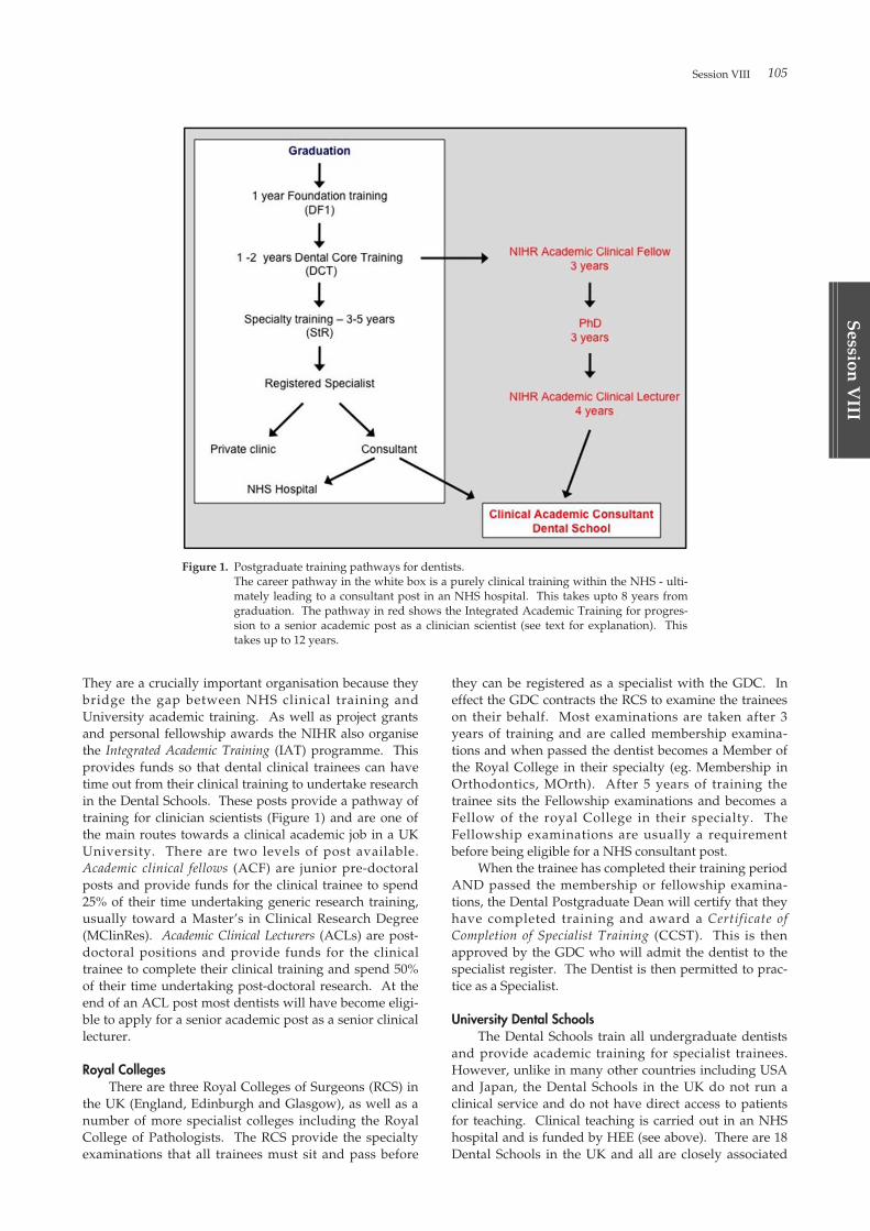
Session VIII 105
Session
VIII
They are a crucially important organisation because theybridge the gap between NHS clinical training andUniversity academic training. As well as project grantsand personal fellowship awards the NIHR also organisethe Integrated Academic Training (IAT) programme. Thisprovides funds so that dental clinical trainees can havetime out from their clinical training to undertake researchin the Dental Schools. These posts provide a pathway oftraining for clinician scientists (Figure 1) and are one ofthe main routes towards a clinical academic job in a UKUniversity. There are two levels of post available.Academic clinical fellows (ACF) are junior pre-doctoralposts and provide funds for the clinical trainee to spend25% of their time undertaking generic research training,usually toward a Master’s in Clinical Research Degree(MClinRes). Academic Clinical Lecturers (ACLs) are post-doctoral positions and provide funds for the clinicaltrainee to complete their clinical training and spend 50%of their time undertaking post-doctoral research. At theend of an ACL post most dentists will have become eligi-ble to apply for a senior academic post as a senior clinicallecturer.
Royal CollegesThere are three Royal Colleges of Surgeons (RCS) in
the UK (England, Edinburgh and Glasgow), as well as anumber of more specialist colleges including the RoyalCollege of Pathologists. The RCS provide the specialtyexaminations that all trainees must sit and pass before
they can be registered as a specialist with the GDC. Ineffect the GDC contracts the RCS to examine the traineeson their behalf. Most examinations are taken after 3years of training and are called membership examina-tions and when passed the dentist becomes a Member ofthe Royal College in their specialty (eg. Membership inOrthodontics, MOrth). After 5 years of training thetrainee sits the Fellowship examinations and becomes aFellow of the royal College in their specialty. TheFellowship examinations are usually a requirementbefore being eligible for a NHS consultant post.
When the trainee has completed their training periodAND passed the membership or fellowship examina-tions, the Dental Postgraduate Dean will certify that theyhave completed training and award a Certificate ofCompletion of Specialist Training (CCST). This is thenapproved by the GDC who will admit the dentist to thespecialist register. The Dentist is then permitted to prac-tice as a Specialist.
University Dental SchoolsThe Dental Schools train all undergraduate dentists
and provide academic training for specialist trainees.However, unlike in many other countries including USAand Japan, the Dental Schools in the UK do not run aclinical service and do not have direct access to patientsfor teaching. Clinical teaching is carried out in an NHShospital and is funded by HEE (see above). There are 18Dental Schools in the UK and all are closely associated
Figure 1. Postgraduate training pathways for dentists.The career pathway in the white box is a purely clinical training within the NHS - ulti-mately leading to a consultant post in an NHS hospital. This takes upto 8 years fromgraduation. The pathway in red shows the Integrated Academic Training for progres-sion to a senior academic post as a clinician scientist (see text for explanation). Thistakes up to 12 years.
02_本文1027(web用追加) 15.10.27 9:05 AM ページ 105
106
Ses
sion
VII
I
with a teaching hospital. All clinical academic staff in theSchools also have a contract with the NHS, so that theycan undertake clinical service and teach clinical subjectsto undergraduate and postgraduate students. Thismeans that, in their capacity as academic consultants,many Dental School clinical staff also train specialists.However the Universities cannot award qualificationsthat enable dentists to register as a specialist with theGDC: this can only be done by the Royal Colleges (seeabove).
The dental schools usually offer three types of post-graduate qualifications:
PhDs : These are conventional research training degreessimilar to those in other parts of the world. They areavailable to clinical and non clinical candidates, and take3-4 years to complete. It is very difficult to get funding todo a PhD and dentists have to apply for fellowships fromthe research councils or from NIHR. The IntegratedAcademic Training pathway (Figure 1) is funded byNIHR and provides support for dentists who wish to doa PhD.
Masters Degrees : Most dental schools offer 1 or 2 yearmasters programmes in specific clinical disciplines.These are often taken by dentists who wish to enhancetheir skills, but they do not lead to a specialist qualifica-tion which the GDC will recognise. However a masterstraining may contribute towards a specialist training, butthe dentists will also have to satisfy the requirements ofthe Dental Postgraduate Dean and sit the examinations ofthe Royal Colleges. Many Schools collaborate with theNHS, the DPGD and the Royal Colleges to provide a uni-fied training, which results in a Master’s degree and eligi-bility to sit the College examinations. Some schools alsooffer MClinRes degrees, which provide generic researchtraining for junior clinicians and ACFs.
Professional Doctorates : Some Schools offer a three yearintegrated clinical and academic training which results ina professional doctorate (Doctorate of Clinical Dentistry,DClinDent). These take from 3-4 years and are usuallytailored to provide the clinical training required to satisfythe DPGD, and enable the candidate to sit the member-ship examinations of the Royal Colleges. Note howeverthat the DClinDent itself is not recognised by the GDCand the dentists must ALSO sit the College examinationsif he or she wishes to be registered as a specialist.
TRAINING PATHWAYSTraining pathways for dentists are summarised in
Figure 1. It can be seen that it may take up to 12 years tobecome a senior clinical academic (clinician scientist) in aUK University. It is also a complex process involvingmultiple organisations and regulators as summarisedabove. Although salaries are reasonable (Specialtytrainees may earn £30,000 - £40,000 GBP (~£50,000USD))they are low compared to private practice and the train-ing is long. This presents a formidable challenge to theaspiring specialist dentist and relatively few ever embarkon this pathway. Less than 5% of dentists pursue an aca-demic career making recruitment into Dental Schools
very difficult.On graduation, all UK dentists must complete a year
of Foundation Training. This is carried out as an employ-ee of the NHS in a dental practice. Foundation Dentistsare taught by experienced dentists and learn manage-ment skills and become more proficient at whole patientcare. The Foundation Programme is organised andassessed by Dental Postgraduate Deans, who work forHEE (see above).
On completion of foundation training the dentist ispermitted to work for the NHS and most will then seekemployment in primary care dental clinics. Only about15% to 20% undertake further training, and overall, onlyabout 10% will become a specialist. Those who wish toundertake further training will work in a hospital as aDental Core Trainee (DCT) for one or two years. Theseposts are at a junior level and usually rotate through anumber of clinical specialties.
Specialty training is undertaken in a Specialty train-ing Registrar (StR) post in an NHS hospital. These arenationally competitive and are funded by the NHSthrough the Dental Postgraduate Deans. Training takesfrom 3-5 years (Table 1) and is supervised by a seniorclinician (consultant) who is qualified to train. Trainingis continuously assessed. To complete the training andbecome a specialist a dentist must:
· be appointed to a recognised StR post· complete the required amount of time in training (3-5years)
· complete and pass about 18 workplace based assess-ments each year
· maintain a portfolio of all work done· attend and satisfactorily pass an Annual Review ofCompetence Progression (ARCP)
· pass the relevant Royal College specialty examina-tions
· present the portfolio of work and pass the final ARCPfor the DPGD to approve the award of a CCST(Certificate of Completion of Specialist Training)
· be placed on the GDC specialist registerOnce registered, the specialist may be eligible to
apply for a Consultant post in the NHS.If the dentist wishes to work in a University and
undertake teaching and research, they may seek a postwhich enables them to undertake research and study fora PhD. Some academic posts are funded by NIHR (ACFsand ACLs) and allow the dentist to do clinical trainingand research at the same time. These are shown in red inTable 1. To do a PhD the dentists must apply for fundsto get a fellowship to allow them to take time out of clini-cal training for research. Organisations such as theNIHR, the Medical Research Council and a number ofcharities provide funds for fellowships. Dental Schoolsmay also provide funding for academic training postsand for PhD study.
On completion of the clinical training and the PhDthe dentist is then a eligible to apply for a post as an aca-demic consultant at the grade of Senior Lecturer(Associate Professor). However, these posts are verycompetitive and you would require a research trackrecord with high quality publications. Many newly qual-ified specialists therefore work in junior university posts
02_本文1027(web用追加) 15.10.27 9:05 AM ページ 106

Session VIII 107
Session
VIII
(lecturer) to build up their research and teaching experi-ence.
REFERENCESSpeight PM, Farthing PM. (2013). Undergraduate dental
education in the United Kingdom: Curriculumdesign and regulation. Proceedings of the 5thHiroshima Conference on Education and Science in
Dentistry. Hiroshima University, Faculty ofDentistry. 31-35.
General Dental Council. Who we are.http://www.gdc-uk.org/Aboutus/Thecouncil/Pages/whoweare.aspx (accessed August 2015)
Dentists Act 1984. UK Government.http://www.legislation.gov.uk/ukpga/1984/24/contents (accessed August 2015)
02_本文1027(web用追加) 15.10.27 9:05 AM ページ 107
108
Ses
sion
VII
I
International Collaborative Program for Human Resources in Dental Medicine at Graduate-level: The Korean PerspectiveJ.I. Lee
Seoul National University, School of Dentistry, Korea
AbstractToday, graduate education in dental school has
become increasingly important for the advancement ofdentistry. The goal of graduate education in dentistry isnow focused on finding new knowledge and developingnew technology for the advancement of human wellbe-ing in the field of dentistry. In Korea, 11 dental schoolsoffer graduate programs. Large proportions of domesticstudents admitted to graduate programs are mainly fromnon-dental fields, such as biology or engineering.International collaboration needs more active engage-ment and mutual communication between multinationalinstitutions. There are many issues in international col-laboration. Interdisciplinary collaboration would be avery important preceding requirement of internationalcollaboration. Dual degree programs, short-termexchange programs, and exchange seminars or confer-ences are possible and executable actions. Web-basedopen communication or on-line research seminars can behelpful for the expansion of collaboration. The mostimportant goal of the graduate program is humanresource development. Our goal of international collabo-ration should be to set balanced networks betweeninvolved nations. The institutional preparedness forinternational collaborations should be properly assessedin terms of expected outcomes and goals of individualschools. For this agenda we need a framework forhuman resource development, a consortium for the prop-er international collaboration and qualification of gradu-ate education.
IntroductionToday, graduate education in dental school has
become increasingly important for the advancement ofdentistry. The goal of the graduate education in den-tistry is now focused on finding new knowledge anddeveloping new technology for the advancement ofhuman wellbeing in the field of dentistry. In Korea, 11dental schools offer graduate program including masterof science (in Dentistry) and Ph. D. (in Dentistry) Thereare also departments of dentistry in many medicalschools offering graduate degree programs in the clinicalfield. In graduate dental education, there are a largenumber of majors in both clinical dentistry and the basicscience field. Degree granting graduate programs arerun by the department of dentistry or dental science inthe graduate school of each university.
In Korea, there are 10 specialties in clinical dentistry
(specialty certificate). All of them also offers degreegranting courses (MS and PhD) - Oral medicine and oraldiagnosis, Oral and maxillofacial radiology, Oral andmaxillofacial surgery, Pediatric dentistry, Orthodontics,Conservative dentistry, Prosthodontics, Periodontology,and Oral Pathology. In the basic science field, there aretraditional majors - Anatomy, Physiology, Pharmacology,Microbiology, Biochemistry, Dental biomaterials, andPreventive dentistry. Some schools offer convergencemajors, such as Head & Neck Anatomy and ImagingScience, Immunology and Molecular Microbiology inDentistry, Molecular genetics, Cell and Developmentalbiology, Neuroscience, Preventive and Social dentistry,Healthcare management and informatics, Cancer anddevelopmental biology, Dental biomaterials science andDental regenerative biotechnology. Each master anddoctorate degree program require a minimum 2 yearscourse. Some master-doctorate combined programs needa minimum of 4 years. But the total course length isdetermined by the research progress of each theme.
The graduate student selects an advisor professor inthe department within 1 year after the student enters theschool. After completion of the required courses, the stu-dent must pass the qualifying exam before submission ofthesis. For international students, they must pass aKorean or a second language exam (except their motherlanguage). The student will be eligible to submit his/herthesis, only if he/she publishes a paper in the interna-tional journal recognized by our graduate school. Duringthe course, the student will engage in a research projectwith their advisor. There will be plenty of chance to par-ticipate in intramural research seminars and national andinternational meetings.
The challenges and perspectives of education at graduateschools
Graduate-level training in Korean dental school pre-viously was mainly focused on developing practice skills.There were also graduate programs for basic scienceresearch, but they were not enough. Many graduateswere looking to other countries to get advanced graduateeducation.
With the economic growth of the nation, the increasein national economic power has brought an improvementin research capacity. In recent years, dentistry hasbecome one of the most rapidly advancing field of pro-fession in Korea. Dentists promptly adopted lots ofnewly developed technology and research results quick-ly. With the economic growth, the opportunities for
02_本文1027(web用追加) 15.10.27 9:05 AM ページ 108
Session VIII 109
Session
VIII
postgraduate education has increased gradually. Thefocus of graduate programs in academic institutions alsoshifted into the field of science and technology. ManyKorean dental education institutions are now trying veryhard to become a research-focused academic institution.Large proportions of domestic students admitted to grad-uate programs are mainly from non-dental fields, such asbiology or engineering.
Graduate education aims to develop dentistrythrough research and education for dentistry, natural sci-ences and engineering and serve the nation and mankindby training talented experts at an international level.
Korean dental schools aim to develop dentistrythrough academic research in which the dentistry is com-bined with students who have various and broad acade-mic backgrounds. We aim to gain international competi-tiveness in each area by creating a healthcare environ-ment in which research, education and treatment are effi-ciently coordinated. We are making efforts to improvethe welfare of mankind by performing research and edu-cation with the responsibility to develop dentistry oncewe have recognized the implementation of educationphilosophy as national project and our responsibility.For these goals, we need an expansion in internationalcollaboration. But until now these efforts have onlygained limited success.
Issues in international collaboration Korean dental education institutions traditionally
sent many graduates to developed countries in both clini-cal and basic science research areas. Majority was post-graduate specialty training, but also some students wentabroad in research fields.
Incoming student population has grown rapidly inrecent years. But our system of opportunity is still limit-ed. We have government-run national scholarship pro-grams. But it has very limited numbers. University-based scholarship programs have also increased signifi-cantly. But both programs are only eligible to limitedcountries. This made studying in Korea unattractive.Now, we are developing school-based scholarships andresearch grant conjunct scholarship programs. But inter-national collaboration of graduate education cannot becompleted only by scholarship programs. Internationalcollaboration needs more active engagement and mutualcommunication between multinational institutions.Though we believe that international collaboration leadsto better dental science, our institutional budget andfunding structures are ‘always’ not enough to supportinternational collaboration. The students need moreopportunity to meet each other. How can it be accom-plished?
There are other important issues in international col-laboration within graduate education. For graduate edu-cation and international collaboration, the clinical fieldalways got caught in license issues. Language is also abig barrier. Cultural issues need to be considered as well.There is also the issue of age and gender. Terms of visitwill also be an important issue. We need to evaluateprospective outcome of short-term (weeks or months) orlong-term (full-course or years) programs. Sometimesthe differences between countries are less serious than
departmental barriers. Interdisciplinary collaborationwould be a very important preceding requirement ofinternational collaboration.
Each country has a different priority in research top-ics. In developed countries the diversity of topics maynot be a problem but developing or underdevelopedcountries see it as an important issue. The meaning ofresearch in each countries with different cultural andsocioeconomic situation will be different. Academic pro-gram may be similar but also very different. Do we needstandardization of the curriculum? Or can we grantdiversity to each education program?
Followings are part of action plans for globalizationof Seoul National University, School of Dentistry.
- To participate in the exchange of human resources(students and faculty)
- To expand a network of dental education institutions.- To acquire advanced technologies and develop edu-
cational programs- To play an active role in advancing dentistry in devel-
oping countries through fund-raising- To host global academic conferences and symposiums- To share knowledge and skills of dentistry globally- To promote sustainable and diversified joint research
But the proclamation is not enough to reach the goal.We should consider detailed issues and make feasibleaction plans. Dual degree programs, short-termexchange programs, and exchange seminars or confer-ences are possible and executable actions. Sometimesweb-based open communication or on-line research sem-inars can be helpful for the expansion of collaboration.
SuggestionGlobalization can be defined as ‘the process by
which markets and production in different countries arebecoming increasingly interdependent due to the dynam-ics of trade in goods and services and flows of capital andtechnology.’ (European Commission, 1997) The mostimportant goal of the graduate program is humanresource development. In any country, qualified humanresources are the most important assets for the nationand the whole world. Internationalization always has aflow — unidirectional or bidirectional. But our goal ofinternational collaboration should be to set balanced net-works between involved nations.
The institutional preparedness for international col-laborations should be properly assessed in terms ofexpected outcomes and goals of individual schools. Forthis agenda we need a framework for a human resourcedevelopment.
I suggest a consortium for the proper internationalcollaboration and qualification of graduate education. Itcan be an international supervisory committee which,works to evaluate each program for its preparedness tomutual understanding and investigates the impact ofinternational collaborations.
Blumenfield T, Nerad M. Assessing international (post)graduate education. A Research agenda. Aus UnivRev 54 (1): 72-82. 2012
European Commission. ‘Annual Economic Report for1997’ European Economy No. 63. 1997
02_本文1027(web用追加) 15.10.27 9:05 AM ページ 109
110
Ses
sion
VII
I
ABSTRACTInternational collaborative management, with the
help of professors from University of Alabama for theundergraduate program, is an integral part of the Facultyof Dentistry, Chulalongkorn University since its incep-tion in 1940. The first graduate program was establishedin 1973 with the support from the University of Alabamaand there have been many graduate programs estab-lished due to the collaborative management with theschool overseas. In 2008, a Master’s Degree in EstheticRestorative and Implant Dentistry (InternationalProgram) was started in collaboration with the School ofDentistry, UCLA. The program integrated advance artand science into several major fields in dentistry toimprove or maintain esthetic condition of the patient.Starting in 2016, a Doctor of Philosophy in Dental Science(Orthodontics) will be started as a result of collaborationbetween Chulalongkorn University and Tokyo Medicaland Dental University and it will be CU-TMDU JointDegree Program. With strong collaboration between uni-versities and the usage modern information technology,this will enable the development of dental education interms of inter-disciplinary education with long-terminternational collaborative management.
INTRODUCTIONThe genesis of the dental curriculum in Thailand was
initiated in 1928 by Professor Colonel Luang VachVidyavaddhana, with the objective of providing dentaleducation and establishing a school of Dentistry inThailand. After he received his medical degree fromSyracuse University he furthered his study in Dentistryat the University of Pennsylvania. When he returned toThailand, he asked Professor Aller Gustin Ellis, the presi-dent of Chulalongkorn University to set up the school ofDentistry, however due to shortage of budget it wasunsuccessful. He then proposed to the head of theMinistry of Defense, Field Marshal P. Pibulsongkram,with the idea of setting up the school of Dentistry to pro-vide services for military personals. He then received atravel grant from the ministry of defense for academictour overseas in order to learn more about what wasneeded to set up a school of Dentistry. After comingback from the academic tour Field Marshal P.Pibulsongkram has became the president ofChulalongkorn, and as a result the project for the armywas shifted to a development of a school of Dentistrywithin Chulalongkorn University instead and in 1940
Professor Colonel Luang Vach Vidyavaddhana wasappointed to be the first head of the new section. In 1943,the University of Medical Sciences was founded and thedental section in Chulalongkorn University was includedso the affiliation of the first dental school was changed.Professor Colonel Luang Vach Vidyavaddhana thenbecame the first dean of the Faculty of Dentistry in theUniversity of Medical Sciences. In 1969 the University ofMedical Sciences was renamed into Mahidol Universityand in 1972 the school of Dentistry returned toChulalongkorn University where it has located until thisday.
At the beginning there was only three faculty staff,Professor Colonel Luang Vach Vidyavaddhana decidedto write a letter to the US government for academic sup-port. Professor Joseph Volker, the dean of the School ofDentistry, University of Alabama, answered his requestand was sent by the US government to Thailand to helptrain the faculty staff and teach the students. At thattime, curriculum development was also conducted. Itcannot be claimed that the first international collabora-tive program was initiated, it was however the first den-tal collaboration between Thailand and the United Statefor the undergraduate program. Many faculty staffsfrom Chulalongkorn University were sent to study in theUniversity of Alabama and later on Professor CharlesMacallum also came to help the Faculty of Dentistry,Chulalongkorn University. As an oral maxillofacial sur-geon he provided knowledge and collaborations, whichleads to the establishment of specialty training inMaxillofacial in 1973 by Chulalongkorn staffs, whichwere inspired by Professor Charles Macallum andProfessor Chuachote Hungsasut was trained at theUniversity of Alabama and finally become the first Thaidentist who got the first Diplomate, American Board inOral and Maxillofacial surgery. With ProfessorMacallum’s enormous support, he was honored with anhonorable degree of Doctor of Philosophy fromChulalongkorn University in 1993. It is the first graduateprogram in Thailand which was established from the col-laboration between Chulalongkorn University and theUniversity of Alabama.
Many more graduate programs ranging from 1 yearup to 5 years were introduced and more than 20 graduateprograms are currently available. In 2008, the first inter-national collaborative master degree program was start-ed. It is a Master’s Degree in Esthetic Restorative andImplant Dentistry (International Program). The objec-tives of this program are to
International Collaborative Graduate Program at the Faculty of Dentistry, Chulalongkorn UniversityS. Poolthong
DDS, MSc (Dent), PhD, Dean, Faculty of Dentistry, Chulalongkorn University, Bangkok 10330, Thailand
02_本文1027(web用追加) 15.10.27 9:05 AM ページ 110
Session VIII 111
Session
VIII
1. Establish the international teaching and learningcourses.
2. Enable the faculty to generate a program that is self-supported.
3. Establish a national standard for international dentaleducation.
4. Establish a prototype for dental service that is differ-ent from the governmental pattern.The esthetic restorative and implant dentistry is the
advanced art and science of integration of several majorfields in dentistry, i.e. operative dentistry, prosthodon-tics, periodontics, surgery, orthodontics and occlusion,involving systematic treatment planning to achieve high-ly successful treatment outcome leading to proper func-tion of temporo-muscular system, as well as improvingor maintaining esthetic condition of the patient. The pro-gram provides services, both by students and facultymembers, to patients that would promote not only theiroral and physical but also mental health. Collaborationwith universities abroad such as University of Californiaat Los Angeles was established. Students attending theprogram will spend at least one semester at sister dentalschool which is at present the students will visit School ofDentistry, UCLA to get extra training for one semester.The students will be able to attend lectures, seminars andalso clinical observation. They will sit in the class withthose students in UCLA.
Curriculum Structure composes of 42 credits withrequired subject of 18, electives of 2, clinical and laborato-ry practices of 10 and 12 for Thesis. For the non-thesisprogram it composes of required subject of 24, electivesof 2, clinical and laboratory practices of 10 and 6 for inde-pendent study (clinical-oriented). The duration of studyis 2 academic years which include 2 summer sessions.The program also produces a number of publications.
For the thesis program, the study plan is as follow:Year 1 Semester 1 (12 credits)
Research Methodology in Dentistry Statistics in DentistryConservative Dentistry Art Appreciation for General Esthetics Principle of Esthetic Dental Treatment I Principle of Implantology I Seminar in Esthetic Dentistry I Seminar in Implantology I Implant Dental Laboratory Esthetic Dental Laboratory
Year 1 Semester 2 (11 credits)Principle of Esthetic Dental Treatment II Diagnosis and Treatment Planning for EstheticRestorative Dentistry and Implantology Dental Material Science for Esthetic DentistryandImplantology Seminar in Esthetic Dentistry II Esthetic and Implantology Dental Clinic I Seminar in Implantology II ThesisElective
Year 2 Semester 1 (9 credits)Principle of Implantology II Seminar in Esthetic Dentistry III Esthetic and Implantology Dental Clinic II
Seminar in Implantology III Thesis Elective
Year 2 Semester 2 (10 credits)Esthetic and Implantology Dental Clinic III Thesis
For non-thesis program, study plan is as follow:Year 1 Semester 1 (10 credits)
Research Methodology in Dentistry Statistics in Dentistry Conservative Dentistry Art Appreciation for General Esthetics Principle of Esthetic Dental Treatment I Seminar in Esthetic Dentistry I Esthetic Dental Laboratory Principle of Implantology I
Year 1 Semester 2 (11 credits)Seminar in Implantology I Implant Dental Laboratory Principle of Esthetic Dental Treatment II Dental Occlusion: Principle and Application forRestorative DentistryDiagnosis and Treatment Planning for EstheticRestorative Dentistry and Implantology Dental Material Science for Esthetic Dentistry andImplantology Seminar in Esthetic Dentistry II Esthetic and Implantology Dental Clinic I Seminar in Implantology II Elective
Year 2 Semester 1 (11 credits)Principle of Implantology II Seminar in Esthetic Dentistry III Seminar in Implantology III Esthetic and Implantology Dental Clinic II Multidisciplinary Approach to Advanced EstheticDentistry Independent Study in Esthetic Restorative andImplant Dentistry I Elective
Year 2 Semester 2 (10 credits)Esthetic and Implantology Dental Clinic III Independent Study in Esthetic Restorative andImplant Dentistry II Technology in Esthetic Dentistry Multidisciplinary Approach to AdvancedImplantology
The program has been continued for 6 classes start-ing from academic year 2008 until at present (stop forcurriculum development for one year). There are 41 stu-dents graduated from this program and another 11 stu-dents are studying in the program now.
In 2013, another idea was initiated for another inter-national collaborative graduate program. It is a PhD pro-gram with collaboration between ChulalongkornUniversity and Tokyo Medical and Dental University.The program is now under development by both univer-sities, affirming its long history of collaboration. It is theDoctor of Philosophy in Dental Science (Orthodontics),CU-TMDU Joint Degree Program. Students can enroll
02_本文1027(web用追加) 15.10.27 9:05 AM ページ 111
112
Ses
sion
VII
I
from both sides but follow the same process. The pro-gram was designed to bring strengths of the two univer-sities and share those strengths for students. The stu-dents will enroll in course work, practical exercise anddissertation seminar in Thailand for 1 year while theywill do qualifying examination and also dissertation sem-inar for the second year. The students will study usingteleconference with Japan through the third and fifthyear so they can concentrate on their course work andclinical training in Thailand, which include proposaldefense, dissertation seminar and dissertation examina-tion. However, the final agreement for all details has notbeen finished yet. The 5 Year Program (72 credits) willcomprise of:
Course work 24 credits Lecture 8 creditsPractical exercise 1 creditSeminar 2 creditsClinical training 12 creditsElective 1 credit
Dissertation 48 credits
This will open a new era of collaborative dental edu-cation, with ample usage of information technologywhile keeping rooted in traditional study and clinicaltrainings. It will expand expertise and share resources to
bring out the most efficient way of teaching and researchon the international level. The program is expected to becompleted and will start at the earliest in the academicyear 2016. There are many international graduate pro-grams at the Faculty of Dentistry, ChulalongkornUniversity in which there is no collaboration with otherlocal or overseas dental institutions. The two programsare academic innovation that differs from other specialtyprograms from other dental schools.
CONCLUSIONIn conclusion, with strong collaboration between
universities and modern information technology, it ispossible to conduct an international collaborative gradu-ate program to strengthen knowledge sharing andresearch collaboration. This will enable the developmentof dental education in terms of inter-disciplinary educa-tion with long-term international collaborative manage-ment.
REFERENCES1. Patanapiradej, V. 70th Anniversary, Faculty of Dentistry,
Chulalongkorn University. 2010, ChulalongkornUniversity Printing House. P. 39-100, 107-110.
2. http://www.estheticchula.com/ (10 August 2015)
02_本文1027(web用追加) 15.10.27 9:05 AM ページ 112
Session
IX
Oral Health Promotion and Functional Rehabilitation in Super-aged SocietyInitiatives toward the Prevention of Long-Term Care Dependency in Japan
Ministry of Health, Labour and Welfare
M. Sakoi
Oral Rehabilitation and Regional Partnerships
Nippon Dental University
F. Tamura and T. Kikutani
What Can the Dentistry Do in Super-aged Society?
Hiroshima University
M. Yoshikawa
The Role of Dental Professionals in Dysphagia Rehabilitation
Tokushima University
M. Matsuyama
Session IX
02_本文1027(web用追加) 15.10.27 9:05 AM ページ 113

02_本文1027(web用追加) 15.10.27 9:05 AM ページ 114

Session IX 115
Session
IX
Initiatives toward the Prevention of Long-Term Care Dependency in JapanM. Sakoi
M.D., M.P.H., Director, Aging and Health Division, Health and Welfare Bureau for the Elderly, Ministry of Health, Labour and Welfare, Government of JAPAN
Changes in Japan’s PopulationBy examining changes in Japan’s demographic
makeup, the number of seniors over age 65 is predictedto reach 36.57 million by 2025 and reach a peak of 38.78million in 2042. Additionally, the percentage of seniorsover age 75 is expected to grow, surpassing 25% by 2055.It can be seen that the current social structure consists of2.6 persons supporting each elderly person. In 2060, withthe progression of the aging population and decreasingbirthrate, it is estimated that 1.2 person will be support-ing one senior citizen. (Fig. 1)
Since the establishment of the long-term care insur-ance system in 2000, the population over age 75, as anage group with high percentage of persons requiringcare, has increased rapidly and the rapid increase willcontinue for the next 10 years to 2025. From about 2030,the rapid growth of the population over age 75 will leveloff but the population over age 85 will continue to
increase for another 10 years. The population over age40, who pay for the long-term care insurance, hasincreased since the establishment of the long-term insur-ance system in 2000 but will start to decrease after 2025.
Among seniors over age 65, those over level II of the“daily life independency level for seniors with dementia”will increase. Individual or couple-only households withhouseholders over age 65 will increase. The number ofseniors over age 75 will rapidly grow in cities and gradu-ally grow in rural areas with originally high senior popu-lation. Tailored response according to regions is neces-sary as aging circumstances differ according to region.
Establishing the Integrated Community Care SystemBy 2025 when the baby boomers will become age 75
and above, we will implement the creation of a structure(comprehensive regional care system) that comprehen-sively ensures the provision of health care, nursing care,
By examining changes in Japan’s demographic makeup , it can be seen that the current social structure consists of 2.6 persons supportingeach elderly person. In 2060, with the progression of the aging population and decreasing birthrate, it is estimated that 1.2 person will be
Changes in Japan’s Population Pyramid (1990–2060)
1990 (Actual figures) 2025 2060supporting one senior citizen.
2010 (Actual figures)
Total population: Total population: Total population: Total population:Age Age Age Age
80
90
100
80
90
100
80
90
100
80
90
100
Age 75 & older597(5%)
123.61 million
Age 75 & older1,407(11%)
128.06 million
Age 75 & older2,179(18%)
120.66 million
Age 75 & older2,336(27%)
86.74 million
Baby Boomers
60
70
80
60
70
80
60
70
80
60
70
80597(5%)
Age 65–74892( 7%)
1,407(11%)
Age 65–741,517(12%)
2,179(18%)
Age 65–741,479(12%)
2,336(27%)
Age 65–741,128(13%)
Baby Boomers(Born 1947–49)
40
50
40
50
40
50
40
50
Age 20–647,590(61%)
Age 20–647,497(59%)
Age 20–646,559(54%)
Age 20–644,105(47%)
20
30
20
30
20
30
20
30
2nd Baby BoomGeneration
(Born 1971–74)
0 50 100 150 200 2500
10
0 50 100 150 200 2500
10
0 50 100 150 200 2500
10
0 50 100 150 200 2500
10
Persons 65 and
Age 0–193,249(26%)
Age 0–192,287(18%)
Age 0–191,849(15%)
Age 0–191,104(13%)
10K 10K 10K 10K
Source: Ministry of Internal Affairs and Communications – Population Census, Population Estimate; National Institute of Population and Social Security Research – “Population Projections for Japan (January 2012): Medium-Fertility & Medium-Mortality Assumption” (Figures as of Oct. 1 of each year)
Persons 65 andolder
Persons aged 20–64
15.1
11.8
11.2
12.6
10Kpeople
10Kpeople
10Kpeople
10Kpeoples
Fig. 1
02_本文1027(web用追加) 15.10.27 9:05 AM ページ 115
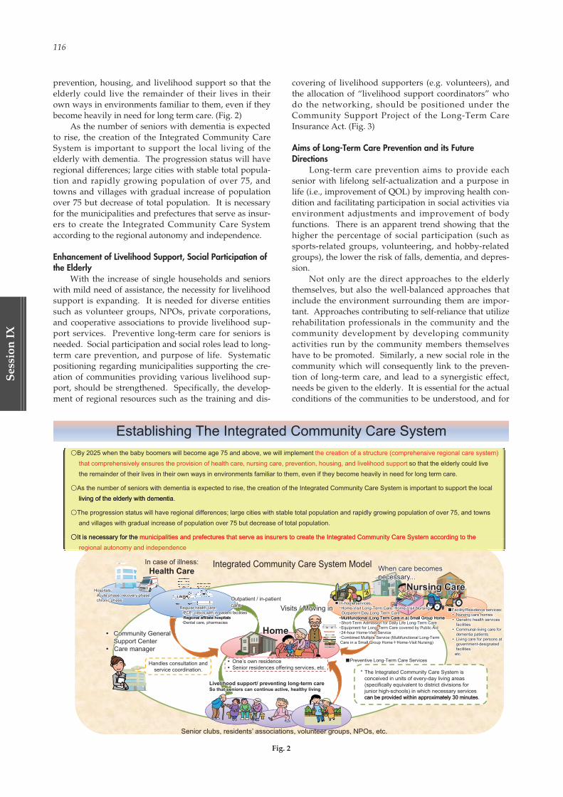
116
Ses
sion
IX
prevention, housing, and livelihood support so that theelderly could live the remainder of their lives in theirown ways in environments familiar to them, even if theybecome heavily in need for long term care. (Fig. 2)
As the number of seniors with dementia is expectedto rise, the creation of the Integrated Community CareSystem is important to support the local living of theelderly with dementia. The progression status will haveregional differences; large cities with stable total popula-tion and rapidly growing population of over 75, andtowns and villages with gradual increase of populationover 75 but decrease of total population. It is necessaryfor the municipalities and prefectures that serve as insur-ers to create the Integrated Community Care Systemaccording to the regional autonomy and independence.
Enhancement of Livelihood Support, Social Participation ofthe Elderly
With the increase of single households and seniorswith mild need of assistance, the necessity for livelihoodsupport is expanding. It is needed for diverse entitiessuch as volunteer groups, NPOs, private corporations,and cooperative associations to provide livelihood sup-port services. Preventive long-term care for seniors isneeded. Social participation and social roles lead to long-term care prevention, and purpose of life. Systematicpositioning regarding municipalities supporting the cre-ation of communities providing various livelihood sup-port, should be strengthened. Specifically, the develop-ment of regional resources such as the training and dis-
covering of livelihood supporters (e.g. volunteers), andthe allocation of “livelihood support coordinators” whodo the networking, should be positioned under theCommunity Support Project of the Long-Term CareInsurance Act. (Fig. 3)
Aims of Long-Term Care Prevention and its FutureDirections
Long-term care prevention aims to provide eachsenior with lifelong self-actualization and a purpose inlife (i.e., improvement of QOL) by improving health con-dition and facilitating participation in social activities viaenvironment adjustments and improvement of bodyfunctions. There is an apparent trend showing that thehigher the percentage of social participation (such assports-related groups, volunteering, and hobby-relatedgroups), the lower the risk of falls, dementia, and depres-sion.
Not only are the direct approaches to the elderlythemselves, but also the well-balanced approaches thatinclude the environment surrounding them are impor-tant. Approaches contributing to self-reliance that utilizerehabilitation professionals in the community and thecommunity development by developing communityactivities run by the community members themselveshave to be promoted. Similarly, a new social role in thecommunity which will consequently link to the preven-tion of long-term care, and lead to a synergistic effect,needs be given to the elderly. It is essential for the actualconditions of the communities to be understood, and for
Visits / Moving in
Community GeneralSupport CenterCare manager
In-home services:Home-Visit Long-Term Care, Home-Visit Nursing,Outpatient Day Long-Term CareMultifunctional (Long Term Care in a) Small Group HomeMultifunctional (Long-Term Care in a) Small Group HomeShort-Term Admission for Daily Life Long-Term CareEquipment for Long-Term Care covered by Public Aid24-hour Home-Visit ServiceCombined Multiple Service (Multifunctional Long-TermCare in a Small Group Home Home-Visit Nursing)
When care becomes...necessary
Nursing Care
Facility/Residence services:Nursing care homesGeriatric health servicesfacilitiesCommunal-living care fordementia patientsLiving care for persons atgovernment-designatedfacilities
etc.
By 2025 when the baby boomers will become age 75 and above, we will implement
Establishing The Integrated Community Care System
so that the elderly could live the remainder of their lives in their own ways in environments familiar to them, even if they become heavily in need for long term care.
As the number of seniors with dementia is expected to rise, the creation of the Integrated Community Care System is important to support the localliving of the elderly with dementialiving of the elderly with dementia.
The progression status will have regional differences; large cities with stable total population and rapidly growing population of over 75, and townsand villages with gradual increase of population over 75 but decrease of total population.
It is necessary for the
Integrated Community Care System ModelIn case of illness:Health Care
It is necessary for the
In-home services:Home-Visit Long-Term Care, Home-Visit Nursing,Outpatient Day Long-Term CareMultifunctional (Long Term Care in a) Small Group Home
Regular health care:PCP, clinics with in-patient facilitiesRegional affiliate hospitals
Outpatient / in-patient care
Hospitals:Acute phase, recovery phase,chronic phase
HomeRegional affiliate hospitalsDental care, pharmacies
Livelihood support/ preventing long-term care So that seniors can continue active, healthy living
Regular health care:PCP, clinics with in-patient facilitiesRegional affiliate hospitals
Outpatient / in-patient care Visits / Moving in
Hospitals:Acute phase, recovery phase,chronic phase
HomeMultifunctional (Long-Term Care in a) Small Group HomeShort-Term Admission for Daily Life Long-Term CareEquipment for Long-Term Care covered by Public Aid24-hour Home-Visit ServiceCombined Multiple Service (Multifunctional Long-TermCare in a Small Group Home Home-Visit Nursing)
Regional affiliate hospitalsDental care, pharmacies
Community GeneralSupport CenterCare manager
Livelihood support/ preventing long-term care So that seniors can continue active, healthy living
* The Integrated Community Care System is conceived in units of every-day living areas (specifically equivalent to district divisions for junior high-schools) in which necessary services can be provided within approximately 30 minutes
Handles consultation andservice coordination.
Preventive Long-Term Care Services
can be provided within approximately 30 minutes.
Senior clubs, residents’ associations, volunteer groups, NPOs, etc.
When care becomes...necessary
Nursing Care
One’s own residenceSenior residences offering services, etc.
Facility/Residence services:Nursing care homesGeriatric health servicesfacilitiesCommunal-living care fordementia patientsLiving care for persons atgovernment-designatedfacilities
etc.
the creation of a structure (comprehensive regional care system)that comprehensively ensures the provision of health care, nursing care, prevention, housing, and livelihood support
municipalities and prefectures that serve as insurers to create the Integrated Community Care System according to themunicipalities and prefectures that serve as insurers to create the Integrated Community Care System according to theregional autonomy and independence
Fig. 2
02_本文1027(web用追加) 15.10.27 9:05 AM ページ 116
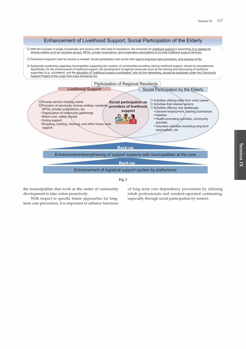
Session IX 117
Session
IX
the municipalities that work as the center of communitydevelopment to take action proactively.
With respect to specific future approaches for long-term care prevention, it is important to enhance functions
of long term care dependency prevention by utilizingrehab professionals and resident-operated commuting,especially through social participation by seniors.
With the increase of single households and seniors with mild need of assistance, the necessity for livelihood support is expanding. It is needed fordiverse entities such as volunteer groups, NPOs, private corporations, and cooperative associations to provide livelihood support services.
Enhancement of Livelihood Support, Social Participation of the Elderly
d e se e t t es suc as o u tee g oups, NPOs, p ate co po at o s, a d coope at e assoc at o s to p o de e ood suppo t se ces
Preventive long-term care for seniors is needed. Social participation and social roles lead to long-term care prevention, and purpose of life.
Systematic positioning regarding municipalities supporting the creation of communities providing various livelihood support, should be strengthened.Specifically, for the enhancement of livelihood support, the development of regional resources such as the training and discovering of livelihood
Participation of Regional Residents SLi lih d S t
ggp,pp,yp gsupporters (e.g. volunteers), and the allocation of “livelihood support coordinators” who do the networking, should be positioned under the CommunitySupport Project of the Long-Term Care Insurance Act.
p y ySocial Participation by the ElderlyppLivelihood Support
Social participation asid f li lih d
Activities utilizing skills from one’s careerActivities that interest seniors
Diverse service meeting needsProvision of service by diverse entities; residents providers of livelihood
supportActivities offering new challenges
General employment, starting businessesHobbiesHealth-promoting activities, communityactivities
Provision of service by diverse entities; residents,NPOs, private corporations, etc.Organization of community gatheringsWatch over, safety checksOuting support
activitiesVolunteer activities excluding long-termcare/welfare, etc.
Shopping, cooking, cleaning, and other house worksupport.
Back-upEnhancement/strengthening of support systems with municipalities at the core
Back-upEnhancement of logistical support system by prefectures 3
Fig. 3
02_本文1027(web用追加) 15.10.27 9:05 AM ページ 117
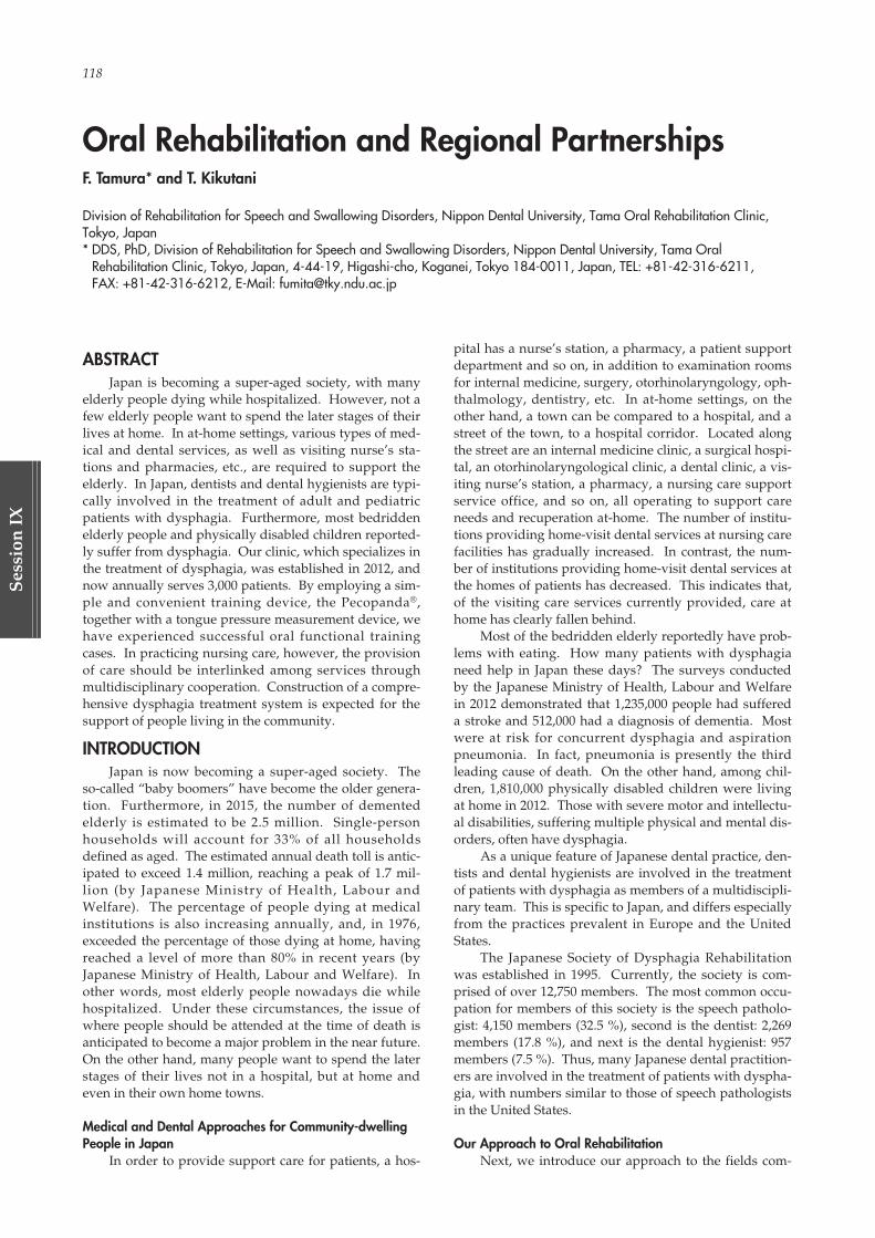
118
Ses
sion
IX
ABSTRACTJapan is becoming a super-aged society, with many
elderly people dying while hospitalized. However, not afew elderly people want to spend the later stages of theirlives at home. In at-home settings, various types of med-ical and dental services, as well as visiting nurse’s sta-tions and pharmacies, etc., are required to support theelderly. In Japan, dentists and dental hygienists are typi-cally involved in the treatment of adult and pediatricpatients with dysphagia. Furthermore, most bedriddenelderly people and physically disabled children reported-ly suffer from dysphagia. Our clinic, which specializes inthe treatment of dysphagia, was established in 2012, andnow annually serves 3,000 patients. By employing a sim-ple and convenient training device, the Pecopanda®,together with a tongue pressure measurement device, wehave experienced successful oral functional trainingcases. In practicing nursing care, however, the provisionof care should be interlinked among services throughmultidisciplinary cooperation. Construction of a compre-hensive dysphagia treatment system is expected for thesupport of people living in the community.
INTRODUCTIONJapan is now becoming a super-aged society. The
so-called “baby boomers” have become the older genera-tion. Furthermore, in 2015, the number of dementedelderly is estimated to be 2.5 million. Single-personhouseholds will account for 33% of all householdsdefined as aged. The estimated annual death toll is antic-ipated to exceed 1.4 million, reaching a peak of 1.7 mil-lion (by Japanese Ministry of Health, Labour andWelfare). The percentage of people dying at medicalinstitutions is also increasing annually, and, in 1976,exceeded the percentage of those dying at home, havingreached a level of more than 80% in recent years (byJapanese Ministry of Health, Labour and Welfare). Inother words, most elderly people nowadays die whilehospitalized. Under these circumstances, the issue ofwhere people should be attended at the time of death isanticipated to become a major problem in the near future.On the other hand, many people want to spend the laterstages of their lives not in a hospital, but at home andeven in their own home towns.
Medical and Dental Approaches for Community-dwellingPeople in Japan
In order to provide support care for patients, a hos-
pital has a nurse’s station, a pharmacy, a patient supportdepartment and so on, in addition to examination roomsfor internal medicine, surgery, otorhinolaryngology, oph-thalmology, dentistry, etc. In at-home settings, on theother hand, a town can be compared to a hospital, and astreet of the town, to a hospital corridor. Located alongthe street are an internal medicine clinic, a surgical hospi-tal, an otorhinolaryngological clinic, a dental clinic, a vis-iting nurse’s station, a pharmacy, a nursing care supportservice office, and so on, all operating to support careneeds and recuperation at-home. The number of institu-tions providing home-visit dental services at nursing carefacilities has gradually increased. In contrast, the num-ber of institutions providing home-visit dental services atthe homes of patients has decreased. This indicates that,of the visiting care services currently provided, care athome has clearly fallen behind.
Most of the bedridden elderly reportedly have prob-lems with eating. How many patients with dysphagianeed help in Japan these days? The surveys conductedby the Japanese Ministry of Health, Labour and Welfarein 2012 demonstrated that 1,235,000 people had suffereda stroke and 512,000 had a diagnosis of dementia. Mostwere at risk for concurrent dysphagia and aspirationpneumonia. In fact, pneumonia is presently the thirdleading cause of death. On the other hand, among chil-dren, 1,810,000 physically disabled children were livingat home in 2012. Those with severe motor and intellectu-al disabilities, suffering multiple physical and mental dis-orders, often have dysphagia.
As a unique feature of Japanese dental practice, den-tists and dental hygienists are involved in the treatmentof patients with dysphagia as members of a multidiscipli-nary team. This is specific to Japan, and differs especiallyfrom the practices prevalent in Europe and the UnitedStates.
The Japanese Society of Dysphagia Rehabilitationwas established in 1995. Currently, the society is com-prised of over 12,750 members. The most common occu-pation for members of this society is the speech patholo-gist: 4,150 members (32.5 %), second is the dentist: 2,269members (17.8 %), and next is the dental hygienist: 957members (7.5 %). Thus, many Japanese dental practition-ers are involved in the treatment of patients with dyspha-gia, with numbers similar to those of speech pathologistsin the United States.
Our Approach to Oral RehabilitationNext, we introduce our approach to the fields com-
Oral Rehabilitation and Regional PartnershipsF. Tamura* and T. Kikutani
Division of Rehabilitation for Speech and Swallowing Disorders, Nippon Dental University, Tama Oral Rehabilitation Clinic,Tokyo, Japan* DDS, PhD, Division of Rehabilitation for Speech and Swallowing Disorders, Nippon Dental University, Tama Oral
Rehabilitation Clinic, Tokyo, Japan, 4-44-19, Higashi-cho, Koganei, Tokyo 184-0011, Japan, TEL: +81-42-316-6211, FAX: +81-42-316-6212, E-Mail: [email protected]
02_本文1027(web用追加) 15.10.27 9:05 AM ページ 118
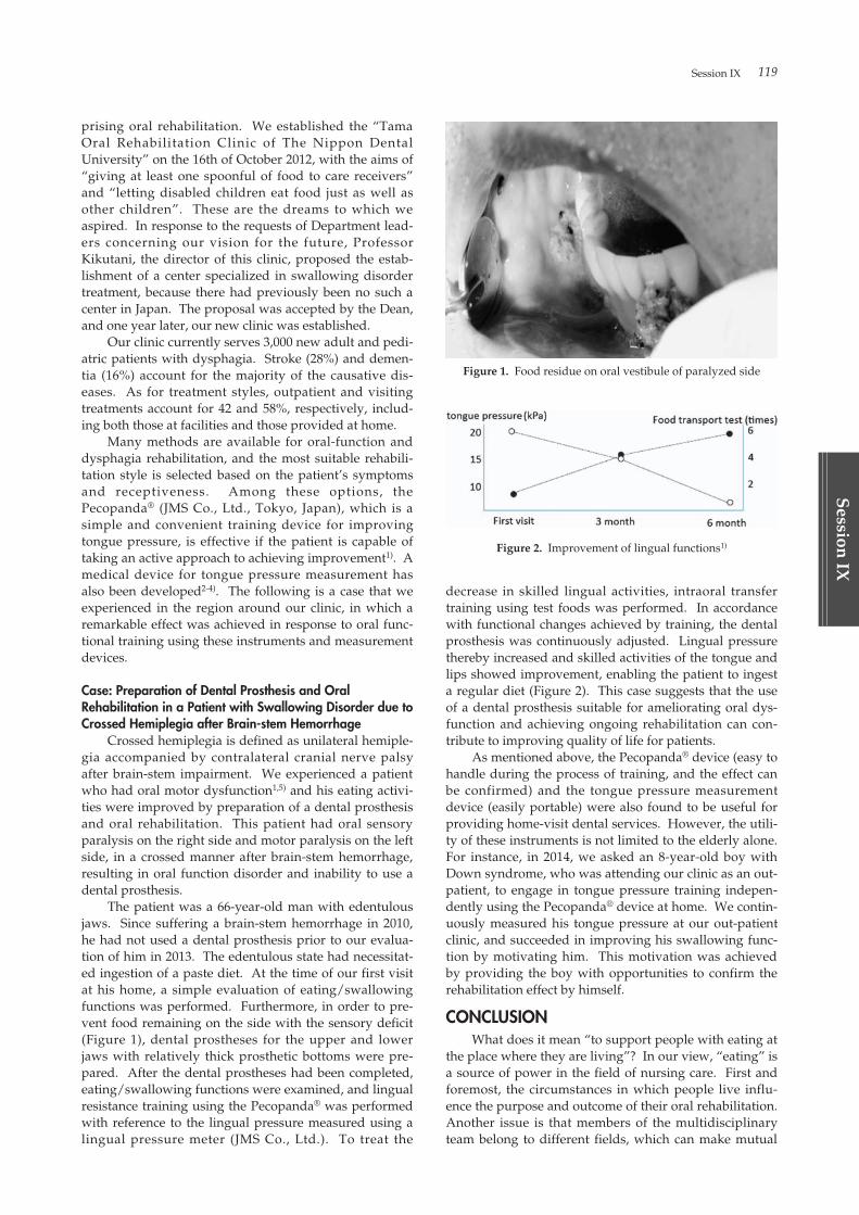
Session IX 119
Session
IX
prising oral rehabilitation. We established the “TamaOral Rehabilitation Clinic of The Nippon DentalUniversity” on the 16th of October 2012, with the aims of“giving at least one spoonful of food to care receivers”and “letting disabled children eat food just as well asother children”. These are the dreams to which weaspired. In response to the requests of Department lead-ers concerning our vision for the future, ProfessorKikutani, the director of this clinic, proposed the estab-lishment of a center specialized in swallowing disordertreatment, because there had previously been no such acenter in Japan. The proposal was accepted by the Dean,and one year later, our new clinic was established.
Our clinic currently serves 3,000 new adult and pedi-atric patients with dysphagia. Stroke (28%) and demen-tia (16%) account for the majority of the causative dis-eases. As for treatment styles, outpatient and visitingtreatments account for 42 and 58%, respectively, includ-ing both those at facilities and those provided at home.
Many methods are available for oral-function anddysphagia rehabilitation, and the most suitable rehabili-tation style is selected based on the patient’s symptomsand receptiveness. Among these options, thePecopanda® (JMS Co., Ltd., Tokyo, Japan), which is asimple and convenient training device for improvingtongue pressure, is effective if the patient is capable oftaking an active approach to achieving improvement1). Amedical device for tongue pressure measurement hasalso been developed2-4). The following is a case that weexperienced in the region around our clinic, in which aremarkable effect was achieved in response to oral func-tional training using these instruments and measurementdevices.
Case: Preparation of Dental Prosthesis and OralRehabilitation in a Patient with Swallowing Disorder due toCrossed Hemiplegia after Brain-stem Hemorrhage
Crossed hemiplegia is defined as unilateral hemiple-gia accompanied by contralateral cranial nerve palsyafter brain-stem impairment. We experienced a patientwho had oral motor dysfunction1,5) and his eating activi-ties were improved by preparation of a dental prosthesisand oral rehabilitation. This patient had oral sensoryparalysis on the right side and motor paralysis on the leftside, in a crossed manner after brain-stem hemorrhage,resulting in oral function disorder and inability to use adental prosthesis.
The patient was a 66-year-old man with edentulousjaws. Since suffering a brain-stem hemorrhage in 2010,he had not used a dental prosthesis prior to our evalua-tion of him in 2013. The edentulous state had necessitat-ed ingestion of a paste diet. At the time of our first visitat his home, a simple evaluation of eating/swallowingfunctions was performed. Furthermore, in order to pre-vent food remaining on the side with the sensory deficit(Figure 1), dental prostheses for the upper and lowerjaws with relatively thick prosthetic bottoms were pre-pared. After the dental prostheses had been completed,eating/swallowing functions were examined, and lingualresistance training using the Pecopanda® was performedwith reference to the lingual pressure measured using alingual pressure meter (JMS Co., Ltd.). To treat the
decrease in skilled lingual activities, intraoral transfertraining using test foods was performed. In accordancewith functional changes achieved by training, the dentalprosthesis was continuously adjusted. Lingual pressurethereby increased and skilled activities of the tongue andlips showed improvement, enabling the patient to ingesta regular diet (Figure 2). This case suggests that the useof a dental prosthesis suitable for ameliorating oral dys-function and achieving ongoing rehabilitation can con-tribute to improving quality of life for patients.
As mentioned above, the Pecopanda® device (easy tohandle during the process of training, and the effect canbe confirmed) and the tongue pressure measurementdevice (easily portable) were also found to be useful forproviding home-visit dental services. However, the utili-ty of these instruments is not limited to the elderly alone.For instance, in 2014, we asked an 8-year-old boy withDown syndrome, who was attending our clinic as an out-patient, to engage in tongue pressure training indepen-dently using the Pecopanda® device at home. We contin-uously measured his tongue pressure at our out-patientclinic, and succeeded in improving his swallowing func-tion by motivating him. This motivation was achievedby providing the boy with opportunities to confirm therehabilitation effect by himself.
CONCLUSIONWhat does it mean “to support people with eating at
the place where they are living”? In our view, “eating” isa source of power in the field of nursing care. First andforemost, the circumstances in which people live influ-ence the purpose and outcome of their oral rehabilitation.Another issue is that members of the multidisciplinaryteam belong to different fields, which can make mutual
Figure 1. Food residue on oral vestibule of paralyzed side
Figure 2. Improvement of lingual functions1)
02_本文1027(web用追加) 15.10.27 9:05 AM ページ 119
120
Ses
sion
IX
cooperation difficult. One of the approaches to solvingthese problems might be the establishment of a compre-hensive dysphagia treatment system in the communitywhere people are actually living. In practicing nursingcare, there should be interlinkage of all forms of care pro-vided, extending from the hospital to the home.Attention should be paid not to “what people can do (areable to eat)”, but to “how people are managing (are cop-ing adequately with eating/dietary needs)”. We believethat narrative-based nursing care is superior to evidence-based nursing care. This viewpoint may provide thefoundation for a positive oral rehabilitation concept inthe super-aged community.
ACKNOWLEDGEMENTThe part of this study was supported by Japan
Agency for Medical Research and development, AMED.We thank Dr. Hiroki Shindo for his fruitful collaboration.
REFERENCES1. Kikutani T. Dental Practice aimed at nutritional
improvement for the motor masticatory disturbance.Ann Jpn Prosthodont 2015: Soc 7: 102-105.
2. Hayashi R, Tsuga K, Yoshida M, Sato Y, Akagawa Y. Anovel handy probe for tongue pressure measurement.Int J Prothodont 2002: 15: 385-388.
3. Yoshida M, Kikutani T, Tsuga K, Utanohara Y,Hayashi R, Akagawa Y. Decreased tongue pressurereflects symptom of dysphagia. Dysphagia 2006: 21: 61-65.
4. Utanohara Y, Hayashi R, Yoshikawa M, Yoshida M,Tsuga K, Akagawa Y. Standard values of maximumtongue pressure taken using newly developed dispos-able tongue pressure measurement device. Dysphagia2008: 23: 286-290.
5. Kikutani T, Tamura F, Nishiwaki K, et al. Oral motorfunction in the masticatory performance in the commu-nity-dwelling elderly. Odontology 2009: 97: 38-42.
02_本文1027(web用追加) 15.10.27 9:05 AM ページ 120
Session IX 121
Session
IX
ABSTRACTThe aim of gerodontology is a “Recovery of eating
function for the eldery” ultimately. And this is possibleto restate “the dentistry for protection, maintaining andimprovement of the oral function in the elderly as to beable to do oral intake until their last days”. Aging ofsociety is a universal problem[1], and Japan is at the fore-front of fighting problems in super-aged society.Dysphagia caused by aging itself and general diseasesissue definitely, when you pursue the Gerodontology.Dentistry can make a contribution to it through the oralrehabilitation[2].
Enhanced support to the burgeoning dementia pop-ulation is also urgent issue in the world. It is predictedthat over 50% of them have dysphagia[3], and immediateresponse to their dysphagia problems will be needed.
The more number of the elderly with care needsincrease, the more number of the elderly who can’t intakeorally with deteriorated swallowing function increase. In
addition, the number of aspiration pneumonia patientswill definitely increase because of their poor oral intake.
I’d like to introduce the current reality in Japanesesuper-aged society and what we have to prepare for it inthe near future.
REFERENCES1) Suh GH, Shah A. A review of the epidemiological tran-
sition in dementia —cross-national comparisons of theindices related to Alzheimer’s disease and vasculardementia. Acta Psychiatr Scand 2001: 104: 4-11.
2) Yoshikawa M, Yoshida M, Nagasaki T, et al. Effects oftooth loss and denture wear on tongue-tip motion inelderly dentulous and edentulous people. J OralRehabil. 2008: 35 (12): 882-8.
3) Alagiakrishnan K, Bhanji RA, Kurian M. Evaluationand management of oropharyngeal dysphagia in dif-ferent types of dementia: a systematic review. ArchGerontol Geriatr. 2013: 56: 1-9.
What Can the Dentistry Do in Super-aged Society?M. Yoshikawa
DDS, PhD, Associated professor, Department of advanced prosthodontics, Graduate school of biomedical and health sciences,Hiroshima University, Kasumi 1-2-3, Minami-ku, Hiroshima City, Hiroshima, JAPAN, 734-8553, TEL: +81-82-257-5677, FAX: 81-82-257-5679, E-Mail: [email protected]
Key words: super-aged society, gerodontology, swallowing disorders, oral rehabilitation
02_本文1027(web用追加) 15.10.27 9:05 AM ページ 121
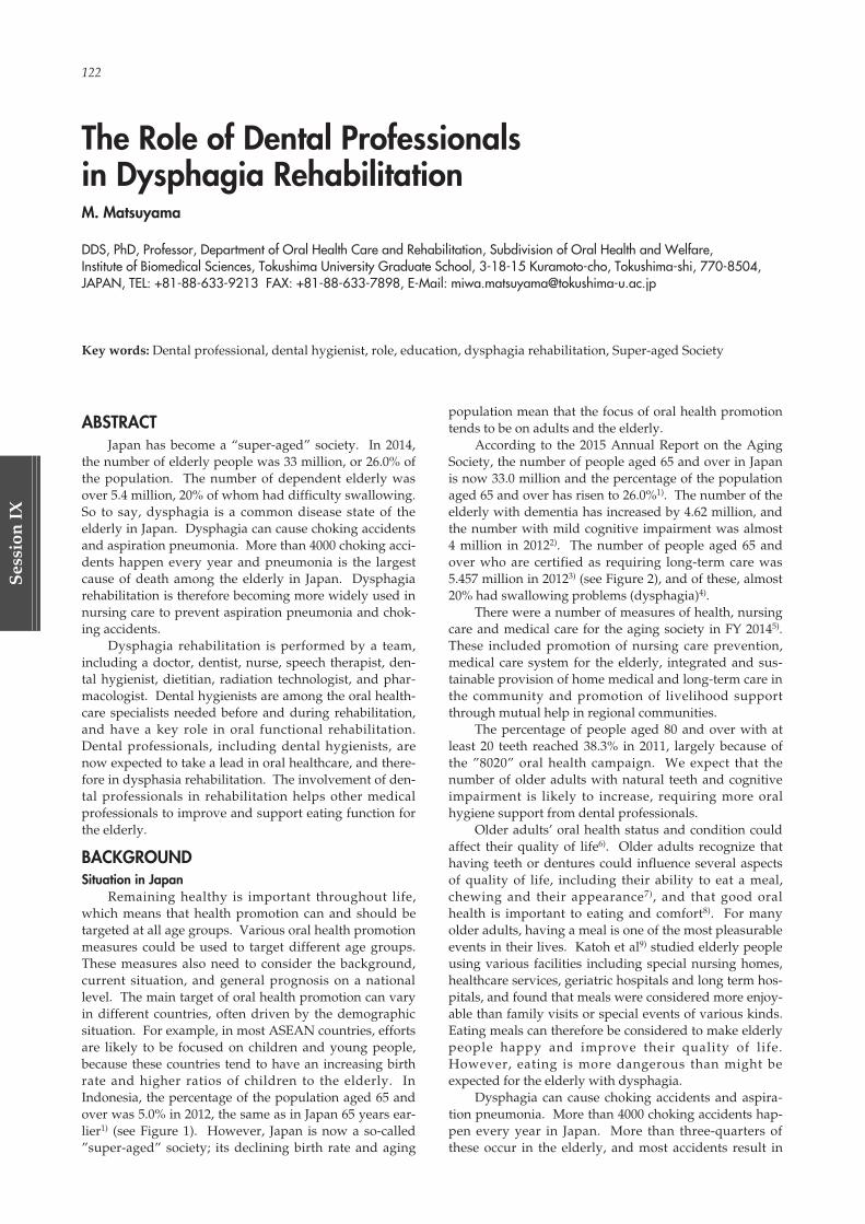
122
Ses
sion
IX
ABSTRACTJapan has become a “super-aged” society. In 2014,
the number of elderly people was 33 million, or 26.0% ofthe population. The number of dependent elderly wasover 5.4 million, 20% of whom had difficulty swallowing.So to say, dysphagia is a common disease state of theelderly in Japan. Dysphagia can cause choking accidentsand aspiration pneumonia. More than 4000 choking acci-dents happen every year and pneumonia is the largestcause of death among the elderly in Japan. Dysphagiarehabilitation is therefore becoming more widely used innursing care to prevent aspiration pneumonia and chok-ing accidents.
Dysphagia rehabilitation is performed by a team,including a doctor, dentist, nurse, speech therapist, den-tal hygienist, dietitian, radiation technologist, and phar-macologist. Dental hygienists are among the oral health-care specialists needed before and during rehabilitation,and have a key role in oral functional rehabilitation.Dental professionals, including dental hygienists, arenow expected to take a lead in oral healthcare, and there-fore in dysphasia rehabilitation. The involvement of den-tal professionals in rehabilitation helps other medicalprofessionals to improve and support eating function forthe elderly.
BACKGROUNDSituation in Japan
Remaining healthy is important throughout life,which means that health promotion can and should betargeted at all age groups. Various oral health promotionmeasures could be used to target different age groups.These measures also need to consider the background,current situation, and general prognosis on a nationallevel. The main target of oral health promotion can varyin different countries, often driven by the demographicsituation. For example, in most ASEAN countries, effortsare likely to be focused on children and young people,because these countries tend to have an increasing birthrate and higher ratios of children to the elderly. InIndonesia, the percentage of the population aged 65 andover was 5.0% in 2012, the same as in Japan 65 years ear-lier1) (see Figure 1). However, Japan is now a so-called”super-aged” society; its declining birth rate and aging
population mean that the focus of oral health promotiontends to be on adults and the elderly.
According to the 2015 Annual Report on the AgingSociety, the number of people aged 65 and over in Japanis now 33.0 million and the percentage of the populationaged 65 and over has risen to 26.0%1). The number of theelderly with dementia has increased by 4.62 million, andthe number with mild cognitive impairment was almost4 million in 20122). The number of people aged 65 andover who are certified as requiring long-term care was5.457 million in 20123) (see Figure 2), and of these, almost20% had swallowing problems (dysphagia)4).
There were a number of measures of health, nursingcare and medical care for the aging society in FY 20145).These included promotion of nursing care prevention,medical care system for the elderly, integrated and sus-tainable provision of home medical and long-term care inthe community and promotion of livelihood supportthrough mutual help in regional communities.
The percentage of people aged 80 and over with atleast 20 teeth reached 38.3% in 2011, largely because ofthe ”8020” oral health campaign. We expect that thenumber of older adults with natural teeth and cognitiveimpairment is likely to increase, requiring more oralhygiene support from dental professionals.
Older adults’ oral health status and condition couldaffect their quality of life6). Older adults recognize thathaving teeth or dentures could influence several aspectsof quality of life, including their ability to eat a meal,chewing and their appearance7), and that good oralhealth is important to eating and comfort8). For manyolder adults, having a meal is one of the most pleasurableevents in their lives. Katoh et al9) studied elderly peopleusing various facilities including special nursing homes,healthcare services, geriatric hospitals and long term hos-pitals, and found that meals were considered more enjoy-able than family visits or special events of various kinds.Eating meals can therefore be considered to make elderlypeople happy and improve their quality of life.However, eating is more dangerous than might beexpected for the elderly with dysphagia.
Dysphagia can cause choking accidents and aspira-tion pneumonia. More than 4000 choking accidents hap-pen every year in Japan. More than three-quarters ofthese occur in the elderly, and most accidents result in
The Role of Dental Professionals in Dysphagia RehabilitationM. Matsuyama
DDS, PhD, Professor, Department of Oral Health Care and Rehabilitation, Subdivision of Oral Health and Welfare, Institute of Biomedical Sciences, Tokushima University Graduate School, 3-18-15 Kuramoto-cho, Tokushima-shi, 770-8504, JAPAN, TEL: +81-88-633-9213 FAX: +81-88-633-7898, E-Mail: [email protected]
Key words: Dental professional, dental hygienist, role, education, dysphagia rehabilitation, Super-aged Society
02_本文1027(web用追加) 15.10.27 9:05 AM ページ 122
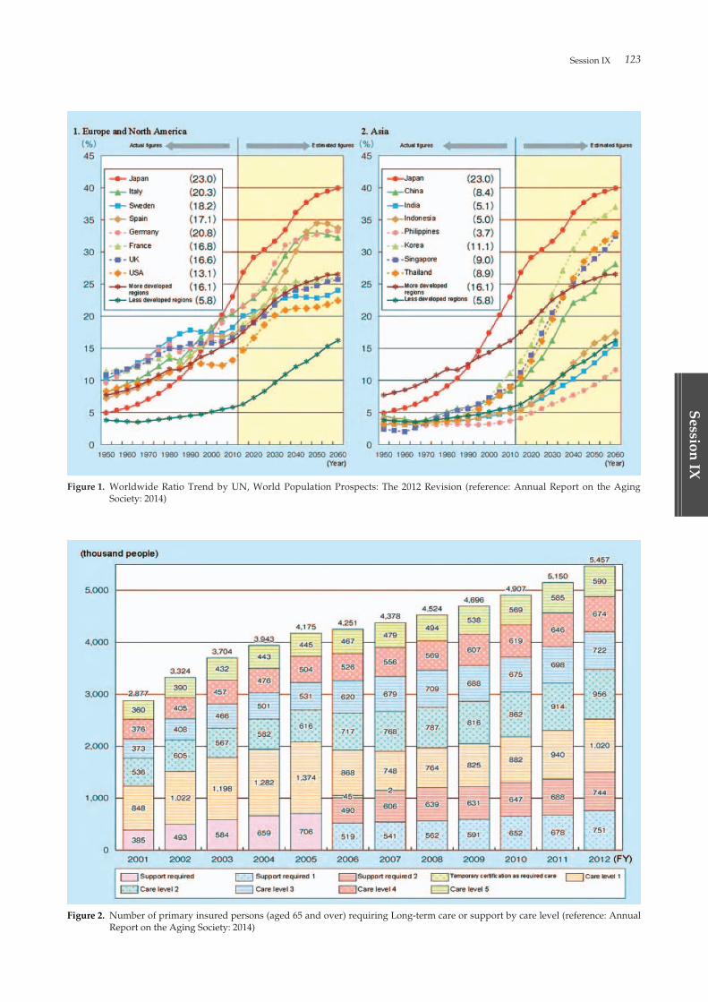
Session IX 123
Session
IX
Figure 2. Number of primary insured persons (aged 65 and over) requiring Long-term care or support by care level (reference: AnnualReport on the Aging Society: 2014)
Figure 1. Worldwide Ratio Trend by UN, World Population Prospects: The 2012 Revision (reference: Annual Report on the AgingSociety: 2014)
02_本文1027(web用追加) 15.10.27 9:05 AM ページ 123
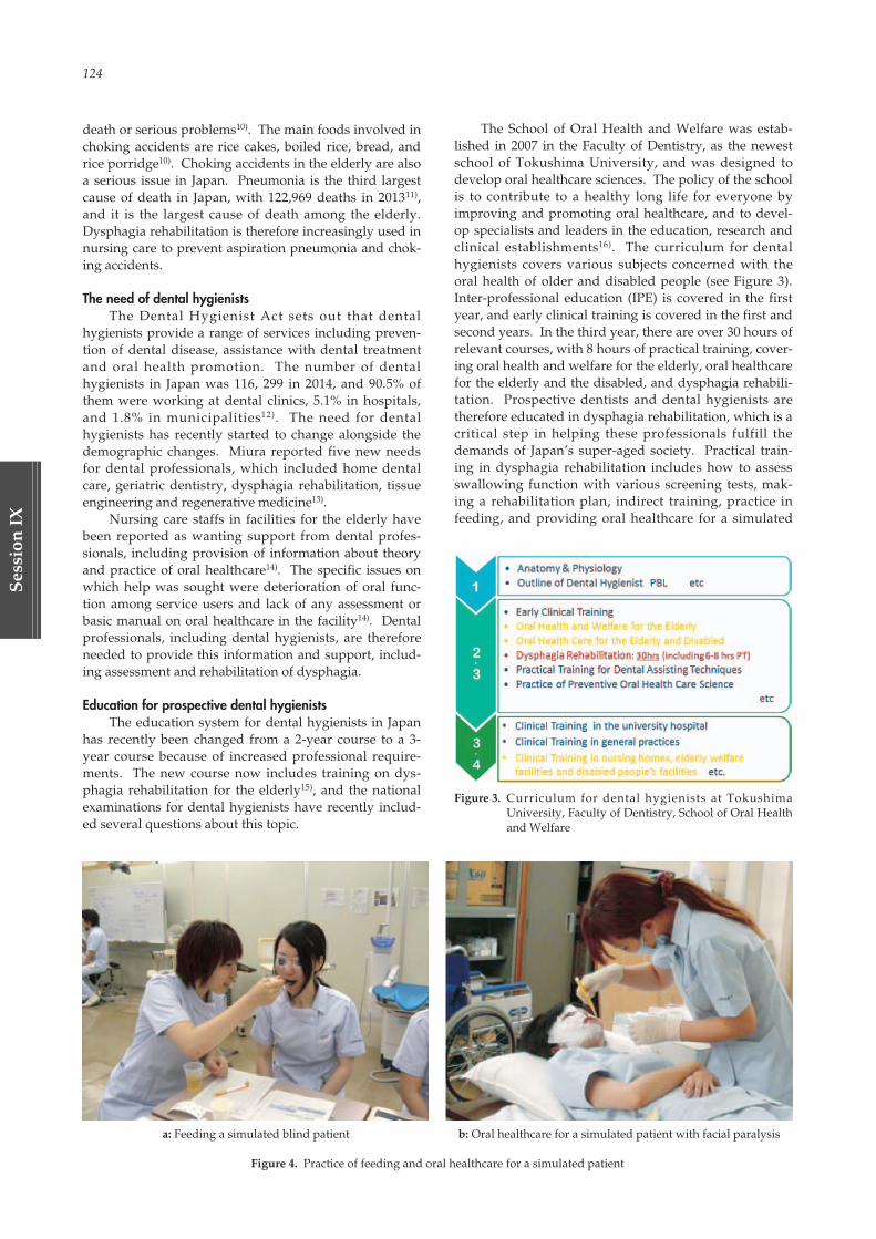
124
Ses
sion
IX
death or serious problems10). The main foods involved inchoking accidents are rice cakes, boiled rice, bread, andrice porridge10). Choking accidents in the elderly are alsoa serious issue in Japan. Pneumonia is the third largestcause of death in Japan, with 122,969 deaths in 201311),and it is the largest cause of death among the elderly.Dysphagia rehabilitation is therefore increasingly used innursing care to prevent aspiration pneumonia and chok-ing accidents.
The need of dental hygienistsThe Dental Hygienist Act sets out that dental
hygienists provide a range of services including preven-tion of dental disease, assistance with dental treatmentand oral health promotion. The number of dentalhygienists in Japan was 116, 299 in 2014, and 90.5% ofthem were working at dental clinics, 5.1% in hospitals,and 1.8% in municipalities12). The need for dentalhygienists has recently started to change alongside thedemographic changes. Miura reported five new needsfor dental professionals, which included home dentalcare, geriatric dentistry, dysphagia rehabilitation, tissueengineering and regenerative medicine13).
Nursing care staffs in facilities for the elderly havebeen reported as wanting support from dental profes-sionals, including provision of information about theoryand practice of oral healthcare14). The specific issues onwhich help was sought were deterioration of oral func-tion among service users and lack of any assessment orbasic manual on oral healthcare in the facility14). Dentalprofessionals, including dental hygienists, are thereforeneeded to provide this information and support, includ-ing assessment and rehabilitation of dysphagia.
Education for prospective dental hygienistsThe education system for dental hygienists in Japan
has recently been changed from a 2-year course to a 3-year course because of increased professional require-ments. The new course now includes training on dys-phagia rehabilitation for the elderly15), and the nationalexaminations for dental hygienists have recently includ-ed several questions about this topic.
The School of Oral Health and Welfare was estab-lished in 2007 in the Faculty of Dentistry, as the newestschool of Tokushima University, and was designed todevelop oral healthcare sciences. The policy of the schoolis to contribute to a healthy long life for everyone byimproving and promoting oral healthcare, and to devel-op specialists and leaders in the education, research andclinical establishments16). The curriculum for dentalhygienists covers various subjects concerned with theoral health of older and disabled people (see Figure 3).Inter-professional education (IPE) is covered in the firstyear, and early clinical training is covered in the first andsecond years. In the third year, there are over 30 hours ofrelevant courses, with 8 hours of practical training, cover-ing oral health and welfare for the elderly, oral healthcarefor the elderly and the disabled, and dysphagia rehabili-tation. Prospective dentists and dental hygienists aretherefore educated in dysphagia rehabilitation, which is acritical step in helping these professionals fulfill thedemands of Japan’s super-aged society. Practical train-ing in dysphagia rehabilitation includes how to assessswallowing function with various screening tests, mak-ing a rehabilitation plan, indirect training, practice infeeding, and providing oral healthcare for a simulated
Figure 3. Curriculum for dental hygienists at TokushimaUniversity, Faculty of Dentistry, School of Oral Healthand Welfare
a: Feeding a simulated blind patient b: Oral healthcare for a simulated patient with facial paralysis
Figure 4. Practice of feeding and oral healthcare for a simulated patient
02_本文1027(web用追加) 15.10.27 9:05 AM ページ 124
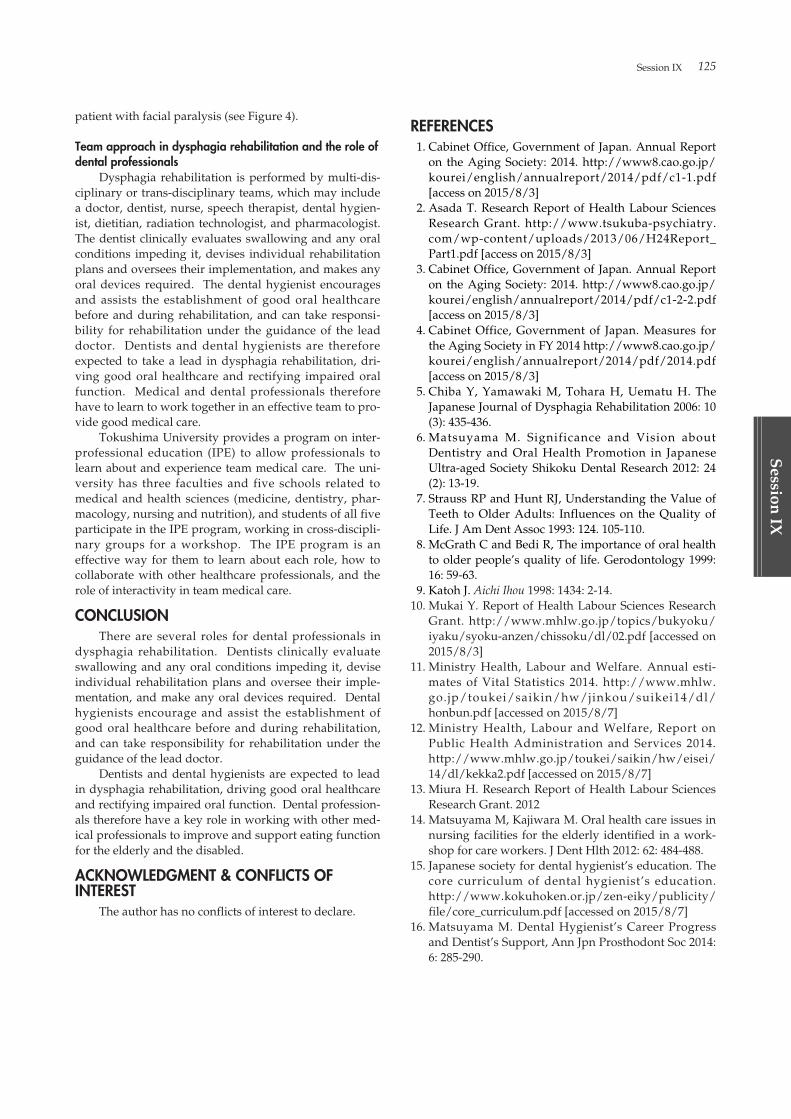
Session IX 125
Session
IX
patient with facial paralysis (see Figure 4).
Team approach in dysphagia rehabilitation and the role ofdental professionals
Dysphagia rehabilitation is performed by multi-dis-ciplinary or trans-disciplinary teams, which may includea doctor, dentist, nurse, speech therapist, dental hygien-ist, dietitian, radiation technologist, and pharmacologist.The dentist clinically evaluates swallowing and any oralconditions impeding it, devises individual rehabilitationplans and oversees their implementation, and makes anyoral devices required. The dental hygienist encouragesand assists the establishment of good oral healthcarebefore and during rehabilitation, and can take responsi-bility for rehabilitation under the guidance of the leaddoctor. Dentists and dental hygienists are thereforeexpected to take a lead in dysphagia rehabilitation, dri-ving good oral healthcare and rectifying impaired oralfunction. Medical and dental professionals thereforehave to learn to work together in an effective team to pro-vide good medical care.
Tokushima University provides a program on inter-professional education (IPE) to allow professionals tolearn about and experience team medical care. The uni-versity has three faculties and five schools related tomedical and health sciences (medicine, dentistry, phar-macology, nursing and nutrition), and students of all fiveparticipate in the IPE program, working in cross-discipli-nary groups for a workshop. The IPE program is aneffective way for them to learn about each role, how tocollaborate with other healthcare professionals, and therole of interactivity in team medical care.
CONCLUSIONThere are several roles for dental professionals in
dysphagia rehabilitation. Dentists clinically evaluateswallowing and any oral conditions impeding it, deviseindividual rehabilitation plans and oversee their imple-mentation, and make any oral devices required. Dentalhygienists encourage and assist the establishment ofgood oral healthcare before and during rehabilitation,and can take responsibility for rehabilitation under theguidance of the lead doctor.
Dentists and dental hygienists are expected to leadin dysphagia rehabilitation, driving good oral healthcareand rectifying impaired oral function. Dental profession-als therefore have a key role in working with other med-ical professionals to improve and support eating functionfor the elderly and the disabled.
ACKNOWLEDGMENT & CONFLICTS OFINTEREST
The author has no conflicts of interest to declare.
REFERENCES01. Cabinet Office, Government of Japan. Annual Report
on the Aging Society: 2014. http://www8.cao.go.jp/kourei/english/annualreport/2014/pdf/c1-1.pdf[access on 2015/8/3]
02. Asada T. Research Report of Health Labour SciencesResearch Grant. http://www.tsukuba-psychiatry.com/wp-content/uploads/2013/06/H24Report_Part1.pdf [access on 2015/8/3]
03. Cabinet Office, Government of Japan. Annual Reporton the Aging Society: 2014. http://www8.cao.go.jp/kourei/english/annualreport/2014/pdf/c1-2-2.pdf[access on 2015/8/3]
04. Cabinet Office, Government of Japan. Measures forthe Aging Society in FY 2014 http://www8.cao.go.jp/kourei/english/annualreport/2014/pdf/2014.pdf[access on 2015/8/3]
05. Chiba Y, Yamawaki M, Tohara H, Uematu H. TheJapanese Journal of Dysphagia Rehabilitation 2006: 10(3): 435-436.
06. Matsuyama M. Significance and Vision aboutDentistry and Oral Health Promotion in JapaneseUltra-aged Society Shikoku Dental Research 2012: 24(2): 13-19.
07. Strauss RP and Hunt RJ, Understanding the Value ofTeeth to Older Adults: Influences on the Quality ofLife. J Am Dent Assoc 1993: 124. 105-110.
08. McGrath C and Bedi R, The importance of oral healthto older people’s quality of life. Gerodontology 1999:16: 59-63.
09. Katoh J. Aichi Ihou 1998: 1434: 2-14.10. Mukai Y. Report of Health Labour Sciences Research
Grant. http://www.mhlw.go.jp/topics/bukyoku/iyaku/syoku-anzen/chissoku/dl/02.pdf [accessed on2015/8/3]
11. Ministry Health, Labour and Welfare. Annual esti-mates of Vital Statistics 2014. http://www.mhlw.go.jp/toukei/saikin/hw/jinkou/suikei14/dl/honbun.pdf [accessed on 2015/8/7]
12. Ministry Health, Labour and Welfare, Report onPublic Health Administration and Services 2014.http://www.mhlw.go.jp/toukei/saikin/hw/eisei/14/dl/kekka2.pdf [accessed on 2015/8/7]
13. Miura H. Research Report of Health Labour SciencesResearch Grant. 2012
14. Matsuyama M, Kajiwara M. Oral health care issues innursing facilities for the elderly identified in a work-shop for care workers. J Dent Hlth 2012: 62: 484-488.
15. Japanese society for dental hygienist’s education. Thecore curriculum of dental hygienist’s education.http://www.kokuhoken.or.jp/zen-eiky/publicity/file/core_curriculum.pdf [accessed on 2015/8/7]
16. Matsuyama M. Dental Hygienist’s Career Progressand Dentist’s Support, Ann Jpn Prosthodont Soc 2014:6: 285-290.
02_本文1027(web用追加) 15.10.27 9:05 AM ページ 125

02_本文1027(web用追加) 15.10.27 9:05 AM ページ 126
Session
X
Oral Cancer in AsiaGlobal Epidemiology of Oral Cancer: One Disease or Two??
University of Sheffield
P.M. Speight
Tobacco and Alcohol Misuse and Risk of Oral Cancer
Okazaki City Hospital
T. Nagao
Oral Cancer in Asia
University of Malaya
R.B. Zain
Areca/betel Quid and Oral Cancer Epidemiology in Asian Pacific Region
Kaohsiung Medical University
Y.H. Yang
Session X
02_本文1027(web用追加) 15.10.27 9:05 AM ページ 127

02_本文1027(web用追加) 15.10.27 9:05 AM ページ 128
Session X 129
Session
X
ABSTRACT“Oral cancer” is ill defined and cancer registries may
record cancers from many different sites in the head andneck under this single umbrella term. Despite a markedreduction in tobacco use in many parts of the world, oralcancer has continued to increase. Careful analysis of thedata however has shown that the increase is mainly seenin younger individuals and in oropharyngeal lesions.Much research has shown that these lesions are associat-ed with HPV infection and provides good evidence thatoral and oropharyngeal cancer are separate diseases withdistinctive clinicopathological and prognostic features. Itis now essential that clinicians, pathologists and epidemi-ologists are more precise in their definitions of these can-cers, since there is clear evidence that they may requiredifferent management strategies.
“Oral cancer” is ill-defined and definitions varyacross the world. Overall, most agencies use the WHOICD system (WHO, 2015) which groups malignant neo-plasms of the lip, oral cavity and pharynx together under
ICD C00 to C14 (Table 1). Cancer registries use thisscheme to code neoplasms for epidemiological purposes,but there is variance in which ICD numbers are included.For example Globocan 2002 defines “Oral Cavity Cancer”as C00 to C08, which includes neoplasm of the salivaryglands, but these are not normally regarded as “oral can-cer”. Cancer Research UK (CRUK, 2015) publish oralcancer statistics, but define oral cancer as C00-C06, C09-C10, and C12-C14, which excludes salivary glands, butincludes oropharynx and hypopharynx. Almost all sta-tistics include oral cavity and pharynx together as “oralcancer”, despite the fact that there is good evidence thatcancers at different sites around the upper aerodigestivetract have different causes and different biological behav-iours. Our current epidemiological data does not allowus to fully understand the full implications of these dif-ferences.
Taking a number of specific examples, we can showthat in the UK the prevalence of cigarette smoking gradu-ally declined (Hindle et al. 2000a) throughout the 20th
century, and now less than 20% of the population usecigarettes. Throughout this period, the incidence of lungcancer fell, but oral cancer continued to increase (Hindleet al. 2000a), leading to speculation that alcohol may playa major role (Hindle et al. 2000b). There is a similar situa-tion in Japan where there has been a gradual decrease incigarette smoking (Funatogawa et al. 2013) but with noreduction in the incidence of oral cancer. This paradoxcannot be easily explained by alcohol consumption alone.
A more careful analysis of the data has shown thatthe greatest increases in oral cancer incidence have beenamong younger individuals (Hindle et al. 1996) (Figure1), and in the oropharynx (Figure 2). In a Swedish popu-lation, Hammarstedt et al. (2006) showed a 280% increasein the incidence of tonsillar cancer between 1970 and2002. Over the same period, the increase in the propor-tion of cancers infected by HPV rose from 23% to 68%,indicating that HPV played a major role in the increasingincidence of oropharynx cancers. It is now well estab-lished that oropharyngeal cancer is associated with infec-tion by high risk human papilloma viruses (HPV)(Gillison et al. 2000; Gillison 2004). Most studies havebeen undertaken in Europe, USA and Australia, and havesuggested that the “HPV epidemic” is confined to west-ern countries. Chaturvedi et al. (2013) evaluated theglobal incidence patterns of oral and oropharyngeal can-
Global Epidemiology of Oral Cancer: One Disease or Two??P.M. Speight
Professor of Oral & Maxillofacial Pathology, School of Clinical Dentistry, University of Sheffield, Sheffield S10 2TA. UK, TEL: +44 (0) 114 271 7960, E-Mail: [email protected]
Key words: Oral cancer, oropharyngeal cancer, human papilloma virus (HPV), epidemiology, cancer statistics
Table 1. ICD (WHO) classification of malignant neoplasms ofthe lip, oral cavity and pharynx - C00-C14
C00 Malignant neoplasm of lipC01 Malignant neoplasm of base of tongueC02 Malignant neoplasm of other and unspecified parts of
tongueC03 Malignant neoplasm of gumC04 Malignant neoplasm of floor of mouthC05 Malignant neoplasm of palateC06 Malignant neoplasm of other and unspecified parts of
mouthC07 Malignant neoplasm of parotid glandC08 Malignant neoplasm of other and unspecified major
salivary glandsC09 Malignant neoplasm of tonsilC10 Malignant neoplasm of oropharynxC11 Malignant neoplasm of nasopharynxC12 Malignant neoplasm of piriform sinusC13 Malignant neoplasm of hypopharynxC14 Malignant neoplasm of other and ill-defined sites in the
lip, oral cavity and pharynx
(WHO 2015)
02_本文1027(web用追加) 15.10.27 9:05 AM ページ 129
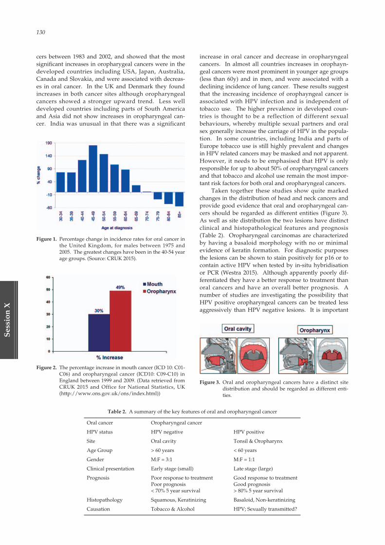
130
Ses
sion
X
cers between 1983 and 2002, and showed that the mostsignificant increases in oropharygeal cancers were in thedeveloped countries including USA, Japan, Australia,Canada and Slovakia, and were associated with decreas-es in oral cancer. In the UK and Denmark they foundincreases in both cancer sites although oropharyngealcancers showed a stronger upward trend. Less welldeveloped countries including parts of South Americaand Asia did not show increases in oropharyngeal can-cer. India was unusual in that there was a significant
increase in oral cancer and decrease in oropharyngealcancers. In almost all countries increases in orophayn-geal cancers were most prominent in younger age groups(less than 60y) and in men, and were associated with adeclining incidence of lung cancer. These results suggestthat the increasing incidence of orophayngeal cancer isassociated with HPV infection and is independent oftobacco use. The higher prevalence in developed coun-tries is thought to be a reflection of different sexualbehaviours, whereby multiple sexual partners and oralsex generally increase the carriage of HPV in the popula-tion. In some countries, including India and parts ofEurope tobacco use is still highly prevalent and changesin HPV related cancers may be masked and not apparent.However, it needs to be emphasised that HPV is onlyresponsible for up to about 50% of oropharyngeal cancersand that tobacco and alcohol use remain the most impor-tant risk factors for both oral and oropharyngeal cancers.
Taken together these studies show quite markedchanges in the distribution of head and neck cancers andprovide good evidence that oral and oropharyngeal can-cers should be regarded as different entities (Figure 3).As well as site distribution the two lesions have distinctclinical and histopathological features and prognosis(Table 2). Oropharyngeal carcinomas are characterizedby having a basaloid morphology with no or minimalevidence of keratin formation. For diagnostic purposesthe lesions can be shown to stain positively for p16 or tocontain active HPV when tested by in-situ hybridisationor PCR (Westra 2015). Although apparently poorly dif-ferentiated they have a better response to treatment thanoral cancers and have an overall better prognosis. Anumber of studies are investigating the possibility thatHPV positive oropharyngeal cancers can be treated lessaggressively than HPV negative lesions. It is important
Table 2. A summary of the key features of oral and oropharyngeal cancer
Oral cancer Oropharyngeal cancer
HPV status HPV negative HPV positive
Site Oral cavity Tonsil & Oropharynx
Age Group > 60 years < 60 years
Gender M:F = 3:1 M:F = 1:1
Clinical presentation Early stage (small) Late stage (large)
Prognosis Poor response to treatment Good response to treatmentPoor prognosis Good prognosis< 70% 5 year survival > 80% 5 year survival
Histopathology Squamous, Keratinizing Basaloid, Non-keratinizing
Causation Tobacco & Alcohol HPV; Sexually transmitted?
Figure 1. Percentage change in incidence rates for oral cancer inthe United Kingdom, for males between 1975 and2005. The greatest changes have been in the 40-54 yearage groups. (Source: CRUK 2015).
Figure 3. Oral and oropharyngeal cancers have a distinct sitedistribution and should be regarded as different enti-ties.
Figure 2. The percentage increase in mouth cancer (ICD 10: C01-C06) and oropharyngeal cancer (ICD10: C09-C10) inEngland between 1999 and 2009. (Data retrieved fromCRUK 2015 and Office for National Statistics, UK(http://www.ons.gov.uk/ons/index.html))
02_本文1027(web用追加) 15.10.27 9:05 AM ページ 130

Session X 131
Session
X
therefore that these lesions are identified as separate enti-ties and that cancer registries properly record the sitesand types of cancer being managed.
In summary, the definition of oral cancer is currentlytoo broad to allow accurate analysis of the different site-specific cancers that may arise in the head and neck. Thediscovery that oropharyngeal cancer is associated withHPV and that it has unique clinicopathological and prog-nostic characteristics, is good evidence that there are atleast two different disease entities in the head and neckwhich have previously been defined under the singleumbrella of “oral cancer”. Going forward it is nowessential that clinicians, pathologists and epidemiologistsare more precise in their definitions of these diseases,since there is clear evidence that they may require differ-ent management strategies. Other specific entitiesalready well defined in the head and neck includesnasopharyngeal and some paranasal undifferentiatedcarcinomas. Further studies may result in better defini-tions of cancers at other sites, for example mobile tongueand lip, and it may well be that these should also beregarded as unique clinicopathological entities. Suchresearch will only be possible if we adopt more accuraterecording and reporting of cancers by site of origin.
REFERENCESChaturvedi AK, Anderson WF, Lortet-Tieulent J, Curado
MP, Ferlay J, Franceschi S, Rosenberg PS, Bray F,Gillison ML. 2013. Worldwide trends in incidencerates for oral cavity and oropharyngeal cancers. JClin Oncol. 31(36): 4550-9.
CRUK, 2015. Oral Cancer Statistics: ht tp ://www.cancerresearchuk.org/heal th-professional/cancer-statistics/statistics-by-cancer-type/oral-cancer
Funatogawa I, Funatogawa T, Yano E. 2013. Trends insmoking and lung cancer mortality in Japan, by birth
cohort, 1949-2010. Bulletin of the World HealthOrganization. 91: 332-340.
Gillison ML, Koch WM, Capone RB, Spafford M, WestraWH, Wu L, Zahurak ML, Daniel RW, Viglione M,Symer DE, Shah KV, Sidransky D. 2000. Evidence fora causal association between human papillomavirusand a subset of head and neck cancers. J Natl CancerInst. 92 (9): 709-20.
Gillison ML. 2004. Human papillomavirus-associatedhead and neck cancer is a distinct epidemiologic,clinical, and molecular entity. Semin Oncol. 31 (6):744-54.
Globocan 2002. Global data on Incidence of oral cancer.http://www.depdb.iarc.fr/globocan2002.htm
Hammarstedt L, Lindquist D, Dahlstrand H, RomanitanM, Dahlgren LO, Joneberg J, Creson N, Lindholm J,Ye W, Dalianis T, Munck-Wikland E. 2006. Humanpapillomavirus as a risk factor for the increase inincidence of tonsillar cancer. Int J Cancer. 119 (11):2620-3.
Hindle I, Downer MC, Speight PM. 2000a. The associa-tion between intra-oral cancer and surrogate mark-ers of smoking and alcohol consumption.Community Dent Health. 17(2):107-13.
Hindle I, Downer MC, Moles DR, Speight PM. 2000b. Isalcohol responsible for more intra-oral cancer? OralOncol. 36 (4): 328-33.
Hindle I, Downer MC, Speight PM. 1996. The epidemiol-ogy of oral cancer. Br J Oral Maxillofac Surg. 34 (5):471-6.
Westra WH. 2015 The pathology of HPV-related headand neck cancer: Implications for the diagnosticpathologist. Semin Diagn Pathol. 32 (1): 42-53.
WHO 2015. International Statistical Classification ofDiseases and Related Health Problems 10th Revision(ICD-10). WHO 2015: http://www.who.int/classifications/icd/en/
02_本文1027(web用追加) 15.10.27 9:05 AM ページ 131
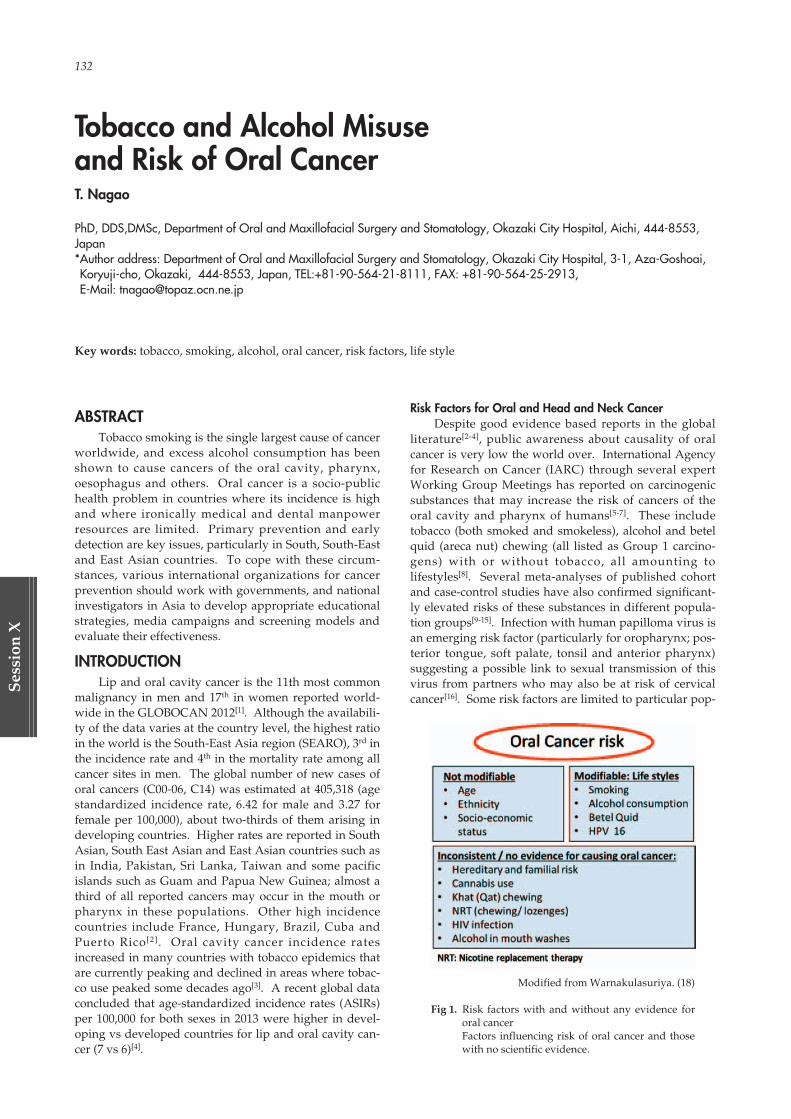
132
Ses
sion
X
ABSTRACTTobacco smoking is the single largest cause of cancer
worldwide, and excess alcohol consumption has beenshown to cause cancers of the oral cavity, pharynx,oesophagus and others. Oral cancer is a socio-publichealth problem in countries where its incidence is highand where ironically medical and dental manpowerresources are limited. Primary prevention and earlydetection are key issues, particularly in South, South-Eastand East Asian countries. To cope with these circum-stances, various international organizations for cancerprevention should work with governments, and nationalinvestigators in Asia to develop appropriate educationalstrategies, media campaigns and screening models andevaluate their effectiveness.
INTRODUCTIONLip and oral cavity cancer is the 11th most common
malignancy in men and 17th in women reported world-wide in the GLOBOCAN 2012[1]. Although the availabili-ty of the data varies at the country level, the highest ratioin the world is the South-East Asia region (SEARO), 3rd inthe incidence rate and 4th in the mortality rate among allcancer sites in men. The global number of new cases oforal cancers (C00-06, C14) was estimated at 405,318 (agestandardized incidence rate, 6.42 for male and 3.27 forfemale per 100,000), about two-thirds of them arising indeveloping countries. Higher rates are reported in SouthAsian, South East Asian and East Asian countries such asin India, Pakistan, Sri Lanka, Taiwan and some pacificislands such as Guam and Papua New Guinea; almost athird of all reported cancers may occur in the mouth orpharynx in these populations. Other high incidencecountries include France, Hungary, Brazil, Cuba andPuerto Rico[2]. Oral cavity cancer incidence ratesincreased in many countries with tobacco epidemics thatare currently peaking and declined in areas where tobac-co use peaked some decades ago[3]. A recent global dataconcluded that age-standardized incidence rates (ASIRs)per 100,000 for both sexes in 2013 were higher in devel-oping vs developed countries for lip and oral cavity can-cer (7 vs 6)[4].
Risk Factors for Oral and Head and Neck CancerDespite good evidence based reports in the global
literature[2-4], public awareness about causality of oralcancer is very low the world over. International Agencyfor Research on Cancer (IARC) through several expertWorking Group Meetings has reported on carcinogenicsubstances that may increase the risk of cancers of theoral cavity and pharynx of humans[5-7]. These includetobacco (both smoked and smokeless), alcohol and betelquid (areca nut) chewing (all listed as Group 1 carcino-gens) with or without tobacco, all amounting tolifestyles[8]. Several meta-analyses of published cohortand case-control studies have also confirmed significant-ly elevated risks of these substances in different popula-tion groups[9-15]. Infection with human papilloma virus isan emerging risk factor (particularly for oropharynx; pos-terior tongue, soft palate, tonsil and anterior pharynx)suggesting a possible link to sexual transmission of thisvirus from partners who may also be at risk of cervicalcancer[16]. Some risk factors are limited to particular pop-
Tobacco and Alcohol Misuse and Risk of Oral CancerT. Nagao
PhD, DDS,DMSc, Department of Oral and Maxillofacial Surgery and Stomatology, Okazaki City Hospital, Aichi, 444-8553,Japan*Author address: Department of Oral and Maxillofacial Surgery and Stomatology, Okazaki City Hospital, 3-1, Aza-Goshoai,Koryuji-cho, Okazaki, 444-8553, Japan, TEL:+81-90-564-21-8111, FAX: +81-90-564-25-2913, E-Mail: [email protected]
Key words: tobacco, smoking, alcohol, oral cancer, risk factors, life style
Modified from Warnakulasuriya. (18)
Fig 1. Risk factors with and without any evidence fororal cancerFactors influencing risk of oral cancer and thosewith no scientific evidence.
02_本文1027(web用追加) 15.10.27 9:05 AM ページ 132
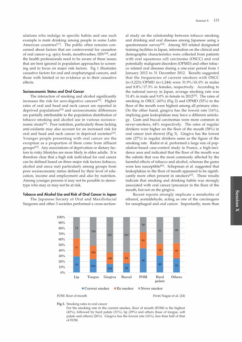
Session X 133
Session
X
ulations who indulge in specific habits and one suchexample is matè drinking among people in some LatinAmerican countries[17]. The public often remains con-cerned about factors that are controversial for causationof oral cancer e.g. spicy foods, mouthwashes, HIV[18], andthe health professionals need to be aware of these issuesthat are best ignored in population approaches to screen-ing and to focus on major risk factors. Fig 1 illustratescausative factors for oral and oropharyngeal cancers, andthose with limited or no evidence as to their causativeeffects.
Socioeconomic Status and Oral CancerThe interaction of smoking and alcohol significantly
increases the risk for aero-digestive cancers[19]. Higherrates of oral and head and neck cancer are reported indeprived populations[20] and socioeconomic differencesare partially attributable to the population distribution oftobacco smoking and alcohol use in various socioeco-nomic strata[21]. Poor nutrition, particularly those lackinganti-oxidants may also account for an increased risk fororal and head and neck cancer in deprived societies[22].Younger people presenting with oral cancer are theexception as a proportion of them come from affluentgroups[23]. Any associations of deprivation or dietary fac-tors to risky lifestyles are more likely in older adults. It istherefore clear that a high risk individual for oral cancercan be defined based on three major risk factors (tobacco,alcohol and areca nut) particularly among groups frompoor socioeconomic status defined by their level of edu-cation, income and employment and also by nutrition.Among younger persons it may not be possible to stereo-type who may or may not be at risk.
Tobacco and Alcohol Use and Risk of Oral Cancer in JapanThe Japanese Society of Oral and Maxillofacial
Surgeons and other 3 societies performed a cross-section-
al study on the relationship between tobacco smokingand drinking and oral diseases among Japanese using aquestionnaire survey[24]. Among 503 related designatedtraining facilities in Japan, information on the clinical anddemographic characteristics were collected from patientswith oral squamous cell carcinoma (OSCC) and oralpotentially malignant disorders (OPMD) and other tobac-co related oral diseases during a one-year period from 1January 2012 to 31 December 2012. Results suggestedthat the frequencies of current smokers with OSCC(n=3,223)/OPMD (n=1,244) were 31.9%/41.0% in malesand 8.8%/17.3% in females, respectively. According tothe national survey in Japan, average smoking rate was31.4% in male and 9.0% in female in 2012[25]. The rates ofsmoking in OSCC (43%) (Fig 2) and OPMD (52%) in thefloor of the mouth were highest among all primary sites.On the other hand, gingiva has the lowest rate (16%),implying gum leukoplakias may have a different aetiolo-gy. Gum and buccal carcinomas were more common innever-smokers, 64% respectively. The rates of regulardrinkers were higher on the floor of the mouth (58%) inoral cancer (not shown) (Fig 3). Gingiva has the lowestrate (27%) in regular drinkers same as the figure of thesmoking rate. Radoï et al. performed a large size of pop-ulation-based case-control study in France, a high-inci-dence area and indicated that the floor of the mouth wasthe subsite that was the most commonly affected by theharmful effects of tobacco and alcohol, whereas the gumswere less susceptible[26]. Schepman et al. suggested thatleukoplakias in the floor of mouth appeared to be signifi-cantly more often present in smokers[27]. These resultsindicate that smoking and drinking habits was stronglyassociated with oral cancer/precancer in the floor of themouth, but not on the gingiva.
Recent reports strongly implicate a metabolite ofethanol, acetaldehyde, acting as one of the carcinogensfor oesophageal and oral cancer. Importantly, more than
FOM: floor of mouth From Nagao et al. (24)
Fig 2. Smoking rates in oral cancerFor the smoking rate in the current smokes, floor of mouth (FOM) is the highest(43%), followed by hard palate (31%), lip (29%) and others (base of tongue, softpalate and others) (26%). Gingiva has the lowest rate (16%), less than half of thatof FOM.
02_本文1027(web用追加) 15.10.27 9:05 AM ページ 133
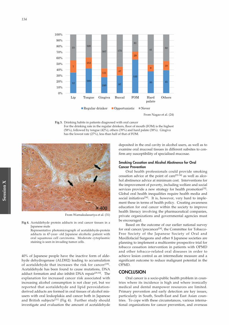
134
Ses
sion
X
40% of Japanese people have the inactive form of alde-hyde dehydrogenase (ALDH2) leading to accumulationof acetaldehyde that increases the risk for cancer[28].Acetaldehyde has been found to cause mutations, DNAadduct formation and also inhibit DNA repair[29,30]. Theexplanation for increased cancer risk associated withincreasing alcohol consumption is not clear yet, but wereported that acetaldehyde and lipid peroxidation-derived adducts are formed in oral tissues of alcohol mis-users with oral leukoplakia and cancer both in Japaneseand British subjects[31] (Fig 4). Further study shouldinvestigate and evaluation the amount of acetaldehyde
deposited in the oral cavity in alcohol users, as well as toexamine oral mucosal tissues in different subsites to con-firm any susceptibility of specialised mucosae.
Smoking Cessation and Alcohol Abstinence for OralCancer Prevention
Oral health professionals could provide smokingcessation advice at the point of care[32-34] as well as alco-hol abstinence advice at minimum cost. Interventions forthe improvement of poverty, including welfare and socialservices provide a new strategy for health promotion[35].Global oral health inequalities require health media andsocial initiatives[36]. It is, however, very hard to imple-ment these in terms of health policy. Creating awarenesseducation for oral cancer within the society to improvehealth literacy involving the pharmaceutical companies,private organizations and governmental agencies mustbe encouraged.
Based on the outcome of our earlier national surveyfor oral cancer/precancer[24], the Committee for Tobacco-Free Society of the Japanese Society of Oral andMaxillofacial Surgeons and other 8 Japanese societies areplanning to implement a multicentre prospective trial fortobacco cessation intervention in patients with OPMDand other tobacco-related oral diseases in order toachieve lesion control as an intermediate measure and asignificant outcome to reduce malignant potential in theOPMD.
CONCLUSIONOral cancer is a socio-public health problem in coun-
tries where its incidence is high and where ironicallymedical and dental manpower resources are limited.Primary prevention and early detection are key issues,particularly in South, South-East and East Asian coun-tries. To cope with these circumstances, various interna-tional organizations for cancer prevention, and overseas
From Nagao et al. (24)
Fig 3. Drinking habits in patients diagnosed with oral cancerFor the drinking rate in the regular drinkers, floor of mouth (FOM) is the highest(58%), followed by tongue (42%), others (39%) and hard palate (38%). Gingivahas the lowest rate (27%), less than half of that of FOM.
From Warnakulasuriya et al. (31)
Fig 4. Acetaldehyde protein adducts in oral cancer tissues in aJapanese maleRepresentative photomicrograph of acetaldehyde-proteinadducts in 47-year- old Japanese alcoholic patient withoral squamous cell carcinoma. Moderate cytoplasmicstaining is seen in invading tumor cells.
02_本文1027(web用追加) 15.10.27 9:05 AM ページ 134
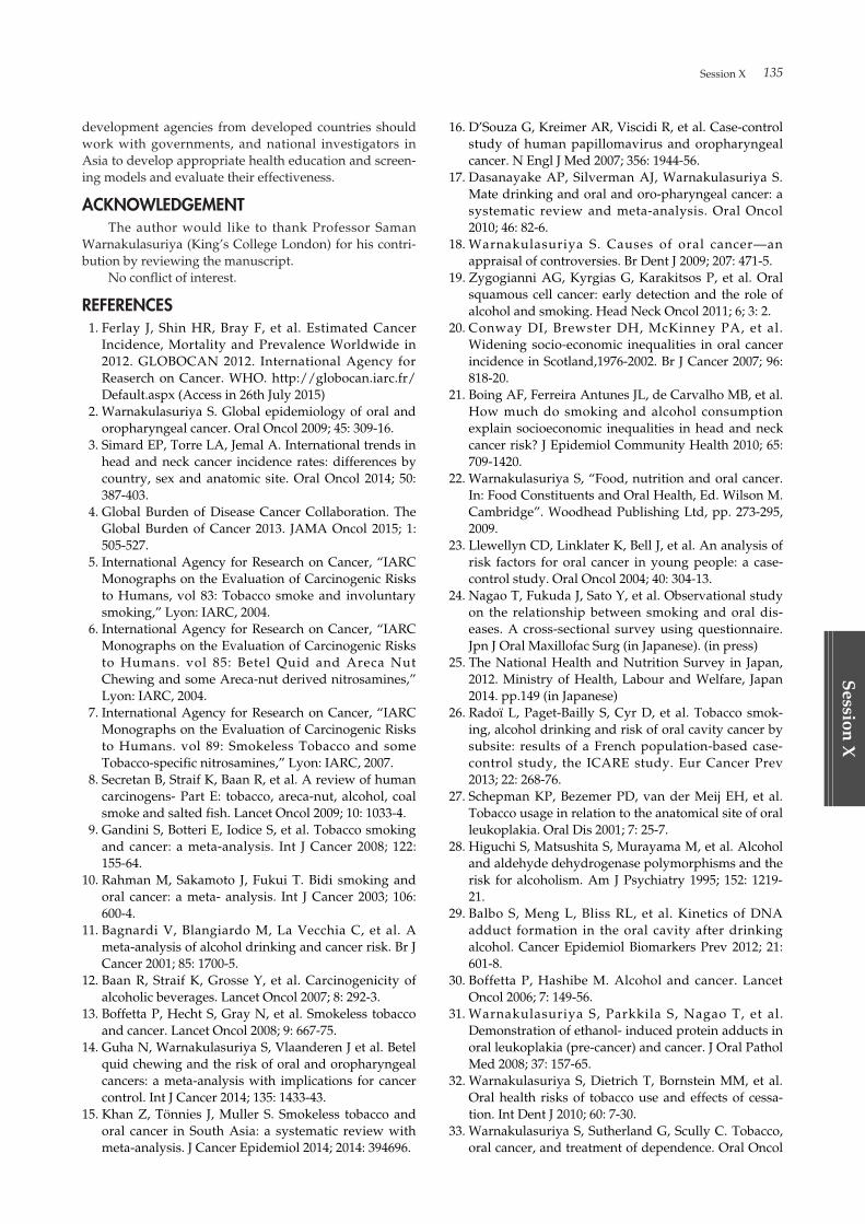
Session X 135
Session
X
development agencies from developed countries shouldwork with governments, and national investigators inAsia to develop appropriate health education and screen-ing models and evaluate their effectiveness.
ACKNOWLEDGEMENTThe author would like to thank Professor Saman
Warnakulasuriya (King’s College London) for his contri-bution by reviewing the manuscript.
No conflict of interest.
REFERENCES01. Ferlay J, Shin HR, Bray F, et al. Estimated Cancer
Incidence, Mortality and Prevalence Worldwide in2012. GLOBOCAN 2012. International Agency forReaserch on Cancer. WHO. http://globocan.iarc.fr/Default.aspx (Access in 26th July 2015)
02. Warnakulasuriya S. Global epidemiology of oral andoropharyngeal cancer. Oral Oncol 2009; 45: 309-16.
03. Simard EP, Torre LA, Jemal A. International trends inhead and neck cancer incidence rates: differences bycountry, sex and anatomic site. Oral Oncol 2014; 50:387-403.
04. Global Burden of Disease Cancer Collaboration. TheGlobal Burden of Cancer 2013. JAMA Oncol 2015; 1:505-527.
05. International Agency for Research on Cancer, “IARCMonographs on the Evaluation of Carcinogenic Risksto Humans, vol 83: Tobacco smoke and involuntarysmoking,” Lyon: IARC, 2004.
06. International Agency for Research on Cancer, “IARCMonographs on the Evaluation of Carcinogenic Risksto Humans. vol 85: Betel Quid and Areca NutChewing and some Areca-nut derived nitrosamines,”Lyon: IARC, 2004.
07. International Agency for Research on Cancer, “IARCMonographs on the Evaluation of Carcinogenic Risksto Humans. vol 89: Smokeless Tobacco and someTobacco-specific nitrosamines,” Lyon: IARC, 2007.
08. Secretan B, Straif K, Baan R, et al. A review of humancarcinogens- Part E: tobacco, areca-nut, alcohol, coalsmoke and salted fish. Lancet Oncol 2009; 10: 1033-4.
09. Gandini S, Botteri E, Iodice S, et al. Tobacco smokingand cancer: a meta-analysis. Int J Cancer 2008; 122:155-64.
10. Rahman M, Sakamoto J, Fukui T. Bidi smoking andoral cancer: a meta- analysis. Int J Cancer 2003; 106:600-4.
11. Bagnardi V, Blangiardo M, La Vecchia C, et al. Ameta-analysis of alcohol drinking and cancer risk. Br JCancer 2001; 85: 1700-5.
12. Baan R, Straif K, Grosse Y, et al. Carcinogenicity ofalcoholic beverages. Lancet Oncol 2007; 8: 292-3.
13. Boffetta P, Hecht S, Gray N, et al. Smokeless tobaccoand cancer. Lancet Oncol 2008; 9: 667-75.
14. Guha N, Warnakulasuriya S, Vlaanderen J et al. Betelquid chewing and the risk of oral and oropharyngealcancers: a meta-analysis with implications for cancercontrol. Int J Cancer 2014; 135: 1433-43.
15. Khan Z, Tönnies J, Muller S. Smokeless tobacco andoral cancer in South Asia: a systematic review withmeta-analysis. J Cancer Epidemiol 2014; 2014: 394696.
16. D’Souza G, Kreimer AR, Viscidi R, et al. Case-controlstudy of human papillomavirus and oropharyngealcancer. N Engl J Med 2007; 356: 1944-56.
17. Dasanayake AP, Silverman AJ, Warnakulasuriya S.Mate drinking and oral and oro-pharyngeal cancer: asystematic review and meta-analysis. Oral Oncol2010; 46: 82-6.
18. Warnakulasuriya S. Causes of oral cancer—anappraisal of controversies. Br Dent J 2009; 207: 471-5.
19. Zygogianni AG, Kyrgias G, Karakitsos P, et al. Oralsquamous cell cancer: early detection and the role ofalcohol and smoking. Head Neck Oncol 2011; 6; 3: 2.
20. Conway DI, Brewster DH, McKinney PA, et al.Widening socio-economic inequalities in oral cancerincidence in Scotland,1976-2002. Br J Cancer 2007; 96:818-20.
21. Boing AF, Ferreira Antunes JL, de Carvalho MB, et al.How much do smoking and alcohol consumptionexplain socioeconomic inequalities in head and neckcancer risk? J Epidemiol Community Health 2010; 65:709-1420.
22. Warnakulasuriya S, “Food, nutrition and oral cancer.In: Food Constituents and Oral Health, Ed. Wilson M.Cambridge”. Woodhead Publishing Ltd, pp. 273-295,2009.
23. Llewellyn CD, Linklater K, Bell J, et al. An analysis ofrisk factors for oral cancer in young people: a case-control study. Oral Oncol 2004; 40: 304-13.
24. Nagao T, Fukuda J, Sato Y, et al. Observational studyon the relationship between smoking and oral dis-eases. A cross-sectional survey using questionnaire.Jpn J Oral Maxillofac Surg (in Japanese). (in press)
25. The National Health and Nutrition Survey in Japan,2012. Ministry of Health, Labour and Welfare, Japan2014. pp.149 (in Japanese)
26. Radoï L, Paget-Bailly S, Cyr D, et al. Tobacco smok-ing, alcohol drinking and risk of oral cavity cancer bysubsite: results of a French population-based case-control study, the ICARE study. Eur Cancer Prev2013; 22: 268-76.
27. Schepman KP, Bezemer PD, van der Meij EH, et al.Tobacco usage in relation to the anatomical site of oralleukoplakia. Oral Dis 2001; 7: 25-7.
28. Higuchi S, Matsushita S, Murayama M, et al. Alcoholand aldehyde dehydrogenase polymorphisms and therisk for alcoholism. Am J Psychiatry 1995; 152: 1219-21.
29. Balbo S, Meng L, Bliss RL, et al. Kinetics of DNAadduct formation in the oral cavity after drinkingalcohol. Cancer Epidemiol Biomarkers Prev 2012; 21:601-8.
30. Boffetta P, Hashibe M. Alcohol and cancer. LancetOncol 2006; 7: 149-56.
31. Warnakulasuriya S, Parkkila S, Nagao T, et al.Demonstration of ethanol- induced protein adducts inoral leukoplakia (pre-cancer) and cancer. J Oral PatholMed 2008; 37: 157-65.
32. Warnakulasuriya S, Dietrich T, Bornstein MM, et al.Oral health risks of tobacco use and effects of cessa-tion. Int Dent J 2010; 60: 7-30.
33. Warnakulasuriya S, Sutherland G, Scully C. Tobacco,oral cancer, and treatment of dependence. Oral Oncol
02_本文1027(web用追加) 15.10.27 9:05 AM ページ 135

136
Ses
sion
X
2005; 41: 244-60.34. Williams M, Scott S. Is there scope for providing oral
cancer health advice in dental access centres? Br DentJ 2008; 205: E16; discussion 452-3.
35. Marmot M. “Fair society, healthy lives,” The Marmotreview Executive Summary. London: The Marmot
Review, 2010.36. Johnson NW, Warnakulasuriya S, Gupta PC et al.
Global oral health inequalities in incidence and out-comes for oral cancer: causes and solutions. Adv DentRes. 2011; 23: 237-46.
02_本文1027(web用追加) 15.10.27 9:05 AM ページ 136
Session X 137
Session
X
ABSTRACTThe epidemiology of Oral Cancer in Asia is unique
and varies from country to country. This paper aims topresent a brief overview of oral cancer status andresearch activities in Asia. The mixture of low, moderateand high-income Asian countries lead to different needsof each country. Research and programmes for oral can-cer in these countries would be in tandem with the coun-tries needs and capabilities ranging from collecting base-line prevalence data to those using advanced technolo-gies towards the development and validation of the bio-markers.
INTRODUCTIONThe prevalence of oral cancer is unique in each coun-
try in Asia due to the variation in lifestyles and habitualrisk factors. The differences in research results maybe atrue difference or maybe due to the differences in diagno-sis and site of the lesions. Research in oral cancerincludes epidemiological research into obtaining baselinedata and risk factors of oral cancer for the country. Earlydetection of oral cancer includes determining level ofawareness of oral cancer and creating awareness amongthe population in particular, for the risk population, con-ducting screening using the clinical oral examination(COE) as the main screening tool (Kao et al., 2015).
Biomarkers may represent as a type of adjunct toscreening and its development may assist in the predic-tion of certain demographic or clinico-pathologic para-meters for example lymph node metastasis while, somemay be potential prognostic biomarkers and for thedevelopment of therapeutics.
Research activities differ from country to country inAsia dependent on the country’s capabilities in terms offunding and research infrastructure. Collaborativeresearch encompasses the research materials and/humanresource crossing borders in the move towards achievingone goal of reducing the burden of oral cancer in theregion. However, despite all these efforts, the survivalrate of patients with oral cancer remained dismal hover-ing around 50% or lower over the last 4 decades. Thelow survival rate has been attributed to the high preva-lence of late stage cancers of over 60% in many Asiancountries. Thus for some countries in Asia, the reasonsfor late stage cancers includes a delay in diagnosis. Manyof these countries needed to have concerted efforts onearly detection of oral cancer.
For sometime, India has been cited the highest inci-dence of oral cancer in the world. However, recentreports showed that Sri Lanka and Pakistan are nowranked at the top. More recent records had identified
South Karachi in Pakistan as having the highest rates(Warnakulasuriya, 2009). Many of the oral cancerpatients in the Southeast Asian region are betel-quid orareca nut chewers. Other risk factors other than betel-quid, which are associated with oral cancer are tobaccosmoking, alcohol drinking with HPV as a debatable riskfactor. Some demographic and clinicopathologic para-meters have also been shown to be prognostic and/orrisk indicators (Krishna et al., 2014). Additionally, anAsian Betel Quid Consortium (ABC) study found that thebetel quid chewing rates among men (10.7-43.6%) weresignificantly higher than women (1.8-34.9%) in Taiwan,Mainland China, Nepal and Sri Lanka. However, thewomen’s rates (29.5-46.8%) were higher than that for men(9.8-12.0%) in Malaysia and Indonesia. This paper aimsto give a brief overview of oral cancer status in Asia.
Early Detection of Oral CancerAmong the reasons for delayed presentation of oral
cancers were inadequate knowledge and skills of dentaland other health professionals in recognizing early can-cer or its precursors. Patients’ delay will also lead tothem presenting with late stage oral cancer. A study ofdelayed presentation of oral cancer showed that patientsthought that their symptoms are trivial and that thelesion would go away (Rogers et al., 2011). Three studiesin India have also shown that while the public are awareof oral cancer and its risk factors, their knowledge aboutearly signs of oral cancer is low and is significantly relat-ed to level of education (Elango et al, 2009, Devadiga andPrasad, 2010, Reddy et al., 2012). Elango et al, 2009 con-ducted a questionnaire survey in a semi-urban area inKerala where the final participants was 845 participants.Devadiga and Prasad, 2010, and Reddy et al, 2012 con-ducted their survey at the dental hospital. In Sri Lanka, astudy on hospital patients showed that three were aware-ness of oral cancer and risk habits but 18% were notknowledgeable about the association of risk habits(Ariyawardana, 2005).
In view of the increasing burden of healthcare costswhen patients have advance oral cancer; or the fact thatmany low income countries do not have enough facilitiesor human resource to manage the oral cancers, there is aneed to improve the delivery of knowledge on oral can-cer to the public.
When knowledge on oral cancer were comparedbetween the undergraduate dental students’ and medicalstudents, a study in Malaysia showed that clinicalchanges of oral cancer were better identified by the den-tal students as compared to the medical students (Awanet al, 2013). It is also equally important to train dentaland medical professionals to recognize clinical changes of
Oral Cancer in AsiaR.B. Zain
BDSc, MS (Oral Pathology & Diagnosis), Head Oral Cancer Research and Coordinating Centre (OCRCC), Faculty of Dentistry,University of Malaya, Kuala Lumpur 50603, Malaysia
02_本文1027(web用追加) 15.10.27 9:05 AM ページ 137
138
Ses
sion
X
the oral mucosa. It has also been shown that dentistsmay also miss oral cancer cases during oral examination(Awan et al, 2013).
While researchers in some Asian countries continuesto explore the areas of biomarkers as predictive or thera-peutic targets, the final development of a target mayrequire 5-10 years to be completed and ready for clinicaltrials. Thus, for many low and middle income Asiancountries, there is a need to take steps in increasing theawareness of oral cancer among the public. In addition,oral cancer screening whether it is targetted or oppur-tunistic are very much needed to ensure early detectionof oral cancer and precursor. Efforts to train dental andhealth professionals in systematic oral examination of theoral mucosa and the neck region need to be enhanced.
Biomarkers for oral cancerThe era of advanced high-throughput technologies
of array CGH, gene expression microarray, RNA-sequencing, exome sequencing and protein array has ledto a ‘boom’ in the number of potential biomarkers beingreported worldwide. These biomarkers have been shownto have enormous potential for cancer grading and stag-ing. Despite this, only few markers have been integratedinto clinical practice. These potential biomarkers need tofirstly be validated such that they are able to differentiatebetween the tumor and normal control tissues, able topredict the clinico-pathological parameters in particularlymph node metastasis and tumor staging and having aprognostic potential through a survival analysis of theseoral cancer patients. Having passed the necessary in-vitro and in-vivo validation tests, consideration can begiven for them to be biomarkers of clinical trials in cancertherapy such that these biomarkers would be able to dif-ferentiate between the malignancy and normal samples.However, especially for validations using immunohisto-chemistry or PCR on patient samples, the criteria ofREporting recommendations for tumor MARKer prog-nostic studies (REMARK), proposed by the StatisticsSubcommittee of the NCI-EORTC Working Group onCancer Diagnostics should be strictly adhered to.
Biomarkers for oral cancer maybe to assist in earlydetection, as an adjunct to diagnosis, prediction andprognosis. Many types of samples can be utilized for bio-marker detection, these are biomarkers in saliva, serumand tissue.
Over 100 potential salivary biomarkers for oral can-cer detection have been reported in the literature. Thesestudies are mainly based on comparing the levels foundin OSCC and non-OSCC normal controls (Cheng et al,2014). A number of categories of biomarkers have beenreported namely, non-organic compunds, peptides, pro-teins, DNAs, RNAs, mRNAS, microRNAs, long non-cod-ing RNAs, oxidative stress-related molecules, glucocorti-coids, metabolomics, glycosylation-related molecules andother molecules for example telomerase. In these sali-vary biomarker research, a number of challenges need tobe considered eg varying collection methods, varyingresults from one researcher to another of the same mark-er for example IL-6 and IL-8 where the levels in normalcontrols differ markedly being indicative of the need toconsider the confounders of the different study popula-
tions. Research in these areas would require high-endfacilities with ample research grants. Thus, only a fewsuch studies are from the Asian region namely fromChina, Taiwan, Korea and Japan.
Several recent studies on serum biomarkers havebeen identified but it is not within the scope of this paperto give an exhaustive list of these available biomarkers.However, some very recently reported serum biomarkersfrom studies in the Asian region included the identifica-tion of a serum chemokine (C-X-C motif) ligand 9(CXCL9), which was found to be associated with tumorprogression and treatment outcome in patients with oralcavity squamous cell carcinoma (Chang et al, 2013). IL-8in serum was also identified and may be involved inpoor clinical outcomes via generation of CD163-positiveM2 macrophages. These factors was thought to haveprognostic value in patients with resectable OSCSS inaddition to N status (Fujita et al 2014). Four candidatebiomarkers from serum proteins namely gelsolin,fibronectin, angiotensinogen, and haptoglobin whichmay be able to predict/detect the presence of lymphnode metastasis in oral cancer patients was also reportedby Chai et al, 2014. In 2015, Xu et al. showed that highserum miR-483-5p expression was an independent ofprognostic factor for OSCC and further suggested thismay be a novel diagnostic and prognostic biomarker forOSCC.
Selected tissue biomarkers with reports originatingfrom Asia include Calreticulin (CRT) an endoplasmicreticulum-resident protein in oral cancer (Chiang et al.,2013). CRT is a potential biomarker and may contributeto the malignant phenotypes of OSCC cells. MMP-9 as apotential tissue biomarker for carcinoma of oral cavityhas recently been reported as a research findings from astudy in eastern India (Nanda et al., 2014). Anotherrecently reported study by the International CancerGenome Consortium (2013) based on tissue samplesreveals molecular subtypes with distinctive mutationalprofiles such as patients predominantly harbouringmutations in CASP8 with or without mutations in FAT1.In some some molecular subgroups, the mean durationof disease-free survival is significantly elevated. Amongthe tissue biomarkers reported by researchers in Japan, arecent study data indicated that Angiopoietin-like 3ANGPTL3 may play a role in OSCCs via MAPK signal-ing cascades, making it a potentially usefuldiagnostic/therapeutic target for use in patients withOSCC. (Koyama et al 2015).
A significant association between the amplificationof EIF3E gene with non-betel quid chewers and the nega-tive lymph node status were shown in Malaysian oralcancer patients. The results suggest that EIF3E may havea role in the carcinogenesis of OSCC in non-betel quidchewers (Yong et al 2014). A recent literature reportingon studies of Malaysian oral cancer patients showed thatco-expression of TWIST1 and ZEB2 in OSCC is associatedwith poor survival (Kong et al 2013). Additionally, dys-regulation of miR-31 and miR-375 expression is associat-ed with clinical outcomes in OSCC of Malaysian patients(Siow et al 2014) and MMP13 is associated with clinicaloutcomes and poor prognosis in OSCC (Vincent-Chonget al 2013). In another Malaysian study, a significant cor-
02_本文1027(web用追加) 15.10.27 9:05 AM ページ 138
Session X 139
Session
X
relation between LOH/MSI at 1q21.3 with clinical out-comes and downregulation of CRNN gene. Thus, theCRNN gene could be considered as a prognostic markerof OSCC (Salahshourofar et al., 2015).
Development of therapeutics and personalized medicineThe biomarker findings open new avenues for bio-
logical characterization and exploration of therapies.Based on the standard methodologies in bringing bio-markers to be useful at the clinical level, the literature onstudies on biomarker development from Asianresearchers in the last 5 years were briefly reviewed andrevealed a number reports suggesting potential therapeu-tic targets.
Among the potential targets, CXC chemokine recep-tor (CXCR4) may be a useful target molecule for the treat-ment of OSCC (Hong et al, 2009 and Yu et al., 2012).Hong et al.’s (2009) study showed that knockdown ofCXCR4 in OSCC cell lines reduced cancer cell prolifera-tion, invasion and migration activity. The result suggeststhat downregulation of CXCR4 induces anti-proliferativeand anti-invasive effects in OSCC. Similarly, Yu et al’s(2012) study provided new evidence of CXCR4 as apotential target where his study demonstrated that RNAinterference-mediated knockdown of CXCR4 in highlyaggressive tumor cells (Tca8113 and SCC-9) significantlyinhibits the proliferation of the two cell lines in vitro andin vivo. More than 1,500 genes involved in cell cycle,apoptosis, and multiple signaling pathways showedaltered expression. These results provide new evidenceof CXCR4 as a promising tumor gene therapeutic target.
Annexin (ANXA10) (Shimizu et al., 2012) has alsobeen reported as potential therapeutic targets for thedevelopment of new treatments for OSCC. Silencing ofANXA10 by shRNA inhibit the cancer cell proliferationactivity through the extracellular regulated kinase (ERK)and cell-cycle pathway. Similarly, silencing ofFibroblasts Activation Protein (FAP) gene by shRNAinhibit the cancer cell proliferation, migration, and inva-sion via the inactivation PTEN/PI3K/AKT and Ras-ERKpathways. FAP also inhibits the tumor growth andmetastasis in xenograft model. Thus, FAP also showpromise as anticancer targets (Wang et al., 2014). LIMand SH3 protein 1 (LASP-1) (Shimizu et al., 2013) alsoinhibits the cancer cell proliferation activity through thecell cycle pathway, suppressing tumor growth by shRNAthat targeted LASP-1.
Additionally, Melanoma Antigen Gene Type II(MAGED4B) (Cheong et al, 2012) and G-Protein SubunitAlpha-12 (GNA12) (Cheong et al., 2014) are 2 potentialtargets being suggested by Malaysian researchers. Over-expression of MAGED4B can promote cancer cell prolif-eration, with evasion towards apoptosis signal andmigration activity. Inhibiting GNA12 expression byshRNA was shown to reduce the cancer cell migrationand invasion in vitro. Also, inhibition of GNA12 sig-nalling inhibited GNA12-mediated RhoA activation,resulting in reduced lymph node metastasis in an ortho-topic xenograft mouse model. Taiwanese researchersreported that Matriptase and Carbonic anhydrase IX(CAIX) have promising roles for OSCC therapy (Chen etal., 2013 and Yang et al., 2015). Matriptase was shown to
also inhibit the cancer cell invasion and migration activi-ty by shRNA treatment while CAIX inhibits the cancercell migration activity by siRNA treatment.
Thus far, there are yet no reports in the literature ofany of these reaching the phase 1 clinical trial stage.
Standardizing criteria in diagnosis of oral cancer typesCriteria in diagnosing the different types of oral can-
cers mostly follow the WHO criteria (2005). However,varied clinical and pathologic profiles of these lesionsmay lead to differences in the interpretation of theseWHO criteria. Thus in the field of biomarker studies orstudies on adjunctive tools, in order to make results com-parable within and across countries, efforts towards stan-dardization of these criteria are needed.
As an example, oral squamous cell carcinoma(OSCC) makes up more than 90% of oral cancer (Ref).However, in interpreting the criteria from the WHO clas-sification (2005), variations may also exist since OSCCcan be divided into 6 types namely, conventional OSCC,verrucous carcinoma, papillary squamous cell carcinoma(PSCC), variants of squamous cell carcinoma such asspindle, basaloid, adenosquamous and acantholytictypes. Whilst the latter 4 variants of OSCC are uncom-mon or rare and have specific features with its relatedprognosis, the verrucopapillary variants seemed to be anarea of controversy as these may be diagnosed inter-changeably with conventional OSCC which exhibits ver-rucopillary features. The true oral PSCC is supposed tobe the counterpart of the laryngeal PSCC, which is associ-ated with HPV (Lewis 2011). However, majority ofOSCC are HPV negative for example in Malaysia, morethan 90% of 215 cases of OSCC examined are HPV nega-tive (unpublished data). A look through the archives ofthe Malaysian verrucopapilary cases from year 2000,revealed that most of these cases with papillary featuresdo not fulfill the criteria of PSCC except for 1 case (Zainet al., 2013). Thus, there is a need to recognize thesetypes as the conventional OSCC with papillary features.In relation to studies in identifying biomarkers, will these2 types have different genetic make-up? Would thesethen lead to different survival rates? Another area ofconcern is the diagnosis of Verrucous Carcinoma, whichis supposed to have a good prognosis with no cervicalnode metastasis, but many have reported otherwise dueto the use of non-strict criteria for its diagnosis or thenon-detection of conventional OSCC foci within the otherparts of the Verrucous Carcinoma. These diagnosticchallenges needed to be addressed and selection of typesof OSCC for many of the molecular based studies in Asianeed to be standardized to ensure an accurate representa-tion of study findings.
CONCLUSIONResearch in oral cancer may need to be prioritized
according to the needs and capability of each country.Collaborative efforts between countries with no or mini-mal facilities but ample study materials and those withstate of art facilities with less study materials would beable to intensify molecular based studies towards bio-markers and therapeutic targets for personalize medi-cine. However, it is now timely to re-look at the Asian
02_本文1027(web用追加) 15.10.27 9:05 AM ページ 139
140
Ses
sion
X
oral cancers and its related data such that accurate sub-classification especially of the spectrum of the oral squa-mous cell carcinoma with verrucopapillary. In the mean-time, efforts for early detection of oral cancer need to beenhanced through networking between Asian specialistsas well as a move to assist these countries to have moretrained personnel.
REFERENCESAriyawardana A, Vithanaarachchi N. Awareness of oral
cancer and precancer among patients attending ahospital in Sri Lanka. Asian Pac J Cancer Prev 2005: 6(1): 58-61.
Awan KH, Khang TW, Yee TK, Zain RB. Assessing oralcancer knowledge and awareness among Malaysiandental and medical students. J Cancer Res Ther 2014:10 (4): 903-7.
Chai YD, Zhang L, Yang Y Su T, Charugundla P, Ai J,Messadi D, Wong DT, Hu S. Discovery of potentialserum protein biomarkers for lymph node metastasisin oral cancer. Head Neck. 2014 doi: 10.1002/hed.23870. [Epub ahead of print]
Chang KP, Wu CC, Fang KH, Tsai CY, Chang YL, Liu SC,Kao HK. Serum levels of chemokine (C-X-C motif)ligand 9 (CXCL9) are associated with tumor progres-sion and treatment outcome in patients with oralcavity squamous cell carcinoma. Oral Oncol 2013: 49:802-7.
Cheng MF, Huang MS, Lin CS, Lin LH, Lee HS, Jiang JC,Hsia KT. Expression of matriptase correlates withtumour progression and clinical prognosis in oralsquamous cell carcinoma. Histopathology. 2014: 65(1): 24-34.
Chiang WF, Hwang TZ, Hour TC, Wang LH, Chiu CC,Chen HR, Wu YJ, Wang CC, Wang LF, Chien CY,Chen JH, Hsu CT, Chen JY. Calreticulin, an endo-plasmic reticulum-resident protein, is highlyexpressed and essential for cell proliferation andmigration in oral squamous cell carcinoma. OralOncol. 2013: 49 (6): 534-41.
Chong CE, Lim KP, Gan CP, Marsh CA, Zain RB,Abraham MT, Prime SS, Teo SH, Silvio Gutkind J,Patel V, Cheong SC. Over expression of MAGED4Bincreases cell migration and growth in oral squa-mous cell carcinoma and is associated with poor dis-ease outcome. Cancer Lett. 2012: 321 (1): 18-26.
Devadiga A, Prasad KV. Knowledge about oral cancer inadults attending a Dental Hospital in India. AsianPac J Cancer Prev 2010: 11 (6): 1609-13.
Elango JK, Sundaram KR, Gangadharan P, Subhas P,Peter S, Pulayath C, Kuriakose MA.Factors affectingoral cancer awareness in a high-risk population inIndia. Asian Pac J Cancer Prev 2009: 10 (4): 627-30.
Fujita Y, Okamoto M, Goda H, Tano T, Nakashiro K,Sugita A, Fujita T, Koido S, Homma S, Kawakami Y,Hamakawa H. Prognostic significance of interleukin-8 and CD163-positive cell-infiltration in tumor tis-sues in patients with oral squamous cell carcinoma.PLoS One. 2014: 9 (12): e110378.
Gan CP, Patel V, Mikelis CM, Zain RB, Molinolo AA,Abraham MT, Teo SH, Abdul Rahman ZA, GutkindJS,Cheong SC. Heterotrimeric G-protein alpha-12
(Gα12) subunit promotes oral cancer metastasis.Oncotarget. 2014: 5 (20): 9626-40.
Ghani WM, Doss JG, Jamaluddin M, Kamaruzaman D,Zain RB. Oral cancer awareness and its determinantsamong a selected Malaysian population. Asian Pac JCancer Prev 2013: 14 (3): 1957-63.
Hong JS, Pai HK, Hong KO, Kim MA, Kim JH, Lee JI,Hong SP, Hong SD. CXCR4 knockdown by smallinterfering RNA inhibits cell proliferation and inva-sion of oral squamous cell carcinoma cells. J OralPathol Med. 2009: 38 (2): 214-9.
India Project Team of the International Cancer GenomeConsortium. Mutational landscape of gingivobuccaloral squamous cell carcinoma reveals new recurrent-ly mutated genes and molecular subgroups. NatCommun. 2013: 4: 2873.
Kao SY, Mao L, Jian XC, Rajan G, Yu GY. Expert consen-sus on the detection and screening of oral cancer andprecancer. Chin J Dent Res 2015: 18 (2): 79-83.
Krishna A, Singh RK, Singh S, Verma P, Pal US, Tiwari S.Demographic risk factors, affected anatomical sitesand clinicopathological profile for oral squamouscell carcinoma in a north Indian population. AsianPac J Cancer Prev 2014: 15 (16): 6755-60.
Kong YH, Syed-Zanaruddin SN, Lau SH, Ramanathan A,Kallarakkal TG, Vincent-Chong VK, Mustafa WMW,Abraham MT, Rahman ZAA, Zain RB, Cheong SC.Co-Expression of TWIST1 and ZEB2 in OralSquamous Cell Carcinoma Is Associated with PoorSurvival. PLoS One. 2015 Jul 27; 10 (7): e0134045.
Koyama T, Ogawara K, Kasamatsu A, Okamoto A,Kasama H, Minakawa Y, Shimada K, Yokoe H,Shiiba M,Tanzawa H, Uzawa K. ANGPTL3 is a novelbiomarker as it activates ERK/MAPK pathway inoral cancer. Cancer Med. 2015: 4 (5): 759-69.
Lee CH, Ko AM, Warnakulasuriya S, Yin BL, Sunarjo,Zain RB, Ibrahim SO, Liu ZW, Li WH, Zhang SS,Kuntoro, Utomo B, Rajapakse PS, WarusavithanaSA, Razak IA, Abdullah N, Shrestha P, Kwan AL,Shieh TY, Chen MK, Ko YC. Intercountry preva-lences and practices of betel-quid use in south,southeast and eastern Asia regions and associatedoral preneoplastic disorders: an international collab-orative study by Asian betel-quid consortium ofsouth and east Asia. Int J Cancer 2011: 129 (7): 1741-51.
Lewis JS. Not your usual cancer case: variants of laryn-geal squamous cell carcinoma. Head and NeckPathol 2011: 5: 23-30.
McShane LM, Altman DG, Sauerbrei W, Taube SE, GionM, Clark GM. Reporting recommendations fortumor MARKer prognostic studies (REMARK). NatClin Pract Urol 2005; 2 (8): 416-22.
Nanda DP, Dutta K, Ganguly KK, Hajra S, Mandal SS,Biswas J, Sinha D. MMP-9 as a potential biomarkerfor carcinoma of oral cavity: a study in eastern India.Neoplasma 2014: 61 (6): 747-57.
Rogers SN, Vedpathak SV, Lowe D. Reasons for delayedpresentation in oral and pharyngeal cancer: thepatient perspective. Br J Oral Maxillofac Surg 2011:49: 349-53.
Salahshourifar I, Vincent-Chong VK, Chang HY, Ser HL,
02_本文1027(web用追加) 15.10.27 9:05 AM ページ 140

Session X 141
Session
X
Ramanathan A, Kallarakkal TG, Rahman ZA, IsmailSM,Prepageran N, Mustafa WM, Abraham MT, TayKK, Zain RB. Downregulation of CRNN gene andgenomic instability at 1q21.3 in oral squamous cellcarcinoma. Clin Oral Investig. 2015: Apr 8. [Epubahead of print]
Shimizu T, Kasamatsu A, Yamamoto A, Koike K, IshigeS, Takatori H, Sakamoto Y, Ogawara K, Shiiba M,Tanzawa H,Uzawa K. Annexin A10 in human oralcancer: biomarker for tumoral growth via G1/S tran-sitionby targeting MAPK signaling pathways. PLoSOne. 2012; 7 (9): e45510.
Shimizu F, Shiiba M, Ogawara K, Kimura R, MinakawaY, Baba T, Yokota S, Nakashima D, Higo M,Kasamatsu A, Sakamoto Y, Tanzawa H, Uzawa K.Overexpression of LIM and SH3 Protein 1 leading toaccelerated G2/M phase transition contributes toenhanced tumourigenesis in oral cancer. PLoS One.2013: 8 (12): e83187.
Siow MY, Ng LP, Vincent-Chong VK, Jamaludin M,Abraham MT, Abdul Rahman ZA, Kallarakkal TG,Yang YH, Cheong SC, Zain RB. Dysregulation ofmiR-31 and miR 375 expression is associated withclinicaloutcomes in oral carcinoma.Oral Dis. 2014: 20(4): 345-51.
Srikanth Reddy B, Doshi D, Padma RM, Kulkarni S,Gaffar A, Ram RV. Oral cancer awareness andknowledge among dental patients in South India. JCraniomaxillofac Surg 2012: 40 (6): 521-4.
Vincent-Chong VK, Salahshourifar I, Karen-Ng LP, SiowMY, Kallarakkal TG, Ramanathan A, Yang YH, KhorGH, Rahman ZA, Ismail SM Prepageran N, MustafaWM, Abraham MT, Tay KK, Cheong SC, Zain RB.Overexpression of MMP13 is associated with clinicaloutcomes and poor prognosis in oral squamous cellcarcinoma. ScientificWorldJournal. 2014: 2014:897523.
Wang H, Wu Q, Liu Z, Luo X, Fan Y, Liu Y, Zhang Y,Hua S, Fu Q, Zhao M, Chen Y, Fang W Lv X.Downregulation of FAP suppresses cell proliferation
and metastasis through PTEN/PI3K/AKT and Ras-ERK signaling in oral squamous cell carcinoma. CellDeath Dis. 2014: 5: e1155.
Warnakulasuriya S. Global epidemiology of oral andoropharyngeal cancer. Oral Oncol 2009: 45: 309-16.
Xu H, Yang Y, Zhao H, Yang X, Luo Y, Ren Y, Liu W, LiN. Serum miR-483-5p: a novel diagnostic and prog-nostic biomarker for patients with oral squamouscell carcinoma. Tumour Biol. 2015 Jul 30. [Epubahead of print]
Yang JS, Lin CW, Chuang CY, Su SC, Lin SH, Yang SF.Carbonic anhydrase IX overexpression regulates themigration and progression in oral squamous cell car-cinoma. Tumour Biol. 2015 Jul 1. [Epub ahead ofprint]
Yong ZW, Zaini ZM, Kallarakkal TG, Karen-Ng LP,Rahman ZA, Ismail SM, Sharifah NA, Mustafa WM,Abraham MT, Tay KK, Zain RB. Genetic alterationsof chromosome 8 genes in oral cancer. Sci Rep. 2014:4: 6073.
Yu T, Wu Y, Huang Y, Yan C, Liu Y, Wang Z, Wang X,Wen Y, Wang C, Li L. RNAi targeting CXCR4inhibits tumor growth through inducing cell cyclearrest and apoptosis. Mol Ther. 2012: 20 (2): 398-407.
Zain RB, Kallarakkal TG, Ramanathan A, Jin Kim J,Tilakaratne WM, Takata T, Warnakulasuriya SHazarey VK, Rich A, Hussaini HM, Jalil A and anInternational Participating Group (IPG). AConsensus report from the first Asian regional meet-ing on the terminology criteria for verruco-papillarylesions of the oral cavity held in Kuala LumpurMalaysia, December 15-18, 2013. Annal Dent UnivMalaya 20 (2): 1-3.
ACKNOWLEDGEMENTSMy gratitude to the staff of the Oral Cancer Research
and Coordinating Centre, Faculty of Dentistry,University of Malaya in particular: Wan Maria Nabillah,Vincent Chong, Wong Gou Rean and Suzanne Edgar fortheir assistance.
02_本文1027(web用追加) 15.10.27 9:05 AM ページ 141
142
Ses
sion
X
Areca/betel Quid and Oral Cancer Epidemiology in Asian Pacific RegionY.H. Yang
PhD, Professor, School of Pharmacy, Kaohsiung Medical University, Taiwan
ABSTRACTOral cancer is a common malignancy in India and
many other countries of South-East Asia and Asia Pacific.Areca/betel quid chewing, with and without tobacco,was considered as having sufficient evidence of carcino-genicity based on the 37th and 85th volumes of the IARCMonographs. Since then, new reports on additional clari-fication of areca/betel quid composition were subse-quently published. The risk on oral cancer from betelquid without tobacco added was further strengthen bytwo meta-analyses. Futuer research agenda may consid-er various types or ways of preparation for using arecanut, since the risk from Taiwan (RR=10.98, 95% CI=4.86~24.84), where betel quid without tobacco is used, wasmuch higher than the pooled estimates from India sub-continent (RR=2.41, 95% CI=1.82~3.19 in users of betelquid without tobacco, and RR=8.47, 95% CI=6.49~11.05for users of betel quid with tobacco). In terms of priori-ties for action, the results of population attributable frac-tion (PAF%=53.7% in Taiwan; PAF%=49.5% in India sub-continent) strongly support the need for betel quid chew-ing intervention. Furthermore, since the study of interac-tion accounted for 72.6% of risk effect, any interventionon the areca/betel quid chewing habit toward oral cancerprevention should also consider concurrent tobacco orcigarette smoking and alcohol drinking.
INTRODUCTIONAccording to GLOBOCAN 20121, the estimated
number of new lip and oral cavity cancer patients was300,373 worldwide. Among them, 168,850 (56.2%) caseswere from Asia, and 77,003 (25.6%) cases were fromIndia. While in most regions of the world, the leadingcancers are those of the lung, breast, prostate and colorec-tum, in India and many other countries of South-EastAsia or Asia Pacific, oral cancer is still a common malig-nancy. Areca/betel quid chewing, with and withouttobacco, was considered as having sufficient evidence ofcarcinogenicity from the 37th and 85th volumes of theInternational Agency for Research on Cancer (IARC)Monographs2,3. The 85th volume of IARC Monographprovides an extensive review on composition, consump-tion and risk to oral cancer for areca/betel quid from epi-demiological studies published in English. Additionalreports on composition of areca/betel quid were laterpublished for countries, including Nepal4, Vietnam5,Micronesia6 and the Solomon islands7.
In terms of investigation on oral cancer risk from“betel quid without added tobacco” and “betel quid withadded tobacco”, two meta-analysis reports8,9 were pub-
lished in 2014. While majority of areca/betel quid chew-ers were also concurrently with smoking or drinkinghabits, the assessment of interaction effect from chew-ing/smoking/drinking is also of interest10.
Areca/Betel Quid Epidemiology in Asian Pacific RegionAreca/betel quid chewing is a prevalent habit in
many Southeast Asian and Asia Pacific communities. Itis chewed or placed in mouth for many reasons, includ-ing its psycho-stimulating effect, as well as a social andcultural practice3. Areca/betel quid has been consideredthe fourth most universally consumed substance aftertobacco, alcohol and caffeine11. It is widely used by atleast 10% of the worlds’ population (600 million persons)globally.
The term of “betel quid” should always be used withcaution. It generally consists of areca nut (from the Arecacatechu tree), betel leaf (from the Piper betle L. vine) andslaked lime (calcium hydroxide) with other local ingredi-ents. In many communities, tobacco is often added to thequid. The term of “betel nut” is not botanically correctand should be avoided in the scientific literatures.
The IARC monograph3 extensively described differ-ent compositions in areca/betel quid as well as and con-sumption in various areas. In brief, areca nut is the seedof the fruit of the oriental palm Areca catechu, which is thebasic ingredient in chewing quid. In different geographi-cal areas, areca/betel quid was used or prepared in dif-ferent forms including unripe/ripe, whole/sliced/chopped,raw/roasted/sun dried, boiled/soaked and fermented.Betel leaf is the most common accompaniment for chew-ing areca nut globally. It is the leaf of Piper betle L. Otherparts of the vine such as the stem, inflorescence (alsocalled flower or pods) or catkins are also consumed withareca nut. Consumption of the inflorescence is commonin Melanesia and in parts of Taiwan, and it is often addedto the quid for its aromatic flavor. Slaked lime (calciumhydroxide) is generally smeared on betel leaf and com-bined with areca/betel quid. In coastal areas, it isobtained by heating the covering of shell fish (sea shells)or is harvested from corals. In central parts of a country,it is quarried from limestone. Tobacco is often added tothe quid mixture (India, Pakistan and Bangladesh)whereas it is never added to the quid in China, Taiwan orPapua New Guinea. Chewing tobacco in the Indian sub-continent is prepared from sun-dried and partly ferment-ed, coarsely cut leaves of Nicotiana rustica and Nicotianatabacum without further processing. Sometimes tobaccois powdered and combined with molasses or boiledbefore use. Other ingredients include catechu (a reddish-brown substance smeared on the betel Leaf), spices
02_本文1027(web用追加) 15.10.27 9:05 AM ページ 142
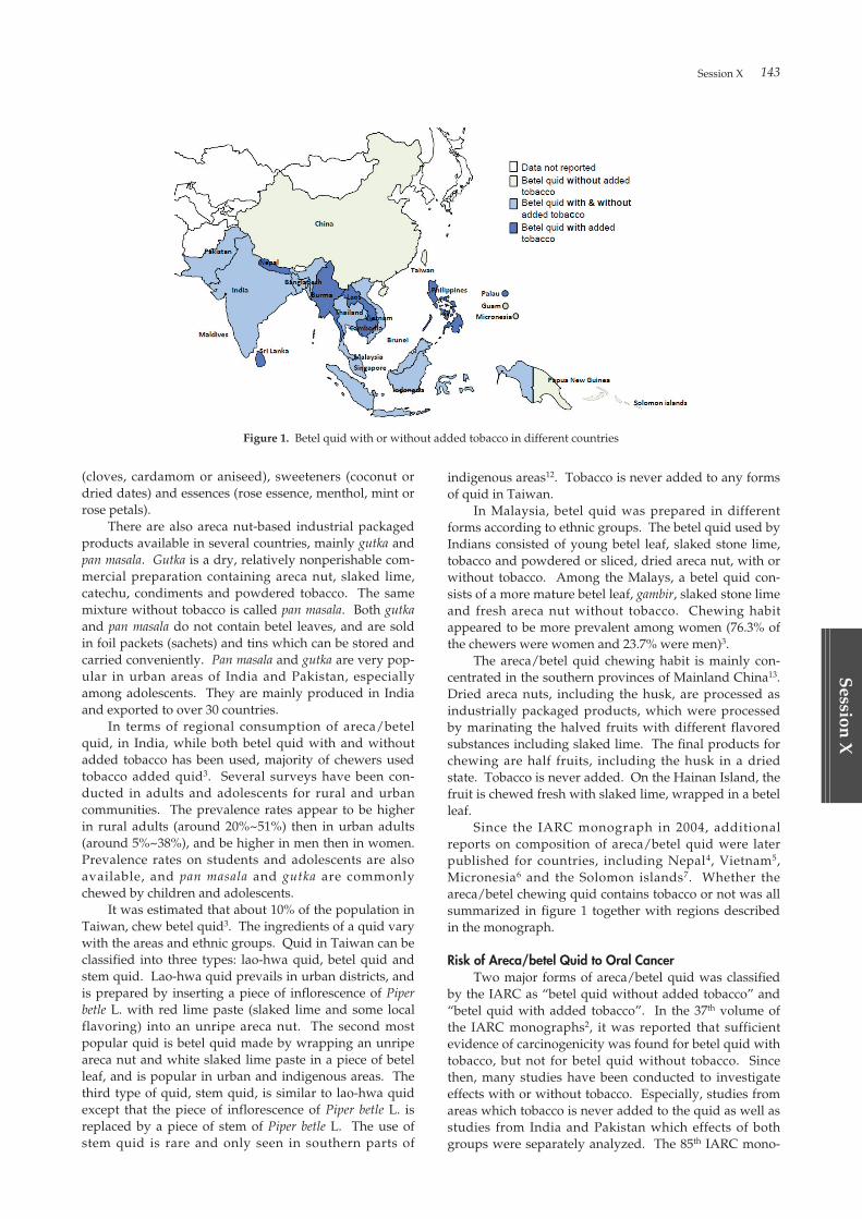
Session X 143
Session
X
(cloves, cardamom or aniseed), sweeteners (coconut ordried dates) and essences (rose essence, menthol, mint orrose petals).
There are also areca nut-based industrial packagedproducts available in several countries, mainly gutka andpan masala. Gutka is a dry, relatively nonperishable com-mercial preparation containing areca nut, slaked lime,catechu, condiments and powdered tobacco. The samemixture without tobacco is called pan masala. Both gutkaand pan masala do not contain betel leaves, and are soldin foil packets (sachets) and tins which can be stored andcarried conveniently. Pan masala and gutka are very pop-ular in urban areas of India and Pakistan, especiallyamong adolescents. They are mainly produced in Indiaand exported to over 30 countries.
In terms of regional consumption of areca/betelquid, in India, while both betel quid with and withoutadded tobacco has been used, majority of chewers usedtobacco added quid3. Several surveys have been con-ducted in adults and adolescents for rural and urbancommunities. The prevalence rates appear to be higherin rural adults (around 20%~51%) then in urban adults(around 5%~38%), and be higher in men then in women.Prevalence rates on students and adolescents are alsoavailable, and pan masala and gutka are commonlychewed by children and adolescents.
It was estimated that about 10% of the population inTaiwan, chew betel quid3. The ingredients of a quid varywith the areas and ethnic groups. Quid in Taiwan can beclassified into three types: lao-hwa quid, betel quid andstem quid. Lao-hwa quid prevails in urban districts, andis prepared by inserting a piece of inflorescence of Piperbetle L. with red lime paste (slaked lime and some localflavoring) into an unripe areca nut. The second mostpopular quid is betel quid made by wrapping an unripeareca nut and white slaked lime paste in a piece of betelleaf, and is popular in urban and indigenous areas. Thethird type of quid, stem quid, is similar to lao-hwa quidexcept that the piece of inflorescence of Piper betle L. isreplaced by a piece of stem of Piper betle L. The use ofstem quid is rare and only seen in southern parts of
indigenous areas12. Tobacco is never added to any formsof quid in Taiwan.
In Malaysia, betel quid was prepared in differentforms according to ethnic groups. The betel quid used byIndians consisted of young betel leaf, slaked stone lime,tobacco and powdered or sliced, dried areca nut, with orwithout tobacco. Among the Malays, a betel quid con-sists of a more mature betel leaf, gambir, slaked stone limeand fresh areca nut without tobacco. Chewing habitappeared to be more prevalent among women (76.3% ofthe chewers were women and 23.7% were men)3.
The areca/betel quid chewing habit is mainly con-centrated in the southern provinces of Mainland China13.Dried areca nuts, including the husk, are processed asindustrially packaged products, which were processedby marinating the halved fruits with different flavoredsubstances including slaked lime. The final products forchewing are half fruits, including the husk in a driedstate. Tobacco is never added. On the Hainan Island, thefruit is chewed fresh with slaked lime, wrapped in a betelleaf.
Since the IARC monograph in 2004, additionalreports on composition of areca/betel quid were laterpublished for countries, including Nepal4, Vietnam5,Micronesia6 and the Solomon islands7. Whether theareca/betel chewing quid contains tobacco or not was allsummarized in figure 1 together with regions describedin the monograph.
Risk of Areca/betel Quid to Oral CancerTwo major forms of areca/betel quid was classified
by the IARC as “betel quid without added tobacco” and“betel quid with added tobacco”. In the 37th volume ofthe IARC monographs2, it was reported that sufficientevidence of carcinogenicity was found for betel quid withtobacco, but not for betel quid without tobacco. Sincethen, many studies have been conducted to investigateeffects with or without tobacco. Especially, studies fromareas which tobacco is never added to the quid as well asstudies from India and Pakistan which effects of bothgroups were separately analyzed. The 85th IARC mono-
Figure 1. Betel quid with or without added tobacco in different countries
02_本文1027(web用追加) 15.10.27 9:05 AM ページ 143
144
Ses
sion
X
graph consists a thorough review of literatures on theirrisks to oral cancer. Recently, two meta-analysisreports8,9 have been conducted to investigate the risk ofbetel quid with or without tobacco to oral cancer.
A meta-analysis9 of 15 case-control studies (4,553cases; 8,632 controls) and 4 cohort studies (15,342)showed that chewing quid with tobacco is significantlyand independently associated with an increased risk ofsquamous-cell carcinoma of the oral cavity (case-controlstudies: OR=7.46, 95% CI=5.86~9.50, and cohort studies:RR=5.48, 95% CI=2.56~11.71). In addition, the 15 case-control studies has shown betel quid without tobacco tohave an independent positive association with oral can-cer, with OR=2.82 (95% CI=2.35~3.40). Presumably, dueto the carcinogenicity of areca nut, betel quid withouttobacco are also strong and independent risk factor fororal cancer.
From a meta-analysis of 50 publications8 on theinvestigation of risk to oral/oropharyngeal cancer, therelative risk (RR) for oral cavity cancer was 2.41 (95%CI=1.82~3.19; 13 studies) in users of betel quid withouttobacco, and was 8.47 (95% CI=6.49~11.05; 25 studies) forusers of betel quid with tobacco. Similarly, both groupshave shown significant associations with oral cavity can-cer. However, in Taiwan, where tobacco is never addedto the quid, the RR was 10.98 (95% CI=4.86~ 24.84; 13studies). The magnitude of RR was much higher inTaiwan. It is speculated that a larger daily amount wasconsumed, or the unripe fruit was used.
In Indian subcontinent, the RR was much higher inwomen (RR=14.56; 95% CI=7.63~27.76) than in men.Dose-response analyses also demonstrated that the riskof oral/oropharyngeal cancer increased with increasingdaily amount and duration (years) of chewingareca/betel quid in India and Taiwan. Around half oforal cancers in these countries could be prevented if peo-ple stopped their chewing habits (population attributablefraction: PAF%=53.7% in Taiwan; PAF%=49.5% in Indiasubcontinent).
While majority of areca/betel quid chewers werealso concurrently with smoking or drinking habits, theassessment of interaction effect from chewing-smoking-drinking is also of interest. One report10 focused on themagnitude of the smoking-drinking-chewing interactioneffect through the pooled Relative Excess Risk due toInteraction (RERI, excess risk in smoking-drinking-chew-ing exposed individuals with respect to the risk expectedfrom the addition of the three individual risks of smok-ing, drinking and chewing). The pooled ORs (14 studies)for smoking, drinking, chewing, smoking-drinking-chewing, respectively were 3.6 (95% CI=1.9~7.0), 2.2 (95%CI=1.6~3.0), 7.9 (95% CI=6.7~9.3), 40.1 (95% CI=35.1~45.8).The pooled RERI was 28.4 (95% CI=22.9~33.7). Amongsmoking-drinking chewing subjects, the individualeffects accounted for 6.7% from smoking, 3.1% fromdrinking, 17.7% from chewing of the risk, while the inter-action effect accounted for the remaining 72.6%. Thestudy results suggested that effective oral cancer controlpolicies must consider concurrent tobacco smoking, alco-hol drinking, betel quid chewing usages as a unique riskfactor for oral cancer.
CONCLUSIONSince the 85th volume of the IARC Monographs3 was
published, many of focus has centered on the clarificationof the composition of areca/betel quid in countries withchewing habits as well as on the strengthening of oralcancer risk from betel quid without added tobacco.Further research agenda may consider various types orways of preparation for using areca nut in quid, since therisk from Taiwan, where betel quid without tobacco isused, was much higher than the pooled estimates fromIndia subcontinent.
In terms of priorities for action, the results of popula-tion attributable fraction (PAF%=53.7% in Taiwan;PAF%=49.5% in India subcontinent) strongly support theneed for betel quid chewing intervention. Furthermore,since the study of interaction accounted for 72.6% of riskeffect, any intervention on the areca/betel quid chewinghabit toward oral cancer prevention should also considerconcurrent tobacco or cigarette smoking and alcoholdrinking.
REFERENCES01. GLOBOCAN 2012 v1.0, Cancer incidence and mortali-
ty worldwide: IARC Cancer Base No. 11 [Internet].International Agency for Research on Cancer; 2013.http://globocan.iarc.fr. Accessed June 30, 2015.
02. Tobacco habits other than smoking; betel-quid andareca-nut chewing; and some related nitrosamines.IARC Working Group. Lyon, 23-30 October 1984.IARC Monogr Eval Carcinog Risk Chem Hum. 1985; 37:1-268.
03. Betel-quid and areca-nut chewing and some areca-nut-derived nitrosamines. IARC Working Group.Lyon, 11-18 June 2003. IARC Monogr Eval CarcinogRisk Hum. 2004; 85: 1-300.
04. Lee CH, Ko AM, Warnakulasuriya S, et al. Populationburden of betel quid abuse and its relation to oral pre-malignant disorders in South, Southeast, and EastAsia: an Asian Betel-quid Consortium Study. Am JPublic Health. 2012; 102 (3): e17-24.
05. Reichart PA, Nguyen XH. Betel quid chewing, oralcancer and other oral mucosal diseases in Vietnam: areview. Journal of oral pathology & medicine: official pub-lication of the International Association of OralPathologists and the American Academy of OralPathology. 2008; 37 (9): 511-514.
06. Oakley E, Demaine L, Warnakulasuriya S. Areca(betel) nut chewing habit among high-school childrenin the Commonwealth of the Northern MarianaIslands (Micronesia). Bulletin of the World HealthOrganization. 2005; 83 (9): 656-660.
07. Tovosia S, Chen PH, Ko AM, Tu HP, Tsai PC, Ko YC.Prevalence and associated factors of betel quid use inthe Solomon Islands: a hyperendemic area for oraland pharyngeal cancer. Am J Trop Med Hyg. 2007; 77(3): 586-590.
08. Guha N, Warnakulasuriya S, Vlaanderen J, Straif K.Betel quid chewing and the risk of oral and oropha-ryngeal cancers: a meta-analysis with implications forcancer control. International journal of cancer. Journalinternational du cancer. 2014; 135 (6): 1433-1443.
02_本文1027(web用追加) 15.10.27 9:05 AM ページ 144
Session X 145
Session
X
09. Gupta B, Johnson NW. Systematic review and meta-analysis of association of smokeless tobacco and ofbetel quid without tobacco with incidence of oral can-cer in South Asia and the Pacific. PLoS One. 2014; 9(11): e113385.
10. Petti S, Masood M, Scully C. The magnitude of tobac-co smoking-betel quid chewing-alcohol drinkinginteraction effect on oral cancer in South-East Asia. Ameta-analysis of observational studies. PLoS One.2013; 8 (11): e78999.
11. Gupta PC, Warnakulasuriya S. Global epidemiology
of areca nut usage. Addict Biol. 2002; 7 (1): 77-83.12. Yang YH, Lee HY, Tung S, Shieh TY. Epidemiological
survey of oral submucous fibrosis and leukoplakia inaborigines of Taiwan. Journal of oral pathology & medi-cine: official publication of the International Association ofOral Pathologists and the American Academy of OralPathology. 2001; 30 (4): 213-219.
13. Zhang X, Reichart PA. A review of betel quid chew-ing, oral cancer and precancer in Mainland China.Oral oncology. 2007; 43 (5): 424-430.
02_本文1027(web用追加) 15.10.27 9:05 AM ページ 145

02_本文1027(web用追加) 15.10.27 9:05 AM ページ 146
Sp
ecial Lectu
re
Special lecture IPatient Simulation and Advanced Clinical Education with SIMROID
J. Morita MFG. CORP.
D.L. Sleeman
Special lecture II“DENTAPAC KOKORO”
—Epochal Solutions to Overcome Barriers to Dental Care
Subject 1: Next Generation Modular Dental Treatment System
THE YOSHIDA DENTAL MFG. CO., LTD.
K. Kuroda
Special lecture III“DENTAPAC KOKORO”
—Epochal Solutions to Overcome Barriers to Dental Care
Subject 2: Development of Dental Equipment and Materials for Home-visiting Dental
Treatment
GC CORPORATION
F. Fusejima
Special lecture IVThe Evaluation of Human Pluripotent Stem Cell Culture
by Advanced Cell Morphological Analysis
National Institutes of Biomedical Innovation
M. Kusuda Furue
Special Lecture
02_本文1027(web用追加) 15.10.27 9:05 AM ページ 147

02_本文1027(web用追加) 15.10.27 9:05 AM ページ 148
Special Lecture 149
Sp
ecial Lectu
re
BACKGROUNDUntil recently, practical dental education has been
performed using sophisticated phantoms and man-nequins which allow educational institutes to objectivelyevaluate technical skills. When it comes to clinical work-flow and patient treatment, this must be done with realpatients who may present with vastly different treatmentneeds making the objective evaluation of a student’s per-formance problematic. A system that combines the bestof training phantoms with the interactive capabilities isneeded to allow the student to practice necessary skilland workflows prior to working with an actual patient.That system is SIMROID.
PRESENTATIONIn our presentation, we will introduce the motiva-
tion behind this innovative project and introduce you tothe people and institutions who collaborated together toachieve this ambitious educational tool.
We will discuss the current gaps in the educationsystem and the underlying motivation that prompted theNippon Dental University to contact Morita and launchwhat will eventually become the future of dental educa-
tion.We will introduce our audience to the SIMROID
dental education system and highlight not only what setsthis system apart from the current education tools avail-able but also show what makes the SIMROID uniquelysuited to modern objective education. From there, wehave an overview of what the patient simulation robotcan actually do. We will highlight the wide range oftreatments that can be simulated and show how manymore are available to students and educators beyondstandard phantoms.
We will conclude with some feedback from our den-tal students who are currently training with SIMROIDsin their schools and share with you their thoughts andreactions to this enhanced education program.
CONCLUSIONThrough our presentation and subsequent discus-
sions and demonstration, we hope that the audience willget a good sense of the potential of such a patient simula-tion system and that they can see the impact trainingwith a SIMROID will have on patient care and ensuringnew dentists are better prepared for the realities of work-ing with real patients in a real clinical setting.
Patient Simulation and Advanced ClinicalEducation with SIMROIDD.L. Sleeman
International Sales, J. Morita MFG. CORP.
02_本文1027(web用追加) 15.10.27 9:05 AM ページ 149

150
Sp
ecia
l Lec
ture
In Japan, a country of super aging society, there areapproximately 6 million elderlies who require nursingcare from the others. 90% of functionally dependentelderly is said to be in need of some type of dental treat-ment, however, the reality is, only 30% of those havebeen able to receive necessary treatment.
One of the reasons that attribute the aforementionedis the physical as well as psychological stress that doctorsexperience in providing dental care due to the insuffi-cient treatment tools required for home-visiting dentalcare.
Under the circumstances, The Japan Dental
Association (JDA), the Japan Association for DentalScience (JADS), and the Japan Dental Trade Association(JDTA) have been promoting the development of theportable home-visiting dental treatment system, and inresponse to a request from the Ministry of Economy,Trade and Industry, the project was actualized by a con-sortium including 10 member companies of the JapanDental Trade Association.
As the deliverable, we have “DENTAPAC KOKORO”the modular-designed portable dental delivery unit,which we will introduce in this presentation.
“DENTAPAC KOKORO”—Epochal Solutions to Overcome Barriers
to Dental CareSubject 1: Next Generation Modular Dental
Treatment SystemK. Kuroda
Division Director, International Business Div., THE YOSHIDA DENTAL MFG. CO., LTD.
02_本文1027(web用追加) 15.10.27 9:05 AM ページ 150
Special Lecture 151
Sp
ecial Lectu
re
The background : In 2008, “New medical equipment andmedical technology industry vision” was formulated bythe Ministry of Health, Labor and Welfare. In the article,there is a “the propulsion of home-visiting dental treat-ment and the development of portable dental instru-ments for the maintenance of dental health”. We chosethree theme of development from eight theme forportable dental instruments and started to development.
• Sub Theme 5 “Kit for impression taking/bite takingrelated materials”
Target : Development of impression tray, impressionmaterials and bite wax for efficient and simple denturefabrication during home-visiting dental treatment.Result : On July 2010, consortium consisted of J.D.A.,J.A.D.S. and J.D.T.A was established and started develop-ment. On July 2014, kit of “Impression taking/bite tak-ing-related materials” was launched.
• Sub Theme 6 “Kit for Denture repairing”Target : Development of self-polymerization resin forefficient and simple denture repair during home-visitingdental treatment.Result : Consortium was established same as above andstarted development. On July 2014, “Denture repair kit”was launched
• Sub Theme 7 “Kit for Oral health care”Target : Development of disposable brush for denturecleaning and oral care brush for oral cleaning for easydenture cleaning during home-visiting dental treatment.Result : Consortium was established same as above andstarted development. On July 2014, “Oral health carekit” was launched.
“DENTAPAC KOKORO”—Epochal Solutions to Overcome Barriers
to Dental CareSubject 2: Development of Dental Equipment
and Materials for Home-visiting Dental Treatment
F. Fusejima
Deputy General Manager, Research & Development Dept., GC CORPORATION
02_本文1027(web用追加) 15.10.27 9:05 AM ページ 151
152
Sp
ecia
l Lec
ture
Human pluripotent stem cells (hPSCs), includingembryonic stem cells (hESCs) or human induced pluripo-tent stem cells (hiPSCs) can proliferate virtually infinitelyand differentiate into almost all cell types. These hPSCscould be a promising new tool both for cell-based regen-erative medicine and pharmaceutical research, includingdrug efficacy and toxicity screening tests. However, itremains challenging to maintain hPSCs in an undifferen-tiated state because of their differentiation potentialand/or genetic instability. Therefore, routine characteri-zation of hPSCs using several standard criteria, such ascell growth, marker expression, or in vitro differentiation,is recommended. In practice, observation under phasecontrast microscope is used to continuously determinethe health condition of hPSCs. When hPSCs are culturedunder proper conditions, they form tight, compact androunded colonies. However, epithelial-mesenchymaltransition (EMT) occurs at the peripheral of the hPSCcolonies where hPSCs differentiate spontaneously.Under improper conditions, hPSCs tend to execute apop-tosis or differentiate, and dynamically change their cell
morphology. Culture-adapted” cells with karyotypicchanges tend to have an increased growth rate orreduced apoptosis. The criterion for evaluating cell mor-phology is not quantitative. Recently, we have devel-oped a non-invasive method to determine the growth ofhPSCs by analyzing morphologies of live, non-labeledhiPSCs in culture without damaging these cells. Whenthe culture conditions become improper, the change incell morphology could be detected by analysis of thephase-contrast images. This method provides real-timeinformation on the growth and quality of hPSCs withoutdamaging or wasting cells and could be useful both forbasic research using hPSCs and for cell processing forhPSC-based therapy.
1. Stem Cells Transl Med. doi: 10.5966/sctm. 2014-02422. Stem Cells and Development 15; 23 (18): 2180-9.3. PLoS One. 8 (1): e54122.4. Stem Cell Res. 5 (2): 157-695. In Vitro Cell Dev Biol Anim. 46: 573-576.6. Proc Natl Acad Sci USA. 105: 13409-14.
The Evaluation of Human Pluripotent Stem Cell Culture by Advanced Cell Morphological AnalysisM. Kusuda Furue
Laboratory of Stem Cell Cultures, National Institutes of Biomedical Innovation, Health and Nutrition. E-Mail: [email protected], [email protected]
02_本文1027(web用追加) 15.10.27 9:05 AM ページ 152
Poster P
resentation
Poster Session
A-1~A-9: Dental Education
B-1~B-28: Frontiers of Biological Science in Dentistry
C-1~C-6: Latest Trends in BioDental Engineering
D-1~D-39: Oral Health and Clinical Treatments
■Airlangga University, Indonesia01-1 [A-1] ······················································································································································· 163
Alteration of Preschool’ Personal Hygiene Behavior: Mouth Hygiene by Health Education with Puzzle
N. Qur’aniati, W. Wigiarti and Nursalam
01-2 [B-1] ························································································································································ 167Modulation Mechanism of Immune Respons From Non Atopy into Atopy Rat After Porphyromonas gingivalis Lipopolysacaride Exposure
N.S. Cornelia
01-3 [B-2] ························································································································································ 168Cellular Changes in the Mucosal Tissue Incision Wound of Wistar Rats AfterAdministered with 40% Topical Gel Bovine Colostrum
T. Saskianti and A. Adlina
01-4 [D-1] ······················································································································································· 185Health Education Strategies to Improve Preschooler’s Parents Behaviour in Oral Health
E.M.M. Has
01-5 [D-2] ······················································································································································· 186Literature Review: Oral Health in Pregnant Women
K. Tiyas
■B P Koirala Institute of Health Science, Nepal02-1 [D-3] ······················································································································································· 186
An Experience of Odontogenic Cysts and Tumors of the Jaw in Eastern NepalA. Shrestha, S. Karki and V. Marla
■Catholic University of Pusan, Korea03-1 [C-1] ······················································································································································· 182
Effect of Aging Treatment on the Flexural Strength of Implant Provisional RestorationMaterials
J.S. Park and M.G. Park
■Chulalongkorn University, Thailand04-1 [B-3] ························································································································································ 168
Basic Fibroblast Growth Factor Induced Interleukin 6 Expression by Stem Cell Isolatedfrom Human Exfoliated Deciduous Teeth
N. Nowwarote, P. Pavasant and T. Osathanon
02_本文1027(web用追加) 15.10.27 9:05 AM ページ 153
Pos
ter
Pre
sen
tati
on
04-2 [B-4] ························································································································································ 169Chemogenomic Analysis of Antifungal Mechanisms of Chitosan
O. Matangkasombut, N. Ungwijarnpanya, P. Katare, D. Supakanjanakanti, T. Chaitrakoonthong, W. Namangkalakul, A. Promchai,
R. Phuprasong and P. Thanyasrisung
04-3 [D-4] ······················································································································································· 187Antifungal Effect of Chitosans on Clinical Candida Isolates
W. Satitviboon, S. Howattanapanich, S. Muangsawat, P. Thanyasrisung and O. Matangkasombut
■International Medical University (IMU), Malaysia05-1 [B-5] ························································································································································ 169
Rugoscopy among the Malaysian Chinese Population: A Pedigree AnalysisT.W. Hong, V. Mutalik, V. Murthy and S.A. Radman
■Kaohsiung Medical University, Taiwan06-1 [D-5] ······················································································································································· 187
Correlation between the Pharyngeal Airway Space and Head Posture After Surgery for Mandibular Prognathism
C.M. Chen, S. Lai, K.K. Chen and H.E. Lee
■Khon Kaen University, Thailand07-1 [A-2] ······················································································································································· 163
Table Clinic Activity in Teaching Preventive Dentistry in Third Year KKU DentalStudents: a Case Study
A. Rattanathongkom, J. Paphangkorakit, S. Luengpailin, T. Damrongrungruang, N. Poomat, T. Plangkoon jorns, R. Noisombut, P. Pisek, P. Phajongviriyatorn,
P. Thanyakob and S. Rattanathongkom
07-2 [A-3] ······················································································································································· 164Accuracy of Intraoral Surveyor Compared to Ney Surveyor on Customized Models
S. Aerarunchot, W. Tangchanyatam, S. Mayurasakorn and W. Pitiphat
07-3 [B-6] ························································································································································ 170Expression of p73 Isoforms and Aurora Kinases in Oral Squamous Cell Carcinoma CellLines
P. Klanrit, P. Chaiyarit, T. Limpaiboon and S.C. Cheong
07-4 [B-7] ························································································································································ 170Anti-fungal Effectiveness of Dental Stone Incorporated with LodopropynylButylcarbamate
P. Kaewpoomhae, R. Akkarapatum and C. Prunkngarmpun
07-5 [B-8] ························································································································································ 171Prevalence of Abnormalities of Teeth and Jaws on Panoramic Radiographs in Thalassaemic Patients
P. Sutthiprapaporn, J. Lertsirivorakul, P. Sukanin, W. Pitiphat and A. Jetsrisuparb
02_本文1027(web用追加) 15.10.27 9:05 AM ページ 154
Poster P
resentation
07-6 [C-2] ······················································································································································· 183Effects of Different Beverages on Color Stability of Heat-cured Acrylic Resin
C. Prunkngarmpun, P. Uttamung and P. Thippa-art
07-7 [C-3] ······················································································································································· 183Anti-adhesion Efficiency of Candida albicans on Curcumin Coated Heat-cured AcrylicResin by Polyelectrolyte Multilayer Technique
T. Angwarawong, P. Seedapol, P. Thanaphuttiwirot and O. Angwaravong
07-8 [D-6] ······················································································································································· 188Treatment of Squamous Papilloma with Double Pedicle Flap and SubepithelialConnective Tissue Graft: A Case Report
C. Tripawat, S. Prajaneh and W. Suwannarong
07-9 [D-7] ······················································································································································· 188Clinical Management of Pyostomatitis vegetans: A Case Report
A. Subarnbhesaj
07-10 [D-8] ····················································································································································· 189Treatment of Gingival Recession with Subepithelial Connective Tissue Graft Combinedwith Double Papillae and Lateral Sliding Flap: A Case Report
M. Tanapoomchai, S. Prajaneh and W. Suwannarong
07-11 [D-9] ····················································································································································· 189Effect of Silver Diamine Fluoride, CPP-ACP, Fluoride Varnish on Surface Microhardnessof Human Dental Enamel
O. Angwaravong, K. Tonggerd, K. Pimpison, W. Weraarchakul and T. Angwarawong
07-12 [D-10] ··················································································································································· 190Translation, Validity and Reliability Testing of the E-San Short-form McGill PainQuestionnaire
T.P. Jorns, S. Puasiri, S. Loppanthong and P. Pansaeng
07-13 [A-10] ··················································································································································· 206PrepLooker: A Mobile Application for Assisting Dental Students in Learning Undercut &Convergence Angles of Crown Preparation
S. Aerarunchot, R. Chotipanvidhayakul, N. Boonpongsathian, T. Songphaeng, K.R. Saikaew and P. Buayai
■Kyungpook National University, Korea08-1 [D-11] ····················································································································································· 190
Aesthetic Rehabilitation of Traumatized Maxillary Anteriors by Surgical Repositionings,Crown Restorations, Bleaching and Laminate Veneers
J.S. Kim, K.M. Son, J.H Ha, Y.K. Kim, S.K. Kim and M.U. Jin
02_本文1027(web用追加) 15.10.27 9:05 AM ページ 155
Pos
ter
Pre
sen
tati
on
■Mahidol University, Thailand09-1 [B-9] ························································································································································ 172
Using Transgenic Mouse Models as an Approach to Study Potential Stem CellSubpopulation in Salivary Gland
K. Janebodin, W. Buranaphatthana, R. Chavanachat, N. Ieronimakis, A. Hays and M. Reyes
09-2 [B-10] ······················································································································································ 173Inhibitory Effect of Moringa oleifera L. Seed Oil on Oral Pathogens
B. Thaweboon, S. Thaweboon and T. Muadcheingka
09-3 [C-4] ······················································································································································· 184Antifungal Activity of Type III Dental Gypsum Incorporated with 3-iodo-2-propynyl-butylcarbamate
T. Riyatanon, P. Sawaengkit, S. Thaweboon, B. Thaweboon and P. Nisalak
09-4 [D-12] ····················································································································································· 191Eagle’s Syndrome Mimicking Orofacial Pain: A Case Report and Review of Literature
S. Visuttiwattanakorn and R. Leelarungsun
09-5 [D-13] ····················································································································································· 191Antimicrobial Effects of Grape Seed and Mangosteen Extracts against MicrofloraAssociated with Percutaneous Skin Reactions Around Craniofacial Implants
B. Shrestha, S. Thaweboon and B. Thaweboon
09-6 [D-14] ····················································································································································· 192Antimicrobial Activities of Clinacanthus nutans (Burm.f) Lindau Leaf Extracts against OralBacteria
R. Srichan, S. Korsuwannawong, T. Muadcheingka and S. Mala
09-7 [D-15] ····················································································································································· 192In vitro Study of Antibacterial Effects of Curcuma Longa Linn Extracts
S. Mala, S. Korsuwannawong, T. Muadcheingka and R. Srichan
09-8 [D-16] ····················································································································································· 193Antibacterial Activities of Piper nigrum Seed Extracts against Oral Bacteria
S. Korsuwannawong, T. Muadcheingka, R. Srichan and S. Mala
■National Hospital of Odonto-Stomatology in Ho Chi Minh City, Viet Nam10-1 [D-17] ····················································································································································· 193
Application of 3D Modeling in Mandibular Tumor’s Treatment: 2 Cases ReportL.P. Pham
10-2 [D-18] ····················································································································································· 194Soft and Hard Tissue Changes After Bimaxillary Surgery in Class III Patient
D.T. Hai and L.H. Phuong
02_本文1027(web用追加) 15.10.27 9:05 AM ページ 156
Poster P
resentation
■Padjadjaran University, Indonesia11-1 [D-19] ····················································································································································· 194
Analise the Ankilylosis in Temporo Mandibular Joint Using CBCT 3D (Case Study)G. Christinne and L. Epsilawati
11-2 [D-20] ····················································································································································· 195The Relationship between Level Vitamin D and Calcium Serum with Mandibular BoneDensity in HIV/AIDS Childrens
R.P. Saptarini, E. Riyanti, I. Sufiawati and Azhari
11-3 [D-21] ····················································································································································· 195Odontoma Cases Base on Cone Beam Computed Tomography (Case Report Study)
F. Septina and L. Epsilawati
11-4 [D-22] ····················································································································································· 196Assesment of Antegonial Index of the Mandible on Panoramic Radiographs
I.M.A. Astika and L. Epsilawati
11-5 [D-23] ····················································································································································· 196Assessment the Shape of Tooth Base on Age Using Panoramic Radiographs of People inBandung Indonesian
I.S. Sasmita and L. Epsilawati
11-6 [D-24] ····················································································································································· 197Assesment of Gonial Index of the Mandible on Panoramic Radiographs
I. Farizka and L. Epsilawati
11-7 [D-25] ····················································································································································· 197Healing Process in Patients with Osteoradionecrosis by Using Oxygen HyperbaricTherapy (Case Study)
L. Epsilawati
11-8 [D-26] ····················································································································································· 198Assessment of Mandible Cortical Bone with Mental Index Base on Age Using PanoramicRadiographs of people in Bandung Indonesian
M. Darjam and L. Epsilawati
11-9 [D-27] ····················································································································································· 198Oral Lesions Related to Food Allergy
N. Nur’aeny
11-10 [D-28] ··················································································································································· 199Assesment of Dental Emerge Base on Age in Sundanes People Using PanoramicRadiograph
Y. Herdiayati and L. Epsilawati
■Prince of Songkla University, Thailand12-1 [D-29] ····················································································································································· 199
Load-deflection Characteristics of Bent Nickel Titanium WireP. Phermsang-ngarm and C. Charoemratrote
02_本文1027(web用追加) 15.10.27 9:05 AM ページ 157
Pos
ter
Pre
sen
tati
on
■Sun Yat-sen University, China13-1 [B-11] ······················································································································································ 173
Expression and Function of Homeobox Gene Lhx8 during Tooth DevelopmentC. Zhou, G. Yang, M. Chen, C. Wang, L. He, L. Xiang, D. Chen, J. Ling and J.J. Mao
■Tohoku University, Japan14-1 [C-5] ······················································································································································· 184
Gene Transfection with the Nanohydroxyapatite-collagen Scaffold, Containing DNA-functionalized Calcium Phosphate Nanoparticles
T. Tenkumo, J.R.V. Saenz, Y. Takada, M. Takahashi, O. Rotan, V. Sokolova, M. Epple and K. Sasaki
■Trisakti University, Indonesia15-1 [D-30] ····················································································································································· 200
Effect of Chitosan in Soft Tissue Healing Post Dental ExtractionA.P.S. Palupi and A.S. Mulyadi
15-2 [D-31] ····················································································································································· 200Effectivity of Andrographis paniculata Mouthwash in Healing of Recurrent AphtousStomatitis Lesion (Preliminary Research)
I. Gunardi and L.T. Christian
■University of Indonesia, Indonesia16-1 [C-6] ······················································································································································· 185
Investigation on Orthodontic Brackets Strength Differences Based on The Design andMaterials Simulation
T. Prasetyadi, B. Irawan, M.K. Purwanagara, B. Suharno, S. Supriadi and T.W. Sitanggang
16-2 [D-32] ····················································································································································· 201Affecting Factors of Bond Failure of Stainless Steel Brackets that Used at Public HealthCenter Kecamatan Jakarta-Indonesia
G. Maringka and E. Herda
■Universitas Sumatera Utara, Indonesia17-1 [D-33] ····················································································································································· 201
Alteration of Mandibular Bone Density in type 2 Diabetes Mellitus Patients Based onRadiograph Examination
T. Boel and Y. Susanti
■University of Dental Medicine, Myanmar18-1 [A-4] ······················································································································································· 164
Comparative Study on Accuracy of Three Electronic Apex Locators Before and AfterExtraction of Lower 1st Permanent Molars
T.S. Mon, W. Lwin, A. Htang and M.M. San
02_本文1027(web用追加) 15.10.27 9:05 AM ページ 158
Poster P
resentation
18-2 [D-34] ····················································································································································· 202Oral Submucous Fibrosis—A Case Report of Young Patient with Stage II Oral Submucous Fibrosis
N. Aung and Z.M. Thein
■University of Malaya, Malaysia19-1 [D-35] ····················································································································································· 203
Collagen Triple Helix Repeat Containing-1 (CTHRC1) Expression in Oral Squamous CellCarcinoma (OSCC): Prognostic Value and Clinico-pathological Implications
C.E. Lee, V.K. Vincent-Chong, A. Ramanathan, T.G. Kallarakkal, L.P. Karen-Ng, W.M.N. Ghani, Z.A.A. Rahman, S.M. Ismail, M.T. Abraham,
K.K. Tay, W.M.W. Mustafa, S.C. Cheong and R.B. Zain
■University of Medicine and Pharmacy, Ho Chi Minh City, VietNam20-1 [D-36] ····················································································································································· 204
The Relationship between Natural Head and Frankfort Planes in Mesh DiagramsAnalysis
L.M. Loc and N.T.Q. Lan
■University of Peradeniya, Sri Lanka21-1 [A-5] ······················································································································································· 165
Metric and Non-metric Characteristics in the Dentition of Children/adolescents withDown Syndrome Attending an Institution in Jaffna, Northern Sri Lanka
A. Neishanthan, D. Ihalagedara, K. Arambawatta, R. Peiris and U. Dissanayake
■Wonkwang University, Korea22-1 [D-37] ····················································································································································· 204
A Patient with Facial Asymmetry and Unilateral Posterior Crossbite Treated withSurgery-first Approach
S.K. Choi, K.Y. Lee, K.H. Kang and S.C. Kim
■Hiroshima University, Japan23-1 [A-6] ······················································································································································· 165
An Assessment of the Class “International Future Dentistry with the Outcome ofResearches 2014 and 2015” with a Video Conference System
H. Oka, M.D. Ariani, P.T.T. Nguyen and K. Kato
23-2 [A-7] ······················································································································································· 166What Do You Think?—A Questionnaire Survey to Dental Students in UMPH and HU by Participants of
Short-term Visit 10 Days Program 2015—M. Kato, K. Okamoto, H. Nagao and H. Oka
23-3 [B-27] ······················································································································································ 181Irradiation Effects of Newly Developed Low Temperature Multi Gas Plasma Jet on OralBacteria
T.R. Abonti, M. Kaku, S. Kojima, H. Sumi, H. Shikata, S. Kojima and K. Tanimoto
02_本文1027(web用追加) 15.10.27 9:05 AM ページ 159
Pos
ter
Pre
sen
tati
on
23-4 [A-8] ······················································································································································· 166The Current Status of Instruction and Curricular Content in Forensic Odontology inAsian Countries I. Results from the Pilot Questionnaire Survey
H. Oka, P.T.T. Nguyen and K. Kato
23-5 [A-9] ······················································································································································· 167How and What We Learned? —HUD Short-term Visit 10days Program 2015—
M. Kawamoto, S. Tsuyama, Y. Kimura, N. Yamakado, T. Akitomo, H. Nakamura, J. Cho, M. Tanaka, K. Tsutiya, M. Kato, K. Okamoto, A. Fujii, M. Kiyama,
H. Nagao, R. Hiura and T. Kobayashi
23-6 [B-12] ······················································································································································ 174Differentiation of Induced Pluripotent Stem Cells into Dental Epithelial-like Cells in aDefined Culture Condition
A.N. Abdullah, K. Tanimoto and K. Kato
23-7 [B-13] ······················································································································································ 174Distinction between Cell Proliferation and Apoptosis Signals Regulated by BDNF inHuman Periodontal Ligament Cells and Gingival Epithelial Cells
K. Kashiwai, M. Kajiya, T. Fujita, S. Matsuda, K. Takeda and H. Kurihara
23-8 [B-14] ······················································································································································ 175Pharmacological Effects of General Anesthetics Altered by the Change of SubunitComposition of GABAA Receptors
M. Hayashiuchi, Y. Yamawaki, K. Oue, K. Harada, S. Asano, M. Irifune, M. Okada and T. Kanematsu
23-9 [B-15] ······················································································································································ 175Effect of Intermittent Parathyroid Hormone Administration on Osseointegration Aspectsin Rabbit Osteoporosis Model
Y. Oki, K. Doi, Y. Makihara, T. Kubo and K. Tsuga
23-10 [B-16] ···················································································································································· 176A Novel Isoform of FGFR2 Modulates FGF10-FGFR Signaling in Osteochondrogenesis
H. Yoshioka, K. Kagawa, S. Okita, K. Kuremoto, Y. Takei, T. Minamizaki, K.Tanimoto, K. Tsuga and Y. Yoshiko
23-11 [B-17] ···················································································································································· 176TLR2 Plays a Key Role in P. gingivalis-induced NASH Progeression
H. Furusho, M. Miyauchi, A. Nagasaki, S. Sakamoto and T. Takata
23-12 [B-18] ···················································································································································· 177Oral Administration of Liposomal Bovine Lactoferrin Suppresses Progression ofRheumatoid Arthritis
S. Yanagisawa, C. Chea, N.F. Ayuningtyas, H. Tsuji, M. Miyauchi, T. Inubushi, A. Ishikado, H. Imanaka, E. Sugiyama and T. Takata
23-13 [B-19] ···················································································································································· 177Development of Calcineurin Inhibitor Induced Gingival Hyperplasia Mice Model
A. Okanobu, S. Matsuda, M. Kajiya, N. Mizuno, T. Fujita and H. Kurihara
02_本文1027(web用追加) 15.10.27 9:05 AM ページ 160
Poster P
resentation
23-14 [B-20] ···················································································································································· 178Biological Effect of Carbon Ion and Gamma Ray Irradiation on Bone Metastasis of BreastCancer Cells
P. Srimawong, M. Sawajiri, H. Terato, K. Maruyama and K. Tanimoto
23-15 [B-21] ···················································································································································· 178The Differences of Electroencephalogram (EEG) Pattern during Sleep between HumanBeings and Mice
T. Yoshinaka and M. Irifune
23-16 [B-22] ···················································································································································· 179A Rhizosphere Microbiota-derived Symbiotic Factor Prevents Intestinal InflammatoryDisease
Y. Asao, A. Hashimoto, D. Yamamoto, S. Teranaka, A. Kodama, Y. Kurashima, H. Kiyono and I. Takahashi
23-17 [B-23] ···················································································································································· 179Bovine Lactoferrin Inhibits Oral Cancer Proliferation and Bone Invasion
C. Chea, N.F. Ayuningtyas, T. Inubushi, A. Subarnbhesaj, M. Miyauchi, A. Ishikado, H. Imanaka and T. Takata
23-18 [B-24] ···················································································································································· 180Odontogenic Infection of Porphyromonas gingivalis Exacerbates Pathological Progressionof Non-alcoholic Steatohepatitis through Activation of Hepatic Stellate Cells byMacrophages
A. Nagasaki, S. Sakamoto, H. Furusho, M. Miyauchi and T. Takata
23-19 [B-25] ···················································································································································· 180MEPE-ASARM, a Substrate of PHEX, Decreases Bone Volume Independently of SerumPhosphate Levels
K. Sakurai, T. Minamizaki, H. Yoshioka, Y. Takei, K. Kozai and Y. Yoshiko
23-20 [B-26] ···················································································································································· 181Porphyromonas gingivalis Infection Exacerbates Rheumatoid Arthritis in ExperimentalMice Model
M. Yamakawa, K. Ouhara, M. Kajiya, M. Kittaka, T. Fujita, K. Takeda, H. Shiba, T. Fujita and H. Kurihara
23-21 [D-38] ··················································································································································· 205Changes of the Airway and Sleep-breathing Patterns of Skeletal II Children UndergoingActivator Therapy
C.C. Medina, H. Ueda, M. Koh, Y. Matsumura and K. Tanimoto
23-22 [D-39] ··················································································································································· 205Inhibitory Effects of Antibiofilm compound-1 on Staphylococcus aureus Biofilm
L. Shrestha, S. Kayama, M. Sasaki, F. Kato, J. Hisatsune, K. Tsuruda, K. Koizumi,N. Tatsukawa, K. Takeda and M. Sugai
02_本文1027(web用追加) 15.10.27 9:05 AM ページ 161

■Stomatological Hospital of Tianjin Medical University, China24-1 [B-28] ······················································································································································ 182
A Novel Intragenic Microdeletion in RUNX2 in a Chinese Family with CleidocranialDysplasia
T. Zhang, Y. Sun, X. Li, H. Zhou, Z. Zhang, J. Zhou and P. Gao
02_本文1027(web用追加) 15.10.27 9:05 AM ページ 162
Poster Presentation 163
Poster P
resentation
Session
IS
ession I
INTRODUCTION : Personal hygiene behaviour such asskin hygiene, mouth hygiene, and hair hygiene is essen-tial for children. In Indonesia, most of children haveproblems in mouth hygiene. This caused by lack of fasili-ties in personal hygiene’s education which cause lessattention to personal hygiene’s behavior in children. Thefunction of health education is to change someone’sbehavior towards personal health. This study was aimedto examine the effect of health education with puzzle toincrease preschool’s personal hygiene: mouth hiygienebehavior at Aisyiyah Bustanul Athfal II kindergarten,Kedurus, Sidoarjo.
METHODS : Design of this study was Quasy Experiment.The population were students B class at Aisyiyah BustanulAthfal II kindergarten, Kedurus, Sidoarjo. The sampleswere taken by simple random sampling technique withthe number thirty respondents. The datas were collectedby using questionnaire and observation sheet, and ana-lyzed by using Wilcoxon Signed Rank Test and MannWhitney U Test with significance level of α ≤ 0.05.
RESULT AND ANALYSIS : The result of this study showed
that preschool knowledge had significance level of p=0.001 in treatment group, while in control group wasp=0.157, for preschool attitude p=0.001, while in controlgroup was p=0.317, for preschool skin hygiene behaviorp=0.001, while in control group was p=1.000, forpreschool mouth hygiene behavior p=0.001, while in con-trol group was p=1.000, for preschool hand hygienebehavior p= 0.001, while in control group was p=0.317,and for preschool hair hygiene behavior p=0.000, whilein control group was p=0.317. The result of MannWhitney U test was p=0.000, it means there was differentin knowledge, attitude and personal hygiene behaviorbetween treatment and control groups.
DISCUSSION : It can be concluded that there is significanteffect of health education by playing puzzle to improvepreschool’s personal hygiene behavior. Puzzle is analternative education media with innovative methods, soit can improve coginitive ability and be exercise to solvetheir own problem.
Key words : Health Education, Personal Hygiene,Preschool, Puzzle
A-1 (01-1) Alteration of Preschool’ Personal Hygiene Behavior:Mouth Hygiene by Health Education with Puzzle
N. Qur’aniati1, W. Wigiarti2 and Nursalam3
1 Lecturer Pediatric of Nursing, Faculty of Nursing, Universitas Airlangga, E-Mail: [email protected], 2 Nurses, Dr.Soetomo Hospital, Surabaya, 3 Professor Faculty of Nursing, Universitas Airlangga
BACKGROUND : Preventive Dentistry Practice is a coursefor teaching third year dental students. These are a lot ofdifficult and complicated contents for students to under-stand. Therefore, a table clinic activity (TCA), brief oralpresentation in areas of clinical application and tech-niques, was applied for teaching.
OBJECTIVE : To evaluate students learning activities,opinion and attitude toward TCA oriented teaching.
METHOD : Seventy third year students, enrolling in thiscourse in second semester 2014, were assigned to activelyparticipate in this process. The following learning out-comes and activities were evaluated by: 1) student behav-ior and classroom activities, 2) individual staff and stu-dent interview, 3) student report and presentation 4)questionnaire. After 10 lectures session in basic knowl-edge given by instructors, all students were divided into7 groups, one patient was assigned for each group. Allpatients had different oral health problems. Each groupof student followed patient in 4 sessions for: 1) assess-ment patient’s problems, 2) obtaining an appropriate pre-
ventive planning programs, 3) applying the preventivetreatment to the patients, 4) re-assessment the patients.One morning-session (TCA) was organized to allow eachgroup of the students to present their results. All stu-dents and teachers discussed and shared knowledge foreach patient case.
RESULTS : The most students (94.2%) actively participatedin learning and were able to understand the whole con-tents; they (95.65%) were able to integrate the concept ofholistic approach into the real patients. During inter-views session, 28 students showed good attitudes andgain experiences toward the learning process, 16 studentsfelt proud to present their cases and shared the experi-ence closely with their teachers.
CONCLUSION : TCA can possibly promote the integrationof holistic approach in practical preventive dentistry forstudents, who have not yet met the real patients.Students can learn cooperatively, and share their opin-ions together and closely with their teachers.
A-2 (07-1) Table Clinic Activity in Teaching Preventive Dentistry in Third Year KKU Dental Students: a Case Study
A. Rattanathongkom1, J. Paphangkorakit1, S. Luengpailin1, T. Damrongrungruang1, N. Poomat1, T. Plangkoon jorns1, R. Noisombut1, P. Pisek1, P. Phajongviriyatorn1, P. Thanyakob1 and S. Rattanathongkom2
1 Faculty of Dentistry, Khon Kaen University, Khon Kaen, Thailand, 2 Faculty of Associated Medical Science, Khon KaenUniversity, Khon Kaen, Thailand
02_本文1027(web用追加) 15.10.27 9:06 AM ページ 163
164
Pos
ter
Pre
sen
tati
on
Preparation of abutment teeth paralleling to the pathof insertion is crucial in the production of removable orfixed partial denture. This step of prosthesis fabricationis generally difficult for dental students and the use ofaiding device such as dental surveyor can be helpful. Wedeveloped a prototype of intraoral surveyor to assist thestudents’ practice in preparation of abutment teeth anddetermination of optimum guiding planes on a man-nequin in dental simulation laboratory. The purpose ofthis study was to determine the accuracy of the intraoralsurveyor compared to standard Ney surveyor. Surveylines were produced on 12 customized models using twodifferent surveyors. The investigator who performed thesurveys was a lecturer in the Department of
Prosthodontics who had more than 15 years of experi-ence in the field. The differences in the survey lines cre-ated by the two surveyors on each sample were mea-sured by a stereomicroscope. The difference of ≤0.2 mmwas considered as acceptable. The Westlake’s test ofequivalence demonstrated that the mean distancebetween the survey lines of the intraoral surveyor andthe Ney surveyor was significantly greater than 0.2 mm(mean = 0.35 mm, 95% confidence interval = 0.24-0.47, p< 0.001). The results suggest that this prototype of theintraoral surveyor was not equivalent to the Ney survey-or. Further development is therefore needed to improvethe accuracy of this intraoral surveyor for use in aidingdental students’ practice.
A-3 (07-2) Accuracy of Intraoral Surveyor Compared to Ney Surveyor on Customized Models
S. Aerarunchot, W. Tangchanyatam, S. Mayurasakorn and W. Pitiphat
Faculty of Dentistry, Khon Kaen University, Thailand
The aim of this study was to compare the accuracy ofthree electronic apex locators (EALs) in determiningworking tooth length. Working length of each tooth wascarried out before and after the tooth was extracted, andthus the measurements in vivo were compared with thereal tooth length (RTL) measured in vitro. Thirty lower1st permanent molars, scheduled for extraction, wereselected for the study. After obtaining the consent fromthe patients, a preoperative radiograph was taken, theaccess opening was prepared, and the occlusal referenceplane was maked in each tooth. Working length wasdetermined by using three apex locators; Sybro Endomini apex locator, Foramatron-D 10 apex locator and CRoot apex locator. For all the electronic measurement ofcanal length, #15 K-file was used. During the measure-ment, the canal was irrigated with 2.5% sodiumhypochlorite. Each tooth was then extracted and the realtooth length was measured by inserting #15 K file into
the root canal until the file tip was just visible at the levelof the apical foramen. This procedure was carried outunder a magnifying glass at a magnification of 5x. Allthe measured lengths were the distance from the coronalreference plane to the apical foramen. The resultsobtained from in vivo measurement were compared withthe corresponding RTL. In locating the apical foramen,the accuracy of Foramatron D10 apex locator was 97.8%,of C Root apex locator was 95.6%, and of SybroEndo miniapex locator was 94.5%. The one-way ANOVA testshowed no significant difference in accuracy of all typesof EALs devices in mesiobuccal, mesiolingual and distalcanals (p=0.987 in mesiobuccal canal, p=0.997 in mesi-olingual & p=0.952 in distal canal of lower 1st permanentmolars). It is concluded that the electronic apex locatorstested in this study exhibited clinically acceptable accura-cy within the range of ±0.5 mm.
A-4 (18-1) Comparative Study on Accuracy of Three Electronic Apex Locators Before and After Extraction of Lower 1st Permanent Molars
T.S. Mon1, W. Lwin1, A. Htang1 and M.M. San2
1 University of Dental Medicine, Mandalay, 2 University of Dental Medicine, Yangon, First authors information: Thet Su Mon,University of Dental Medicine, Mandalay, 62nd street, Chan Mya Tharsi Township, Myanmar, TEL: +959 2024269, E-Mail:[email protected]
02_本文1027(web用追加) 15.10.27 9:06 AM ページ 164
Poster Presentation 165
Poster P
resentation
Session
IS
ession I
BACKGROUND : Abnormal growth in Down syndrome isreflected by variable reduction in size and simplificationin form of many physical traits.OBJECTIVES : This study aimed to investigate the metricand non-metric dental characteristics of a group of Downsyndrome individuals in northern Sri Lanka. MATERIALS AND METHOD : Twenty six dental casts collectedfrom children/adolescents with Down syndrome were used.Buccolingual and mesiodistal crown diameters of teeth weremeasured using digital venire calliper to the nearest 0.01mm.Fifteen non-metric features were recorded using the ArizonaState University Dental Anthropology System and fre-quencies of occurrence were calculated. Sexual dimor-phism of metric data and fluctuating bilateral asymmetryof metric and non-metric features were also analysed.RESULTS : Teeth were small in Down syndrome individu-als. Tooth dimensions were generally greater in male thanfemale. However, statistically significant differences wereobserved only in mandibular central incisor, andmandibular first and second molars. Fluctuating asymme-
try was high in metric dimensions. They also showedhigher prevalence of winging (16%), shovelling (30%) anddouble shovelling (15%) in maxillary central incisors thanthose in Sri Lankan people. They showed a high preva-lence of cusp 5 (90.2%) and cusp of Carabelli (80.9%) inmaxillary first molars, multiple lingual cusps (89.4%) inmandibular second premolars, hypocone (83.3%) in maxil-lary second molar and anterior fovea (70%) in mandibularfirst molar. Deflecting wrinkle in mandibular first molarshowed the lowest prevalence (5%). Down syndromeindividuals showed high bilateral asymmetry in Y-shapedgroove pattern, and in cusp 6 in mandibular first molars.CONCLUSION : Down syndrome individuals showmicrodontia, and minimal sexual dimorphism. Non-met-ric traits of them show both the features of indodont andsinodont dental pattern. They show high fluctuationbilateral asymmetry in both metric and non-metric char-acteristics. These results are consistent with the conceptof amplifies developmental disturbances and instabilityfor dental traits in Down syndrome.
A-5 (21-1) Metric and Non-metric Characteristics in the Dentition of Children/adolescents with Down Syndrome Attending an Institution in Jaffna, Northern Sri Lanka
A. Neishanthan1, D. Ihalagedara2, K. Arambawatta2, R. Peiris2 and U. Dissanayake1
1 Faculty of Dental Sciences, University of Peradeniya, Sri Lanka, 2 Department of Basic Sciences, Faculty of Dental Sciences,University of Peradeniya, Sri Lanka
BACKGROUND : Information and CommunicationTechnology has developed and we can apply this technol-ogy to our education. Since 2012, Hiroshima UniversityFaculty of Dentistry (HUD) has shared some lectures indental schools with our oversea sister schools by videoconference systems. Since 2014, we have established theclass “International Future Dentistry with the Outcome ofResearches,” and shared the lectures with AirlanggaUniversity in Indonesia, University of Medicine andPharmacy at Ho Chi Minh City in Vietnam and Universityof Sheffield in the United Kingdom. The class objectivesare; being able to (1) understand the topics in dentistrywith global view point, (2) understand the relationshipbetween clinic and the research outcomes, (3) explain howto apply the result of researches, (4) develop internationaldental education for undergraduate dental students. Thelectures in the class conducted in English and have thetopics related to trend of biology and genome medicine,and practice evidence-based and patient-oriented den-tistry. Each lecture includes contents about the relation-
ship between clinical treatment and the research outcomes.
METHODS : To assess the new class and the facilities, wedid a questionnaire survey to the HUD students in theclass at the end of the class 2015.
RESULTS : 30% of HUD students answered they couldreceive many opinions/ideas from different viewpointsfor one topics in this class. About the facilities, 60% ofthe HUD students didn’t have big problems to follow theslides and to catch the sounds through the video confer-ence systems. However, 50% of them answered that theycould not feel the lecturers’ attention to them throughvideo conference system clearly.
CONCLUSION : To share classes among several schools ismeaningful not only for receiving lectures from speakersin abroad but also for understanding different view-points through questions and discussions. We need todevelop appropriate conditions to share live lecturesmore effectively from abroad.
A-6 (23-1) An Assessment of the Class “International Future Dentistry with the Outcome of Researches 2014 and 2015” with a Video Conference System
H. Oka1, M.D. Ariani1, P.T.T. Nguyen1 and K. Kato1,2
1 Department of International Collaboration Development for Dentistry (ICDD), Institute of Biomedical & Health Sciences, HiroshimaUniversity, Japan, 2 Department of Biomaterials, Institute of Biomedical & Health Sciences, Hiroshima University, Japan
02_本文1027(web用追加) 15.10.27 9:06 AM ページ 165
166
Pos
ter
Pre
sen
tati
onS
essi
on I
BACKGROUND : Hiroshima University (HU) Faculty ofDentistry has newly established the International DentalCourse program since 2011. This is the program that stu-dents from oversea partner universities study dental sub-jects in Hiroshima for four years. In addition, “Short-term Visit (SV) 10 days programs” has been conductedduring the same periods. SV 10 days programs areexchange programs which HU dental students visit part-ner universities for 10 days. The aims of those programsare not only the friendship between the institutionsbecomes closer but also students can understand theimportance of mutual understanding, experience harmo-nious coexistence and develop each other. In 2014, threeHU dental students visited University of Medicine andPharmacy at Ho Chi Minh City (UMPH).
STUDY OBJECTIVE : The objective of this study is to under-stand the general views and their backgrounds of dentalstudents in UMPH and HU.
DESIGN AND METHODS : We did a questionnaire survey
with 10 questions to dental students in UMPH and HU.The questions included their English skills and dentalproblems in the country, features of the university, futurecareer plans.
RESULTS : 174 UMPH students (the 4-6th grade) and 35HU students (the 5th grade) replied. As dental problemsin the country, UMPH students answered “high preva-lence of dental caries.” On the other hands, HU studentspayed attention to the influence of the super aging soci-ety. UMPH students recognized that they frequentlyused English “reading” skills and HU students usedEnglish “listening and reading” skills.
CONCLUSIONS : Through SV program 2015, we noticedcultural and national differences between UMPH andHU. Features of university and future career plans mightbe related to education system of each university. Byconducting this questionnaire survey to many dental stu-dents both universities, we could know general viewsand backgrounds more widely.
A-7 (23-2) What Do You Think?—A Questionnaire Survey to Dental Students in UMPH and HU
by Participants of Short-term Visit 10 Days Program 2015—M. Kato1, K. Okamoto1, H. Nagao1 and H. Oka2
1 School of Dentistry, Hiroshima University, 2 Institute of Biomedical and Health Sciences, Hiroshima University
BACKGROUND : Dentists play an important role locallyand nationally in assisting in the identification of the vic-tim of the mass fatality incidents. Moreover, incidencescan involve people of different nationalities in this glob-alization era. Therefore, it is very important to share thecommon knowledge and skills about forensic odontologyamong dentists in the world and same region for the col-laboration. This is the first survey, which was conductedto assess the current status and curricular content ofinstruction in forensic odontology education in dentalschools of Asian countries.METHODS : The questionnaire was sent to twelve dentalschools.RESULTS : Ten schools from Cambodia, Indonesia, Malaysia,Philippine, Taiwan, Thailand, Vietnam and Japan replied.Our results indicate 20% of dental schools who respondedto the survey still did not offer any formal education inforensic odontology. The other 80% of those had some den-tal association/societies/institutions in the country working
for forensic issue and they also had the contents related to“Individual identification” in DDS or BDS curriculumregardless to the exist of a subject “forensic odontology”.Three schools answered that they have departments relatedto forensic odontology in their hospital or college. In addi-tion, seven schools from five countries answered that theyknew some trained forensic odontologists in their countries. CONCLUSION : Although some forensic dental faculties aretrained to assist law enforcement agencies, it seems to bethere are differences in the depth of the forensic dental edu-cation among Asian dental schools. Based on the results ofthis survey, a series of recommendation for structuring theforensic science curriculum in dental schools is necessary.In the case of Japan, there is the Model Core Curriculum fordental education. However, although some schools have asubject, “Forensic Odontology”, others only have speciallectures about topics related to that. Therefore, we suggestthat formal instruction in the principles of forensic odontol-ogy should be a goal of organized dentistry in Asia.
A-8 (23-4) The Current Status of Instruction and Curricular Content in Forensic Odontology in Asian Countries I. Results from the Pilot Questionnaire Survey
H. Oka1, P.T.T. Nguyen1 and K. Kato1,2
1 Department of International Collaboration Development for Dentistry (ICDD), Institute of Biomedical & Health Sciences,Hiroshima University, Japan, 2 Department of International Collaboration Development for Dentistry (ICDD), Institute ofBiomedical & Health Sciences, Hiroshima University, Japan, Department of Biomaterials, Institute of Biomedical & HealthSciences, Hiroshima University, Japan
02_本文1027(web用追加) 15.10.27 9:06 AM ページ 166
Poster Presentation 167
Poster P
resentation
Session
I
BACKGROUND : Short-term Visit (SV) 10days Programsof Hiroshima University Faculty of Dentistry (HUD) is aglobal personnel training program by collaborations withHUD sister schools. At HUD, we have learnt the dentalsubjects with a Japanese-English dual linguistic educa-tion system; all the lectures given in these two languagesbecause students need to understand special Japaneseterm for learning about Japanese system. SV 10daysPrograms link to the education system and we can devel-op dental networks with multicultural dental and socialexperiences in abroad. In this presentation, we wouldlike to report the SV 10days Program 2015 and show thesplendor of the studying abroad.
SUMMARY OF WORK : We separately joined the programat Taipei Medical University in Taiwan (3 students),University of Sheffield in England (3 students),University of British Columbia in Canada (4 students),
University of Medicine or Pharmacy Ho Chi Minh City inVietnam (3 students), Khon Kaen University in Thailand(3 students). We participated in this program with vari-ous aims and purposes; to improve the ability of Englishconversation, to visit the dentistry spot like dental clinicor hospital, to experience foreign cultures, to considerabout the future life and so on.
SUMMARY OF RESULTS : We learned about culture, tradi-tion and of course dentistry of each country. We wereable to exchange many opinions by meeting various pro-fessors, teachers and students at each university.
CONCLUSION : It’s very important to share the informa-tion and develop networking among the future profes-sionals. The SV program gave us a big opportunity tothink about the future concretely.
A-9 (23-5) How and What We Learned? —HUD Short-term Visit 10days Program 2015—
M. Kawamoto, S. Tsuyama, Y. Kimura, N. Yamakado, T. Akitomo, H. Nakamura, J. Cho, M. Tanaka, K. Tsutiya, M. Kato, K. Okamoto, A. Fujii, M. Kiyama, H. Nagao, R. Hiura and T. Kobayashi
School of Dentistry, Hiroshima University
BACKGROUND AND RATIONALE : In pediatric dentistry,gingivitis is a common disease, where one of thepathogens causing the problem is Porphyromonas gingi-valis (Pg). It is indicated in some research that Pg has asignificant role in activating cytokines which are respon-sible for allergies. Allergic asthma is a respiratory dis-ease that often affects the various age, but the prevalenceis higher in children. During these allergic asthma is stillconsidered as a genetic disease and it is believed that thepathogenesis of asthma occurs only in atopic individuals.However, some previous studies found that asthma canbe found in subjects who previously had no atopy histo-ry.
STUDY OBJECTIVE is an attempt to explain the mechanismof pathogenesis of asthma in non-atopic subjects due toexposure to the bacteria Pg and their products.Porphyromonas gingivalis which has a form of endotoxinlipopolysaccharide has a role to activate TLR2 and
change Th0 to Th2 direction, so previously non-atopicsubjects become atopy. Porphyromonas gingivalis bacteriacan be passed among individuals, hence the LPS in it wasalso moved. With a certain dose of non atopic, it isproved that individuals who are exposed to LPS intoatopy can develop into asthma.
METHODS : Wistar rats were used in this study, whichrepresents non-atopic subjects as experimental animals,given the exposure to LPS Pg then four days later exam-ined levels of Th1, Th2, and Elisa were used in this study.
RESULT : There is a shifting from a non-atopy rat becamean atopy rat. This is evidenced by increasing Th2 in nonatopy rat cause of LPS Pg exposure.
CONCLUSION : Clinicians can reduce the prevalence ofasthma by controlling amount of Pg bacteria.
Key words : LPS, Porphyromonas gingivalis, Asthma.
B-1 (01-2) Modulation Mechanism of Immune Respons From Non Atopy into Atopy Rat After Porphyromonas gingivalis LipopolysacarideExposure
N.S. Cornelia
Pediatric Dentistry Department, Faculty of Dentistry, Airlangga University, Surabaya-Indonesia. +62315028867, E-Mail: [email protected]
02_本文1027(web用追加) 15.10.27 9:06 AM ページ 167
168
Pos
ter
Pre
sen
tati
onS
essi
on I
BACKGROUND : Povidone iodine as topical administra-tion may be toxic and has a bad taste for intra oral lesionin children. Thus, alternatives of topical administrationis essential to shorten the wound healing proses whilereducing discomfort that arise in children.
PURPOSE : The objective of this research was to determinethe number of inflammatory cells, fibroblast, and newblood vessel on rat mucosa wound, after previouslyadministered with topical administration of 40% purebovine colostrum gel.
METHODS : A total sample of 9 strains of male Wistar rats(Rattusnorvegicus L) were divided into 3 groups. Thefirst group was controlled group, rat mucosa wereincised followed by irrigation with sterile distilled water.The second group, incision followed by irrigation withpovidone iodine 10%. The third group, incision followedby irrigation with 40% pure bovine colostrum gel.Irrigation was conducted twice a day on all rats. Woundtissues biopsy were performed at 48 hours after incision.
Neutrophils (polymorphonuclear), lymphocytes, fibrob-last, and new blood vessels were counted under micro-scope. Data were tested using One way Anova anddTukey HSD with significance level of 95%.
RESULT : The number of lymphocytes, fibroblast, and newblood vessels were higher on 40% pure bovine colostrumgel administration than that in the other groups. Thenumber of neutrophils in the group with 40% purebovine colostrum gel was lower compared to the othergroups.
CONCLUSION : This study showed that 40% pure bovinecolostrum gel administration increased lymphocytes,fibroblast, and new blood vessels number and loweringneutrophils, which might be indicators of wound healing.
Key words : Bovine colostrum, wound healing, polymor-phonuclear (PMN), lymphocytes, fibroblast, new bloodvessel
B-2 (01-3) Cellular Changes in the Mucosal Tissue Incision Wound of WistarRats After Administered with 40% Topical Gel Bovine Colostrum
T. Saskianti and A. Adlina
Department of Pediatric Dentistry, Airlangga University, Surabaya-Indonesia, Correspondence: Tania Saskianti, c/o:Kedokteran Gigi Anak, Fakultas Kedokteran Gigi Universitas Airlangga. Jl. Prof DR. Moestopo no. 47, Surabaya-Indonesia. E-Mail: [email protected]
BACKGROUND : Basic fibroblast growth factor (bFGF)and interleukin-6 (IL-6) regulate pluripotent stem cellsexpression, particularly REX-1, in stem cells isolatedfrom human exfoliated deciduous teeth (SHEDs).Though, their interaction was not yet unknown.
OBJECTIVE : The aim of present study was to investigatethe effect of bFGF on IL-6 expression in SHEDs.
METHODS : Cells were isolated from dental pulp tissuesof human deciduous teeth. Flow cytometry was used todetermined mesenchymal stem cell marker expressionand the osteogenic and adipogenic differentiation poten-tial was also determined. IL-6 mRNA and proteinexpression was determined using real-time quantitativepolymerase chain reaction and enzyme linked
immunosorbent assay, respectively.
RESULTS : Cells expressed CD44, CD73, CD90, CD105 butnot CD45. Further, the upregulation of osteogenic andadipogenic marker genes was observed upon maintain-ing cells in osteogenic and adipogenic inductive medium,respectively. bFGF induced IL-6 mRNA and proteinexpression in dose dependent manner. The significantupregulation of IL-6 expression was noted at the bFGFconcentration of 10 ng/mL. The intracellular mechanismof bFGF-induced IL-6 expression was also determined.
CONCLUSION : bFGF enhanced IL-6 expression bySHEDs and this mechanism may participate in bFGF reg-ulating stemness maintenance.
B-3 (04-1) Basic Fibroblast Growth Factor Induced Interleukin 6 Expression by Stem Cell Isolated from Human Exfoliated Deciduous Teeth
N. Nowwarote1,2, P. Pavasant2,3 and T. Osathanon2,3
1 Graduate Program in Oral Biology, Faculty of Dentistry, Chulalongkorn University, Bangkok, 10330 Thailand, 2 ResearchUnit of Mineralized Tissue, Faculty of Dentistry, Chulalongkorn University, Bangkok, 10330 Thailand, 3 Department ofAnatomy, Faculty of Dentistry, Chulalongkorn University, Bangkok, 10330 Thailand
02_本文1027(web用追加) 15.10.27 9:06 AM ページ 168
Poster Presentation 169
Poster P
resentation
Session
I
BACKGROUND AND RATIONALE : Oral candidiasis is acommon opportunistic infection in immunocompromisedpatients. With increasing prevalence of antifungal drugresistance, alternative/adjunctive antifungal agents are indemand. OBJECTIVE : To use a chemogenomic approach inSaccharomyces cerevisiae model to investigate the antifun-gal mechanisms of chitosan, a natural product with highbiocompatibility and antimicrobial activity.MATERIALS AND METHODS : Broth dilution and agar dilu-tion assays were employed to determine the MinimumFungicidal Concentration (MFC) and sublethal concen-tration, respectively, of a water-soluble chitosan (molecu-lar weight 150-200 kD) against S. cerevisiae. The sublethalconcentration was used to screen the haploid yeast dele-tion library, a collection of yeast strains with single dele-tions of virtually all open reading frames in the yeastgenome. Deletion strains that showed significantly lessgrowth than the wild type were selected and rescreened.The list of genes whose deletion led to hypersensitive tochitosan was analyzed using Gene ontology (GO) analy-
sis tools on Saccharomyces genome database. To deter-mine the effect of chitosan on susceptibility of commonoral Candida species to amphotericin B and fluconazole,e-test strips was used to determine the minimuminhibitory concentration (MIC) of the drugs in the pres-ence and absence of chitosan in the media. RESULTS : We identified yeast deletion strains with hyper-sensitivity to the hight molecular weight water-solublechitosan. The group was enriched for genes involved inmembrane biosynthesis and endosomal sorting complex-es. This result suggested that the antifungal activitymight be enhanced when we combine chitosan with anti-fungal drugs that target the yeast cell membrane. Thishypothesis was explored in several Candida species.Interestingly, the MICs of fluconazole and amphotericinB for certain Candida species were decreased by 1.5-4folds in the presence of chitosan. CONCLUSION : Water-soluble chitosan likely targets thefungal cell membrane and could enhance the activity ofamphotericin B and fluconazole. Key words : Antifungal agent, Candida, chitosan
B-4 (04-2) Chemogenomic Analysis of Antifungal Mechanisms of ChitosanO. Matangkasombut1,2, N. Ungwijarnpanya3,4, P. Katare5, D. Supakanjanakanti3, T. Chaitrakoonthong3,W. Namangkalakul3, A. Promchai3, R. Phuprasong3 and P. Thanyasrisung1
1 Department of Microbiology and DRU on Oral Microbiology, Faculty of Dentistry, Chulalongkorn University, 2 Laboratory ofBiotechnology, Chulabhorn Research Institute, 3 DDS program, 4 Department of Orthodontics, 5 Graduate program in OralBiology, Faculty of Dentistry, Chulalongkorn University, Bangkok, Thailand 10330
BACKGROUND : Rugoscopy is the study of palatal rugae.It is found to be unique to individuals and can be usedfor personal identification which is important for anyforensic investigation. However, palatal rugae for pedi-gree analysis is yet to be explored.
OBJECTIVES : To determine the relationship and similari-ties between palatal rugae type among female studentsand their parents. The objective is to identify the majori-ty rugae type among the Malaysian Chinese population.
EXPERIMENTAL METHOD : The sample size constituted of23 families accounting for a total of 69 individuals (23fathers, 23 mothers and 23 daughters). Palatal rugaewere analysed using the dental casts. The parametersrecorded were length, shape, unification, direction, site ofrugae and total number. Mann-Whitney U test was usedto test the relationship between both parents and child.
RESULTS : It was found that the father’s palatal rugae typesimilar to his child in terms of primary length, curved,
wavy, circular, total of unification, diverging, converg-ing, forwardly and total number on the right side. Themother’s palatal rugae type is similar to her child’s interms of primary, curved, wavy, straight, circular, total ofunification, diverging, converging, forwardly, left, rightand total of rugae. It was found that primary length,straight shape, diverging unification, forwardly directedpattern and total number of rugae found on the left sideof the palate is predominant for Malaysian Chinese pop-ulation.
CONCLUSION : Pedigree analysis can be done by deter-mining the similarities of those parameters of palatalrugae and personal identification is possible as it isunique to every individual. These features can definitelyaid as a supplemental tool for forensic identification.
Key words : Rugoscopy, Palatal Rugae, PedigreeAnalysis, Malaysian Chinese
B-5 (05-1) Rugoscopy among the Malaysian Chinese Population: A Pedigree Analysis
T.W. Hong1, V. Mutalik2, V. Murthy3 and S.A. Radman4
1 Student, BDS, International Medical University, Kuala Lumpur, Malaysia., 2 Oral Pathology Resident, The Ohio StateUniversity, Ohio, United States of America., 3 Department of Pathology, School of Medicine, International Medical University,Kuala Lumpur, Malaysia., 4 Department of Community Medicine, School of Medicine, International Medical University, KualaLumpur, Malaysia.
02_本文1027(web用追加) 15.10.27 9:06 AM ページ 169
170
Pos
ter
Pre
sen
tati
onS
essi
on I
BACKGROUND : As the expression levels of Aurora kinas-es, particularly Aurora-A and Aurora-B, are up-regulatedin a broad range of human cancer cell lines and tissuesincluding oral cancer, many studies have thus consideredthem as potential prognostic markers and targets for can-cer treatment. Furthermore, some studies have demon-strated the interaction between p53 family members andAurora kinases. However, differential expression pro-files of Aurora kinases and p73 isoforms in oral cancercell lines are not fully investigated.
OBJECTIVE : To investigate the expression of Aurorakinase family members including Aurora-A and Aurora-Bas well as p73 isoforms in p53-mutated oral squamous cellcarcinoma (OSCC) cell lines, ORL-48T and ORL-136T ascompared to the normal human gingival epithelial cells.
EXPERIMENTAL METHODS : The expression levels ofAurora-A, Aurora-B, and p73 isoforms in p53-mutatedOSCC cell lines, ORL-48T and ORL-136T as well as the nor-mal human gingival epithelial cells were examined by real-
time reverse transcription PCR and Western blot analysis.
RESULTS : ORL-136T, but not ORL-48T, expressed a sig-nificant increase in mRNA and protein levels of Aurora-A as compared to the normal gingival epithelial cells.Although ORL-136T did not demonstrate a significantlyincreased level of Aurora-B mRNA, this cell line demon-strated a remarkable higher level of Aurora-B proteinthan the normal gingival epithelial cells. In addition,ORL-48T and ORL-136T expressed higher mRNA levelsof p73 isoforms, including TAp73, ∆Ex2p73, and∆Ex2/3p73, than the normal gingival epithelial cells.Both oral cancer cell lines expressed TAp73 and ∆Np73protein and they expressed higher level of ∆Np73 proteinthan the normal gingival epithelial cells.
CONCLUSION : Aurora kinases and p73 isoforms areaberrantly expressed in these two oral cancer cell lines.These data support an essential role of Aurora kinasesand p73 isoforms in tumourigenesis of oral cancer(MRG5680015 to PK).
B-6 (07-3) Expression of p73 Isoforms and Aurora Kinases in Oral Squamous Cell Carcinoma Cell Lines
P. Klanrit1, P. Chaiyarit1, T. Limpaiboon2 and S.C. Cheong3
1 Department of Oral Diagnosis, Research group of chronic inflammatory oral diseases and systemic diseases associated withoral health, Faculty of Dentistry, Khon Kaen University, Khon Kaen 40002, Thailand, 2 Department of Clinical Chemistry, Facultyof Associated Medical Sciences, Khon Kaen University, Khon Kaen 40002, Thailand, 3 Oral Cancer Research Team, CancerResearch Malaysia, 2nd Floor Outpatient Centre, Sime Darby Medical Centre, Subang Jaya, Selangor 47500, Malaysia
BACKGROUND : The dental stone is widely used in den-tistry for study and master model. However, when den-tal stone be exposed to moisture or contaminated fromremained food in the saliva, owing to the impressionmaking, the fungi will grow and result in the corrosion ofthe model surface. If the fungus left out to proliferateand produce numerous spores that can spread in the airwill harm to the health of anyone who inhale or contactwith the spores.
OBJECTIVE : This research studied in the effectiveness ofgrowth inhibition of fungus in dental stone mixed withIodopropynyl butylcarbamate (IPBC), an antifungalagent. The objective is to compare the effectiveness ofgrowth inhibition of Aspergillus fumigatus in dental stoneincorporated with IPBC to the conventional.
METHOD : The modified IPBC dental stones at concentra-tion of 100 1,000 and 10,000 ppm by weight were testedwith the zone of fungal growth inhibition by Agar well
diffusion assay and chose the least concentration thateffected to be tested with the period of resisting.
RESULT : The modified IPBC dental stones at concentra-tion of 100 1,000 and 10,000 ppm have the average diame-ters of the zone of fungal growth inhibition were12.73±0.06 21.43±0.35 and 28.22±0.14 millimeters, respec-tively. In the test of period of resisting, the concentrationof 100 ppm was chosen and found that it can inhibit thefungal growth during all the time for 6 weeks of test.
CONCLUSION : The dental stones incorporated with IPBCat concentration range of 100 to 10,000 ppm can inhibitthe fungal growth. The increasing of the zone of inhibi-tion will be raised according to the concentration level ofIPBC. In practically, we can use the dental stone incorpo-rated with IPBC at concentration of 100 ppm at least 6weeks long, safely, without the growth of Aspergillusfumigatus.
B-7 (07-4) Anti-fungal Effectiveness of Dental Stone Incorporated with Lodopropynyl Butylcarbamate
P. Kaewpoomhae, R. Akkarapatum and C. Prunkngarmpun
Faculty of Dentistry, Khon Kaen University, Thailand
02_本文1027(web用追加) 15.10.27 9:06 AM ページ 170

Poster Presentation 171
Poster P
resentation
INTRODUCTION : Thalassaemia is a form of inheritedautosomal recessive blood disorder characterized byabnormal formation of hemoglobin and considered themost common genetic disorder world-wide. Previous lit-erature reviewed a little information about the panoramicradiographic changes of teeth and jaws in thalassaemia.
OBJECTIVES : The purpose of the present study, therefore,was to evaluate the abnormalities of teeth and jaws onpanoramic radiographs in thalassaemic patients.
MATERIAL AND METHODS : 163 patients (male=72(44.2%), female=91 (55.8%); aged 1.9-28.6 year-old(mean±SD; 12.4±5.8 year-old) were studied. Only 148(90.8%) patients, however, were taken the panoramicradiographs. The radiographs were reviewed the occur-rence of any of the following radiographic sign: 1. Rootlengths and crown-body ratio, 2. Supernumerary teeth, 3.Taurodontism, 4. Germination, 5. Dental age estimation,6. Small maxillary sinuses, 7. Identification of the inferioralveolar canal, and 8. Mandibular cortical erosion. Alldata were presented in descriptive data. The relationshipbetween thalassaemia grouped by genetics and abnor-
malities of teeth and jaws on panoramic radiographswere evaluated by logistic regression analysis.
RESULTS : The prevalence of abnormalities of teeth, smallmaxillary sinuses, absence of inferior alveolar canal andmandibular cortical erosion were 68.2%, 52.7%, 50.7%,and 29.7%, respectively. Root lengths and crown-bodyratio were 2.25±0.41 and 2.38±0.39 mm in male andfemale, respectively. The chronological and dental agedifferences were 0.23±1.08 and 0.72±1.07 in male andfemale, respectively. Alpha-thalassaemia group wasfound abnormalities of teeth more than Beta-thalas-saemia group 3.3 times. (OR=3.3, 95% CI=1.2-9.5; p=0.02).Furthermore, Alpha and Beta Thalassemia group wasfound small maxillary sinuses less than Beta-thalas-saemia group 0.1 times (OR=0.1, 95% CI=0-0.4; p<0.001).
CONCLUSION : The thalassaemic patients may be associ-ated with abnormalities of teeth and small maxillarysinuses on panoramic radiographs.
Key words : thalassaemia, panoramic radiograph, jawabnormalities, tooth
B-8 (07-5) Prevalence of Abnormalities of Teeth and Jaws on Panoramic Radiographs in Thalassaemic Patients
P. Sutthiprapaporn1, J. Lertsirivorakul2, P. Sukanin2, W. Pitiphat3 and A. Jetsrisuparb1 Department of Oral Diagnosis, Faculty of Dentistry, Khon Kaen University, Thailand, 2 Department of Pediatric, Faculty of Dentistry,Khon Kaen University, Thailand, 3 Department of Community Dentistry, Faculty of Dentistry, Khon Kaen University, Thailand
02_本文1027(web用追加) 15.10.27 9:06 AM ページ 171
172
Pos
ter
Pre
sen
tati
onS
essi
on I
BACKGROUND : Salivary gland damage decreases salivasecretion, resulting in difficulties in mastication and oraldiseases, thereby patient’s poor quality of life. Tissueengineering is a promising approach for salivary glandregeneration but its success is still limited by an effectivesource of stem/progenitor cells, inducers, and scaffolds.Salivary gland mesenchymal cells were distinguishedfrom epithelium by a neural crest marker, plateletderived growth factor receptor-alpha (PDGFRa), and pre-viously shown to support salivary gland differentiation.Nevertheless, a stem/progenitor cell function of salivarygland mesenchymal cells is still undetermined.
OBJECTIVES : To investigate if PDGFRa+ subpopulationof neural crest-derived cells, in addition to a role as sup-portive cells, may function as stem/progenitor cells insalivary gland regeneration.
EXPERIMENTAL METHODS : PDGFRa transgenic mice wereused to localize PDGFRa expressing mesenchymal cellsin submandibular salivary glands. These PDGFRa+ cellswere isolated, expanded, characterized, and examinedtheir in vitro differentiation capacity. PDGFRa transgenicmice were irradiated for the whole body with the lowdose and then observed for one month post-operation todetermine a derivative of mesenchymal cells to acinar tis-sues by the genetic lineage tracing.
RESULTS : Histology of submandibular salivary glandsdemonstrated that PDGFRa expressing cells were locatedin mesenchymal stromal tissues, indicating that PDGFRawas able to use as a mesenchymal marker distinguishedfrom acinar epithelial cells. We successfully isolated andexpanded PDGFRa+ cells from submandibular salivaryglands in our culture condition and showed that these
cells represented spindle-shaped mesenchymal cells.These isolated cells also expressed neural crest (PDGFRaand SOX10) and mesenchymal (COL1 and SMA) mark-ers, but not epithelial marker (E-cad). This indicated thatour culture maintained a majority of neural crest-derivedsalivary gland mesenchymal cells. In vitro 3D differentia-tion on matrigel (MG), cells on MG formed acinar-likestructures whereas undifferentiated cells on tissue cul-ture polystyrene (TCPS) formed only a confluent mono-layer. In addition, these acinar-like structures on MG,but not on TCPS were positively stained for amylase-1,an enzymatic marker in salivary glands. Q-RT-PCR alsoshowed higher level of specific salivary gland gene mark-ers (Amy-1, Aqp-5, and ZO-1) in that on MG, compared toTCPS, confirming a differentiation capacity of PDGFRaexpressing cells. Interestingly, irradiated murine sub-mandibular, sublingual, and parotid salivary glands inPDGFRa transgenic mice showed that some acinar tissueswere derived from PDGFRa expressing mesenchymalcells. Those acinar tissues also showed positive stainingfor amylase-1 and aquaporin-5, suggesting that they arefunctional. This genetic tracing data confirmed our invitro differentiation’s result and indicated the potentialstem/progenitor cell function of salivary gland mes-enchymal cells.
CONCLUSION : We accomplished to isolate, and expandPDGFRa expressing salivary gland mesenchymal cells.The in vitro and in vivo studies suggest that PDGFRa cellsubpopulation functions as stem/progenitor cells in thesalivary gland. This result would give us a great hope touse this cell subpopulation as a stem/progenitor cellsource in salivary gland regeneration and tissue engi-neering.
B-9 (09-1) Using Transgenic Mouse Models as an Approach to Study Potential Stem Cell Subpopulation in Salivary Gland
K. Janebodin1,2, W. Buranaphatthana2,3, R. Chavanachat4, N. Ieronimakis4, A. Hays5 and M. Reyes5
1 Mahidol University, Faculty of Dentistry, Bangkok, Thailand, 2 University of Washington, School of Dentistry, Seattle, USA, 3 Chiang Mai University, Faculty of Dentistry, Chiang Mai, Thailand, 4 University of Washington, School of Medicine, Seattle,USA, 4 Montefiore Medical Center, Bronx, New York, USA
02_本文1027(web用追加) 15.10.27 9:06 AM ページ 172
Poster Presentation 173
Poster P
resentation
Session
I
BACKGROUND : Moringa oleifera L. or drumstick tree is asmall deciduous tree found in many tropic and subtrop-ics regions worldwide. It is used as a highly nutritivevegetable in many countries. Various parts of M. oleiferatree such as root, root bark, leaves, flowers, and unripepods are commonly consumed and they are having somemedicinal properties.
OBJECTIVE : Essential oil extracted from M. oleifera seedswas assayed for the evaluation of antimicrobial activitiesagainst oral pathogens.
EXPERIMENTAL METHODS : Essential oil extracted from M.oleifera seed was obtained from Tea oil and Plant OilsDevelopment Center, Thailand. The stock solution of theoil was prepared in 10% dimethyl sulfoxide (DMSO) andfurther diluted in distilled water to give the final concen-tration of 50% (v/v) for the use in the experiment. Oralpathogens used for the evaluation were Streptococcusmutans KPSK2, Lactobacillus casei ATCC 6363, Candidaalbicans ATCC 10231, Porphyromonas gingivalis W50, and
Actinomyces israelii ATCC 10048. The microbial growth inhibitory potential of the oil
was initially determined using agar disk diffusionmethod. Then the minimum inhibitory concentration(MIC) values were also evaluated using agar dilutionmethod. A 0.2% chlorhexidine gluconate and 5% DMSOsolution served as positive control and negative control,respectively.
RESULTS : M. oleifera oil had antimicrobial activity againstC. albicans, P. gingivalis and A. israelii with the zones ofinhibition ranged from 10 to 13 mm. MIC values for C.albicans, P. gingivalis and A. israelii were 3.12, 0.32 and0.39 % v/v, respectively. No inhibitory effects wereobserved on S. mutans and L. casei.
CONCLUSION : Essential oil extracted from M. oleiferaseeds can inhibit the in vitro growth of some oralpathogens. It appears to be a promising source of newalternative antimicrobial agent that may be used for theprevention or treatment of some oral infections.
B-10 (09-2) Inhibitory Effect of Moringa oleifera L. Seed Oil on Oral PathogensB. Thaweboon, S. Thaweboon and T. Muadcheingka
Department of Oral Microbiology, Faculty of Dentistry, Mahidol University, Thailand
Lhx8 is a transcription factor of the LIM-homeoboxgene family, and plays pivotal roles in the morphogene-sis and cell fate specification of multiple organs.Previous study using Lhx8 and Lhx6 double knockoutmice revealed that deletion of Lhx6/8 interrupts toothinitiation and dental mesenchymal specification, with thephenotype of missing molars. However, Lhx8 functionsand signaling pathways are largely elusive.
In this study, we sought to probe Lhx8 expression inthe embryonic and postnatal tooth organ; explore Lhx8function in tooth development, odontoblast differentiationand dentinogenesis by silencing and overexpressing Lhx8in different models; clarify the transcriptome change inLhx8 overexpressed human dental pulp stem/progenitorcells (hDPSCs) and investigate the related signal pathwaysessential for the function of Lhx8. Our study here pro-vides essential information for understanding the role ofLhx8 in tooth development and thus tooth regeneration.
In the process of tooth development, Lhx8 was foundto be intensively and exclusively expressed in neural crest
derived ectomesenchyme and dental mesenchyme, contin-uing to restrict to dental papilla and odontoblast and grad-ually decrease over time. Lhx8 knockdown leads to accel-erated tooth development and dentinogenesis, whereasLhx8 overexpression delays/inhibits the process, suggest-ing that Lhx8 negatively regulate odontoblast differentia-tion and dentinogenesis. Excessive Lhx8 negatively regu-lates mineralization, odontoblast differentiation anddentin maturation, whereas exerts minor effects on cellproliferation and migration. Lhx8 activates Wnt and TGFβsignaling pathways, which contribute importantly in Lhx8mediated fine-tuning of tooth development. During earlytooth development, high-level Lhx8 specifies dental mes-enchyme by activating both pathways, whereas in laterdifferentiation stage, the waning of Lhx8 decreases theactivation of Wnt and TGFβ signaling pathway, guarantee-ing odontoblast differentiation and tooth development.
Together, Lhx8 plays an important role in regulationof tooth development and regeneration by fine-turningWnt and TGFb signaling.
B-11 (13-1) Expression and Function of Homeobox Gene Lhx8 during Tooth Development
C. Zhou1,2***, G. Yang1***, M. Chen1, C. Wang1, L. He1,2, L. Xiang1,2, D. Chen1, J. Ling2** and J.J. Mao1*1 Guanghua School of Stomatology, Hospital of Stomatology, Guangdong Provincial Key Laboratory of Stomatology, Sun Yat-sen University, 56 Lingyuanxi Road, Guangzhou 510055, China, 2 Center for Craniofacial Regeneration, Columbia UniversityMedical Center, 630 W. 168 St. -PH7E- CDM, New York, NY 10032, USA, * Corresponding author, ** Correspondingauthor: E-Mail: [email protected] (J. Ling), [email protected] (J.J. Mao)., *** Equal contribution.
02_本文1027(web用追加) 15.10.27 9:06 AM ページ 173
174
Pos
ter
Pre
sen
tati
onS
essi
on I
Ses
sion
I
Tooth regeneration is one of the most significantchallenges in regenerative dentistry. Recent studies havesuccessfully generated tooth-like structure by mimickingthe reciprocal interaction between dental epithelial andmesenchymal cells in tooth organogenesis. However,clinical applications of such technologies for tooth regen-eration are limited primarily due to the lack of appropri-ate source of dental epithelial cells. Induced pluripotentstem cells (iPS) are attractive as an alternative source fordental epithelial cells due to their unique characteristics.Although there were several reports that dealt with iPScells as the source of dental epithelial cells, however, anefficient procedure for dental epithelial differentiationremains to be established for the future applications inregenerative dentistry. In the present study, we exam-ined the effect of neurotrophin-4 (NT-4) on the differenti-ation of iPS cells into dental epithelial cells. Addition ofNT-4 during the formation of embryoid body (EB) trig-
gered up-regulation of epithelial markers (p63 and CK14)significantly, suggesting that NT-4 provides an inductivemicroenvironment for the differentiation of iPS cells intodental epithelial cells. Expansion under serum-free cul-ture conditions improve the formation of cells with cob-blestone-like morphology and significantly down-regu-lated the expression of Oct4 and K18. Phenotypic analy-sis revealed high expression of a dental epithelial surfacemarker, CD49f, on these cells. Formation of iPS-deriveddental epithelial-like cells was further confirmed by thehigh expressions of ameloblast-specific markers (AMBN,AMG, DSPP and DMP-1). These results suggest that theaddition of NT-4 during the formation of EB togetherwith the serum-free culture condition promoted the dif-ferentiation of iPS cells into dental epithelial-like cells.
Key words : Induced pluripotent stem cells, dental-epithelial-like cells, neurotophin-4
B-12 (23-6) Differentiation of Induced Pluripotent Stem Cells into Dental Epithelial-like Cells in a Defined Culture Condition
A.N. Abdullah1,2, K. Tanimoto2 and K. Kato1
1 Department of Biomaterials, Institute of Biomedical & Health Sciences, Hiroshima University, 2 Department of Orthodontics,Institute of Biomedical & Health Sciences, Hiroshima University
BACKGROUND : Brain-derived neurotrophic factor(BDNF) is known to play a role in proliferation and dif-ferentiation in various types of cells. Previously, we havereported that BDNF enhances periodontal tissue regener-ation in dog periodontal defect model. Importantly, thedown growth of gingival epithelium which comprises amajor obstacle to the regeneration was not observed. Forthe clinical application of BDNF, it is indispensable toreveal the molecular mechanism underlying this keyfinding. On the other hands, it is reported that BDNFinduces neuronal cells apoptosis via p75-JNK signaling.
OBJECTIVE : The aim of this study is to test whetherBDNF facilitates apoptosis via p75-JNK cascade ofhuman gingival epithelial cells in comparison withhuman periodontal ligament cells.
EXPERIMENTAL METHODS : Immortalized human gingivalepithelial cells (OBA9) and immortalized human peri-odontal ligament cells (HPL cells) were employed. The
cells were exposed to BDNF. The cell proliferation andapoptosis was determined by BrdU assay and TUNELstaining, respectively. In addition, phosphorylation ofERK, JNK and cleaved-caspase3 were detected byimmunoblotting. Furthermore, an inhibition assay wasconducted by using JNK inhibitor, SP600125 or p75siRNA transfection.
RESULTS : BDNF treatment facilitated cell proliferationand phosphorylation of ERK in HPL cells. However,contrary to HPL cells, OBA9 showed neither cell prolifer-ation nor phosphorylation of ERK but demonstrated cellapoptosis by BDNF stimulation. BDNF increasedcleaved-caspase3 and phosphorylation of JNK expressionin OBA9 and these elevations were clearly diminished byJNK and p75 siRNA transfection.
CONCLUSION : These findings suggested that BDNF canactivate p75-JNK-caspase3 signaling cascade in humangingival epithelial cells to induce cell apoptosis.
B-13 (23-7) Distinction between Cell Proliferation and Apoptosis Signals Regulated by BDNF in Human Periodontal Ligament Cells and Gingival Epithelial Cells
K. Kashiwai, M. Kajiya, T. Fujita, S. Matsuda, K. Takeda and H. Kurihara
Department of periodontal medicine, applied life sciences, institute of biomedical and health sciences, Hiroshima University
02_本文1027(web用追加) 15.10.27 9:06 AM ページ 174
Poster Presentation 175
Poster P
resentation
Session
I
BACKGROUND : GABAA receptor is the main inhibitoryreceptor in the central nervous system and is moleculartarget of many general anesthetics. GABAA receptorscomprise a heteropentameric protein complex assembledfrom 16 different subunits. The subunit constitution deter-mines the pharmacological properties of the GABAA
receptors. Therefore, genetically modified animals in amolecule related to the GABAergic neurotransmissionexhibit different pharmacological responses to the anes-thetic drugs. We have clarified that phospholipase C-relat-ed but catalytically inactive protein (PRIP) plays importantroles in the intracellular transport of GABAA receptors.OBJECTIVES : In this study, we investigated the pharma-cological responses of anesthetic drugs in Prip-KO mice. MATERIALS & METHODS : We homogenized the wholebrain of Prip-KO and wild-type mice and fractionatedinto whole tissue fraction and plasma membrane fractionby centrifugation method. The expression of each sub-unit of GABAA and NMDA receptors in those fractionswas analyzed by immunoblotting using each specificantibody. Propofol, etomidate, pentobarbital, and keta-
mine were intraperitoneally injected into Prip-KO andwild-type mice, and onset and duration time of loss-of-righting reflex were analyzed. Furthermore, mice wereimplanted with electroencephalogram and electromyo-gram electrodes for polysomnographic recordings. Afterrecovery period, the mice were performed thepolysomnography by administration of propofol andpentobarbital, and sleep-wake stages were analyzed. RESULTS : Immunoblot analyses showed that the expressionof β3 subunit of GABAA receptors was specificallydecreased in the plasma membrane fractions of Prip-KOmice. Propofol- and etomidate-induced hypnosis were sig-nificantly decreased in Prip-KO mice, and sleep time mea-sured by polysomnographic recordings was dramaticallyreduced in Prip-KO mice by administration of propofol.CONCLUSION : Since the cell surface expression of β3 sub-unit of GABAA receptors was significantly reduced in Prip-KO mice compared with wild-type mice, the pharmaco-logical effects of propofol and etomidate was significantlyattenuated in Prip-KO mice. Therefore, PRIP may regulatethe intracellular trafficking of GABAA receptor β subunit.
B-14 (23-8) Pharmacological Effects of General Anesthetics Altered by the Change of Subunit Composition of GABAA Receptors
M. Hayashiuchi1,2, Y. Yamawaki1, K. Oue1,3, K. Harada1, S. Asano1, M. Irifune3, M. Okada2 and T. Kanematsu1
1 Department of Cellular and Molecular Pharmacology, Institute of Biomedical and Health Sciences, Hiroshima University, Hiroshima,734-8553, Japan, 2 Department of Special Care Dentistry, Hiroshima University Hospital, Hiroshima, 734-8551, Japan, 3 Departmentof Dental Anesthesiology, Institute of Biomedical and Health Sciences, Hiroshima University, Hiroshima, 734-8553, Japan
BACK GROUND : Bone quality is an important predictivefactor in the prognosis of dental implant. At low bonedensity sites such as found in osteoporosis patients, it isdifficult to achieve favourable primary stability andosseointegration. Recently, intermittent parathyroid hor-mone administration is available to increase the bonedensity by enhancing the bone remodelling. Althoughparathyroid hormone administration could be possibletreatment to improve osseontegration at dental implants,the details remain unknown.
OBJECTIVES : The aim of present study was to evaluatethe effect of parathyroid hormone on the osseointegra-tion in an osteoporosis model.
EXPERIMENTAL METHODS : Fifteen female New Zealandwhite rabbits (17weeks, 3.0-3.5 kg) underwent ovariecto-my and administrated glucocorticoid to induce osteo-porosis (OP). They were divided in three groups. Thefirst group was injected parathyroid hormone intermit-tently for 4 weeks until implant placement (Gr. A) andthe second group was injected parathyroid hormone
intermittently for 8 weeks until end point (Gr. B). Theremaining group was injected saline for 4 weeks untilimplant placement as control. After intermittent admin-istration of parathyroid hormone or saline, titaniumimplants were inserted into the distal femur epiphyses ofeach animal. After 4 weeks of implant placement,implant stability quotient (ISQ) and removal torque (RT)were measured to evaluate the osseointegration.
RESULTS : ISQ values of Gr. B (80.9±1.9) were significantlyhigher than that of Gr. A (69.3±5.9) and control (66.6±5.3).RT values of Gr. B (77.0±29.2 Ncm) were significantlyhigher than that of Gr. A (36.0±8.4 Ncm) and control(31.6±15.2 Ncm).
CONCLUSION : In the limited results of this study, inter-mittent parathyroid hormone administration improvesthe placed implant stability in osteoporosis model. It issuggested that parathyroid hormone administrationcould be effective therapy to achieve the favourableosseointegration at low bone density sites.
B-15 (23-9) Effect of Intermittent Parathyroid Hormone Administration on Osseointegration Aspects in Rabbit Osteoporosis Model
Y. Oki, K. Doi, Y. Makihara, T. Kubo and K. Tsuga
Department of Advanced Prosthodontics, Hiroshima University Graduate School of Biomedical & Health Sciences
02_本文1027(web用追加) 15.10.27 9:06 AM ページ 175
176
Pos
ter
Pre
sen
tati
on
The fibroblast growth factor (FGF) family consists of22 members and plays a key role in many aspects ofdevelopment including cell proliferation and differentia-tion through FGF receptor (FGFR) tyrosine kinases andtheir coupled intracellular signaling pathways. Gain-of-function mutations in the common extracellular domainof FGFR2 isoforms (type b and c) cause craniosynostosisand chondrodysplasia syndromes. FGF10, a major ligandfor FGFR2b, is well known to regulate the proliferation ofepithelial cells. Meanwhile, abrogation of FGF10 in anApert syndrome mouse model rescues skeletal defects.Taken together with a possible role of FGF10 in chondro-genesis in vitro, these results lead us to hypothesize thatFGF10-FGFR signaling is involved in bone and/or carti-lage formation. To test this hypothesis, we generatedtransgenic mice overexpressing mouse FGF10 under thecontrol of doxycycline. Transgenic (TG) pups overex-pressing FGF10 from embryonic day 12.5 were smaller inbody size and had shorter cranium in the rostro-caudal
axis, compared with control littermates. Whole mountskeletal staining and micro-CT revealed skeletal dyspla-sia including dwarfed mandible and cleft palate inFGF10-TG fetuses. In analysis of a casual link betweenFGF10 and FGFR2, we found the additional alternativeRNA splicing isoform of Fgfr2b expressed particularly inbone, cartilage, and mouse chondrogenic ATDC5 cells.The predicted translation product of this isoform appearsto include the amino acid sequence encoded by exons 8and 9 (type b and c, respectively) and lack membrane-spanning and tyrosine kinase domains. As expected, weconfirmed that this isoform was translated as a soluble form of FGFR2b (sFGFR2b) in ATDC5 cells.Overexpressing sFGFR2b in ATDC5 cells increased cellproliferation, suggesting that sFGFR2b may act as adecoy receptor for FGFs. Thus, a large amount of FGF10may trap sFGFR2b and allow for enhancing other FGF-FGFR signaling pathways, with resultant skeletal anom-alies in FGF10-TG mice.
B-16 (23-10) A Novel Isoform of FGFR2 Modulates FGF10-FGFR Signaling in Osteochondrogenesis
H. Yoshioka1, K. Kagawa1,2, S. Okita1,3, K. Kuremoto2, Y. Takei1, T. Minamizaki1, K. Tanimoto3, K. Tsuga2 and Y. Yoshiko1
1 Department of Calcified Tissue Biology, Hiroshima University Institute of Biomedical & Health Sciences, Hiroshima, Japan, 2 Department of Advanced Prosthodontics, Hiroshima University Institute of Biomedical & Health Sciences, Hiroshima, Japan, 3 Department of Orthodontics and Craniofacial Developmental Biology, Hiroshima University Institute of Biomedical & HealthSciences, Hiroshima, Japan
OBJECTIVES : Non-alcoholic steatohepatitis (NASH)develops from fatty liver. In addition, NASH has possi-bilities to develop into lethal diseases (cirrhosis andhepatic carcinoma). Therefore, prevention and interven-tion are needed. Recently, we reported that odonoto-genic infection of Porphyromonas gingivalis (P.g.) exacer-bated pathological progression of NASH. Moreover, wenoticed that upregulation of TLR2 (P.g. -LPS receptor) infatty livers strengthened sensitivity to P.g. -LPS. The aimof the study is to clarify the critical role of TLR2 signalingin pathological progression of NASH.
METHODS : In vitro experiments: human hepatocytes(HC-3716hTERT) induced fatty deposition by palmitate-treatment, were used as steatotic hepatocytes. We exam-ined the effect of palmitate-induced steatosis on TLR2expression. Moreover, the effect of TLR2 inhibitor on P.g.-LPS induced cytokines and inflammasome with or with-out palmitate-treatment. In vivo experiments: C57BL/6J(WT) mice and TLR2KO mice were used. They fed eitherchow-diet (CD) or high-fat diet (HFD) for 8 weeks andthen half of the mice in each group were infected P.g.
from pulp. HFD-P.g. (–), HFD-P.g. (+), CD-P.g. (–) andCD-P.g. (+) in each strain were prepared. Histologicalfindings in liver were analyzed.
RESULTS : In vitro experiments: In steatotic hepatocytes,TLR2 mRNA and protein expression levels were upregu-lated. P.g. -LPS induced mRNA expressions of inflam-masome (NLRP3 and caspase-1) and cytokines (IL-1β, IL-6, IL-8, MCP-1, TNF-α) were further upregulated insteatotic hepatocytes. Furthermore, TLR2 inhibitor inten-sively suppressed the expression of them. In vivo exper-iments: Histologically, in WT- HFD-P.g. (+) group, P.g.odonotogenic infection accelerated lipid deposition andmacrophage infiltration compared with WT- HFD-P.g. (–)group. However, in TLR2KO HFD groups, the findingwas not prominent.
CONCLUSIONS : Innate immune responses through TLR2signaling play important roles in exacerbation of inflam-mation and lipid deposition by odonotogenic infection ofP.g.. It is suggested that a possible novel therapy target-ing TLR2 may be beneficial on NASH treatment.
B-17 (23-11) TLR2 Plays a Key Role in P. gingivalis-induced NASH ProgeressionH. Furusho, M. Miyauchi, A. Nagasaki, S. Sakamoto and T. Takata
Department of Oral and Maxillofacial Pathobiology, Hiroshima University
Ses
sion
IS
essi
on I
02_本文1027(web用追加) 15.10.27 9:06 AM ページ 176
Poster Presentation 177
Poster P
resentation
Session
I
Bovine lactoferrin (bLF) is a multifunctional protein hav-ing modulatory effect of inflammation and immune response.Rheumatoid arthritis (RA) is autoimmune disease showinginflammatory bone destruction. It is well accepted that TNF-αis a key molecule for RA progression. Furthermore an imbal-ance of Th17 cells and regulatory T-cells is critical for RA pro-gression. The purpose of this study is, therefore, to clarify theinhibitory effects of liposomal bLF (LbLF), which wasdesigned to deliver bLF to small intestine, on RA progression.In vivo EXPERIMENT : Mannan (RA inducer) wasintraperitoneally injected to SKG mice (genetic RAmodel). Orally applied LbLF reduced joint swelling andinhibited bone destruction of joints. Histologically, pan-nus and osteoclastic bone destruction were suppressed inLbLF-applied animals. Moreover, flow cytometry analy-sis showed that orally applied LbLF improved the imbal-ance of Th17 cells and regulatory T-cells in splenocytesand lymphocytes. IL-17 production by Th17 cells ofLbLF animals was significantly reduced.In vitro EXPERIMENT : Anti-inflammatory effects of bLF
were examined with human synovial fibroblasts from a RApatient (RASF) and macrophages (THP1), which arecytokine producing cells in pannus. Western blot andELISA analyses showed that bLF pre-treatment suppressedTNF-α production from RASF and THP1 through inhibi-tion of NF-κB and MAPK pathways. Moreover, immuno-precipitation analysis revealed that direct binding betweenbLF and TRAF2 (an adapter protein/ubiquitin ligase) wasessential in bLF-induced TNF-α down-regulation. CLINICAL TRIAL : 20 patients with RA were orally applied270mg/day of LbLF for 3 months. Average of diseaseactivity score 28 (DAS28) and CRP were graduallyreduced and TNF-α and IL-17 levels were significantlydownregulated by LbLF-supplementation.
We clarified that LbLF effectively prevented patho-logical progression of RA via suppressing TNF-α produc-tion and improving imbalance of Th17 cells and regulato-ry T-cells. Moreover LbLF-supplementation for RApatients showed improvement of RA. Therefore LbLF isa novel beneficial preventive/therapeutic reagent for RA.
B-18 (23-12) Oral Administration of Liposomal Bovine Lactoferrin Suppresses Progression of Rheumatoid Arthritis
S. Yanagisawa1, C. Chea1, N.F. Ayuningtyas1, H. Tsuji1, M. Miyauchi2, T. Inubushi3, A. Ishikado4, H. Imanaka4, E. Sugiyama5 and T. Takata2
1 Department of Oral and Maxillofacial Pathobiology, Graduate School of Biomedical & Health Sciences, Hiroshima University, 2 Departmentof Oral & Maxillofacial Pathobiology, Institute of Biomedical & Health Sciences, Hiroshima University, 3 Sanford Burnham MedicalResearch Institute, 4 R&D Department, Sunstar Inc., 5 Department of Clinical immunology and Rheumatology, Hiroshima University Hospital
BACKGROUND : Drug-induced gingival hyperplasia(DiGH) is a side effect principally associated with threetypes of drugs: anticonvulsant (e.g. phenytoin), immuno-suppressant (e.g. cyclosporine A) and various calciumchannel blockers (e.g. nifedipine). Novel treatmentmethod for DiGH is required because of recurrence aftersurgical treatment or difficulty of dose or drug changes.To develop novel treatment method, animal model withaccurate and certainly occurred disease is necessary toelucidate the mechanism of DiGH. DiGH mice model isideal for clarifying the mechanism because of the consid-erable background information, a wide range of geneti-cally engineered strains and many high quality immuno-chemical and cellular reagents for experiments.
STUDY OBJECTIVE : The aim of this study is to develop themice model and to elucidate of DiGH mechanisms.
EXPERIMENTAL METHODS : A sterile ligature (5-0 silkthread) was placed around the maxillary second molar ofC57BL/6j mice (6- to 8-week-old). After a week,Cyclosporine A (CsA, 50 mg/kg/day), Tacrolims (FK506,30 mg/kg/day) or Mizoribine (30 mg/kg/day) were
injected to intraperitoneal for 4 weeks. The influence ofbacterial infection on onset of DiGH was also examinedby administration of antibiotics cocktail (ampicillin,metronidazole, vancomycin and neomycin sulfate). Thedegree of gingival hyperplasia was assessed by the per-centage of gingival width of buccal side to width of sec-ond molar. The gingival tissue was histologically ana-lyzed after HE staining.
ESSENTIAL RESULTS : Only the combination of ligatureand calcineurin inhibitor (CsA or FK506) administrationinduced the gingival hyperplasia. Pretreatment withantibiotics suppressed the onset of gingival hyperplasia.In histological analysis, calcineurin inhibitor with liga-ture showed gingival connective tissue enlargement withincreased collagen fibers.
CONCLUSIONS : The mice gingival hyperplasia modelwas successfully developed by the combination with lig-ature application (local bacterial infection) and cal-cineurin inhibitor administration. This model may leadto elucidate the mechanisms and be useful for develop-ment of novel treatment.
B-19 (23-13) Development of Calcineurin Inhibitor Induced Gingival Hyperplasia Mice Model
A. Okanobu, S. Matsuda, M. Kajiya, N. Mizuno, T. Fujita and H. Kurihara
02_本文1027(web用追加) 15.10.27 9:06 AM ページ 177
178
Pos
ter
Pre
sen
tati
on
BACKGROUND AND RATIONALE : Periostin (POSTN) orig-inally isolated as an osteoblast specific factor functions asa cell adhesion molecule. Overexpression of POSTN hasbeen observed in the bone metastatic cancer. Many stud-ies indicated that POSTN plays an important role in bonemetastasis. Radiotherapy significantly improves localtumor control, but recent evidence suggests that radiationexposure to the tumor target can promote tumor invasionand metastasis. Irradiation to bone metastatic breast can-cer cells induced expression of periostin increase in cancercells. However, the role of POSTN in microenvironmentof breast cancer cells invading area is still unclear.STUDY OBJECTIVE : To examine the irradiation effects ofcarbon ion and gamma ray on POSTN expression in thebreast cancer cells and bone invasion cytokine, such asReceptor activator of nuclear factor kappa-B ligand(RANKL) and Vascular endothelial growth factor (VEGF)in osteoblast cells. MATERIAL AND METHOD : Breast cancer cells (FM3A/R)were exposed to carbon ion and gamma ray. Irradiated
breast cancer cells were co-cultured with osteoblasticcells (MC3T3-E1). POSTN expression was examined inbreast cancer cells, using the RT-PCR. Bone invasioncytokines, RANKL and VEGF were identified and isolat-ed from osteoblastic cells, using the western blotting.RESULTS : POSTN expression in breast cancer cells wasincreased within three days after irradiation of carbonion and gamma ray. Carbon ion irradiated cells expressPOSTN less than gamma ray irradiated cells. TheRANKL and VEGF of osteoblasts in co-culture assaywere harmoniously enhanced with the expression ofPOSTN in breast cancer cells.CONCLUSION : Radiotherapy often associated with riskof metastasis to bone that has not been overcome.Carbon ion irradiation could reduce the expression ofPOSTN in breast cancer cells compared to conventionalgamma ray irradiation. These observations suggest thatcarbon ion irradiation may suppress the cancer invasionand metastasis to bone.
B-20 (23-14) Biological Effect of Carbon Ion and Gamma Ray Irradiation on Bone Metastasis of Breast Cancer Cells
P. Srimawong1,4, M. Sawajiri1, H. Terato2, K. Maruyama3 and K. Tanimoto1
1 Department of Oral & Maxillofacial Radiology, Institute of Biomedical & Health Sciences, Hiroshima University, Japan, 2 Analytical Research Center for Experimental Sciences, Saga University, Japan, 3 Research Center for Radiation Protection,National Institute of Radiological Sciences, Japan, 4 Department of Oral & Maxillofacial Radiology, Faculty of Dentistry,Mahidol University, Thailand
BACKGROUND : Loss of consciousness is one of theimportant end points in general anesthesia. AlthoughEEG analysis is used as an index of loss of consciousnesswith either entropy® or BIS® monitoring during clinicalgeneral anesthesia, the differences of EEG patternbetween human beings and animals have not been clear.
STUDY OBJECTIVE : We measured and analyzed EEG inmice, which are commonly used in animal experiments,and compared the analyzed EEG of mice with that ofhuman beings to identify the validity of animal experi-ments for “drug discovery” of general anesthetics.
EXPERIMENTAL METHODS : We recorded both EEG andEMG in adult male ddY mice. Electrodes for EEG or forEMG were placed in anterior and posterior parts of skullor in muscle of neck under pentobarbital anesthesia,respectively. Recording time is 24 hours during bothnighttime (20:00-08:00) and daytime (08:00-20:00).
Another experiment was performed to examine the
effects of the intravenous anesthetic propofol on EEG andEMG. All data were analyzed using the software of SleepSign® (KISSEI COMTEC).
RESULTS : The summation of waking time in mice wassignificantly increased in the night time. There is no sig-nificant difference between the average of non-rapid eyemovement (NREM) sleep and REM sleep time in the bothday- and nighttime. After propofol injection, the increasein δ and θ wave bands and the reduction of EMG ampli-tude were observed.
DISCUSSION AND CONCLUSION : A significant increasein the summation of waking time during the night timesuggested that mouse is nocturnal animal. The character-istics of sleep in daytime are similar to those in nighttime.This may be involved that mice follow polyphasic sleeppatterns, while human beings follow monophasic sleep.These findings suggest that the characteristics of sleep inmice are different from those in human beings.
B-21 (23-15) The Differences of Electroencephalogram (EEG) Pattern during Sleep between Human Beings and Mice
T. Yoshinaka and M. Irifune
Faculty of Dentistry, The University of Indonesia, Jakarta, Indonesia
02_本文1027(web用追加) 15.10.27 9:06 AM ページ 178
Poster Presentation 179
Poster P
resentation
Our previous study showed one of the most fre-quently isolated species in plant rhizosphere microbiota,Stenotrophomonas maltophilia, can symbiotically inhabitcytosol compartment of murine bone marrow (BM)derived macrophages. The species produce a uniquetype II secretion system (T2SS) protein, smlt2713, whichlikely enhances anti-inflammatory IL-10 production bycolonic macrophages under a MyD88-dependent manner.We also observed the smlt2713 defective mutant is notable to inhabit cytosol compartment, instead makes aber-rantly giant cohabitation niche in endosome/phagosome.Thus, we speculate that the smlt2713 protein should playa crucial role for the symbiotic cohabitation of the bacte-ria in intestinal macrophage and would contribute to cre-ate/maintain immunologically homeostatic environmentin the intestinal mucosa. To explore in vivo effect of thesmlt2713 transduced macrophage on chronic inflamma-
tion, we devised the optimal expression system of thesmlt2713 gene in BM-macrophage, which preferentiallyproduces IL-10 and exhibits other immunoregulatoryphenotypes, and adapted the smlt2713 transducedmacrophage into murine CD4+CD45RBhigh T-cell inducedcolitis model. Our immunopathological analysisrevealed that intraperitoneal administration of thesmlt2713 transduced macrophage ameliorated inflamma-tory reactions in the colonic mucosa—infiltration ofinflammatory cells and fibrosis of colonic mucosa as wellas death caused by the chronic inflammation—developedby adoptive transfer of CD45RBhigh T cell population.Thus, the rhizosphere-derived symbiotic factor—smlt2713—might potentially provide a therapeutic forchronic inflammatory disease on the basis of an entirelyunique mechanism.
B-22 (23-16) A Rhizosphere Microbiota-derived Symbiotic Factor Prevents Intestinal Inflammatory Disease
Y. Asao1, A. Hashimoto1, D. Yamamoto1, S. Teranaka1, A. Kodama1, Y. Kurashima2, H. Kiyono2 and I. Takahashi1,2
1 Department of Mucosal Immunology, Hiroshima University Dental School, 2 Division of Mucosal Immunology, Institute ofMedical Science, The University of Tokyo
Lactoferrin (LF) has been reported to have potentialeffects on tumor growth. Some studies have shown theeffects of LF on oral squamous cell carcinoma (OSCC),but its effects and mechanism remain to be clarified. Wefound new insights of effects of bLF and its subcellularmechanism, both in vivo and in vitro studies. In Vivo, 104
of SCCVII cells were transplanted to masseter region ofC3H/HeN mice. The animals were orally administratedwith liposomalized bLF (L-bLF) at 100mg/kgbw/dayand 500mg/kgbw/day for 3 weeks. Micro-CT andhistopathological observation showed that bLF signifi-cantly decreased tumor size and osteoclastic bonedestruction of zygoma and mandible. In Vitro, bLF inhib-ited osteoclast differentiation in co-culture by using bonemarrow cells. It was also demonstrated that bLFdowregulated expression of IL-1β and RANKL osteoclast
inducers, and IL-1β-stimulated IL-1β and RANKL pro-ductions of OSCC by using RT-PCR and ELISA, throughthe inhibition of NF-kB and MAPK pathways.Furthermore, through LRP1, bLF significantly inhibitedtissue plasminogen activator (tPA)-induced cells invasion by inactivation of ERK1/2 and some MMPs.Interestingly, bLF increased E-Cadherin expressions inboth mRNA and protein levels, thereby inhibited EMTand induced its reverse program, mesenchymal-epithelialtransition (MET), in the metastatic process, which thenled an inhibition of cells migration and cells invasion.Our data suggested that bLF suppressed OSCC inducedosteoclastogenesis, cells proliferation, migration, andinvasion through LRP1. Thus bLF could be used as apreventive and supplemented agent for controllingOSCC.
B-23 (23-17) Bovine Lactoferrin Inhibits Oral Cancer Proliferation and Bone Invasion
C. Chea1, N.F. Ayuningtyas1, T. Inubushi2, A. Subarnbhesaj3, M. Miyauchi1, A. Ishikado4, H. Imanaka4 and T. Takata1
1 Department of Oral & Maxillofacial Pathobiology, Institute of Biomedical and Health Sciences, Hiroshima University, 2 Sanford Burnham Medical Research Institute, 3 Department of Oral Diagnostic Science, Faculty of Dentistry, Khon KaenUniversity, 4 R&D Department, Sunstar Inc.
02_本文1027(web用追加) 15.10.27 9:06 AM ページ 179
180
Pos
ter
Pre
sen
tati
on
Hepatic stellate cell activation by macrophages (MΦ)plays important roles in pathological progression of non-alcoholic steatohepatitis (NASH). Although we previ-ously reported odontogenic infection of Porphyromonasgingivalis (P.g.) exacerbated inflammation and fibrosis inNASH, underlining mechanism is still unclear. In thepresent study, we investigated importance of MΦ-hepaticstellate cell interaction in P.g.-odontogenic infectioninduced pathological progression of NASH.
1) The cytokine expression in human MΦ (PMA-stimulated THP-1 cells) with P.g.-LPS stimulation wasexamined with/without palmitate-treatment. P.g.-LPSupregulated TNF-α and IL-1β expressions at mRNA andprotein levels in both conditions. TLR2 (a P.g.-LPS recep-tor) inhibitor significantly inhibited P.g.-LPS inducedcytokine production such as TNF-α and IL-1β. 2)Fibrosis-relating markers like α-smooth muscle actinwere upregulated in hepatic stellate cells cultured withconditioned medium of P.g.-LPS stimulated MΦ, indicat-ing that cytokines produced by P.g.-LPS stimulated MΦcontribute to hepatic stellate cell activation. 3) At 6-week
after P.g.-infection from dental pulp of High Fat Diet(HFD)-induced fatty liver mouse model, effects of oraladministration of antibiotic (azithromycin 100µg/body)with or without root canal treatment (azithromycin50µg/ml) were examined to evaluate usefulness of thera-peutic intervention. After 3 weeks, MΦ localization andfibrosis area were analyzed with immunohistochemicalstaining for Mac2 and Sirius red staining. The P.g.-odon-togenic infection-increased MΦ foci (hepatic crown-likestructures: hCLS), MΦ infiltrating area and fibrosis areawere significantly reduced in treatment groups (hCLS;P<0.01, MΦ infiltrating area; P<0.01, fibrosis area;P<0.05).
Therefore, it is suggested that MΦ increased in theliver by P.g.-odontogenic infection exacerbates pathologi-cal progression of NASH through stimulating fibrogenicactivity of hepatic stellate cell via excessive cytokine pro-duction. Moreover, elimination of P.g.-infection by den-tal treatment/antibiotic application may be a beneficialimpact on NASH through reducing MΦ in the liver.
B-24 (23-18) Odontogenic Infection of Porphyromonas gingivalis Exacerbates Pathological Progression of Non-alcoholic Steatohepatitis through Activation of Hepatic Stellate Cells by Macrophages
A. Nagasaki, S. Sakamoto, H. Furusho, M. Miyauchi and T. Takata
Department of Oral and Maxillofacial Pathobiology Institute of Biomedical and Health Sciences Hiroshima University
BACK GROUND : Bone matrix protein MEPE (matrix extra-cellular phosphoglycoprotein) is involved in phosphatemetabolism and contains the acidic serine- and aspartate-rich motif (ASARM) that is produced by proteolytic pro-cessing and released into the extracellular compartment.Phosphorylated ASARM (pASARM) is degraded by phos-phate regulating endopeptidase, X-linked (PHEX), whichis expressed in osteoblasts and odontoblasts. Loss-of-func-tion mutation in PHEX causes bone and tooth mineraliza-tion defects. Therefore, the accumulation of pASARM inbone with loss-of-function mutations in PHEX may causemineralization defects. Recently, we showed thatpASARM inhibited matrix mineralization in osteoblastcultures, and PHEX partially rescued the effect.OBJECTIVES : In this study, we examined the effect ofexogenously added pASARM on bone metabolism in vivo.EXPERIMENTAL METHODS : We administrated ASARM,pASARM or vehicle alone to C57BL/6 male mice (6-week-old) for two weeks via micro osmotic pumps. Afteradministration, several bone parameters were measuredby µCT. Serum and urine biochemical analysis were per-
formed. mRNA levels related in bone and phosphatemetabolism were determined by real time PCR. Bonehistomorphometry were performed by calcein doublelabeling and villanueva staining.RESULTS : pASARM did not impinge on body weight,serum calcium and phosphate levels, and renal Slc34a3and Cyp27b1 mRNA levels. In parallel with these,pASARM did not change urine calcium and phosphatelevels. Neither gene expression profiling in bone norserum markers showed significant effects of pASARM onbone formation and resorption. Interestingly, however,µCT analysis indicated that pASARM decreased tissuevolume, bone volume, tissue surfaces, bone surfaces andtrabecular thickness and increased trabecular bone pat-tern factor, with a concomitant decrease in calcein label-ing of bone surfaces. Because non-phosphorylatedASARM has little effect on bone, the phosphorylation isnecessary for ASARM to exert its actions in bone.CONCLUSION : These results suggest that pASARM mayinhibit bone mineralization without changes in serumphosphate and calcium levels.
B-25 (23-19) MEPE-ASARM, a Substrate of PHEX, Decreases Bone Volume Independently of Serum Phosphate Levels
K. Sakurai1, T. Minamizaki2, H. Yoshioka2, Y. Takei2, K. Kozai3 and Y. Yoshiko2
1 Department of Pediatric Dentistry, Graduate School of Biomedical & Health Sciences, Hiroshima University, 2 Department ofCalcified Tissue Biology, Institute of Biomedical & Health Sciences, Hiroshima University, 3 Department of Pediatric Dentistry,Institute of Biomedical & Health Sciences, Hiroshima University
02_本文1027(web用追加) 15.10.27 9:06 AM ページ 180
Poster Presentation 181
Poster P
resentation
BACKGROUND : The epidemiological studies haverevealed the relationship between periodontitis andrheumatoid arthritis (RA). However, the detail of themechanism is still unclear. Recently, Porphyromonas gin-givalis (Pg) was reported to produce citrullinated protein(CP), a causative factor of RA.
STUDY OBJECTIVE : To clarify the role of Pg infection onthe enhancement of RA in mice model.
MATERIALS AND METHODS : RA model mice (SKG mice,CLEA Japan) were established by intraperitoneal injec-tion of laminarin (LA). Mice were divided into 4 groups[(A) PBS injection, (B) LA injection, (C) Pg+LA injection,(D) Pg injection]. The joint swelling was evaluated byarthritis score. The bone morphology was observed bymicro CT. The ankle joint tissue was stained with hema-toxylin and eosin. Anti-cyclic-citrullinated protein (CCP)antibody and matrix metalloproteinase-3 (MMP-3) inserum were measured by ELISA. The cytokine profile inserum was analyzed by Bio-Plex system (Bio-Rad).
Osteoclast differentiation of bone marrow cells (BMCs)from leg bone in each group of mice was examined. Wealso examined the localization of Pg and the presence ofCP in joint tissue by Western blotting.
ESSENTIAL RESULTS : In group C, arthritis score was 3-foldhigh compared to group B. The micro CT image in groupC clearly showed the decrease of bone density at joint ofankle. The infiltration of inflammatory cells and the bonedestruction were also observed in ankle tissue. Anti-CCPantibody and MMP-3 production in serum from group Cwere highest among the groups. The levels of IL-2, IL-6,CXCL1 and MIP-1α in serum from group C mice was sig-nificantly high compared with the other groups. Theosteoclastogenesis of BMCs was increased in group Cmice. Furthermore, in group C, Pg components andhigher protein level of CP were detected in joint tissue.
CONCLUSION : Pg infection has a potential for RA exacer-bation.
B-26 (23-20) Porphyromonas gingivalis Infection Exacerbates Rheumatoid Arthritis in Experimental Mice Model
M. Yamakawa, K. Ouhara, M. Kajiya, M. Kittaka, T. Fujita, K. Takeda, H. Shiba, T. Fujita and H. Kurihara
Department of Periodontal Medicine, Division of Applied Life Sciences, Institute of Biomedical & Health Sciences, HiroshimaUniversity
BACKGROUND AND RATIONALE : It is important to con-sider oral hygiene during orthodontic treatment as oraltissue and teeth are subjected to long term compromisedoral environment due to use of various types of ortho-dontic appliances and become susceptible to plaque,caries, gingivitis. Plasma sterilization has attracted atten-tion in dental community for low frequency pressureplasma jets in order to sterilize the infected dental tissue.
STUDY OBJECTIVE : We carried out experiments to evalu-ate the sterilization effects of this technology on oralpathogenic microorganisms (S. mutans, Lactobacillus, A.Actinomycetemcomitans) to determine its potential for clin-ical application.
DESIGN AND EXPERIMENTAL METHODS USED : Experimentswere conducted using a newly developed low tempera-ture multigas plasma jet. We used O2, N2, Ar and (O2,50%+N2, 50%). The microbes were cultured in BHI broth.Plasma irradiation time were 0 (control), 10 and 60s from2 and 20mm on the agar plates. CFU was counted after24 hours. Next, 24 well plates were filled with 50µl of
microbial suspension with 450µl of citrate buffer (pH 3.5,5.0, 7.5). Then, plasma was applied for 0 (control), 1and 5min. After irradiation, CFU was counted after 2 days.The 3rd experiment involved direct application of plasmaon human extracted tooth. Plaque was initiated on theenamel surface of tooth before applying plasma. Plasmairradiation time was 60s and continued for 21 days. Thedepth of enamel resorption area was noted by ananoscale hybrid microscope.
ESSENTIAL RESULTS : O2 had the best sterilizing effect and60sec irradiation time from 20mm was significant. In theliquid phase sterilizing effect became prominent at pH3.5. Decalcification of enamel was noted significantlylower in plasma irradiated tooth surface compared tocontrol.
CONCLUSION : The experiments revealed that multi gasplasma jet had best sterilizing effect with O2 plasma onmicroorganisms and it has great potential to be used fordental treatment.
B-27 (23-3) Irradiation Effects of Newly Developed Low Temperature Multi Gas Plasma Jet on Oral Bacteria
T.R. Abonti1, M. Kaku2, S. Kojima2, H. Sumi2, H. Shikata1, S. Kojima1 and K. Tanimoto2
1 Department of Orthodontics, Hiroshima University Graduate School of Biomedical & Health Sciences, 2 Department ofOrthodontics, Hiroshima University Institute of Biomedical & Health Sciences
02_本文1027(web用追加) 15.10.27 9:06 AM ページ 181
182
Pos
ter
Pre
sen
tati
onS
essi
on I
Ses
sion
I
BACKGROUND : Cleidocranial dysplasia (CCD) is a rarehuman autosomal dominant skeletal dysplasia character-ized by cranial, clavicular, and dental anomalies. CCD isusually caused by mutations in the runt-related transcrip-tion factor 2 gene (RUNX2), which was mapped to chro-mosome 6p21. However, the pathogenesis of CCD isalways different in different races. Most of the data onRUNX2 mutations in CCD patients have been generatedfrom Caucasian populations, and few RUNX2 mutationshave been reported in Chinese CCD patients. And thestringent genotype-phenotype correlations also have notbeen found to date.
OBJECTIVES : Present study was designed to investigatethe aberration in the RUNX2 gene in a Chinese family withCCD.
EXPERIMENTAL METHODS : Clinical evaluations were per-formed on all participants in the CCD family by radi-ographic techniques. Genomic DNA was isolated fromthe blood samples of all 11 participants in this family,
including 3 patients. Mutation analysis of RUNX2 wasperformed using amplified polymerase chain reaction anddirect sequencing. To determine the copy number of theexons of RUNX2, real-time quantitative PCR was per-formed for all 3 patients and the 8 unaffected individuals.
RESULTS : A previously reported SNP (rs6921145) wasfound, but no causative mutation was detected in the cod-ing regions of RUNX2 by direct sequencing analysis. Real-time quantitative PCR revealed a novel exon 4 to exon 7intragenic deletion in RUNX2 in all 3 affected family mem-bers.
CONCLUSION : Our findings suggest that the intragenicdeletion from exon 4 to exon 7 in the RUNX2 gene is thecause of CCD in this family and most likely causes CCDby altering the protein structure of RUNX2, which thenfails to regulate the transcription of RUNX2-regulatedgenes. We therefore highlight the importance of consider-ing deletions and duplications in patients without acausative mutation identified by DNA sequencing.
B-28 (24-1) A Novel Intragenic Microdeletion in RUNX2in a Chinese Family with Cleidocranial Dysplasia
T. Zhang1, Y. Sun1, X. Li1, H. Zhou1, Z. Zhang2, J. Zhou1 and P. Gao1
1 Department of Prosthodontics, Stomatological Hospital of Tianjin Medical University, Tianjin , P.R. China, 2 Division ofPediatric Orthopaedics, TianJin Hospital, Tianjin, P.R. China
OBJECTIVES : The aim of this study was to investigate themechanical properties of resins for implant provisionalrestoration. After aging treatment on some kind of resinsand CAD/CAM PMMA block for Implant provisionalrestorations flexural strength, flexural modulus was mea-sured and the fracture surface of specimens was observed.
METHODS : Flexural strength and flexural modulus ofVertex self curing resin, Luxatemp automix solar andDmax PMMA block was measured by UTM. The frac-ture surface of specimens was observed by SEM.
RESULTS : 1. Flexural strength and flexural modulus val-ues decreased in the order of Dmax PMMA block,Luxatemp automix solar, Vertex self curing resin and sig-nificant differences were found (p<0.05).
2. Flexural strength values of the experimental groupto dried for 24 hours at 37˚C and the experimental groupto immersed in 37˚C distilled water for 14 days, Vertexself curing resin and Luxatemp automix solar showeddecreases of the flexural strength values. Dmax PMMAblock showed increases in the flexural strength valuse.Vertex self curing resin and Dmax PMMA block showedsignificant differences (p<0.05).
3. Flexural modulus values of the experimentalgroup to dried for 24 hours at 37˚C and the experimentalgroup to immersed in 37˚C distilled water for 14 days,flexural modulus values decreased in all experimentalgroups. Vertex self curing resin and Dmax PMMA blockshowed significant differences (p<0.05).
4. In the observation of fracture surface of specimens,the difference between the experimental group to dried for24 hours at 37˚C and the experimental group to immersedin 37˚C distilled water for 14 days did not appeared.
CONCLUSIONS : Among experimental groups, CAD/CAM PMMA block was significantly highest flexuralstrength and flexural modulus values followed by dualcuring resin, and self curing resin (p<0.05). After agingtreatment, self curing resin and dual curing resin showeddecreases in the flexural strength, but CAD/CAM PMMAblock showed increases in the flexural strength. Allexperimental group (self curing resin and CAD/CAMPMMA block) except for dual curing resin showed signifi-cant differences by aging treatment (p<0.05). In the obser-vation of fracture surface of specimens, there were noapparent differences by aging treatment.
C-1 (03-1) Effect of Aging Treatment on the Flexural Strength of Implant Provisional Restoration Materials
J.S. Park and M.G. Park
Department of Dental Laboratory Science Graduate School, Catholic University of Pusan, Pusan, Korea
02_本文1027(web用追加) 15.10.27 9:06 AM ページ 182
Poster Presentation 183
Poster P
resentation
Session
I
Discoloration of dental materials, especially for den-ture base, is one of the causes of patient dissatisfactionand time expense for replacement the new one.Therefore, the study to determine the color stability ofheat-cured denture base acrylic resin (Meliodent®) afterimmersed in different kind of beverages for a period oftime were observed. Fifty heat-cured denture baseacrylic resin disc specimens (50.0 mm diameter, 0.5 mmthick) were prepared, then stored in distilled water at37˚C for 24 hours. Color of all specimens was measuredwith a spectrophotometer using CIE L*a*b* relative. Thespecimens were divided into 5 groups (n=10) and thenstored in 5 different kind of beverages: vegetable andfruit juice, coffee, green tea, carbonated drink and dis-tilled water as a control for 30 and 45 days. After storage,the color of the specimens was measured again and colorchanges (∆E) were calculated. Mean data of all groups
were compared with One-Way ANOVA and multiplecomparisons by Bonferroni test. The result of 30 daysimmersion showed no significant different in mean colorchanged (p>0.05) with vegetable and fruit juice grouphad the most color changed. By the way, the result of 45days immersion the most color changes was the veg-etable and fruit juice group, followed by coffee, carbonat-ed drink, green tea and then distilled water group (10.77,10.01, 9.30, 8.90, 5.71) with statistically significant differ-ent (p=0.00). When compared to clinical acceptable valueof National Bureau of Standard units (∆E>3.3), all speci-mens both 30 days and 45 days immersion color changedwere exceeded. Avoidance or minimize of drinking col-ored beverages such as green tea, carbonated drink, cof-fee and vegetable and fruit juice may be suggested fordenture wearing patients to keep satisfaction color stabil-ity of denture base.
C-2 (07-6) Effects of Different Beverages on Color Stability of Heat-cured Acrylic Resin
C. Prunkngarmpun1, P. Uttamung2 and P. Thippa-art2
1 Faculty of Dentistry, Khon Kaen University, Thailand, 2 Ministry of public health, Thailand
BACKGROUND : Post-surgical maxillary defects can be imme-diately reconstructed using an acrylic resin obturator torestore function and esthetics in maxillofacial defects. Long-term use of acrylic resin obturator can accumulate microor-ganisms resulting in biofilm formation such as Candidabiofilm, which is initially a pathogenesis of infections in theoral cavity of immunocompromised or poor oral hygienepatients. Currently, there have been many studies to amelio-rate anti-microbial properties of acrylic resin. The fabricationof polyelectrolyte multilayer (PEM) films is one of surfacemodification techniques using Layer-by-Layer (LbL) deposi-tion of oppositely charged polyelectrolytes. Several reportsshowed good properties of curcumin for examples, anti-microbial, anti-inflammation and healing effects. However,there is no recent study of curcumin as surface modifyingagent on acrylic resin coated via PEM technique.
OBJECTIVE : This study evaluated the inhibitory effect ofcurcumin PEM films coated heat-cured acrylic resin onthe adhesion of Candida albicans.
EXPERIMENTAL METHODS : The heat-cured acrylic resinsamples (3x6x1mm) were prepared and coated with cur-cumin at concentrations of 0.01, 0.1, 1 and 10 mM by PEMtechnique. The adhesion of Candida albicans was evaluatedby adhesion assay and cell adhesion morphology wasexamined by scanning electron microscopy (SEM) analysis.
RESULTS : The uncoated acrylic resin discs had significantlymore microorganism adhesion (100±0%) than those coatedwith curcumin at concentration of 0.01 (56.58+5.91%), 0.1(39.44±0.68%), 1 (38.50±1.50%) and 10 mM (29.24±2.52%),respectively (p<0.05). In addition, increasing concentrationsof curcumin showed a significant reduction in the percent-age of microorganism adhesion. Moreover, the result formSEM analysis was corresponded to the adhesion assay.
CONCLUSION : The curcumin coated acrylic resin byPEM technique reduces the adhesion of Candica albicansand can be further used to reduce infectious complicationin patients with maxillary defects.
C-3 (07-7) Anti-adhesion Efficiency of Candida albicans on Curcumin CoatedHeat-cured Acrylic Resin by Polyelectrolyte Multilayer Technique
T. Angwarawong1,2, P. Seedapol3, P. Thanaphuttiwirot3 and O. Angwaravong4
1 Department of Prosthodontics, Faculty of Dentistry, Khon Kaen University, Khon Kaen, Thailand, 2 Research group of chronicinflammatory oral diseases and systemic disease associated with oral health, Faculty of dentistry, Khon Kaen University. KhonKaen, Thailand, 3 sixth year dental student, Faculty of Dentistry, Khon Kaen University, Khon Kaen, Thailand, 4 Department ofPediatric Dentistry, Faculty of Dentistry, Khon Kaen University, Khon Kaen, Thailand
02_本文1027(web用追加) 15.10.27 9:06 AM ページ 183
184
Pos
ter
Pre
sen
tati
onS
essi
on I
Ses
sion
I
BACKGROUND : The growth of fungi on dental model candamage the gypsum and affect the physical appearanceof the dental model. Moreover, it can be transferredamong patients and dental personnel. Fungi also relateto numerous acute and chronic illnesses. Thus, the devel-opment of antifungal dental gypsum is required to avoidthe fungal growth on the dental model.
OBJECTIVE : This study aims to evaluate the antifungalactivity of type III dental gypsum incorporated with 3-iodo-2-propynyl-butylcarbamate (IPBC).
EXPERIMENTAL METHODS : The antifungal activities oftype III dental gypsum (The Siam Moulding Plaster Co.,Ltd, Thailand) incorporated with 0.005% w/w IPBC weretested against Penicillium notatum MI-311 and Aspergillusflavus MI-321 obtained from culture collection of OralMicrobiology Department, Faculty of Dentistry, MahidolUniversity, Thailand, and Aspergillus spp. isolated fromdental model of orthodontic patient using techniquemodified from ASTM G21. Type III dental gypsum was
prepared and poured into the perti dishes and allowed toset for 30 min. Fifty µL of spore suspension of each fun-gus (104 CFU/mL) was dropped on the surface of gyp-sum and incubated at room temperature, ≥85% relativehumidity for 28 days. Fungal growth was visually scoredaccording to ASTM G21 compared with the controlgroup (gypsum without IPBC).
RESULTS : Type III dental gypsum incorporated withIPBC showed a significant antifungal activity against alltested fungi. No fungal growth was found on the testedgypsum, as opposed to the control group which showedscore 3 and 4 of fungal growth.
CONCLUSION : Type III dental gypsum incorporatedwith 3-iodo-2-propynyl-butylcarbamate (IPBC) had anti-fungal effect on all tested fungi under the environment ofthe experiment. This developed antifungal dental gyp-sum could be used to fabricate dental models, which canbe preserved overtime for the purpose of patients’ treat-ment planning and evaluation.
C-4 (09-3) Antifungal Activity of Type III Dental Gypsum Incorporated with 3-iodo-2-propynyl-butylcarbamate
T. Riyatanon, P. Sawaengkit, S. Thaweboon, B. Thaweboon and P. Nisalak
Faculty of Dentistry, Mahidol University, Bangkok, Thailand
BACKGROUND : Numerous scaffolds have been developedand reported successful tissue regeneration in combinationwith growth factors. However the activity and the concentra-tion of growth factors locally is not enough in the later stagesof tissue repair process, since the duration and volume ofrelease of growth factor mainly depends on the based scaffoldgraduation. The scaffold including gene transfection agentsloaded scaffold might make the cells invade the scaffold andtake up the agents into the cytoplasm; this will induce the cellto release the objective growth factor with activity.
STUDY OBJECTIVE : This study aimed to fabricate the genereleasing biodegradable scaffold for bone regenerationusing gene transfection vector and investigate the trans-fection efficiency.
METHODS : For the preparation of calcium phosphate vector(hereafter called as CaP), a dispersion of Ca(NO3)2 and(NH4)2HPO4 were mixed with DNA plasmid encodedhuman BMP-2 and protamine complex and finally, accom-plished by the addition of either an aqueous solution of prot-
amine, polyethyleneimine and Octa-arginine. For the prepa-ration of nanohydroxyapatite-collagen (hereafter called asnHAC), type I atelocollagen gel was dissolved in 0.01 M HCland added KH2PO4/K2HPO4 and Tris-buffer with CaCl2 solu-tion at 4˚C. Finally, CaP add into nHAC and accomplishedby freeze-drying process for 12 hours (0.08mbar, -80˚C). Thetransfection efficiency and cell viability of nCaP/nHACwas investigated on human MSCs by ELISA test.
RESULTS : The scaffolds had high biocompatibility andsucceeded to transport DNA into nucleus of attachedcells. Especially, the scaffold with the CaP vector con-taining protamine showed the highest biocompatibilityand transfection efficiency.
CONCLUSION : We demonstrated the successful genetransfection of DNA-functionalized calcium phosphatenanoparticles loaded on nanohydroxyapatite-collagenscaffolds. This modified nHAC scaffold can serve as anefficient gene releasing tool in tissue engineering fortreating various bone defects.
C-5 (14-1) Gene Transfection with the Nanohydroxyapatite-collagen Scaffold, Containing DNA-functionalized Calcium Phosphate Nanoparticles
T. Tenkumo1, J.R.V. Saenz2, Y. Takada3, M. Takahashi3, O. Rotan4, V. Sokolova4, M. Epple4 and K. Sasaki1,2
1 Division of liaison center for innovative dentistry, Tohoku University Graduate school of Dentistry, 2 Advanced ProstheticDentistry, Tohoku University Graduate school of Dentistry, 3 Division of Dental Biomaterials, Tohoku University Graduate Schoolof Dentistry, 4 Institute for Inorganic Chemistry and Center for Nanointegration Duisburg-Essen (CeNIDE), University ofDuisburg-Essen, Universitaetsstr. 5-7, 45117 Essen, Germany
02_本文1027(web用追加) 15.10.27 9:06 AM ページ 184
Poster Presentation 185
Poster P
resentation
INTRODUCTION : In Indonesia today ortodontic bracketsare still imported from other countries, they are not wellsuited with the Indonesian anatomical tooth. This condi-tion was viewed from the contour of the labial teeth, toothsize and the angle of inclination teeth. The proper brack-ets design would deliver force from arch wire to the teeth,then moved to the desired position. In order to producean orthodontic brackets that suited with the Indonesianpeople, therefore it was necessary to investigate theimported brackets in order to acknowledge the advantageand disadvantages of the design and materials.OBJECTIVES : The purpose of this study is to investigatethe strenght of two different bracket designs in receivingforce from the archwire through computer simulation.MATERIAL DAN METHODS : Two types of imported stain-less steel orthodontic brackets with different designs (MiniSprint Forestadent (Roth) (A) and Geminy 3M (B) with slot0.022 x 0.028 inch (Roth) were used in this study, thebrackets were redrawn using Autocad Inventor software2013®., then simulated by gived a pressure of 80 g of arch-
wire (0.019 x 0.025 inch and a length of 9 mm) to the twosides of the wing brackets in the opposite direction. thissituation was analogous as the archwire deflection whenexpecting to move teeth tipping in the oral environment.RESULT : When the wing parts of orthodontics bracket receiveda force in the opposite direction, Bracket A receives pressureforce that was 42.75 MPa larger than the Bracket B was 21.47MPa. This differences evident were described in the simula-tion, which shows the color indicators on the brackets.CONCLUSION : The orthodontic brackets strength is shown inthe concept of design and the selected material. But this soft-ware has limitations in selecting materials which well suitedwith the original product, therefore the simulation can onlyviewed from the design of the brackets. The main differencesof the two brackets design are shown on wing and the size ofbrackets. The more larger the brackets size then the forcereceived can be divided into a large area and would mini-mized the stress. The results of this investigation also showedthat the failure of the production process also shows influenceto the designs precision and orthodontic brackets strength.
C-6 (16-1) Investigation on Orthodontic Brackets Strength Differences Based on The Design and Materials Simulation
T. Prasetyadi1, B. Irawan2, M.K. Purwanagara3, B. Suharno4, S. Supriadi5 and T.W. Sitanggang6
1 Resident of Doctoral Dentistry programme at University of Indonesia, 2 Department of Dental Material, University ofIndonesia, 3 Department of Orthodontics, University of Indonesia, 4 Department of Metalurgy and Material Engineering,University of Indonesia, 5 Department of Mechanical Engineering, University of Indonesia, 6 Resident of Master EngineeringProgramme at division of mechanical engineering, University of Indonesia
BACKGROUND : The prevalence of dental caries amongpreschooler is still high in Indonesia. Lack of parents’behaviour has contributed to the rise of dental cariesprevalence because preschooler still needs help fromtheir parents in oral health care. During this time, oralhealth education is carried out only for children. Thenew oral health education strategy to improve preschool-er’s parents’ behaviour in oral health need to be devel-oped.
OBJECTIVES : To explore health education strategy inimproving preschooler’s parents’ behaviour in oralhealth.
DESIGN : This was a literature review. Articles publishedfrom 2005 to 2015 in English and indexed in SAGE,EBSCO host, Science Direct, Online Wiley Library, andProQuest databases were searched by using the key termoral health, oral health education, oral health behaviour,and parents of preschool children. Finally, the GoogleScholar search engine was used to do a comprehensivesearch to ensure completeness of the search. Only stud-ies that indicate a significant change after oral health edu-cation on parents behaviour in oral health were
reviewed.
RESULTS : The search yielded 7 articles: randomised con-trolled trial (3); quasy experiment (2); pre experiment (1);and comparative (1). The longest intervention was con-ducted five years (since birth until 5 years old). Theprovider of health education were dentist, dental hygien-ist, health volunteers, and interprofessionals (includesnurses). The type of health education strategy werehome visit and consultation, lecture, and hands on train-ing on brushing child’s teeth. The type of media usedwere printed materials (booklet, brochure, pamphlet),video, and mobile phone text messaging. Material onoral health education includes oral hygiene habit, dietaryhabit, and dental attendance pattern.
CONCLUSION : Rigorous study about health educationstrategy in improving preschooler’s parents’ behaviour inoral health is scarce. Future study can use these findingsto develop effective health education strategy to improvepreschooler’s parents’ behaviour in oral health.
Key words : health education, oral health behaviour,preschooler’s parents
D-1 (01-4) Health Education Strategies to Improve Preschooler’s Parents Behaviour in Oral Health
E.M.M. Has1
Departement of Psychiatric and Community Health Nursing, Faculty of Nursing, Universitas Airlangga, Surabaya, Indonesia
02_本文1027(web用追加) 15.10.27 9:06 AM ページ 185

186
Pos
ter
Pre
sen
tati
on
BACKGROUND : Oral health problem usually happenedduring pregnancy. It is important to improve oral healthin women with pregnancy to prevent serious dental dis-ease during pregnancy (Bansal and Gupta, 2013).However maintaining oral health in pregnancy hadn’t bethe main concern of women and family. Health care pro-fessional including nurses and midwives should buildpartnership with dentist to improve oral health in preg-nant mother.
OBJECTIVES : The purpose of this literature review was toidentify women behavior in maintaining oral health dur-ing pregnancy and women oral health status.
METHODS : A literature research was conducted throughPortal Garuda and Google Scholar database using the keywords pregnancy, pregnant women, and oral health. Theliterature research use articles between 2010 to 2015.There were 10 articles and four was reviewed.
RESULTS : A study at medan suggested that pregnantwomen have lack of knowledge about maintaining oral
health in pregnancy and only 8% had visited dentist(Diana and Hasibuan, 2010). An observational researchusing dental examination showed that caries prevalenceon pregnant women was 41.9% with the average ofdecayed, missing, and filled teeth (DMFT) index was 6.44(Lestari, 2012). Other study conduct on 30 pregnantwomen in Puskesmas jember found that less than 50% ofwomen answer correctly about periodontal disease butfound that 92.2% answer correctly on taking care ofmouth and dental health; and found 56.7% had periodon-tal disease (Saadiyah, 2014). On contrary a study atPuskesmas Bahu Menado on 50 pregnant women foundthat pregnant women had good knowledge, attitude, andaction on oral health care (Arisanty, 2013).
CONCLUSION : Periodontal problem during pregnancystill high and pregnant women awareness about peri-odontal disease and treatment still low. Health care pro-fessional involvement in improving pregnant womenawareness of their oral health is inevitable.
D-2 (01-5) Literature Review: Oral Health in Pregnant WomenK. Tiyas
Universitas Airlangga, Surabaya, Indonesia
BACKGROUND AND RATIONALE : Odontogenic cysts andtumors are those arising from the tooth forming elementsof the jaw. The prevalence, nature and site of predilec-tion of these lesions are variable. The basic informationof these lesions are very important to know as it bestdescribes the nature and trend of these diseases and itsimpact on human health.
OBJECTIVE : To survey the characteristics of odontogeniccysts and tumors of the jaw.
DESIGN AND EXPERIMENTAL METHODS USED : This was aretrospective study and involved collection of data from1998-2012; pertaining to odontogenic cysts and tumors ofjaw from the archives of department of pathology. Thedemographic and clinico-pathological data were collect-ed and entered in MS Excel sheet 2000 and subjected todescriptive statistical analysis.
RESULTS : A total of 2767 cases were encountered of theoral and maxillofacial region of which 6.14% (170 cases)were of odontogenic origin. 61.7% and 38.3% were odon-togenic cysts and tumors respectively. More females
showed predilection for odontogenic cysts (52.38%) andtumors (69.23%) as compared to males (47.61 % & 30.76%respectively). Among the odontogenic cysts, radicularwas the most frequent (52.38%) followed by dentigerouscysts (29.52%). The most frequent odontogenic tumorswere of ameloblastoma (35.38%) followed by keratocysticodontogenic tumor (18.46%). Follicular followed by plex-iform was the most frequent histopathological variant ofameloblastoma. Both odontogenic cysts and tumorsoccurred during the second to fourth decade of life withposterior mandible being the most common site.
CONCLUSION : This study suggests radicular cyst andameloblastoma to be the most frequent odontogenic cystand tumor. All the lesions diagnosed are benign lesions.Both odontogenic cysts and tumors were found to bemore prevalent in females compared to males. With thelack of information regarding the trend of cysts andtumors of odontogenic origin of the jaw in Nepal, thisstudy attempts to highlight the nature and behavior ofthe pathology.
SESSION SUB-THEME : Oral Health and Clinical Treatments
D-3 (02-1) An Experience of Odontogenic Cysts and Tumors of the Jaw in Eastern Nepal
A. Shrestha1, S. Karki2 and V. Marla3
1 Oral Pathology, B P Koirala Institute of Health Science, Nepal, 2 Department of Pathology, B P Koirala Institute of HealthScience, Nepal, 3 Department of Oral Pathology, B P Koirala Institute of Health Science, Nepal
02_本文1027(web用追加) 15.10.27 9:06 AM ページ 186
Poster Presentation 187
Poster P
resentation
Session
I
BACKGROUND : Oral candidiasis is a common oppor-tunistic infection. Since the emergence of antifungal drugresistant strains, a new antifungal agent is required.Chitosans are deacetylated derivatives of chitin which isthe exoskeletons of Crustaceans. They have antimicro-bial effects, biodegradable, biocompatible, and low toxic.
OBJECTIVE : To examine an antifungal activity of highmolecular weight (HMWC), N-succinyl (NSC), oligomerand polymer chitosans against clinical Candida isolates.
METHODS : Minimal fungicidal concentrations (MFC) offour chitosan derivatives; HMWC (150-200 kDa), NSC,oligomer (7-9 kDa; 80 mesh) and polymer (900-1,000 kDa;40mesh) were investigated by using agar dilutionmethod. In the same time, minimum inhibitory concen-tration (MIC) of two antifungal drugs; amphotericin B(AP) and fluconazole (FL), against clinical Candida iso-lates were determined by Epsilometer test (E-test).
RESULTS : The MFC of HMWC was different amongspecies, even among strains within range 2.5-5 mg/ml.
The highest concentration of NSC (4 mg/ml) was unableto kill any clinical isolated strains. Oligomer and poly-mer chitosans had an antifungal activity towards majori-ty of oral Candida species with variation. The MIC of APwas range from 0.032 to 6 mg/ml. There were 4 strainsof C. parapsilosis and 1 strain of C. glabrata showed resis-tant to AP (MIC ≥ 2 mg/ml). Fluconazole had the MICrange between 0.032-24 mg/ml against all tested isolatedstrains except C. krusei that has natural resistance to thedrug (MIC ≥ 64 mg/ml). Interestingly, HMWC with theconcentration between 2.5-5 mg/ml can kill most antifun-gal drug-resistant strains of C. parapsilosis, C. glabrata andC. krusei.
CONCLUSION : HMWC has an antifungal activity againstclinical Candida isolates. Moreover, it can kill AP- or FL-resistant strains that found in the study. This findingindicates a possibility to develop HMWC as a synergisticantifungal agent against antifungal drug-resistant strains.However, further studies on synergistic effect should becontinued.
D-4 (04-3) Antifungal Effect of Chitosans on Clinical Candida IsolatesW. Satitviboon, S. Howattanapanich, S. Muangsawat, P. Thanyasrisung and O. Matangkasombut
Department of Microbiology and DRU on Oral Microbiology, Faculty of Dentistry, Chulalongkorn University, Bangkok,Thailand, E-Mail: [email protected]
PURPOSE : The aim of this study was to determine thecorrelation between the pharyngeal airway space andhead posture after mandibular setback surgery formandibular prognathism.
MATERIALS AND METHODS : Serial lateral cephalogramsof 37 patients with mandibular prognathism who under-went intraoral vertical ramus osteotomy (IVRO) wereevaluated before (T1) and immediately (T2), between 6weeks and 3 months (T3), and more than 1 year (T4) aftersurgery. Paired t-tests and Pearson’s correlation analysiswere used to evaluate the postoperative changes in allcephalometric parameters, including the mandible,hyoid, head posture (craniocervical angle), and pharyn-geal airway space.
RESULTS : The mandible and hyoid were setback by 12.8mm and 4.9 mm, respectively, at T2. Furthermore, thehyoid showed significant inferior movement of 10.7 mm,with an 8-mm increase in the tongue depth. The upperoropharyngeal airway (UOP) shortened by 4.1mm, thelower oropharyngeal airway (LOP) by 1.7 mm, and thelaryngopharyngeal airway by 2 mm. The craniocervicalangle showed a significant increase of 2.8˚. UOP andLOP showed a significant correlation with the craniocer-vical angle at T2 and T4.
CONCLUSIONS : Our findings conclude that the oropha-ryngeal airway space is significantly decreased and cor-related with a change in the head posture after mandibu-lar setback surgery.
D-5 (06-1) Correlation between the Pharyngeal Airway Space and Head Posture After Surgery for Mandibular Prognathism
C.M. Chen1, S. Lai1, K.K. Chen2 and H.E. Lee3
1 Department of Oral and Maxillofacial Surgery, Kaohsiung Medical University Hospital, Kaohsiung Medical University, 2 Department of Conservative Dentistry, Kaohsiung Medical University Hospital, Kaohsiung Medical University, 3 GraduateInstitute of Dental Sciences, College of Dental Medicine, Kaohsiung Medical University, Presenter: Chun-Ming Chen, DDS,MDS., Graduate Institute of Dental Sciences, College of Dental Medicine, Kaohsiung Medical, University No. 100, Shih-Chuan1st Road, Kaohsiung, Taiwan., E-Mail: [email protected]
02_本文1027(web用追加) 15.10.27 9:06 AM ページ 187
188
Pos
ter
Pre
sen
tati
onS
essi
on I
BACKGROUND : Oral squamous papilloma (OSP) is abenign lesion that shows the proliferation of stratifiedsquamous epithelium related to human papilloma virus(HPV) subtypes 6 and 11. Surgical removal is the treat-ment of choice. Complete removal of OSP may result inmucogingival dehiscence defect which can cause estheticproblems, hypersensitivity and ineffective oral hygienecare.
OBJECTIVE : This report describes a combination of twosurgical procedures: (1) excisional biopsy of OSP in themaxillary anterior teeth and (2) double pedicle flap withsubepithelial connective tissue graft to restore the defectafter complete removal of OSP.
METHODS : A 61 year old Thai female patient presentedwith a rubbery, pink-to-white, cauliflower-like, asympto-matic, 3 x 3.5 mm lesion that extends from the gingivalmargin of the maxillary left central incisor to mucogingi-
val junction. The lesion was completely excised resultingin a mucogingival dehiscence defect. A double pedicleflap with subepithelial connective tissue graft was per-formed to treat the defect. The excised tissue was sent toDepartment of Pathology. The patient was seen 10 dayspost-op for suture removal and oral hygiene instructions.Follow-up assessments and professional dental cleaningwere conducted every month for up to 6 months.
RESULTS : At 6 months, clinical follow-up showed com-plete coverage of the surgical site. No recurrence of thelesion was observed. The biopsy result revealed squa-mous papilloma.
CONCLUSION : Treatment of OSP with surgical removalmay result in a defect in the gingiva. Double pedicle flapwith subepithelial connective tissue graft should be con-sidered as an alternative treatment to manage the defectespecially in the esthetic zone.
D-6 (07-8) Treatment of Squamous Papilloma with Double Pedicle Flap and Subepithelial Connective Tissue Graft: A Case Report
C. Tripawat, S. Prajaneh and W. Suwannarong
Department of Periodontology, Faculty of Dentistry, Khon Kaen University, Thailand
Pyostomatitis vegetans (PV) is a rare oral disorderfirst introduced by McCarthy, who believed the lesion tobe the oral counterpart of pyoderma vegetans, a diseasecharacterized by pustules, erythema and brown annularvegetating plaques affecting mainly the intertriginousareas resolving with post-inflammatory hyperpigmenta-tion. PV is considered a highly specific marker forinflammatory bowel disease (IBD) particularly ulcerativecolitis. The disease is most commonly found in youngand middle-aged adults with M:F ratio = 3:1. In mostcases, bowel disease precedes the onset of oral lesions bymonths or years but the symptoms may be minimal andcan go undetected. The pathogenesis is as yet unknownalthough immunological disorders, microbial factors andnutritional deficiency have been suggested as possibleetiologic factors.
This case of a 54-year-old woman presented withmultiple friable pustules on both maxillary and mandibu-lar attached gingiva was reported. These pustules werepainless, but when they ruptured, areas of ulcerationcausing some discomforts were observed. This patient
also reported some mild symptoms of bowel disease. Abiopsy from the maxillary right palatal gingiva was per-formed. Histopathological findings consist of numerousintraepithelial and subepithelial microabscesses contain-ing a large number of neutrophils and eosinophils withinthe stratum spinosum and in the connective tissue papillaareas. Finally, the definitive diagnosis of Pyostomatitisvegetans was made.
Management of PV, in the absence of IBD, therapywith topical corticosteroids can be successful but sys-temic corticosteroids, dapsone and sulphasalazine mayalso be useful. Treatment of any associated bowel dis-ease may be effective in controlling oral and skin lesionsbut lesions may recur after treatment is reduced orstopped. In this case, our treatment includes a combina-tion of systemic corticosteroid (Prednisolone), topical cor-ticosteroid (Fluocinolone acetonide using customizedtrays) and Metronidazole. Spontaneous remissionoccurred after 6 months of treatment when the symptomsof IBD were resolved.
D-7 (07-9) Clinical Management of Pyostomatitis vegetans: A Case ReportA. Subarnbhesaj
DDS, GradDip (Oral Med), MDSC (Oral Pathol), FRCDS (Oral Pathol), PhD (Dental Science), Department of Oral Diagnosis,Faculty of Dentistry, Khon Kaen University, Thailand
02_本文1027(web用追加) 15.10.27 9:06 AM ページ 188
Poster Presentation 189
Poster P
resentation
BACKGROUND : Gingival recession is a multifactorialperiodontal problem that results in aesthetic deficiencyand hypersensitivity. According to many systematicreviews, subepithelial connective tissue graft is a treat-ment that has the highest success rate. Thin keratinizedtissue is more prone to inflammation. There are varioustechniques to gain the keratinized tissue.
OBJECTIVE : The purpose of this case report is to presentthe treatment of gingival recession with inadequate kera-tinized tissue at the same time by using subepithelialconnective tissue graft combined with double papillaeand lateral sliding flap.
METHOD : This case report is on a 22-year-old Thai malepatient with a chief complaint of aesthetic deficiency ontooth 23 and 24 after he had orthodontic treatment about8 years ago. Clinically, 3-4 millimeters of gingival reces-sion was present on the buccal aspect of tooth 23 andtooth 24. No loss of proximal support on both teeth.Keratinized tissue on tooth 24 was 1 millimeter. No
hypersensitivity was detected. Class I Miller’s classifica-tion of gingival recession on tooth 23 and 24 wereobserved. The patient was treated with subepithelialconnective tissue graft harvested from the palate com-bined with partial thickness double papillae to coverrecession of tooth 23 and lateral sliding flap to coverrecession of tooth 24.
RESULT : 10 weeks post-op, result showed complete rootcoverage on both teeth with similar colour to adjacentarea. The zone of keratinized tissue on tooth 24 increasedby 4 millimeters. The gingival thickness was alsoincreased clinically comparing to non-grafted area.
CONCLUSION : Miller class I gingival recession has a pre-dictable outcome for complete root coverage when treat-ed with connective tissue graft. To gain keratinized tis-sue simultaneously with root coverage, double papillaeand lateral sliding flap should be taken into considera-tion when the interdental papillae and keratinized tissueof the adjacent tooth are available.
D-8 (07-10) Treatment of Gingival Recession with Subepithelial Connective Tissue Graft Combined with Double Papillae and Lateral Sliding Flap: A Case Report
M. Tanapoomchai, S. Prajaneh and W. Suwannarong
Department of Periodontology, Faculty of Dentistry, Khon Kaen University, Thailand
BACKGROUND : Calcium fluoride is a major reaction prod-uct formed during topical treatment of dental hard tissueby various fluoride containing topical materials, and actsas a reservoir of fluoride. To date, there is no availabledata on silver diamine fluoride as compared to the othermaterials, regarding its effect on surface microhardness.
OBJECTIVE : This study was to compare the effect of silverdiamine fluoride, casein phosphopeptide amorphous cal-cium phosphate (CPP-ACP), fluoride varnish on surfacemicrohardness of enamel.
EXPERIMENTAL METHODS : 120 human premolar specimenswere randomly divided into 4 groups as follows: 1. silverdiamine fluoride, 2. CPP-ACP, 3. fluoride varnish, 4. controlgroup (artificial saliva). Extracted premolars were cutmesio-distally with a slow speed diamond saw. One side oftooth specimens was randomly assigned. Each specimenwas embedded in acrylic resin and the outer enamel wasground flat. Microhardness measurements at before andafter treatment were performed using a Vicker microhard-
ness test. After baseline microhardness was recorded, speci-mens were applied by each assigned material for 3 minutesand then stored in artificial saliva at 37˚C for 6 hours. Afterspecimens were subjected to microhardness testing. Themedian of the mean difference of the surface hardness wasanalyzed by Kruskal-Wallis test among the 4 groups andthe surface hardness between two groups was compared byMann-Whitney U test at a significance level of 0.05.
RESULTS : This study showed that silver diamine fluoride,CPP-ACP and fluoride varnish had significantly positiveeffect on enamel hardness (p<0.001). The no significantdifference in hardness between silver diamine fluorideand fluoride varnish (p=0.807). However, there were sig-nificant differences between CPP-ACP and silverdiamine fluoride (p=0.017), and between between CPP-ACP and fluoride varnish (p=0.012).
CONCLUSION : CPP-ACP showed the highest significantincrease in surface microhardness of enamel followed by sil-ver diamine fluoride, fluoride varnish and control groups.
D-9 (07-11) Effect of Silver Diamine Fluoride, CPP-ACP, Fluoride Varnish on Surface Microhardness of Human Dental Enamel
O. Angwaravong1, K. Tonggerd2, K. Pimpison3, W. Weraarchakul4 and T. Angwarawong5
1 Department of Pediatric Dentistry, Faculty of Dentistry, Khon Kaen university, Khon Kean, Thailand, 2 Dental department,Ranod hospital, Songkhla, Thailand, 3 Dental department, Kaedam hospital, Mahasarakham, Thailand, 4 Department ofCommunity Dentistry, Faculty of Dentistry, Khon Kaen university, Khon Kean, Thailand, 5 Department of Prosthodontics, Facultyof Dentistry, Khon Kaen university, Khon Kean, Thailand
02_本文1027(web用追加) 15.10.27 9:06 AM ページ 189
190
Pos
ter
Pre
sen
tati
onS
essi
on I
BACKGROUND : Short Form McGill Pain Questionnaire(SFMPQ) is a useful instrument for the assessment of pain.However it has not been translated into E-san language,which is the main language used in northeast Thailand.
OBJECTIVE : The objective of this study was to translateand test the validity and reliability of the E-san version ofthe SFMPQ (E-SFMPQ).
METHODS : This study was a descriptive study involvingblinded and independent back to back translation of theEnglish version of the SFMPQ by five expert individualsand subsequently tested for content validity. Pearsoncorrelation coefficient, interclass correlation coefficient,cronbach’s alpha coefficient were used to test the predic-tive validity, reliability and internal consistency of the E-SFMPQ respectively. Moreover, sensitivity and specifici-ty of the E-SFMPQ were also assessed.
RESULTS : 186 orofacial pain patients were assessed byone interviewer and re-assessed again after 15 minuteswith E-SFMPQ. Pearson correlation coefficient of muscu-
loskeletal pain group and odontogenic pain group werehigh (>0.7) but neuropathic pain group was quite moder-ate (0.68). For reliability test of the total, sensory, affec-tive, and evaluate total pain intensity, high intraclass cor-relations were demonstrated (>0.9 all group). Internalconsistency was found to be high with Cronbach’s alphamore than 0.8 for test and retest in musculoskeletal andodontogenic pain group. Comparing the sensitivitybetween the 3 groups, we found that the sensitivity ofneuropathic pain and musculoskeletal pain were moder-ate but low sensitivity in odontogenic pain group.Specificity of neuropathic pain and odontogenic painwere high but low specificity in musculoskeletal pain.However, excluding the odontogenic pain group, thesensitivity of the neuropathic and musculoskeletal paingroup was high at 0.8, and 0.9 respectively.
CONCLUSION : The E-SFMPQ developed is reliable, validand cross-culturally equivalent to the original SF-MPQquestionnaire and responsive for the assessment ofpatients with neuropathic and musculoskeletal pains.
D-10 (07-12) Translation, Validity and Reliability Testing of the E-San Short-form McGill Pain Questionnaire
T.P. Jorns1, S. Puasiri2, S. Loppanthong3 and P. Pansaeng3
1 Assistant Professor, Department of Oral Biology, Faculty of Dentistry, Khon Kaen University, Thailand, 2 Assistant Professor,Department of Community Dentistry, Faculty of Dentistry, Khon Kaen University, Thailand, 3 6th year dental student, Faculty ofDentistry, Khon Kaen University, Thailand
Aesthetic rehabilitation of severely traumatizedmaxillary anteriors presents extreme challenges to thedentist. The prognosis of traumatized tooth depends onaccurate diagnosis and physiological status of theinvolved tooth. Sometimes we should include adjacenttooth in treatment plan for aesthetic improvement. Thiscase report describes aesthetic managements of trauma-tized maxillary anteriors using surgical repositioning andcrown restorations, and of the adjacent teeth using vitaltooth bleaching and laminate veneers. A 19-year-oldfemale patient was presented with traumatized bothupper central incisors. Clinical and radiographic exami-nations revealed complicated crown-root fractures withpulp exposure on both maxillary central incisors andintact both upper lateral incisors of peg-shape. After root
canal treatment, surgical repositioning and crownrestoration of the both upper central incisors, anterioraesthetic rehabilitation was completed by adding bleach-ing and laminate veneers on both maxillary lateralincisors. In the present case, even though the dentitionpossessed pre-existing aesthetic problems of peg-shapedadjacent teeth, more satisfying esthetic results could beobtained with special concern, such as bleaching andlaminate veneers on the adjacent teeth following surgicalrepositioning and crown restoration of traumatized teeth.Therefore, it can be concluded that when we meetpatients presenting traumatized teeth, a multidiscipli-nary approach will be necessary for the restoration ofentire esthetics.
D-11 (08-1) Aesthetic Rehabilitation of Traumatized Maxillary Anteriors by Surgical Repositionings, Crown Restorations, Bleaching and Laminate Veneers
J.S. Kim, K.M. Son, J.H Ha, Y.K. Kim, S.K. Kim and M.U. Jin
Department of Conservative Dentistry, Kyungpook National University, Daegu, Republic of Korea
02_本文1027(web用追加) 15.10.27 9:06 AM ページ 190
Poster Presentation 191
Poster P
resentation
Session
I
BACKGROUND : Severe skin reactions around craniofacialimplants utilized for the retention of facial prosthesishave been noted to cause patient discomfort and canjeopardize treatment outcomes if left untreated.Staphylococcus aureus is one of the microorganisms associ-ated with the skin reactions. OBJECTIVE : To determine the antimicrobial effects ofgrape seed and mangosteen extracts against microfloraassociated with percutaneous skin reactions around cran-iofacial implants.STUDY DESIGN : The extracts were initially screened fortheir antimicrobial properties against predominantlyfound microflora around craniofacial implants by diskdiffusion test. The test specimens included referencestrains of Staphylococcus aureus (ATCC 6538), Escherichiacoli (ATCC 25922), and Candida albicans (ATCC 10231) andclinical strains of Klebsiella pneumonia and Candida parap-silosis. Modified agar dilution millipore method was usedto determine minimum inhibitory concentrate (MIC) andminimum cidal concentration (MCC) of the extracts and
to test the efficacy of a 50-50 admixture of polyethyleneglycol and propylene glycol as a potential drug deliverymedia by combining it with various concentrations of theextracts. All tests were triplicated for confirmation.RESULTS : Grape seed extract showed the most potentactivity against S. aureus at MIC of 0.625 mg/ml andMCC of 1.25 mg/ml, whereas mangosteen extractshowed inhibitory activity against S. aureus at MIC of1.25 mg/ml and MCC of 2.5 mg/ml. On the contrary,both extracts showed low or no reactivity against E. coli,K. pneumonia, C. albicans, and C. parapsilosis. The admix-ture of polyethylene glycol and propylene glycol showedno antagonistic effects to the extracts and could be usedas a potential drug delivery media. CONCLUSION : Grape seed and mangosteen extractsalong with their combination with polyethylene glycoland propylene glycol showed antimicrobial propertiesagainst S. aureus. These extracts can be studied furtherand used as natural alternatives to commercial drugs inthe treatment of percutaneous skin reactions.
D-13 (09-5) Antimicrobial Effects of Grape Seed and Mangosteen Extracts against Microflora Associated with Percutaneous Skin Reactions Around Craniofacial Implants
B. Shrestha1, S. Thaweboon2 and B. Thaweboon2
1 Maxillofacial Prosthetic Service, Department of Prosthodontics, Faculty of Dentistry, Mahidol University, Bangkok, Thailand, 2 Department of Oral Microbiology, Faculty of Dentistry, Mahidol University, Bangkok, Thailand
Eagle’s syndrome, which is associated with elongat-ed styloid process, can lead to craniofacial and dentalpain. It can also lead to recurrent pain at the oropharynx.It is often difficult to diagnose for the clinicians due itsrare incidence and limited literature. Failure to give anaccurate diagnosis will result in a loss of time and
expenses and will cause undue stress to the patient andthe clinician. This clinical report presents a patient withEagle’s syndrome who presented with painful headache,otalgia, and pain during swallowing. Its diagnosis andmanagement through intra-oral approach is explained.
D-12 (09-4) Eagle’s Syndrome Mimicking Orofacial Pain: A Case Report and Review of Literature
S. Visuttiwattanakorn and R. Leelarungsun
Faculty of Dentistry, Mahidol University
02_本文1027(web用追加) 15.10.27 9:06 AM ページ 191
192
Pos
ter
Pre
sen
tati
on
Clinacanthus nutans is commonly used as herbalmedicines in Thailand. Previous studies have shown itsanti-inflammatory effects and its promotion of thewound healing process. However, In Thailand, scientistsfound that dysentery and fever can be treated by thisplant. Due to its anticell lysis property, the plant hasbeen used as anti-venom for snake and scorpion bitesand also removes nettle rashes. Therefore, the aim of thiswork is to study the antibacterial activities ofClinacanthus nutans extracts against S. mutan strainKPSK2, E. faecalis ATCC 19433, and clinical isolate of L.casei.
METHODS : The herb was extracted with 95% ethanol andwas tested for antibacterial activities by agar disc diffu-sion method and and Minimum Inhibition Concentration(MIC) value against the bacteria.
RESULTS : The extracts of Clinacanthus nutans showedantibacterial activities against E. faecalis and L. caseistrains in the range of 14.97-24.86 mm. S. mutans showedthe most antibacterial activities (38 mm) in comparison tothe control group; 0.2% Chlorhexidine mouthwash solu-tion. The MIC value of Clinacanthus nutans extractsagainst E. faecalis, L. casei, and S. mutans were 15.63µg/ml, 3.91 µg/ml and 0.49 µg/ml, respectively.
CONCLUSION : All bacteria tested were inhibited by theextract of Clinacanthus nutans with MIC that ranged from0.976-31.25 µg/ml. The extracts from C. nutans werepotential antimicrobial agents to control dental caries andperiodontopathic bacteria.
Key words : Antibacterial activities, biological activities,Clinacanthus nutans extracts
D-14 (09-6) Antimicrobial Activities of Clinacanthus nutans (Burm.f) Lindau Leaf Extracts against Oral Bacteria
R. Srichan1, S. Korsuwannawong2, T. Muadcheingka3 and S. Mala4
Faculty of Dentistry, Mahidol University, Bangkok, Thailand, 1 [email protected], 2 [email protected], 3 [email protected], 4 [email protected]
Curcuma Longa Linn, widely known as tumeric, is arhizomatous herbaceous plant. It has long been used intraditional Asian medicines, it was used for treatment ofviral, bacterial, and protozoan infection. Odontogenicinfection causes a major dental problems. The odonto-genic infection commonly occurred from both aerobicand anaerobic bacteria. Therefore, the oral bacteria asso-ciated with dental caries such as Streptococcus mutan,Lactobacillus casei, and Enterococcus faecalis were tested inthis study.
OBJECTIVE : To study the antibacterial activities of theextracts against bacteria that associated with dentalcaries.
METHODS : Curcuma Longa Linn was performed bydried rhizome of turmeric at 40˚C for 3 days. The pow-der sieving system could be used for separating sizedpowder. The test organisms were S. mutans strainKPSK2, E. faecalis ATCC 19433, and clinical isolate of L.casei. All organisms were tested with different concentra-
tions of the extracts. The MIC (minimtum inhibitory con-centration) was determined using micro-broth dilutionmethod.
RESULTS : The extracts showed antibacterial activitiesagainst E. faecalis and L. casei in the range of 15.03-33.00mm. S. mutans showed the most antibacterial activities(38 mm) in comparison to the control group; 0.2%Chlorhexidine. The MIC of the extracts against and S.mutans was 0.976 µg/ml. On the other hand, the MIC ofL. casei and E. faecalis were 7.82 and 31.25 µg/ml, respec-tively.
CONCLUSION : All bacteria tested were inhibited by theextracts with MIC that ranged from 0.976-31.25 µg/ml.The extract showed good Antibacterial effect against S.mutans. The extract could inhibit bacteria causing dentalcaries.
Key words : Antibacterial, biological activities, CurcumaLonga Linn extracts
D-15 (09-7) In vitro Study of Antibacterial Effects of Curcuma Longa Linn Extracts
S. Mala1, S. Korsuwannawong2, T. Muadcheingka3 and R. Srichan4
Faculty of Dentistry, Mahidol University, Bangkok, Thailand, 1 [email protected], 2 [email protected], 3 [email protected], 4 [email protected]
02_本文1027(web用追加) 15.10.27 9:06 AM ページ 192
Poster Presentation 193
Poster P
resentation
Session
I
BACKGROUND : Recently, Three-Dimensional (3D) mod-eling has been particularly widely adopted in medicalfields. Rapid prototyping biomodeling technologiesusing high resolution CT can apply to many aspects ofOral Maxillofacial Surgery. After osteotomies resectionof mandibular tumour, bending of reconstruction platesis time-consuming and depends on the skill of surgeons.Bending plates on the 3D models prior to the surgeryreduce operating times.
METHODS : A series of 2 patients with large tumors of themandible underwent resection and simultaneous recon-struction with reconstruction plates. 2 patients weretreated with 3D modeling and their reconstruction plateswere bent prior to the surgery.
RESULT : Bending plates on the 3D models prior to thesurgery significantly reduce operating times by savingtime of repeated bending and adapting of plates. Themandibular symmetry achieved easily.
CONCLUSION : Using 3D modeling can accurately repro-duce the morphology of an anatomic structure in oder toproduce a real sized physical copy in polylactide (PLA),which is necessary to design treatment planning moreexactly. 3D modeling defined the border of bone cuttingline clearly, assisted surgeons in bending recontrustru-tion plates prior to the surgery to reduce operative timeand improve esthetic quality.
D-17 (10-1) Application of 3D Modeling in Mandibular Tumor’s Treatment: 2 Cases Report
L.P. Pham
Maxillofacial Surgery Department, National Hospital of Odonto-Stomatology in Ho Chi Minh City, Viet Nam
Piper nigrum belonging to Piperaceae family is one ofthe most commonly used spices and is well-known astraditional medicine as well as its usage in the pharma-ceutical and food industries. Streptococci mutans and lac-tobacilli are strong acid producers and hence cause anacidic environment creating the risk for cavities. E. fae-calis has been frequently found in root canal treated teethin prevalence values ranging from 30% to 90% of thecases. Root canal-treated teeth are about nine times morelikely to harbor E. faecalis than cases of primary infec-tions.
OBJECTIVE : To study the antibacterial activities of Pipernigrum seeds against Streptococcus mutan, Lactobacilluscasei, and Enterococcus faecalis.
METHODS : The herb was tested for antibacterial activitiesby agar disc diffusion method and Minimum InhibitionConcentration (MIC) against E. faecalis ATCC 19433, and
clinical isolate of L. casei and S. mutan strain KPSK2.
RESULTS : Piper nigrum seed extracts showed antibacterialactivities against E. faecalis, L. casei, and S. mutans to inhi-bition zone 15.12, 24.99 and 34.39 mm, respectively. S.mutans showed the most antibacterial activities (34.39mm) in comparison to the control group; 0.2%Chlorhexidine mouthwash solution 18.64 mm. The MICof extracts of Piper nigrum seeds against E. faecalis and S.mutans was 31.25 µg/ml. On the other hand, the MIC ofL. casei was 7.81 µg/ml.
CONCLUSION : All bacteria tested were inhibited by thePiper nigrum seed extracts with MIC concentration 7.81-31.25 µg/ml. Piper nigrum seed extracts could inhibitbacteria causing dental caries and periodontal diseases.
Key words : Antibacterial activities, biological activities,Piper nigrum seed extracts
D-16 (09-8) Antibacterial Activities of Piper nigrum Seed Extracts against Oral Bacteria
S. Korsuwannawong1, T. Muadcheingka2, R. Srichan3 and S. Mala4
Faculty of Dentistry, Mahidol University, Bangkok, Thailand, 1 [email protected], 2 [email protected], 3 [email protected], 4 [email protected]
02_本文1027(web用追加) 15.10.27 9:06 AM ページ 193
194
Pos
ter
Pre
sen
tati
on
BACKGROUND : In the modern society, the beauty is con-cerned by most people and more especially the face withharmonious shape and symmetry is very important if thepatient had the malformation jaw. One of the most seriu-os malformation of jaws is the class III. The proceess oftreat this malfomation relative to bone and hard tissue.However, after sugery, the patient is not interested inhard tissue which change or not, they concern about thechanging of soft tissue. Currently, in Vietnamese com-munity, we has not found any researchs which focusabout the changing of hard tissue and soft tissue, as wellas correlations between changes in hard tissue-soft tissueafter orthognathic sugery.
OBJECTIVES : The aims of this retrospective cephalometricstudy were to assess the results of bimaxillary surgery onsubjects presenting with Class III patients and to evaluatethe correlation between soft and hard tissue change.
MATERIAL AND METHOD : Soft and hard tissue changeswere recorded by measurements of presurgical and post-surgical lateral cephalograms in acetate paper. 30 lateral;cephalograms (15patients) were selected which satifiedthe sample selection. Pearson correlation coefficient wereused to assess the degree of correlation in terms of soft tohard tissue changes between the two cephalograms.
RESULTS : The results showed that there was normaliza-tion of the cephalometric variables after surgery.Maxillary hard and soft tissue move forward. Otherwise,mandibular soft and hard tissue move backward. Thechanging of position showed a moderate correlation attwo position Sn, A and Si, B in the horizontal direction.
CONCLUSION : The hard tissue was changed from ClassIII to Class I after surgery. The movement of soft tissuefollow the changing position of hard tissue but thismovement cannot be predicted.
D-18 (10-2) Soft and Hard Tissue Changes After Bimaxillary Surgery in Class III Patient
D.T. Hai1 and L.H. Phuong2
1 National Hospital of Odonto-Stomatology at Ho Chi Minh city, Vietnam, 2 Faculty of Dentistry-University of Medicine andPharmacy at Ho Chi Minh city, Vietnam
BACKGROUND : Radiographic examination of the tem-poromandibular joint in the field of dentistry that is usedas the investigation plays an important role in the diag-nosis, determine the treatment plan, and evaluate theresults of treatment. 3D CBCT tool able to demonstrateanatomical structures temporomandibular joint is clearlyof the view coronal, sagittal and axial. The purpose ofthis study is to look at the ability of CBCT in interpretingabnormalities in the TMJ.
CASE : A patient came to the hospital dnegan complaintsof pain and could not open his mouth. The doctor whoexamined suspected abnormalities in the TMJ and the
patient is asked to do the CBCT.
RESULT : After CBCT where carried out at the open andclosed positions, look for changes in shape and reducedflatness of articular eminantia position. Look also theconditions in which the distance between the fossa andcondyle head attached. Physicians ensure that patientssuffering from TMJ Ankylosis.
CONSLUTION : CBCT able to analyze abnormalities in theTMJ with good and accurate.
Key words : ankylosis, CBCT
D-19 (11-1) Analise the Ankilylosis in Temporo Mandibular Joint Using CBCT 3D (Case Study)
G. Christinne and L. Epsilawati
Dentomaxillfacial Radilogy Departemant, Faculty of Dentistry, Padjadjaran University, Bandung, Indonesia, Coresspondent:Grace C Simatupang, Lusi Epsilawati, Department of Dentmaxill facial Radiology, Faculty of Dentistry, Padjadjaran University,Sekeloa Selatan I Bandung, West Java, Indonesia, 40132, TEL/FAX: +62-22-2532683
02_本文1027(web用追加) 15.10.27 9:06 AM ページ 194
Poster Presentation 195
Poster P
resentation
Session
I
BACKGROUND : Human immunodeficiency virus (HIV)is a virus attacks the immune system of the body, ussualywas caused by HIV type 1. The proportion of women innew HIV infections in Indonesia has grown from 34 per-cent in 2008 to 44 percent in 2011 will lead a rise infec-tions among children. There is an association betweenlow vitamin D and HIV disease progression. Vitamin Dis not only involve in calcium homeostasis which is havea negative impact on bone health, but also in regulationof immune system. Bone alteration have been observedin the course of HIV which reduced bone mineral densityis the common bone lesion found in HIV patients. Bonemineral density is a parameter that predict fracture riskwhich in turn correlates with a shorter life expectancy.This research will study the relationship between levelvitamin D and calcium serum with mandibular bonedensity in HIV/AIDS childrens.
METHOD : The research method is cross sectional study,serum 1,25-dihydroxyvitamin D and calcium levels wereassessed from blood for randomly selected subject of HIVinfected children enrolled treatment at Klinik TerataiFKUP Rumah Sakit Hasan Sadikin Bandung, West Java,Indonesia during March-June 2015. Panoramic radi-ograph were taken for measuring mandibular bone den-sity.
RESULT : All 30 subject HIV/AIDS children showedserum 1,25-dihydroxyvitamin D were classified as vita-min D deficient (≤ 20nm/ml). A few subject showed aninsuficient serum calcium level and all patient have lowmandibular bone density.
CONCLUSION : Deficient vitamin D levels may lead lowermandibular bone density in HIV/AIDS children.
D-20 (11-2) The Relationship between Level Vitamin D and Calcium Serum with Mandibular Bone Density in HIV/AIDS Childrens
R.P. Saptarini, E. Riyanti, I. Sufiawati and Azhari
Departement of Pediatric Dentistry, Oral Medicine and Oral Radiology, Faculty of Dentistry, Padjadjaran University,Correspodency: Risti Saptarini, Department of Pedodontik Dentistry, Faculty of Dentistry, Padjadjaran University, SekeloaSelatan I Bandung, West Java, Indonesia, 40132, TEL/Fax: +62-22-2532683, E-Mail: [email protected]
OBJEKTIVE : To detect abnormalities in the oral cavity,particularly the swelling using radiography CBCT 3D.
CASE : Patient male Age 13 years, came to the Hospital ofFaculty of Dentistry University of Padjadjaran with com-plaints gear 21 does not grow. Then do the photo to see3D CBCT abnormalities impaction 21.
RESULT : The 3D CBCT radiographs found Odontoma onthe impacted tooth 21. CBCT is not only used for the
assessment of dental implant, but also various cases ofdisorders in the oral cavity, in this case the location andangulation of impacted teeth, cysts, abscesses, mesio-dens, swelling and malignancy, making it easier for thesurgeon to perform the operation.
CONCLUSION : CBCT can diagnose abnormalities odon-toma.
Key words : Odontoma, CBCT
D-21 (11-3) Odontoma Cases Base on Cone Beam Computed Tomography (Case Report Study)
F. Septina and L. Epsilawati
Department of Dentomaxillofacial Radiology, Faculty of Dentistry, Padjadajaran University, Bandung, Indonesia,Correspondence: Farihah Septina, Lusi Epsilawati, Department of Dentmaxill facial Radiology, Faculty of Dentistry, PadjadjaranUniversity, Sekeloa Selatan I Bandung, West Java, Indonesia, 40132, TEL/FAX: +62-22-2532683, E-Mail: [email protected]
02_本文1027(web用追加) 15.10.27 9:06 AM ページ 195

196
Pos
ter
Pre
sen
tati
on
BACKGROUND : A number of measurements of jaw bonehave been developed for use with panoramic radi-ographs. Antegonion index the best straight line wasplotted along the anterior border of the ascending ramusand extended down to cross the lower border ofmandible. The tangent to the lower border was drawnand a perpendicular to the tangent plotted, dotted line.Measurement of antegonion cortical thickness (antego-nion index) was made along this perpendicular.
OBJECTIVE : The aim of this study was to assess AntegonialIndex of the mandible on panoramic radiographs.
MATERIALS AND METHODS : This description studyincluded 200digital panoramic radiographs of female andmale patients that were grouped into 2 age groups(group 1: age 20-35; group 2: age 36-55). Index of the cor-tical width or thickness at the antegonion (Antegonial
Index) were measured on both sides of the mandible inall panoramic radiographs.
RESULTS : The results of measured Antegonial Indexwascalculated, whereas in group 1 of female patients showedmean values 2,49 on the left side and 2,45 on the rightside while in group 2 showed 2,73 on the left side and2,55 on the right side.The index of male patients in group1 had mean values 2,30 on the left side and 2,31 on theright side, while in group 2 showed 2,69 on the left sideand 2,81 on the right side.
CONCLUSION : According to this study,male patientsdemonstrated lower mean value of Antegonial Indexthan female patients in age group 1. In addition, group 2showed higher mean value than group 1 in male andfemale patients on both sides.
D-22 (11-4) Assesment of Antegonial Index of the Mandible on Panoramic Radiographs
I.M.A. Astika and L. Epsilawati
Dentomaxillofacial Radiology Department, Faculty of Dentistry, Padjajaran University, Bandung, Indonesia, Correspondent: IMade Agus Astika, DDS, E-Mail: [email protected]
OBJECTIVE : The aim of this study was to assessment theshape of the tooth base on age using radiographpanoramic in people, Bandung, Indonesia. Using thepanoramic radiographs of people in Bandung Indonesia.
MATERIAL AND METHODS : 50 panoramic radiographs foreach age group were observed. Cortical bone thicknesswas measured with mental index, base on Age in radiog-araphy on both sides of mandible. Average from all datafor each group observed.
RESULT : Based on the survey results revealed that therewere differences in the growth of every age ranging fromages 8-13, and can seemingly also that there are growthand development in line with in accordance with age.
CONCLUSION : The more we age; it also increased thegrowth and development of teeth.
Key words : Mental index (MI), Panoramic radiography
D-23 (11-5) Assessment the Shape of Tooth Base on Age Using Panoramic Radiographs of People in Bandung Indonesian
I.S. Sasmita1 and L. Epsilawati2
1 Department of Pedodontik Dentistry, Faculty of Dentistry, Padjadajaran University, Bandung, Indonesia, 2 Department ofDentomaxillofacial Radiology, Faculty of Dentistry, Padjadjaran University, Bandung, Indonesia, Correspondence: InneSuherna Sasmita, Lusi Epsilawati, Department Pedodontik Dentistry and Dentomaxillo Facial Radiology, Faculty of Dentistry,Padjadjaran University, Sekeloa Selatan I Bandung, West Java, Indonesia, 40132, TEL/FAX: +62-22-2532683, E-Mail: [email protected]
02_本文1027(web用追加) 15.10.27 9:06 AM ページ 196
Poster Presentation 197
Poster P
resentation
BACKGROUND : A number of measurements of jaw bonehave been developed for use with panoramic radi-ographs. The Gonial Index was introduced first and rep-resents the thickness of the inferior mandibular cortex ofthe gonion region.
OBJECTIVE : The aim of this study was to assess GonialIndex of the mandible on panoramic radiographs.
MATERIALS AND METHODS : This description studyincluded 200digital panoramic radiographs of female andmale patients that were grouped into 2 age groups(group 1: age 20-35; group 2: age 36-55). Index of the cor-tical width or thickness at the gonion (Gonial Index) weremeasured on both sides of the mandible in all panoramicradiographs.
RESULTS : The results of measured Gonial Indexwas cal-culated, whereas in group 1 of female patients showedmean values 1,44 on the left side and 1,49 on the rightside while in group 2 showed 1,28 on the left side and1,55 on the right side.The index of male patients in group1 had mean values 1,61 on the left side and 1,57 on theright side, while in group 2 showed 1,25 on the left sideand 1,28 on the right side.
CONCLUSION : According to this study, male patientsdemonstrated higher mean value of Gonial Index thanfemale patients in age group 1. In addition, group 2showed lower mean value than group 1 in male patientson both sides while in female patients only on the leftside.
D-24 (11-6) Assesment of Gonial Index of the Mandible on Panoramic Radiographs
I. Farizka1 and L. Epsilawati2
1 Dentomaxillofacial Radiology Department, Faculty of Dentistry, Trisakti University, Jakarta, Indonesia, 2 DentomaxillofacialRadiology Department, Faculty of Dentistry, Padjajaran University, Bandung, Indonesia, Correspondent: Intan Farizka, DDS, E-Mail: [email protected]
BACK GROUND : Osteoradinecrosis is an infectious dis-ease and inflammation that occurs in the jaw after doingradio therapy. Radiographic picture of the disease is visi-ble lesions lucent extensive radio in the jaw. In Clinicalcondition, seen fistula and abscess in the jaw. One thera-py for the treatment of this condition is the HyperbaricOxygen. The purpose of this paper is to look case reportwork hyperbaric oxygen therapy in treating osteora-dionecrosis.
CASE : Patient age 55-year-old, woman, came with com-plaints of pain in left and right lower jaw, since onemonth ago, after doing radiotherapy for 6 months.Patients diagnosed osteoradionecrosis. For the healing,
selected medical therapy and patients are advised fordoing oxygen hyperbaric therapy in AL Minto Harjo hos-pital Jakarta.
RESULT : One month after doing hyperbaric oxygen thera-pies seen change and healing. Lesions that form still hasthe same broad but disappeared fistula, abscess dries,necrotic tissue disappeared/reduced as well as the for-mation of new bone matrix.
CONCLUSION : Oxygen therapy hyperbarik, obviouslyable to heal lesions caused osteomielitys osteoradionecro-sis.
D-25 (11-7) Healing Process in Patients with Osteoradionecrosis by Using Oxygen Hyperbaric Therapy (Case Study)
L. Epsilawati
Dentomaxillofacial Radiology Department, Faculty of Dentistry, Padjadjaran University, Bandung, Indonesia, Correspondent:Lusi Epsilawati, DDS., M.Kes., Sp.RKG, E-Mail: [email protected]
02_本文1027(web用追加) 15.10.27 9:06 AM ページ 197
198
Pos
ter
Pre
sen
tati
on
OBJECTIVE : The aim of this study was to assessment ofmandibular cortical bone with mental index (MI), usingthe panoramic radiographs of people in BandungIndonesia.
MATERIAL AND METHODS : 50 panoramic radiographs foreach group were observed. Cortical bone thickness wasmeasured with mental index base on Age in radiogara-phy on both sides of mandible. Average from all data foreach group observed.
RESULT : There were significant different of mandible
bone quality based on the tree technique, for each: group5-11, 12-25, 26-45, 46-55 dan up 55 years old. The bonecortikal thickness rich on maximal in 26-45 years old thendecline until this edge.
CONCLUSION : The bone cortikal using mental indexwith panoramic radiograph, shown diferent for eachgroup.
Key words : Mental index (MI), Panoramic radiography
D-26 (11-8) Assessment of Mandible Cortical Bone with Mental Index Base on Age Using Panoramic Radiographs of people in Bandung Indonesian
M. Darjam1 and L. Epsilawati2
1 Department of Oral Biology, Faculty of Dentistry, Padjadajaran University, Bandung, Indonesia, 2 Department ofDentomaxillofacial Radiology, Faculty of Dentistry, Padjadjaran University, Bandung, Indonesia, Correspondence: MurnsariDarjan, Lusi Epsilawati, Department of oral Biology and Dentomaxillo facial Radiology, Faculty of Dentistry, PadjadjaranUniversity, Sekeloa Selatan I Bandung, West Java, Indonesia, 40132, TEL/FAX: +62-22-2532683, E-Mail: [email protected]
BACKGROUND : The prevalence of allergies are increas-ing throughout the world in both developed and devel-oping countries, including food allergies. According toWorld Allergy Organization, data in year 2013, as manyas 240-550 million people may suffer from food allergies.The condition of food allergies can also occur in the oralmucosa, as oral allergy syndrome, recurrent aphthousstomatitis, and geographic tongue.
STUDY OBJECTIVE : Furthermore author intends to delivera literature review regarding some food allergy-relatedoral lesions.
METHODS : Informations related to the topic were collect-ed from scientific journals published on internet and alsotextbooks of Oral Medicine.
RESULTS : The characteristics of food allergies is the rapidonset following to the allergen and often belongs to TypeI, IgE-mediated hypersensitivity. Some oral lesions dueto food allergies, especially those found in adults such asOral allergy syndrome (OAS) which is a class 2 foodallergy, develops after aeroallergen sensibilization, as the
allergens are mostly protein from plant origin (freshfruits, vegetables, pollens, latex). Recurrent Aphthousstomatitis (RAS) also has been investigated to find outrelation with some foods such as cow’s milk, gluten,chocolate, nuts, or food additives. Other common orallesion related to food allergy is geographic tongue (GT), acondition that has similarities in pathogenesis with rhini-tis or asthma that both as a result of an acute inflammato-ry reaction. RAS dan GT has often been associated withatopy, which is a hypersensitivity condition to certainantigens include food allergens, characterized with exag-gerated production of IgE, and have an inherited tenden-cy.
CONCLUSION : The increasing incidence of food allergyin the world can lead to the emergence of oral lesionsrelated to food allergy reactions, so as dentist should beable to recognize these lesions and perform proper treat-ment.
Key words : Oral lesion, food allergy
D-27 (11-9) Oral Lesions Related to Food AllergyN. Nur’aeny
Oral Medicine Department, Faculty of Dentistry, Universitas Padjadjaran, E-Mail: [email protected]
02_本文1027(web用追加) 15.10.27 9:06 AM ページ 198
Poster Presentation 199
Poster P
resentation
Session
I
BACKGROUND : Determination of the age, in some casesexample disaster it is necessary, considering thatIndonesia is a country vulnerable to natural disasters.Determination of the age can be made by several tech-niques which are based on the age of the existing teeth inthe mouth. Research on the growth and development ofthe anterior teeth is sufficient a lot, as well as the molarteeth, but in contrast to the premolars. Research on thegrowth and development of the premolars are still rare.The aim of this research is to assesment of dental EmergeBase on Age in Sundanes People Using PanoramicRadiograph.
MATERIALS AND METHODS : This study used a methoddescription with technical survey, conducted in the firstand second premolars. The population is all of the data
radiographs were aged 9-14 years. Data obtained byEmerge of the first and second premolars associated withage. The data is then processed and displayed in theform of tables and graphs.
RESULTS : Based on the results shown there are differ-ences in dental Emerge different picture from every dif-ferent age groups, and the increasing age, the dentalemerge differently.
CONCLUSION : Growth and development of the first andsecond premolars using panoramic radiographs mayhelp determine the age of a person.
Key words : Dental Emerge, age, growth and develop-mental of the teeth
D-28 (11-10) Assesment of Dental Emerge Base on Age in Sundanes People Using Panoramic Radiograph
Y. Herdiayati1 and L. Epsilawati2
1 Pedodontic Dentistry, Faculty of Dentistry, Padjadjaran University, Bandung, West Java, Indonesia, 2 DentomaxillofacialRadiology, Faculty of Dentistry, Padjadjaran University, West Java, Indonesia, Correspondent: Yetti Herdijdaty Sumantadiredjaand Lusi Epsilawati, Department Pedodontik Dentistry and Dentomaxillo Facial Radiology, Faculty of Dentistry, PadjadjaranUniversity, Sekeloa Selatan I Bandung, West Java, Indonesia, 40132, TEL/FAX: +62-22-2532683, E-Mail: [email protected]
BACKGROUND : When leveling severe crowded teethwith nickel titanium (NiTi) wire, the more severity of dis-placement, the high force the wire exerts. Resulting inhigh chance of root resorption and periodontal destruc-tion. Bent NiTi could produce the lower deactivationforce with the less side effects.
OBJECTIVES : The study was designed to determine thenatural characteristics of load-deflection rate of bent NiTiin different amount of bending and amount of deflectionat 0.5 mm after deactivation.
METHODS : The three-bracket bend test was performedwith an Intron universal testing machine (AMETEKLloyd Instrument Ltd., Hamphshire, UK) in an environ-ment of 37˚C with the 10 N load cell and the crossheadspeed of 1 mm/min. Two brackets were bonded on an
acrylic block with 8 mm span and the central one wasbonded on the acrylic crosshead.
0.012” bent superelastic NiTi (SuperElastic RegularForce, Highland Metals) with the placement of bends at 1mm, 2 mm and 3 mm were tested at the same amount oftotal deflection at 1, 2, 3, 4 mm.
RESULTS : No hysteresis presented from 1,2 and 3 mmbent NiTi at 1 mm deflection. When amount of bendsand the amount of deflections increased, the wiresshowed no springback characteristics. Bent NiTi with theplacement of 1 and 2 mm bends produced lower forces.
CONCLUSION : To reduce the force of superelastic NiTi,to reach 50 to 60 g, bending of 1 and 2 mm in NiTi is rec-ommended.
D-29 (12-1) Load-deflection Characteristics of Bent Nickel Titanium WireP. Phermsang-ngarm and C. Charoemratrote
Prince of Songkla University, Songkla, Thailand
02_本文1027(web用追加) 15.10.27 9:06 AM ページ 199
200
Pos
ter
Pre
sen
tati
on
BACKGROUND : Chitosan is a deacetylated derivative ofchitin, obtained from the hard outer skeleton of crus-tacean. In medical, it has been widely used for woundhealing, skin grafts, haemodialysis and haemostasis.Whereas its utilization in post tooth extraction is limited.
OBJECTIVE : To evaluate the effectivity of chitosan onwound healing post simple dental extraction.
METHOD : Case control study on 20 subjects (male:female= 12:8) age 35-50, were divided equally into chitosan andcontrol groups. All subjects were extracted 1 non-vitaltooth. Post extraction clinical signs such as calor, rubor,tumor and dolor were recorded at day 2 and day 7.Chitosan gel was inserted into dental socket after toothremoval. No medication was given to all subjects.
Wilcoxon Singed-Rank test was used for data analysis.Ethical clearance No.144/KE/FKG/10/2014.
RESULTS : There are significant difference between chi-tosan and control group on dolor and tumor signs on day2 (P=0.001; P=0.03) and day 7 (P=0.012; P=0.029).Whereas significant difference on rubor sign was foundonly on day 7 between both groups (P=0.024). There isno difference between both groups on calor on both days.
CONCLUSIONS : Chitosan could suppress inflammationafter dental extraction especially reducing pain, oedemaand redness of the injury tissue.
Key words : chitosan, dental extraction
D-30 (15-1) Effect of Chitosan in Soft Tissue Healing Post Dental ExtractionA.P.S. Palupi1 and A.S. Mulyadi2
1 Oral Maxillofacial Surgery Dept, Faculty of Dentistry, Trisakti University, 2 Undergraduate of Faculty of Dentistry, TrisaktiUniversity
BACKGROUND : In dental practice, patients often com-plaint of recurrent oral ulceration. One of common oralulceration is recurrent aphthous stomatitis (RAS). Theprevalence of this condition is around 20% in population.Andrographis paniculata is a plant that widely used in themedical field because of several effects such as antihepa-toxic, anti-malarial, anti-inflammatory, antioxidant,antimicrobial, anti-cancer, antiviral, antivenom, analgesicand antipyretic. In oral mucosa, these A. paniculata hasnot been evaluated. Even more, mouthwash medicamentfor oral lesion is limited to several products.
OBJECTIVE : to identify A. paniculata effect on recovery ofRAS lesion compared to chlorhexidine gluconate andtetracycline as a mouthwash.
METHOD : this experimental research used double blindrandomized clinical trial design approach. The subjectswere 15 patients having episode of RAS from TrisaktiDental Hospital, Indonesia. Subjects were divided into 3groups, A. paniculata 0.6%, chlorhexidine gluconate 0.1 %,and tetracycline 1.6%. Pain score was assessed everyday.
Ulcer size was measured on day 1, 7 and 10. Ethicalclearance No.156/KE/FKG/10/2014. Kruskal-Wallis testis used to analyze data.
RESULT : There is a difference found in reduction of painscore before and during therapy within each groups(P=0.066; P=0.043; P=0.039); but there was no differencein pain score before and during therapy between groups(P=0.255). There is no difference on duration of healingbetween all groups (P=0.527). Mean of duration of heal-ing in A. paniculata, chlorhexidine gluconate and tetracy-cline groups are 4.8 days, 6.2 days, and 4.2 days. Basedon the size of the lesion, there was no difference in lesionsize reduction from pretreatment to 7 days treatment(P=0.291).
CONCLUSION : A. paniculata effect on the healing of RASlesion similar to chlorhexidine gluconate and tetracy-cline.
Key words : Andrographis paniculata, aphtous stomati-tis, healing.
D-31 (15-2) Effectivity of Andrographis paniculata Mouthwash in Healing of Recurrent Aphtous Stomatitis Lesion (Preliminary Research)
I. Gunardi1 and L.T. Christian2
1 Oral Medicine Dept, Faculty of Dentistry, Trisakti University, 2 Undergraduate of Faculty of Dentistry, Trisakti University
02_本文1027(web用追加) 15.10.27 9:06 AM ページ 200
Poster Presentation 201
Poster P
resentation
Session
I
Panoramic and periapical radiography serve asadjunct to establish the diagnosis of a disease. One of thefeatures that can be observed in radiographs is the alter-ation of mandibular bone density. The determination ofmandibular density is essential in diagnostic process,treatment planning and management of several treat-ment procedures such as implant placement, bone graft-ing and periodontal treatment. One of the systemic dis-eases which affect bone density is Diabetes mellitus(DM). Diabetes mellitus constitutes a group of commonmetabolic disorders that share the phenotype of hyper-glycemia caused by impairment in insulin secretion,insulin action or both. The study aimed to determine thecorrelation of Diabetes mellitus by mandibular bone den-sity. The decreasing of bone density in diabetic patientscan be seen in radiograph. If the alteration of mandibularradiographic density in diabetic patients in relation to theduration of DM can be determined, it can serve as aguideline for dentists to manage DM patients. A total of65 patients with a history of type 2 diabetes mellitus were
taken from secondary data, (supplemented with inter-views in the form of questionnaire). Periapical andpanoramic radiograph examinations were made. Toobserve the mandibular bone density, the estimation wasdone by grading method. The results of mandibularbone density were correlated with age, hereditary, med-ication and duration of disease. Mandibular bone densi-ty was not significantly altered in type 2 diabetes mellituspatients (p>0.05). Similarly, mandibular density did notdiffer in patients with longer duration of DM type 2(p>0.05), but the results showed that there was a relationbetween drug intake and mandibular bone damage(p=0.006). In conclusion was no alteration of mandibularbone density in type 2 DM patients according to age,duration of disease and familial history of DM, only drugintake history was significantly correlated with mandibu-lar density change.
Key words : Diabetes mellitus, bone density, bone loss
D-33 (17-1) Alteration of Mandibular Bone Density in type 2 Diabetes Mellitus Patients Based on Radiograph Examination
T. Boel and Y. Susanti
Departemen Radiologi Dental, Fakultas Kedokteran Gigi, Universitas Sumatera Utara, Jl. Alumni No.2 Kampus USU Medan
BACKGROUND : There have been many research on thefactors causing the bond failure (released of stainlesssteel brackets) in terms of its bond strength but there isno data that provide information about the factors thatled to the released of stainless steel brackets in terms ofpatient.
OBJECTIVES : The objective of this study was to obtaindata about the factors that affect the release of stainlesssteel orthodontic brackets in terms of the patient’s diet,activities, the time and teeth’s regio that stainless steelbrackets often released.
EXPERIMENTAL METHODS : The study was conducted bymeans of a survey using a questionnaire containing ques-tions regarding the factors that led to the released ofstainless steel brackets in terms of patients’ diet, activi-ties, the time and teeth’s regio that stainless steel bracketsoften released. Questionnaires (already qualified ethicalclearance) were distributed at 10 of 44 (Simple Random
Sampling) Public Health Center Kecamatan Jakarta-Indonesia in the period from April to June 2015. Twentyfive subjects were taken from each health center so thetotal subjects were 250. Statistical Analysis of data is con-ducted by using Chi-square (p <0.005).
RESULTS : Statistical analysis indicated that 240 patientshad experience released stainless steel brackets, and sig-nificantly 150 patients experience released in the period1-3 weeks after treatment. The teeth regio that stainlesssteel brackets often released are upper Premolar.The hardand sticky foods, activities such as sports, accidents andbad habits are also affect the released of stainless steelbrackets.
CONCLUSION : The frequency of stainless steel bracketsreleased is quite high mainly occurred in the period 1-3weeks after treatment and the most released on upperPremolar. The diet and activities patient’s are also affect-ed the released of stainless steel brackets.
D-32 (16-2) Affecting Factors of Bond Failure of Stainless Steel Brackets that Used at Public Health Center Kecamatan Jakarta-Indonesia
G. Maringka1 and E. Herda2
1 Doctoral Student at Faculty of Dentistry, Universitas Indonesia, 2 Department Dental Materials, Faculty of Dentistry, UniversitasIndonesia
02_本文1027(web用追加) 15.10.27 9:06 AM ページ 201
202
Pos
ter
Pre
sen
tati
on
Oral Submucous Fibrosis (OSMF) is a potentiallymalignant disorder having higher risk of developing can-cer of oral cavity if untreated at early stages. The fre-quency of cancerous changes is reported to be 3% to 6%on OSMF cases. It is the most common precancerouslesion of the oral cavity in the South-East Asian, wherethe habit of betel quid chewing and its commercialpreparations are high. Early diagnosis and prompt treat-ment is very important to prevent the lesion to be malig-nant. This is the case report of Oral Submucous Fibrosis
occuring in 23 year old male with a history of betel quidchewing for five years duration. According to PindborgJJ (1989), it is diagnosed clinically as stage II OralSubmucous Fibrosis. Scapel incisional biopsy was per-formed, the specimen was done for histopathologicalexamination and immunohistochemical studying withVimentin and Alpha Smooth Muscle Actin. Treatmentwith physiotherapy, nutritional support, intralesionalsteroid injection with quitting of areca nut chewing habitwill be described in this case report.
D-34 (18-2) Oral Submucous Fibrosis—A Case Report of Young Patient
with Stage II Oral Submucous FibrosisN. Aung and Z.M. Thein
University of Dental Medicine (Yangon), Myanmar, First author’s information: Nandar Aung, Room 53, Building 20, Pyi YeikMon Housing, 7th Quarter, Kamayut,Yangon, Myanmar, Myanmar, TEL: +9595337348, E-Mail: [email protected]
02_本文1027(web用追加) 15.10.27 9:06 AM ページ 202
Poster Presentation 203
Poster P
resentation
Session
I
BACKGROUND AND RATIONALE : Collagen Triple HelixRepeat Containing 1 (CTHRC1) is a 30kDa secreted andpro-migratory protein. This gene often found to be over-expressed in various types of human cancers. However,the correlation between CTHRC1 expression level withclinico-pathological parameters and prognosis in oralcancer remains unclear.
STUDY OBJECTIVES : Hence, this study aimed to deter-mine mRNA and protein expression of CTHRC1 in oralsquamous cell carcinoma (OSCC) and to evaluate theclinical and prognostic impact of this gene in OSCC.
DESIGN AND EXPERIMENTAL METHODS : In this study,mRNA and protein expression of CTHRC1 in OSCCswere determined by quantitative PCR (q-PCR) andimmunohistochemistry (IHC), respectively. The associa-tion between CTHRC1 and clinico-pathological parame-ters were evaluated by univariate and multivariate bina-ry logistic regression analyses. Correlation betweenCTHRC1 protein expressions with survival wereanalysed using Kaplan-Meier and Multivariate Coxregression models.
RESULTS : Current study demonstrated CTHRC1 was sig-nificantly overexpressed at the mRNA level in OSCC (p =0.001). Univariate analyses indicated high-expression ofCTHRC1 was significantly associated with positivelymph node metastasis (p = 0.034), advanced stagepTNM staging (p = 0.01), and tumour size ≥ 4 cm (p =0.011). However, only positive lymph node metastasisstill remain significantly after adjusted with other con-founder factors in multivariate logistic regression analy-ses (p = 0.044). Kaplan-Meier survival analyses and Coxmodel demonstrated that patients with high-expressionof CTHRC1 protein were associated with poor prognosisand is an independent prognostic factor in OSCC.
CONCLUSION : This study indicated that CTHRC1expression was significantly over-expressed in OSCCcompared to normal oral mucosa samples. The over-expression of CTHRC1 protein could be an independentpredictor for nodal metastasis and low CTHRC1 expres-sion level is a significant good prognostic marker forOSCC.
D-35 (19-1) Collagen Triple Helix Repeat Containing-1 (CTHRC1) Expression in Oral Squamous Cell Carcinoma (OSCC): Prognostic Value and Clinico-pathological Implications
C.E. Lee1,2, V.K. Vincent-Chong1,2, A. Ramanathan1,2, T.G. Kallarakkal1,2, L.P. Karen-Ng1, W.M.N. Ghani1,Z.A.A. Rahman1,2, S.M. Ismail1,2, M.T. Abraham3, K.K. Tay4, W.M.W. Mustafa5, S.C. Cheong2,6 and R.B. Zain1,2*1 Oral Cancer Research and Coordinating Centre (OCRCC), Faculty of Dentistry, University of Malaya, Kuala Lumpur,Malaysia, 2 Department of Oro-Maxillofacial Surgical and Medical Sciences, Faculty of Dentistry, University of Malaya, KualaLumpur, Malaysia, 3 Department of Oral and Maxillofacial Surgery, Hospital Tengku Ampuan Rahimah, Selangor Darul Ehsan,Klang, Malaysia, 4 Department of Oral Surgery, Hospital Umum Kuching, Sarawak, Malaysia, 5 Department of Oral andMaxillofacial Surgery, Hospital Kuala Lumpur, Kuala Lumpur, Malaysia, 6 Oral Cancer Research Team, Cancer ResearchInitiatives Foundation (CARIF), Selangor Darul Ehsan, Malaysia, *Corresponding author: Rosnah Binti Zain, Oral CancerResearch and Coordinating Centre (OCRCC), Faculty of Dentistry, University of Malaya, 50603 Kuala Lumpur, Malaysia, TEL:+60379674896, FAX: +60379547301, E-Mail: [email protected], [email protected]
02_本文1027(web用追加) 15.10.27 9:06 AM ページ 203
204
Pos
ter
Pre
sen
tati
onS
essi
on I
Ses
sion
I
OBJECTIVES : The aim of this study was to investigate thecorrelation between Na’Sn-natural head plane andNa’Sn-Frankfort plane in mesh diagrams analysis inVietnamese adults, in order to find a interswitchingmethod between these two angles from Frankfort planein lateral cephalograms to natural head plane.
MATERIALS AND METHOD : This descriptive cross-section-al study was conducted on 68 lateral cephalometric radi-ographs (32 males and 36 females, aged between 18 to 25years-old, evaluated by 10 orthodontists independentlyin 1999 as having harmonious appearance). Lateralcephalometric radiographs were taken in natural headposition, traced, scanned with a 1:1 ratio, and measuredwith AutoCAD 2010 software.
RESULTS : Three characteristic angles (Na’Sn-natural headplane, Na’Sn-Frankfort plane and Pog’’Pn-Frankfortplane) were selected to determine the relationshipbetween natural head and Frankfort plane. A multi-vari-able regression equation estimating the correlation wasestablished as follows: Na’Sn-natural head plane =0.665×Na’Sn-Frankfort plane -0.347×Pog’’Pn-Frankfortplane + 55.488.
CONCLUSION : The signihicant quantitative relationshipbetween natural head and Frankfort planes through aregression equation can be efficiently utilized in meshdiagrams analysis among Vietnamese adults.
D-36 (20-1) The Relationship between Natural Head and Frankfort Planes in Mesh Diagrams Analysis
L.M. Loc and N.T.Q. Lan
Faculty of Odonto-Stomatology, University of Medicine and Pharmacy, Ho Chi Minh City, VietNam
BACKGROUND AND RATIONALE : Compared with con-ventional surgico-orthodontic approach, having surgeryfirst eliminates the unsightly pre-surgical appearance andallows the chief complaint of the patient to be addressedat the beginning of treatment. In addition, immediateresolution of the soft tissue and skeletal imbalance is anadded advantage in surgery first approach.
TREATMENT OBJECTIVE : In conventional pre-surgicalorthodontics, the orthodontist tries to achieve a preopera-tive occlusion which is against what the soft tissue andskeletal components dictate. This has been thought of asone of the challenges in decompensating the arches priorto surgery. When surgery is completed first, the skeletaland soft tissue discrepancy is relieved and the teeth canbe aligned without the need to fight with the physiologi-cal limitation.
Especially, In orthognathic surgery for correctingfacial asymmetry, It is hard to decompensate teethbecause of surrounding structures like tongue and cheek.Although dental decompensation is done, this structuralimbalance could evoke trauma from occlusion or tem-
poromandibular disorder.In surgery first orthodontic treatment, structural bal-
ance and transient muscle atrophy induced from orthog-nathic surgery make it easier to decompensate teethagainst occlusal forces or structural limitation.
TREATMENT DESIGN : To resolve the facial asymmetry,differential set-back of mandible body was planned.After BSSRO was performed first, postoperative ortho-dontic treatment was carried over. Transverse dentaldecompensation was done by posterior intrusion viamini-implants unilaterally.
TREATMENT RESULTS : The total period of active treatmentwas 18 months. Both patient’s occlusion and facialappearance were significantly improved by the surgery-first approach.
CONCLUSION : We can definitely achieve treatment out-comes balanced with facial esthetics, functions, and sta-bilities through this surgery-first orthodontic treatmentonce a proper case-selection, diagnosis, and treatmentplanning are established.
D-37 (22-1) A Patient with Facial Asymmetry and Unilateral Posterior Crossbite Treated with Surgery-first Approach
S.K. Choi, K.Y. Lee, K.H. Kang and S.C. Kim
College of dentisty, Wonkwang university, Iksan, Jeonlabookdo, Korea
02_本文1027(web用追加) 15.10.27 9:06 AM ページ 204
Poster Presentation 205
Poster P
resentation
Session
I
OBJECTIVE : Due to their similar mechanics by which boththe Andresen Activator and the oral appliance forObstructive Sleep Apnea Syndrome treatment advancethe mandible forward, the aim of this study is to confirmif besides the intended inducement of development of themandible the Activator may also help improving healthysleep breathing patterns in children.
MATERIALS AND METHODS : Eight children aged 9 to 13years currently undergoing activator therapy atHiroshima University Hospital took part in this study. Aportable sleep monitor was administered with therequest of using it two different times, once wearing theactivator and the second time without the appliance,indicators of severity included respiratory disturbanceindex, sleep fragmentation and cumulative time in respi-ratory effort, also obstructive, central and mixed eventswere considered; likewise two cephalometric radi-
ographs were required following the previous requisites.Cephalometric measures included upper, middle andlower airway space width.
RESULTS : The results of these tests were analyzed andcompared, from the sleep monitor tests it was found thatsleep-breathing indicators of severity as well as all eventswere significantly decreased when the children slept withthe activator in mouth rather than when not. From thecephalometric radiographs, it was found that when theappliance is in mouth there is a marked widening of theairways rather than when not wearing the activator.
CONCLUSION : The use of the Andresen activator notonly induces proper development of the mandible but italso improves sleep breathing on children undergoingthis kind of orthopedic therapy.
D-38 (23-21) Changes of the Airway and Sleep-breathing Patterns of Skeletal II Children Undergoing Activator Therapy
C.C. Medina, H. Ueda, M. Koh, Y. Matsumura and K. Tanimoto
Department of Orthodontics, Applied Life Sciences, Hiroshima University, Institute of Biomedical & Health Sciences
BACKGROUND : A novel benzimidazole molecule, knownas antibiofilm compound 1 (ABC-1) was found to preventbacterial biofilm formation in various bacterialpathogens, without affecting the growth of bacteria(Sambanthamoorthy et al, 2011) while the targets of ABC-1 were unknown.OBJECTIVE : We used S. aureus clinical isolates to studythe effect of ABC-1 on biofilm components including sur-face proteins, Polysaccharide Intercellular Adhesin (PIA)and extracellular DNA (eDNA) during the biofilm forma-tion process.METHOD : 55 biofilm-forming clinical strains were select-ed from S. aureus collection of Department ofBacteriology, Hiroshima University. ABC-1 was synthe-sized in Department of Synthetic Organic Chemistry.The biofilm forming capacity was analyzed by microtiterplate assay. PIA production was detected by dot-blotusing anti-PIA sera. Surface proteins were extracted bylysostaphin treatment in hypertonic condition and ana-lyzed by SDS-PAGE. Expression of protein was analyzed
by real-time PCR or by Western blotting. eDNA wasextracted and quantitated by quantitative PCR usinghousekeeping genes. RESULTS : Biofilm inhibition was observed in most of ourstrains upon treatment with 156 µM ABC-1. Dot blot analy-sis revealed that PIA production was reduced after thetreatment of ABC-1. Real-time PCR and Western blotanalysis indicated Protein A (SpA) expression was selective-ly down regulated. spa knockout mutant showed signifi-cant but not complete decrease of biofilm production sug-gesting involvement of other factors. ABC-1 treatment alsodecreased eDNA release, which was confirmed by qPCR.CONCLUSION : Our results demonstrated that ABC-1inhibited biofilm formation of S. aureus through affectingmultiple components including SpA, PIA, and eDNA dur-ing early phases of biofilm formation without affecting thegrowth of bacteria. The detailed mechanism of multipleeffects on biofilm components remains to be elucidated.However, ABC-1 could be a potential candidate as a newpreventive agent to inhibit biofilm formation of S. aureus.
D-39 (23-22) Inhibitory Effects of Antibiofilm compound-1 on Staphylococcus aureus Biofilm
L. Shrestha1, S. Kayama1, M. Sasaki2, F. Kato1, J. Hisatsune1, K. Tsuruda3, K. Koizumi1, N. Tatsukawa1, K. Takeda2 and M. Sugai1
1 Department of Bacteriology, Hiroshima University Graduate School of Biomedical Sciences, 2 Department of SyntheticOrganic Chemistry, Hiroshima University Graduate School of Biomedical Sciences, 3 Department of Maxillofacial FunctionalDevelopment, Hiroshima University Graduate School of Biomedical Sciences
02_本文1027(web用追加) 15.10.27 9:06 AM ページ 205
206
Pos
ter
Pre
sen
tati
on
ABSTRACT : The undercut and convergence angle are thefactors affecting retention and resistance to the crown,but the accuracy of its visual estimation has not beendetermined. It is difficult for the inexperienced dentalstudents to evaluate the undercut and convergence angle,so they might not place the abutments for porcelain fusedto metal crown properly. The objective of this study wasto evaluate the effectiveness of a mobile application,PrepLooker, in learning about undercut and convergenceangles. This study conducted an experiment with twentyof the 6th year dental students who did the tests beforeusing the application. The tests were the ten artificialteeth prepared to be the abutments for porcelain fused tometal crown, with convergence angle ranging between 0-20 degrees and more than 20 degrees and with and with-
out undercut. All of the abutment teeth were checked forthe validity and reliability by three experts. After that,the dental students studied how to determine and esti-mate undercut and convergence by using the applicationfor one hour. Then the 6th year dental students did thepost-test without using the application. The averagescores before and after using application were comparedwith Paired T-test (P-value < 0.025). This study resultshowed that the application can assist the 6th year dentalstudents to understand undercut & convergence anglesof the crown preparation. The average scores after usingthe application were increased significantly by 4.5%. Inthe future, the application would be developed for actualusage in clinical situations.
A-10 (07-13) PrepLooker: A Mobile Application for Assisting Dental Students in Learning Undercut & Convergence Angles of Crown Preparation
S. Aerarunchot, R. Chotipanvidhayakul, N. Boonpongsathian, T. Songphaeng, K.R. Saikaew and P. Buayai
Ses
sion
I
02_本文1027(web用追加) 15.10.27 11:32 AM ページ 206
Au
ther In
dex
Abdullah, A.N. 174Abonti, T.R. 181Abraham, M.T. 203Adlina, A. 168Aerarunchot, S. 164, 206Akitomo, T. 167Akkarapatum, R. 170Angwaravong, O. 183, 189Angwarawong, T. 183, 189Arambawatta, K. 165Ariani, M.D. 165Asano, S. 175Asao, Y. 179Astika, I.M.A. 196Aung, N. 202Ayuningtyas, N.F. 177, 179Azhari 195Boel, T. 201Boonpongsathian, N. 206Buayai, P. 206Buranaphatthana, W. 172Chaitrakoonthong, T. 169Chaiyarit, P. 170Chan, D. 60Charoemratrote, C. 199Chavanachat, R. 172Chea, C. 177, 179Chen, C.M. 187Chen, D. 173Chen, K.K. 187Chen, M. 173Cheong, S.C. 170, 203Cho, J. 167Choi, S.K. 204Chotipanvidhayakul, R. 206Christian, L.T. 200Christinne, G. 194Cornelia, N.S. 167Damrongrungruang, T. 163Darjam, M. 198Dissanayake, U. 165Doi, K. 175Epple, M. 184Epsilawati, L. 194, 195, 196, 197, 198, 199Eyal, S. 56Farizka, I. 197Fujii, A. 167Fujii, N. 78Fujita, Ta. 181
Fujita, Ts. 22, 174, 177, 181Furusho, H. 18, 176, 180Fusejima, F. 151Gao, P. 182Ghani, W.M.N. 203Gunardi, I. 200Ha, J.H 190Hai, D.T. 194Harada-Hada, K. 35, 175Has, E.M.M. 185Hashimoto, A. 179Hayashiuchi, M. 175Hays, A. 172He, L. 173Herda, E. 201Herdiayati, Y. 199Hisatsune, J. 205Hiura, R. 167Hong, G. 78Hong, T.W. 169Howattanapanich, S. 187Htang, A. 164Ieronimakis, N. 172Ihalagedara, D. 165Imanaka, H. 177, 179Inubushi, T. 177, 179Irawan, B. 185Irifune, M. 175, 178Ishida, Y. 78Ishikado, A. 177, 179Ismail, S.M. 203Janebodin, K. 172Jetsrisuparb, A. 171Jin, M.U. 190Jorns, T.P. 190Kaewpoomhae, P. 170Kagawa, K. 176Kajiya, M. 174, 177, 181Kaku, M. 181Kallarakkal, T.G. 203Kanai, T. 42Kanematsu, T. 35, 175Kang, K.H. 204Karen-Ng, L.P. 203Karki, S. 186Kashiwai, K. 174Katare, P. 169Kato, F. 205Kato, K. 85, 165, 166, 174
Auther Index
03_索引1027(web用追加) 15.10.27 9:02 AM ページ 1
Au
ther
In
dex
Kato, M. 166, 167Kawamoto, M. 167Kayama, S. 205Kayashita, J. 91Kikutani, T. 118Kim, J.S. 190Kim, S.C. 204Kim, S.K. 190Kim, Y.K. 190Kimura, Y. 167Kittaka, M. 181Kiyama, M. 167Kiyono, H. 44, 179Klanrit, P. 170Kobayashi, T. 167Kodama, A. 179Koh, M. 205Koizumi, K. 205Kojima, Sho. 181Kojima, Shu. 181Korsuwannawong, S. 192, 193Kozai, K. 180Kubo, T. 175Kurashima, Y. 179Kuremoto, K. 176Kurihara, H. 18, 22, 174, 177, 181Kuroda, K. 150Kusiak, J.W. 3Kusuda Furue, M. 152Lai, S. 187Lan, N.T.Q. 204Lee, C.E. 203Lee, H.E. 187Lee, J.I. 108Lee, K.Y. 204Lee, W.F. 81Leelarungsun, R. 191Lertsirivorakul, J. 171Li, X. 182Limpaiboon, T. 170Lin, C.K. 81Lin, C.T. 81Ling, J. 173Loc, L.M. 204Loppanthong, S. 190Luengpailin, S. 163Lwin, W. 164Maeda, T. 78Makihara, Y. 175
Mala, S. 192, 193Mao, J.J. 173Maringka, G. 201Marla, V. 186Maruyama, K. 178Matangkasombut, O. 169, 187Matsuda, S. 174, 177Matsumura, Y. 205Matsushita, K. 25Matsuyama, M. 122Mayurasakorn, S. 164Medina, C.C. 205Mimura, S. 78Minamizaki, T. 71, 176, 180Miyauchi, M. 18, 176, 177, 179, 180Mizuno, N. 177Mon, T.S. 164Muadcheingka, T. 173, 192, 193Muangsawat, S. 187Mulyadi, A.S. 200Munenaga, S. 22Murthy, V. 169Mustafa, W.M.W. 203Mutalik, V. 169Nagao, H. 166, 167Nagao, T. 132Nagasaki, A. 18, 176, 180Nakamura, H. 167Nakamura, T. 94Nakao, M. 65Namangkalakul, W. 169Neishanthan, A. 165Nguyen, P.T.T. 165, 166Nikawa, H. 78Nisalak, P. 184Noisombut, R. 163Nur’aeny, N. 198Nursalam 163O’Hayre, M. 3Oishi, K. 96Oka, H. 78, 165, 166Okada, M. 175Okamoto, K. 166, 167Okanobu, A. 177Oki, Y. 175Okita, S. 176Ono, K. 78Osathanon, T. 168Oue, K. 175
03_索引1027(web用追加) 15.10.27 9:02 AM ページ 2
Au
ther In
dex
Ouhara, K. 18, 22, 181Palupi, A.P.S. 200Pansaeng, P. 190Paphangkorakit, J. 163Park, J.S. 182Park, M.G. 182Pavasant, P. 168Peiris, R. 165Phajongviriyatorn, P. 163Pham, L.P. 193Phermsang-ngarm, P. 199Phuong, L.H. 194Phuprasong, R. 169Pimpison, K. 189Pisek, P. 163Pitiphat, W. 164, 171Plangkoon jorns, T. 163Poolthong, S. 110Poomat, N. 163Prajaneh, S. 188, 189Prasetyadi, T. 185Promchai, A. 169Prunkngarmpun, C. 170, 183Puasiri, S. 190Purwanagara, M.K. 185Qur’aniati, N. 163Radman, S.A. 169Rahman, Z.A.A. 203Ramanathan, A. 203Rattanathongkom, A. 163Rattanathongkom, S. 163Reyes, M. 172Riyanti, E. 195Riyatanon, T. 184Rotan, O. 184Saenz, J.R.V. 184Saikaew, K.R. 206Sakamoto, S. 18, 176, 180Sakoi, M. 115Sakurai, K. 180San, M.M. 164Saptarini, R.P. 195Sasaki, K. 78, 184Sasaki, M. 205Saskianti, T. 168Sasmita, I.S. 196Satitviboon, W. 187Sawaengkit, P. 184Sawajiri, M. 178
Seedapol, P. 183Septina, F. 195Shiba, H. 181Shikata, H. 181Shrestha, A. 186Shrestha, B. 191Shrestha, L. 205Shukunami, C. 53Shuler, C. 99Shuto, T. 78Sitanggang, T.W. 185Sleeman, D.L. 149Sokolova, V. 184Somerman, M.J. 3Son, K.M. 190Songphaeng, T. 206Speight, P.M. 103, 129Srichan, R. 192, 193Srimawong, P. 178Subarnbhesaj, A. 179, 188Sufiawati, I. 195Sugai, M. 78, 85, 205Sugiyama, E. 177Suharno, B. 185Sukanin, P. 171Sumi, H. 181Sun, Y. 182Supakanjanakanti, D. 169Supriadi, S. 185Susanti, Y. 201Sutthiprapaporn, P. 171Suwannarong, W. 188, 189Taji, T. 78Takada, Y. 184Takahashi, I. 179Takahashi, M. 184Takahashi, N. 78Takata, T. 18, 176, 177, 179, 180Takeda, Ka. 174, 181Takeda, Ke. 205Takei, Y. 71, 176, 180Takeishi, R. 78Takeuchi, Y. 78Tamura, F. 118Tanaka, M. 167Tanapoomchai, M. 189Tangchanyatam, W. 164Tanimoto, Ke. 178Tanimoto, Ko. 174, 176, 181, 205
03_索引1027(web用追加) 15.10.27 9:02 AM ページ 3
Au
ther
In
dex
Tatsukawa, N. 205Tay, K.K. 203Tenkumo, T. 78, 184Terakado, S. 75Teranaka, S. 179Terato, H. 178Thanaphuttiwirot, P. 183Thanyakob, P. 163Thanyasrisung, P. 169, 187Thaweboon, B. 173, 184, 191Thaweboon, S. 173, 184, 191Thein, Z.M. 202Thippa-art, P. 183Tiyas, K. 186Tomonaga, K. 38Tonggerd, K. 189Tripawat, C. 188Tsuga, K. 78, 175, 176Tsuji, H. 177Tsuruda, K. 205Tsuruta, J. 82Tsutiya, K. 167Tsuyama, S. 167Ueda, H. 205Ungwijarnpanya, N. 169Uoshima, K. 78Uttamung, P. 183Van Dyke, T.E. 11Vincent-Chong, V.K. 203Visuttiwattanakorn, S. 191Wang, C. 173Weraarchakul, W. 189Wigiarti, W. 163Xiang, L. 173Yamagata, K. 67Yamakado, N. 167Yamakawa, M. 22, 181Yamamoto, D. 179Yamawaki, Y. 175Yamazaki, K. 28Yanagisawa, S. 177Yang, G. 173Yang, Y.H. 142Yoshiba, K. 78Yoshikawa, M. 121Yoshiko, Y. 71, 176, 180Yoshinaka, T. 178Yoshioka, H. 71, 176, 180Zain, R.B. 137, 203
Zelzer, E. 56Zhang, T. 182Zhang, Z. 182Zhou, C. 173Zhou, H. 182Zhou, J. 182
03_索引1027(web用追加) 15.10.27 9:02 AM ページ 4


Related Documents