Ž . Psychiatry Research: Neuroimaging Section 98 2000 163]175 Hippocampal pathology in schizophrenia: magnetic resonance imaging and spectroscopy studies Lawrence S. Kegeles a,c, U , Dikoma C. Shungu b , Satish Anjilvel a,c , Stephen Chan b , Steven P. Ellis a,c , Eric Xanthopoulos a,c , Dolores Malaspina a,d , Jack M. Gorman a,d , J. John Mann a,b,c , Marc Laruelle a,b,c , Charles A. Kaufmann a a Department of Psychiatry, Columbia Uni ¤ ersity, New York, NY, USA b Department of Radiology, Columbia Uni ¤ ersity, New York, NY, USA c Department of Neuroscience, Brain Imaging Di ¤ ision, New York State Psychiatric Institute, New York, NY 10032, USA d Department of Clinical Psychobiology, Brain Imaging Di ¤ ision, New York State Psychiatric Institute, New York, NY 10032, USA Received 6 April 1999; received in revised form 30 December 1999; accepted 13 January 2000 Abstract The hippocampus is a site of previously reported structural and functional abnormalities in schizophrenia. We Ž . Ž . used magnetic resonance imaging MRI and proton magnetic resonance spectroscopy MRS to measure gray matter Ž . Ž . Ž . volumes, the neuronal marker N-acetylaspartate NAA , and the combination of glutamate Glu , glutamine Gln , Ž . and g-aminobutyric acid GABA , designated Glx. Measurements were obtained of the medial temporal lobe, Ž . centered on the hippocampus, in 10 male patients with schizophrenia 3 neuroleptic-medicated and 7 medication-free , and 10 matched normal volunteers. MRI volumetric measurements and MRS data obtained with short echo time Ž . Ž . TE s20 ms one-dimensional STEAM chemical shift imaging CSI on a GE 1.5 Tesla Signa system were analyzed. wŽ . Ž .x Ž . A laterality index L] R r L qR was generated from the ratio of Glx to choline-containing compounds Cho to test asymmetry changes. Reliability of the MRS measures was assessed with five test ] retest studies of healthy Ž . volunteers and showed coefficients of variation CV in the range of 36 ] 44% for the MRS ratios and standard Ž . deviations S.D. of 0.15] 0.17 for the laterality indices. The GlxrCho laterality index showed a relative right-sided Ž . Ž . excess in this region in the patients y0.23 "0.20 compared to the controls q0.06 "0.20 , which was not confounded by tissue composition or placement variability of the MRS voxels. Hippocampal volume deficit and U Corresponding author. Department of Neuroscience, 1051 Riverside Drive, Box a42, New York, NY 10032, USA. Tel.: q1-212-543-5497; fax: q1-212-568-6171. Ž . E-mail address: [email protected] L.S. Kegeles 0925-4927r00r$ - see front matter Q 2000 Elsevier Science Ireland Ltd. All rights reserved. Ž . PII: S 0 9 2 5 - 4 9 2 7 00 00044-5

Welcome message from author
This document is posted to help you gain knowledge. Please leave a comment to let me know what you think about it! Share it to your friends and learn new things together.
Transcript
Ž .Psychiatry Research: Neuroimaging Section 98 2000 163]175
Hippocampal pathology in schizophrenia: magneticresonance imaging and spectroscopy studies
Lawrence S. Kegelesa,c,U, Dikoma C. Shungub, Satish Anjilvela,c,Stephen Chanb, Steven P. Ellisa,c, Eric Xanthopoulosa,c,
Dolores Malaspinaa,d, Jack M. Gormana,d, J. John Manna,b,c,Marc Laruellea,b,c, Charles A. Kaufmanna
aDepartment of Psychiatry, Columbia Uni ersity, New York, NY, USAbDepartment of Radiology, Columbia Uni ersity, New York, NY, USA
cDepartment of Neuroscience, Brain Imaging Di ision, New York State Psychiatric Institute, New York, NY 10032, USAdDepartment of Clinical Psychobiology, Brain Imaging Di ision, New York State Psychiatric Institute, New York,
NY 10032, USA
Received 6 April 1999; received in revised form 30 December 1999; accepted 13 January 2000
Abstract
The hippocampus is a site of previously reported structural and functional abnormalities in schizophrenia. WeŽ . Ž .used magnetic resonance imaging MRI and proton magnetic resonance spectroscopy MRS to measure gray matter
Ž . Ž . Ž .volumes, the neuronal marker N-acetylaspartate NAA , and the combination of glutamate Glu , glutamine Gln ,Ž .and g-aminobutyric acid GABA , designated Glx. Measurements were obtained of the medial temporal lobe,
Ž .centered on the hippocampus, in 10 male patients with schizophrenia 3 neuroleptic-medicated and 7 medication-free ,and 10 matched normal volunteers. MRI volumetric measurements and MRS data obtained with short echo timeŽ . Ž .TEs20 ms one-dimensional STEAM chemical shift imaging CSI on a GE 1.5 Tesla Signa system were analyzed.
wŽ . Ž .x Ž .A laterality index L]R r LqR was generated from the ratio of Glx to choline-containing compounds Cho totest asymmetry changes. Reliability of the MRS measures was assessed with five test]retest studies of healthy
Ž .volunteers and showed coefficients of variation CV in the range of 36]44% for the MRS ratios and standardŽ .deviations S.D. of 0.15]0.17 for the laterality indices. The GlxrCho laterality index showed a relative right-sided
Ž . Ž .excess in this region in the patients y0.23"0.20 compared to the controls q0.06"0.20 , which was notconfounded by tissue composition or placement variability of the MRS voxels. Hippocampal volume deficit and
U Corresponding author. Department of Neuroscience, 1051 Riverside Drive, Box a42, New York, NY 10032, USA. Tel.:q1-212-543-5497; fax: q1-212-568-6171.
Ž .E-mail address: [email protected] L.S. Kegeles
0925-4927r00r$ - see front matter Q 2000 Elsevier Science Ireland Ltd. All rights reserved.Ž .PII: S 0 9 2 5 - 4 9 2 7 0 0 0 0 0 4 4 - 5
( )L.S. Kegeles et al. r Psychiatry Research: Neuroimaging 98 2000 163]175164
asymmetry were not significant, and other MRS measures showed no differences between patients and controls. Thepreliminary finding of a lateralized abnormality in Glx is consistent with postmortem findings of asymmetricneurochemical temporal lobe abnormalities in schizophrenia. Q 2000 Elsevier Science Ireland Ltd. All rightsreserved.
Keywords: Schizophrenia; Hippocampus; Magnetic resonance imaging; Proton magnetic resonance spectroscopy; Neurochemistry
1. Introduction
Abnormalities in the hippocampal formation inschizophrenia have been revealed by multiple ap-
Ž .proaches for review, see Lillrank et al., 1995 .Cytoarchitectonic abnormalities have been re-ported in postmortem studies in this regionŽKovelman and Scheibel, 1984; Jakob and Beck-
.mann, 1986 . Reduced hippocampal size has beenŽreported in postmortem Bogerts et al., 1985;
. ŽHyde et al., 1991 and neuroimaging Bogerts etal., 1990; Suddath et al., 1990; Ellison et al., 1997;
. Ž .Stefanis et al., 1997 studies. Bogerts et al. 1990found left-sided hippocampal volume reduction inmale patients. Reduction of temporal lobe gray
Žmatter has also been found Suddath et al., 1989;.Shenton et al., 1992 , and one study found a
left-sided hippocampal gray matter reduction un-accompanied by changes in overall temporal lobe
Ž .volume Shenton et al., 1992 . However, severalstudies have not found hippocampal volume re-
Žduction in patients Kelsoe et al., 1988; Swayze etal., 1992; Zipursky et al., 1994; DeLisi et al., 1995;
.Torres et al., 1997 . A recent meta-analysis ofhippocampal volumetrics in schizophrenia as-
Ž .sessed by magnetic resonance imaging MRI re-ported a significant bilateral reduction of 4%,with an effect size of 0.37 on the left and 0.39 on
Ž .the right Nelson et al., 1998 .Many postmortem neurochemical studies have
Ž .found altered glutamate Glu function, predomi-nantly in the left hippocampus, in schizophrenia.
Ž .Kerwin et al. 1988 found a left-sided reductionŽ .in Glu receptor binding, and Harrison et al. 1991
found decreased receptor gene expression in hip-Ž .pocampus. Ulas and Cotman 1993 found no
significant difference from controls in Glu recep-tor binding in the right hemisphere, but did not
Ž .examine the left side. Deakin et al. 1989 foundreduced binding to Glu uptake sites in left tem-
poral cortex in schizophrenia, and Tsai et al.Ž . Ž .1995 identified non-lateralized lower levels of
ŽGlu and the related enzyme NAALADase N-.acetyl-a-linked acidic dipeptidase in the hip-
pocampus.Ž .Magnetic resonance spectroscopy MRS pro-
vides a means for direct in vivo measurement ofneurochemicals, and MRI provides a means foranatomic registration of the relatively largeŽ .several ml volumes from which the neurochemi-cal data are obtained. Proton or 1H-MRS yieldsinformation on the neurochemicals N-acetylas-
Ž .partate NAA , creatine and phosphocreatineŽ . Ž .Cr , choline containing compounds Cho , andthe amino acids and amino acid derivatives gluta-
Ž .mate, glutamine Gln , and g-aminobutyric acidŽ .GABA , whose resonance signals are con-founded in a single peak at 2.1 parts per millionŽ .ppm chemical shift at the usual clinical mag-netic field strength of 1.5 T.
Studies of NAA in schizophrenia have beenused to estimate neuronal density or integrity andhave generally shown reductions in temporallobes, but produced mixed results in the pre-
Žfrontal cortex for review, see Kegeles et al.,.1998 . Several studies have found NAA deficits in
Žtemporal lobes Nasrallah et al., 1994, 1995;Fukuzako et al., 1995; Renshaw et al., 1995;Bertolino et al., 1996; Yurgelun-Todd et al., 1996;
. Ž .Cecil et al., 1999 . Buckley et al. 1994 found noalterations in the left temporal lobe and Maier et
Ž .al. 1995 , using unsuppressed water as internalreference, found left hippocampal decreases inNAA, Cr, and Cho but not in the metaboliteratios since all concentrations decreased in com-parable measure.
The combined proton MRS peak containingGlu, Gln, and GABA is designated Glx, and in
Ž .vitro simulation studies Stanley et al., 1995b
( )L.S. Kegeles et al. r Psychiatry Research: Neuroimaging 98 2000 163]175 165
have suggested relative abundances of approxi-mately 52% Glu, 27% Gln, and 21% GABA inthis peak in brain. Most proton MRS studies ofGlu and Glu-containing resonances in schizo-
Žphrenia have investigated the frontal region for.review, see Kegeles et al., 1998 , where they have
Žgenerated mixed results Choe et al., 1994; Stan-.ley et al., 1995a; Bartha et al., 1997 . One study
has reported measurements of these resonancesin temporal lobes and found an increase in this
Ž .region Cecil et al., 1999 . Our purpose in thisstudy was to measure hippocampal Glx concen-tration in schizophrenia and to test its possibleasymmetry in comparison with normal controls, inview of the above postmortem findings.
2. Methods and materials
2.1. Subjects
We studied 10 male, right-handed patients withDSM-IV diagnoses of schizophrenia, 7 of whom
Žwere unmedicated 6 were medication-withdrawn.and 1 was medication-naive , and 3 of whom were
medicated with neuroleptics at the time of study.The medicated patients had been treated prior tothe scan for 31, 53, and 79 days, respectively, after
Ž .being medication-naive first case or after medi-cation-withdrawn periods of 28 days and 35 daysin the latter two cases, respectively. At the timeof study, these three patients were taking olanza-pine 5 mgrday, haloperidol 15 mgrday, andclozapine 300 mgrday, respectively. For thepatients who were medication-withdrawn, themedication-free period was 37"30 days, mini-
Ž .mum of 12 days ns6 . We also studied 10 male,right-handed healthy volunteers, matched for age,
Žmean parental education level patients, 15"2.years; controls, 13"3 years, Ps0.5 , and ethnic-
ity. All healthy volunteers were without lifetimepersonal history or first-degree relatives with his-tories of schizophrenia or psychotic mood dis-order, and were free of personal Axis I diagnosis
Ž .by SCID Spitzer et al., 1987 . All subjects werefree of current or prior history of substance abuse.All subjects were rated on handedness with the
Ž .Edinburgh inventory Oldfield, 1971 , and all
patients received psychiatric assessments with theŽ . ŽPANSS Kay et al., 1987 , BPRS Overall and
. ŽGorham, 1962 , and DIGS Diagnostic Interview. Ž .for Genetic Studies Nurnberger et al., 1994 as
well as neuropsychological evaluations includingverbal, performance, and full-scale IQ and verbal
Ž .memory Wechsler, 1981 . All patients were med-ically healthy by history, physical examination androutine hospital laboratory evaluation, includingurine toxicology examination, and all control sub-jects were medically healthy by history. All sub-jects were free of conditions that would precludean MRI scan, and MRI scans of all subjects wereread by a neuroradiologist as normal. All controlsubjects were found on urine toxicology examina-tion immediately prior to MR examination to befree of acute intoxication with substances ofabuse. All subjects received lorazepam 1 mg orally20 min prior to the MR study to assist withanxiety or claustrophobia caused by the noise andconfining space of the scanner. All subjects gavewritten informed consent for the examination,which followed a protocol approved by the hospi-tal Institutional Review Board. Subject character-istics are summarized in Table 1.
2.2. MR examination
The combined MRI and MRS study was ofapproximately 1 h duration.
2.2.1. MRSMRS examinations were performed on a 1.5-T
GE Signa system, and included a T2-weightedcoronal MRI localizer sequence with TRs4500ms, TEs105 ms, flip angles908. The MRS study
Ž .consisted of short echo TEs20 ms one-dimen-sional proton stimulated echo acquisition modeŽ . Ž .STEAM chemical-shift imaging CSI , with 32phase-encoded steps across a preselected rec-tangular volume. Shimming was performed by acombination of the two automated methods avail-able on the Signa system. The first of these ap-proaches, the field map or phase map method,produces a map of the field homogeneity from thephase difference between two echoes acquiredwith a gradient echo sequence at two differentecho times, TE. This map is then used to calcu-
( )L.S. Kegeles et al. r Psychiatry Research: Neuroimaging 98 2000 163]175166
Table 1Study sample characteristics
Patients ControlsŽ . Ž .ns10 ns10
aHandedness 89"17 92"16Age 28"7 years 29"5 years
Ž .Gender MrF 10r0 10r0Parental education 15"2 years 13"3 years
bŽ .Ethnicity AArHrC 3r3r4 3r3r4Diagnosis DSM-IV schizophrenia ]
Duration of illness 8"5 years ]cMedication status 7 medication-free, 3 medicated ]
BPRS 33"4 ]
a Ž . Ž .Edinburgh inventory, rated on a scale from y100 left-handed to q100 right-handed .bAfrican AmericanrHispanicrCaucasian.c Ž . ŽOne drug-naive; medication-free periods37"30 days, minimum 12 days ns6 ; 3 recently medicated see text for further
.details .
late the values of the shim currents needed tocorrect for field homogeneity. The second ap-proach, available only with spectroscopy se-quences, performs voxel shimming by automati-cally adjusting the shim currents to minimize theline width of the water resonance; this is done byusing a simplex algorithm to find the combinationof shim currents that results in improved homo-geneity. We used the field map method, which
Žtakes less than 1 min to perform, repeatedly usu-.ally two or three times until a reasonably homo-
geneous field was obtained over the region ofinterest. Next, the simplex voxel-shimming ap-proach was implemented during the automaticpre-scanning procedure which precedes every ac-quisition on the Signa system. With this combina-tion of shimming procedures, we routinely ob-tained water proton line widths of 0.1]0.25 ppm.Residual water signal resulting from incompletesuppression at acquisition was removed by a stan-
Ždard post-processing technique Marion et al.,.1989 . CSI localization was performed by scrolling
through the slices of a coronal T2-weighted local-izer image and selecting a coronal slice in mid-head of the hippocampus as anterior]posteriorcenter of the 2-cm deep CSI region. This resultedin rectangular volume placement across the ante-rior hippocampi bilaterally, with anterior face 6.5"5.9 mm posterior to the anterior commissure.
ŽThis distance did not differ by group 6.7"6.7
mm in the patients and 6.2"5.3 mm in the.controls, Ps0.86 . This volume was subdivided
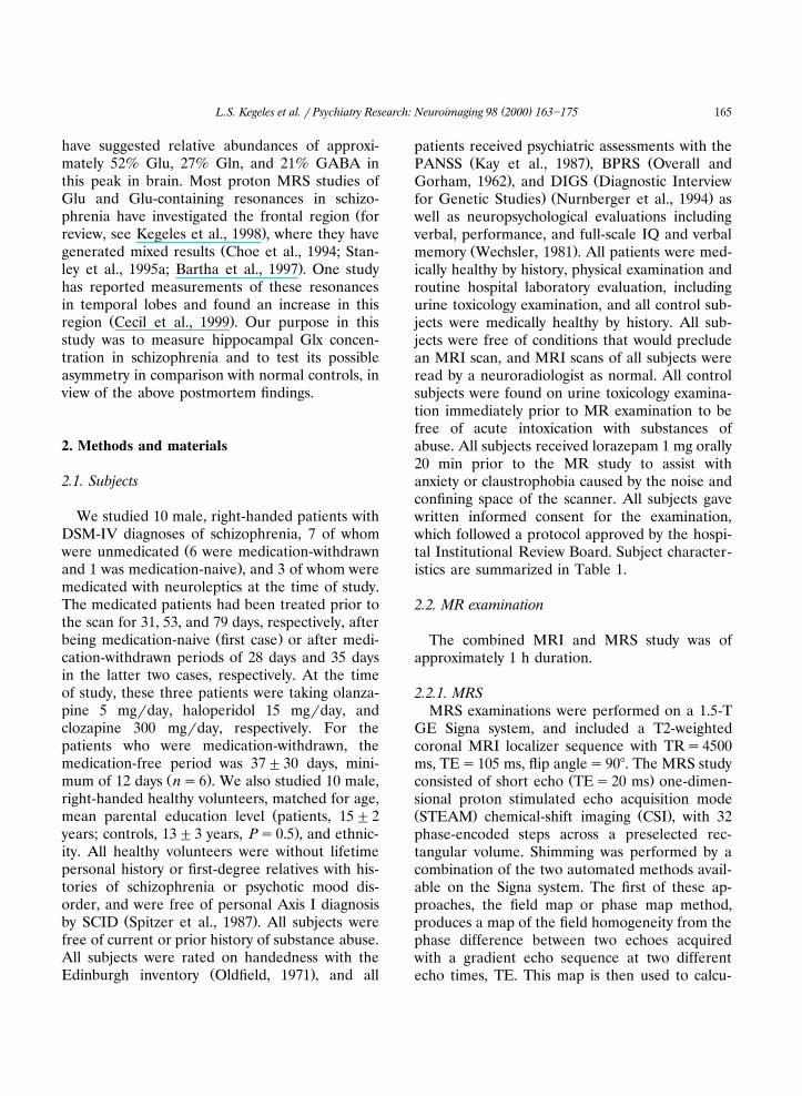
by the phase encoding to provide spectral datafrom individual voxels of dimensions ranging from0.6=0.7=2.0 cm to 1.0=1.0=2.0 cm, resultingin an overall voxel size of 1.32"0.38 cm3, with no
Ž 3difference between the groups 1.40"0.37 cm inthe patients, 1.24"0.39 cm3 in the controls, Ps
. Ž .0.35 Fig. 1 . Voxel size was varied to accommo-date variation in hippocampal cross-section asevident on the coronal localizer, for the purposeof improving the percent overlap of the spectros-copy voxel with hippocampal tissue. Data fromeach of the voxels best centered on the hippo-campi bilaterally were selected for analysis. Inmost cases, one voxel per side per subject wasselected; however, in rare cases when the coronalcross-section of the hippocampus had substantialoverlap with two adjacent voxels, both were se-
Žlected and the data were averaged this occurredin one patient and one control bilaterally, and in
.two patients on the left only . The resulting num-ber of voxels selected did not differ by group orby side: x
2 for the distribution of cases in whichŽone voxel was selected left: 7 patients and 9
.controls; right: 9 patients and 9 controls was 0.13Ž .Fisher’s exact test, Ps0.74 . Spectra were quan-tified using a least-squares non-linear fitting algo-
Ž .rithm Marquardt, 1963 adapted to a LorentzianŽ . Ž .spectral line shape Shungu et al., 1992 Fig. 2 .
()
L.S.K
egelesetal.r
Psychiatry
Research:N
euroimaging
982000
163]175
167
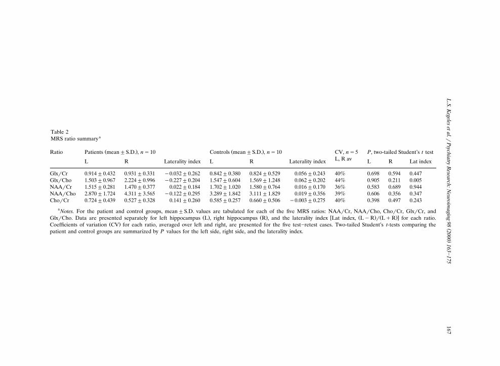
Table 2aMRS ratio summary
Ž . Ž .Ratio CV, ns5Patients mean"S.D. , ns10 Controls mean"S.D. , ns10 P, two-tailed Student’s t testL, R avL R Laterality index L R Laterality index L R Lat index
GlxrCr 0.914"0.432 0.931"0.331 y0.032"0.262 0.842"0.380 0.824"0.529 0.056"0.243 40% 0.698 0.594 0.447GlxrCho 1.503"0.967 2.224"0.996 y0.227"0.204 1.547"0.604 1.569"1.248 0.062"0.202 44% 0.905 0.211 0.005NAArCr 1.515"0.281 1.470"0.377 0.022"0.184 1.702"1.020 1.580"0.764 0.016"0.170 36% 0.583 0.689 0.944NAArCho 2.870"1.724 4.311"3.565 y0.122"0.295 3.289"1.842 3.111"1.829 0.019"0.356 39% 0.606 0.356 0.347ChorCr 0.724"0.439 0.527"0.328 0.141"0.260 0.585"0.257 0.660"0.506 y0.003"0.275 40% 0.398 0.497 0.243
aNotes. For the patient and control groups, mean"S.D. values are tabulated for each of the five MRS ratios: NAArCr, NAArCho, ChorCr, GlxrCr, andŽ . Ž . w Ž . Ž .xGlxrCho. Data are presented separately for left hippocampus L , right hippocampus R , and the laterality index Lat index, LyR r LqR for each ratio.
Ž .Coefficients of variation CV for each ratio, averaged over left and right, are presented for the five test]retest cases. Two-tailed Student’s t-tests comparing thepatient and control groups are summarized by P values for the left side, right side, and the laterality index.
( )L.S. Kegeles et al. r Psychiatry Research: Neuroimaging 98 2000 163]175168
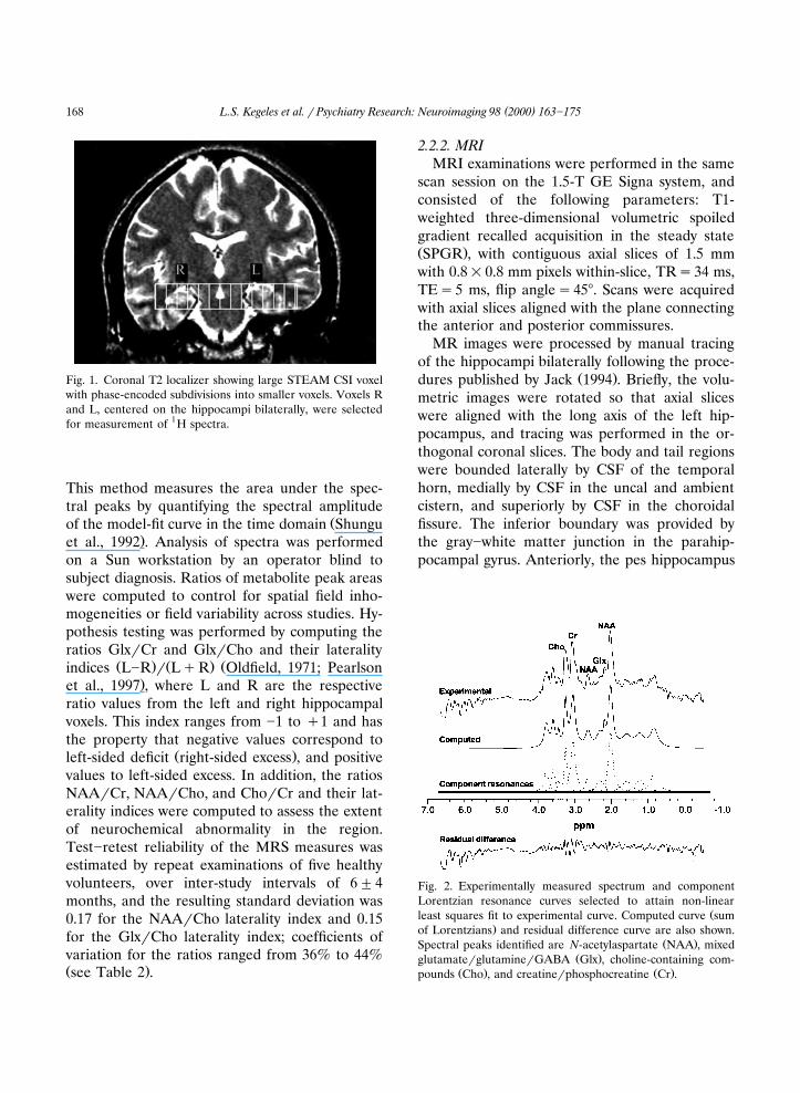
Fig. 1. Coronal T2 localizer showing large STEAM CSI voxelwith phase-encoded subdivisions into smaller voxels. Voxels Rand L, centered on the hippocampi bilaterally, were selectedfor measurement of 1H spectra.
This method measures the area under the spec-tral peaks by quantifying the spectral amplitude
Žof the model-fit curve in the time domain Shungu.et al., 1992 . Analysis of spectra was performed
on a Sun workstation by an operator blind tosubject diagnosis. Ratios of metabolite peak areaswere computed to control for spatial field inho-mogeneities or field variability across studies. Hy-pothesis testing was performed by computing theratios GlxrCr and GlxrCho and their laterality
Ž . Ž . Žindices L]R r LqR Oldfield, 1971; Pearlson.et al., 1997 , where L and R are the respective
ratio values from the left and right hippocampalvoxels. This index ranges from ]1 to q1 and hasthe property that negative values correspond to
Ž .left-sided deficit right-sided excess , and positivevalues to left-sided excess. In addition, the ratiosNAArCr, NAArCho, and ChorCr and their lat-erality indices were computed to assess the extentof neurochemical abnormality in the region.Test]retest reliability of the MRS measures wasestimated by repeat examinations of five healthyvolunteers, over inter-study intervals of 6"4months, and the resulting standard deviation was0.17 for the NAArCho laterality index and 0.15for the GlxrCho laterality index; coefficients ofvariation for the ratios ranged from 36% to 44%Ž .see Table 2 .
2.2.2. MRIMRI examinations were performed in the same
scan session on the 1.5-T GE Signa system, andconsisted of the following parameters: T1-weighted three-dimensional volumetric spoiledgradient recalled acquisition in the steady stateŽ .SPGR , with contiguous axial slices of 1.5 mmwith 0.8=0.8 mm pixels within-slice, TRs34 ms,TEs5 ms, flip angles458. Scans were acquiredwith axial slices aligned with the plane connectingthe anterior and posterior commissures.
MR images were processed by manual tracingof the hippocampi bilaterally following the proce-
Ž .dures published by Jack 1994 . Briefly, the volu-metric images were rotated so that axial sliceswere aligned with the long axis of the left hip-pocampus, and tracing was performed in the or-thogonal coronal slices. The body and tail regionswere bounded laterally by CSF of the temporalhorn, medially by CSF in the uncal and ambientcistern, and superiorly by CSF in the choroidalfissure. The inferior boundary was provided bythe gray]white matter junction in the parahip-pocampal gyrus. Anteriorly, the pes hippocampus
Fig. 2. Experimentally measured spectrum and componentLorentzian resonance curves selected to attain non-linear
Žleast squares fit to experimental curve. Computed curve sum.of Lorentzians and residual difference curve are also shown.
Ž .Spectral peaks identified are N-acetylaspartate NAA , mixedŽ .glutamaterglutaminerGABA Glx , choline-containing com-
Ž . Ž .pounds Cho , and creatinerphosphocreatine Cr .
( )L.S. Kegeles et al. r Psychiatry Research: Neuroimaging 98 2000 163]175 169
was included and was disarticulated from theamygdala by the uncal recess of the temporalhorn. Posteriorly, the last coronal section in-cluded was the one providing a full profile view ofthe crus of the fornix. This procedure includesthe regions CA-1 through CA-4 of the hippocam-pus, the dentate gyrus, the subiculum, and thealveus. Tracings were performed and resultinghippocampal volumes were measured by tworaters using the MEDx software system for image
Ž .analysis Sensor Systems, Sterling, VA . Interraterreliability after initial reliability training, derivedfrom two raters who duplicated 16 of the cases,
Ž .showed intraclass correlation coefficient ICCŽ .Kirk, 1982 s0.96.
Tissue segmentation of these T1-weightedSPGR MRI scans was performed using a combi-nation of in-house and commercial software byfirst editing extracerebral tissue from the images,deriving CSFrgray matterrwhite matter thresh-olds from a triple Gaussian curve fit to the imageintensity histograms, and applying masks withthese thresholds to the MRI restricted to theMRS voxel. Tissue editing was performed using
Žan automatic edge detection algorithm IMIPS,.Inc., Pembroke, MA . Gaussian modeling of the
histograms was performed with in-house algo-Ž .rithms developed using MATLAB Natick, MA .
The points of intersection of the Gaussian func-tions were computed numerically and imported asthreshold values into the MEDx system to per-form the segmentation of the MRI into the gray,white, and CSF compartments. Coordinates of theMRS voxels were transferred to the SPGR MRimages in the MEDx system, which was then usedto compute the gray matterrwhite matterrCSFvolumes within each MRS voxel.
The percent overlap between the MRS voxelsand the manually traced hippocampal structuresŽvoxel volume contained in the hippocampus as a
.percent of total voxel volume was also computedusing the MEDx system to check voxel placement.
2.3. Statistical analysis
Laterality hypothesis testing was performedwith two-tailed Student t-test for group differ-ences in the laterality index values for GlxrCr
and GlxrCho. This was supplemented by mixedeffects modeling using the likelihood ratio testŽ .Bickel and Doksum, 1977 to control for effectson GlxrCho of potential confounding variablesŽgray matter content, voxel overlap with the hip-
.pocampus . Repeated measures ANOVA wasused to test the effects of side, diagnosis, and
Žtheir interaction on MRI measures tissue seg-.mentation, hippocampal volume . Regression
analysis was performed to explore the correla-tions of the MRI and MRS outcome measureswith patient clinical data.
3. Results
3.1. MRS
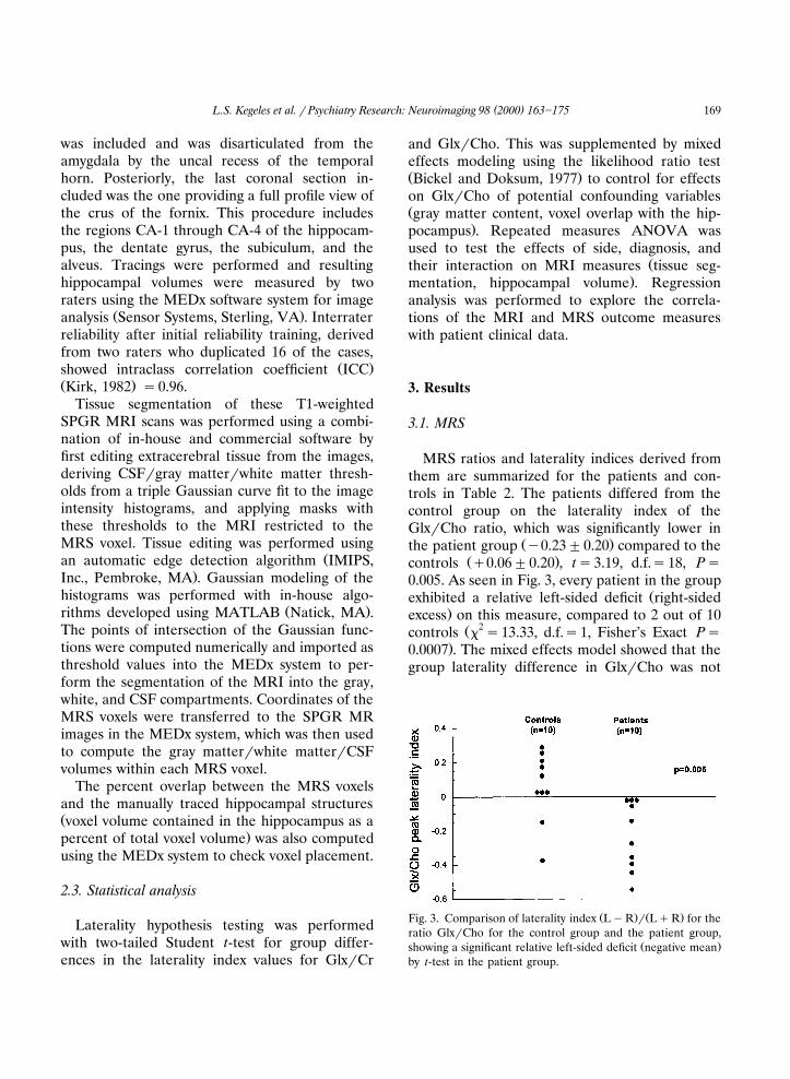
MRS ratios and laterality indices derived fromthem are summarized for the patients and con-trols in Table 2. The patients differed from thecontrol group on the laterality index of theGlxrCho ratio, which was significantly lower in
Ž .the patient group y0.23"0.20 compared to theŽ .controls q0.06"0.20 , ts3.19, d.f.s18, Ps
0.005. As seen in Fig. 3, every patient in the groupŽexhibited a relative left-sided deficit right-sided
.excess on this measure, compared to 2 out of 10Ž 2controls x s13.33, d.f.s1, Fisher’s Exact Ps
.0.0007 . The mixed effects model showed that thegroup laterality difference in GlxrCho was not
Ž . Ž .Fig. 3. Comparison of laterality index LyR r LqR for theratio GlxrCho for the control group and the patient group,
Ž .showing a significant relative left-sided deficit negative meanby t-test in the patient group.
( )L.S. Kegeles et al. r Psychiatry Research: Neuroimaging 98 2000 163]175170
confounded by voxel gray matter content or byvoxel placement relative to the hippocampus: thelikelihood ratio test showed x
2 for main effect ofgray matter percent was 0.46, d.f.s1, Ps0.49; x
2
for main effect of percent overlap was 0.35, d.f.s1, Ps0.55. The effect of side showed a trendtoward significance, with x
2 s3.26, d.f.s1, Ps0.07, while the side by diagnosis interaction wassignificant, with x
2 s4.23, d.f.s1, Ps0.040, ad-justing for side, diagnosis, gray matter percent,and percent overlap. No difference was seen for
Žthe GlxrCr laterality index ]0.03"0.26 for thepatients, q0.06"0.24 for the controls, ts0.78,
.d.f.s18, Ps0.45 , nor for the NAArCho later-Žality index y0.12"0.29 for the patients, q0.02
"0.36 for the controls, ts0.96, d.f.s18, Ps.0.35 . The ratio of NAArCr was lower in the
Žpatients by 11% on the left and by 7% on the.right , but did not attain significance in this sam-
ple.The statistical power to test medication effects
was limited by the small size of the medicated andunmedicated patient subsamples in this study.Nevertheless, both patient subgroups showed sig-nificantly lower values of the laterality index of
ŽGlxrCho than the controls medicated patientsvs. controls, ts4.22, d.f.s11, P-0.01; andunmedicated patients vs. controls, ts2.12, d.f.s
.15, Ps0.05 . The laterality index of GlxrChoŽ .was lower in medicated ns3, y0.46"0.08Ž .compared to unmedicated ns7, y0.13"0.15
Ž .patients ts3.46, d.f.s8, P-0.01 .
3.2. MRI
Hippocampal volumes of patients were 4121"551 mm3 on the left and 4322"647 mm3 on theright; volumes of controls were 4579"372 mm3
on the left and 4554"459 mm3 on the right.Thus, mean hippocampal volumes of patients were10% smaller on the left and 5% smaller on theright compared to the corresponding side in con-trols. Repeated measures ANOVA showed no
Ž Ž .main effect of side Fs1.13, d.f.s 1,18 , Ps. Ž Ž .0.30 or diagnosis Fs2.56, d.f.s 1,18 , Ps. Ž0.13 , and no side by diagnosis interaction Fs
Ž . .1.86, d.f.s 1,18 , Ps0.19 . The laterality indexof hippocampal volume showed no group differ-
Žence patients, y0.02"0.04; controls, q0.001".0.05, Ps0.24 . Total brain volumes were also
Žnon-significantly lower in the patients 1417.0"154.4 cm3 in the patients; 1522.1"149.7 cm3 in
.the controls, Ps0.14 , and hippocampal volumesas percent of total brain volumes similarly showedno significant differences by repeated measures
Ž Ž .ANOVA side: Fs1.40, d.f.s 1,18 , Ps0.25;Ž .diagnosis: Fs0.03, d.f.s 1,18 , Ps0.87; side by
Ž .diagnosis interaction: Fs1.63, d.f.s 1,18 , Ps.0.22 .
Accuracy of MRS voxel placement relative tothe hippocampus was evaluated by a percent
Žoverlap measure voxel volume contained in the.hippocampus as a percent of total voxel volume .
Accuracy of voxel placement did not differŽbetween patients and controls on the left pa-
tients: 40"15%; controls: 47"7%, t s1.31,.d.f.s18, Ps0.21 but was found to differ on the
Žright patients: 34"9%; controls: 48"9% ts.3.35, d.f.s18, P-0.01 . However, the mixed ef-
fects model reported above established theabsence of a confounding impact of this measureon GlxrCho, in that there was no main effect ofpercent overlap on GlxrCho.
In the patients, gray matter accounted for 87"7% of the voxel volumes on the left and 82"9%on the right, while the controls had 83"12%gray matter on the left and 85"15% on theright. Repeated measures ANOVA showed, forgray matter content, no main effect of groupŽ Ž . . ŽFs0.007, d.f.s 1,18 , Ps0.94 or side Fs
Ž . .0.51, d.f.s 1,18 , Ps0.48 , with a trend toward aŽ Ž .group by side interaction Fs3.17, d.f.s 1,18 ,
.Ps0.09 . The mixed effects model reported aboveshowed that the GlxrCho group laterality effectremained significant after correcting for gray mat-ter content.
3.3. Clinical measures
On a post-hoc basis, we examined a series ofpsychiatric and neuropsychological measureswithin the patient group in relation to the hip-pocampal volumes and the laterality indices ofthe MRS measures NAArCho and GlxrCho. Noregression results survived Bonferroni correctionfor multiple comparisons.
( )L.S. Kegeles et al. r Psychiatry Research: Neuroimaging 98 2000 163]175 171
4. Discussion
Our MRS measurements indicated a relativeŽ .asymmetry right-sided excess of GlxrCho, a
Glu-containing measure, in hippocampus inpatients. Evidence for a glutamatergic deficit inschizophrenia was suggested by the finding of low
Ž . Ž .cerebrospinal fluid CSF Glu by Kim et al. 1980 .While subsequent studies failed to replicate this
Žfinding Perry, 1982; Gattaz et al., 1985; Korpi et.al., 1987 , this study has stimulated other investi-
gations of possible Glu abnormalities inschizophrenia, and has led to several Glu and
Ž .N-methyl D-aspartate NMDA -related hypothe-Žses of schizophrenia Wachtel and Turski, 1990;
Javitt and Zukin, 1991; Ulas and Cotman, 1993;.Olney and Farber, 1995; Coyle, 1996 . A recent
postmortem study found Glu deficits in the hip-pocampus in schizophrenia without alterations in
Ž .NAA levels Tsai et al., 1995 . However, the cor-relation of postmortem neurochemical measuresto those obtained through MRS remains to beelucidated. Other recent data have suggested thatGlu excess, rather than deficit, may result fromNMDA Glu receptor blockade or deficitŽ .Moghaddam et al., 1997 , and that Glu excessmay be present in vivo in temporal lobes in
Ž .schizophrenia Cecil et al., 1999 . In view of thelimitations in interpreting the Glx peak, a conclu-
Ž .sion that the findings of Cecil et al. 1999 orthose reported here represent a Glu elevation isvery preliminary. Furthermore, relating such
Ž .findings to the work of Moghaddam et al. 1997and to the NMDA receptor deficit hypotheses of
Žschizophrenia Javitt and Zukin, 1991; Olney and.Farber, 1995 requires an understanding of the
relationship of acute to chronic NMDA receptorhypofunction, which is not fully developedŽ .Jentsch and Roth, 1999 .
The potential for asymmetry in the Cho reso-nance to confound the GlxrCho measurementshould be considered. While lower Cho values onthe right appear to contribute to the relativeright-sided elevation of GlxrCho, these are notsufficient to result in significant differences inNAArCho or ChorCr or their laterality indices.This is in contrast to the laterality index of
GlxrCho, suggesting a contribution from Glx aswell. In particular, the choice of two voxels on theleft and one on the right in two patients formetabolite analysis does not appear to have con-tributed to lower right-sided Cho values: thesetwo patients had ChorCr laterality indices ofy0.018 and q0.087, both lower than the patientgroup mean of 0.141, i.e. both had relatively moreChorCr on the right than the average of thepatients. The absence of a difference for theChorCr ratio is consistent with most published
Ž .studies including Renshaw et al. 1995 , BertolinoŽ . Ž .et al. 1996 , and Yurgelun-Todd et al. 1996 .
Studies of the Cr and Cho resonances in temporallobes in schizophrenia that have claimed absolutemetabolite concentration measurements have also
Ž .yielded negative results. Buckley et al. 1994 ,who quantified by computing the peak ratio to themean background, found no group differences;
Ž .Maier et al. 1995 , using unsuppressed water asinternal reference, found comparable left-sideddecreases in Cho, Cr, and NAA, and therefore nochanges in the ratios.
Our results suggest that the reduced GlxrCholaterality index may be present in both medicatedand unmedicated patients with schizophrenia.Nevertheless, the small subsample sizes mean thatfurther studies will be needed to rule out a neu-roleptic medication effect. Medication effects onthe Cho peak produced by antidepressants have
Žbeen suggested in other brain regions Renshaw.et al., 1997 , but no patient in our sample was
taking antidepressant medication any fewer than38 days prior to the MRS examination. Theadministration of lorazepam is an unlikely poten-tial confounder of our results. Lorazepam acts ona specific site within the GABA receptor complexto potentiate the inhibitory effects of that tran-smitter. However, a lateralized effect produced bya systemically administered agent would not beexpected, unless there were a preexisting lateral-ized abnormality in the GABA or Glu systemassociated with schizophrenia. The lateralizedGlxrCho results are consistent with the possibil-ity that there may be no Glx group difference at
Ž .baseline prior to administration of lorazepam ,but that acute benzodiazepine agonist challenge

( )L.S. Kegeles et al. r Psychiatry Research: Neuroimaging 98 2000 163]175172
may cause an excess right-sided Glx response inthe patients. This interpretation is limited by theabsence of the baseline measurement, but is alsoconsistent with abnormalities in GlurGlnrGABAtransmission in the patients. Attrition of subjectsdue to anxiety and claustrophobia in the MRIscanner is much greater without lorazepam; nev-ertheless, a study without lorazepam would bedesirable.
Our NAA measurements did not attain statisti-cal significance in terms of group differences,although they suggested deficits in the NAArCrratio in the hippocampal region in the patient
Žgroup, consistent with previous reports Nasrallahet al., 1994, 1995; Fukuzako et al., 1995; Renshawet al., 1995; Bertolino et al., 1996; Yurgelun-Todd
.et al., 1996; Cecil et al., 1999 . Deficits are consis-tent with neuronal loss or non-specific dysfunc-tion in this region.
Although the differences were not statisticallyŽ .significant, lower mean left-sided 10% and
Ž .right-sided 5% hippocampal volumes in theschizophrenic patient group are comparable inmagnitude with many prior studies including the
Ž .meta-analysis of Nelson et al. 1998 . The absenceof a significant lateralization difference in thepatient volumes is suggestive that volume averag-ing did not confound the lateralized MRS finding.The voxel placement and segmentation analysesalso support the absence of these potential arti-facts contributing to the finding of neurochemicalasymmetry.
Anterior]posterior variability in voxel place-ment probably did not contribute to lateralized
Ždifferences in the MRS measures e.g. throughanterior]posterior differences in metabolite con-
.centrations , since the placement measure did notdiffer by group, and left and right voxels were inthe same coronal plane in each subject. Whilevoxel placement did not explain the asymmetryfound in GlxrCho, it illustrates that a potentialpitfall of single-voxel or small-voxel-number MRSstudies is poor reliability of voxel placement acrosssubjects. This problem can be addressed by accu-rate use of landmarks at acquisition time, anapproach not generally adopted in the MRSschizophrenia literature or, as done here, by
post-acquisition investigation of placement vari-ability and its relationship to neurochemistry re-sults. More difficult to control at acquisition isthe tissue composition of the MRS voxels, whichmust be addressed post-acquisition to rule outthis potential confound. Other authors have ad-dressed tissue segmentation post-acquisitionŽHetherington et al., 1996; Lim and Spielman,
.1997; Renshaw et al., 1997 .A limitation of this study is the relatively low
test]retest reliability for the Glx measurementsperformed at 1.5 T reported here and elsewhereŽ .Stanley et al., 1995b , and is a reason for inter-preting the GlxrCho laterality finding as prelimi-nary. Test]retest reliability data for 1H-MRS re-ported by other investigators indicate consider-
Žable variability Renshaw et al., 1995; Stanley etal., 1995b; Charles et al., 1996; Bertolino et al.,
. Ž .1998 . An ICC of 0.59 S.D.s0.16 was found byŽ .Renshaw et al. 1995 in temporal lobe for the
NAArCr ratio, comparable to our value of 0.64Ž .S.D.s0.17 for the laterality index of NAArCho.
Ž .Stanley et al. 1995b reported coefficient of vari-Ž .ation CV values ranging from 20% to over 50%
for the components of the Glx peak in frontalŽlobes, comparable to our range of 36]44% Table
. 32 . In smaller regions covered by 0.8 cm voxels,Ž .Bertolino et al. 1998 measured CV values rang-
ing from 15 to 25% for the NAArCho ratio.Contributing to the relatively low reliability oftemporal lobe Glx-containing ratio measurements
Ž .are the combined factors of small ROI voxelsize, the low signal intensity of the Glx measure,and the high magnetic susceptibility variation ofthe temporal lobe region. It is possible that im-provements in standardization of voxel location atacquisition time will improve test]retest reliabil-ity of MRS measures in smaller anatomic regions.
ŽUltimately, higher magnetic field strengths Roth-man et al., 1992, 1993; Mason et al., 1994; Heth-
.erington et al., 1997 and spectroscopic editingŽmethods Rothman et al., 1992, 1993; Keltner et
.al., 1996; Hetherington et al., 1997 can provideimproved spectral and spatial resolution as wellas sensitivity, and are strategies for use in futurestudies to further investigate Glu abnormalities inhippocampus in schizophrenia.
( )L.S. Kegeles et al. r Psychiatry Research: Neuroimaging 98 2000 163]175 173
Acknowledgements
This research was supported in part by a YoungInvestigator Award from the National Alliancefor Research on Schizophrenia and DepressionŽ .NARSAD , by a grant from the G. Harold andLeila Y. Mathers Charitable Trust, and by PublicHealth Service Grants MH46745, MH50727, and1-R01-MH54192. We thank Mindy Kroll, AnnShinn, and Sheldon Yao for their assistance withsubject recruitment and escorting, and RaymondGoetz for his assistance with the NYSPISchizophrenia Research Unit database.
References
Bartha, R., Williamson, P.C., Drost, D.J., Malla, A., Carr, T.J.,Cortese, L., Canaran, G., Rylett, R.J., Neufeld, R.W.J.,1997. Measurement of glutamate and glutamine in themedial prefrontal cortex of never-treated schizophrenicpatients and healthy controls by proton magnetic resonancespectroscopy. Archives of General Psychiatry 54, 959]965.
Bertolino, A., Callicott, J.H., Nawroz, S., Mattay, V.S., Duyn,J.H., Tedeschi, G., Frank, J.A., Weinberger, D.R., 1998.Reproducibility of proton magnetic resonance spectros-copic imaging in patients with schizophrenia. Neuropsy-chopharmacology 18, 1]9.
Bertolino, A., Nawroz, S., Mattay, V.S., Barnett, A.S., Duyn,J.H., Moonen, C.T.W., Frank, J.A., Tedeschi, G., Wein-berger, D.R., 1996. Regionally specific pattern of neu-rochemical pathology in schizophrenia as assessed by multi-slice proton magnetic resonance spectroscopic imaging.American Journal of Psychiatry 153, 1554]1563.
Bickel, P.J., Doksum, K.A., 1977. Mathematical Statistics:Basic Ideas and Selected Topics. Holden-Day, San Fran-cisco.
Bogerts, B., Ashtari, M., Degreef, G., Alvir, J.M., Bilder, R.M.,Lieberman, J.A., 1990. Reduced temporal limbic structurevolumes on magnetic resonance images in first episodeschizophrenia. Psychiatry Research 35, 1]13.
Bogerts, B., Meertz, E., Schonfeldt-Bausch, R., 1985. Basalganglia and limbic system pathology in schizophrenia.Archives of General Psychiatry 42, 784]791.
Buckley, P.F., Moore, C., Long, H., Larkin, C., Thompson, P.,Mulvany, F., Redmond, O., Stack, J.P., Ennis, J.T.,Waddington, J.L., 1994. 1H-magnetic resonance spectros-copy of the left temporal and frontal lobes in schizophre-nia: clinical, neurodevelopmental, and cognitive correlates.Biological Psychiatry 36, 792]800.
Cecil, K.M., Lenkinski, R.E., Gur, R.E., Gur, R.C., 1999.Proton magnetic resonance spectroscopy in the frontal andtemporal lobes of neuroleptic naive patients withschizophrenia. Neuropsychopharmacology 20, 131]140.
Charles, H.C., Lazeyras, F., Tupler, L.A., Krishnan, K.R.R.,
1996. Reproducibility of high spatial resolution protonmagnetic resonance spectroscopic imaging in the humanbrain. Magnetic Resonance in Medicine 35, 606]610.
Choe, B.Y., Kim, K.T., Suh, T.S., Lee, C., Paik, I.H., Bahk,Y.W., Shinn, K.S., Lenkinski, R.E., 1994. 1H magneticresonance spectroscopy characterization of neuronal dys-function in drug-naive, chronic schizophrenia. AcademicRadiology 1, 211]216.
Coyle, J.T., 1996. The glutamatergic dysfunction hypothesisfor schizophrenia. Harvard Review of Psychiatry 3, 241]253.
Deakin, J.F., Slater, P., Simpson, M.D., Gilchrist, A.C., Skan,W.J., Royston, M.C., Reynolds, G.P., Cross, A.J., 1989.Frontal cortical and left temporal glutamatergic dysfunc-tion in schizophrenia. Journal of Neurochemistry 52,1781]1786.
DeLisi, L.E., Tew, W., Xie, S., Hoff, A.L., Sakuma, M., Kush-ner, M., Lee, G., Shedlack, K., Smith, A.M., Grimson, R.,1995. A prospective follow-up study of brain morphologyand cognition in first-episode schizophrenic patients: pre-liminary findings. Biological Psychiatry 38, 349]360.
Ellison, Z.R., Wright, I.C., McGuire, P.K., 1997. Neuroimage5, S273.
Fukuzako, H., Takeuchi, K., Hokazono, Y., Fukuzako, T.,Yamada, K., Hashiguchi, T., Obo, Y., Ueyama, K., Taki-gawa, M., Fujimoto, T., 1995. Proton magnetic resonancespectroscopy of the left medial temporal and frontal lobesin chronic schizophrenia: preliminary report. PsychiatryResearch: Neuroimaging 61, 193]200.
Gattaz, W.F., Gasser, T., Beckmann, H., 1985. Multidimensio-nal analysis of the concentrations of 17 substances in theCSF of schizophrenics and controls. Biological Psychiatry20, 360]366.
Harrison, P.J., McLaughlin, D., Kerwin, R.W., 1991. De-creased hippocampal expression of a glutamate receptorgene in schizophrenia. Lancet 337, 450]452.
Hetherington, H.P., Newcomer, B.R., Pan, J.W., 1997. Mea-Ž .surements of human cerebral GABA at 4T abstract .
Proceedings of the International Society for Magnetic Res-onance in Medicine 1, 240.
Hetherington, H.P., Pan, J.W., Mason, G.F., Adams, D.,Vaughn, M.J., Twieg, D.B., Pohost, G.M., 1996. Quantita-tive 1H spectroscopic imaging of human brain at 4.1 Tusing image segmentation. Magnetic Resonance inMedicine 36, 21]29.
Hyde, T., Casanova, M., Kleinman, J.E., Weinberger, D.R.,1991. Neuroanatomical and neurochemical pathology inschizophrenia. American Psychiatric Press Review in Psy-chiatry 10, 7]23.
Jack, C.R., 1994. MRI-based hippocampal volume measure-ments in epilepsy. Epilepsia 35, S21]S29.
Jakob, H., Beckmann, H., 1986. Prenatal developmental dis-turbances in the limbic allocortex in schizophrenics. Jour-nal of Neural Transmission 65, 303]326.
Javitt, D.C., Zukin, S.R., 1991. Recent advances in the phency-clidine model of schizophrenia. American Journal of Psy-chiatry 148, 1301]1308.
Jentsch, J.D., Roth, R.H., 1999. The neuropsychopharma-
( )L.S. Kegeles et al. r Psychiatry Research: Neuroimaging 98 2000 163]175174
cology of phencyclidine: from NMDA receptor hypofunc-tion to the dopamine hypothesis of schizophrenia. Neu-ropsychopharmacology 20, 201]225.
Kay, S.R., Fiszbein, A., Opler, L.A., 1987. The positive andŽ .negative syndrome scale PANSS for schizophrenia.
Schizophrenia Bulletin 13, 261]276.Kegeles, L.S., Humaran, T.J., Mann, J.J., 1998. In vivo neu-
rochemistry of the brain in schizophrenia as revealed bymagnetic resonance spectroscopy. Biological Psychiatry 44,382]398.
Kelsoe Jr., J.R., Cadet, J.L., Pickar, D., Weinberger, D.R.,1988. Quantitative neuroanatomy in schizophrenia. A con-trolled magnetic resonance imaging study. Archives ofGeneral Psychiatry 45, 533]541.
Keltner, J.R., Wald, L.L., Christensen, J.D., Maas, L.C., Moore,C.M., Cohen, B.M., Renshaw, P.F., 1996. A technique fordetecting GABA in the human brain with PRESS localiza-tion and optimized refocusing spectral editing radiofre-quency pulses. Magnetic Resonance in Medicine 36,458]461.
Kerwin, R.W., Patel, S., Meldrum, B.S., Czudek, C., Reynolds,G.P., 1988. Asymmetrical loss of glutamate receptor sub-type in left hippocampus in schizophrenia. Lancet 1,583]584.
Kim, J.S., Kornhuber, H.H., Schmid-Burgk, W., Holzmuller,B., 1980. Low cerebrospinal fluid glutamate in schizophrenicpatients and a new hypothesis on schizophrenia. Neuro-science Letters 20, 379]382.
Kirk, R.E., 1982. Experimental Design: Procedures for theBehavioral Sciences. BrooksrCole Publishing Company,Pacific Grove, California.
Korpi, E.R., Kaufmann, C.A., Marnela, K.-M., Weinberger,D.R., 1987. Cerebrospinal fluid amino acid concentrationsin chronic schizophrenia. Psychiatry Research 20, 337]345.
Kovelman, J.A., Scheibel, A.B., 1984. A neurohistological cor-relate of schizophrenia. Biological Psychiatry 19, 1602]1621.
Lillrank, S.M., Lipska, B.K., Weinberger, D.R., 1995. Neu-rodevelopmental animal models of schizophrenia. ClinicalNeuroscience 3, 98]104.
Lim, K.O., Spielman, D.M., 1997. Estimating NAA in corticalgray matter with applications for measuring changes due toaging. Magnetic Resonance in Medicine 37, 372]377.
Maier, M., Ron, M.A., Barker, G.J., Tofts, P.S., 1995. Protonmagnetic resonance spectroscopy: an in vivo method ofestimating hippocampal neuronal depletion in schizophre-nia. Psychological Medicine 25, 1201]1209.
Marion, D., Ikura, M., Bax, A., 1989. Improved solvent sup-pression in one- and two-dimensional NMR spectroscopyby convolution of time-domain data. Journal of MagneticResonance 84, 425]430.
Marquardt, D., 1963. An algorithm for least-squares estima-tion of nonlinear parameters. Journal of Applied Mathe-matics 11, 431]441.
Mason, G.F., Pan, J.W., Ponder, S.L., Twieg, D.B., Pohost,G.M., Hetherington, H.P., 1994. Detection of brain gluta-mate and glutamine in spectroscopic images at 4.1 T.Magnetic Resonance in Medicine 32, 142]145.
Moghaddam, B., Adams, B., Verma, A., Daly, D., 1997. Acti-vation of glutamatergic neurotransmission by ketamine: Anovel step in the pathway from NMDA receptor blockadeto dopaminergic and cognitive disruptions associated withthe prefrontal cortex. Journal of Neuroscience 17,2921]2927.
Nasrallah, H.A., Skinner, T.E., Schmalbrock, P., Robitaille,Ž1P.M., 1994. Proton magnetic resonance spectroscopy H
.MRS of the hippocampal formation in schizophrenia: apilot study. British Journal of Psychiatry 165, 481]485.
Nasrallah, H.A., Skinner, T.E., Schmalbrock, P., Robitaille,P.M., 1995. In vivo 1H-NMR-spectroscopy of the limbictemporal lobe in patients with schizophrenia. In: Nasrallah,
Ž .H.A., Pettegrew, J.W. Eds. , NMR Spectroscopy in Psychi-atric Brain Disorders. American Psychiatric Press, Inc,Washington, DC, pp. 1]20.
Nelson, M.D., Saykin, A.J., Flashman, L.A., Riordan, H.J.,1998. Hippocampal volume reduction in schizophrenia asassessed by magnetic resonance imaging: a meta-analyticstudy. Archives of General Psychiatry 55, 433]440.
Nurnberger, J., Blehar, M.C., Kaufmann, C.A., York-Cooler,C., Simpson, S.G., Harkavy-Friedman, J., Severe, J.B.,Malaspina, D., Reich, T., 1994. Diagnostic interview forgenetic studies. Rationale, unique features, and training.NIMH Genetics Initiative. Archives of General Psychiatry51, 849]859.
Oldfield, R.C., 1971. The assessment and analysis of handed-ness: the Edinburgh inventory. Neuropsychologia 9, 97]113.
Olney, J.W., Farber, N.B., 1995. Glutamate receptor dysfunc-tion and schizophrenia. Archives of General Psychiatry 52,998]1007.
Overall, J.E., Gorham, D.R., 1962. The brief psychiatric ratingscale. Psychological Reports 10, 799]812.
Pearlson, G.D., Barta, P.E., Powers, R.E., Menon, R.R.,Richards, S.S., Aylward, E.H., Federman, E.B., Chase, G.A.,Petty, R.G., Tien, A.Y., 1997. Medial and superior tem-poral gyral volumes and cerebral asymmetry in schizophre-nia versus bipolar disorder. Biological Psychiatry 41, 1]14.
Perry, T.L., 1982. Normal cerebrospinal fluid and brain gluta-mate levels in schizophrenia do not support the hypothesisof glutamatergic neuronal dysfunction. Neuroscience Let-ters 28, 81]85.
Renshaw, P.F., Lafer, B., Babb, S.M., Fava, M., Stoll, A.L.,Christensen, J.D., Moore, C.M., Yurgelun-Todd, D.A.,Bonello, C.M., Pillay, S.S., Rothschild, A.J., Nierenberg,A.A., Rosenbaum, J.F., Cohen, B.M., 1997. Basal gangliacholine levels in depression and response to fluoxetinetreatment: an in vivo proton magnetic resonance spectros-copy study. Biological Psychiatry 41, 837]843.
Renshaw, P.F., Yurgelun-Todd, D.A., Tohen, M., Gruber, S.,Cohen, B.M., 1995. Temporal lobe proton magnetic reso-nance spectroscopy of patients with first-episode psychosis.American Journal of Psychiatry 152, 444]446.
Rothman, D.L., Hanstock, C.C., Petroff, O.A., Novotny, E.J.,Prichard, J.W., Shulman, R.G., 1992. Localized 1H NMRspectra of glutamate in the human brain. Magnetic Reso-nance Medicine 25, 94]106.
( )L.S. Kegeles et al. r Psychiatry Research: Neuroimaging 98 2000 163]175 175
Rothman, D.L., Petroff, O.A., Behar, K.L., Mattson, R.H.,1993. Localized 1H NMR measurements of gamma-aminobutyric acid in human brain in vivo. Proceedings ofthe National Academy of Sciences USA 90, 5662]5666.
Shenton, M.E., Kikinis, R., Jolesz, F.A., Pollak, S.D., LeMay,M., Wible, C.G., Hokama, H., Martin, J., Metcalf, D.,Coleman, M., 1992. Abnormalities of the left temporal lobeand thought disorder in schizophrenia. A quantitative mag-netic resonance imaging study. New England Journal ofMedicine 327, 604]612.
Shungu, D.C., Bhujwalla, Z.M., Li, S.J., Rose, L.M., Wehrle,J.P., Glickson, J.D., 1992. Determination of absolute phos-phate metabolite concentration in RIF-1 tumors in vivo by31 P-1H-2 H NMR spectroscopy using water as an internalintensity reference. Magnetic Resonance in Medicine 28,105]121.
Spitzer, R.L., Williams, J.B.W., Gibbon, N., First, M.D., 1987.Ž .Structured Clinical Interview for DSM-III-R SCID . New
York State Psychiatric Institute, Biometrics Research De-partment, New York.
Stanley, J.A., Drost, D.J., Williamson, P.C., Carr, T.J., 1995a.In vivo proton MRS study of glutamate and schizophrenia.
Ž .In: Nasrallah, H.A., Pettegrew, J.W. Eds. , NMR Spectros-copy in Psychiatric Brain Disorders. American PsychiatricPress, Inc, Washington, DC, pp. 21]44.
Stanley, J.A., Drost, D.J., Williamson, P.C., Thompson, R.T.,1995b. The use of a priori knowledge to quantify short echoin vivo 1H MR spectra. Magnetic Resonance in Medicine34, 17]24.
Stefanis, N., Yakely, J., Frangou, S., Sharma, T., O’Connell, P.,Morgan, K., Murray, R., 1997. Pregnancy and birth compli-
Ž .cations PBC -associated hippocampal volume reduction insporadic schizophrenia. Schizophrenia Research 24, S157.
Suddath, R.L., Casanova, M.F., Goldberg, T.E., Daniel, D.G.,Kelsoe Jr., J., Weinberger, D.R., 1989. Temporal lobepathology in schizophrenia: a quantitative magnetic reso-
nance imaging study. American Journal of Psychiatry 146,464]472.
Suddath, R.L., Christison, G.W., Torrey, E.F., Casanova, M.F.,Weinberger, D.R., 1990. Anatomical abnormalities in thebrains of monozygotic twins discordant for schizophrenia.New England Journal of Medicine 322, 789]794.
Swayze, V.W.I., Andreasen, N.C., Alliger, R.J., Yuh, W.T.,Ehrhardt, J.C., 1992. Subcortical and temporal structures inaffective disorder and schizophrenia: a magnetic resonance
w ximaging study see comments . Biological Psychiatry 31,221]240.
Torres, I.J., Flashman, L.A., O’Leary, D.S., Swayze II, V.,Andreasen, N.C., 1997. Lack of an association betweendelayed memory and hippocampal and temporal lobe sizein patients with schizophrenia and healthy controls. Biolog-ical Psychiatry 42, 1087]1096.
Tsai, G., Passani, L.A., Slusher, B.S., Carter, R., Baer, L.,Kleinman, J.E., Coyle, J.T., 1995. Abnormal excitatory neu-rotransmitter metabolism in schizophrenic brains. Archivesof General Psychiatry 52, 829]836.
Ulas, J., Cotman, C.W., 1993. Excitatory amino acid receptorsin schizophrenia. Schizophrenia Bulletin 19, 105]117.
Wachtel, H., Turski, L., 1990. Glutamate: a new target inschizophrenia? Trends in Pharmacology 11, 219]220.
Wechsler, D., 1981. WAIS-R Manual. Psychological Corpora-tion, New York.
Yurgelun-Todd, D.A., Renshaw, P.F., Gruber, S.A., Water-naux, C., Cohen, B.M., 1996. Proton magnetic resonancespectroscopy of the temporal lobes in schizophrenics andnormal controls. Schizophrenia Research 19, 55]59.
Zipursky, R.B., Marsh, L., Lim, K.O., Dement, S., Shear, P.K.,Sullivan, E.V., Murphy, G.M., Csernansky, J.G., Pfeffer-baum, A., 1994. Volumetric MRI assessment of temporallobe structures in schizophrenia. Biological Psychiatry 35,501]551.
Related Documents


![Gut microbiome and magnetic resonance spectroscopy study of … · of schizophrenia has many classic hypotheses, such as the dopamine (DA) hypothesis [11], serotonin (5-HT) hypothesis](https://static.cupdf.com/doc/110x72/5fd7f81d71c34432dc2ad713/gut-microbiome-and-magnetic-resonance-spectroscopy-study-of-of-schizophrenia-has.jpg)