Hindawi Publishing Corporation Stem Cells International Volume 2013, Article ID 205878, 14 pages http://dx.doi.org/10.1155/2013/205878 Research Article Hippocampal Neurogenesis and the Brain Repair Response to Brief Stereotaxic Insertion of a Microneedle Shijie Song, 1,2 Shuojing Song, 3 Chuanhai Cao, 4 Xiaoyang Lin, 4 Kunyu Li, 1 Vasyl Sava, 1,2 and Juan Sanchez-Ramos 1,2,4 1 Department of Neurology, University of South Florida, 13220 Laurel Drive, Tampa, FL 33612, USA 2 Research Service, James A. Haley VA Medical Center, Tampa, FL 33612, USA 3 Feinberg School of Medicine, Northwestern University, Chicago, IL 60611, USA 4 Department of Molecular Pharmacology and Physiology, University of South Florida, Tampa, FL 33612, USA Correspondence should be addressed to Juan Sanchez-Ramos; [email protected] Received 28 June 2012; Revised 23 November 2012; Accepted 14 January 2013 Academic Editor: Joshua J. Breunig Copyright © 2013 Shijie Song et al. is is an open access article distributed under the Creative Commons Attribution License, which permits unrestricted use, distribution, and reproduction in any medium, provided the original work is properly cited. We tested the hypothesis that transient microinjury to the brain elicits cellular and humoral responses that stimulate hippocampal neurogenesis. Brief stereotaxic insertion and removal of a microneedle into the right hippocampus resulted in (a) significantly increased expression of granulocyte-colony stimulating factor (G-CSF), the chemokine MIP-1a, and the proinflammatory cytokine IL12p40; (b) pronounced activation of microglia and astrocytes; and (c) increase in hippocampal neurogenesis. is study describes immediate and early humoral and cellular mechanisms of the brain’s response to microinjury that will be useful for the investigation of potential neuroprotective and deleterious effects of deep brain stimulation in various neuropsychiatric disorders. 1. Background Deep brain stimulation through chronically implanted metal electrodes into specific brain regions is becoming a com- mon therapeutic choice for medication refractory movement disorders such as Parkinson’s disease (PD), tremors, and dystonia (see reviews [1–3]). More recently, DBS has been applied to psychiatric and behavioral disorders including depression, obsessive compulsive disorder, and addiction and most recently to disorders of consciousness [4–9]. Long-term implantation of a fine metal electrode, even without chronic electrical stimulation may produce unwanted effects. Neuropathological examination of brain tissue from patients with DBS revealed activated astrocytes and microglia regardless of the underlying disease [10–15]. Electrical stimulation is not required to see signs of neu- roinflammation; inflammatory changes have been observed around recording electrodes used for characterizing epilep- togenic tissue and around CSF fluid shunt catheters [16, 17]. To understand the earliest reactions to implantation of a metal electrode, we studied the cellular and cytokine responses over time to transient insertion of a fine needle (maximum diameter of 200 m) into the dorsal hippocampus of the mouse. We tested the hypothesis that the creation of a focal microlesion in hippocampus elicits self-repair mechanisms mediated by cytokines which activate microglia, promote astrocytosis, and stimulate stem/progenitor cells to proliferate and generate new neurons. 2. Materials and Methods All procedures described here were reviewed and approved by the IACUC Committee of the University of South Florida and the Haley VA Research Service. 2.1. Animals. C57BL/6 mice, 8–10 weeks old, were pur- chased from Harlan Laboratories, and transgenic GFP mice (C57BL/6-Tg [ACTB-EGFP] 1Osb/J, 003291) were purchased from Jackson Laboratory (Bar Harbor, ME). Most of the experiments utilized groups of C57BL/6 mice, and one experiment utilized chimeric mice (C57BL/6 mice trans- planted with green fluorescent protein expressing (GFP+) bone marrow).

Welcome message from author
This document is posted to help you gain knowledge. Please leave a comment to let me know what you think about it! Share it to your friends and learn new things together.
Transcript

Hindawi Publishing CorporationStem Cells InternationalVolume 2013 Article ID 205878 14 pageshttpdxdoiorg1011552013205878
Research ArticleHippocampal Neurogenesis and the Brain Repair Response toBrief Stereotaxic Insertion of a Microneedle
Shijie Song12 Shuojing Song3 Chuanhai Cao4 Xiaoyang Lin4 Kunyu Li1
Vasyl Sava12 and Juan Sanchez-Ramos124
1 Department of Neurology University of South Florida 13220 Laurel Drive Tampa FL 33612 USA2 Research Service James A Haley VA Medical Center Tampa FL 33612 USA3 Feinberg School of Medicine Northwestern University Chicago IL 60611 USA4Department of Molecular Pharmacology and Physiology University of South Florida Tampa FL 33612 USA
Correspondence should be addressed to Juan Sanchez-Ramos jsramoshealthusfedu
Received 28 June 2012 Revised 23 November 2012 Accepted 14 January 2013
Academic Editor Joshua J Breunig
Copyright copy 2013 Shijie Song et al This is an open access article distributed under the Creative Commons Attribution Licensewhich permits unrestricted use distribution and reproduction in any medium provided the original work is properly cited
We tested the hypothesis that transient microinjury to the brain elicits cellular and humoral responses that stimulate hippocampalneurogenesis Brief stereotaxic insertion and removal of a microneedle into the right hippocampus resulted in (a) significantlyincreased expression of granulocyte-colony stimulating factor (G-CSF) the chemokine MIP-1a and the proinflammatory cytokineIL12p40 (b) pronounced activation ofmicroglia and astrocytes and (c) increase in hippocampal neurogenesisThis study describesimmediate and early humoral and cellularmechanisms of the brainrsquos response tomicroinjury that will be useful for the investigationof potential neuroprotective and deleterious effects of deep brain stimulation in various neuropsychiatric disorders
1 Background
Deep brain stimulation through chronically implanted metalelectrodes into specific brain regions is becoming a com-mon therapeutic choice for medication refractory movementdisorders such as Parkinsonrsquos disease (PD) tremors anddystonia (see reviews [1ndash3]) More recently DBS has beenapplied to psychiatric and behavioral disorders includingdepression obsessive compulsive disorder and addiction andmost recently to disorders of consciousness [4ndash9]
Long-term implantation of a fine metal electrodeeven without chronic electrical stimulation may produceunwanted effects Neuropathological examination of braintissue from patients with DBS revealed activated astrocytesand microglia regardless of the underlying disease [10ndash15]Electrical stimulation is not required to see signs of neu-roinflammation inflammatory changes have been observedaround recording electrodes used for characterizing epilep-togenic tissue and around CSF fluid shunt catheters [16 17]
To understand the earliest reactions to implantation ofa metal electrode we studied the cellular and cytokineresponses over time to transient insertion of a fine needle
(maximumdiameter of 200120583m) into the dorsal hippocampusof the mouse We tested the hypothesis that the creationof a focal microlesion in hippocampus elicits self-repairmechanismsmediated by cytokines which activate microgliapromote astrocytosis and stimulate stemprogenitor cells toproliferate and generate new neurons
2 Materials and Methods
All procedures described here were reviewed and approvedby the IACUC Committee of the University of South Floridaand the Haley VA Research Service
21 Animals C57BL6 mice 8ndash10 weeks old were pur-chased from Harlan Laboratories and transgenic GFP mice(C57BL6-Tg [ACTB-EGFP] 1OsbJ 003291) were purchasedfrom Jackson Laboratory (Bar Harbor ME) Most of theexperiments utilized groups of C57BL6 mice and oneexperiment utilized chimeric mice (C57BL6 mice trans-planted with green fluorescent protein expressing (GFP+)bone marrow)
2 Stem Cells International
22 Generation of Chimeric Mice The procedure for bonemarrow harvesting from tg GFP+ mice has been previouslypublished by Sanchez-Ramos and coworkers [18 19] Brieflybone marrow cells are collected from femurs and tibias ofadult male GFP transgenic mice by flushing the bone shaftwith PBS + 05 bovine serum albumin (BSA) + 2mMethylenediaminetetraacetic acid (EDTA) (Sigma)
To generate chimeric mice C57BL6J mice were lethallyirradiated with 8Gy total body irradiation (delivered in twofractions of 4Gy an interval of 4 hours) at dose rate of103Gymin in a Gammacell 40 Extractor [20] Followingirradiation the mice were given a bone marrow transplant(10 times 106 mononuclear cells) from transgenic GFP miceinfused via tail vein Bone marrow-derived cells in therescuedmice were readily tracked by virtue of their green flu-orescence Examination of blood smears from tail clippingsfor the presence of green monocytes confirmed successfulengraftment
23 Stereotaxic Insertion and Removal of MicroneedleAnimals were anesthetized with sodium pentobarbital(50mgkg ip) and placed into a stereotactic frame Usingbregma as the reference point a trephine hole was thendrilled in the skull and the needle was gently inserted intothe hippocampus (AP 25mm ML 13mm DV 35mm)Mice received 5-bromo-21015840-deoxyuridine (BrdU) (Sigma)injections (100mgkg ip Bid immediately after the surgeryand 2 days after surgery) to label nascent cells during a 3-dayperiod
24 Tissue Preparation and Sectioning At one two and fourweeks after needle stimulation mice were anesthetized with10 chloral hydrate and a transcardial perfusion of the brainwith 20mL saline and 50mL of 4 paraformaldehyde wasdone The brain was removed and fixed for 48 hours inthe same solution After fixing the brains were immersedovernight in 20 sucrose in PBS Thirty 120583m frozen sec-tions through the striatum hippocampus midbrain andcerebellum were prepared and stored in vials containing acryopreservation solution
25 Immunohistochemistry Brain sections were preincu-bated in PBS containing 10 normal serum (goat or donkeyVector) and 03 Triton X-100 (Sigma) for 30min The sec-tions were then transferred to a solution containing primaryantibodies in 1 normal serum and 03 triton X-100PBSand incubated overnight at 4∘C The specific antibodies usedin each experiment were rat anti-BrdU (Serotec) 1 100mouse anti-NeuN (Chemicon) 1 50 mouse antinestin (BDBiosciences) rabbit anti-DCX (Abcam Inc) 1 1000 rabbitanti-Iba1(Wako Chemicals USA Inc) 1 500 rabbit anti-GFAP (BioGenex) 1 50 in PBS containing 1 100 normalserum without Triton X-100 After incubation with primaryantibody the sections were washed and incubated for 1 hourwith Alexa Fluor 488 goat anti-mouse IgG diluted 1 400 inPBS or Alexa Fluor 546 goat anti-rabbit IgG diluted 1 600in PBS (Invitrogen) at room temperature Isotype controlsmatching the primary antibodyrsquos host species (mouse) were
used in place of the primary antibody (monoclonal toNeuN and Nestin) to check for specificity of the stain Thesections were then rinsed in PBS three times and coveredwith a cover glass Some sections were stained (after allother staining) with DAPI (300 nM) for nuclear stainingFluorescent signals from the labeled cells were visualizedwithfluorescence microscopy using appropriate filters or a ZeissLSM510 confocal microscope
26 Image Analysis and Cell Counts Quantitation ofmicrogliosis and astrogliosis was made by computerizedimage analysis Images at 20x magnification were acquiredas digitized tagged-image format files to retain maximumresolution using an Olympus BX60 microscope with anattached digital camera system (DP-70 Olympus) Digitalimages were routed into a Windows PC for quantitativeanalyses using ImageJ software (NIH) Images of six sections(180 120583m apart) were captured from serially sectionedhippocampus Color images were separated into green redand blue channels The monochrome image for green (eitherIba1 or GFAP) was then processed by setting a thresholdto discriminate staining from background Each field ofinterest was manually edited to eliminate artifacts Forthe Iba1 (microgliosis) and GFAP (astrocytosis) burdenanalyses data are reported as the percentage of labeled areacaptured (positive pixels) divided by the full area captured(total pixels) Bias was eliminated by analyzing each entireregion of interest represented by the sampling of 6 sectionsper hippocampus A total of 6ndash8 mice hippocampi wereanalyzed
Unbiased estimates of the number of immature neu-rons dentate gyrus (DG) were made by counting DCX-immunoreactive cells in serially sectioned hippocampusaccording to the method previously described [21 22] Esti-mates of numbers of BrdU labeled microglia (Iba1-BrdU+)cells in hippocampuswere also determined Briefly positivelylabeled cells were counted in every 6th section (each sectionseparated by 180 120583m) using a modification to the opticaldissector method cells on the upper and lower planes werenot counted to avoid counting partial cells The number ofDCX+ cells counted in every 6th section was multiplied by 6to get the total number of DCX cells in the DG and Iba1 cellsin hippocampus The total number of hippocampi analyzedwas 3 for each time period The unlesioned left hippocampusserved as control
27 Cytokine Assay After creating a right-side hippocampusmicrolesion mice were euthanized at 6 12 24 48 and 72hours (119899 = 3 mice per time interval) followed by perfusionwith saline Frontal cortex and hippocampus of the left andright brains were dissected and kept in freezer for cytokineassay Levels of 17 cytokines were measured using Bio-RadBio-Plex kits (Bio-Rad catalogue number 171F11181) Samplesand standards were prepared using company protocols withthe initial concentration of standards ranging from 32 ngmLto 195 pgmL Samples were prepared for analysis by diluting1 volume of the tissue sample with three volumes of the Bio-Plex mouse sample diluent Using the microplate readout
Stem Cells International 3
lowast
(a) (b)
(c) (d)
(e) (f)
(g) (h)
Figure 1 Cellular response to insertion and removal of amicroneedle (a) Lowpower viewof cells labeledwithBrdU in region of hippocampusandmidbrain (BrdU = red NeuN = green) needle was inserted on the right side of brain (yellow line) BrdU injections were given on the dayof lesion and subsequent two days Image taken one week after lesion BrdU+ cells are found along the needle track and in the subarachnoidspace and vasculature on both sides of brain Scale bar = 200 120583m for panels (a) (b) (c) and (d) (b) Hippocampus (rostral to section in(a)) from the same animal on the unlesioned side BrdU+ cells are seen in cortex corpus callosum the subarachnoid space dentate gyrussubgranular zone and stratum lacunosum molecular of the hippocampus (one week after lesion) (c) Nonlesioned hippocampus oppositethe lesioned hippocampus in panel (d) (2 wks after lesion) (d) Site and track of needle insertion (yellow line) Two weeks after lesion (e)Nonlesioned hippocampus at higher power (Iba1 = green scale bar = 20 120583m for panels (e) (f) (g) and (h)) (f) Iba1+ cells in lesionedhippocampus (g) BrdU+ cells on nonlesioned side corresponding to panel (e) (h) BrdU+ cells on lesioned side corresponding to panel (f)
4 Stem Cells International
(a) (b)
(c) (d)
(e) (f)
(g) (h)
40
30
20
10
0
LesionControl
2wks 4wks
Iba1 hippocampus
Iba1
signa
l(
area
of fi
eld)
lowast
lowast
lowastlowast
(i)
4
3
2
1
0
LesionControl
BrdU hippocampus
2wks 4wks
BrdU
sign
al(
area
of fi
eld)
lowast
lowast
lowastlowast
(j)
Figure 2 Continued
Stem Cells International 5
5000
4000
3000
2000
1000
0
LesionControl
Num
ber o
f cel
ls
2wks 4wks
Cell count (Iba1)
lowast
lowast
(k)
1500
100
500
0
LesionControl
Num
ber o
f cel
ls
2wks 4wks
Cell count (Iba1BrdU)
lowast
lowast
(l)
Figure 2 Microgliosis indicated by Iba1 immunostaining in hippocampus at 2 wks and 4wks after microlesion Panels on the left ((a) (c)(e) and (g)) illustrate the microglial response on the unlesioned control side and the panels on the right ((b) (d) (f) and (h)) are thecorresponding lesioned sides Panels (a) (b) = Iba1 immunostaining (c) (d) = BrdU+ cells corresponding to sections (a) and (b) respectivelyPanels (e) (f) are merged images of Iba1-BrdU signal 2 wks after lesion Panels (g) (h) are merged images of Iba1-BrdU signals 4 wks afterlesion Insert box shows a merged confocal microscopic image of double-labeled Iba-1-BrdU (i) Microgliosis assessed by image analysis ofIba1 signal 119910-axis = mean Iba1 signal area (as percent of hippocampal field at 20x magnification) Microglial signal was 16 times greater onthe lesioned side than control at 2 wks (lowast119875 lt 0001)Themicroglial signal on the lesioned side declined significantly after 4wks (lowastlowast119875 lt 0001)but remained significantly elevated compared to the unlesioned side (j) BrdU signal area was 3 times greater on the lesioned side at 2 wksand declined after 4wks (k) A number of Iba1+ cells were approximately 245 times greater on the lesioned hippocampus compared to thenonlesioned control at both 2 and 4wks (lowast119875 lt 001) Notice that microglia in the lesioned hippocampus are larger than on the control sideand therefore the total Iba1 signal area is much greater than total number of cells (l) A number of double-labeledmicroglia (Iba1BrdU) werealso greater on the lesioned side than controls at both 2wks (lowast119875 lt 005) Double-labeled microglia comprised sim36 of the total number ofIba1+ cells Scale bar = 20 120583m
each cytokine level was calculated based on its own standardcurve
28 Statistical Analysis Neurohistologic measures wereexpressed as mean plusmn SEM and statistically evaluated using 2-way ANOVA followed by Bonferroni corrections for multiplecomparisons (GraphPad version 501) The time course ofcytokines release was analyzed using 2-way ANOVA Allcomparisons were considered significant at 119875 lt 005
3 Results
Insertion and immediate removal of a fine needle to thehippocampus on one side of brain resulted in mobilizationof cells along the needle track BrdU+ cells labeled in vivoduring the 3 days after placement of the lesion were foundalong the microinjury track through cerebral cortex to thehippocampus and to a lesser extent were observed alongthe corpus callosum on both sides of the brain (Figure 1)Although many of the BrdU+ cells appear to be derived fromperipheral blood any cell with proliferative capacity withinbrain and its liningmembranes were also labeled BrdU+ cellswere found in the needle-breeched subarachnoid space andcerebrospinal fluid (CSF) from where they have access to
hippocampus by way of the CA3-dentate gyral border withthe ventricle even on the nonlesioned side (Figure 1(b))
The labeling of tissue sections with anti-Iba1 antibodiesrevealed both a significant proliferation and enlargement ofmicroglial cells (Figure 2) Two weeks after placement ofthe lesion the mean Iba1 signal area per field reflectingboth size and number of cells was 16 times the signal onthe unlesioned control side (Figures 2(a) 2(b) and 2(i))At four weeks the signal on the lesioned side remainedelevated but was decreased compared to the signal at 2 wkssuggesting a time-dependent downregulation of microgliosisin this model (Figures 2(h) and 2(i)) The mean BrdU signalarea was 3 times greater on the lesioned side than thecontrol side but like the Iba1 signal BrdU area decreasedsignificantly from 2wks to 4wks (Figure 2(j)) The numberof Iba1+ cells was approximately 245 times greater on thelesioned hippocampus compared to the nonlesioned controlat both 2 and 4wks (Figure 2(k)) The microglia in thelesioned hippocampus were morphologically larger than onthe control side and therefore the total Iba1 signal area ismuch greater than the total number of counted cells TheIba1+ cell counts likely underestimated the true number ofmicroglia because individual cells were difficult to distinguishin regions where the intense microgliosis resulted in clus-ters of Iba1+ staining However when Iba1+ cells that had
6 Stem Cells International
a nucleus labeled with BrdU (Iba1BrdU) were counted therewas clearly a significantly greater number of Iba1BrdU+cells on the lesioned side than in controls at 2 wks (lowast119875 lt005) Double-labeled microglia comprised sim36 of the totalnumber of Iba1+ cells suggesting that a significant proportionof microglia were born after the 3 days of labeling withBrdU
The contribution of blood-derived cells (GFP+ cells inchimeric mice) to the total microglial population is shownin Figure 3 The image analysis of GFP+ and Iba1 signalson the lesioned side revealed a mean GFP+ signal equal to26 of the total Iba1 signal (ratio of 134516) suggestingthat approximately one-fourth of the microglial signal comesfrom the peripheral blood (GFP+ bone marrow-derivedcells) Cells counts of double-labeled Iba1GFP cells confirma significantly increased number of blood-derived microgliaon the lesioned side compared to the unlesioned side
The microlesion also triggered significant astrocytosisindicated by GFAP immunoreactivity (Figure 4) GFAP sig-nal on the lesioned side was 6 times that of the nonlesionedcontrol side Like themicrogliosis theGFAP signal decreasedby 4wks after the lesion (Figure 4(g)) Counts of GFAP+ cellswere not done because of difficulty in discerning individualGFAP+ astrocytes in many regions of astrocytosis
Insertion and removal of the needle stimulated neuroge-nesis in the subgranular zone of hippocampus indicated byimmunostaining for doublecortin (DCX) a marker of imma-ture neurons (Figure 5) The mean DCX signal in dentategyrus was significantly increased at 2 weeks and remainedincreased at 4wks compared to the nonlesioned control side(Figure 5(d)) Unbiased estimates of cell counts of DCX-BrdU colabeled cells were also increased significantly at 2 and4 weeks (Figure 5(e)) The total number of double-labeledcells was diminished at 4wks compared to 2wks suggestingthat many new neurons born in the immediate days afterlesion placement undergo subsequent apoptosis HoweverDCX+ cells unlabeled with BrdU were clearly maintained atapproximately the same level at 2 and 4wks suggesting thereare cytokine signals that continue to stimulate generation ofnew neurons beyond the time frame of BrdU injections (iefirst 3 days of microlesioning)
The contribution of blood-born GFP+ cells to increasedneurogenesis was examined (Figure 6) Rare GFP+ cellswere found to coexpress nestin in the neurogenic niche(Figure 6(d)) but these cells did not express the typicalfibrillary processes of neural progenitors in the subgranularzon [23] Hence increased neurogenesis triggered by thelesion could not be attributed to transdifferentiation of blood-derived cells
Within 6 hrs of creating the microlesion 3 out of 17soluble cytokines were significantly increased in hippocam-pus and frontal cortex (along the path of needle insertion)Granulocyte-colony stimulating factor (G-CSF) MIP-1a andIL12p40 were increased in both hippocampus and frontalcortex (Figure 7) G-CSF levels peaked at 6 hrs after place-ment of the lesion and returned to levels measured on theunlesioned control side by 24 hrs IL12p40 concentrationspeaked at 12 hrs and were back to baseline by 72 hrs MIP-1apeaked at 12 hrs and remained elevated until 72 hrs
4 Discussion
Simple insertion and immediate removal of a sterile fineneedle into the dorsal hippocampus triggered a robust cel-lular response characterized by proliferation of microgliaand astrocytes The microgliosis and astrocytosis remainedprominent up to 4wks though it declined from the maxi-mum intensity at 2 wks after the lesion Of the total microglialsignal in hippocampus approximately 36 of these cells wereborn during the 3-day period after the lesion placement (indi-cated by double-labeled BrdU-Iba1 cells) The contributionof blood-born cells (indicated by GFP+ cells in chimericmice) to total microglial burden was approximately 26Thecontribution of blood monocytes to the brain population ofmicroglia is dynamic and can change dramatically followinginjury infection or neurodegenerative processes [24ndash29]As shown here even a brief microinjury as represented byinsertion and immediate removal of a fine sterile needleinto brain triggers a significant infiltration and activation ofblood-derived microglia
A potentially beneficial consequence of the microlesionwas the stimulation of neurogenesis in the subgranular zoneof the dentate gyrus evidenced by the significant increasein total DCX signal a marker of immature neurons [30]The DCX signal at 4wks remained elevated even as thenumbers of double-labeled BrdU-DCXdecreased by 4weeksThe discrepancy between total DCX and BrdU-DCX labeledcells remained high at 4wks but the decrease in double-labeled BrdU-DCX at 4 weeks may be explained by (a)programmed cell death of new neurons born during theimmediate postlesion period and (b) continued neurogenesisin the period after BrdU injections The contribution ofblood-derived cells (GFP+) to neurogenesis in chimericmiceas indicated by GFP-Nestin coexpression in subgranularzone was negligible suggesting that the source of cells forneurogenesis was within the neurogenic niche itself ratherthan recruitment of exogenous cells It is notable that theseGFP-Nestin expressing cells did not exhibit radial fiberstypical of neural progenitors in the subgranular zone [23]Nestin-GFP+ coexpressionmay also indicate development ofendothelial cells from bone marrow-derived GFP+ cells
The cascade of events that resulted in these cellularresponses is complex but the findings here identify afew salient cytokines that may contribute to the cellularmobilization Macrophage inflammatory protein-1a (MIP-1aalso known as CCL-3) is known for its chemotactic andproinflammatory effects Levels of MIP-1a remained elevatedfor 3 days and most likely played a role in the activationof microglia and astrocytes and also in the recruitmentof blood-born monocytes to the site of injury IL12-p40(also known as cytotoxic lymphocyte maturation factor 2) isa proinflammatory cytokine with immunoregulatory prop-erties especially in promoting Th1 cell-mediated immuneresponses [31] G-CSF is a hematopoietic cytokine thatincreases proliferation of blood stem stems and results inincreased number of polymorphonuclear leukocytes [32]More recently it has been recognized as a neurotrophicfactorwith antiapoptotic effects and direct actions to promoteneurogenesis [33] In the present study elevated G-CSF levels
Stem Cells International 7
(a) (b)
(c)
(d)
40
30
20
10
0
Lesion Control
GFP+ and Iba1 hippocampus
GFP+ cellsIba1+ cells
Sign
al(
area
of fi
eld)
(e)
1000
800
600
200
0
Num
ber o
f cel
ls
Cell count (GFPIba1+)
Lesion Control
400
(f)
Figure 3 Contribution of blood-derived cells to microgliosis in chimeric mice 2 weeks after lesion placement (a) Blood-derived GFP+cells in hippocampal region (b) Microgliosis two weeks after the lesion is indicated by Iba1 immunoreactivity (c) Merged images of GFP+(A) and Iba1+ cells (B) (d) Merged confocal image showing double-labeled microglia (yellow) at a higher magnification (e) Mean Iba1 andGFP+ signals On the lesioned side the mean GFP+ signal is 26 of the total Iba1 signal (ratio of 134516) (f) Cells count of double-labeledIba1GFP cells is significantly greater on the lesioned side than control
8 Stem Cells International
(a) (b)
(c) (d)
(e) (f)
LesionControl
40
30
20
10
0
2wks 4wks
GFAP hippocampus
lowast
lowastlowast
GFA
P sig
nal
( ar
ea)
(g)
Figure 4 Astrocytosis in hippocampus indicated by GFAP immunoreactivity Panels on the left ((a) (c) and (e)) depict the unlesionedcontrol side panels (b) (d) and (f) show the lesioned side (a) (b) = GFAP (green channel) (c) (d) = BrdU (red channel) (e) (f) mergedchannels (GFAP and BrdU) (g) Astrocytosis measured as extent of GFAP immunoreactivity (mean area of field) was increased 6 timesthat of control (119899 = 3mice at 2 wks 3 mice at 4wks 6 sections per mouse) 2-way ANOVA showed that both treatment and time contributesignificantly to the variance Signal was significantly higher on lesioned side at 2 weeks At 4 weeks themean signal had declined significantlylowast119875 lt 0001 lowastlowast119875 lt 0001 using Bonferroni correction for multiple comparisons
Stem Cells International 9
(a) (b)
(c)
4
3
2
1
0
DCX
sign
al(
area
of D
G fi
eld)
DCX signal DG
2 wks 4 wks
lowast lowast
Lesion sideControl side
(d)
1000
800
600
400
200
0
2 wks 4 wks
Double-labeled immature neurons(DCXBrdU)
lowast
lowast
Num
ber o
f cel
ls pe
r DG
LesionControl
(e)
Figure 5 Microlesion stimulates neurogenesis (a) Merged image of DCX and BrdU immunoreactive cells on the unlesioned control side(2wks after the lesion) (b) Lesioned side illustrates increased DCX and BrdU (merged image) (c) Same as panel (b) but magnified scalebar = 20 120583m Doublecortin (DCX) Immunoreactive cells in the subgranular zone of the dentate gyrus extend processes into the granularzone The box inserted in (c) depicts confocal images of double-labeled DCX-BrdU cell at a higher power Upper two panels are isolated forDCX (green) and BrdU (red) immunofluorescence and the lower panel is the merged image (scale bar = 10 120583m) (d) Summary data of DCXsignal expressed as percent of DG field Lesioned side exhibits a significantly increased DCX signal compared to control at both 2 and 4wksafter the microlesion Unlike microgliosis and astrocytosis the DCX signal does not decline after 4wks (e) Cell counts of double-labeledimmature neurons (DCXBrdU) born within 2 days of lesion placement The lesion significantly increased birth of new neurons comparedto unlesioned control side 119875 lt 0001 However the number of double-labeled cells was significantly less at 4wks than observed at 2 wks
10 Stem Cells International
(a)
(b) (c)
lowastlowast
(d)
Figure 6 Chimeric mice stained for GFP (green) and Nestin (red) (a) GFP+ cells can be seen along the needle track and infiltrating in thesub-granular zone (SGZ) of the dentate gyrus (DG) and the CSF fluid space ventral to the hippocampus (scale bar = 200 120583m) (b) GFP+ cellsin the SGZ of the DG (c) Nestin+ cells in SGZ of the DG Most of the GFP+ cells are in the hilus (also known as CA4) (d) Merged imageillustrating coexpression of GFP and nestin in two cells (asterisks) in the immediate SGZ (scale bar = 20 120583m)
Stem Cells International 11
150
125
100
75
50
25
0
0 6 12 18 24 30 36 42 48 54 60 66 72 78
G-CSF hippocampus
Time (hrs)
(pg
mL)
(a)
G-CSF frontal cortex
0 6 12 18 24 30 36 42 48 54 60 66 72 78
Time (hrs)
150
125
100
75
50
25
0
(pg
mL)
(b)
100
75
50
25
0
IL12p40 hippocampus
0 6 12 18 24 30 36 42 48 54 60 66 72 78
Time (hrs)
(pg
mL)
(c)
IL12p40 frontal cortex
0 6 12 18 24 30 36 42 48 54 60 66 72 78
Time (hrs)
100
75
50
25
0
(pg
mL)
(d)
75
50
25
0
MIP-1a hippocampus
LeftRight
0 6 12 18 24 30 36 42 48 54 60 66 72 78
Time (hrs)
(pg
mL)
(e)
MIP-1a frontal cortex
LeftRight
0 6 12 18 24 30 36 42 48 54 60 66 72 78
Time (hrs)
75
50
25
0
(pg
mL)
(f)
Figure 7Three of the 17 cytokines measured in hippocampus and frontal cortex (path of the needle track) were significantly changed on thelesioned side compared to the control side Each time point was determined from 119899 = 3 mice (total of 18 pairs of hippocampi) and assayswere run in triplicate 2-way ANOVA (treatment versus time) revealed a significant effect (119875 lt 005) for each of the cytokines except forG-CSF levels in frontal cortex
12 Stem Cells International
may have contributed to hippocampal neurogenesis Othercytokines weremeasured (including EGF BDNF and variouspro- and anti-inflammatory cytokines) but were not foundto be significantly altered A limitation in the present studyis that immediate cellular responses to microinjury (hoursto several days) were not studied and so the relationship ofthe acute cytokine release profile to the immediate cellularresponse pattern is not available Nevertheless the cellularresponses documented here at 2 and 4wks can be seenas a consequence of the acute humoral reaction to themicrolesion More mechanistic studies in the future willbe designed to determine the effects of blocking specificcytokines on the cellular responses
These findings may be relevant to the growing clinicalpractice of DBS through chronically implanted metal elec-trodes into specific brain regions Electrical stimulation is notrequired to see signs of neuroinflammation inflammatorychanges have been observed around recording electrodesused for characterizing epileptogenic tissue and around CSFfluid shunt catheters [16 17]The animal literature also revealssimilar activation of microglia and astrocytes followinginsertion of electrodes and other intracerebral implants [34ndash38] Recently a study of electrode implantation withoutelectrical stimulation has revealed persistent and widespreadneuroinflammation in rats which extends beyond the elec-trode track in a region-selective manner [39] Widespreadneuroinflammation appears to be a general feature of thechronic implantation procedure since it was found in ratsimplanted with three different types of electrodes varying inthickness and shape
On the other hand the enhanced hippocampal neuroge-nesis elicited by microlesions in young adult mice may notbe completely applicable to human patients who are typicallyolder and suffer from conditions such as AD in whichneurogenesis is impaired However research with transgenicmouse models of AD (tg APPPS1) has revealed that thehippocampus retains competency to generate new neuronsespecially when triggered by administration of G-CSF orwhen mice are provided enriched environments and exercise[18 40]
5 Conclusions
Microinjury was produced by insertion and removal of afine needle targeting the hippocampus on one side Thelesion caused a time-dependent increase in levels of severalinflammatory and anti-inflammatory cytokines Subsequenthistological analysis at 2 and 4 weeks revealed microglio-sis and astrocytosis Microgliosis was a prominent cellularresponse and though bonemarrow-derived cells contributedto this population of cells themajority of activated microgliawere endogenous to the brainThemicrolesion also increasedhippocampal neurogenesis indicated by the increased num-bers of immature neurons (DCX+ cells) counted in the sub-granular zone Based on what is known in the literature aboutthe cytokines (MIP-1a IL12-p40 and G-CSF) their increasedlevels very likely contributed to the cellular inflammatoryresponse around and distant from the lesion These findings
are relevant to the growing clinical practice of DBS throughchronically implanted metal electrodes into specific brainregions Electrical stimulation is not required to see signsof neuroinflammation G-CSF which has neuromodulatoryeffects has previously been shown to increase hippocampalneurogenesis in mice models of Alzheimerrsquos disease and thiscorrelated with improved performance in a hippocampal-dependent learning task [41] G-CSF is increasingly recog-nized as a neurotrophic factor that attenuates neuronal deathand enhances functional recovery in various animal modelsof neurological disorders and is being explored in clinicaltrials [33 42ndash45]
Acknowledgments
This paper is supported by a VA Merit Grant to S Song Thecontents of this research paper do not represent the viewsof the Department of Veterans Affairs or the United StatesGovernment
References
[1] J M Bronstein M Tagliati R L Alterman et al ldquoDeep brainstimulation for Parkinson disease an expert consensus andreview of key issuesrdquo Archives of Neurology vol 68 no 2 pp165ndash171 2011
[2] E D Flora C L Perera A L Cameron and G J MaddernldquoDeep brain stimulation for essential tremor a systematicreviewrdquoMovement Disorders vol 25 no 11 pp 1550ndash1559 2010
[3] P Krack and L Vercueil ldquoReview of the functional surgicaltreatment of dystoniardquo European Journal of Neurology vol 8no 5 pp 389ndash399 2001
[4] L B Marangell M Martinez R A Jurdi and H ZboyanldquoNeurostimulation therapies in depression a review of newmodalitiesrdquo Acta Psychiatrica Scandinavica vol 116 no 3 pp174ndash181 2007
[5] A Conca J Di Pauli H Hinterhuber and H P KapfhammerldquoDeep brain stimulation a review on current researchrdquo Neu-ropsychiatrie vol 25 no 1 pp 1ndash8 2011
[6] P P de Koning M Figee P van den Munckhof P R Schuur-man and D Denys ldquoCurrent status of deep brain stimulationfor obsessive-compulsive disorder a clinical review of differenttargetsrdquo Current Psychiatry Reports vol 13 no 4 pp 274ndash2822011
[7] J Luigjes W van den Brink M Feenstra et al ldquoDeep brainstimulation in addiction a review of potential brain targetsrdquoMolecular Psychiatry vol 17 no 6 pp 572ndash583 2012
[8] A N Sen P G Campbell S Yadla J Jallo and A D SharanldquoDeep brain stimulation in the management of disorders ofconsciousness a review of physiology previous reports andethical considerationsrdquo Neurosurgical Focus vol 29 no 2 pE14 2010
[9] S A Shah and N D Schiff ldquoCentral thalamic deep brain stim-ulation for cognitive neuromodulationmdasha review of proposedmechanisms and investigational studiesrdquo European Journal ofNeuroscience vol 32 no 7 pp 1135ndash1144 2010
[10] P Burbaud A Vital A Rougier et al ldquoMinimal tissue damageafter stimulation of the motor thalamus in a case of chorea-acanthocytosisrdquo Neurology vol 59 no 12 pp 1982ndash1984 2002
Stem Cells International 13
[11] K L Chou M S Forman J Q Trojanowski H I Hurtig andGH Baltuch ldquoSubthalamic nucleus deep brain stimulation in apatient with levodopa-responsive multiple system atrophy casereportrdquo Journal of Neurosurgery vol 100 no 3 pp 553ndash5562004
[12] J M Henderson D J OrsquoSullivan M Pell et al ldquoLesion ofthalamic centromedian-parafascicular complex after chronicdeep brain stimulationrdquoNeurology vol 56 no 11 pp 1576ndash15792001
[13] M S Nielsen C R Bjarkam J C Soslashrensen M Bojsen-MoslashllerN A Sunde and K Oslashstergaard ldquoChronic subthalamic high-frequency deep brain stimulation in Parkinsonrsquos diseasemdashahistopathological studyrdquo European Journal of Neurology vol 14no 2 pp 132ndash138 2007
[14] J G Pilitsis Y Chu J Kordower D C Bergen E J Cochranand R A E Bakay ldquoPostmortem study of deep brain stimula-tion of the anterior thalamus case reportrdquoNeurosurgery vol 62no 2 pp E530ndashE532 2008
[15] V Vedam-Mai A Yachnis M Ullman S P Javedan and M SOkun ldquoPostmortem observation of collagenous lead tip regionfibrosis as a rare complication ofDBSrdquoMovementDisorders vol27 no 4 pp 565ndash569 2012
[16] P S Hughes J P Krcek D E Hobson and M R Del BigioldquoAn unusual inflammatory response to implanted deep brainelectrodesrdquo Canadian Journal of Neurological Sciences vol 38no 1 pp 168ndash170 2011
[17] C L Stephan J J Kepes K Santacruz S B Wilkinson BFegley and I Osorio ldquoSpectrum of clinical and histopathologicresponses to intracranial electrodes from multifocal asepticmeningitis to multifocal hypersensitivity-type meningovasculi-tisrdquo Epilepsia vol 42 no 7 pp 895ndash901 2001
[18] J Sanchez-Ramos S Song V Sava et al ldquoGranulocyte colonystimulating factor decreases brain amyloid burden and reversescognitive impairment in Alzheimerrsquos micerdquo Neuroscience vol163 no 1 pp 55ndash72 2009
[19] S Song and J Sanchez-Ramos ldquoPreparation of neural progeni-tors from bone marrow and umbilical cord bloodrdquo Methods inMolecular Biology vol 438 pp 123ndash134 2008
[20] T Furuya R Tanaka T Urabe et al ldquoEstablishment ofmodifiedchimeric mice using GFP bone marrow as a model for neuro-logical disordersrdquoNeuroReport vol 14 no 4 pp 629ndash631 2003
[21] T J Shors G Miesegaes A Beylin M Zhao T Rydel and EGould ldquoNeurogenesis in the adult is involved in the formationof tracememoriesrdquoNature vol 410 no 6826 pp 372ndash376 2001
[22] T J Shors D A Townsend M Zhao Y Kozorovitskiy and EGould ldquoNeurogenesis may relate to some but not all types ofhippocampal-dependent learningrdquo Hippocampus vol 12 no 5pp 578ndash584 2002
[23] J M Encinas and G Enikolopov ldquoIdentifying and quantitatingneural stem and progenitor cells in the adult brainrdquoMethods inCell Biology vol 85 pp 243ndash272 2008
[24] A R Simard D Soulet G Gowing J P Julien and SRivest ldquoBone marrow-derived microglia play a critical rolein restricting senile plaque formation in Alzheimerrsquos diseaserdquoNeuron vol 49 no 4 pp 489ndash502 2006
[25] T M Malm M Koistinaho M Parepalo et al ldquoBone-marrow-derived cells contribute to the recruitment of microglial cellsin response to 120573-amyloid deposition in APPPS1 double trans-genic Alzheimer micerdquo Neurobiology of Disease vol 18 no 1pp 134ndash142 2005
[26] M Djukic A Mildner H Schmidt et al ldquoCirculating mono-cytes engraft in the brain differentiate into microglia and
contribute to the pathology following meningitis in micerdquoBrain vol 129 no 9 pp 2394ndash2403 2006
[27] E Kokovay and L A Cunningham ldquoBone marrow-derivedmicroglia contribute to the neuroinflammatory response andexpress iNOS in theMPTPmousemodel of Parkinsonrsquos diseaserdquoNeurobiology of Disease vol 19 no 3 pp 471ndash478 2005
[28] AMildnerH SchmidtMNitsche et al ldquoMicroglia in the adultbrain arise from Ly-6ChiCCR2+monocytes only under definedhost conditionsrdquo Nature Neuroscience vol 10 no 12 pp 1544ndash1553 2007
[29] R Tanaka M Komine-Kobayashi H Mochizuki et al ldquoMigra-tion of enhanced green fluorescent protein expressing bonemarrow-derived microgliamacrophage into the mouse brainfollowing permanent focal ischemiardquo Neuroscience vol 117 no3 pp 531ndash539 2003
[30] S S Magavi B R Leavitt and J D Macklis ldquoInduction ofneurogenesis in the neocertex of adult micerdquo Nature vol 405no 6789 pp 951ndash955 2000
[31] L Bao J U Lindgren P Van Der Meide S W Zhu HG Ljunggren and J Zhu ldquoThe critical role of IL-12p40 ininitiating enhancing and perpetuating pathogenic events inmurine experimental autoimmune neuritisrdquo Brain Pathologyvol 12 no 4 pp 420ndash429 2002
[32] A D Ho D Young M Maruyama et al ldquoPluripotent andlineage-committed CD34+ subsets in leukapheresis productsmobilized by G-CSF GM-CSF vs a combination of bothrdquoExperimental Hematology vol 24 no 13 pp 1460ndash1468 1996
[33] A Schneider C Kruger T Steigleder et al ldquoThe hematopoieticfactor G-CSF is a neuronal ligand that counteracts programmedcell death and drives neurogenesisrdquo The Journal of ClinicalInvestigation vol 115 no 8 pp 2083ndash2098 2005
[34] G C McConnell T M Schneider D J Owens and R VBellamkonda ldquoExtraction force and cortical tissue reaction ofsilicon microelectrode arrays implanted in the rat brainrdquo IEEETransactions on Biomedical Engineering vol 54 no 6 pp 1097ndash1107 2007
[35] B K Leung R Biran C J Underwood and P A TrescoldquoCharacterization ofmicroglial attachment and cytokine releaseon biomaterials of differing surface chemistryrdquo Biomaterialsvol 29 no 23 pp 3289ndash3297 2008
[36] P Stice A Gilletti A Panitch and J Muthuswamy ldquoThinmicroelectrodes reduce GFAP expression in the implant site inrodent somatosensory cortexrdquo Journal of Neural Engineeringvol 4 no 2 pp 42ndash53 2007
[37] M Lenarz H H Lim T Lenarz et al ldquoAuditory midbrainimplant histomorphologic effects of long-term implantationand electric stimulation of a new deep brain stimulation arrayrdquoOtology and Neurotology vol 28 no 8 pp 1045ndash1052 2007
[38] R Biran D C Martin and P A Tresco ldquoThe brain tissueresponse to implanted siliconmicroelectrode arrays is increasedwhen the device is tethered to the skullrdquo Journal of BiomedicalMaterials Research A vol 82 no 1 pp 169ndash178 2007
[39] Y K Hirshler U Polat and A Biegon ldquoIntracranial electrodeimplantation produces regional neuroinflammation and mem-ory deficits in ratsrdquo Experimental Neurology vol 222 no 1 pp42ndash50 2010
[40] B J Catlow A R Rowe C R Clearwater M Mamcarz GW Arendash and J Sanchez-Ramos ldquoEffects of environmentalenrichment and physical activity on neurogenesis in transgenicPS1APP micerdquo Brain Research vol 1256 pp 173ndash179 2009
[41] J Sanchez-Ramos S Song V Sava et al ldquoGranulocyte colonystimulating factor decreases brain amyloid burden and reverses
14 Stem Cells International
cognitive impairment in Alzheimerrsquos micerdquo Neuroscience vol163 no 1 pp 55ndash72 2009
[42] W R Schabitz and A Schneider ldquoNew targets for establishedproteins exploring G-CSF for the treatment of strokerdquo Trendsin Pharmacological Sciences vol 28 no 4 pp 157ndash161 2007
[43] L Tonges J C Schlachetzki J H Weishaupt and M BahrldquoHematopoietic cytokinesmdashon the verge of conquering neurol-ogyrdquoCurrentMolecularMedicine vol 7 no 2 pp 157ndash170 2007
[44] Y Nishio M Koda T Kamada et al ldquoGranulocyte colony-stimulating factor attenuates neuronal death and promotesfunctional recovery after spinal cord injury in micerdquo Journal ofNeuropathology and Experimental Neurology vol 66 no 8 pp724ndash731 2007
[45] T Duning H Schiffbauer T Warnecke et al ldquoG-CSF preventsthe progression of structural disintegration of white mattertracts in amyotrophic lateral sclerosis a pilot trialrdquo PLoS ONEvol 6 no 3 Article ID e17770 2011
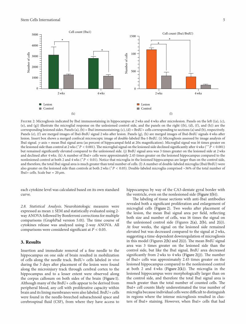
Related Documents